Embed Size (px)
Citation preview
International Journal of Clinical Medicine, 2013, 4, 273-281 http://dx.doi.org/10.4236/ijcm.2013.45048 Published Online May 2013 (http://www.scirp.org/journal/ijcm)
273
Concurrent Multi-Modality Treatment of Keloids (CMTK) Not Manageable by Conventional Postoperative Radiotherapy
Kamal Malaker1*, Mustafa Zaidi2, Mohamad Rida Franka2, Tawfik Al Yafi3
1Introduction to Clinical Medicine, Ross University School of Medicine, Roseau, Dominica; 2Burns and Plastic Surgery Centre, Burns and Plastic Surgery Hospital, Tripoli, Libya; 3Plastic Surgery, King Khalid National Guard Hospital, Jeddah, KSA. Email: *[email protected] Received February 21st, 2013; revised March 12th, 2013; accepted May 2nd, 2013 Copyright © 2013 Kamal Malaker et al. This is an open access article distributed under the Creative Commons Attribution License, which permits unrestricted use, distribution, and reproduction in any medium, provided the original work is properly cited.
ABSTRACT
Objective: To design and test a treatment regimen which is clinically responsive, readily available, cost effective, and applicable especially to children and women of child bearing age. Design Setting: A prospective cohort study. Setting: Two major postgraduate teaching hospitals: one in Tripoli, Libya and the other in Jeddah, Saudi Arabia. Participants: Fifty-seven patients with 79 keloids, referred from Plastic Surgery Units between April 1996 and January 2005. Main Outcome Measure: Degree of flattening of the keloidal lesion and symptomatic recovery. Results: Result of treatment has been analyzed using unified set criteria. Seventy-seven percent of this cohort had complete response. 19% of cases had partial response, 50% acknowledged the treatment outcome had been “satisfactory” and 44% had an “acceptable” outcome. There was no significant acute or delayed reaction. Conclusion: The technique appears universally adaptable, cost effective, and can safely be prescribed for children and women of child-bearing age. In spite of prolonged treat- ment course, compliance was excellent. Keywords: Unresceptable Keloids; Non-Surgical Treatment
1. Introduction
Postoperative radiotherapy is generally effective for ke- loid treatment [1], but in several situations neither sur- gery nor radiotherapy is applicable. Symptomatic keloids could seriously affect a patient’s life. Only sufferers can understand the agony; the itching and irritation is deep and there is no relief by scratching or from any topical applications. Clinical experience suggests that the psy- chological effect is usually devastating, especially with extensive keloids on exposed areas. This is due to the physical disfigurement and silent rejection by society, even at times by family and friends. To the medical pro- fession, not being a life threatening condition, it is of minimal consequence, and patients are frequently passed from generalist to specialist doctors, in vain.
Although the etiology of keloids remains an enigma, in the last 10 - 15 years significant advances have been made in understanding the natural history of keloids and the biogenetic processes involved [2]. As a result, the
current approach to prevention of keloid formation is bio- logical, for example, interference with TNF, PDGF, ILI, cytokine activity [3], Tamoxifen analog that down regu- lates TGF [4], anti-allergic drug Tronilast that suppresses release of cytokines as PDGF, TGF, and Interlukin-1b [5] are currently under trial. Unfortunately, none of these biological developments are accessible to patients where they are needed most, namely in the developing world. The research however, is not directed against well-in- formed matured keloids for its resolution.
There is ample clinical evidence to suggest that: Intra-lesional infiltration of steroids can alleviate sym-
ptoms of keloids, and can even induce atrophy and wound dehiscence [6-8].
Persistent pressure can induce atrophy of keloidal scars [9-11].
Silicone sheets and gels cause symptomatic relief and softening of keloids by moisture retention [12-15].
Clinical application of post-operative radiotherapy for the prevention of keloids is well established [16].
Carcinogenic effect of radiation in children [17] and *Corresponding author.
Copyright © 2013 SciRes. IJCM

Concurrent Multi-Modality Treatment of Keloids (CMTK) Not Manageable by Conventional Postoperative Radiotherapy
274
women (breast) is well documented [10,18]. Radia- tion induced skeletal growth defect in children is also well recognized [19].
With the above basic understanding we set out to make best use of the above factors in developing a technique to treat large symptomatic keloids, which are not amenable to any form of therapeutic intervention, to date.
2. Objective
This study was undertaken to develop a method of treat- ment for patients with recurrent, symptomatic (physical or psychological) keloids, which could not be treated due to location, size, or extent by surgery and/or radiotherapy especially due to young age and women of childbearing age. The primary objective was to relieve symptoms, be it physical, cosmetic, or psychosocial, using a combina- tion of available treatment modalities.
2.1. Rationale for Development of the Technique
Intra-lesional infiltration of a long acting steroid has a well-established place in the treatment of symptomatic keloid and hypertrophic scars [6-8]. Cortisone is known to be a potent anti-angiogenic agent [14,20,21]. Heparin has shown to possess strong anti-angiogenic property in association with cortisone [7,11,15,22].
Physical pressure applied on the keloidal lesions, by dint of causing hypoxia, we presume, would induce at- rophy of the hypertrophic scar or matured keloid. Thus, it is hoped, if pressure is applied along with intra-lesional long-acting steroid in combination with Heparin, the pos- sibility of inducing atrophy is expected to be substan- tially increased. It is hoped that the anti-angiogenic pro- perty of steroid and heparin would work along with is- chemia, induced by physical pressure leading to atrophy and eventual resolution of keloidal tissue.
Silicone sheet dressing apparently causes softening of keloids by retaining moisture [12-14]. It has been sug- gested that this is induced by static electricity generated by the sheet and skin [15]. Retention of interstitial fluid might improve keloidal oxygen diffusion and reduce hy- poxia. Hypoxia is a strong potentiator of tissue angio- genesis [6,23,24] and, at the same time, fibrosis, which is evident in many clinical conditions [1,6,24,25]. Softer or oedematous keloids are likely to be more amenable to dissemination of medication i.e., instilled steroids and/or Heparin. Added pressure assists in dissemination of drugs more efficiently than it would through tightly packed bundles of fibrous tissue in keloids, without oedema or softer consistency.
To achieve this, pressure needs to be reasonably high. It must be continuous for 2 - 3 weeks, by which time tissue reconstitution following any injury would have
been completed [26]. Unfortunately, constant pressure on the skin associated with steroid injection poses a risk of petechiae, micro-haemorrhagic spots, dense pigmentation, atrophy, and even ulceration. This was a handicap in one of the earlier patients treated. After some consideration it was decided to use Vitamin E on the skin surface before placing the moisture-retaining dressing. Vitamin E is a highly potent free radical scavenger and an antioxidant [27,28]. Vitamin E is also known to be a cell membrane lipid stabilizer, which protects cell membrane from lipid peroxidation [27,29]. Since addition of Vitamin E to the protocol there have been no episodes of acute skin prob- lems, thus it appears that topical application of Vitamin E protects from pressure-induced damage to the skin.
2.2. Materials and Methods (Tripoli Protocol)
Between August 1996 and July 2005, 60 patients with 79 keloids were accrued from Tripoli Medical Centre in Tripoli, Libya and the Princess Norah Oncology Centre in Jeddah, Saudi Arabia. Age of this group of patients is between six and 70 (one patient) years. The majority of patients were between 21 and 30 years. The male to fe- male ratio was 1:2. From this group 71 lesions in 57 pa- tients could be accepted as per our criteria for inclusion. Remaining lesions were found not suitable mainly due to the anatomical site of the lesion where pressure could not effectively be applied i.e., anterior abdominal wall, the neck region, or because of the large size of the lesions itself.
3. Patient Selection
There were a huge number of referrals for treatment in this protocol. We set the inclusion and exclusion criteria as follows.
3.1. Criteria for Eligibility
Patients who had recurrence after surgery and/or ra- diotherapy.
Patients who could not be treated primarily by radio- therapy due to anatomical site of the lesions, i.e., breast, anterior neck, abdomen, etc.
Reasonable volume and size of the lesion, which can be effectively infiltrated by drugs as prescribed (6 × 6 × 2 cm average volume).
Young children and adolescents with only symptom- matic keloids, where radiation is not recommended.
3.2. Criteria for Ineligibility
Patients with lesions where effective pressure cannot be applied; i.e., anterior abdominal wall, anterior neck etc.
Copyright © 2013 SciRes. IJCM
Concurrent Multi-Modality Treatment of Keloids (CMTK) Not Manageable by Conventional Postoperative Radiotherapy
275
Steroid allergy or known intolerance to steroids. Skin condition, which precludes pressure application
namely, Psoriasis or Dermatitis close to the lesion. Patients with known coagulopathy.
3.3. Criteria for Exclusion
Intolerance to regimen at any time during the course of the treatment.
Physical or symptomatic deterioration at any stage during the course of treatment.
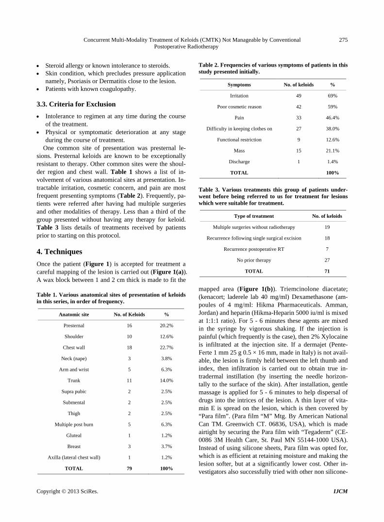
One common site of presentation was presternal le- sions. Presternal keloids are known to be exceptionally resistant to therapy. Other common sites were the shoul- der region and chest wall. Table 1 shows a list of in- volvement of various anatomical sites at presentation. In- tractable irritation, cosmetic concern, and pain are most frequent presenting symptoms (Table 2). Frequently, pa- tients were referred after having had multiple surgeries and other modalities of therapy. Less than a third of the group presented without having any therapy for keloid. Table 3 lists details of treatments received by patients prior to starting on this protocol.
4. Techniques
Once the patient (Figure 1) is accepted for treatment a careful mapping of the lesion is carried out (Figure 1(a)). A wax block between 1 and 2 cm thick is made to fit the Table 1. Various anatomical sites of presentation of keloids in this series, in order of frequency.
Anatomic site No. of Keloids %
Presternal 16 20.2%
Shoulder 10 12.6%
Chest wall 18 22.7%
Neck (nape) 3 3.8%
Arm and wrist 5 6.3%
Trunk 11 14.0%
Supra pubic 2 2.5%
Submental 2 2.5%
Thigh 2 2.5%
Multiple post burn 5 6.3%
Gluteal 1 1.2%
Breast 3 3.7%
Axilla (lateral chest wall) 1 1.2%
TOTAL 79 100%
Table 2. Frequencies of various symptoms of patients in this study presented initially.
Symptoms No. of keloids %
Irritation 49 69%
Poor cosmetic reason 42 59%
Pain 33 46.4%
Difficulty in keeping clothes on 27 38.0%
Functional restriction 9 12.6%
Mass 15 21.1%
Discharge 1 1.4%
TOTAL 100%
Table 3. Various treatments this group of patients under- went before being referred to us for treatment for lesions which were suitable for treatment.
Type of treatment No. of keloids
Multiple surgeries without radiotherapy 19
Recurrence following single surgical excision 18
Recurrence postoperative RT 7
No prior therapy 27
TOTAL 71
mapped area (Figure 1(b)). Triemcinolone diacetate; (kenacort; laderele lab 40 mg/ml) Dexamethasone (am- poules of 4 mg/ml: Hikma Pharmaceuticals. Amman, Jordan) and heparin (Hikma-Heparin 5000 iu/ml is mixed at 1:1:1 ratio). For 5 - 6 minutes these agents are mixed in the syringe by vigorous shaking. If the injection is painful (which frequently is the case), then 2% Xylocaine is infiltrated at the injection site. If a dermajet (Pente- Ferte 1 mm 25 g 0.5 × 16 mm, made in Italy) is not avail- able, the lesion is firmly held between the left thumb and index, then infiltration is carried out to obtain true in- tradermal instillation (by inserting the needle horizon- tally to the surface of the skin). After installation, gentle massage is applied for 5 - 6 minutes to help dispersal of drugs into the intrices of the lesion. A thin layer of vita- min E is spread on the lesion, which is then covered by “Para film”. (Para film “M” Mtg. By American National Can TM. Greenwich CT. 06836, USA), which is made airtight by securing the Para film with “Tegaderm” (CE- 0086 3M Health Care, St. Paul MN 55144-1000 USA). Instead of using silicone sheets, Para film was opted for, which is as efficient at retaining moisture and making the lesion softer, but at a significantly lower cost. Other in- vestigators also successfully tried with other non silicone-
Copyright © 2013 SciRes. IJCM

Concurrent Multi-Modality Treatment of Keloids (CMTK) Not Manageable by Conventional Postoperative Radiotherapy
276
(a) (b)
(c) (d)
Figure 1. Demonstrating the steps of the procedure of con- current multi-modality treatment of keloid. (a) Presternal butterfly keloid before treatment; (b) Following injection occlusive dressing with polyfilm and Tagaderm. The wax block to be used for compression is also seen; (c) Wax blocks secured on the lesion above the occlusive dressing, compressed with Elastoplast; (d) Complete flattening of the lesion with residual skin pigmentation. 3 months after com- pletion of 3 injections. based materials [30]. “Para film” is readily available in most clinical laboratories. The wax mould is placed on the lesion to match the outline. The wax moulding is se- cured and pressure dressing applied with elastoplasts (Figure 1(c)). Patients are reviewed weekly and injected after 3 weeks. Three injections are normally planned. However, application of pressure continues, at times, for 24 - 30 weeks to obtain maximal flattening.
5. Results
It has been difficult to quantitate and measure the re- sponse of therapy in this situation; therefore, an objective classification system of response of keloids to treatment was devised. Measured regression of the lesion, docu- mented symptomatic improvement, and also index of satisfaction were collected for analysis. Results of which are as follows.
Definition of Response
Complete Response: Unlike many other clinical situa- tions, the texture of the skin surface cannot be changed. Therefore, in this group, it is considered a complete re- sponse when:
The thickness of the lesion is less than 25% of its presenting height or completely flattened; All presenting symptoms have disappeared; Patient is “satisfied” with the cosmetic result (op-
tional). Partial Response: When thickness of the lesion remains 25% - 75% of
its original presentation; Persistence of any presenting symptoms or all pre-
senting symptoms persist with less intensity; Cosmetic result is at least “acceptable” as opposed
to “satisfactory” (optional). No response or progression: When there is no change in the thickness of the le-
sion (0% - 25% reduction); No change or exacerbation of presenting symptoms; Both of the above.
6. Index of Satisfaction
The majority of the patients presented with multiple sym- ptoms and had undergone repeated treatment without any benefit except deteriorating cosmesis. We asked all pa- tients (or parents) about their level of satisfaction fol- lowing completion of treatment. They had to respond with a “” in an unmarked questionnaire whether the post treatment appearance was “satisfactory”, “accept- able”, or “no comment”.
7. Response to Treatment
The result of the treatment is presented in Table 4. We made every endeavor to assess the response to treatment objectively by using the above criteria and by assessing patients’ index of satisfaction in this group of 71 patients, who received treatment with this technique (CMTK).
Results
The tolerance and compliance to treatment was accept- able:
As per our criteria complete response was achieved in 55 (77%) lesions, 13 (19%) lesions had some relief of symptoms and three (3) had no response (4%) (Table 4).
Cosmetic result was “satisfactory” amongst 36 lesions (50%), “acceptable” amongst another 21 lesions (44%) and “no comment” was expressed regarding four (4) keloids (6%) (Table 4).
Lesions in the limb, gluteal region, and neck presented with painful restriction of movement. Twelve (12) ke- loids presented with functional restriction. Following completion of therapy, full functional recovery was achieved in three (3) out of 12 lesions. Partial functional recovery was achieved in the remaining nine (9). It is
Copyright © 2013 SciRes. IJCM

Concurrent Multi-Modality Treatment of Keloids (CMTK) Not Manageable by Conventional Postoperative Radiotherapy
277
Table 4. Gives details of response to treatment using CMTK Protocol (Tripoli Protocol). 71 keloidal lesions were treated according to the Protocol.
Parameter No. of keloids
%
Complete response 55 77%
Partial response 13 19%
No response 3 4%
Cosmesis “Satisfactory” per patient 36 50%
Cosmesis “Acceptable” 31 44%
Cosmesis “Unacceptable” 4 6%
Pre-treatment functional impairment 12/71 16.6%
Post therapy full recover 3/12 25%
Post therapy partial functional recovery 9/12 75%
interesting to note that all lesions had at least some de- gree of functional recovery.
Figure 1(a) shows a 28-year-old male infantry soldier, who developed a keloid in the presternal region on an acne site, which had developed to the presenting size over a period of 3 - 4 years. Six months previously he started to have severe itching, pain, and discomfort while wearing clothes. He had, on two occasions, intra-lesional triamcinolone injections, with only a temporary period of relief. The lesion increased slightly in size with clinical diagnosis of keloid. He was started on the concurrent multimodality regimen; Figure 1(b) shows the mapping of the lesion, covered by polyfilm and Tegaderm occlu- sive (airtight) dressing. This also demonstrates the cus- tomized wax block to be placed on the mapped area of the lesion to exert continuous pressure (compressive dressing). Figure 1(c) shows the wax block in place and pressure applied by Elastoplast, which is secured by a second layer of Tegaderm. Figure 1(d) shows the lesion after six (6) months of treatment. This is after four (4) applications of concurrent multimodality (CMTK) regi- men; the lesion is almost flat, slightly pigmented, and totally asymptomatic.
Figure 2(a) shows post-burn keloids in the scapular® region in a 38-year-old male Filipino patient. Figure 2(b) after 2 episodes of multimodality therapy. The lesion has been completely flattened, normal skin covering, and ex- cellent cosmesis
Figure 3(a) shows recurrence of a left deltoid region keloid following primary resection of a vaccination ke- loid in a 35-year-old Saudi male. Figure 3(n) shows the lesion completely flat with some pigmentation and as- ymptomatic 3 months after completion of 3 episodes of multimodality treatment (CMTK).
(a) (b)
Figure 2. Post burn keloid. (a) Post burn keloid in the shoul- der (left), before treatment; (b) Complete resolution follow- ing the first treatment.
(a) (b)
Figure 3. (a) The lesion before treatment; (b) Complete flat- tening of the lesion after treatment with residual pigmenta- tion.
Figure 4(a) post-sternotomy severely symptomatic ke- loid in an 18-year-old female. Figure 4(b) shows com- plete regression and symptomatic relief of the keloid after 3 treatments.
Figure 5(a) shows multiple post varicella keloids in a 19-year-old Saudi girl. She presented with 15 lesions, all were treated in stages and all responded completely with some residual pigmentation (Figure 5(b)).
Tolerance to the concurrent multimodality (CMTK) re- gimen has been excellent. There were minor side effects, namely pigmentation, skin atrophy, etc., which have been documented in Table 5.
8. Discussion
Keloids are not life threatening and, in a way, self-limit- ing. Dismal results of treatment of recurrent, large, and bulky lesions have so far alienated the medical commu- nity. The underlying mechanism of keloid formation is unclear. Significant strides have been made to understand the molecular mechanism [2]. As a result, post-operative recurrence can now be effectively eliminated, but a cure for matured unresectable keloids still remains elusive. The result of postoperative external beam radiotherapy
Copyright © 2013 SciRes. IJCM

Concurrent Multi-Modality Treatment of Keloids (CMTK) Not Manageable by Conventional Postoperative Radiotherapy
278
(a) (b)
Figure 4. Post sternotomy keloid. (a) Before treatment; (b) Six months after treatment complete regression with resi- dual depigmentation seen.
(a) (b)
Figure 5. Multiple keloids following chicken pox. (a) Several keloids in the chest wall before treatment (see text), patient; (b) Complete regressions of all the lesions with faint resid- ual pigmentation, 6 months after completion of scheduled treatment. Table 5. Indicated complication and side efects of CMTK regimen.
Complication No. of Lesions
Pigmentation 28
Skin trophy 3
Wound dehiscence 0
Steroid induced toxicity 0
Wound healing problem 0
No response 3
Petechiae 2
Bleeding/Haematoma 0
[31] or brachytherapy [16,24] is excellent. On the other hand, primary radiotherapy for treatment of keloids until
recently proved to be essentially ineffective. Malaker, et al. demonstrated a hypo-fractionated radiation therapy regimen treated successfully as primary treatment for un- resectable keloids. They used once weekly 750 cGy on Kilovoltage or 3 - 6 MeV electrons to treat primarily with radiation, to a total dose of 3750 cGy in five weeks. Epilation was the most common acute and late toxicity. Tolerance and compliance were excellent [32]. In an at- tempt to get the greatest benefit, the agents known to be effective have been used in this protocol in a concerted fashion. Three agents, namely long acting steroids, oc- clusive dressing, and pressure, are all known to have be- neficial effect against keloids. The addition of Heparin may have enhanced the effect of steroids to reduce neo- vascularity and induce regression. Unlike malignant tu- mours, in keloids, angiogenicity is not an issue. It is known that hypoxia is a strong trigger for neo-angio- genesis [33-35]. Chronic hypoxia is also known to cause fibroblastic proliferation [1,25,36,37]. By applying pres- sure to a keloidal lesion, it is possible to initiate exacer- bation of keloidal growth. On the other hand, if the me- chanical pressure is high enough, it is possible to occlude keloidal vessels to such an extent that nutrition and oxy- gen supply will be restricted sufficiently to induce atro- phy. The amount of pressure needed to achieve this ef- fect, may also cause atrophy or even necrosis of the su- perficial layer of the overlying skin. In fact, the first pa- tient in this group did have significant petechiae, des- quamation, and minute focal ulceration after the first ap- plication of pressure. Therefore, we opted for less pres- sure and topical vitamin E application on the skin to pre- vent atrophy and/or necrosis. This appears to have worked for this cohort. By inducing hypoxia, if angiogenesis is provoked, it would further enhance the growth of the le- sion. In fact, it was noted that if only pressure is applied for these lesions for a week then removed, not immedi- ately but after 5 - 6 days, the lesion increases in size, be- comes hyperemic, tender, and appears more active. This led us to postulate that pressure induced hypoxia may have provoked neo-angiogenesis in the lesion, which is why they became active 5 - 6 days after releasing the pressure. Under light microscopy keloidal tissues have much less vascularity than normal skin, but under elec- tron microscopy Kischer, et al. [22,23] noticed rich mi- cro-vascularity in the keloidal tissue. Significant numbers of these vessels remained completely or partly obliterated and had dense walls. It is possible that added hypoxia due to pressure induces further production of vascular endothelial growth factors (VEGF) and thus neo-angio- genesis and further proliferation of microvasculature and keloidal regrowth to be clinically evident as we noticed in some of our patients when application of pressure was abandoned after 1 or 2 treatments.
Copyright © 2013 SciRes. IJCM

Concurrent Multi-Modality Treatment of Keloids (CMTK) Not Manageable by Conventional Postoperative Radiotherapy
279
It is through the combined anti-angiogenic effect of long acting steroid and heparin infiltration in the lesion that should be able to counteract pressure-induced neo- angiogenesis, at least during the early phase of treatment when it is most needed. It appears that this hypothesis may be true in our patients who achieved an excellent re- gression.
Silicone sheet and silicon gel seems to produce symp- tomatic relief and also softening of the keloidal lesion, by moisture retention [12-14]. This is likely to loosen the tightly packed collagen bundles in the keloidal lesion and improve oxygenation and nutrition by diffusion. This may also cause diffusion of symptoms causing agents out of the lesion resulting in symptomatic relief [21,38]. Sili- cone sheets and silicone gel are expensive and are not readily available. Therefore, we opted to use “Para film” which is commonly used in all clinical laboratories, and is cheap and easily available. This has the similar mois- ture-retaining property as the “Silicone products”.
To increase durability of Parafilm we have used 3 - 4 folds of the material. To ensure air-tightness of the dress- ing we used Tegaderm, but any type of tape (non allergic) can be as effective as Tegaderm. It appears that several factors are working at cross purposes in this protocol. However, the net outcome is more favorable than one could have envisaged. By avoiding use of ionizing radia- tion, we have made this protocol applicable to children and women. There is no risk of carcinogenesis, growth impairment, or organ failure as known to occur in case of treatment associated with ionizing radiation.
Frequently in our geographical environment, distance of the clinic from the patient’s hometown is a deterring factor for patients to come for regular follow-up. Fast progressing and universally available communication sys- tems hopefully will bridge the gap not only in follow-up of this group of patients, but also for all patient popula- tions in general. Late complications of atrophy, ulcera- tion, recurrence, or malignant transformation have not been reported by any of our patients in the study.
Maintaining a regular follow-up for these patients has been more difficult because otherwise physically well and active patients are generally non-compliant for fol- low-ups [31,38-40]. Further trials with a larger patient population and long-term follow up would certainly give a proper perspective of this regime.
9. Conclusion
This is a simple, affordable, and universally applicable technique to offer some comfort to hitherto untreatable symptomatic keloids. The procedure is acceptable. Re- sponse rate is significant at 77%, and patient compliance is also very good. Toxicities associated with the tech- nique are minimal. Most importantly this can be safely
used for children, unlike radiation. The follow-up has been from 6 - 60 months. Further studies with a larger population in the prevalent areas for keloid i.e. Sub-Sa- haran Africa and Southern Asia should be useful. With some perseverance one can master this technique easily. This technique can be applied in an outpatient clinic or for that matter in the office of a General Practitioner or a primary health care facility.
10. Acknowledgements
The authors wish to thank Sister Kim Geong Ye, Chief Radiotherapy Nurse at Tripoli Medical Centre, Sister Rene Cornellissen and Fuad Salameh, Staff Nurse of Ra- diotherapy Section of Princess Norah Oncology Centre for their meticulous care of our patients, and Mrs. Noura Pellicci, Princess Norah Oncology Centre secretariat for preparing the manuscript. Special thanks to Miss Joan Joseph and Mr. Irus Toussaint of Ross University School of Medicine for administrative and technical support.
REFERENCES [1] S. Anderson and B. M. Branner, “Progressive Renal Dis-
ease: A Disorder of Adaptation,” Quarterly Journal of Medicine, Vol. 70, No. 263, 1989, pp. 185-189.
[2] P. Rubin, A. Soni and J. P. Williams, “The Molecular and Cellular Biologic Basis for the Radiation Treatment of Benign Proliferative Disease,” Seminars in Radiation On- cology, Vol. 9, No. 2, 1999, pp. 203-214. doi:10.1016/S1053-4296(99)80010-1
[3] A. E. Brissett and D. A. Sherris, “Scar Contractures, Hy- pertrophic Scars, and Keloids,” Facial Plastic Surgery, Vol. 17, No. 4, 2001, pp. 263-272. doi:10.1055/s-2001-18827
[4] W. G. Payne, F. Ko, S. Anspaugh, et al., “Down-Regu- lating Causes of Fibrosis with Tamoxifen: A Possible Cellular/Molecular Approach to Treat Rhinophyma,” An- nals of Plastic Surgery, Vol. 56, No. 3, 2006, pp. 301- 305. doi:10.1097/01.sap.0000199155.73000.2f
[5] H. Tamai, O. Katoh, S. Suzuki, et al., “Impact of Tranil- ast on Restenosis after Coronary Angioplasty: Tranilast Restensosis Following Angioplasty Trial (TREAT),” Ame- rican Heart Journal, Vol. 138, No. 5, 1999, pp. 968-975. doi:10.1016/S0002-8703(99)70025-6
[6] G. Muneuchi, S. Suzuki, M. Onodera, O. Ito, Y. Hata and H. H. Igawa, “Long-Term Outcome of Intralesional Injec- tion of Triamcinolone Acetonide for the Treatment of Keloid Scars in Asian Patients,” Scandinavian Journal of Plastic and Reconstructive Surgery and Hand Surgery, Vol. 40, No. 2, 2006, pp. 111-116. doi:10.1080/02844310500430003
[7] G. G. Gauglitz, H. C. Korting, T. Pavicic, et al., “Hyper- trophic Scarring and Keloids: Pathomechanisms and Cur- rent and Emerging Treatment Strategies,” Molecular Me- dicine, Vol. 17, No. 1-2, 2011, pp. 113-125. doi:10.2119/molmed.2009.00153
Copyright © 2013 SciRes. IJCM

Concurrent Multi-Modality Treatment of Keloids (CMTK) Not Manageable by Conventional Postoperative Radiotherapy
280
[8] S. Mutalik, “Treatment of Keloids and Hypertrophic Scars,” Indian Journal of Dermatology, Venereology and Leprology, Vol. 71, No. 1, 2005, pp. 3-8. doi:10.4103/0378-6323.13777
[9] A. Goel and P. Shrivastava, “Post-Burn Scars and Scar Contractures,” Indian Journal of Plastic Surgery, Vol. 43, 2010, pp. S63-S71. doi:10.4103/0970-0358.70724
[10] B. Berman and H. C. Bieley, “Keloids,” Journal of the American Academy of Dermatology, Vol. 33, No. 1, 1995, pp. 117-123. doi:10.1016/0190-9622(95)90035-7
[11] T. S. Alster and E. L. Tanzi, “Hypertrophic Scars and Keloids: Etiology and Management,” American Journal of Clinical Dermatology, Vol. 4, No. 4, 2003, pp. 235- 243. doi:10.2165/00128071-200304040-00003
[12] G. L. Dockery and R. Z. Nilson, “Treatment of Hypertro- phic and Keloid Scars with Silastic Gel Sheeting,” Jour- nal of Foot and Ankle Surgery, Vol. 33, No. 2, 1994, pp. 110-119.
[13] K. Perkins, R. B. Davey and K. A. Wallis, “Silicone Gel: A New Treatment for Burn Scars and Contractures,” Burns, Including Thermal Injury, Vol. 9, No. 3, 1983, pp. 201-204. doi:10.1016/0305-4179(83)90039-6
[14] M. Gibbons, R. Zuker, M. Brown, et al., “Experience with Silastic Gel Sheeting in Pediatric Scarring,” Journal of Burn Care & Rehabilitation, Vol. 15, No. 1, 1994, pp. 69-73. doi:10.1097/00004630-199401000-00013
[15] B. Hirshowitz, E. Lindenbaum, Y. Har-Shai, et al., “Sta- tic-Electric Field Induction by a Silicone Cushion for the Treatment of Hypertrophic and Keloid Scars,” Plastic and Reconstructive Surgery, Vol. 101, No. 5, 1998, pp. 1173- 1183.
[16] L. Narkwong and P. Thirakhupt, “Postoperative Radio- therapy with High Dose Rate Iridium 192 Mould for Pre- vention of Earlobe Keloids,” Journal of the Medical As- sociation of Thailand, Vol. 89, No. 4, 2006, pp. 428-433.
[17] E. Willich, H. Kuttig, G. Pfeil, et al., “Pathological Chan- ges (Developmental/Growth Disturbances) of the Spine after Radiation Therapy of Nephroblastomas during Early Childhood. A Retrospective Long-Term Follow-Up Study in 82 Children,” Strahlentherapie und Onkologie, Vol. 166, 1990, pp. 815-821.
[18] D. L. Preston, A. Mattsson, E. Holmberg, et al., “Radia- tion Effects on Breast Cancer Risk: A Pooled Analysis of Eight Cohorts,” Radiation Research, Vol. 158, No. 2, 2002, pp. 220-235. doi:10.1667/0033-7587(2002)158[0220:REOBCR]2.0.CO;2
[19] F. Nguyen, C. Rubino, S. Guerin, et al., “Risk of a Sec- ond Malignant Neoplasm after Cancer in Childhood Treated with Radiotherapy: Correlation with the Integral Dose Restricted to the Irradiated Fields,” International Journal of Radiation Oncology Biology Physics, Vol. 70, No. 3, 2008, pp. 908-915. doi:10.1016/j.ijrobp.2007.10.034
[20] T. A. Mustoe, “Evolution of Silicone Therapy and Me- chanism of Action in Scar Management,” Aesthetic Plas- tic Surgery, Vol. 32, No. 1, 2008, pp. 82-92. doi:10.1007/s00266-007-9030-9
[21] P. W. Grigsby, A. Russel, D. Bruner, et al., “Late Injury of Cancer Therapy on the Female Reproductive Tract,” International Journal of Radiation Oncology Biology Phy- sics, Vol. 31, No. 5, 1995, pp. 1281-1299. doi:10.1016/0360-3016(94)00426-L
[22] C. W. Kischer, M. R. Shetlar and M. Chvapil, “Hyper- trophic Scars and Keloids: A Review and New Concept Concerning Their Origin,” Scanning Electron Microscopy, Vol. 4, 1982, pp. 1699-1713.
[23] D. Deveci, J. M. Marshall and S. Egginton, “Chronic Hy- poxia Induces Prolonged Angiogenesis in Skeletal Mus- cles of Rat,” Experimental Physiology, Vol. 87, No. 3, 2002, pp. 287-291. doi:10.1113/eph8702377
[24] B. Liu and M. K. Connolly, “The Pathogenesis of Cuta- neous Fibrosis,” Seminars in Cutaneous Medicine and Surgery, Vol. 17, No. 1, 1998, pp. 3-11. doi:10.1016/S1085-5629(98)80055-2
[25] C. T. Taylor and S. P. Colgan, “Therapeutic Targets for Hypoxia-Elicited Pathways,” Pharmaceutical Research, Vol. 16, No. 10, 1999, pp. 1498-1505. doi:10.1023/A:1011936016833
[26] P. Martin, “Wound Healing—Aiming for Perfect Skin Regeneration,” Science, Vol. 276, No. 5309, 1997, pp. 75-81. doi:10.1126/science.276.5309.75
[27] M. G. Traber and L. Packer, “Vitamin E: Beyond Anti- oxidant Function,” The American Journal of Clinical Nu- trition, Vol. 62, No. 6, 1995, pp. 1501S-1509S.
[28] S. Khanna, S. Roy, H. Ryu, P. Bahadduri, P. W. Swaan, R. R. Ratan and C. K. Sen, “Molecular Basis of Vitamin E Action. Tocotrienol Modulates 12-Lipoxygenase, a Key Mediator of Glutamate-Induced Neurodegeneration,” The Journal of Biological Chemistry, Vol. 278, No. 44, 2003, pp. 43508-43515. doi:10.1074/jbc.M307075200
[29] L. Ernster, P. Forsmark and K. Nordenbrand, “The Mode of Action of Lipid-Soluble Antioxidants in Biological Membranes: Relationship between the Effects of Ubiqui- nol and Vitamin E as Inhibitors of Lipid Peroxidation in Submitochondrial Particles,” Biofactors, Vol. 3, No. 4, 1992, pp. 241-248.
[30] V. Jones, J. E. Grey and K. G. Harding, “ABC of Wound Healing: Wound Dressings,” British Medical Journal, Vol. 332, No. 7544, 2006, pp. 777-780. doi:10.1136/bmj.332.7544.777
[31] K. Malaker, K. Vijayraghavan, I. Hodson and T. Al Yafi, “Retrospective Analysis of Treatment of Unresectable Keloids with Primary Radiation over 25 Years,” Clinical Oncology (Royal College of Radiologists), Vol. 16, No. 4, 2004, pp. 290-298. doi:10.1016/j.clon.2004.03.005
[32] T. S. Alster and T. B. West, “Treatment of Scars: A Re- view,” Annals of Plastic Surgery, Vol. 39, No. 4, 1997, pp. 418-432. doi:10.1097/00000637-199710000-00014
[33] B. J. Moeller, Y. Cao, Z. Vujaskovic, C. Y. Li, Z. A. Haroon and M. W. Dewhirst, “The Relationship between Hypoxia and Angiogenesis,” Seminars in Radiation On- cology, Vol. 14, No. 3, 2004, pp. 215-221. doi:10.1016/j.semradonc.2004.04.005
[34] C. W. Pugh and P. J. Ratcliffe, “Regulation of Angio-
Copyright © 2013 SciRes. IJCM

Concurrent Multi-Modality Treatment of Keloids (CMTK) Not Manageable by Conventional Postoperative Radiotherapy
Copyright © 2013 SciRes. IJCM
281
genesis by Hypoxia: Role of the HIF System,” Nature Medicine, Vol. 9, No. 6, 2003, pp. 677-684. doi:10.1038/nm0603-677
[35] J. M. Gleadle, B. L. Ebert, J. D. Firth and P. J. Ratcliffe, “Regulation of Angiogenic Growth Factor Expression by Hypoxia, Transition Metals, and Chelating Agents,” Ame- rican Journal of Physiology, Vol. 268, No. 6, 1995, pp. C1362-C1368.
[36] J. T. Norman, I. M. Clark and P. L. Garcia, “Hypoxia Promotes Fibrogenesis in Human Renal Fibroblasts,” Kidney International, Vol. 58, No. 6, 2000, pp. 2351- 2366. doi:10.1046/j.1523-1755.2000.00419.x
[37] Y. Ganat, S. Soni, M. Chacon, M. L. Schwartz and F. M. Vaccarino, “Chronic Hypoxia Up-Regulates Fibroblast Growth Factor Ligands in the Perinatal Brain and Induces Fibroblast Growth Factor-Responsive Radial Glial Cells
in the Sub-Ependymal Zone,” Neuroscience, Vol. 112, No. 4, 2002, pp. 977-991. doi:10.1016/S0306-4522(02)00060-X
[38] A. P. Kelly, “Medical and Surgical Therapies for Ke- loids,” Dermatology and Therapy, Vol. 17, No. 2, 2004, pp. 212-218. doi:10.1111/j.1396-0296.2004.04022.x
[39] Q. Dinh, M. Veness and S. Richards, “Role of Adjuvant Radiotherapy in Recurrent Earlobe Keloids,” Austral- asian Journal of Dermatology, Vol. 45, No. 3, 2004, pp. 162-166. doi:10.1111/j.1440-0960.2004.00079.x
[40] E. E. Tredget, B. Nedelec, P. G. Scott and A. Ghahary, “Hypertrophic Scars, Keloids, and Contractures. The Cel- lular and Molecular Basis for Therapy,” Surgical Clinics of North America, Vol. 77, No. 3, 1997, pp. 701-730. doi:10.1016/S0039-6109(05)70576-4