Embed Size (px)
Citation preview
247/ Acta Chir Orthop Traumatol Cech. 84, 2017, No. 4p. 247–253 CURRENT CONCEPTS REVIEW
SOUBORNÝ REFERÁT
INTRODUCTION
Mild traumatic brain injury (mTBI) is defined with aGlasgow Coma Scale (GCS) of 13 to 15 points. Brainconcussion represents the predominant portion of mTBI(Fig. 1). The patient is usually conscious, being awake,with open eyes, responds adequately to speech and canperform targeted movements on request.
During the last 2 to 3 decades, intensive discussionsand analyses were performed on sport-related concussionsin the anglo-american scientific literature. This resultedin several consensus recommendations during the last10 years (44). The topic of brain concussion became in-creasingly popular even in Germany, especially in highcontact sports, e.g. ice hockey (58).
Thus, the present overview will give basic knowledgeon this relevant injury regarding recognition, acutemana gement and potential long-term problems.
DefinitionConcussion is a brain injury! According to the Consensus
statement on concussion in sport from 2012, a concussionis defined as „a complex pathophysiological process af-fecting the brain, induced by biomechanical forces“.(44). The latter can act directly or indirectly against thehead. A concussion typically results in a rapid, shortimpairment of neurological functions that typicallyresolve spontaneously but can also lead to neuropatho-logical changes.
Concussion in Sports – What Trauma/OrthopedicSurgeons Need to Know!
Otřes mozku ve sportu – co by měl traumatolog a ortoped znát
A. GÄNSSLEN1, T. NEUBAUER2, W. KRUTSCH3
1 Trauma Department, Klinikum Wolfsburg, Wolfsburg, Germany; Initiative „Schütz Deinen Kopf“ der ZNS Hannelore KohlStiftung
2 Trauma Department, Landesklinikum Horn, Horn, Germany3 Department of Trauma Surgery, University Medical Centre Regensburg, FIFA Medical Centre of Excellence, Regensburg,
Germany; Initiative „Schütz Deinen Kopf“ der ZNS Hannelore Kohl Stiftung
SUMMARY
Concussion in sport is often underdiagnosed with the potential risk of long-term sequelae. This article presents themechanisms, the underlying pathophysiology and typical primary signs and symptoms. The recognition and resulting medicalmeasures including the present recommendations and decisions on return to play are described.
The majority of patients with concussion present with clinical and cognitive symptoms only for short time intervals. As arule a complete subjective recovery is observed within a few hours or days. Although neurocognitive impairments can persistin individual cases, they also show a good tendency to heal. Thus, after 1 year nearly all patients no longer have any relevantdisorders.
In a few cases long-term disturbances can occur in the presence of certain risk factors and/or after repetitive concussion.Unspecific symptoms and some cognitive impairments are the main reported problems; however, there is also a potentialbut individually unpredictable risk of developing neurodegenerative alterations and diseases.
Acute clinical symptoms indicate a more functionalproblem than a structural damage. Correspondingly,standard image techniques (CT/MRI) usually show nostructural pathologies (44).
Injury mechanismAccording to the above mentioned definition, a con-
cussion may be caused by a direct blow or indirectforce acting against the head and the brain (46).
The exact injury mechanism is unknown as severaldifferent force vectors can result in a concussion. Adirect blow against the head can lead to a local braininjury at the impact site, resulting in some intracranialshaking of the whole brain. Brain parts can bounceagainst the bone even on the side opposite to the primaryforce induction. Additionally, injuries due to shearingforces and tissue deceleration along the irregularlyshaped cranial base and the falx cerebri and tentoriumcerebelli may occur. Many direct mechanisms are obvious(e.g. head impact against the ground etc.). The indirectinjury is a result from transferred forces, not directlystriking the head, which are usually transmitted to thebrain via the body's trunk.
The force can be directed along an imaginary straightline (linear force effect) or it results in a rotationalmovement of the head and brain. In the majority a com-bination of both potential force mechanisms are present.
247_253_ganslen 14.8.17 14:55 Stránka 247
248/ Acta Chir Orthop Traumatol Cech. 84, 2017, No. 4 CURRENT CONCEPTS REVIEWSOUBORNÝ REFERÁT
In particular, the accelerating torsional forces seem tobe the major cause of neurological disturbances.
As a result of these effects, different brain movementscan occur within the bony skull, leading to involvement/in-jury of different brain regions, which can explain thewide range of different symptoms to be observed. Themechanism can be subtle and clinically not obvious.Therefore, a positive correlation between the force extendand the resulting symptoms is often unusual (40).
Despite standard imaging techniques fail to showstructural injury, „nonvisible injuries” to the neuronsand the smallest blood vessels must always be assumed.These cell injuries lead to complex changes in the cells,which can trigger inflammatory reactions in the tissues,restrict chemical changes and cell nutrition.
PathophysiologyThe mechanical deformation of the brain tissue during
trauma can result in shear injuries to the brain cells(38). In consequence, local cellular injury can be present,axons can be damaged or even disrupted from the cellbodies or even apoptosis can occur.
According to the primary injury mechanism, focal/directand diffuse brain injuries can be distinguished.
The local direct (focal) damage to the brain tissueleads to excessive excitation of the cells. As a result,substances enter the surrounding tissue, which negativelyinfluence the energy supply of the neurons. The adjacentsupporting cells in the brain are also damaged resulting
in functional impairment. As a result of cell membranechanges, mitochondrial dysfunction and cellular swellingoccurs, leading to an energy crisis, which ultimatelycan result in complete loss of neuronal function. Theextent of mitochondrial damage correlates with thelong-term (persistent consequences) of the brain damage.Additionally, the local cerebral blood flow is diminished,supporting the energy crisis. This pathophysiologicalneurometabolic cascade is supposed to be a main reasonof functional neurological impairment (25).
Diffuse brain tissue injury leads to axonal injury,which can sometimes be visualized with new imagingtechniques. A stretching of 10% within 100 ms canlead to permanent axonal damage. Myelination of the
Table 1. Typical symptoms after concussions
Physical Cognitive Emotional Sleepheadache feeling foggy sadness trouble falling
asleepdizziness slowed down irritability impaired sleep
durationbalance impaired emotional less sleep
problems concentration than usualvoimiting difficulty nervousness more sleep
remembering than usualsensitivity repeating
to light/noise questionsvisual problems
fatigue
Fig. 1. Concussion as the major part of mTBI.
247_253_ganslen 14.8.17 14:55 Stránka 248
249/ Acta Chir Orthop Traumatol Cech. 84, 2017, No. 4 CURRENT CONCEPTS REVIEWSOUBORNÝ REFERÁT
axons can be a protective mechanism. Myelination ofaxons usually provides faster information transportbetween the cells. After injury, this information transportis disturbed (38). In children, during brain development,myelination is incomplete, a possible reason that thechildren s brain is more vulnerable, potentially ex-plaining the prolonged recovery phase. Histologically,comparable changes like in Alzheimer’s disease couldbe detected.
In addition to these neuronal consequences, local cir-culatory disturbances in the injured brain causes increasedoxygen demand, which is present for several days. Thecardiovascular system can show heart rate variabilityduring physical and mental stress.
Signs and symptomsSigns (objective detection, observation) of concussion
can include confusion, loss of consciousness, gait balancedisturbances and swaying, pupil difference, a dazzed,blank or vaccant look and/or grabbing/clutching of head.
Classical symptoms such as unconsciousness andamnesia are reported in athletes in about 20% (4) and< 10% in recreational sports (47).
The subjective symptom complex is broard and includesclinical, neurocognitive symptoms and behavioral andsleep changes (40, 44, Table 1). The most commonprimary symptoms include headache (70–80%), dizziness(34–70%), nausea/vomiting (20–40%), weakness andfatigue (20–50%), visual disturbances (approx. 20%)and sensitivity to light and noise (10–60%) (4, 35).
In the acute assessment, the focus is generally not yetfocused on neurocognitive symptoms, behavioral andsleep changes.
The frequency problemConcussion are underreported and misunderstood by
all involved persons as they are underestimated in itsconsequences (17, 42). Athletes perform a return-to-play often too early, despite adequate knowledge aboutthe injury, due to „external pressure" by coaches, athletictrainers and even team physicians (11, 31, 34).
The overall rate of overlooked orunreported concussions is high withan average of 40% (30.5–81.5%)(18, 22, 36, 40, 48). In recreationalsports a significantly higher rate ofunrecognized injuries is reported(69).
Immediate recognition =sideline evaluation
It is generally accepted, that ath-letes with suspected concussionhave to immediately stop their par-ticipation in sports and should bejudged by a physician on the sameday (44).
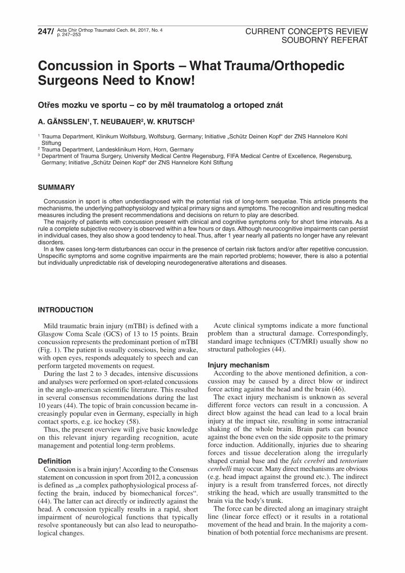
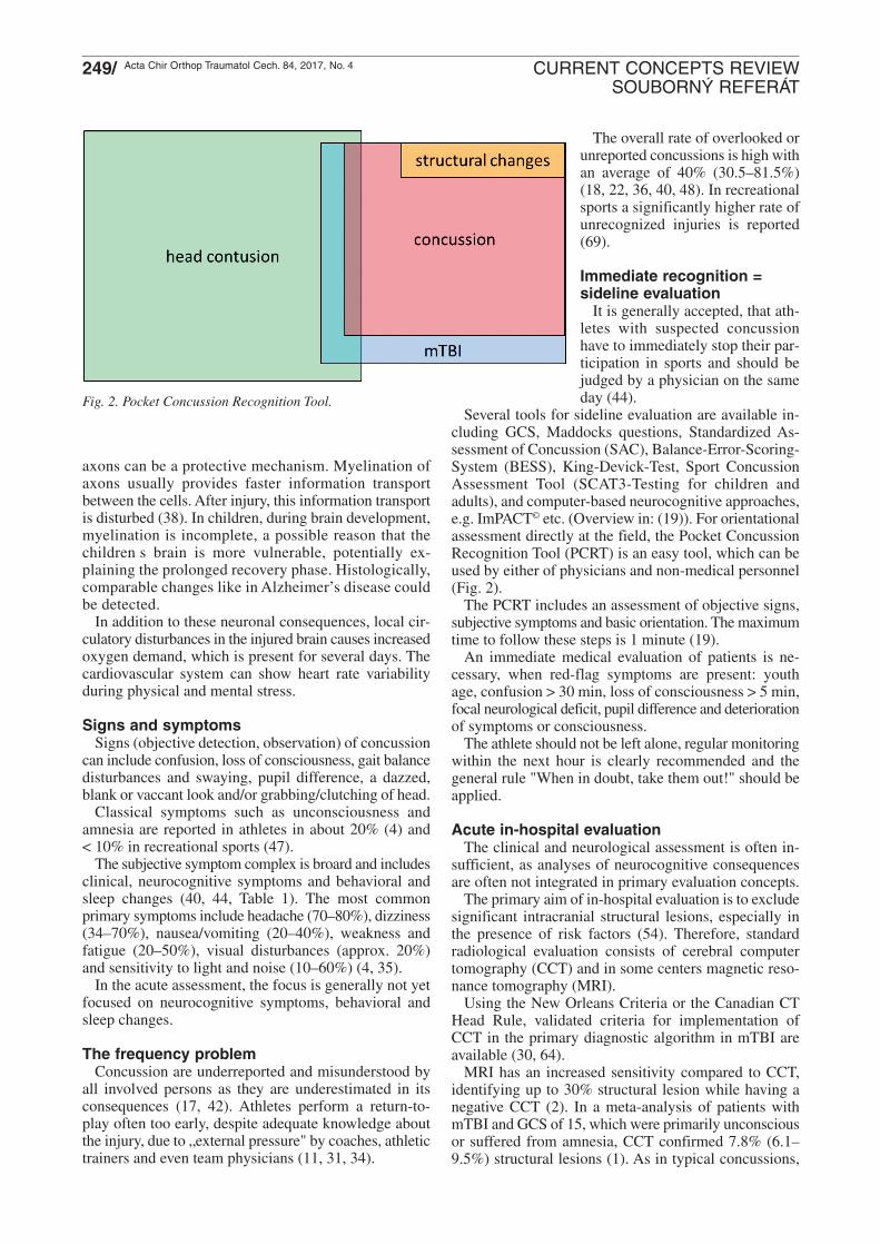
Several tools for sideline evaluation are available in-cluding GCS, Maddocks questions, Standardized As-sessment of Concussion (SAC), Balance-Error-Scoring-System (BESS), King-Devick-Test, Sport ConcussionAssessment Tool (SCAT3-Testing for children andadults), and computer-based neurocognitive approaches,e.g. ImPACT© etc. (Overview in: (19)). For orientationalassessment directly at the field, the Pocket ConcussionRecognition Tool (PCRT) is an easy tool, which can beused by either of physicians and non-medical personnel(Fig. 2).
The PCRT includes an assessment of objective signs,subjective symptoms and basic orientation. The maximumtime to follow these steps is 1 minute (19).
An immediate medical evaluation of patients is ne -cessary, when red-flag symptoms are present: youthage, confusion > 30 min, loss of consciousness > 5 min,focal neurological deficit, pupil difference and deteriorationof symptoms or consciousness.
The athlete should not be left alone, regular monitoringwithin the next hour is clearly recommended and thegeneral rule "When in doubt, take them out!" should beapplied.
Acute in-hospital evaluationThe clinical and neurological assessment is often in-
sufficient, as analyses of neurocognitive consequencesare often not integrated in primary evaluation concepts.
The primary aim of in-hospital evaluation is to excludesignificant intracranial structural lesions, espe cially inthe presence of risk factors (54). Therefore, standardradio logical evaluation consists of cerebral computertomography (CCT) and in some centers magnetic reso-nance tomography (MRI).
Using the New Orleans Criteria or the Canadian CTHead Rule, validated criteria for implementation ofCCT in the primary diagnostic algorithm in mTBI areavailable (30, 64).
MRI has an increased sensitivity compared to CCT,identifying up to 30% structural lesion while having anegative CCT (2). In a meta-analysis of patients withmTBI and GCS of 15, which were primarily unconsciousor suffered from amnesia, CCT confirmed 7.8% (6.1–9.5%) structural lesions (1). As in typical concussions,
Fig. 2. Pocket Concussion Recognition Tool.
247_253_ganslen 14.8.17 14:55 Stránka 249
250/ Acta Chir Orthop Traumatol Cech. 84, 2017, No. 4 CURRENT CONCEPTS REVIEWSOUBORNÝ REFERÁT
the presence of these clinical signs is low (10–20%), indicating a minor risk in concussions.
Clinically, a detailed head-neck evaluation is recom-mended including an orientated neurological evaluationwith testing the mental status, cognitive functions andbalance and a re-analysis of present symptoms (44).Early assessment of cognitive functions seems to be anessential component of primary evaluation (44).
A useful tool can be the SCAT3 which is available forchildren aged 5–12 years and older persons > 12 years(44). The SCAT3 screening includes analysis of clinicalsymptoms, the GCS, Maddock s-Questions, SAC-Testingand modified BESS-Testing. Testing balance is supposedof relevant value in the primary and secondary evaluationof these patients (16).
Due to its prognostic relevance, a detailed medicalhistory has to be analyzed including number of pre-existing concussions/TBI, duration of symptoms, presenceand type of amnesia (retrograde/antegrade) (44).
Blood biomarkers (e.g. S100B) are presently not re -commended for general use, as they fail to clearly dis-tinguish between concussed and non-concussed patients,especially in sports (40, 61). S100B was able to identifypatients "not at risk" (21, 40).
Acute treatmentBased on the knowledge of the pathophysiological
changes at the cellular level, primary physical and mentalrest was recommended (44) in order not to stress the dis-turbed cells with cognitive work, thus extending the re-covery process. Cognitive rest involves the reduction ofreading, computer use, texting, television or films, videogames and other mental activities. The complete eliminationof cognitive stimuli is not recommended (56).
Present knowledge indicates, that complete mentaland physical rest does not appear to be meaningful butrather an intellectual and physical activity can improvethe recovery phase at an early stage (24, 67). A prolongedcomplete resting phase can lead to other problems, e.g.depressive mood and fatigue (5). The primary goal is tomodify these stimuli not to trigger or worsen symptoms.
All other treatment modalities in this initial phase aremore or less symptomatic treatments of special symptoms.
Standard recoveryComplete subjective recovery of clinical symptoms is
usually present within one week after trauma in approx-imately 85% of cases. 97% fully recover within 1 month.Complete symptom recovery is typically within 3–12months (40, 41). It has to be noticed, that clinical symp-toms (Table 1) typically show faster recovery than neu-rocognitive problems (39). Despite this positive prognosis,one year after trauma, about 15% of patients report onrelevant symptoms, mostly headaches and motion dis-turbances (55). Recent studies show that, especially inchildren and younger people, symptom duration can beprolonged. In children, an average symptom duration of4 to 6 weeks was reported (7, 27, 53).
Various investigations could detect relevant neurocog-nitive disorders for a longer period of time. Using com-
puter-based neuropsychological testing, the sensitivityand specificity of the recovery assessment could be op-timized, in contrast to single clinical symptom analysis(33, 59).
Therefore, additive neuropsychological testing, espe-cially with baseline examination is supposed to be animportant part of follow-up examination getting a re-turn-to-play decision. It should always be used in com-bination with other evaluation tools and not as an isolateddecision-making tool (44).
Risk factors of prolonged recoverySeveral risk factors affecting the healing process are
identified. Primary presence of significant headaches,weakness/fatigue and the presence of amnesia as wellas a pathological neurological examination may resultin a prolonged recovery phase.
Other factors associated with prolongation of symptomswere female sex, the initial presence of a retrograde orantegrade amnesia, preexisting brain function disorders,anxiety, depression, learning disabilities and/or migraine.
In young and middle-aged children a statistically sig-nificant extended rehabilitation phase was observedcompared to adolescents and adults (40).
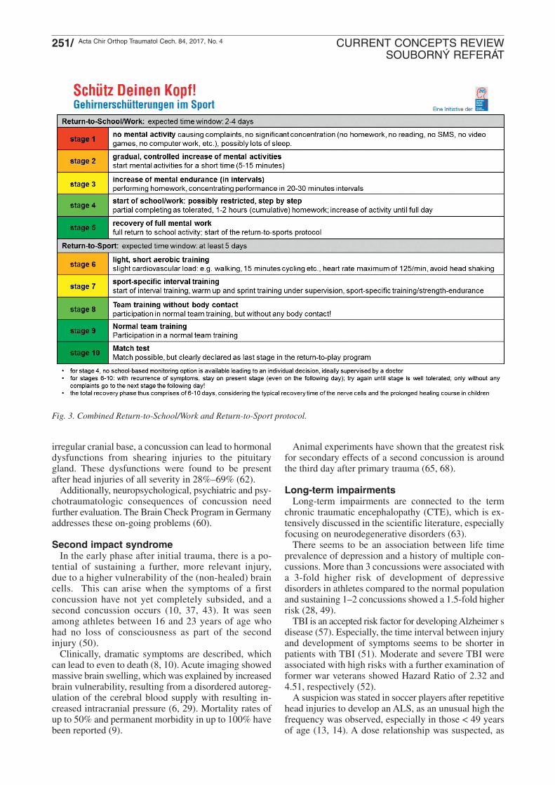
Return-to-work/school and return-to-playConsideration of the pathophysiology and the natural
recovery process is important in deciding return-to-play(44). An athlete should be at rest and after exercise clin-ically and cognitively symptom-free before competi-tiveness consists!
Prerequisite for return-to-play is the complete recoveryduring school or professional working. Therefore, a re-turn-to-play at the day of trauma is the absolute excep-tion.
Accordingly, a step-by-step recovery concept is rec-ommended, integrating the return-to school/work andreturn-to-sports (Fig. 3).
At least 6 to 10 days are normally necessary beforefull return-to sport is possible, which corresponds to theminimum recovery time of the affected neurons.
Post-concussion syndromeIn some patients, symptoms remain observable over a
longer time period (> 3 months, post-concussion syndrome– PCS). Overall, these symptoms are considered non-specific as many of these symptoms are present afterother injuries or diseases. Accordingly, a high prevalenceof PCS symptoms is observed in the normal population(26).
Because many of these symptoms are of differentorigin, specialized disciplines, e.g. neuropsychology,neurology, physiotherapy, otorhinolaryngology etc.)should be integrated in an interdisciplinary managementconcept, if symptom prolongation is extended > 4–6weeks after primary injury (3, 15).
Special focus should concentrate on vestibulo-oculardysfunction, which is observed, especially in childrenin up to 30%, resulting in a 2-fold prolongation ofrecovery (20). Due to force transmission along the
247_253_ganslen 14.8.17 14:55 Stránka 250
251/ Acta Chir Orthop Traumatol Cech. 84, 2017, No. 4 CURRENT CONCEPTS REVIEWSOUBORNÝ REFERÁT
irregular cranial base, a concussion can lead to hormonaldysfunctions from shearing injuries to the pituitarygland. These dysfunctions were found to be presentafter head injuries of all severity in 28%–69% (62).
Additionally, neuropsychological, psychiatric and psy-chotraumatologic consequences of concussion needfurther evaluation. The Brain Check Program in Germanyaddresses these on-going problems (60).
Second impact syndromeIn the early phase after initial trauma, there is a po-
tential of sustaining a further, more relevant injury,due to a higher vulnerability of the (non-healed) braincells. This can arise when the symptoms of a firstconcussion have not yet completely subsided, and asecond concussion occurs (10, 37, 43). It was seenamong athletes between 16 and 23 years of age whohad no loss of consciousness as part of the secondinjury (50).
Clinically, dramatic symptoms are described, whichcan lead to even to death (8, 10). Acute imaging showedmassive brain swelling, which was explained by increasedbrain vulnerability, resulting from a disordered autoreg-ulation of the cerebral blood supply with resulting in-creased intracranial pressure (6, 29). Mortality rates ofup to 50% and permanent morbidity in up to 100% havebeen reported (9).
Animal experiments have shown that the greatest riskfor secondary effects of a second concussion is aroundthe third day after primary trauma (65, 68).
Long-term impairmentsLong-term impairments are connected to the term
chronic traumatic encephalopathy (CTE), which is ex-tensively discussed in the scientific literature, especiallyfocusing on neurodegenerative disorders (63).
There seems to be an association between life timeprevalence of depression and a history of multiple con-cussions. More than 3 concussions were associated witha 3-fold higher risk of development of depressivedisorders in athletes compared to the normal populationand sustaining 1–2 concussions showed a 1.5-fold higherrisk (28, 49).
TBI is an accepted risk factor for developing Alzheimer sdisease (57). Especially, the time interval between injuryand development of symptoms seems to be shorter inpatients with TBI (51). Moderate and severe TBI wereassociated with high risks with a further examination offormer war veterans showed Hazard Ratio of 2.32 and4.51, respectively (52).
A suspicion was stated in soccer players after repetitivehead injuries to develop an ALS, as an unusual high thefrequency was observed, especially in those < 49 yearsof age (13, 14). A dose relationship was suspected, as
Fig. 3. Combined Return-to-School/Work and Return-to-Sport protocol.
247_253_ganslen 14.8.17 14:55 Stránka 251

252/ Acta Chir Orthop Traumatol Cech. 84, 2017, No. 4 CURRENT CONCEPTS REVIEWSOUBORNÝ REFERÁT
playing > 5 years was identified as a risk factor. Com-parable data were found in an US analysis, where a TBIsustained within the last 10 years increased the odds forALS development 11-fold (12). A meta-analysis reporteda 1.7-fold higher risk after TBI (12).
Whereas early results were indifferent in their conclusionon association between TBI and suicidality (32, 45, 66),a recent analysis on 235,119 patients after concussionsfound a 3-fold increased risk compared to the normalpopulation (23).
CONCLUSIONS
It can be concluded from these analyses, that there isa potential risk, even after sport-related concussion, butno clear recommendations can be made to the individualpatient. Recent results found a threshold dose-responserelationship between concussion and later life risk forcognitive impairment, subjective executive dysfunction,depression, apathy, and behavioral dysregulation.
References
1. af Geijerstam J, Britton M. Mild head injury - mortality and com-plication rate: meta-analysis of findings in a systematic literaturereview. Acta Neurochir (Wien). 2003;145:843–850.
2. Bazarian J. Use of computed tomographic scans for patients withminor head injury. Ann Emerg Med. 2002;39:348–349.
3. Benedict P, Baner N, Harrold G, Moehringer N, Hasanaj L,Serrano L, Sproul M, Pagnotta G, Cardone D, Flanagan S, RuckerJ, Galetta S, Balcer L. Gender and age predict outcomes ofcognitive, balance and vision testing in a multidisciplinaryconcussion center. J Neurol Sci. 2015; 353:11–115.
4. Benson B, Meeuwisse W, Rizos J, Kang J, Burke C. A prospectivestudy of concussions among National Hockey League playersduring regular season games: the NHLNHLPA Concussion Program.CMAJ. 2011;183:905–911.
5. Berlin A, Kop W, Deuster P. Depressive mood symptoms andfatigue after exercise withdrawal: the potential role of decreasedfitness. Psychosom Med. 2006;68:224–230.
6. Bowen A. Second impact syndrome: a rare, catastrophic, preventablecomplication of concussion in young athletes. J Emerg Nurs.2003;29:287–289.
7. Brown N, Mannix R, O'Brien M, Gostine D, Collins M, MeehanW. Effect of cognitive activity level on duration of post-concussionsymptoms. Pediatrics. 2014;133:e299-304.
8. Cantu R. Head injuries in sport. Br J Sports Med. 1996;30:289–296.
9. Cantu R. Athletic head injuries. Clin Sports Med. 1997;16:531–542.
10. Cantu R. Second-impact syndrome. Clin J Sports Med. 1998;17:37–44.
11. Carson J, Lawrence D, Kraft S, Garel A, Snow C, Chatterjee A,Libfeld P, MacKenzie H, Thornton J, Moineddin R, Frémont P.Premature return to play and return to learn after a sport-relatedconcussion. Physician’s chart review. Can Fam Phys. 2014;60:e310–e315.
12. Chen H, Richard M, Sandler D, Umbach D, Kamel F. Head injuryand amyotrophic lateral sclerosis. Am J Epidemiol. 2007;166:810–816.
13. Chio A, Benzi G, Dossena M, Mutani R, Mora G. Severelyincreased risk of amyotrophic lateral sclerosis among Italian pro-fessional foot-ball players. Brain. 2005;128:472–476.
14. Chio A, Calvo A, Dossena M, Ghiglione P, Mutani R, Mora G.ALS in Italian professional soccer players: the risk is still presentand could be soccer-specific. Amyotroph Lateral Scler. 2009;10:205–209.
15. Collins M, Kontos A, Reynolds E, Murawski C, Fu F. A compre-hensive, targeted approach to the clinical care of athletes followingsport-related concussion. Knee Surg Sports Traumatol Arthrosc.2014;22:235–246.
16. Davis G, Iverson G, Guskiewicz K, Ptito A, Johnston K. Contributionsof neuroimaging, balance testing, electrophysiology and bloodmarkers to the assessment of sport-related concussion. Br J SportsMed. 2009;43(Suppl 1):i36–45.
17. Delaney, J, Lacroix, V, Leclerc, S and Johnston, K. Concussionsamong university football and soccer players. Clin J Sports Med,2002. 12.331–338.
18. Delaney J, Lamfookon C, Bloom G, Al-Kashmiri A, Correa J. Whyuniversity athletes choose not to reveal their concussion symptomsduring a practice or game. Clin J Sports Med. 2015;25:113–125.
19. Dziemianowicz M, Kirschen M, Pukenas B, Laudano E, Balcer L,Galetta S. Sports-related concussion testing. Curr Neurol NeurosciRep. 2012;12:547–549.
20. Ellis M, Cordingley D, Vis S, Reimer K, Leiter J, Russell K.Vestibulo-ocular dysfunction in pediatric sports-related concussion.J Neurosurg Pediatr. 2015;16:248–255.
21. Filippidis A, Papadopoulos D, Kapsalaki E, Fountas K. Role ofthe S100B serum biomarker in the treatment of children sufferingfrom mild traumatic brain injury. Neurosurg Focus. 2010;29:E2.
22. Fraas M, Coughlan G, Hart E, McCarthy C. Concussion historyand reporting rates in elite Irish rugby union players. Phys TherSport. 2014;15:136–142.
23. Fralick M, Thiruchelvam D, Tien H, Redelmeier D. Risk ofsuicide after a concussion. CMAJ. 2016;188:497–504 [Epub 2016Feb 8].
24. Gibson S, Nigrovic L, O’Brien M, Meehan W. The effect of rec-ommending cognitive rest on recovery from sport-related concussion.Brain Inj. 2013;27:839–842.
25. Giza C, Hovda D. The new neurometabolic cascade of concussion.Neurosurgery. 2014;(Suppl 4):S24–S33.
26. Gouvier W, Cubic B, Jones G, Brantley P, Cutlip Q. Postconcussionsymptoms and daily stress in normal and head-injured collegepopulations. Arch Clin Neuropsychol. 1992;7:93–211.
27. Grubenhoff J, Deakyne S, Brou L, Bajaj L, Comstock R, KirkwoodM. Acute concussion symptom severity and delayed symptomresolution. Pediatrics 2014;134:54–62.
28. Guskiewicz K, Marshall S, Bailes J, McCrea M, Harding HP Jr,Matthews A, Mihalik JR, Cantu R. Recurrent concussion and riskof depression in retired professional football players. Med SciSports Exerc. 2007;39:903–909.
29. Guskiewicz K, McCrea M, Marshall S, Cantu R, Randolph C,Barr W. Cumulative effects associated with recurrent concussionin collegiate football players: the NCAA Concussion Study.JAMA. 2003;290:2549–2555.
30. Haydel M, Preston C, Mills T, Luber S, Blaudeau E, DeBlieux P.Indications for computed tomog- raphy in patients with minorhead injury. N Engl J Med. 2000;343:100–105.
31. Hwang V, Trickey A, Lormel C, Bradford A, Griffen M, LawrenceC, Sturek C, Stacey E, Howell J. Are pediatric concussion patientscompliant with discharge instructions? J Trauma Acute Care Surg.2014;77:117–122.
32. Iverson G. Chronic traumatic encephalopathy and risk of suicidein former athletes. Br J Sports Med. 2014;48:162–165.
33. Kontos A, Braithwaite R, Dakan S, Elbin R. Computerized neu-rocognitive testing within 1 week of sport-related concussion:meta-analytic review and analysis of moderating factors. J IntNeuropsychol Soc. 2014;20:324–332.
34. Kroshus E, Baugh C, Daneshvar D, Stamm J, Laursen R, AustinS. Pressure on Sports Medicine Clinicians to Prematurely ReturnCollegiate Athletes to Play After Concussion. J Athl Train.2015;50:944–951.
35. Lau B, Kontos A, Collins M, Mucha A, Lovell M. Which on-fieldsigns/symptoms predict protracted recovery from sport-relatedconcussion among high school football players? Am J SportsMed. 2011;39:2311-2318 [Epub 2011 Jun 28].
36. Llewellyn T, Burdette G, Joyner A, Buckley T. Concussionreporting rates at the conclusion of an intercollegiate athleticcareer. Clin J Sports Med. 2014;24:76–79.
247_253_ganslen 14.8.17 14:55 Stránka 252
253/ Acta Chir Orthop Traumatol Cech. 84, 2017, No. 4 CURRENT CONCEPTS REVIEWSOUBORNÝ REFERÁT
37. Lovell M, Fazio V. Concussion management in the child and ado-lescent athlete. Curr Sports Med Rep. 2008;7:12–15.
38. MacFarlane M, Glenn T. Neurochemical cascade of concussion.Brain Inj. 2015;29:1391–1352.
39. Makdissi M, Darby D, Maruff P, Ugoni A, Brukner P, McCrory P.Natural history of concussion in sport: markers of severity andimplications for management. Am J Sports Med. 2010;38:464–471.
40. McCrea M. Mild traumatic brain injury and postconcussion syn-drome. Oxford University Press, Oxford, 2008.
41. McCrea M, Guskiewicz K, Marshall S. Acute effects and recoverytime following concussion in collegiate football players: theNCAA Concussion Study. JAMA. 2003;290:2556–2563.
42. McCrea M, Hammeke T, Olsen G, Leo P, Guskiewicz K. Unreportedconcussion in high school football players: implications for pre-vention. Clin J Sports Med. 2004;14:13–17.
43. McCrory P, Berkovic S. Second impact syndrome. Neurology.1998;50:677–683.
44. McCrory P, Meeuwisse W, Aubry M, Cantu R, Dvorak J, EchemendiaR, Engebretsen L, Johnston K, Kutcher J, Raftery M, Sills A,Schneider K, Tator C, Benson B, Davis G, Ellenbogan R, GuskiewiczK, Herring S, Iverson G, Jordan B, Kissick J, McCrea M, McIntoshA, Maddocks D, Makdissi M, Purcell L, Putukian M, SchneiderK, Tator C. Consensus statement on concussion in sport: the 4th
International Conference on Concussion in Sport held in Zurich,November 2012. Br J Sports Med. 2013;47:250–258.
45. McKee A, Stern R, Nowinski C, Stein T, Alvarez V, Daneshvar D,Lee H, Wojtowicz S, Hall G, Baugh C, Riley D, Kubilus C,Cormier K, Jacobs M, Martin B, Abraham C, Ikezu T, Reichard R,Wolozin B, Budson A, Goldstein L, Kowall N, Cantu R. Thespectrum of disease in chronic traumatic encephalopathy. Brain.2013;136:43–64.
46. Meaney D, Smith D. Biomechanics of concussion. Clin SportsMed. 2011;30:19–31.
47. Meehan WR, d'Hemecourt P, Comstock R. High school concussionsin the 2008–2009 academic year: mechanism, symptoms, andmanagement. Am J Sports Med. 2010;38:2405–2409.
48. Meehan, Wr, Mannix, R, O'Brien, M and Collins, M. The prevalenceof undiagnosed concussions in athletes. Clin J Sports Med, 2013.23.339–342.
49. Montenigro P, Alosco M, Martin B, Dneshvar D, Mez J, ChaissonC, Nowinski C, Au R, McKee A, Cantu R, McClean M, Stern R,Tripodis Y. Cumulative Head impact exposure predicts later-lifedepression, apathy, executive dysfunction, and cognitive impairmentin former high school and college football players. J Neurotrauma.2017;34:328-340 [Epub March 30, 2016].
50. Mori T, Katayama Y, Kawamata T. Acute hemispheric swellingassociated with thin subdural hematomas: pathophysiology ofrepetitive head injury in sports. Acta Neurochir. 2006;96(Sup-pl):40–43.
51. Nemetz P, Leibson C, Naessens J, Beard M, Kokmen E, AnnegersJ, Kurland L. Traumatic brain injury and time to onset of Alzheimer'sdisease: a population-based study. Am J Epidemiol. 1999;149:32–40.
52. Plassman B, Havli, R, Steffen, D, Helm, M, Newma, T, Drosdic,D, Phillip, C, Ga, B, Welsh-Bohme, K, Burk, J, Guralnik J,Breitner J. Documented head injury in early adulthood and risk ofAlzheimer's disease and other dementias. Neurology. 2000;55:1158–1166.
53. Ransom D, Vaughan C, Pratson L, Sady M, McGill C, Gioia G.Academic effects of concussion in children and adolescents. Pe -diatrics. 2015;135:1043–1050.
54. Rickels E. Diagnostik und Therapie von Schädel-Hirn-Traumen.Chirurg. 2009;80:153–163.
55. Rickels E, von Wild K, Wenzlaff P, Bock W (Hrsg.). Schädel-Hirn-Verletzung – Epidemiologie und Versorgung: Ergebnisse einerprospektiven Studie. Zuckschwerdt; Auflage 1, 2006.
56. Rose S, McNally K, Heyer G. Returning the student to schoolafter concussion: what do clinicians need to know?. Concussion(Future Medicine). 2015;1: [doi 10.2217/cnc.15.4].
57. Rubenstein R. Traumatic brain injury: risk factors and biomarkersof Alzheimer's disease and chronic traumatic encephalopathy.Curr Tran Geriatr Gerontol Rep. 2012;1:143–148.
58. Ruhe A, Gänsslen A, Klein W. The incidence of concussion inprofessional and collegiate ice hockey: are we making progress?A systematic review of the literature. Br J Sports Med. 2014;48:102–106.
59. Schatz P, Pardini, J, Lovell, M, Collins, M and Podell, K. Sensitivityand specificity of the ImPACT Test Battery for concussion inathletes. Arch Clin Neuropsychol. 2006;21:91–99.
60. Schmehl I, Johl U, Sparenberg P, Kinze S, Dahne F, Rogge W.Brain-Check nach Schadel-Hirn-Trauma. Diagnostisches Modulzur Ermittlung von Folgeschaden. Trauma Berufskrankh. 2011;13:12–17.
61. Schulte S, Podlog L, Hamson-Utley J, Strathmann F, Strüder H. Asystematic review of the biomarker S100B: implications for sport-related concussion management. J Athl Train. 2014;49:830–850.
62. Scranton R, Baskin D. Impaired pituitary axes following traumaticbrain injury. J Clin Med. 2015;4:1463–1479.
63. Stein T, Alvarez V, McKee A. Concussion in chronic traumatic en-cephalopathy. Curr Pain Headache Rep. 2015;19:47 [doi10.1007/s11916-015-0522-z].
64. Stiell I, Wells G, Vandemheen K, Clement C, Lesiuk H, LaupacisA. The Canadian CT Head Rule for patients with minor headinjury. Lancet. 2001;357:1391–1396.
65. Tavazzi B, Vagnozzi R, Signoretti S, Amorini A, Belli A, CimattiM, Delfini R, Di Pietro V, Finocchiaro A, Lazzarino G. Temporalwindow of metabolic brain vulnerability to concussions: oxidativeand nitrosative stresses – part II. Neurosurgery. 2007;61:390–396.
66. Teasdale T, Engberg A. Suicide after traumatic brain injury: apopulation study. J Neurol Neurosurg Psychiatry. 2001;71:436–440.
67. Thomas D, Apps J, Hoffmann R, McCrea M, Hammeke T. Benefitsof strict rest after acute concussion: a randomized controlled trial.Pediatrics. 2015;135:213–223.
68. Vagnozzi R, Tavazzi B, Signoretti S, Amorini A, Belli A, CimattiM, Delfini R, Di Pietro V, Finocchiaro A, Lazzarino G. Temporalwindow of metabolic brain vulnerability to concussions: mito-chondrial-related impairment - part I. Neurosurgery. 2007;61:379–389.
69. Williamson I, Goodman D. Converging evidence for the under-re-porting of concussions in youth ice hockey. Br J Sports Med.2006;40:128–132.
Corresponding authorDr. med. A. GänsslenTrauma Department, Klinikum WolfsburgSauerbruchstrasse 7D-38440 Wolfsburg, GermanyE-mail: [email protected]
247_253_ganslen 14.8.17 14:55 Stránka 253