Embed Size (px)
Citation preview
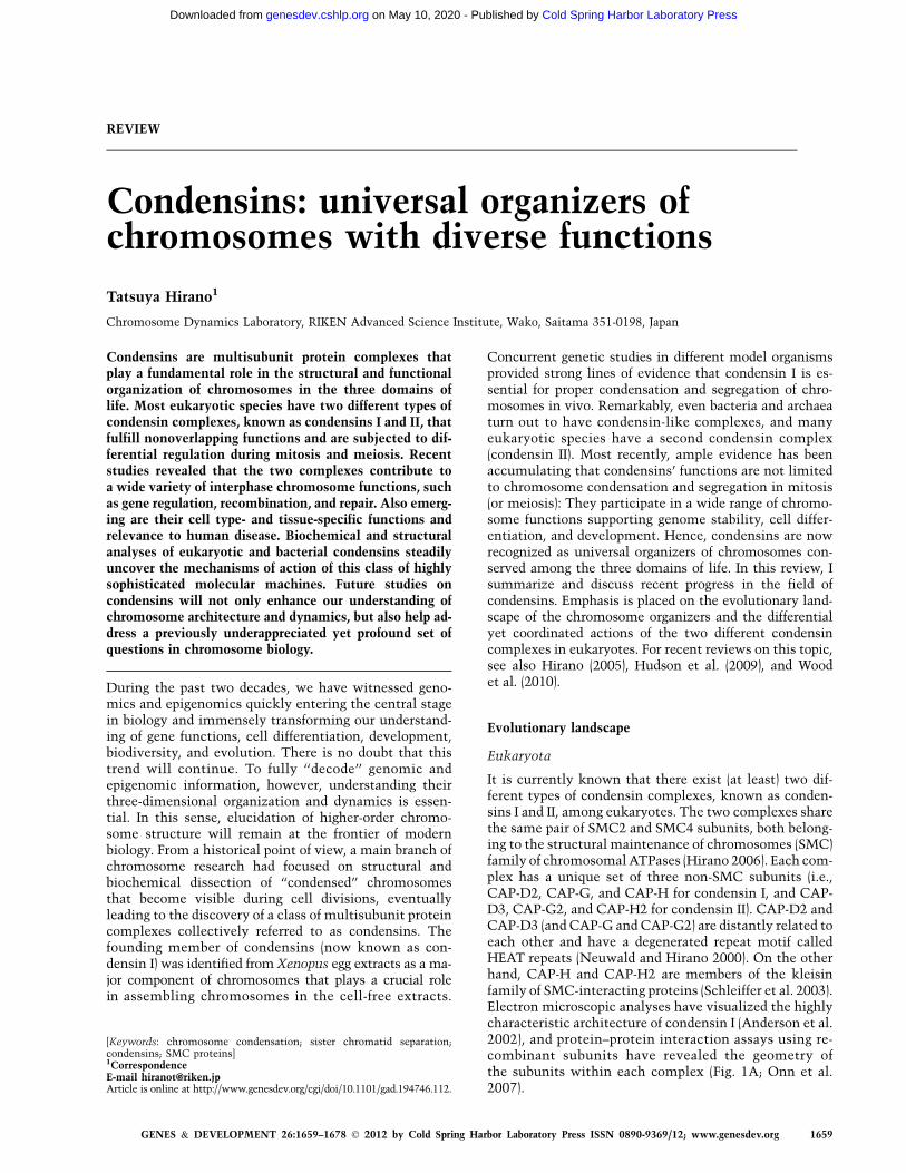
REVIEW
Condensins: universal organizers ofchromosomes with diverse functions
Tatsuya Hirano1
Chromosome Dynamics Laboratory, RIKEN Advanced Science Institute, Wako, Saitama 351-0198, Japan
Condensins are multisubunit protein complexes thatplay a fundamental role in the structural and functionalorganization of chromosomes in the three domains oflife. Most eukaryotic species have two different types ofcondensin complexes, known as condensins I and II, thatfulfill nonoverlapping functions and are subjected to dif-ferential regulation during mitosis and meiosis. Recentstudies revealed that the two complexes contribute toa wide variety of interphase chromosome functions, suchas gene regulation, recombination, and repair. Also emerg-ing are their cell type- and tissue-specific functions andrelevance to human disease. Biochemical and structuralanalyses of eukaryotic and bacterial condensins steadilyuncover the mechanisms of action of this class of highlysophisticated molecular machines. Future studies oncondensins will not only enhance our understanding ofchromosome architecture and dynamics, but also help ad-dress a previously underappreciated yet profound set ofquestions in chromosome biology.
During the past two decades, we have witnessed geno-mics and epigenomics quickly entering the central stagein biology and immensely transforming our understand-ing of gene functions, cell differentiation, development,biodiversity, and evolution. There is no doubt that thistrend will continue. To fully ‘‘decode’’ genomic andepigenomic information, however, understanding theirthree-dimensional organization and dynamics is essen-tial. In this sense, elucidation of higher-order chromo-some structure will remain at the frontier of modernbiology. From a historical point of view, a main branch ofchromosome research had focused on structural andbiochemical dissection of ‘‘condensed’’ chromosomesthat become visible during cell divisions, eventuallyleading to the discovery of a class of multisubunit proteincomplexes collectively referred to as condensins. Thefounding member of condensins (now known as con-densin I) was identified from Xenopus egg extracts as a ma-jor component of chromosomes that plays a crucial rolein assembling chromosomes in the cell-free extracts.
Concurrent genetic studies in different model organismsprovided strong lines of evidence that condensin I is es-sential for proper condensation and segregation of chro-mosomes in vivo. Remarkably, even bacteria and archaeaturn out to have condensin-like complexes, and manyeukaryotic species have a second condensin complex(condensin II). Most recently, ample evidence has beenaccumulating that condensins’ functions are not limitedto chromosome condensation and segregation in mitosis(or meiosis): They participate in a wide range of chromo-some functions supporting genome stability, cell differ-entiation, and development. Hence, condensins are nowrecognized as universal organizers of chromosomes con-served among the three domains of life. In this review, Isummarize and discuss recent progress in the field ofcondensins. Emphasis is placed on the evolutionary land-scape of the chromosome organizers and the differentialyet coordinated actions of the two different condensincomplexes in eukaryotes. For recent reviews on this topic,see also Hirano (2005), Hudson et al. (2009), and Woodet al. (2010).
Evolutionary landscape
Eukaryota
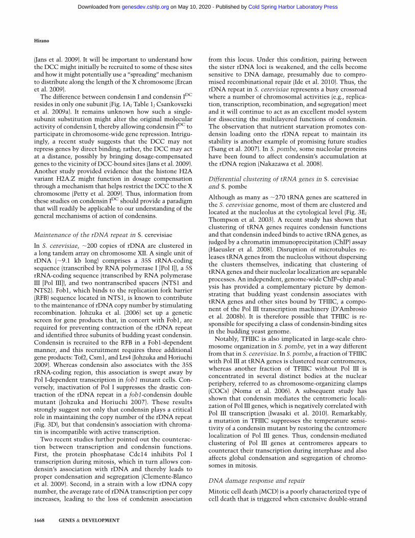
It is currently known that there exist (at least) two dif-ferent types of condensin complexes, known as conden-sins I and II, among eukaryotes. The two complexes sharethe same pair of SMC2 and SMC4 subunits, both belong-ing to the structural maintenance of chromosomes (SMC)family of chromosomal ATPases (Hirano 2006). Each com-plex has a unique set of three non-SMC subunits (i.e.,CAP-D2, CAP-G, and CAP-H for condensin I, and CAP-D3, CAP-G2, and CAP-H2 for condensin II). CAP-D2 andCAP-D3 (and CAP-G and CAP-G2) are distantly related toeach other and have a degenerated repeat motif calledHEAT repeats (Neuwald and Hirano 2000). On the otherhand, CAP-H and CAP-H2 are members of the kleisinfamily of SMC-interacting proteins (Schleiffer et al. 2003).Electron microscopic analyses have visualized the highlycharacteristic architecture of condensin I (Anderson et al.2002), and protein–protein interaction assays using re-combinant subunits have revealed the geometry ofthe subunits within each complex (Fig. 1A; Onn et al.2007).
[Keywords: chromosome condensation; sister chromatid separation;condensins; SMC proteins]1CorrespondenceE-mail [email protected] is online at http://www.genesdev.org/cgi/doi/10.1101/gad.194746.112.
GENES & DEVELOPMENT 26:1659–1678 � 2012 by Cold Spring Harbor Laboratory Press ISSN 0890-9369/12; www.genesdev.org 1659
Cold Spring Harbor Laboratory Press on May 10, 2020 - Published by genesdev.cshlp.orgDownloaded from
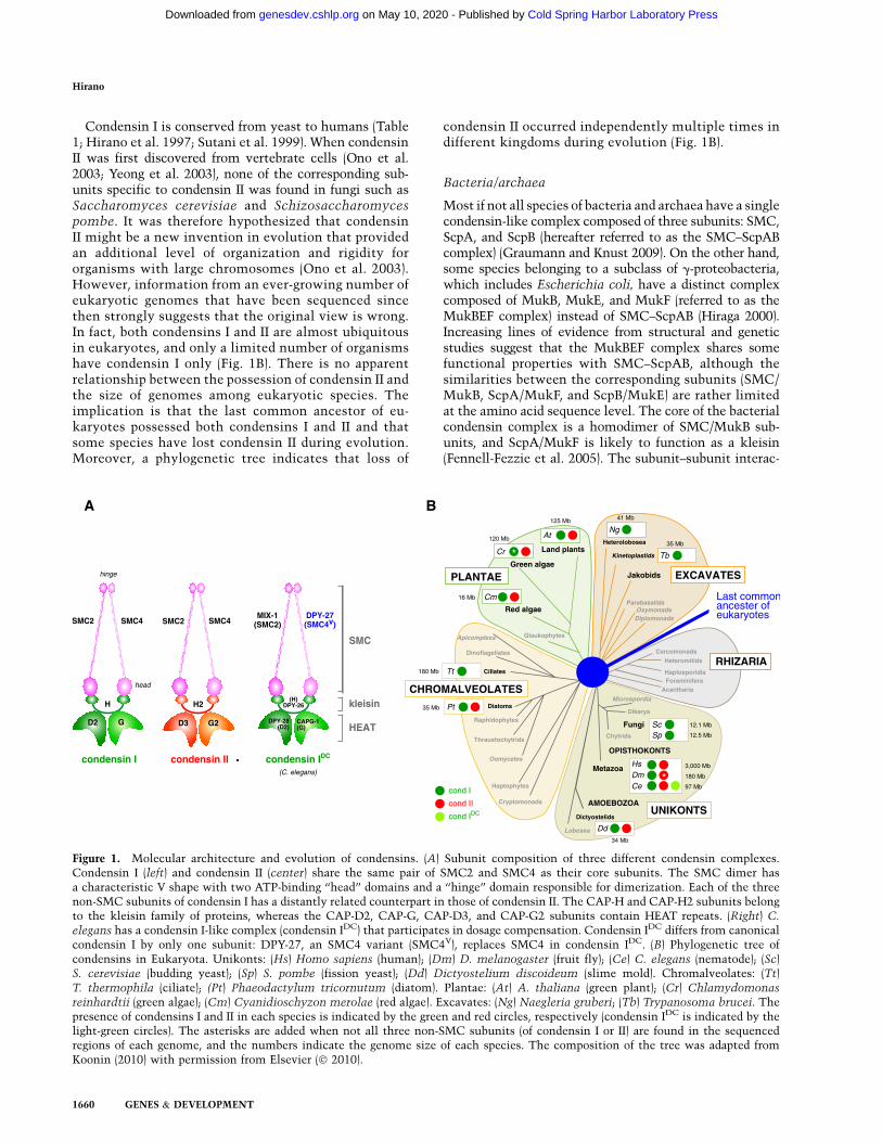
Condensin I is conserved from yeast to humans (Table1; Hirano et al. 1997; Sutani et al. 1999). When condensinII was first discovered from vertebrate cells (Ono et al.2003; Yeong et al. 2003), none of the corresponding sub-units specific to condensin II was found in fungi such asSaccharomyces cerevisiae and Schizosaccharomycespombe. It was therefore hypothesized that condensinII might be a new invention in evolution that providedan additional level of organization and rigidity fororganisms with large chromosomes (Ono et al. 2003).However, information from an ever-growing number ofeukaryotic genomes that have been sequenced sincethen strongly suggests that the original view is wrong.In fact, both condensins I and II are almost ubiquitousin eukaryotes, and only a limited number of organismshave condensin I only (Fig. 1B). There is no apparentrelationship between the possession of condensin II andthe size of genomes among eukaryotic species. Theimplication is that the last common ancestor of eu-karyotes possessed both condensins I and II and thatsome species have lost condensin II during evolution.Moreover, a phylogenetic tree indicates that loss of
condensin II occurred independently multiple times indifferent kingdoms during evolution (Fig. 1B).
Bacteria/archaea
Most if not all species of bacteria and archaea have a singlecondensin-like complex composed of three subunits: SMC,ScpA, and ScpB (hereafter referred to as the SMC–ScpABcomplex) (Graumann and Knust 2009). On the other hand,some species belonging to a subclass of g-proteobacteria,which includes Escherichia coli, have a distinct complexcomposed of MukB, MukE, and MukF (referred to as theMukBEF complex) instead of SMC–ScpAB (Hiraga 2000).Increasing lines of evidence from structural and geneticstudies suggest that the MukBEF complex shares somefunctional properties with SMC–ScpAB, although thesimilarities between the corresponding subunits (SMC/MukB, ScpA/MukF, and ScpB/MukE) are rather limitedat the amino acid sequence level. The core of the bacterialcondensin complex is a homodimer of SMC/MukB sub-units, and ScpA/MukF is likely to function as a kleisin(Fennell-Fezzie et al. 2005). The subunit–subunit interac-
Figure 1. Molecular architecture and evolution of condensins. (A) Subunit composition of three different condensin complexes.Condensin I (left) and condensin II (center) share the same pair of SMC2 and SMC4 as their core subunits. The SMC dimer hasa characteristic V shape with two ATP-binding ‘‘head’’ domains and a ‘‘hinge’’ domain responsible for dimerization. Each of the threenon-SMC subunits of condensin I has a distantly related counterpart in those of condensin II. The CAP-H and CAP-H2 subunits belongto the kleisin family of proteins, whereas the CAP-D2, CAP-G, CAP-D3, and CAP-G2 subunits contain HEAT repeats. (Right) C.
elegans has a condensin I-like complex (condensin IDC) that participates in dosage compensation. Condensin IDC differs from canonicalcondensin I by only one subunit: DPY-27, an SMC4 variant (SMC4V), replaces SMC4 in condensin IDC. (B) Phylogenetic tree ofcondensins in Eukaryota. Unikonts: (Hs) Homo sapiens (human); (Dm) D. melanogaster (fruit fly); (Ce) C. elegans (nematode); (Sc)S. cerevisiae (budding yeast); (Sp) S. pombe (fission yeast); (Dd) Dictyostelium discoideum (slime mold). Chromalveolates: (Tt)T. thermophila (ciliate); (Pt) Phaeodactylum tricornutum (diatom). Plantae: (At) A. thaliana (green plant); (Cr) Chlamydomonas
reinhardtii (green algae); (Cm) Cyanidioschyzon merolae (red algae). Excavates: (Ng) Naegleria gruberi; (Tb) Trypanosoma brucei. Thepresence of condensins I and II in each species is indicated by the green and red circles, respectively (condensin IDC is indicated by thelight-green circles). The asterisks are added when not all three non-SMC subunits (of condensin I or II) are found in the sequencedregions of each genome, and the numbers indicate the genome size of each species. The composition of the tree was adapted fromKoonin (2010) with permission from Elsevier (� 2010).
Hirano
1660 GENES & DEVELOPMENT
Cold Spring Harbor Laboratory Press on May 10, 2020 - Published by genesdev.cshlp.orgDownloaded from
tions are possibly highly dynamic, and the subunit stoi-chiometry in a single complex might be either (SMC/MukB)2(ScpA/MukF)2(ScpB/MukE)2 or (SMC/MukB)2(ScpA/MukF)2(ScpB/MukE)4 (Hirano and Hirano 2004; Gloyd et al.2007; Woo et al. 2009).
Overview of essential functions
Recent series of genetic and cell biological studies revealthat condensins I and II fulfill nonoverlapping functionsin many aspects of chromosome biology. Intriguingly,however, the relative contribution of the two condensincomplexes to mitotic chromosome assembly and segre-gation apparently varies among different eukaryotic spe-cies, as discussed below.
Mammals
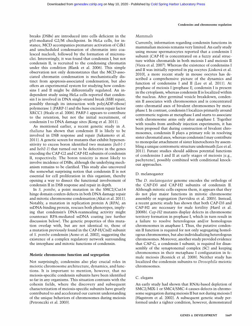
Early studies employing siRNA-mediated gene knock-down in human tissue culture cells demonstrated thatcondensins I and II have differential contributions tomitotic chromosome architecture and segregation, im-plying that both complexes play essential roles in mitosis(Ono et al. 2003, 2004; Hirota et al. 2004). These studiesalso showed that the two condensin complexes displaystrikingly different behaviors during the cell cycle (Fig.2A, from top to right). Condensin I is sequestered in thecytoplasm during interphase and gains access to chromo-somes only after the nuclear envelope breaks down inprometaphase. In contrast, condensin II localizes to thenucleus from interphase through prophase and participatesin an early stage of chromosome condensation within theprophase nucleus. After nuclear envelope breakdown(NEBD), condensins I and II collaborate to support properassembly of chromosomes in which sister chromatids arewell resolved by metaphase and to promote faithfulsegregation in anaphase. The characteristic behaviors ofthe two condensin complexes are most likely to be centralto our understanding of their action and regulation. At thelevel of ontogeny, knocking out the gene encoding theCAP-G2 subunit in mice has been shown to cause embry-onic lethality (Smith et al. 2004; Xu et al. 2006). Whereas
knockout mice for the other subunits have not yet beendescribed, all of them are anticipated to be essential forembryogenesis. Remarkably, recent studies started to un-cover tissue-specific developmental defects caused by a hy-pomorphic mutation or misregulation of condensin sub-units, which will be discussed in later sections.
Chicken DT40 cells
Generation of conditional knockout cell lines by usingchicken DT40 cells offers an alternative method forsilencing target genes of interest and is arguably superiorto siRNA-mediated knockdown techniques widely usedin mammalian cells. Such cell lines can be established bythe introduction of a tetracycline-regulated transgene,followed by disruption of the authentic loci throughhomologous recombination. Hudson et al. (2003) appliedthis technique to the condensin core subunit SMC2 (alsoknown as ScII) and showed that it is required for non-histone protein assembly and structural integrity of mi-totic chromosomes. A subsequent mutational study dem-onstrated that ATP binding, but not hydrolysis, by SMC2is required for its stable association with chromosomes(Hudson et al. 2008). In DT40 cells depleted of SMC2,defects in chromosome condensation are relatively modestin metaphase but become very severe in anaphase, leadingto the proposal that a hypothetical factor termed regulatorof chromosome architecture (RCA) might cooperate withcondensins to preserve the characteristic shape of meta-phase chromosomes (Vagnarelli et al. 2006).
Xenopus laevis (cell-free egg extracts)
Metaphase chromosomes can be reconstituted in vitrostarting from simple substrates, such as sperm chroma-tin, in Xenopus cell-free egg extracts. Biochemical char-acterization of the chromosomes assembled in the cell-free extracts led to the discovery of the first condensincomplex, now known as condensin I (Hirano and Mitchison1994; Hirano et al. 1997). A subsequent study showedthat condensin II is also present in the cell-free extracts,albeit as a less abundant component compared with
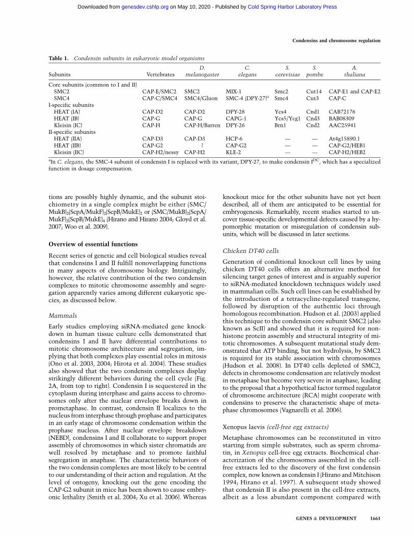
Table 1. Condensin subunits in eukaryotic model organisms
Subunits VertebratesD.
melanogasterC.
elegansS.
cerevisiaeS.
pombeA.
thaliana
Core subunits (common to I and II)SMC2 CAP-E/SMC2 SMC2 MIX-1 Smc2 Cut14 CAP-E1 and CAP-E2SMC4 CAP-C/SMC4 SMC4/Gluon SMC-4 (DPY-27)a Smc4 Cut3 CAP-C
I-specific subunitsHEAT (IA) CAP-D2 CAP-D2 DPY-28 Ycs4 Cnd1 CAB72176HEAT (IB) CAP-G CAP-G CAPG-1 Ycs5/Ycg1 Cnd3 BAB08309Kleisin (IC) CAP-H CAP-H/Barren DPY-26 Brn1 Cnd2 AAC25941
II-specific subunitsHEAT (IIA) CAP-D3 CAP-D3 HCP-6 — — At4g15890.1HEAT (IIB) CAP-G2 ? CAP-G2 — — CAP-G2/HEB1Kleisin (IIC) CAP-H2/nessy CAP-H2 KLE-2 — — CAP-H2/HEB2
aIn C. elegans, the SMC-4 subunit of condensin I is replaced with its variant, DPY-27, to make condensin IDC, which has a specializedfunction in dosage compensation.
Condensins and chromosome regulation
GENES & DEVELOPMENT 1661
Cold Spring Harbor Laboratory Press on May 10, 2020 - Published by genesdev.cshlp.orgDownloaded from
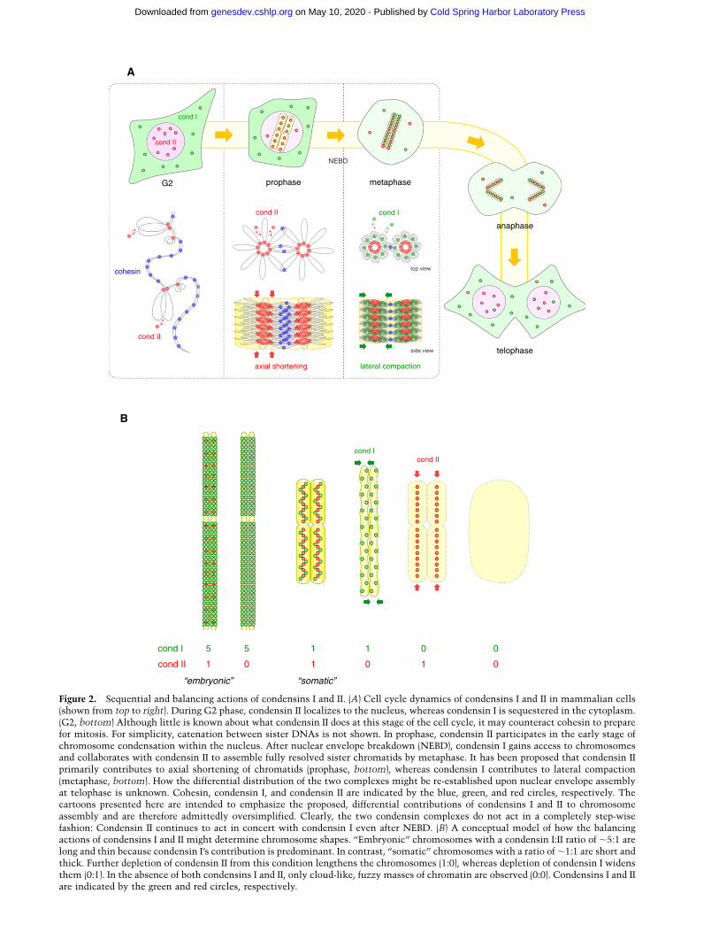
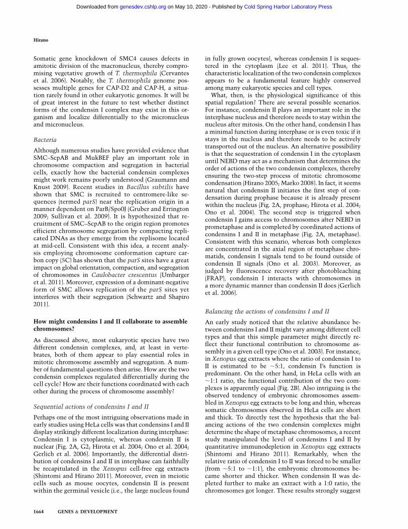
Figure 2. Sequential and balancing actions of condensins I and II. (A) Cell cycle dynamics of condensins I and II in mammalian cells(shown from top to right). During G2 phase, condensin II localizes to the nucleus, whereas condensin I is sequestered in the cytoplasm.(G2, bottom) Although little is known about what condensin II does at this stage of the cell cycle, it may counteract cohesin to preparefor mitosis. For simplicity, catenation between sister DNAs is not shown. In prophase, condensin II participates in the early stage ofchromosome condensation within the nucleus. After nuclear envelope breakdown (NEBD), condensin I gains access to chromosomesand collaborates with condensin II to assemble fully resolved sister chromatids by metaphase. It has been proposed that condensin IIprimarily contributes to axial shortening of chromatids (prophase, bottom), whereas condensin I contributes to lateral compaction(metaphase, bottom). How the differential distribution of the two complexes might be re-established upon nuclear envelope assemblyat telophase is unknown. Cohesin, condensin I, and condensin II are indicated by the blue, green, and red circles, respectively. Thecartoons presented here are intended to emphasize the proposed, differential contributions of condensins I and II to chromosomeassembly and are therefore admittedly oversimplified. Clearly, the two condensin complexes do not act in a completely step-wisefashion: Condensin II continues to act in concert with condensin I even after NEBD. (B) A conceptual model of how the balancingactions of condensins I and II might determine chromosome shapes. ‘‘Embryonic’’ chromosomes with a condensin I:II ratio of ;5:1 arelong and thin because condensin I’s contribution is predominant. In contrast, ‘‘somatic’’ chromosomes with a ratio of ;1:1 are short andthick. Further depletion of condensin II from this condition lengthens the chromosomes (1:0), whereas depletion of condensin I widensthem (0:1). In the absence of both condensins I and II, only cloud-like, fuzzy masses of chromatin are observed (0:0). Condensins I and IIare indicated by the green and red circles, respectively.
Cold Spring Harbor Laboratory Press on May 10, 2020 - Published by genesdev.cshlp.orgDownloaded from
condensin I (Ono et al. 2003). In accordance with therelative ratio between the two complexes (condensinI:condensin II = ;5:1), condensin I plays a predominantrole in chromosome assembly, whereas condensin II hasa minor contribution to this process in this cell-freeextract. As discussed later in this review, a more recentstudy using this experimental system has demonstratedthat the relative ratio of condensin I to II indeed acts asa critical factor that determines the shape of mitoticchromosomes (Shintomi and Hirano 2011).
Drosophila melanogaster
Early genetic studies in Drosophila showed that properassembly and segregation of mitotic chromosomes requirecondensin I subunits, including SMC4/Gluon (Steffensenet al. 2001), CAP-H/Barren (Bhat et al. 1996), CAP-G (Dejet al. 2004), and CAP-D2 (Savvidou et al. 2005). It has alsobeen shown that condensin I contributes to proper locali-zation of topoisomerase II (topo II) to the chromosomeaxis (Coelho et al. 2003) and maintenance of the struc-tural integrity of centromeric heterochromatin (Oliveiraet al. 2005). It remains to be determined whether the five-subunit condensin II complex is present in Drosophilabecause the gene encoding CAP-G2 has not yet beenfound in the sequenced region of the Drosophila genome.Currently available evidence suggests that the putativecondensin II subunits (CAP-D3 and CAP-H2) may notplay a major role in mitotic chromosome organizationand segregation (Savvidou et al. 2005; Oliveira et al. 2007)and that they have meiotic functions instead, makinga crucial contribution to anaphase I chromosome segre-gation (Hartl et al. 2008b; see below).
Caenorhabditis elegans
Unlike other eukaryotes, C. elegans has three condensincomplexes (Csankovszki et al. 2009a). In addition tocondensins I and II, this organism possesses a condensinI-like complex (known as condensin IDC) that constitutesa part of the large protein complex involved in dosagecompensation. Condensin IDC differs from the canonicalcondensin I complex by only one subunit (Fig. 1A; Table 1).It is assumed that duplication of SMC4 during evolutiongave rise to DPY-27, leading to the creation of condensinIDC with a specialized function of chromosome-wide generepression. Notably, the mitotic phenotypes observed incondensin I-deficient cells are far less severe than those incondensin II-deficient cells, implying that condensin IIplays a dominant role during mitosis in this organism.This atypical observation could be related to the fact thatC. elegans has a unique holocentric structure in whichcentromeres/kinetochores assemble and function alongthe entire chromosome arms (Hagstrom et al. 2002).
Fungi
None of the genes encoding condensin II-specific sub-units can be found in the genome of fungi. In S. pombe,the five-subunit condensin I complex localizes to thecytoplasm during interphase and relocates to the nucleus
in mitosis to support chromosome condensation andsegregation (Sutani et al. 1999). Unlike in S. pombe, thecondensin subunits localize to the nucleus throughoutthe cell cycle in S. cerevisiae (Freeman et al. 2000).Although this behavior is reminiscent of that of con-densin II in human cells, the amino acid sequences of thesubunits of S. cerevisiae condensin clearly indicate thatit is condensin I. The constitutive nuclear localization ofcondensin I in S. cerevisiae could be related to its ratherpeculiar cell cycle in which S-phase and M-phase eventspartially overlap with each other (e.g., the spindle beginsto form in late S phase, and there is no clear G2 phase).Whereas many early studies on S. cerevisiae condensinfocused on its role in promoting the condensation andsegregation of rDNA (e.g., Freeman et al. 2000; Lavoieet al. 2004), more recent, state-of-the-art imaging tech-niques have uncovered the function of condensin I asa chromatin ‘‘spring’’ at pericentromeric regions in meta-phase (Stephens et al. 2011) and its contribution to‘‘recoiling’’ of stretched chromosome arms in anaphase(Renshaw et al. 2010).
Arabidopsis thaliana
The genome of A. thaliana encodes all subunits of con-densins I and II (Hirano 2005). Whereas the A. thalianagenome has a single essential gene (AtCAP-C) encodingSMC4 (Siddiqui et al. 2006), it possesses two paralogousgenes for SMC2, known as AtCAP-E1 and AtCAP-E2 (Siddiqui et al. 2003). The two SMC2 proteins arefunctionally redundant: Single-homozygous mutants(E1�/� and E2�/�) are viable, whereas double-homozygous(E1�/�E2�/�) and E1�/�E2+/� plants are both embryonic-lethal. On the other hand, an E1+/�E2�/� plant results inmeristem disorganization and fasciation, indicating thatSMC2 activity above a certain threshold is required forproper development of A. thaliana. Very recently, a geneticscreen for mutants that exhibit hypersensitivity to excessboron has led to the somewhat surprising conclusion thatcondensin II is nonessential for mitosis and instead playsa role in alleviating DNA damage (Sakamoto et al. 2011).Whereas genetic studies have yet to be reported for thegenes encoding condensin I-specific subunits, it is antici-pated that condensin I is sufficient for mitotic chromosomeassembly and segregation in this organism.
Tetrahymena thermophila
T. thermophila is a ciliate that maintains two nuclei,a germline micronucleus and a somatic macronucleus, ina cell. The micronucleus divides by mitosis in a mannersimilar to that observed in many other eukaryotes. Onthe other hand, the formation and division of the macro-nucleus is highly unique: It is derived from the micronu-cleus through genomic rearrangement, contains >200 chro-mosome fragments ranging in size from 20 to >3000 kb,and divides by a poorly characterized mechanism of‘‘amitosis’’ (Yao and Chao 2005). The macronucleusgenome of T. thermophila encodes SMC2, SMC4, andall condensin I-specific subunits but lacks the genesfor condensin II-specific subunits (Eisen et al. 2006).
Condensins and chromosome regulation
GENES & DEVELOPMENT 1663
Cold Spring Harbor Laboratory Press on May 10, 2020 - Published by genesdev.cshlp.orgDownloaded from
Somatic gene knockdown of SMC4 causes defects inamitotic division of the macronucleus, thereby compro-mising vegetative growth of T. thermophila (Cervanteset al. 2006). Notably, the T. thermophila genome pos-sesses multiple genes for CAP-D2 and CAP-H, a situa-tion rarely found in other eukaryotic genomes. It will beof great interest in the future to test whether distinctforms of the condensin I complex may exist in this or-ganism and localize differentially to the micronucleusand micronucleus.
Bacteria
Although numerous studies have provided evidence thatSMC–ScpAB and MukBEF play an important role inchromosome compaction and segregation in bacterialcells, exactly how the bacterial condensin complexesmight work remains poorly understood (Graumann andKnust 2009). Recent studies in Bacillus subtilis haveshown that SMC is recruited to centromere-like se-quences (termed parS) near the replication origin in amanner dependent on ParB/Spo0J (Gruber and Errington2009; Sullivan et al. 2009). It is hypothesized that re-cruitment of SMC–ScpAB to the origin region promotesefficient chromosome segregation by compacting repli-cated DNAs as they emerge from the replisome locatedat mid-cell. Consistent with this idea, a recent analy-sis employing chromosome conformation capture car-bon copy (5C) has shown that the parS sites have a greatimpact on global orientation, compaction, and segregationof chromosomes in Caulobacter crescentus (Umbargeret al. 2011). Moreover, expression of a dominant-negativeform of SMC allows replication of the parS sites yetinterferes with their segregation (Schwartz and Shapiro2011).
How might condensins I and II collaborate to assemblechromosomes?
As discussed above, most eukaryotic species have twodifferent condensin complexes, and, at least in verte-brates, both of them appear to play essential roles inmitotic chromosome assembly and segregation. A num-ber of fundamental questions then arise. How are the twocondensin complexes regulated differentially during thecell cycle? How are their functions coordinated with eachother during the process of chromosome assembly?
Sequential actions of condensins I and II
Perhaps one of the most intriguing observations made inearly studies using HeLa cells was that condensins I and IIdisplay strikingly different localization during interphase:Condensin I is cytoplasmic, whereas condensin II isnuclear (Fig. 2A, G2; Hirota et al. 2004; Ono et al. 2004;Gerlich et al. 2006). Importantly, the differential distri-bution of condensins I and II in interphase can faithfullybe recapitulated in the Xenopus cell-free egg extracts(Shintomi and Hirano 2011). Moreover, even in meioticcells such as mouse oocytes, condensin II is presentwithin the germinal vesicle (i.e., the large nucleus found
in fully grown oocytes), whereas condensin I is seques-tered in the cytoplasm (Lee et al. 2011). Thus, thecharacteristic localization of the two condensin complexesappears to be a fundamental feature highly conservedamong many eukaryotic species and cell types.
What, then, is the physiological significance of thisspatial regulation? There are several possible scenarios.For instance, condensin II plays an important role in theinterphase nucleus and therefore needs to stay within thenucleus after mitosis. On the other hand, condensin I hasa minimal function during interphase or is even toxic if itstays in the nucleus and therefore needs to be activelytransported out of the nucleus. An alternative possibilityis that the sequestration of condensin I in the cytoplasmuntil NEBD may act as a mechanism that determines theorder of actions of the two condensin complexes, therebyensuring the two-step process of mitotic chromosomecondensation (Hirano 2005; Marko 2008). In fact, it seemsnatural that condensin II initiates the first step of con-densation during prophase because it is already presentwithin the nucleus (Fig. 2A, prophase; Hirota et al. 2004;Ono et al. 2004). The second step is triggered whencondensin I gains access to chromosomes after NEBD inprometaphase and is completed by coordinated actions ofcondensins I and II in metaphase (Fig. 2A, metaphase).Consistent with this scenario, whereas both complexesare concentrated in the axial region of metaphase chro-matids, condensin I signals tend to be found outside ofcondensin II signals (Ono et al. 2003). Moreover, asjudged by fluorescence recovery after photobleaching(FRAP), condensin I interacts with chromosomes ina more dynamic manner than condensin II does (Gerlichet al. 2006).
Balancing the actions of condensins I and II
An early study noticed that the relative abundance be-tween condensins I and II might vary among different celltypes and that this simple parameter might directly re-flect their functional contribution to chromosome as-sembly in a given cell type (Ono et al. 2003). For instance,in Xenopus egg extracts where the ratio of condensin I toII is estimated to be ;5:1, condensin I’s function ispredominant. On the other hand, in HeLa cells with an;1:1 ratio, the functional contribution of the two com-plexes is apparently equal (Fig. 2B). Also intriguing is theobserved tendency of embryonic chromosomes assem-bled in Xenopus egg extracts to be long and thin, whereassomatic chromosomes observed in HeLa cells are shortand thick. To directly test the hypothesis that the bal-ancing actions of the two condensin complexes mightdetermine the shape of metaphase chromosomes, a recentstudy manipulated the level of condensins I and II byquantitative immunodepletion in Xenopus egg extracts(Shintomi and Hirano 2011). Remarkably, when therelative ratio of condensin I to II was forced to be smaller(from ;5:1 to ;1:1), the embryonic chromosomes be-came shorter and thicker. When condensin II was de-pleted further to make an extract with a 1:0 ratio, thechromosomes got longer. These results strongly suggest
Hirano
1664 GENES & DEVELOPMENT
Cold Spring Harbor Laboratory Press on May 10, 2020 - Published by genesdev.cshlp.orgDownloaded from
that condensin II’s action primarily contributes to axialshortening of chromatids, whereas condensin I supportstheir lateral compaction (Fig. 2A,B).
This idea has gained additional support from recentstudies in vivo. First, when cells are arrested at meta-phase for a long period, condensin I is inactivated throughcaspase-dependent cleavage, resulting in the formation ofshort and thick chromosomes (Lai et al. 2010). Second,abnormally short and wavy chromosomes are observedwhen condensin II is hyperactivated in cells deficient inits negative regulator, MCPH1 (Yamashita et al. 2011).Third, a more recent study has depleted condensin I- or II-specific subunits from chicken DT40 cells and has shownthat condensin II is required for establishing rigid chro-mosome axes whereas condensin I helps organize chro-matin loops around the axes (Green et al. 2012).
Toward mechanistic understanding of the assemblyand shaping of chromosome arms
Although the recent studies discussed above start to shednew light on a previously underappreciated set of ques-tions, it remains unclear mechanistically how the se-quential and balancing actions of condensins I and IImight support chromosome assembly and shaping. Fu-ture experiments should critically test what would hap-pen if the order of actions were reversed experimentally,for example. It will also be of great importance to un-derstand why the shape of metaphase chromosomesmight change during development and vary among dif-ferent cell types. Conceivably, the ratio of condensin I toII would not be the sole parameter that affects chromo-some shaping. Its relationships with other activities, suchas gene expression and replication programs, need to beexplored rigorously. Finally and most importantly, an in-depth comparison of biochemical activities associatedwith condensins I and II would be vital in fully under-standing the mechanistic basis of chromosome assemblyand shaping.
The outcome from these efforts should eventually beintegrated with structural studies of chromosomes,which themselves have a long history yet remain contro-versial (for review, see Belmont 2006; Marko 2008). Forexample, on the basis of the visualization of early phasesof chromosome condensation by light and electron mi-croscopy, one study proposed a hierarchical folding, axialglue model (Kireeva et al. 2004). According to this model,condensation starts with progressive folding of a 30-nmfiber into fibers with increasing diameters by middleprophase. The next step involves large-scale coiling ofa 200- to 250-nm fiber in late prophase, followed bystabilization by ‘‘glue’’ distributing along the chromatidaxis in metaphase. On the other hand, another studyusing cryoelectron microscopy and X-ray scattering doesnot support the existence of the 30-nm fiber in mitoticchromosomes, arguing that chromosomes are primarilycomposed of irregularly folded nucleosome fibers (Nishinoet al. 2012). Despite the apparent differences, the twomodels share the common view that condensins becomeenriched at the chromatid axis and thereby play crucial
roles in establishing rod-shaped chromatids observed inmetaphase. From a biochemical point of view, it is im-portant to note that metaphase chromosomes, when iso-lated with great caution, have a surprisingly simple pro-tein composition (Gasser and Laemmli 1987; Hirano andMitchison 1994). Therefore, seemingly bold attempts toreconstitute a ‘‘chromosome’’ from purified componentswould not be completely unrealistic, possibly offeringa powerful approach to filling the gap that currently existsbetween the biochemical and structural studies of chro-mosomes. Other important lines of experimental ap-proaches would include state-of-the-art microscopicobservations of chromosomes in living cells (Mora-Bermudez et al. 2007; Renshaw et al. 2010) and mechan-ical and enzymatic manipulations of native chromo-somes in vitro (Pope et al. 2006; Kawamura et al. 2010).
Roles of condensins I and II in centromere/kinetochoreorganization
Condensins play crucial roles in the structural and func-tional organization of the centromere/kinetochore regionof chromosomes. In animal cells, for instance, condensin I(but not condensin II) regulates the stiffness of centro-meric heterochromatin, the loss of which loosens cen-tromeric cohesion and leads to abnormal orientationsand/or movements of sister kinetochores (Ono et al.2004; Oliveira et al. 2005; Gerlich et al. 2006; Ribeiroet al. 2009). In S. cerevisiae, condensin is responsible foraxial compaction of pericentric chromatin and acts aspart of a molecular ‘‘spring’’ that generates tension be-tween bioriented sister centromeres (Stephens et al.2011).
C. elegans has unique chromosome structures, knownas holocentric chromosomes, in which numerous centro-meres/kinetochores assemble along the entire length ofeach chromatid. In this organism, although condensin Idistributes to entire chromosomes (Csankovszki et al.2009a), condensin II is largely confined to the centro-meres, and its mutation causes massive merotelic attach-ments and chromosome segregation defects (Stear andRoth 2002). Interestingly, a subfraction of condensin II isalso found to be enriched at or near the inner kinetochoreplate on monocentric chromosomes in humans (Onoet al. 2004), Xenopus (Shintomi and Hirano 2011), andDrosophila (Savvidou et al. 2005). To what extent con-densin II might contribute to the assembly and structuralmaintenance of kinetochores remains unclear. For in-stance, although kinetochores are apparently normal bothstructurally and functionally in SMC2-depleted DT40cells (Ribeiro et al. 2009), a recent study using Xenopusegg extracts reports that condensin II is required forefficient loading of CENP-A and contributes to retentionof CENP-A nucleosomes at centromeres (Bernad et al.2011).
Cell cycle regulators of condensins
Given the multisubunit architecture of condensins andtheir diverse functions in chromosome dynamics through-
Condensins and chromosome regulation
GENES & DEVELOPMENT 1665
Cold Spring Harbor Laboratory Press on May 10, 2020 - Published by genesdev.cshlp.orgDownloaded from
out the cell cycle, it is anticipated that a wide variety ofregulatory signals are imposed on condensin subunits tocontrol their subcellular localization, chromosomal load-ing/unloading, activation/inactivation, and fine-tuning ofall these events. It seems clear, however, that even themost updated list of their phosphorylation sites, created bymodern phosphoproteomics approaches, remains incom-plete (Nousiainen et al. 2006; Hegemann et al. 2011;Pagliuca et al. 2011). On the other hand, multiple mitotickinases such as cyclin-dependent kinase 1 (Cdk1), auroraB, and polo-like kinases (Plks) have been shown to phos-phorylate and regulate condensins, although their relativecontributions apparently vary among different organisms.
Cdk1
Condensin I purified from Xenopus egg extracts displaysan ability to introduce positive superhelical tensioninto dsDNA in vitro (Kimura and Hirano 1997). This andother related activities are stimulated by Cdk1–cyclinB-dependent phosphorylation of the regulatory subunits(Kimura et al. 1998, 1999), implying that these activitiesmight represent physiologically relevant ones that helppromote mitotic chromosome condensation in vivo.Evidence is also available that in S. pombe, Cdk1 phos-phorylation of the SMC4/Cut3 subunit promotes con-densin’s entry into the nucleus during mitosis (Sutaniet al. 1999). A recent study in HeLa cells has shown thatCdk1 phosphorylates the CAP-D3 subunit of condensin IIat Thr 1415 and thereby promotes the early stage ofchromosome condensation (Abe et al. 2011). It will be ofgreat importance to understand precisely how differentcombinations of Cdks and mitotic cyclins might regulatethe spatiotemporal activation of condensins I and II. It ispossible, for instance, that Cdk1/2–cyclin A and Cdk1–cyclin B sequentially phosphorylate condensin II to co-ordinate the early and late stages of chromosome con-densation (Hirano 2005). On the basis of a quantitativeproteomics approach, Pagliuca et al. (2011) have recentlyproposed that a class of Cdk substrates might interactfirst with cyclin A and later with cyclin B through the so-called ‘‘handover’’ mechanism. Condensin II is indeed anexcellent candidate for such Cdk substrates, althoughdirect evidence supporting this idea remains to beobtained.
Aurora B
An early study reported that siRNA-mediated depletionof aurora B from Drosophila S2 cells causes a drasticdecrease of a condensin I subunit associated with mitoticchromosomes (Giet and Glover 2001). However, theextent of the functional contribution of aurora B to chro-mosomal loading of condensins appears to vary amongdifferent species or cell types. For example, depletion ofaurora B from Xenopus egg extracts barely or only subtlyaffects the level of condensin I loaded onto metaphasechromosomes and the resulting chromosome morphol-ogy (MacCallum et al. 2002; Takemoto et al. 2007). InHeLa cells, inhibition or depletion of aurora B reducesthe amount of condensin I, but not of condensin II,
loaded onto chromosomes in prometaphase (Lipp et al.2007). Conceivably, aurora B’s impact on condensin Iwould be maximal in anaphase when an additional amountof condensin I becomes associated with the chromosomearms (Gerlich et al. 2006; Tada et al. 2011). Evidence is alsoavailable that aurora B might have a role in anaphasechromatid contraction through a mechanism independentof condensins (Mora-Bermudez et al. 2007). In C. elegans,a requirement for aurora B in condensin I loading appearsmuch more severe than in vertebrate cells (Hagstrom et al.2002). Although bulk loading of condensin II is barelyaffected in the absence of aurora B, its localization atkinetochores appears compromised (Collette et al. 2011),as has been reported previously in HeLa cells (Ono et al.2004). In S. cerevisiae, it was proposed that mitotic foldingof the rDNA repeat involves two steps: an aurora B/Ipl1-independent step from G2 through metaphase and anaurora B/Ipl1-dependent step in anaphase (Lavoie et al.2004). In S. pombe, aurora B/Ark1 phosphorylates threespecific residues at the N-terminal domain of CAP-H/Cnd2, a condensin I subunit, to support proper chromo-some segregation (Nakazawa et al. 2011), possibly bymodulating condensin–chromatin interactions (Tadaet al. 2011).
Plks
A recent study has shown that in S. cerevisiae, the majorkinase that phosphorylates condensin subunits in ana-phase is in fact Polo/Cdc5, rather than aurora B/Ipl1 (St-Pierre et al. 2009). Polo/Cdc5-mediated phosophorylationcauses hyperactivation of condensin’s supercoiling activ-ity in vitro and is required for proper anaphase condensa-tion in vivo. It is very important to note, however, thatPolo/Cdc5-mediated phosphorylation alone is insuffi-cient to activate the supercoiling activity and must actin concert with Cdk1-mediated phosphorylation. Possi-ble cooperative actions of Polo/Plk1 and Cdk1 in theregulation of condensin II have been suggested in a recentstudy using HeLa cells (Abe et al. 2011). Thus, anemerging view is that a single dominant kinase is notenough to regulate the highly complex and dynamicactions of condensins in any organisms (Bazile et al.2010). It will be important in the future to fully un-derstand how multiple phosphorylation events catalyzedby the mitotic kinases help coordinate the spatiotempo-ral loading and activation of condensins in each species.
Other positive and negative regulators
In addition to the mitotic kinases discussed above, a seriesof recent studies has identified other positive and nega-tive regulators of condensins. For example, casein kinase2 (CK2) phosphorylates condensin I and negatively regu-lates its functions during interphase (Takemoto et al.2006). Notably, CK2-mediated phosphorylation of con-densin I reduces its DNA supercoiling activity in vitro.According to another study, protein phosphatase 2A(PP2A) facilitates chromosomal recruitment of conden-sin II in Xenopus egg extracts as well as in human cells ina manner independent of its own catalytic activity
Hirano
1666 GENES & DEVELOPMENT
Cold Spring Harbor Laboratory Press on May 10, 2020 - Published by genesdev.cshlp.orgDownloaded from
(Takemoto et al. 2009). Moreover, the monomethylationof histone H4 at Lys 20 (H4K20me1) has been reported toplay a role in recruiting condensin II to prophase chro-mosomes (Liu et al. 2010). Finally, it has been shownthat MCPH1, a BRCA1 C-terminal (BRCT) domain-containing protein responsible for primary microceph-aly, acts as a highly specific, potent inhibitor of con-densin II and helps prevent premature condensation ofchromosomes during G2 phase (Yamashita et al. 2011).Its potential involvement in the etiology of microceph-aly and in brain development are discussed below.
Gene regulation and other chromosomal functions
Accumulating lines of evidence demonstrate that con-densins participate in a wide variety of nonmitotic chro-mosome functions in many different model organisms.Such examples are as diverse as gene regulation (trans-vection and dosage compensation), recombination (rDNArepeat maintenance), DNA damage response, and repair.
Polytene disassembly and transvection controlin Drosophila
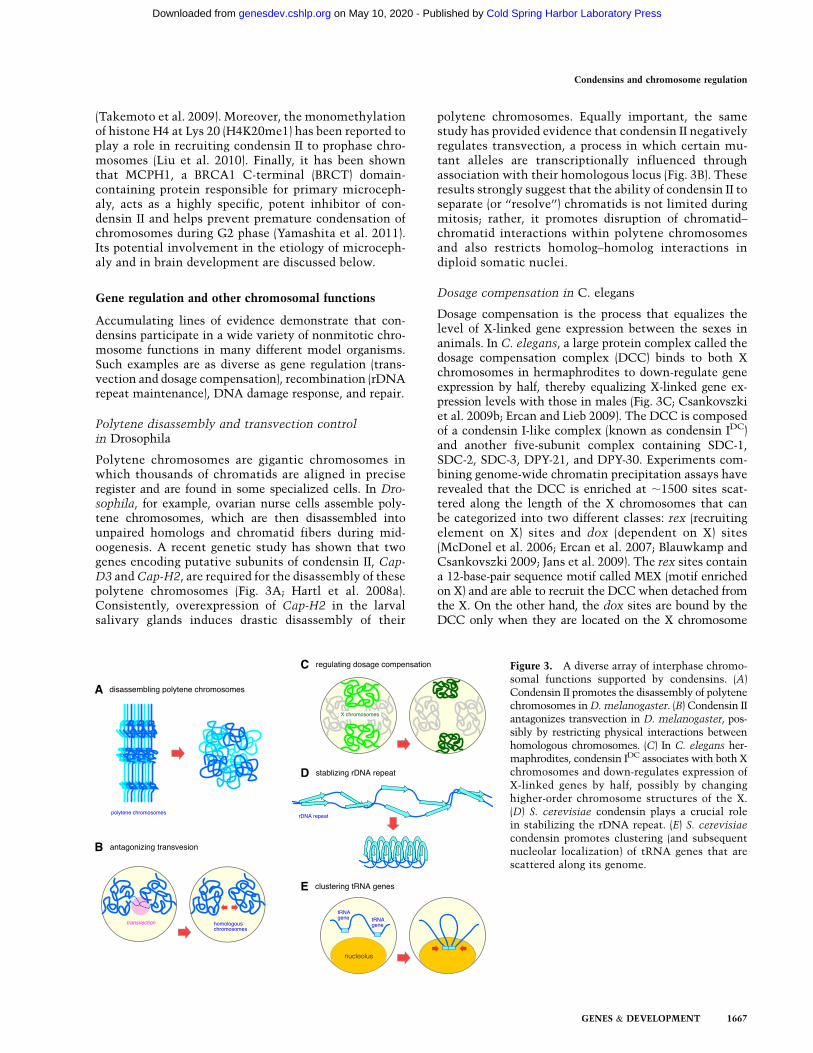
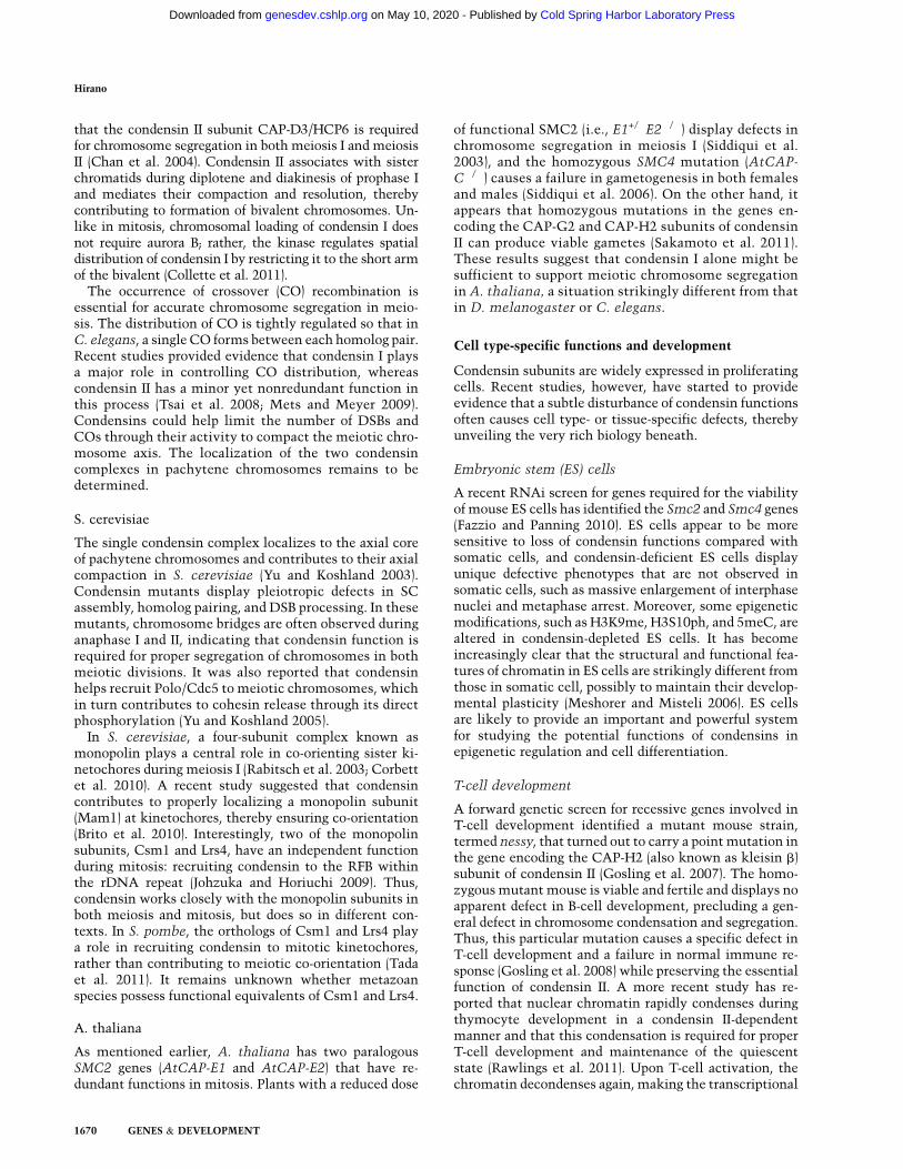
Polytene chromosomes are gigantic chromosomes inwhich thousands of chromatids are aligned in preciseregister and are found in some specialized cells. In Dro-sophila, for example, ovarian nurse cells assemble poly-tene chromosomes, which are then disassembled intounpaired homologs and chromatid fibers during mid-oogenesis. A recent genetic study has shown that twogenes encoding putative subunits of condensin II, Cap-D3 and Cap-H2, are required for the disassembly of thesepolytene chromosomes (Fig. 3A; Hartl et al. 2008a).Consistently, overexpression of Cap-H2 in the larvalsalivary glands induces drastic disassembly of their
polytene chromosomes. Equally important, the samestudy has provided evidence that condensin II negativelyregulates transvection, a process in which certain mu-tant alleles are transcriptionally influenced throughassociation with their homologous locus (Fig. 3B). Theseresults strongly suggest that the ability of condensin II toseparate (or ‘‘resolve’’) chromatids is not limited duringmitosis; rather, it promotes disruption of chromatid–chromatid interactions within polytene chromosomesand also restricts homolog–homolog interactions indiploid somatic nuclei.
Dosage compensation in C. elegans
Dosage compensation is the process that equalizes thelevel of X-linked gene expression between the sexes inanimals. In C. elegans, a large protein complex called thedosage compensation complex (DCC) binds to both Xchromosomes in hermaphrodites to down-regulate geneexpression by half, thereby equalizing X-linked gene ex-pression levels with those in males (Fig. 3C; Csankovszkiet al. 2009b; Ercan and Lieb 2009). The DCC is composedof a condensin I-like complex (known as condensin IDC)and another five-subunit complex containing SDC-1,SDC-2, SDC-3, DPY-21, and DPY-30. Experiments com-bining genome-wide chromatin precipitation assays haverevealed that the DCC is enriched at ;1500 sites scat-tered along the length of the X chromosomes that canbe categorized into two different classes: rex (recruitingelement on X) sites and dox (dependent on X) sites(McDonel et al. 2006; Ercan et al. 2007; Blauwkamp andCsankovszki 2009; Jans et al. 2009). The rex sites containa 12-base-pair sequence motif called MEX (motif enrichedon X) and are able to recruit the DCC when detached fromthe X. On the other hand, the dox sites are bound by theDCC only when they are located on the X chromosome
Figure 3. A diverse array of interphase chromo-somal functions supported by condensins. (A)Condensin II promotes the disassembly of polytenechromosomes in D. melanogaster. (B) Condensin IIantagonizes transvection in D. melanogaster, pos-sibly by restricting physical interactions betweenhomologous chromosomes. (C) In C. elegans her-maphrodites, condensin IDC associates with both Xchromosomes and down-regulates expression ofX-linked genes by half, possibly by changinghigher-order chromosome structures of the X.(D) S. cerevisiae condensin plays a crucial rolein stabilizing the rDNA repeat. (E) S. cerevisiae
condensin promotes clustering (and subsequentnucleolar localization) of tRNA genes that arescattered along its genome.
Condensins and chromosome regulation
GENES & DEVELOPMENT 1667
Cold Spring Harbor Laboratory Press on May 10, 2020 - Published by genesdev.cshlp.orgDownloaded from
(Jans et al. 2009). It will be important to understand howthe DCC might initially be recruited to some of these sitesand how it might potentially use a ‘‘spreading’’ mechanismto distribute along the length of the X chromosome (Ercanet al. 2009).
The difference between condensin I and condensin IDC
resides in only one subunit (Fig. 1A; Table 1; Csankovszkiet al. 2009a). It remains unknown how such a single-subunit substitution might alter the original molecularactivity of condensin I, thereby allowing condensin IDC toparticipate in chromosome-wide gene repression. Intrigu-ingly, a recent study suggests that the DCC may notrepress genes by direct binding; rather, the DCC may actat a distance, possibly by bringing dosage-compensatedgenes to the vicinity of DCC-bound sites (Jans et al. 2009).Another study provided evidence that the histone H2Avariant H2A.Z might function in dosage compensationthrough a mechanism that helps restrict the DCC to the Xchromosome (Petty et al. 2009). Thus, information fromthese studies on condensin IDC should provide a paradigmthat will readily be applicable to our understanding of thegeneral mechanisms of action of condensins.
Maintenance of the rDNA repeat in S. cerevisiae
In S. cerevisiae, ;200 copies of rDNA are clustered ina long tandem array on chromosome XII. A single unit ofrDNA (;9.1 kb long) comprises a 35S rRNA-codingsequence (transcribed by RNA polymerase I [Pol I]), a 5SrRNA-coding sequence (transcribed by RNA polymeraseIII [Pol III]), and two nontranscribed spacers (NTS1 andNTS2). Fob1, which binds to the replication fork barrier(RFB) sequence located in NTS1, is known to contributeto the maintenance of rDNA copy number by stimulatingrecombination. Johzuka et al. (2006) set up a geneticscreen for gene products that, in concert with Fob1, arerequired for preventing contraction of the rDNA repeatand identified three subunits of budding yeast condensin.Condensin is recruited to the RFB in a Fob1-dependentmanner, and this recruitment requires three additionalgene products: Tof2, Csm1, and Lrs4 (Johzuka and Horiuchi2009). Whereas condensin also associates with the 35SrRNA-coding region, this association is swept away byPol I-dependent transcription in fob1 mutant cells. Con-versely, inactivation of Pol I suppresses the drastic con-traction of the rDNA repeat in a fob1-condensin doublemutant (Johzuka and Horiuchi 2007). These resultsstrongly suggest not only that condensin plays a criticalrole in maintaining the copy number of the rDNA repeat(Fig. 3D), but that condensin’s association with chroma-tin is incompatible with active transcription.
Two recent studies further pointed out the counterac-tion between transcription and condensin functions.First, the protein phosphatase Cdc14 inhibits Pol Itranscription during mitosis, which in turn allows con-densin’s association with rDNA and thereby leads toproper condensation and segregation (Clemente-Blancoet al. 2009). Second, in a strain with a low rDNA copynumber, the average rate of rDNA transcription per copyincreases, leading to the loss of condensin association
from this locus. Under this condition, pairing betweenthe sister rDNA loci is weakened, and the cells becomesensitive to DNA damage, presumably due to compro-mised recombinational repair (Ide et al. 2010). Thus, therDNA repeat in S. cerevisiae represents a busy crossroadwhere a number of chromosomal activities (e.g., replica-tion, transcription, recombination, and segregation) meetand it will continue to act as an excellent model systemfor dissecting the multilayered functions of condensin.The observation that nutrient starvation promotes con-densin loading onto the rDNA repeat to maintain itsstability is another example of promising future studies(Tsang et al. 2007). In S. pombe, some nucleolar proteinshave been found to affect condensin’s accumulation atthe rDNA region (Nakazawa et al. 2008).
Differential clustering of tRNA genes in S. cerevisiaeand S. pombe
Although as many as ;270 tRNA genes are scattered inthe S. cerevisiae genome, most of them are clustered andlocated at the nucleolus at the cytological level (Fig. 3E;Thompson et al. 2003). A recent study has shown thatclustering of tRNA genes requires condensin functionsand that condensin indeed binds to active tRNA genes, asjudged by a chromatin immunoprecipitation (ChIP) assay(Haeusler et al. 2008). Disruption of microtubules re-leases tRNA genes from the nucleolus without dispersingthe clusters themselves, indicating that clustering oftRNA genes and their nucleolar localization are separableprocesses. An independent, genome-wide ChIP–chip anal-ysis has provided a complementary picture by demon-strating that budding yeast condensin associates withtRNA genes and other sites bound by TFIIIC, a compo-nent of the Pol III transcription machinery (D’Ambrosioet al. 2008b). It is therefore possible that TFIIIC is re-sponsible for specifying a class of condensin-binding sitesin the budding yeast genome.
Notably, TFIIIC is also implicated in large-scale chro-mosome organization in S. pombe, yet in a way differentfrom that in S. cerevisiae. In S. pombe, a fraction of TFIIICwith Pol III at tRNA genes is clustered near centromeres,whereas another fraction of TFIIIC without Pol III isconcentrated in several distinct bodies at the nuclearperiphery, referred to as chromosome-organizing clamps(COCs) (Noma et al. 2006). A subsequent study hasshown that condensin mediates the centromeric locali-zation of Pol III genes, which is negatively correlated withPol III transcription (Iwasaki et al. 2010). Remarkably,a mutation in TFIIIC suppresses the temperature sensi-tivity of a condensin mutant by restoring the centromerelocalization of Pol III genes. Thus, condensin-mediatedclustering of Pol III genes at centromeres appears tocounteract their transcription during interphase and alsoaffects global condensation and segregation of chromo-somes in mitosis.
DNA damage response and repair
Mitotic cell death (MCD) is a poorly characterized type ofcell death that is triggered when extensive double-strand
Hirano
1668 GENES & DEVELOPMENT
Cold Spring Harbor Laboratory Press on May 10, 2020 - Published by genesdev.cshlp.orgDownloaded from
breaks (DSBs) are introduced into cells deficient in thep53-mediated G2/M checkpoint. In HeLa cells, for in-stance, MCD accompanies premature activation of Cdk1and unscheduled condensation of chromatin into coa-lesced nucleoli, followed by the formation of micronu-clei. Interestingly, it was found that condensin I, but notcondensin II, is recruited to the condensing chromatinunder this condition (Blank et al. 2006). This simpleobservation not only demonstrates that the MCD-asso-ciated chromatin condensation is mechanistically dis-tinct from apoptosis-associated condensation, but alsooffers an experimental system for studying how conden-sins I and II might be differentially regulated. An in-dependent study using HeLa cells reported that conden-sin I is involved in DNA single-strand break (SSB) repair,possibly through its interaction with poly(ADP-ribose)polymerase 1 (PARP-1) and the base excision repair factorXRCC1 (Heale et al. 2006). PARP-1 appears to contributeto the retention, but not the initial recruitment, ofcondensin I to DNA damage sites (Kong et al. 2011).
As mentioned earlier, a recent genetic study in A.thaliana has shown that condensin II is likely to beinvolved in DSB response and repair (Sakamoto et al.2011). A genetic screen for mutants that exhibit hypersen-sitivity to excess boron identified two mutants (heb1-1and heb2-1) that turned out to be defective in the genesencoding the CAP-G2 and CAP-H2 subunits of condensinII, respectively. The boron toxicity is most likely toinvolve incidence of DSBs, although the underlying mech-anism remains to be clarified. This study also uncoveredthe somewhat surprising notion that condensin II is notessential for cell proliferation in this organism, therebyopening a way to dissect the functional involvement ofcondensin II in DSB response and repair in depth.
In S. pombe, a point mutation in the SMC2/Cut14hinge domain confers defects in both DNA damage repairand mitotic chromosome condensation (Akai et al. 2011).Notably, a mutation in replication protein A (RPA), anssDNA-binding protein, rescues both phenotypes, imply-ing that condensin’s DNA-reannealing activity mightcounteract RPA-mediated ssDNA coating (see furtherdiscussion below). The genetic properties of this muta-tion overlap with, but are not identical to, those ofa mutation previously found in the CAP-H/Cnd2 subunitof S. pombe condensin (Aono et al. 2002), suggesting theexistence of a complex regulatory network surroundingthe interphase and mitotic functions of condensin.
Meiotic chromosome function and segregation
Not surprisingly, condensins also play crucial roles inmeiotic chromosome architecture, dynamics, and func-tions. It is important to mention, however, that nomeiosis-specific condensin subunits have been identifiedso far in any organisms. This situation contrasts with thecohesin fields, where the discovery and subsequentcharacterization of meiosis-specific subunits have greatlycontributed to and accelerated our current understandingof the unique behaviors of chromosomes during meiosis(Petronczki et al. 2003).
Mammals
Currently, information regarding condensin functions inmammalian meiosis remains very limited. An early studyusing mouse spermatocytes reported that a condensin Isubunit (CAP-H) is concentrated on a fuzzy axial struc-ture within chromatids in both meiosis I and meiosis II(Viera et al. 2007). Whereas the existence of condensins Iand II was initially reported in pig oocytes (Liskova et al.2010), a more recent study in mouse oocytes has de-scribed a comprehensive picture of the dynamics andfunction of condensins I and II (Lee et al. 2011). Atprophase of meiosis I (prophase I), condensin I is presentin the cytoplasm, whereas condensin II is localized withinthe nucleus. After germinal vesicle breakdown, conden-sin II associates with chromosomes and is concentratedonto chromatid axes of bivalent chromosomes by meta-phase I. In contrast, condensin I localizes primarily aroundcentromeric regions at metaphase I and starts to associatewith chromosome arms only after anaphase I. Togetherwith results from antibody injection experiments, it hasbeen proposed that during construction of bivalent chro-mosomes, condensin II plays a primary role in resolvingsister chromatid axes, whereas condensin I may contributeto monopolar attachment of sister kinetochores by assem-bling a unique centromeric structure underneath (Lee et al.2011). Future studies should address potential functionsof condensins I and II at early stages of meiosis (e.g.,pachytene), possibly combined with conditional knock-out approaches.
D. melanogaster
The D. melanogaster genome encodes the orthologs ofthe CAP-D3 and CAP-H2 subunits of condensin II.Although mitotic cells express them, it appears that theydo not play major functions in mitotic chromosomeassembly or segregation (Savvidou et al. 2005). Instead,a recent genetic study has shown that both CAP-D3 andCAP-H2 are necessary for male fertility (Hartl et al.2008b). Cap-H2 mutants display defects in chromosometerritory formation in prophase I, which in turn result ina failure to resolve heterologous and/or homologouschromosomes in anaphase I. Thus, the putative conden-sin II function is required for not only segregating homol-ogous chromosomes, but also individualizing heterologouschromosomes. Moreover, another study provided evidencethat CAP-G, a condensin I subunit, is required for disas-sembly of the synaptonemal complex (SC) and keepingchromosomes in their metaphase I configuration in fe-male meiosis (Resnick et al. 2008). Neither study haslocalized the condensin subunits to Drosophila meioticchromosomes.
C. elegans
An early study had shown that RNAi-based depletion ofSMC2/MIX-1 or SMC4/SMC-4 causes defects in chromo-some segregation during meiosis II but not during meiosis I(Hagstrom et al. 2002). A subsequent genetic study per-formed under a tighter condition, however, demonstrated
Condensins and chromosome regulation
GENES & DEVELOPMENT 1669
Cold Spring Harbor Laboratory Press on May 10, 2020 - Published by genesdev.cshlp.orgDownloaded from
that the condensin II subunit CAP-D3/HCP6 is requiredfor chromosome segregation in both meiosis I and meiosisII (Chan et al. 2004). Condensin II associates with sisterchromatids during diplotene and diakinesis of prophase Iand mediates their compaction and resolution, therebycontributing to formation of bivalent chromosomes. Un-like in mitosis, chromosomal loading of condensin I doesnot require aurora B; rather, the kinase regulates spatialdistribution of condensin I by restricting it to the short armof the bivalent (Collette et al. 2011).
The occurrence of crossover (CO) recombination isessential for accurate chromosome segregation in meio-sis. The distribution of CO is tightly regulated so that inC. elegans, a single CO forms between each homolog pair.Recent studies provided evidence that condensin I playsa major role in controlling CO distribution, whereascondensin II has a minor yet nonredundant function inthis process (Tsai et al. 2008; Mets and Meyer 2009).Condensins could help limit the number of DSBs andCOs through their activity to compact the meiotic chro-mosome axis. The localization of the two condensincomplexes in pachytene chromosomes remains to bedetermined.
S. cerevisiae
The single condensin complex localizes to the axial coreof pachytene chromosomes and contributes to their axialcompaction in S. cerevisiae (Yu and Koshland 2003).Condensin mutants display pleiotropic defects in SCassembly, homolog pairing, and DSB processing. In thesemutants, chromosome bridges are often observed duringanaphase I and II, indicating that condensin function isrequired for proper segregation of chromosomes in bothmeiotic divisions. It was also reported that condensinhelps recruit Polo/Cdc5 to meiotic chromosomes, whichin turn contributes to cohesin release through its directphosphorylation (Yu and Koshland 2005).
In S. cerevisiae, a four-subunit complex known asmonopolin plays a central role in co-orienting sister ki-netochores during meiosis I (Rabitsch et al. 2003; Corbettet al. 2010). A recent study suggested that condensincontributes to properly localizing a monopolin subunit(Mam1) at kinetochores, thereby ensuring co-orientation(Brito et al. 2010). Interestingly, two of the monopolinsubunits, Csm1 and Lrs4, have an independent functionduring mitosis: recruiting condensin to the RFB withinthe rDNA repeat (Johzuka and Horiuchi 2009). Thus,condensin works closely with the monopolin subunits inboth meiosis and mitosis, but does so in different con-texts. In S. pombe, the orthologs of Csm1 and Lrs4 playa role in recruiting condensin to mitotic kinetochores,rather than contributing to meiotic co-orientation (Tadaet al. 2011). It remains unknown whether metazoanspecies possess functional equivalents of Csm1 and Lrs4.
A. thaliana
As mentioned earlier, A. thaliana has two paralogousSMC2 genes (AtCAP-E1 and AtCAP-E2) that have re-dundant functions in mitosis. Plants with a reduced dose
of functional SMC2 (i.e., E1+/�E2�/�) display defects inchromosome segregation in meiosis I (Siddiqui et al.2003), and the homozygous SMC4 mutation (AtCAP-C�/�) causes a failure in gametogenesis in both femalesand males (Siddiqui et al. 2006). On the other hand, itappears that homozygous mutations in the genes en-coding the CAP-G2 and CAP-H2 subunits of condensinII can produce viable gametes (Sakamoto et al. 2011).These results suggest that condensin I alone might besufficient to support meiotic chromosome segregationin A. thaliana, a situation strikingly different from thatin D. melanogaster or C. elegans.
Cell type-specific functions and development
Condensin subunits are widely expressed in proliferatingcells. Recent studies, however, have started to provideevidence that a subtle disturbance of condensin functionsoften causes cell type- or tissue-specific defects, therebyunveiling the very rich biology beneath.
Embryonic stem (ES) cells
A recent RNAi screen for genes required for the viabilityof mouse ES cells has identified the Smc2 and Smc4 genes(Fazzio and Panning 2010). ES cells appear to be moresensitive to loss of condensin functions compared withsomatic cells, and condensin-deficient ES cells displayunique defective phenotypes that are not observed insomatic cells, such as massive enlargement of interphasenuclei and metaphase arrest. Moreover, some epigeneticmodifications, such as H3K9me, H3S10ph, and 5meC, arealtered in condensin-depleted ES cells. It has becomeincreasingly clear that the structural and functional fea-tures of chromatin in ES cells are strikingly different fromthose in somatic cell, possibly to maintain their develop-mental plasticity (Meshorer and Misteli 2006). ES cellsare likely to provide an important and powerful systemfor studying the potential functions of condensins inepigenetic regulation and cell differentiation.
T-cell development
A forward genetic screen for recessive genes involved inT-cell development identified a mutant mouse strain,termed nessy, that turned out to carry a point mutation inthe gene encoding the CAP-H2 (also known as kleisin b)subunit of condensin II (Gosling et al. 2007). The homo-zygous mutant mouse is viable and fertile and displays noapparent defect in B-cell development, precluding a gen-eral defect in chromosome condensation and segregation.Thus, this particular mutation causes a specific defect inT-cell development and a failure in normal immune re-sponse (Gosling et al. 2008) while preserving the essentialfunction of condensin II. A more recent study has re-ported that nuclear chromatin rapidly condenses duringthymocyte development in a condensin II-dependentmanner and that this condensation is required for properT-cell development and maintenance of the quiescentstate (Rawlings et al. 2011). Upon T-cell activation, thechromatin decondenses again, making the transcriptional
Hirano
1670 GENES & DEVELOPMENT
Cold Spring Harbor Laboratory Press on May 10, 2020 - Published by genesdev.cshlp.orgDownloaded from
activator Stat5 accessible to its target promoters. Theseresults suggest that condensin II regulates a developmen-tally programmed cycle of chromatin condensation anddecondensation that has a great impact on T-cell differ-entiation. Intriguingly, similar if not identical chromatincondensation is observed during erythroid cell differenti-ation, and the condensin II subunit CAP-G2 is implicatedin this process and accompanying transcriptional repres-sion (Xu et al. 2006).
Primary microcephaly
Primary microcephaly is a neurodevelopmental disordercharacterized by reduced brain size and mental retarda-tion in humans (Thornton and Woods 2009). MCPH1(also known as microcephalin) is one of the eight geneproducts known to be responsible for this disease. In-triguingly, cells from MCPH1 patients (but not fromother MCPH patients) display abnormal chromosomedynamics, including premature chromosome condensa-tion (PCC) in G2 phase (Trimborn et al. 2004). Althoughthe PCC phenotype may be a secondary consequence ofa compromised G2/M checkpoint observed in MCPH1-deficient cells (Alderton et al. 2006; Tibelius et al. 2009),other lines of evidence suggest that MCPH1 may workvery closely with condensin II (Trimborn et al. 2006;Wood et al. 2008). In fact, a more recent study has demon-strated that the N-terminal domain of human MCPH1(hMCPH1) is able to specifically inhibit the action ofcondensin II in Xenopus egg extracts and that its centraldomain has an auxiliary role in shaping metaphase chro-mosomes by physically interacting with condensin II(Yamashita et al. 2011). Despite these observations, it israther surprising to find that MCPH1-deficient cellssegregate their ill-shaped chromosomes in a normal timeframe. Mcph1 knockout mice are viable (Liang et al. 2010)and display microcephaly (Gruber et al. 2011). It iscurrently unknown whether the MCPH1’s ability toregulate condensin II functions might be directly relevantto the etiology of microcephaly. The prevalent view in thefield is that loss of MCPH gene functions causes an un-coupling between cell cycle progression and the centro-some cycle, which in turn compromises proliferation ofneuroprogenitor cells with a highly unique division mode(Thornton and Woods 2009). Condensin II and MCPH1could, in fact, function as components of the signalingnetwork that regulates mitotic entry in response to a rangeof cellular stresses (Hirano 2005; Chin and Yeong 2010).Whatever the mechanism might be, it is important toemphasize that MCPH1 microcephaly represents the firstexample of human diseases that accompany misregulationof the chromosome condensation machinery.
Cancers and tissue growth
Genome instability is a hallmark of cancers. Given thecritical role of condensins in proper assembly and segre-gation of chromosomes, it is formally possible thatsubtle perturbations of their functions might promotegenome instability, eventually contributing to tumori-genesis. Although no direct evidence for this idea has
been reported so far, potentially interesting observationscan be found in the literature. For instance, somaticpoint mutations in the SMC2/CAP-E and SMC4/CAP-Csubunits have been identified in several cases of pyothorax-associated lymphoma in humans (Ham et al. 2007). Inzebrafish, a mutation in the condensin I CAP-G subunitincreases genomic imbalances and the rate of apoptosisin the retina (Seipold et al. 2009). In Drosophila, it hasbeen reported that a retinoblastoma family protein (Rfb1)is required for efficient localization of condensin II withchromatin by physically interacting with its subunit,CAP-D3 (Longworth et al. 2008). Finally, a mutation inthe condensin I CAP-G subunit has been found to betightly associated with an increase in body size at pubertyin cattle (Setoguchi et al. 2011). The correspondingsyntenic region in human chromosome 4 is also knownbe to be associated with adult height, raising the possi-bility that a condensin I variant might affect skeletalgrowth in mammals.
Molecular mechanisms of action
Despite the rapid accumulation of information regardingthe biology of condensins, our understanding of theirmolecular mechanisms of action remains poor. The largesize and intricate subunit architecture of the complexeshave made it a big challenge to reconstitute active com-plexes from recombinant subunits (Onn et al. 2007).Nonetheless, extensive efforts using a wide variety ofapproaches are now gathering valuable information aboutthe structure and mechanics of condensins. For reviews onthe mechanistic aspects of condensins, see also Swedlowand Hirano (2003), Hirano (2006), Cuylen and Haering(2011), and Baxter and Aragon (2012).
Ensuring dsDNA: a preparatory stepfor subsequent coiling?
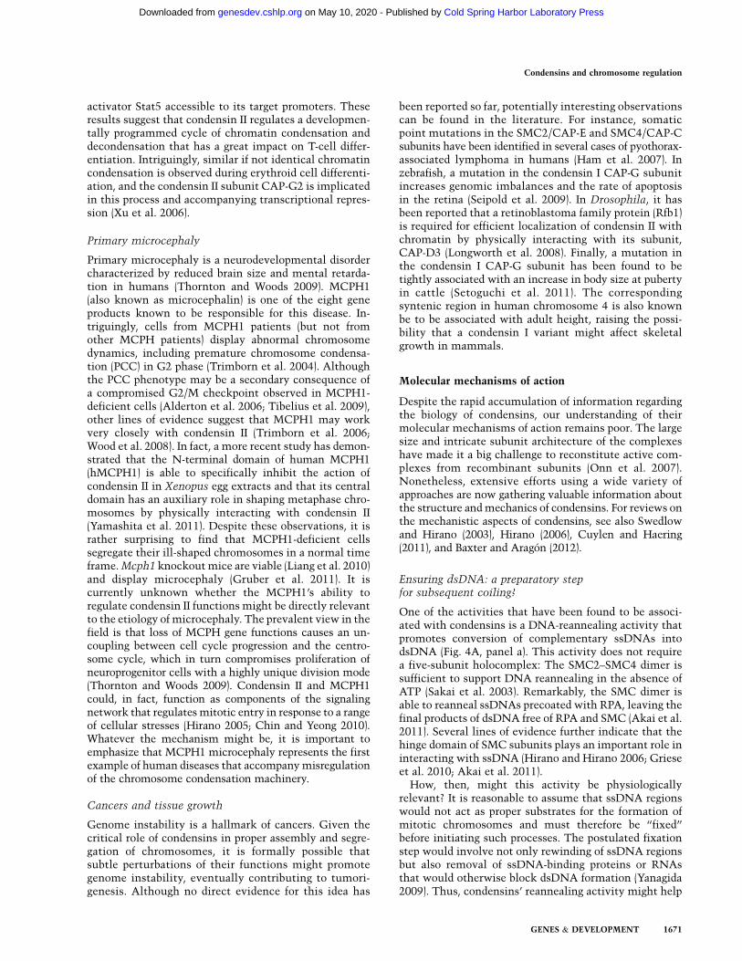
One of the activities that have been found to be associ-ated with condensins is a DNA-reannealing activity thatpromotes conversion of complementary ssDNAs intodsDNA (Fig. 4A, panel a). This activity does not requirea five-subunit holocomplex: The SMC2–SMC4 dimer issufficient to support DNA reannealing in the absence ofATP (Sakai et al. 2003). Remarkably, the SMC dimer isable to reanneal ssDNAs precoated with RPA, leaving thefinal products of dsDNA free of RPA and SMC (Akai et al.2011). Several lines of evidence further indicate that thehinge domain of SMC subunits plays an important role ininteracting with ssDNA (Hirano and Hirano 2006; Grieseet al. 2010; Akai et al. 2011).
How, then, might this activity be physiologicallyrelevant? It is reasonable to assume that ssDNA regionswould not act as proper substrates for the formation ofmitotic chromosomes and must therefore be ‘‘fixed’’before initiating such processes. The postulated fixationstep would involve not only rewinding of ssDNA regionsbut also removal of ssDNA-binding proteins or RNAsthat would otherwise block dsDNA formation (Yanagida2009). Thus, condensins’ reannealing activity might help
Condensins and chromosome regulation
GENES & DEVELOPMENT 1671
Cold Spring Harbor Laboratory Press on May 10, 2020 - Published by genesdev.cshlp.orgDownloaded from
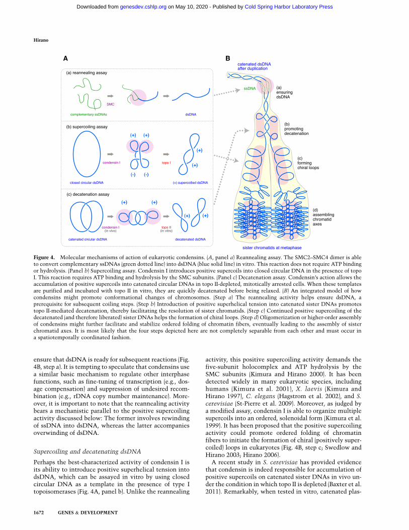
ensure that dsDNA is ready for subsequent reactions (Fig.4B, step a). It is tempting to speculate that condensins usea similar basic mechanism to regulate other interphasefunctions, such as fine-tuning of transcription (e.g., dos-age compensation) and suppression of undesired recom-bination (e.g., rDNA copy number maintenance). More-over, it is important to note that the reannealing activitybears a mechanistic parallel to the positive supercoilingactivity discussed below: The former involves rewindingof ssDNA into dsDNA, whereas the latter accompaniesoverwinding of dsDNA.
Supercoiling and decatenating dsDNA
Perhaps the best-characterized activity of condensin I isits ability to introduce positive superhelical tension intodsDNA, which can be assayed in vitro by using closedcircular DNA as a template in the presence of type Itopoisomerases (Fig. 4A, panel b). Unlike the reannealing
activity, this positive supercoiling activity demands thefive-subunit holocomplex and ATP hydrolysis by theSMC subunits (Kimura and Hirano 2000). It has beendetected widely in many eukaryotic species, includinghumans (Kimura et al. 2001), X. laevis (Kimura andHirano 1997), C. elegans (Hagstrom et al. 2002), and S.cerevisiae (St-Pierre et al. 2009). Moreover, as judged bya modified assay, condensin I is able to organize multiplesupercoils into an ordered, solenoidal form (Kimura et al.1999). It has been proposed that the positive supercoilingactivity could promote ordered folding of chromatinfibers to initiate the formation of chiral (positively super-coiled) loops in eukaryotes (Fig. 4B, step c; Swedlow andHirano 2003; Hirano 2006).
A recent study in S. cerevisiae has provided evidencethat condensin is indeed responsible for accumulation ofpositive supercoils on catenated sister DNAs in vivo un-der the condition in which topo II is depleted (Baxter et al.2011). Remarkably, when tested in vitro, catenated plas-
Figure 4. Molecular mechanisms of action of eukaryotic condensins. (A, panel a) Reannealing assay. The SMC2–SMC4 dimer is ableto convert complementary ssDNAs (green dotted line) into dsDNA (blue solid line) in vitro. This reaction does not require ATP bindingor hydrolysis. (Panel b) Supercoiling assay. Condensin I introduces positive supercoils into closed circular DNA in the presence of topoI. This reaction requires ATP binding and hydrolysis by the SMC subunits. (Panel c) Decatenation assay. Condensin’s action allows theaccumulation of positive supercoils into catenated circular DNAs in topo II-depleted, mitotically arrested cells. When these templatesare purified and incubated with topo II in vitro, they are quickly decatenated before being relaxed. (B) An integrated model of howcondensins might promote conformational changes of chromosomes. (Step a) The reannealing activity helps ensure dsDNA, aprerequisite for subsequent coiling steps. (Step b) Introduction of positive superhelical tension into catenated sister DNAs promotestopo II-mediated decatenation, thereby facilitating the resolution of sister chromatids. (Step c) Continued positive supercoiling of thedecatenated (and therefore liberated) sister DNAs helps the formation of chiral loops. (Step d) Oligomerization or higher-order assemblyof condensins might further facilitate and stabilize ordered folding of chromatin fibers, eventually leading to the assembly of sisterchromatid axes. It is most likely that the four steps depicted here are not completely separable from each other and must occur ina spatiotemporally coordinated fashion.
Hirano
1672 GENES & DEVELOPMENT
Cold Spring Harbor Laboratory Press on May 10, 2020 - Published by genesdev.cshlp.orgDownloaded from
mids with positive supercoils are more susceptible totopo II-mediated decatenation than those with no super-coils (Fig. 4A, panel c), suggesting that condensin-medi-ated positive supercoiling could facilitate decatenation ofsister DNA molecules during mitosis (Fig. 4B, step b). It isreasonable to speculate that when this action of conden-sin is compromised, catenanes between sister DNAs failto be removed completely, thereby causing defects in sisterchromatid resolution and segregation in anaphase (e.g.,Coelho et al. 2003; Hudson et al. 2003; D’Ambrosio et al.2008a). In the future, it will be important to address howpositive superhelical tension imposed by condensin mightalter nucleosome structures (Bancaud et al. 2007) and howtopo II might act on such nucleosome templates (Roca 2009).
A pair of recent studies has shown that E. coli MukBphysically interacts with ParC, the DNA-binding subunitof a type II topoisomerase (topo IV), and stimulates itsactivity in vitro (Hayama and Marians 2010; Li et al.2010). Although these observations are highly illuminat-ing, careful consideration will be required before integrat-ing all data currently available because there exist somemechanistic differences in the eukaryotic and prokaryoticdecatenation systems (Hirano 2000; Postow et al. 2001).
How might condensins work?
The mechanism by which the condensin complexesmight interact with DNA to induce its conformationalchanges is unknown. A recent study using minichromo-somes isolated from yeast cells has provided evidencethat condensin encircles dsDNA, thereby forming topo-logical links—a mechanism similar to that proposed forcohesin–DNA interactions (Cuylen et al. 2011). It isattractive to speculate that whereas cohesin holds twodifferent sister DNA strands together, a single condensincomplex might link two distant segments of a sisterchromatid to promote its folding and compaction. Indeed,this idea is not incompatible with the chiral loopingmodel of condensin’s action, although condensin–DNAinteractions are clearly more complex than those pre-dicted from a simple topological entrapment (Stray andLindsley 2003; Stray et al. 2005; Hudson et al. 2008).
Some of the previous models predicted that condensinscould oligomerize or form a higher-order assembly to folda chromatin fiber during the process of chromosomecondensation (Swedlow and Hirano 2003; Hirano 2006;Graumann and Knust 2009). In fact, positive supercoilingof DNA is likely to be a net product of reactions involvingmultiple condensin I complexes, and a single DNA mol-ecule manipulation assay using magnetic tweezers alsoprovides strong support for cooperative actions of con-densin I (Strick et al. 2004). It is possible that highlydynamic condensin–DNA interactions are further modi-fied or stabilized by condensin–condensin interactions,which in turn contribute to assembling sister chromatidaxes (Fig. 4B, step d; Maeshima and Laemmli 2003; Onoet al. 2003; Kireeva et al. 2004). Moreover, the idea ofoligomerization gains support from several different ex-periments using bacterial condensins, including electronmicroscopy (Matoba et al. 2005; Fuentes-Perez et al.
2012), a single DNA molecule assay (Cui et al. 2008), anda combination of structural and biochemical analyses(Woo et al. 2009). Particularly important is the proposalthat ATP-dependent engagement of MukB head domainsdisrupts a MukB–MukF interaction, thereby providingan opportunity to initiate oligomerization of multipleMukBEF complexes (Woo et al. 2009). Future studiesusing the eukaryotic and prokaryotic complexes shouldclarify exactly how the ATP-binding and hydrolysis cycleof their SMC core subunits might be coupled with con-densin–DNA interactions as well as putative condensin–condensin interactions.
Conclusions and perspective
Although the condensin complexes were originally dis-covered as central players in mitotic chromosome assem-bly and segregation, recent studies revealed that theyparticipate in an amazingly diverse array of chromosomalevents. Evidence is also accumulating that condensins Iand II fulfill nonoverlapping functions, behave differen-tially during the cell cycle, and are under the control ofdistinct sets of specific regulators. From an evolutionarypoint of view, the principal position of condensin I inmitotic segregation is conserved among virtually alleukaryotes. In contrast, condensin II is apparently usedin different contexts among different species. Althoughthese conceptual advances provide a valuable guide forfuture studies in the field, a number of outstanding ques-tions remain to be answered. For example, the molecularmechanisms of action of condensins must be investigatedand understood in depth. Because most of our currentknowledge comes from analyses of condensin I, it will beof great importance to critically address to what extentthe two condensin complexes might differentiate fromeach other at a mechanistic level. Moreover, it will be ofgreat interest to explore cell type- and tissue-specificfunctions of condensins and their relevance to the de-velopment of organisms. Along these lines, functionalcross-talk of condensins with epigenetic control will bean exciting area in future studies. The integration ofknowledge from multidisciplinary approaches shouldeventually shed light on the evolutionary questions ofhow a primitive form of life might have devised a strat-egy for handling the long molecules of DNA and howsuch an early principle of genome organization mighthave evolved to help create the astonishing diversity oflife that we see today on Earth.
Acknowledgments
I thank members of the Hirano laboratory for critically readingthe manuscript. I am also grateful to many colleagues in thecondensin field for stimulating discussions. The work donein my laboratory was supported by Grant-in-Aid for SpeciallyPromoted Research (20002010).
References
Abe S, Nagasaka K, Hirayama Y, Kozuka-Hata H, Oyama M,Aoyagi Y, Obuse C, Hirota T. 2011. The initial phase of
Condensins and chromosome regulation
GENES & DEVELOPMENT 1673
Cold Spring Harbor Laboratory Press on May 10, 2020 - Published by genesdev.cshlp.orgDownloaded from
chromosome condensation requires Cdk1-mediated phos-phorylation of the CAP-D3 subunit of condensin II. GenesDev 25: 863–874.
Akai Y, Kurokawa Y, Nakazawa N, Tonami-Murakami Y,Suzuki Y, Yoshimura S-H, Iwasaki H, Shiroiwa Y, NakamuraT, Shibata E, et al. 2011. Opposing role of condensin hingeagainst replication protein A in mitosis and interphase throughpromoting DNA annealing. Open Biol 1: 110023. doi: 10.1098/rsob.110023.
Alderton GK, Galbiati L, Griffith E, Surinya KH, Neitzel H,Jackson AP, Jeggo PA, O’Driscoll M. 2006. Regulation ofmitotic entry by microcephalin and its overlap with ATPsignalling. Nat Cell Biol 8: 725–733.
Anderson DE, Losada A, Erickson HP, Hirano T. 2002. Con-densin and cohesin display different arm conformations withcharacteristic hinge angles. J Cell Biol 156: 419–424.
Aono N, Sutani T, Tomonaga T, Mochida S, Yanagida M. 2002.Cnd2 has dual roles in mitotic condensation and interphase.Nature 417: 197–202.
Bancaud A, Wagner G, Conde E, Silva N, Lavelle C, Wong H,Mozziconacci J, Barbi M, Sivolob A, Le Cam E, et al. 2007.Nucleosome chiral transition under positive torsional stressin single chromatin fibers. Mol Cell 27: 135–147.
Baxter J, Aragon L. 2012. A model for chromosome condensa-tion based on the interplay between condensin and topo-isomerase II. Trends Genet 28: 110–117.
Baxter J, Sen N, Martınez VL, De Carandini MEM, SchvartzmanJB, Diffley JFX, Aragon L. 2011. Positive supercoiling ofmitotic DNA drives decatenation by topoisomerase II ineukaryotes. Science 331: 1328–1332.
Bazile F, St-Pierre J, D’Amours D. 2010. Three-step model forcondensin activation during mitotic chromosome condensa-tion. Cell Cycle 9: 3243–3255.
Belmont AS. 2006. Mitotic chromosome structure and conden-sation. Curr Opin Cell Biol 18: 1–7.
Bernad R, Sanchez P, Rivera T, Rodrıguez-Corsino M, BoyarchukE, Vassias I, Ray-Gallet D, Arnaoutov A, Dasso M, AlmouzniG, et al. 2011. Xenopus HJURP and condensin II are requiredfor CENP-A assembly. J Cell Biol 192: 569–582.
Bhat MA, Philp AV, Glover DM, Bellen HJ. 1996. Chromatidsegregation at anaphase requires the barren product, a novelchromosome associated protein that interacts with topoiso-merase II. Cell 87: 1103–1114.
Blank M, Lerenthal Y, Mittelman L, Shiloh Y. 2006. Condensin Irecruitment and uneven chromatin condensation precedemitotic cell death in response to DNA damage. J Cell Biol174: 195–206.
Blauwkamp TA, Csankovszki G. 2009. Two classes of dosagecompensation complex binding elements along Caenorhabdi-
tis elegans X chromosomes. Mol Cell Biol 29: 2023–2031.Brito IL, Yu H-G, Amon A. 2010. Condensins promote coori-
entation of sister chromatids during meiosis I in buddingyeast. Genetics 185: 55–64.
Cervantes MD, Coyne RS, Xi X, Yao M-C. 2006. The condensincomplex is essential for amitotic segregation of bulk chro-mosomes, but not nucleoli, in the ciliate Tetrahymena
thermophila. Mol Cell Biol 26: 4690–4700.Chan RC, Severson AF, Meyer BJ. 2004. Condensin restructures
chromosomes in preparation for meiotic divisions. J Cell Biol
167: 613–625.Chin CF, Yeong FM. 2010. Safeguarding entry into mitosis: The
antephase checkpoint. Mol Cell Biol 30: 22–32.Clemente-Blanco A, Mayan-Santos M, Schneider D, Machın F,
Jarmuz A, Tschochner H, Aragon L. 2009. Cdc14 inhibitstranscription by RNA polymerase I during anaphase. Nature458: 219–222.
Coelho P, Queiroz-Mechado J, Sunkel CE. 2003. Condensin-dependent localisation of topoisomerase II to an axial chro-mosomal structure is required for sister chromatid resolu-tion during mitosis. J Cell Sci 116: 4763–4776.
Collette KS, Petty EL, Golenberg N, Bembenek JN, CsankovszkiG. 2011. Different roles for Aurora B in condensin targetingduring mitosis and meiosis. J Cell Sci 124: 3684–3694.
Corbett KD, Yip CK, Ee L-S, Walz T, Amon A, Harrison SC.2010. The monopolin complex crosslinks kinetochore com-ponents to regulate chromosome-microtubule attachments.Cell 142: 556–567.
Csankovszki G, Collette K, Spahl K, Carey J, Snyder M, Petty E,Patel U, Tabuchi T, Liu H, McLeod I, et al. 2009a. Threedistinct condensin complexes control C. elegans chromo-some dynamics. Curr Biol 19: 9–19.
Csankovszki G, Petty EL, Collette KS. 2009b. The worm solu-tion: A chromosome-full of condensin helps gene expressiongo down. Chromosome Res 17: 621–635.
Cui Y, Petrushenko ZM, Rybenkov VV. 2008. MukB acts asa macromolecular clamp in DNA condensation. Nat StructMol Biol 15: 411–418.
Cuylen S, Haering CH. 2011. Deciphering condensin actionduring chromosome segregation. Trends Cell Biol 21: 552–559.
Cuylen S, Metz J, Haering CH. 2011. Condensin structureschromosomal DNA through topological links. Nat Struct
Mol Biol 18: 894–901.D’Ambrosio C, Kelly G, Shirahige K, Uhlmann F. 2008a. Con-
densin-dependent rDNA decatenation introduces a temporalpattern to chromosome segregation. Curr Biol 18: 1084–1089.
D’Ambrosio C, Schmidt CK, Katou Y, Kelly G, Itoh T, ShirahigeK, Uhlmann F. 2008b. Identification of cis-acting sites forcondensin loading onto budding yeast chromosomes. Genes
Dev 22: 2215–2227.Dej KJ, Ahn C, Orr-Weaver TL. 2004. Mutations in the Dro-
sophila condensin subunit dCAP-G: Defining the role of con-densin for chromosome condensation in mitosis and geneexpression in interphase. Genetics 168: 895–906.
Eisen JA, Coyne RS, Wu M, Wu D, Thiagarajan M, Wortman JR,Badger JH, Ren Q, Amedeo P, Jones KM, et al. 2006. Mac-ronuclear genome sequence of the ciliate Tetrahymena ther-mophila, a model eukaryote. PLoS Biol 4: e286. doi: 10.1371/journal.pbio.0040286.
Ercan S, Lieb JD. 2009. C. elegans dosage compensation: Awindow into mechanisms of domain-scale gene regulation.Chromosome Res 17: 215–227.
Ercan S, Giresi PG, Whittle CM, Zhang X, Green RD, Lieb JD.2007. X chromosome repression by localization of theC. elegans dosage compensation machinery to sites of tran-scription initiation. Nat Genet 39: 403–408.
Ercan S, Dick L, Lieb J. 2009. The C. elegans dosage compen-sation complex propagates dynamically and independently ofX chromosome sequence. Curr Biol 19: 1777–1787.
Fazzio TG, Panning B. 2010. Condensin complexes regulatemitotic progression and interphase chromatin structure inembryonic stem cells. J Cell Biol 188: 491–503.
Fennell-Fezzie R, Gradia SD, Akey D, Berger JM. 2005. TheMukF subunit of Escherichia coli condensin: Architectureand functional relationship to kleisins. EMBO J 24: 1921–1930.
Freeman L, Aragon-Alcaide L, Strunnikov AV. 2000. The con-densin complex governs chromosome condensation and mi-totic transmission of rDNA. J Cell Biol 149: 811–824.
Fuentes-Perez ME, Gwynn EJ, Dillingham MS, Moreno-HerreroF. 2012. Using DNA as a fiducial marker to study SMC
Hirano
1674 GENES & DEVELOPMENT
Cold Spring Harbor Laboratory Press on May 10, 2020 - Published by genesdev.cshlp.orgDownloaded from
complex interactions with the atomic force microscope.Biophys J 102: 839–848.
Gasser SM, Laemmli UK. 1987. Improved methods for theisolation of individual and clustered mitotic chromosomes.Exp Cell Res 173: 85–98.
Gerlich D, Hirota T, Koch B, Peters J-M, Ellenberg J. 2006.Condensin I stabilizes chromosomes mechanically througha dynamic interaction in living cells. Curr Biol 16: 333–344.
Giet R, Glover DM. 2001. Drosophila Aurora B kinase isrequired for histone H3 phosphorylation and condensin re-cruitment during chromosome condensation and to organizethe central spindle during cytokinesis. J Cell Biol 152: 669–682.
Gloyd M, Ghirlando R, Matthews LA, Guarne A. 2007. MukEand MukF form two distinct high affinity complexes. J Biol
Chem 282: 14373–14378.Gosling KM, Makaroff LE, Theodoratos A, Kim Y-H, Whittle B,
Rui L, Wu H, Hong NA, Kennedy GC, Fritz J-A, et al. 2007. Amutation in a chromosome condensin II subunit, kleisin b,specifically disrupts T cell development. Proc Natl Acad Sci104: 12445–12450.
Gosling KM, Goodnow CC, Verma NK, Fahrer AM. 2008.Defective T-cell function leading to reduced antibody pro-duction in a kleisin-b mutant mouse. Immunology 125: 208–217.
Graumann PL, Knust T. 2009. Dynamics of the bacterial SMCcomplex and SMC-like proteins involved in DNA repair.Chromosome Res 17: 265–275.
Green LC, Kalitsis P, Chang TM, Cipetic M, Kim JH, MarshallO, Turnbull L, Whitchurch CB, Vagnarelli P, Samejima K,et al. 2012. Contrasting roles of condensin I and II in mitoticchromosome formation. J Cell Sci 125: 1591–1604.
Griese JJ, Witte G, Hopfner K-P. 2010. Structure and DNAbinding activity of the mouse condensin hinge domain high-light common and diverse features of SMC proteins. NucleicAcids Res 38: 3454–3465.
Gruber S, Errington J. 2009. Recruitment of condensin to rep-lication origin regions by ParB/SpoOJ promotes chromosomesegregation in B. subtilis. Cell 137: 685–696.
Gruber R, Zhou Z, Sukchev M, Joerss T, Frappart P-O, WangZ-Q. 2011. MCPH1 regulates the neuroprogenitor divisionmode by coupling the centrosomal cycle with mitotic entrythrough the Chk1–Cdc25 pathway. Nat Cell Biol 13: 1325–1334.
Haeusler RA, Pratt-Hyatt M, Good PD, Gipson TA, Engelke DR.2008. Clustering of yeast tRNA genes is mediated by specificassociation of condensin with tRNA gene transcription com-plexes. Genes Dev 22: 2204–2214.
Hagstrom KA, Holmes VF, Cozzarelli NR, Meyer BJ. 2002. C.
elegans condensin promotes mitotic chromosome architec-ture, centromere organization, and sister chromatid segre-gation during mitosis and meiosis. Genes Dev 16: 729–742.
Ham MF, Takakuwa T, Rahadiani N, Tresnasari K, Nakajima H,Aozasa K. 2007. Condensin mutations and abnormal chro-mosomal structures in pyothorax-assocaited lymphoma. Can-
cer Sci 98: 1041–1047.Hartl TA, Smith HF, Bosco G. 2008a. Chromosome alignment
and transvection are antagonized by condensin II. Science
322: 1384–1387.Hartl TA, Sweeney SJ, Knepler PJ, Bosco G. 2008b. Condensin II
resolves chromosomal associations to enable anaphase I seg-regation in Drosophila male meiosis. PLoS Genet 4: e1000228.doi: 10.1371/journal.pgen.1000228.
Hayama R, Marians KJ. 2010. Physical and functional interac-tion between the condensin MukB and the decatenase topo-
isomerase IV in Escherichia coli. Proc Natl Acad Sci 107:18826–18831.
Heale JT, Ball AR, Schmiesing JA, Kim J-S, Kong X, Zhou S,Hudson DF, Earnshaw WC, Yokomori K. 2006. Condensin Iinteracts with the PARP-1–XRCC1 complex and functions inDNA single-strand break repair. Mol Cell 21: 837–848.
Hegemann B, Hutchins JRA, Hudecz O, Novatchkova M,Rameseder J, Sykora MM, Liu S, Mazanek M, Lenart P,Heriche J-K, et al. 2011. Systematic phosphorylation analysisof human mitotic protein complexes. Sci Signal 4: rs12. doi:10.1126/scisignal.2001993.
Hiraga S. 2000. Dynamic localization of bacterial and plasmidchromosomes. Annu Rev Genet 34: 21–59.
Hirano T. 2000. Chromosome cohesion, condensation and sepa-ration. Annu Rev Biochem 69: 115–144.
Hirano T. 2005. Condensins: Organizing and segregating thegenome. Curr Biol 15: R265–R275. doi: 10.1016/j.cub.2005.03.037.
Hirano T. 2006. At the heart of the chromosome: SMC proteinsin action. Nat Rev Mol Cell Biol 7: 311–322.
Hirano M, Hirano T. 2004. Positive and negative regulation ofSMC–DNA interactions by ATP and accessory proteins.EMBO J 23: 2664–2673.
Hirano M, Hirano T. 2006. Opening closed arms: Long-distanceactivation of SMC ATPase by hinge–DNA interaction. Mol
Cell 21: 175–186.Hirano T, Mitchison TJ. 1994. A heterodimeric coiled-coil
protein required for mitotic chromosome condensation invitro. Cell 79: 449–458.
Hirano T, Kobayashi R, Hirano M. 1997. Condensins, chromo-some condensation protein complexes containing XCAP-C,XCAP-E and a Xenopus homolog of the Drosophila Barrenprotein. Cell 89: 511–521.
Hirota T, Gerlich D, Koch B, Ellenberg J, Peters JM. 2004.Distinct functions of condensin I and II in mitotic chromo-some assembly. J Cell Sci 117: 6435–6445.
Hudson DF, Vagnarelli P, Gassmann R, Earnshaw WC. 2003.Condensin is required for nonhistone protein assembly andstructural integrity of vertebrate chromosomes. Dev Cell 5:323–336.
Hudson DF, Ohta S, Freisinger T, Macisaac F, Sennels L, Alves F,Lai F, Kerr A, Rappsilber J, Earnshaw W. 2008. Molecular andgenetic analysis of condensin function in vertebrate cells.Mol Biol Cell 19: 3070–3079.
Hudson DF, Marshall KM, Earnshaw WC. 2009. Condensin:Architect of mitotic chromosomes. Chromosome Res 17:131–144.
Ide S, Miyazaki T, Maki H, Kobayashi T. 2010. Abundance ofribosomal RNA gene copies maintains genome integrity.Science 327: 693–696.
Iwasaki O, Tanaka A, Tanizawa H, Grewal S, Noma K. 2010.Centromeric localization of dispersed pol III genes in fissionyeast. Mol Biol Cell 21: 254–265.
Jans J, Gladden JM, Ralston EJ, Pickle CS, Michel AH, PferdehirtRR, Eisen MB, Meyer BJ. 2009. A condensin-like dosagecompensation complex acts at a distance to control expres-sion throughout the genome. Genes Dev 23: 602–618.
Johzuka K, Horiuchi T. 2007. RNA polymerase I transcriptionobstructs condensin association with 35S rRNA codingregions and can cause contraction of long repeat in Saccha-
romyces cerevisiae. Genes Cells 12: 759–771.Johzuka K, Horiuchi T. 2009. The cis element and factors
required for condensin recruitment to chromosomes. Mol
Cell 34: 26–35.Johzuka K, Terasawa M, Ogawa H, Ogawa T, Horiuchi T. 2006.
Condensin loaded onto the replication fork barrier site in the
Condensins and chromosome regulation
GENES & DEVELOPMENT 1675
Cold Spring Harbor Laboratory Press on May 10, 2020 - Published by genesdev.cshlp.orgDownloaded from
rRNA gene repeats during S phase in a FOB1-dependentfashion to prevent contraction of a long repetitive array inSaccharomyces cerevisiae. Mol Cell Biol 26: 2226–2236.
Kawamura R, Pope LH, Christensen MO, Sun M, Terekhova K,Boege F, Mielke C, Andersen AH, Marko JF. 2010. Mitoticchromosomes are constrained by topoisomerase II-sensitiveDNA entanglements. J Cell Biol 188: 653–663.
Kimura K, Hirano T. 1997. ATP-dependent positive supercoilingof DNA by 13S condensin: A biochemical implication forchromosome condensation. Cell 90: 625–634.
Kimura K, Hirano T. 2000. Dual roles of the 11S regulatorysubcomplex in condensin functions. Proc Natl Acad Sci 97:11972–11977.
Kimura K, Hirano M, Kobayashi R, Hirano T. 1998. Phosphor-ylation and activation of 13S condensin by Cdc2 in vitro.Science 282: 487–490.
Kimura K, Rybenkov VV, Crisona NJ, Hirano T, Cozzarelli NR.1999. 13S condensin actively reconfigures DNA by introduc-ing global positive writhe: Implications for chromosomecondensation. Cell 98: 239–248.
Kimura K, Cuvier O, Hirano T. 2001. Chromosome condensa-tion by a human condensin complex in Xenopus egg extracts.J Biol Chem 276: 5417–5420.
Kireeva N, Lakonishok M, Kireev I, Hirano T, Belmont AS.2004. Visualization of early chromosome condensation: Ahierarchical folding, axial glue model of chromosome struc-ture. J Cell Biol 166: 775–785.
Kong X, Stephens J, Ball AR, Heale JT, Newkirk DA, Berns MW,Yokomori K. 2011. Condensin I recruitment to base damage-enriched DNA lesions is modulated by PARP1. PLoS ONE 6:e23548. doi: 10.1371/journal.pone.0023548.
Koonin EV. 2010. Preview. The incredible expanding ancestor ofeukaryotes. Cell 140: 606–608.
Lai S-K, Wong C-H, Lee Y-P, Li H-Y. 2010. Caspase-3-mediateddegradation of condensin Cap-H regulates mitotic cell death.Cell Death Differ 18: 996–1004.
Lavoie BD, Hogan E, Koshland D. 2004. In vivo requirements forrDNA chromosome condensation reveal two cell-cycle-reg-ulated pathways for mitotic chromosome folding. Genes Dev18: 76–87.
Lee J, Ogushi S, Saitou M, Hirano T. 2011. Condensins I and IIare essential for construction of bivalent chromosomes inmouse oocytes. Mol Biol Cell 22: 3465–3477.
Li Y, Stewart NK, Berger AJ, Vos S, Schoeffler AJ, Berger JM,Chait BT, Oakley MG. 2010. Escherichia coli condensinMukB stimulates topoisomerase IV activity by a directphysical interaction. Proc Natl Acad Sci 107: 18832–18837.
Liang Y, Gao H, Lin S-Y, Peng G, Huang X, Zhang P, Goss JA,Brunicardi FC, Multani AS, Chang S, et al. 2010. BRIT1/MCPH1 is essential for mitotic and meiotic recombinationDNA repair and maintaining genomic stability in mice. PLoS
Genet 6: e1000826. doi: 10.1371/journal.pgen.1000826.Lipp JJ, Hirota T, Poser I, Peters J-M. 2007. Aurora B controls the
association of condensin I but not condensin II with mitoticchromosomes. J Cell Sci 120: 1245–1255.
Liskova L, Susor A, Pivonkova K, Saskova A, Karabınova P,Kubelka M. 2010. Detection of condensin I and II in matur-ing pig oocytes. Reprod Fertil Dev 22: 644–652.
Liu W, Tanasa B, Tyurina OV, Zhou TY, Gassmann R, Liu WT,Ohgi KA, Benner C, Garcia-Bassets I, Aggarwal AK, et al.2010. PHF8 mediates histone H4 lysine 20 demethylationevents involved in cell cycle progression. Nature 466: 508–512.
Longworth MS, Herr A, Ji JY, Dyson NJ. 2008. RBF1 promoteschromatin condensation through a conserved interaction withthe Condensin II protein dCAP-D3. Genes Dev 22: 1011–1024.
MacCallum DE, Losada A, Kobayashi R, Hirano T. 2002. ISWIremodeling complexes in Xenopus egg extracts: Identifica-tion as major chromosomal components that are regulatedby INCENP-aurora B. Mol Biol Cell 13: 25–39.
Maeshima K, Laemmli UK. 2003. A two-step scaffolding modelfor mitotic chromosome assembly. Dev Cell 4: 467–480.
Marko JF. 2008. Micromechanical studies of mitotic chromo-somes. Chromosome Res 16: 469–497.
Matoba K, Yamazoe M, Mayanagi K, Morikawa K, Hiraga S.2005. Comparison of MukB homodimer versus MukBEFcomplex molecular architectures by electron microscopy re-veals a higher-order multimerization. Biochem Biophys Res
Commun 333: 694–702.McDonel P, Jans J, Peterson BK, Meyer BJ. 2006. Clustered DNA
motifs mark X chromosomes for repression by a dosage com-pensation complex. Nature 444: 614–618.
Meshorer E, Misteli T. 2006. Chromatin in pluripotent embry-onic stem cells and differentiation. Nat Rev Mol Cell Biol 7:540–546.
Mets D, Meyer B. 2009. Condensins regulate meiotic DNAbreak distribution, thus crossover frequency, by controllingchromosome structure. Cell 139: 73–86.
Mora-Bermudez F, Gerlich D, Ellenberg J. 2007. Maximalchromosome compaction occurs by axial shortening in ana-phase and depends on Aurora kinase. Nat Cell Biol 9: 822–831.
Nakazawa N, Nakamura T, Kokubu A, Ebe M, Nagao K,Yanagida M. 2008. Dissection of the essential steps forcondensin accumulation at kinetochores and rDNAs duringfission yeast mitosis. J Cell Biol 180: 1115–1131.
Nakazawa N, Mehrotra R, Ebe M, Yanagida M. 2011. Condensinphosphorylated by the Aurora-B-like kinase Ark1 is contin-uously required until telophase in a mode distinct from Top2.J Cell Sci 124: 1795–1807.
Neuwald AF, Hirano T. 2000. HEAT repeats associated withcondensins, cohesins and other chromosome-related com-plexes. Genome Res 10: 1445–1452.
Nishino Y, Eltsov M, Joti Y, Ito K, Takata H, Takahashi Y,Hihara S, Frangakis AS, Imamoto N, Ishikawa T, et al. 2012.Human mitotic chromosomes consist predominantly ofirregularly folded nucleosome fibres without a 30-nm chro-matin structure. EMBO J 31: 1644–1653.
Noma K, Cam HP, Marata RJ, Grewal SI. 2006. A role for TFIIICtranscription factor complex in genome organization. Cell
125: 859–872.Nousiainen M, Sillje HHW, Sauer G, Nigg EA, Korner R. 2006.
Phosphoproteome analysis of the human mitotic spindle.Proc Natl Acad Sci 103: 5391–5396.
Oliveira RA, Coelho PA, Sunkel CE. 2005. The condensin Isubunit Barren/CAP-H is essential for the structural integ-rity of centromeric heterochromatin during mitosis. Mol
Cell Biol 25: 8971–8984.Oliveira RA, Heidmann S, Sunkel CE. 2007. Condensin I binds
chromatin early in prophase and displays a highly dynamicassociation with Drosophila mitotic chromosomes. Chro-
mosoma 116: 259–274.Onn I, Aono N, Hirano M, Hirano T. 2007. Reconstitution and
subunit geometry of human condensin complexes. EMBO J
26: 1024–1034.Ono T, Losada A, Hirano M, Myers MP, Neuwald AF, Hirano T.
2003. Differential contributions of condensin I and conden-sin II to mitotic chromosome architecture in vertebrate cells.Cell 115: 109–121.
Ono T, Fang Y, Spector D, Hirano T. 2004. Spatial and temporalregulation of condensins I and II in mitotic chromosomeassembly in human cells. Mol Biol Cell 15: 3296–3308.
Hirano
1676 GENES & DEVELOPMENT
Cold Spring Harbor Laboratory Press on May 10, 2020 - Published by genesdev.cshlp.orgDownloaded from
Pagliuca FW, Collins MO, Lichawska A, Zegerman P, ChoudharyJS, Pines J. 2011. Quantitative proteomics reveals the basis forthe biochemical specificity of the cell-cycle machinery. Mol
Cell 43: 406–417.Petronczki M, Siomos MF, Nasmyth K. 2003. Un menage a
quatre: The molecular biology of chromosome segregation inmeiosis. Cell 112: 423–440.
Petty EL, Collette KS, Cohen AJ, Snyder MJ, Csankovszki G.2009. Restricting dosage compensation complex binding tothe X chromosomes by H2A.Z/HTZ-1. PLoS Genet 5:e1000699. doi: 10.1371/journal.pgen.1000699.
Pope LH, Xiong C, Marko JF. 2006. Proteolysis of mitoticchromosomes induces gradual and anisotropic decondensa-tion correlated with a reduction of elastic modulus and struc-tural sensitivity to rarely cutting restriction enzymes. Mol
Biol Cell 17: 104–113.Postow L, Crisona NJ, Peter BJ, Hardy CD, Cozzarelli NR. 2001.
Topological challenges to DNA replication: Conformationsat the fork. Proc Natl Acad Sci 98: 8219–8226.
Rabitsch KP, Petronczki M, Javerzat JP, Genier S, Chwalla B,Schleiffer A, Tanaka TU, Nasmyth K. 2003. Kinetochorerecruitment of two nucleolar proteins is required for homo-log segregation in meiosis I. Dev Cell 4: 535–548.
Rawlings JS, Gatzka M, Thomas PG, Ihle JN. 2011. Chromatincondensation via the condensin II complex is required forperipheral T-cell quiescence. EMBO J 30: 263–276.
Renshaw MJ, Ward JJ, Kanemaki M, Natsume K, Nedelec FJ,Tanaka TU. 2010. Condensins promote chromosome recoil-ing during early anaphase to complete sister chromatidseparation. Dev Cell 19: 232–244.
Resnick T, Dej K, Xiang Y, Hawley R, Ahn C, Orr-Weaver T.2008. Mutations in the chromosomal passenger complex andthe condensin complex differentially affect synaptonemal com-plex disassembly and metaphase I configuration in Drosophila
female meiosis. Genetics 181: 875–887.Ribeiro S, Gatlin J, Dong Y, Joglekar A, Cameron L, Hudson D,
Farr C, McEwen B, Salmon E, Earnshaw W, et al. 2009.Condensin regulates the stiffness of vertebrate centromeres.Mol Biol Cell 20: 2371–2380.
Roca J. 2009. Topoisomerase II: A fitted mechanism for thechromatin landscape. Nucleic Acids Res 37: 721–730.
Sakai A, Hizume K, Sutani T, Takeyasu K, Yanagida M. 2003.Condensin but not cohesin SMC heterodimer induces DNAreannealing through protein-protein assembly. EMBO J 22:2764–2775.
Sakamoto T, Inui YT, Uraguchi S, Yoshizumi T, Matsunaga S,Mastui M, Umeda M, Fukui K, Fujiwara T. 2011. Condensin IIalleviates DNA damage and is essential for tolerance of boronoverload stress in Arabidopsis. Plant Cell 23: 3533–3546.
Savvidou E, Cobbe N, Steffensen S, Cotterill S, Heck MMS.2005. Drosophila CAP-D2 is required for condensin complexstability and resolution of sister chromatids. J Cell Sci 118:2529–2543.
Schleiffer A, Kaitna S, Maurer-Stroh S, Glotzer M, Nasmyth K,Eisenhaber F. 2003. Kleisins: A superfamily of bacterial andeukaryotic SMC protein partners. Mol Cell 11: 571–575.
Schwartz MA, Shapiro L. 2011. An SMC ATPase mutant disruptschromosome segregation in Caulobacter. Mol Microbiol 82:1359–1374.
Seipold S, Priller F, Goldsmith P, Harris W, Baier H, Abdelilah-Seyfried S. 2009. Non-SMC condensin I complex proteinscontrol chromosome segregation and survival of proliferatingcells in the zebrafish neural retina. BMC Dev Biol 9: 40. doi:10.1186/1471-213X-9-40.
Setoguchi K, Watanabe T, Weikard R, Albrecht E, Kuhn C,Kinoshita A, Sugimoto Y, Takasuga A. 2011. The SNP
c.1326T>G in the non-SMC condensin I complex, subunitG (NCAPG) gene encoding a p.Ile442Met variant is associ-ated with an increase in body frame size at puberty in cattle.Anim Genet 42: 650–655.
Shintomi K, Hirano T. 2011. The relative ratio of condensin I toII determines chromosome shapes. Genes Dev 25: 1464–1469.
Siddiqui NU, Stronghill PE, Dengler RE, Hasenkampf CA, RiggsCD. 2003. Mutations in Arabidopsis condensin genes disruptembryogenesis, meristem organization and segregation of ho-mologous chromosomes during meiosis. Development 130:3283–3295.
Siddiqui NU, Rusyniak S, Hasenkampf CA, Riggs CD. 2006.Disruption of the Arabidopsis SMC4 gene, AtCAP-C, com-promises gametogenesis and embryogenesis. Planta 223:990–997.
Smith ED, Xu Y, Tomson BN, Leung CG, Fujikawa Y, Orkin SH,Crispino JD. 2004. More than blood, a novel gene required formammalian postimplantation development. Mol Cell Biol
24: 1168–1173.Stear JH, Roth MB. 2002. Characterization of HCP-6, a C.
elegans protein required to prevent chromosome twistingand merotelic attachment. Genes Dev 16: 1498–1508.
Steffensen S, Coelho PA, Cobbe N, Vass S, Costa M, Hassan B,Prokopenko SN, Bellen H, Heck MMS, Sunkel CE. 2001. Arole for Drosophila SMC4 in the resolution of sister chro-matids in mitosis. Curr Biol 11: 295–307.
Stephens AD, Haase J, Vicci L, Taylor RM, Bloom K. 2011.Cohesin, condensin, and the intramolecular centromere looptogether generate the mitotic chromatin spring. J Cell Biol
193: 1167–1180.St-Pierre J, Douziech M, Bazile F, Pascariu M, Bonneil E,
Sauve V, Ratsima H, D’Amours D. 2009. Polo kinaseregulates mitotic chromosome condensation by hyperactiva-tion of condensin DNA supercoiling activity. Mol Cell 34:416–426.
Stray JE, Lindsley JE. 2003. Biochemical analysis of the yeastcondensin Smc2/4 complex: An ATPase that promotesknotting of circular DNA. J Biol Chem 278: 26238–26248.
Stray JE, Crisona NJ, Belotserkovskii BP, Lindsley JE, CozzarelliNR. 2005. The Saccharomyces cerevisiae Smc2/4 condensincompacts DNA into (+) chiral structures without net super-coiling. J Biol Chem 280: 34723–34734.
Strick TR, Kawaguchi T, Hirano T. 2004. Real-time detection ofsingle-molecule DNA compaction by condensin I. Curr Biol
14: 874–880.Sullivan NL, Marquis KA, Rudner DZ. 2009. Recruitment of
SMC by ParB–parS organizes the origin region and promotesefficient chromosome segregation. Cell 137: 697–707.
Sutani T, Yuasa T, Tomonaga T, Dohmae N, Takio K, YanagidaM. 1999. Fission yeast condensin complex: Essential roles ofnon-SMC subunits for condensation and cdc2 phosphoryla-tion of Cut3/SMC4. Genes Dev 13: 2271–2283.
Swedlow JR, Hirano T. 2003. The making of the mitoticchromosome: Modern insights into classical questions. Mol
Cell 11: 557–569.Tada K, Susumu H, Sakuno T, Watanabe Y. 2011. Condensin
association with histone H2A shapes mitotic chromosomes.Nature 474: 477–483.
Takemoto A, Kimura K, Yanagisawa J, Yokoyama S, Hanaoka F.2006. Negative regulation of condensin I by CK2-mediatedphosphorylation. EMBO J 25: 5339–5349.
Takemoto A, Murayama A, Katano M, Urano T, Furukawa K,Yokoyama S, Yanagisawa J, Hanaoka F, Kimura K. 2007.Analysis of the role of Aurora B on the chromosomal targetingof condensin I. Nucleic Acids Res 35: 2403–2412.
Condensins and chromosome regulation
GENES & DEVELOPMENT 1677
Cold Spring Harbor Laboratory Press on May 10, 2020 - Published by genesdev.cshlp.orgDownloaded from
Takemoto A, Maeshima K, Ikehara T, Yamaguchi K, MurayamaA, Imamura S, Imamoto N, Yokoyama S, Hirano T, WatanabeY, et al. 2009. The chromosomal association of condensin II isregulated by a noncatalytic function of PP2A. Nat Struct Mol
Biol 16: 1302–1308.Thompson M, Haeusler RA, Good PD, Engelke DR. 2003.
Nucleolar clustering of dispersed tRNA genes. Science 302:1399–1401.
Thornton GK, Woods CG. 2009. Primary microcephaly: Do allroads lead to Rome? Trends Genet 25: 501–510.
Tibelius A, Marhold J, Zentgraf H, Heilig C, Neitzel H, DucommunB, Rauch A, Ho A, Bartek J, Kramer A. 2009. Microcephalin andpericentrin regulate mitotic entry via centrosome-associatedChk1. J Cell Biol 185: 1149–1157.
Trimborn M, Bell SM, Felix C, Rashid Y, Jafri H, Griffith PD,Neumann LM, Krebs A, Reis A, Sperling K, et al. 2004.Mutations in microcephalin cause aberrant regulation ofchromosome condensation. Am J Hum Genet 75: 261–266.
Trimborn M, Schindler D, Neitzel H, Hirano T. 2006. Misregu-lated chromosome condensation in MCPH1 primary micro-cephaly is mediated by condensin II. Cell Cycle 5: 322–326.
Tsai CJ, Mets DG, Albrecht MR, Nix P, Chan A, Meyer BJ. 2008.Meiotic crossover number and distribution are regulated bya dosage compensation protein that resembles a condensinsubunit. Genes Dev 22: 194–211.
Tsang CK, Li H, Zheng XFS. 2007. Nutrient starvation promotescondensin loading to maintain rDNA stability. EMBO J 26:448–458.
Umbarger MA, Toro E, Wright MA, Porreca GJ, Bau D, HongS-H, Fero MJ, Zhu LJ, Marti-Renom MA, McAdams HH,et al. 2011. The three-dimensional architecture of a bacterialgenome and its alteration by genetic perturbation. Mol Cell
44: 252–264.Vagnarelli P, Hudson D, Ribeiro SA, Trinkle-Mulcahy L, Lai F,
Farr CJ, Lamond AI, Earnshaw WC. 2006. Condensin andRepo-Man–PP1 co-operate in the regulation of chromosomearchitecture during mitosis. Nat Cell Biol 8: 1133–1142.
Viera A, Gomez R, Parra MT, Schmiesing JA, Yokomori K, RufasJS, Suja JA. 2007. Condensin I reveals new insights on mousemeiotic chromosome structure and dynamics. PLoS ONE 2:e783. doi: 10.1371/journal.pone.0000783.
Woo J-S, Lim J-H, Shin H-C, Suh M-K, Ku B, Lee K-H, Joo K,Robinson H, Lee J, Park S-Y, et al. 2009. Structural studies ofa bacterial condensin complex reveal ATP-dependent disrup-tion of intersubunit interactions. Cell 136: 85–96.
Wood J, Li K, Liang Y, Chen J. 2008. Microcephalin/MCPH1associates with the condensin ii complex to function inhomologous recombination repair. J Biol Chem 283: 29586–29592.
Wood AJ, Severson AF, Meyer BJ. 2010. Condensin and cohesincomplexity: The expanding repertoire of functions. Nat Rev
Genet 11: 391–404.Xu Y, Leung CG, Kennedy BK, Crispino JD. 2006. MTB, the
murine homologue of condensin II subunit CAP-G2, re-presses transcription and promotes erythroid cell differenti-ation. Leukemia 20: 1261–1269.
Yamashita D, Shintomi K, Ono T, Gavvovidis I, Schindler D,Neitzel H, Trimborn M, Hirano T. 2011. MCPH1 regulateschromosome condensation and shaping as a composite mod-ulator of condensin II. J Cell Biol 194: 841–854.
Yanagida M. 2009. Clearing the way for mitosis: Is cohesina target? Nat Rev Mol Cell Biol 10: 489–496.
Yao M-C, Chao J-L. 2005. RNA-guided DNA deletion in Tetra-
hymena: An RNAi-based mechanism for programmed ge-nome rearrangements. Annu Rev Genet 39: 537–559.
Yeong FM, Hombauer H, Wendt KS, Hirota T, Mudrak I,Mechtler K, Loregger T, Marchler-Bauer A, Tanaka K, PetersJ-M, et al. 2003. Identification of a subunit of a novel kleisin-b/SMC complex as a potential substrate of protein phospha-tase 2A. Curr Biol 13: 2058–2064.
Yu H-G, Koshland DE. 2003. Meiotic condensin is required forproper chromosome compaction, SC assembly, and resolu-tion of recombination-dependent chromosome linkages.J Cell Biol 163: 937–947.
Yu H-G, Koshland D. 2005. Chromosome morphogenesis: Con-densin-dependent cohesin removal during meiosis. Cell 123:397–407.
Hirano
1678 GENES & DEVELOPMENT
Cold Spring Harbor Laboratory Press on May 10, 2020 - Published by genesdev.cshlp.orgDownloaded from
10.1101/gad.194746.112Access the most recent version at doi: 26:2012, Genes Dev.
Tatsuya Hirano functionsCondensins: universal organizers of chromosomes with diverse
References
http://genesdev.cshlp.org/content/26/15/1659.full.html#ref-list-1
This article cites 168 articles, 67 of which can be accessed free at:
License
ServiceEmail Alerting
click here.right corner of the article or
Receive free email alerts when new articles cite this article - sign up in the box at the top
Copyright © 2012 by Cold Spring Harbor Laboratory Press
Cold Spring Harbor Laboratory Press on May 10, 2020 - Published by genesdev.cshlp.orgDownloaded from