Embed Size (px)
Citation preview
Dottorato di Ricerca
in
Organismi Modello
nella Ricerca
Biomedica e Veterinaria
XXVI ciclo
Tesi di Dottorato
“Conditional activation of AKT1E17K
promotes breast tumorigenesis in a knock-in
mouse model”
Tutor: Candidata:
Ch.mo Prof Giuseppe Viglietto Fabiana Colelli
Coordinatore:
Ch.mo Prof. Paolo De Girolamo
ANNO ACCADEMICO 2013/2014
2
INDEX
ABSTRACT pg 5
RIASSUNTO pg 6
INTRODUCTION pg 7
BREAST CANCER pg 7
EPIDEMIOLOGY pg 7
HISTOLOGICAL CLASSIFICATION pg 8
IN SITU BREAST CANCER pg 8
INVASIVE BREAST CANCER pg 8
INVASIVE DUCTAL CARCINOMA pg 9
INVASIVE LOBULAR CARCINOMA pg 9
GENETICS OF BREAST CANCER pg 10
MOLECULAR CLASSIFICATION pg 12
BREAST CANCER METASTASIS pg 13
BREAST CANCER STEM CELLS pg 15
RISK FACTORS FOR BREAST CANCER pg 17
THE PROTEIN SERINE / THREONINE KINASE B (PKB / AKT) pg 19
IN BREAST CANCER
GENETICALLY ENGINEERED MOUSE MODELS OF PI3K/AKT pg 25
SIGNALING IN BREAST CANCER
PROJECT’S AIM pg 28
MATERIALS AND METHODS pg 29
RESTRICTION DIGEST OF PLASMID DNA pg 29
PURIFYING DNA FROM AGAROSE GEL pg 29
3
DNA LIGATION pg 30
TRANSFORMATION OF ELECTROCOMPETENT BACTERIAL CELLS pg 31
RECOVERING PLASMID DNA FROM BACTERIAL CULTURE pg 31
(MINIPREP)
SITE DIRECTED MUTAGENESIS pg 32
SOUTHERN BLOT pg 32
ISOLATION OF GENOMIC DNA FROM MOUSE TAILS pg 33
PCR AMPLIFICATION FOR GENOTYPING pg 34
FIXATION AND PARAFFIN EMBEDDING OF TISSUE pg 35
HEMATOXYLIN AND EOSIN STAINING PROTOCOL pg 36
RNA EXTRACTION pg 37
RT-PCR pg 37
REAL-TIME PCR pg 37
TOTAL PROTEIN EXTRACTION pg 39
WESTERN BLOT pg 40
IHC-PARAFFIN PROTOCOL (IHC-P) pg 41
ISOLATION OF VIABLE EPITHELIAL CELLS FROM MURINE pg 43
MAMMARY TISSUE AND MAMMOSPHERE CULTURES
EVEROLIMUS TREATMENT pg 44
RESULTS pg 46
GENERATION OF A Cre-INDUCIBLE AKT1E17K
MAMMARY pg 46
MOUSE MODEL
CHARACTERIZATION OF AKT1E17K
EXPRESSION IN pg 48
MAMMARY TISSUE
AKT1E17K
EXPRESSION IS ONCOGENIC IN MAMMARY TISSUE pg 50
AKT1E17K
CONTRIBUTION IN THE GENERATION AND pg 54

4
MAINTENANCE OF PUTATIVE BREAST CANCER STEM CELLS
AKT1E17K
IS A PUTATIVE TARGET IN BREAST CANCER THERAPY pg 56
DISCUSSION pg 58
BIBLIOGRAPHY pg 64
5
ABSTRACT
Activating mutations in the PI3K/AKT pathway are present in majority of breast cancer. The
gain of function mutation E17K of AKT1, was found in 8% of breast cancers, especially
ductal carcinomas, but several studies performed so far have failed to define the real role of
this mutation in the breast tissue trnsformation. To investigate the role of the AKT1E17K
in
breast tumorigenesis, we explored the phenotype of a new mouse model which express the
mutant transgene in mammary epithelium. The expression of AKT1E17K
enhances the activity
of the kinase and the phosphorilation status of downstream substrates, such as FOXO1 and
GSK3α/β. In addition, transgenic mice showed an increased cellularity 8-10 times higher than
control mice breast tissues. Moreover 70% of transgenic mice expressing the mutant form of
AKT1 develop ductal carcinomas from medium to high grade. We have identified also the
contribution of AKT1E17K
in the generation and maintenance of putative breast cancer stem
cells. Finally, using a pharmacological study, we were able to slow down tumor formation by
inhibiting downstream effect of AKT1 pathway. All together these data have allowed us to
demonstrate that AKT1E17K
is itself capable to induct the onset of ductal carcinoma in
transgenic mice.
6
RIASSUNTO
Mutazioni attivanti nel pathway PI3K/AKT sono stati trovati nella maggior parte dei
carcinomi mammari. La mutazione E17K nel gene AKT1, che ne determina un guadagno di
funzione è stata trovata nell’8% dei casi di cancro al seno, specialmente in carcinomi duttali,
ma i diversi studi condotti finora non sono riusciti a definire il vero ruolo di questa mutazione
nel processo di trasformazione tumorale del tessuto mammario. Per studiare il ruolo di
AKT1E17K
nella tumorigenesi mammaria, abbiamo esplorato il fenotipo di un nuovo modello
murino che esprime il transgene in maniera specifica nell’ epitelio mammario. L’espressione
di AKT1E17K
aumenta l’attività della chinasi e dunque il grado di fosforilazione di substrati a
valle, come FOXO1 e GSK3α/β.inoltre i topi transgenici mostrano una cellularità tissutale 8-
10 volte superiore a quella dei topi di controllo. Il 70% dei topi transgenici che esprimono la
forma mutante di AKT1 sviluppano carcinoma duttale ad alto e medio grado. Abbiamo anche
identificato il contributo di AKT1E17K
nella generazione e mantenimento delle putative cellule
tumorali staminali mammarie. Infine, medinate uno studio farmacologico siamo riusciti a
ritardare la formazione dei tumori inibendo il pathway a valle di AKT1. Tutti questi dati ci
hanno consentito di dimostrare che AKT1E17K
è di per sé capace di indurre l’insorgenza di
carcinoma mammario duttale in topi transgenici.
7
INTRODUCTION
BREAST CANCER
EPIDEMIOLOGY
Breast cancer is the third most frequent cancer in the world (after lung and gastric cancer) and
most common female malignancy. It is the fifth cause of death from cancer overall (after lung,
stomach, colorectal and liver cancers) and leading cause of cancer death among females,
accounting for 23% of the total cancer cases and 14% of the cancer deaths. [1][2] The areas of
high risk are represented by the populations of North America, Europe and Australia, where
6% of women develop invasive breast cancer before age 75. The risk of breast cancer is low in
the less developed regions of sub-Saharan Africa and Southern and Eastern Asia, including
Japan, where the probability of developing breast cancer by age 75 is one third respect to rich
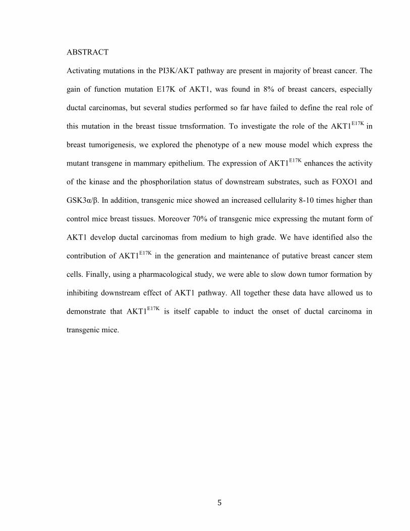
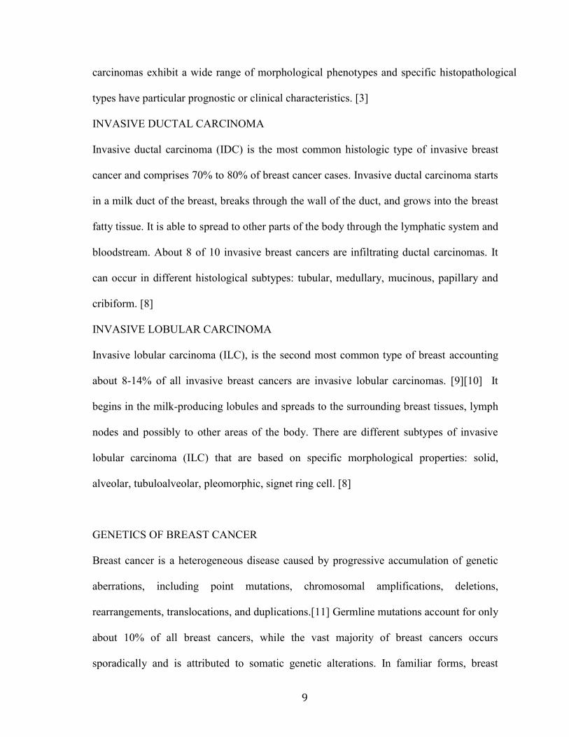
countries. [3] In 2013, in USA, 232.340 new cases of invasive breast cancer have been
diagnosed among women, as well as 64.640 additional cases of in situ breast cancer. In 2013,
have been estimated approximately 39.620 women deaths from breast cancer and 2.240 men
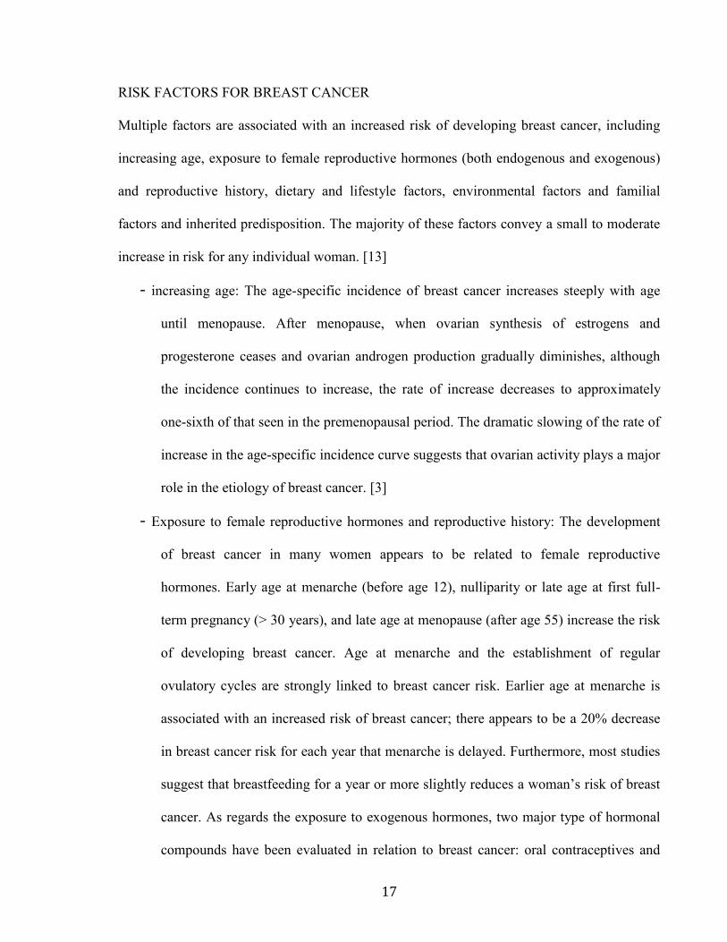
have been diagnosed with breast cancer and 410 men are dead from the disease. (figure 1). [4]
Figure 1: estimated new female in situ and invasive breast cancer cases and death by age, US, 2013. [4]
8
HISTOLOGICAL CLASSIFICATION
Breast cancer originates from the epithelial cells of the glandular tree and may give rise to
different histotypes. The most common are the lobular and ductal carcinomas, of which there
are in situ and invasive forms.
IN SITU BREAST CANCER
- Ductal carcinoma in situ (DCIS) is the most common type of in situ breast cancer,
accounting for about 83% of in situ cases diagnosed during 2006-2010. DCIS may or may not
progress to invasive cancer; in fact, it consists of cancerous cells that proliferate within the
lumens of the breast duct with no invasion. It grows so slowly that even without treatment
does not affect the women health. [5][6] Clinical studies suggest, however, that about one-
third, and possibly more, of DCIS cases will progress to invasive cancer if left untreated. [5]
- Lobular carcinoma in situ (LCIS, also known as lobular neoplasia) is not a true cancer or
precancer, but an indicator of increased risk for developing invasive cancer. It is represented
by the presence of unusual cells in the lobules of the breast. LCIS is much less common than
DCIS, accounting for about 12% of female in situ breast cancers diagnosed during 2006-2010.
[7]
- Other in situ breast cancers have characteristics of both ductal and lobular carcinomas or
have unknown origins.[4]
INVASIVE BREAST CANCER
Invasive breast carcinoma is a group of malignant epithelial tumors characterized by invasion
of adjacent tissues and a marked tendency to metastasize to distant sites. Invasive breast
9
carcinomas exhibit a wide range of morphological phenotypes and specific histopathological
types have particular prognostic or clinical characteristics. [3]
INVASIVE DUCTAL CARCINOMA
Invasive ductal carcinoma (IDC) is the most common histologic type of invasive breast
cancer and comprises 70% to 80% of breast cancer cases. Invasive ductal carcinoma starts
in a milk duct of the breast, breaks through the wall of the duct, and grows into the breast
fatty tissue. It is able to spread to other parts of the body through the lymphatic system and
bloodstream. About 8 of 10 invasive breast cancers are infiltrating ductal carcinomas. It
can occur in different histological subtypes: tubular, medullary, mucinous, papillary and
cribiform. [8]
INVASIVE LOBULAR CARCINOMA
Invasive lobular carcinoma (ILC), is the second most common type of breast accounting
about 8-14% of all invasive breast cancers are invasive lobular carcinomas. [9][10] It
begins in the milk-producing lobules and spreads to the surrounding breast tissues, lymph
nodes and possibly to other areas of the body. There are different subtypes of invasive
lobular carcinoma (ILC) that are based on specific morphological properties: solid,
alveolar, tubuloalveolar, pleomorphic, signet ring cell. [8]
GENETICS OF BREAST CANCER
Breast cancer is a heterogeneous disease caused by progressive accumulation of genetic
aberrations, including point mutations, chromosomal amplifications, deletions,
rearrangements, translocations, and duplications.[11] Germline mutations account for only
about 10% of all breast cancers, while the vast majority of breast cancers occurs
sporadically and is attributed to somatic genetic alterations. In familiar forms, breast
10
cancer susceptibility genes can be categorized into three classes according to their
frequency and level of risk that they confer:
- rare high-penetrance genes, in particular BRCA1 and BRCA2, which encode large
proteins with multiple functions which act as classic tumor suppressor genes that maintain
genomic stability by facilitating double-strand DNA repair through homologous
recombination. [12]. When loss of heterozygosis (LOH) occurs via loss, mutation or
silencing of the wild type BRCA1 and BRCA2 allele, the resultant defective DNA repair
system leads to rapid acquisition of additional mutations, particularly during DNA
replication. [13]. BRCA1 and BRCA2 mutations account for approximately half of all
dominantly inherited hereditary breast cancers. These mutations confer a relative risk of
breast cancer 10 to 30 times higher that of women in the general population, resulting in a
nearly 85% lifetime risk of breast cancer development. [14]. Other high-penetrance genes
are TP53, PTEN, STK11/LKB1, and CDH1. These high-penetrance genes confer an eight-
ten fold increase in risk of breast cancer as compared to non- carriers, but they collectively
account for less than 1% of cases of breast cancer. Like BRCA1 and BRCA2, these genes
are inherited in an autosomal dominant manner and function as tumor suppressors. [15].
- rare intermediate-penetrance genes. Four genes that confer an elevated but moderate risk of
developing breast cancer have been identified, namely CHEK2, ATM, BRIP1, and PALB2,
involved in signal transduction and DNA repair. Each of these genes confers approximately a
two-three fold relative risk of breast cancer.[13].
- common low-penetrance genes and loci, include approximately ten different alleles and loci
in 15% to 40% of women with breast cancer. [14] Despite their frequency, the relative risk of
breast cancer conferred by any one of these genetic variants alone is minimal, less than 1.5
fold.[16] Nevertheless, these alleles and loci may become clinically relevant in interaction
11
with other high-, moderate-, and low-risk genes; these additive or multiplicative relationships
could account for a measurable fraction of population risk.
For example, association studies of FGFR2 and MAP3K1 within BRCA families showed that
these single nucleotide polymorphisms (SNPs) conferres an increased risk in the presence of
BRCA2 mutations. Recent studies suggest that microRNA (miRNA) SNPs may also contribute
to breast cancer susceptibility, and miRNAs appear to regulate many tumor suppressor genes
and oncogenes via degradation of target mRNAs or repression of their translation. [13]
The vast majority of breast cancers are sporadic, caused by accumulation of several somatic
genetic alterations. Recent data suggest that a typical individual breast cancer harbors
anywhere from 50 to 80 different somatic mutations. [11] Many of these mutations occur as a
result of erroneous DNA replication; others may occur through exposure to exogenous and
endogenous mutagens. [13] Among gene amplifications, the most frequent in breast cancer
regard HER-2/Neu, growth receptor that activates the Ras-MEK and PI3K pathway, amplified
in about 13% of the breast cancer. Cyclin D1, amplified in about 10-12% of the breast cancers,
WIP1 (13%) and GASC1, amplified in about 5-10% of total breast cancers and in the 20-25%
of the basal breast cancer. [17]
Inactivation of gene functions by deletion or other mechanisms commonly occurs in PTEN
and p53 in HER2/neu positive breast cancers, triple negative breast cancers, and BRCA-
associated breast and ovarian cancers. PI3K amplifications and activating mutations are
common in breast cancers and several genes such as AKT and STAT3 are often expressed at
high activities but without detectable amplifications of those genes. [18] Epigenetic
alterations, such as methylation of cytosine residues in CpG dinucleotides, can bring about
gene inactivation, for example p16 gene in breast cancers. [17] A substantial number of these
somatic mutations sort out among a much smaller number of biological groups and cell
12
signaling pathways that are known to be pathogenetic in breast cancer, thereby vastly reducing
the complexity of the genomic landscape. Examples of such pathways include interferon
signaling, cell cycle checkpoint, BRCA1/2- related DNA repair, p53, transforming growth
factor-β (TGF-β) signaling, Notch, epidermal growth factor receptor (EGFR), FGF, ERBB2,
RAS, and PI3K-AKT. [13]
MOLECULAR CLASSIFICATION
The accumulation of different mutations has significant effects on the expression of important
tissue-specific genes. Distinct molecular subtypes of breast cancer have been identified using
biological markers, including the presence or absence of estrogen receptors (ER+/ ER-),
progesterone receptors (PR+/PR-), and human epidermal growth factor receptor 2
(HER2+/HER2-). [4][13]
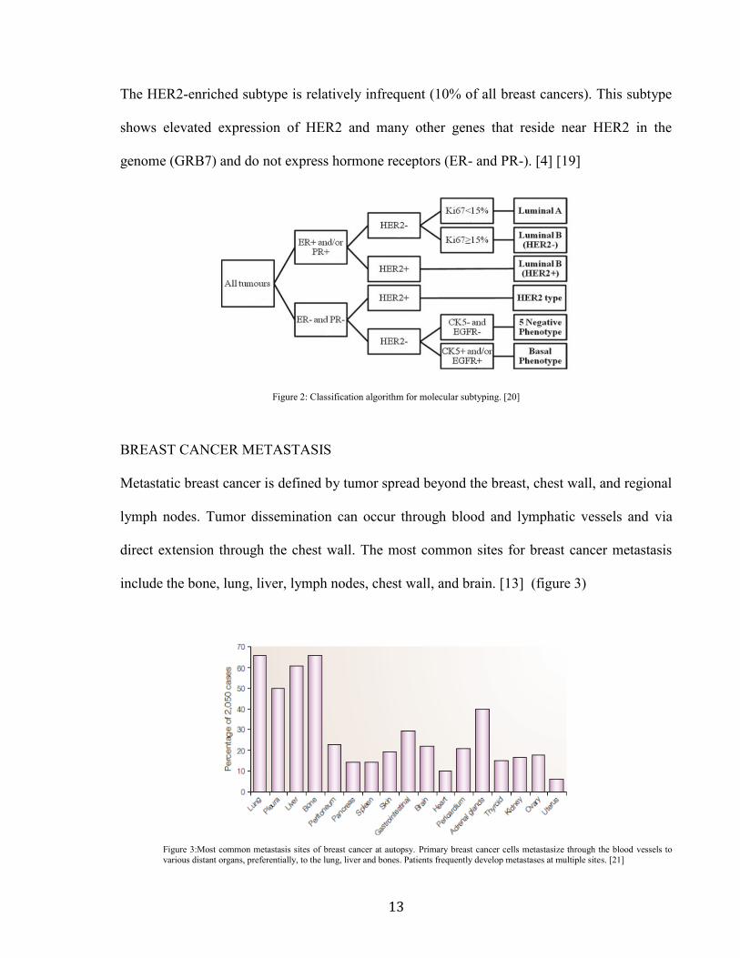
We can distinguish four different types of breast cancer: luminal A, luminal B, Basal-like and
HER2 enriched. (figure 2)
Luminal A tumors have high expression of ER and ER-regulated genes, low expression of the
HER2 cluster and proliferation-associated genes. Luminal B tumors tend to be highly
proliferative, express mutant form of TP53, show lower expression of ER and ER-regulated
genes and can be HER2+ or HER2-. [19][20] Basal-like breast cancer are referred to as “triple
negative” because they are ER-, PR-, and HER2-. The basal- like subtype is characterized by
low expression of the luminal genes, low expression of the HER2 gene cluster, high
expression of the proliferation cluster, and high expression of a unique cluster of genes called
the basal cluster (cytokeratins 5, 6, 14,17, c-Kit; Vimentin; P-Cadherin). Several risk factors
for developing basal-like tumors have been identified, among which the most interesting being
the link between the basal-like subtype and BRCA1 mutation carriers. [19]
13
The HER2-enriched subtype is relatively infrequent (10% of all breast cancers). This subtype
shows elevated expression of HER2 and many other genes that reside near HER2 in the
genome (GRB7) and do not express hormone receptors (ER- and PR-). [4] [19]
Figure 2: Classification algorithm for molecular subtyping. [20]
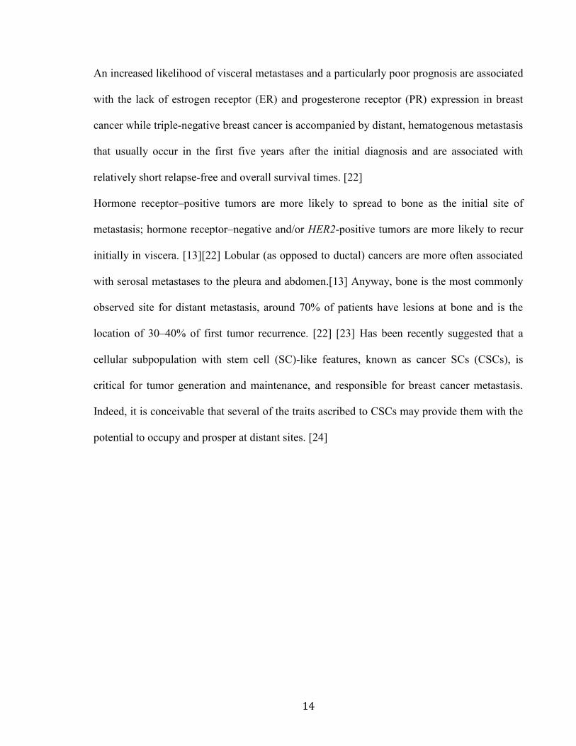
BREAST CANCER METASTASIS
Metastatic breast cancer is defined by tumor spread beyond the breast, chest wall, and regional
lymph nodes. Tumor dissemination can occur through blood and lymphatic vessels and via
direct extension through the chest wall. The most common sites for breast cancer metastasis
include the bone, lung, liver, lymph nodes, chest wall, and brain. [13] (figure 3)
Figure 3:Most common metastasis sites of breast cancer at autopsy. Primary breast cancer cells metastasize through the blood vessels to
various distant organs, preferentially, to the lung, liver and bones. Patients frequently develop metastases at multiple sites. [21]
14
An increased likelihood of visceral metastases and a particularly poor prognosis are associated
with the lack of estrogen receptor (ER) and progesterone receptor (PR) expression in breast
cancer while triple-negative breast cancer is accompanied by distant, hematogenous metastasis
that usually occur in the first five years after the initial diagnosis and are associated with
relatively short relapse-free and overall survival times. [22]
Hormone receptor–positive tumors are more likely to spread to bone as the initial site of
metastasis; hormone receptor–negative and/or HER2-positive tumors are more likely to recur
initially in viscera. [13][22] Lobular (as opposed to ductal) cancers are more often associated
with serosal metastases to the pleura and abdomen.[13] Anyway, bone is the most commonly
observed site for distant metastasis, around 70% of patients have lesions at bone and is the
location of 30–40% of first tumor recurrence. [22] [23] Has been recently suggested that a
cellular subpopulation with stem cell (SC)-like features, known as cancer SCs (CSCs), is
critical for tumor generation and maintenance, and responsible for breast cancer metastasis.
Indeed, it is conceivable that several of the traits ascribed to CSCs may provide them with the
potential to occupy and prosper at distant sites. [24]
15
BREAST CANCER STEM CELLS
Cancer stem cells (CSCs) are tumor cells with enhanced capacity for tumor generation. CSCs
possess several fundamental attributes similar to normal adult stem cells. They are capable of
dividing asymmetrically to produce one stem cell, characterized by self-renewal, and one
progenitor cell, which allows to produce phenotypically diverse cancer cells that constitute
tumors. [25] In breast cancer has been isolated a small population of tumorigenic cells with
stem cell (SC)-like features, capable of regenerating the phenotypic heterogeneity of the
original tumor when injected subcutaneously into NOD/SCID mice. [26][27] These breast
cancer stem cells (BCSCs) are characterized by the cell-surface markers ESA+/CD44
+/CD24
-
/low , devoid of the expression of the lineage markers CD2, CD3, CD10, CD 16, CD18, CD31,
CD64, and CD140b (Lin−) and bear high ALDH1 activity. [25][26][28] Putative breast CSCs
have also been isolated from patient samples after in vitro propagation and from breast cancer
cell lines, through their ability to proliferate in suspension as non adherent spheres
(mammospheres). Because the capacity to form mammospheres is increased in early
progenitor/stem cells, this system has been widely used as an indirect measurement of the
number of cells with self-renewal capability. [29] The origin of breast CSCs is controversial.
Current experimental evidence supports two different, but not exclusive, theories (figure 4).
Figure 1. Or igin of the breast cancer stem cells (BCSCs)
BCSCs may arise from normal mammary stem cells (A) or from non-stem tumor cells that
have gained the ability for self-renewal (B) by epithelial to mesenchymal transition (EMT)
and oncogenic transformation. Both of these hypotheses consider that the phenotypic
characteristics of BCSCs are caused by genetic alterations and/or EMT. As result, BCSCs
display alterations in signaling pathways controlling the cell cycle, differentiation, and
survival (C).
Velasco-Velázquez et al. Page 10
Int J Biochem Cell Biol . Author manuscript; available in PMC 2013 April 01.
NIH
-PA
Au
tho
r Ma
nu
scrip
tN
IH-P
A A
uth
or M
an
uscrip
tN
IH-P
A A
uth
or M
an
uscrip
t
Figure 4. Origin of the breast cancer stem cells (BCSCs). BCSCs may arise from normal mammary stem cells (A) or from non-
stem tumor cells that have gained the ability for self-renewal (B) by epithelial to mesenchymal transition (EMT) and oncogenic transformation. Both of these hypotheses consider that the phenotypic characteristics of BCSCs are caused by genetic alterations
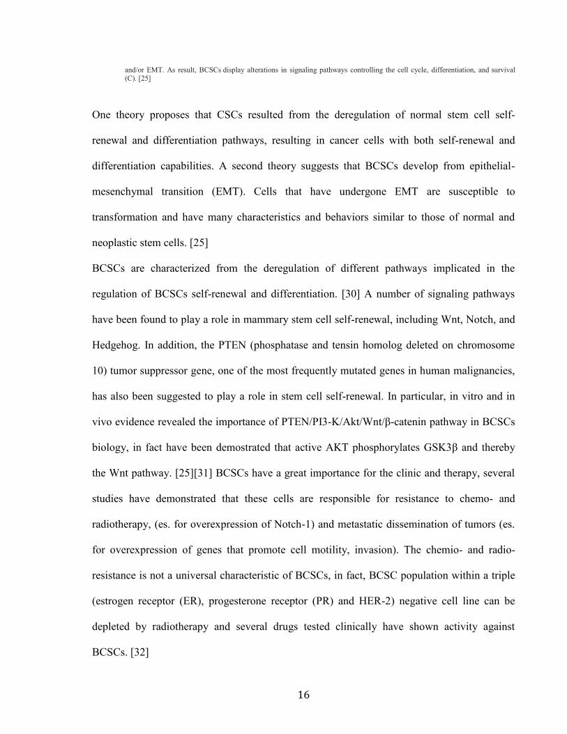
16
and/or EMT. As result, BCSCs display alterations in signaling pathways controlling the cell cycle, differentiation, and survival (C). [25]
One theory proposes that CSCs resulted from the deregulation of normal stem cell self-
renewal and differentiation pathways, resulting in cancer cells with both self-renewal and
differentiation capabilities. A second theory suggests that BCSCs develop from epithelial-
mesenchymal transition (EMT). Cells that have undergone EMT are susceptible to
transformation and have many characteristics and behaviors similar to those of normal and
neoplastic stem cells. [25]
BCSCs are characterized from the deregulation of different pathways implicated in the
regulation of BCSCs self-renewal and differentiation. [30] A number of signaling pathways
have been found to play a role in mammary stem cell self-renewal, including Wnt, Notch, and
Hedgehog. In addition, the PTEN (phosphatase and tensin homolog deleted on chromosome
10) tumor suppressor gene, one of the most frequently mutated genes in human malignancies,
has also been suggested to play a role in stem cell self-renewal. In particular, in vitro and in
vivo evidence revealed the importance of PTEN/PI3-K/Akt/Wnt/β-catenin pathway in BCSCs
biology, in fact have been demostrated that active AKT phosphorylates GSK3β and thereby
the Wnt pathway. [25][31] BCSCs have a great importance for the clinic and therapy, several
studies have demonstrated that these cells are responsible for resistance to chemo- and
radiotherapy, (es. for overexpression of Notch-1) and metastatic dissemination of tumors (es.
for overexpression of genes that promote cell motility, invasion). The chemio- and radio-
resistance is not a universal characteristic of BCSCs, in fact, BCSC population within a triple
(estrogen receptor (ER), progesterone receptor (PR) and HER-2) negative cell line can be
depleted by radiotherapy and several drugs tested clinically have shown activity against
BCSCs. [32]
17
RISK FACTORS FOR BREAST CANCER
Multiple factors are associated with an increased risk of developing breast cancer, including
increasing age, exposure to female reproductive hormones (both endogenous and exogenous)
and reproductive history, dietary and lifestyle factors, environmental factors and familial
factors and inherited predisposition. The majority of these factors convey a small to moderate
increase in risk for any individual woman. [13]
- increasing age: The age-specific incidence of breast cancer increases steeply with age
until menopause. After menopause, when ovarian synthesis of estrogens and
progesterone ceases and ovarian androgen production gradually diminishes, although
the incidence continues to increase, the rate of increase decreases to approximately
one-sixth of that seen in the premenopausal period. The dramatic slowing of the rate of
increase in the age-specific incidence curve suggests that ovarian activity plays a major
role in the etiology of breast cancer. [3]
- Exposure to female reproductive hormones and reproductive history: The development
of breast cancer in many women appears to be related to female reproductive
hormones. Early age at menarche (before age 12), nulliparity or late age at first full-
term pregnancy (> 30 years), and late age at menopause (after age 55) increase the risk
of developing breast cancer. Age at menarche and the establishment of regular
ovulatory cycles are strongly linked to breast cancer risk. Earlier age at menarche is
associated with an increased risk of breast cancer; there appears to be a 20% decrease
in breast cancer risk for each year that menarche is delayed. Furthermore, most studies
suggest that breastfeeding for a year or more slightly reduces a woman’s risk of breast
cancer. As regards the exposure to exogenous hormones, two major type of hormonal
compounds have been evaluated in relation to breast cancer: oral contraceptives and
18
menopausal replacement therapy. The evidence suggests a small increase in the
relative risk associated with the use of combined oral contraceptives and
postmenopausal hormone replacement therapy (HRT). [3][4]
- dietary and lifestyle factors: Observational studies suggested that high-caloric diets rich
in animal fat and proteins were associated with higher rates of breast cancer ,
combined with consumption of alcohol, a lack of physical exercise, overweight and
obesity (in particular for postmenopausal breast cancer). [3][4][13]
- environmental factors: only limited data are available on specific exposures in relation to
breast cancer. Long-term follow-up of women exposed to the Hiroshima or Nagasaki
nuclear explosions indicates an increased risk of breast cancer, in particular for women
exposed around puberty. Similarly, exposure as a result of treatment and surveillance
of tuberculosis is associated with risk. Other environmental factors, including exposure
to electromagnetic fields and organochlorine pesticides, have been suggested to
increase breast cancer risk. [3][13]
- familial factors and inherited predisposition: Women (as well as men) with a family
history of breast cancer, especially in a first-degree relative (mother, sister, daughter,
father, or brother), are at increased risk of developing breast cancer; this risk is higher
if more than one first-degree relative developed breast cancer. Compared to women
without a family history, risk of breast cancer is 1.8 times higher for women with one
first-degree female relative who has been diagnosed, nearly 3 times higher for women
with two relatives, and nearly 4 times higher for women with three or more relatives.
Risk is further increased when the affected relative was diagnosed at a young age. It is
important to note that the majority of women with one or more affected first-degree
relatives will never develop breast cancer and that most women who develop breast
19
cancer do not have a family history of the disease. It is estimated that 5% to 10% of
breast cancer cases results from inherited mutations, including those in the breast
cancer susceptibility genes BRCA1 and BRCA2 .[4][13] Other inherited conditions
associated with smaller increased breast cancer risk include Li-Fraumeni and Cowden
syndromes and a number of more common genetic mutations [4]
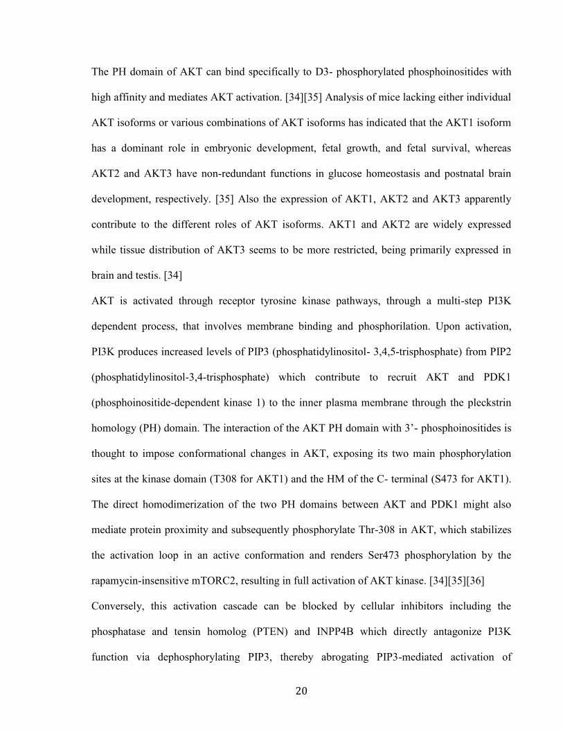
THE PROTEIN SERINE / THREONINE KINASE B (PKB / AKT) IN BREAST CANCER
AKT, a downstream effector of phosphatidylinositol-3 kinase (PI3K), is one of the most
frequently hyperactivated protein kinase in human cancer .[33] It is a serine/threonine protein
kinase and its hyperactivation is associated with resistance to apoptosis, increased cell growth,
cell proliferation and cell energy metabolism. In mammalian cells AKT comprises three
highly homologous members (>80% protein sequence identity) termed AKT1/PKBα,
AKT2/PKBβ and AKT3/PKBγ, encoded by three different genes located on chromosomes
14q32, 19q13 and 1q43. AKT kinases share the same structural organization, containing an N-
terminal pleckstrin homology (PH) domain, a central catalytic domain and a C-terminal
regulatory domain that contains the hydrofobic motif (HM). (Figure 5)
Figure 5: Akt domains and comparison of Akt isoforms (% of homology). Chromosome location of each Akt
isoform in human and phosphorylation sites in Akt1. [35]
20
The PH domain of AKT can bind specifically to D3- phosphorylated phosphoinositides with
high affinity and mediates AKT activation. [34][35] Analysis of mice lacking either individual
AKT isoforms or various combinations of AKT isoforms has indicated that the AKT1 isoform
has a dominant role in embryonic development, fetal growth, and fetal survival, whereas
AKT2 and AKT3 have non-redundant functions in glucose homeostasis and postnatal brain
development, respectively. [35] Also the expression of AKT1, AKT2 and AKT3 apparently
contribute to the different roles of AKT isoforms. AKT1 and AKT2 are widely expressed
while tissue distribution of AKT3 seems to be more restricted, being primarily expressed in
brain and testis. [34]
AKT is activated through receptor tyrosine kinase pathways, through a multi-step PI3K
dependent process, that involves membrane binding and phosphorilation. Upon activation,
PI3K produces increased levels of PIP3 (phosphatidylinositol- 3,4,5-trisphosphate) from PIP2
(phosphatidylinositol-3,4-trisphosphate) which contribute to recruit AKT and PDK1
(phosphoinositide-dependent kinase 1) to the inner plasma membrane through the pleckstrin
homology (PH) domain. The interaction of the AKT PH domain with 3’- phosphoinositides is
thought to impose conformational changes in AKT, exposing its two main phosphorylation
sites at the kinase domain (T308 for AKT1) and the HM of the C- terminal (S473 for AKT1).
The direct homodimerization of the two PH domains between AKT and PDK1 might also
mediate protein proximity and subsequently phosphorylate Thr-308 in AKT, which stabilizes
the activation loop in an active conformation and renders Ser473 phosphorylation by the
rapamycin-insensitive mTORC2, resulting in full activation of AKT kinase. [34][35][36]
Conversely, this activation cascade can be blocked by cellular inhibitors including the
phosphatase and tensin homolog (PTEN) and INPP4B which directly antagonize PI3K
function via dephosphorylating PIP3, thereby abrogating PIP3-mediated activation of
21
downstream signaling events such as PDK1 and AKT. However, in vitro engineered AKT
kinase can override this regulatory mechanism and maintain it in a “supercharged” stage. This
can be done by insertion of myristoylated (Myr) tag at its N-terminus which results in AKT
anchoring in plasma membrane and constitutive AKT activation independently of PI3K
activity. [36]
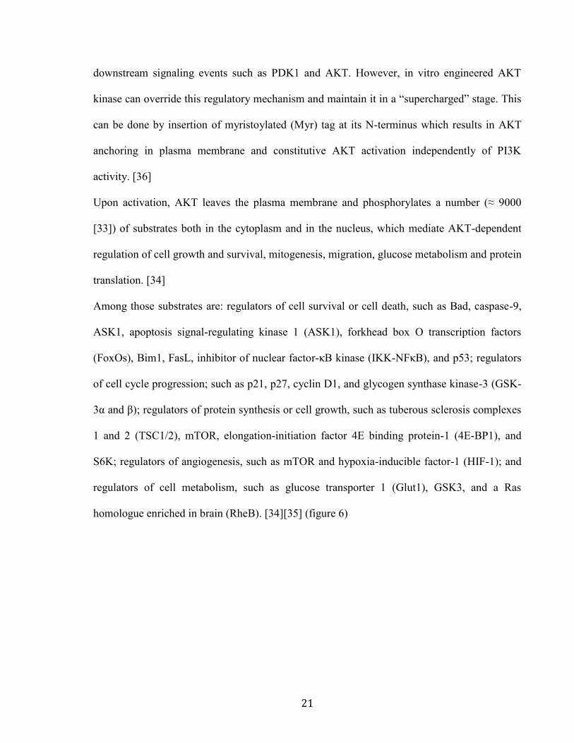
Upon activation, AKT leaves the plasma membrane and phosphorylates a number (≈ 9000
[33]) of substrates both in the cytoplasm and in the nucleus, which mediate AKT-dependent
regulation of cell growth and survival, mitogenesis, migration, glucose metabolism and protein
translation. [34]
Among those substrates are: regulators of cell survival or cell death, such as Bad, caspase-9,
ASK1, apoptosis signal-regulating kinase 1 (ASK1), forkhead box O transcription factors
(FoxOs), Bim1, FasL, inhibitor of nuclear factor-κB kinase (IKK-NFκB), and p53; regulators
of cell cycle progression; such as p21, p27, cyclin D1, and glycogen synthase kinase-3 (GSK-
3α and β); regulators of protein synthesis or cell growth, such as tuberous sclerosis complexes
1 and 2 (TSC1/2), mTOR, elongation-initiation factor 4E binding protein-1 (4E-BP1), and
S6K; regulators of angiogenesis, such as mTOR and hypoxia-inducible factor-1 (HIF-1); and
regulators of cell metabolism, such as glucose transporter 1 (Glut1), GSK3, and a Ras
homologue enriched in brain (RheB). [34][35] (figure 6)
22
Figure 6: Akt signaling pathway. [37]
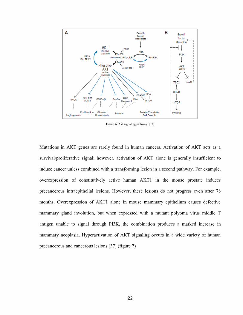
Mutations in AKT genes are rarely found in human cancers. Activation of AKT acts as a
survival/proliferative signal; however, activation of AKT alone is generally insufficient to
induce cancer unless combined with a transforming lesion in a second pathway. For example,
overexpression of constitutively active human AKT1 in the mouse prostate induces
precancerous intraepithelial lesions. However, these lesions do not progress even after 78
months. Overexpression of AKT1 alone in mouse mammary epithelium causes defective
mammary gland involution, but when expressed with a mutant polyoma virus middle T
antigen unable to signal through PI3K, the combination produces a marked increase in
mammary neoplasia. Hyperactivation of AKT signaling occurs in a wide variety of human
precancerous and cancerous lesions.[37] (figure 7)
23
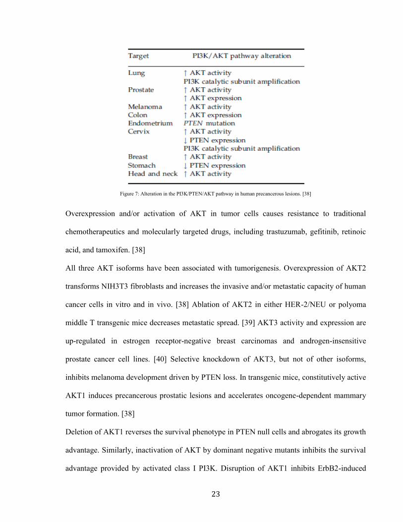
Figure 7: Alteration in the PI3K/PTEN/AKT pathway in human precancerous lesions. [38]
Overexpression and/or activation of AKT in tumor cells causes resistance to traditional
chemotherapeutics and molecularly targeted drugs, including trastuzumab, gefitinib, retinoic
acid, and tamoxifen. [38]
All three AKT isoforms have been associated with tumorigenesis. Overexpression of AKT2
transforms NIH3T3 fibroblasts and increases the invasive and/or metastatic capacity of human
cancer cells in vitro and in vivo. [38] Ablation of AKT2 in either HER-2/NEU or polyoma
middle T transgenic mice decreases metastatic spread. [39] AKT3 activity and expression are
up-regulated in estrogen receptor-negative breast carcinomas and androgen-insensitive
prostate cancer cell lines. [40] Selective knockdown of AKT3, but not of other isoforms,
inhibits melanoma development driven by PTEN loss. In transgenic mice, constitutively active
AKT1 induces precancerous prostatic lesions and accelerates oncogene-dependent mammary
tumor formation. [38]
Deletion of AKT1 reverses the survival phenotype in PTEN null cells and abrogates its growth
advantage. Similarly, inactivation of AKT by dominant negative mutants inhibits the survival
advantage provided by activated class I PI3K. Disruption of AKT1 inhibits ErbB2-induced
24
mammary tumorigenesis. AKT1 deficiency delays tumor growth and reduces metastasis.
AKT1 null mammary epithelial tumor cells have also reduced proliferative capability with
reduced cyclin D1 and increased p27. These data suggest that AKT1 plays an important role
in mammary tumorigenesis. [41]
The oncogenic activation of AKT1 can be induced by several means, most commonly
occurring either due to the compromise in its membrane-targeting by PH domain, or due to the
pathological conformational changes occurring in the mutant structure. Genetic mutations in
the PH domain alter AKT1 localization and sensitivity to the PtdIns bringing serious
consequences for its activity.[42]
A dominant hotspot mutation at nucleotide 49 (G>A) of the gene encoding AKT1 results in
the substitution of a lysine for glutamic acid at the amino acid 17 (Akt1-E17K). [34][42][43]
In the apo conformation, Glu 17 occupies the phosphoinositide-binding pocket and forms a
network of hydrogen bonds. The Lys 17 substitution results in a shift in the surface charge
around the pocket from negative with Glu 17 to effectively neutral in the mutant. [42][43] The
AKT1E17K
mutation alters the electrostatic interactions of the pocket, activates AKT1 in a
PI3K-independent manner, increasing level of AKT1 phosphorylation on Thr 308 and Ser 473
as compared to wild-type. [42][44] AKT1E17K
kinase activity was shown to be approximately
four fold higher than that of AKT1 wild type, suggesting that the mutation alters AKT1
regulation and hence it enhances cellular activity. [42][43] Furthermore, it has also been
proposed that the mutation induces large affinity increase for PI(4,5)P2 which is essential to
the constitutive plasma membrane targeting of the mutant PH domain and thus to the
oncogenic nature of the full-length AKT1E17K
protein. [42] Moreover, it was also suggested
that the E17K PH domain mutation causes structural changes in the PH domain, which further
hinders its interaction with AKT1/2 inhibitor VIII. [43] All these observations strongly
25
suggest that the damaging conformational changes in mutant PH domain might cause such
pathological outcomes. Functionally, the AKT1E17K
mutation stimulates AKT signaling,
induces cellular transformation and produces leukaemia in mice. [44][45] The AKT1E17K
mutation was found in 5 out of 61 (8.2%) breast cancers, 3 out of 51 (5.9%) colorectal
cancers, 1 out of 50 (2.0%) ovarian cancers [43] and 3 out 105 (2.9%) lung cancer. [46]
GENETICALLY ENGINEERED MOUSE MODELS OF PI3K/AKT SIGNALING IN
BREAST CANCER
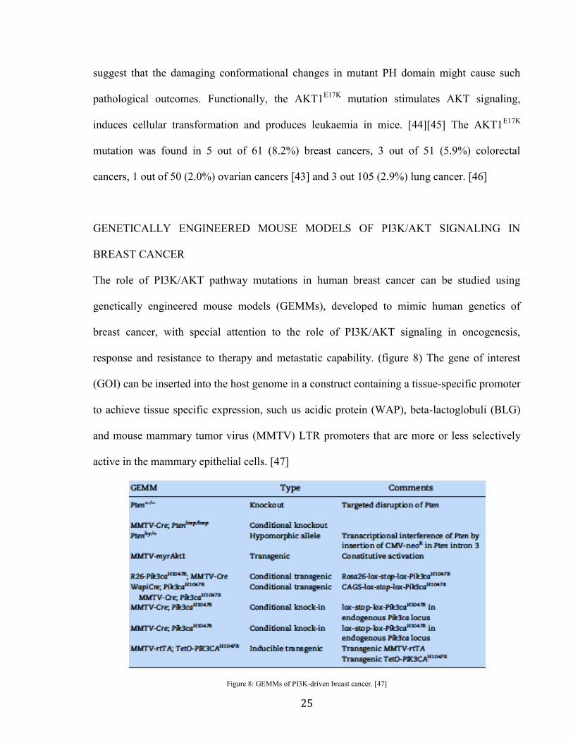
The role of PI3K/AKT pathway mutations in human breast cancer can be studied using
genetically engineered mouse models (GEMMs), developed to mimic human genetics of
breast cancer, with special attention to the role of PI3K/AKT signaling in oncogenesis,
response and resistance to therapy and metastatic capability. (figure 8) The gene of interest
(GOI) can be inserted into the host genome in a construct containing a tissue-specific promoter
to achieve tissue specific expression, such us acidic protein (WAP), beta-lactoglobuli (BLG)
and mouse mammary tumor virus (MMTV) LTR promoters that are more or less selectively
active in the mammary epithelial cells. [47]
Figure 8: GEMMs of PI3K-driven breast cancer. [47]
26
Several GEMMs with tissue-specific mutation of PIK3CA, which encodes the
phosphoinositide-3-kinase (PI3K) catalytic subunit p110α have been published. Several
groups have shown that expression of the H1047R PIK3CA mutant in luminal mammary
epithelium results in the formation of mammary tumors of several phenotypes, in particular
adenosquamous carcinoma or adenomyoepithelioma phenotype. Genetic interaction between
PIK3CAH1047R
and p53 loss-of-function mutations in R26-PIK3CAH1047R
;p53loxP/+
;MMTV-Cre
mice leads to the reduction of survival of double-mutant animals, which developed lymphoma
and mammary tumors with rapid kinetics. R26-PIK3CAH1047R
;p53loxP/+
;MMTV-Cre mammary
tumors were mostly adenosquamous carcinoma or spindle cell/EMT indicating that double-
mutant mice develop a distinct spectrum of mammary tumors. [47][48]
In Pten heterozygous knockout (Pten+/-
) the loss of PTEN expression is associated with basal-
like tumors. [49] Deletion of the Pten gene in mammary epithelium in conditional Pten gene
knock-out mice, generated by flanking exon 5, which encodes the phosphatase domain of
PTEN, with LoxP sequences, causes increased cell proliferation, hyper-branched ductal
structure, precocious development, delayed involution and severely impaired apoptosis.
PTEN-deficient mammary epithelium also displays remarkable neoplastic changes. [50]
To examine the role of AKT1 in the etiology of mammary tumorigenesis, transgenic mice
were generated that express human AKT1 under the control of the mouse mammary tumor
virus (MMTV) LTR. Ackler et al. have demonstrated for the first time that AKT1 expression
during lactation results in a pronounced delay in involution, associated with hyperplasia and
marked expression of cyclin D1. [51]
Addition of a myristoylation signal to the murine AKT1 gene, which results in AKT1
anchoring in plasma membrane and constitutive AKT1 activation independently of PI3K
27
activity, increases the incidence of benign lesions, delay in mammary involution and
susceptibility to epithelial mammary tumor (ER-positive adenocarcinomas or adenosquamous
tumors) formation induced by the carcinogen 9,10-dimethyl-1,2 benzanthracene (DMBA).
[52]
Finally, in double myrAKT;p53(R172H) mice p53 inactivation by R172H point mutation
combined with myrAKT transgenic expression significantly increases the percentage and size
of mammary carcinoma, but was not sufficient to promote full penetrance of the tumorigenic
phenotype. [41]
28
PROJECT'S AIM
Data from the literature have shown a significant involvement of the PI3K/Akt pathway in the
onset of breast cancer. The role of AKT1 is controversial, the kinase seems to have a minor
role, and limited to the ductal breast carcinoma. The purpose of this research project is to
identify the oncogenic role of AKT1E17K
in the mammary tumorigenesis by generating a
mouse model that expresses AKT1E17K
specifically in the breast, with particular attention to
the effect on cell proliferation and breast cancer stem cells (BCSCs) maintenance.
Furthermore this project comprises a pharmacological study in order to identify AKT1 as a
putative target in breast cancer therapy.
29
MATERIALS AND METHODS
RESTRICTION DIGEST OF PLASMID DNA
Digestion of plasmid DNA was performed using different NEB enzymes. It is preferable to
digest 0.2-1.5 µg DNA with a 2-fold to 10-fold excess of enzyme in a total volume of 20 µl. In
the case of double digestion use the most compatible buffer with all the enzymes and it is
important that the total volume of enzymes add to reaction is not more than 1/10 of the total
reaction volume. A
typical restriction enzyme digestion protocol is below:
- In a 1.5mL tube combine the following:
DNA
Restriction Enzyme(s)
10X Buffer
10X BSA (if recommended by manufacturer)
dH2O up to total volume (20 µl)
- Mix gently and spin down briefly
- Incubate at the optimal reaction temperature (usually 37°C) for 2 hours
The samples were run in an 1% agarose gel electrophoresis with ethidium bromide, using the
non-digested plasmid as negative control.
PURIFYING DNA FROM AGAROSE GEL
Gel purification allows to isolate and purify DNA fragments based on size. The procedure
starts with standard agarose gel electrophoresis, which separates DNA by their length in base
pairs. Following electrophoresis, cut DNA bands out of the agarose gel and purify the DNA
30
samples. In our case, for this purpose, we used the QIAGEN QIAquick Gel Extraction Kit,
using the standard protocol provided by the manufacturer.
DNA LIGATION
The final step in the construction of a recombinant plasmid is connecting the insert DNA
(gene or fragment of interest) into a compatibly digested vector backbone. This is
accomplished by covalently connecting the sugar backbone of the two DNA fragments. This
reaction, called ligation, is performed by the T4 DNA ligase enzyme. The DNA ligase
catalyzes the formation of covalent phosphodiester linkages, which permanently join the
nucleotides together. This experiment was carried out using NEB T4 DNA Ligase (M0202).
Before setting up the ligation reaction itself, it is important to determine the amount of cut
insert and vector to use for the ligation reaction. The volume of vector DNA and insert DNA
used in the ligation will vary depending on the size of each and their concentration. However,
for most standard cloning (where the insert is smaller than the vector) a 3 insert : 1 vector ratio
will work just fine.
To calculate the volume of the insert for the ligation reaction must use the following formula:
X ng of insert = (3) (bp insert) (50 ng linearized plasmid-) ÷ (size of plasmid in bp)
- Set up the typical ligation reaction as follows:
10X T4 DNA ligase buffer
vector DNA
insert DNA
dH2O up to total volume (20 µl)
- Gently mix the reaction by pipetting
- Incubate at 16°C overnight
31
- Chill on ice and transform 1-5 μl of the reaction into 50 μl competent cells.
TRANSFORMATION OF ELECTROCOMPETENT BACTERIAL CELLS
The electrical transformation refers ingestion of foreign DNA in competent bacterical cells,
that are able to take exogenous DNA, through the creation of pores in the bacterial cell walls
using an electrical pulse.
The transformation protocol requires several steps:
- Thaw competent cells on ice for defrosting and mix 1 to 5μl of DNA (usually 10pg to
100ng) into 50μL of competent cells;
- Add cell/DNA mixture to the electroporation cuvette;
- Place cuvette in electroporator and shock cells at 2500 V;
- Remove cuvette from the chamber and immediately add SOC (SOB + glucose 2M). This
step should be done as quickly as possible to prevent cells from dying off;
- Transfer SOC-cell mixture to an eppendorf tube and incubate tube in 37°C shaker for at
least 1 hr to permit expression of antibiotic resistance gene;
- Centrifuge at 4500 rpm, discard the supernatant and resuspend the bacterial pellet in 100
μl of LB;
- Plate transformation onto prewarmed LB-agar plate supplemented with appropriate
antibiotic and Incubate overnight at 37°C.
RECOVERING PLASMID DNA FROM BACTERIAL CULTURE (MINIPREP)
After liquid bacteric culture, at 37°C for overnight in a shacking incubator, bacterial cells were
pelleted by centrifugation at 5000 rpm for 10 minutes. Afterwards, the QIAprep Spin
Miniprep kit by QIAGEN was used to extract plasmid DNA as manufacturer’s
32
recommendations and resuspended in 30 µl of H2O milliQ.
SITE DIRECTED MUTAGENESIS
In vitro site-directed mutagenesis has been used to insert the specific mutation in the gene of
interest. This experiment was performed using stratagene QuikChange Lightning Site-Directed
Mutagenesis Kit, following the protocol provided and specific oligonucleotides specially
designed, whose sequences are shown below:
Mut hAKT1 Fow TGCACAAACGAGGGAAGTACATCAAGACCTG
Mut hAKT1 Rev CAGGTCTTGATGTACTTCCCTCGTTTGTGCA
SOUTHERN BLOT
- digest 30μg of genomic DNA overnight with desired enzyme;
- prepare a 0,7% agarose gel with EtBr 0,25μg7μl;
- load samples with Sounthern Dye;
- run overnight at 20-50V;
- take a picture using a ruler;
- cut marker and wasteful agarose gel;
- place gel in a bowl with DEPURNATION BUFFER (0,2 N HCl) for the time necessary
to make the color of the BBF turn from blue to yellow;
- wash with H2O (2 x 2 min);
- cut BBF and measure the gel;
- wash with TRANSFER BUFFER 1 (0,4 NaOH-1 M NaCl) (2 x 15 min);
- cut a nitrocellulose membrane (Hybond N+) of same dimension of gel, 3MM paper and
blotting paper;
33
- set up transfer apparatus:elettroforetic apparatus, glass, 3 MM paper;
- up side down agarose gel and place on nylon membrane, blotting paper and weight;
- transfer overnight in TRANSFER BUFFER 2 (10X SSC- NaOH 0,1 M);
- disassemble the blot and mark with a pencil well location on membrane;
- wash 15 min with NEUTRALIZATION BUFFER (0,5 M Tris-Hcl pH 7,5);
- dry membrane with 3MM paper;
- pre-hybridize using dig easy hyb granules roche kit cat. No. 11796895001, for 30 min at
55°C;
- hybridize overnight in agitation at 55°C with labeled and denatured probe;
- wash with WASH BUFFER 1 (2X SSC-0,1% SDS) (2 x 5 min) RT in agitation;
- wash with WASH BUFFER 2 (0,5X SSC-0,1% SDS) (2 x 15 min) at 65°C in agitation;
- blocking 30 min with BLOCKING SOLUTION (Blocking Reagent Roche Kit Cat. No.
11096176001 in 1X Maleic Acid 1:10);
- hybridize 30 min with anti-digoxigenin antibody alkaline phosphatase conjugated;
- wash with WASH BUFFER (1X Maleic Acid-TRITON) (2 x 15 min);
- equilibrate with DETECTION BUFFER (0,1 M Tris-HCl-0,1 M NaCl pH 7,5);
- detect hybridization by chemioluminescence witn CDP-Star kit Roche;
Previous probe labeling require PCR reaction with PCR DIG Probe Synthesis Kit
Roche Cat. No. 11636090910, and digoxigenin-conjugated dideoxynucleotides (Dig-
ddUTP) and probe denaturation is carried out at 100°C for 5 minutes.
ISOLATION OF GENOMIC DNA FROM MOUSE TAILS
- Cut tail pieces (5mm);
- Add 750 μl lysis buffer (0,05M Tris pH8.0, 1M EDTA, 0,1M NaCl, 1% SDS) and
34
Proteinase K (0,5 mg/ml);
- Incubate at 60°C overnight;
- Shake the samples at 1000 rpm at room temperature for 10 minutes;
- Add 250 μl of 6M NaCl and shake the samples at 1000 rpm at room temperature for 10
minutes;
- Centrifuge at 4° C for 10 minutes at full speed;
- Recover the liquid phase and add dropwise 500 μl of isopropanol;
- Precipitate DNA by inverting the tube;
- Spin down DNA at 4° C for 10 minutes at full speed and remove supernatant;
- Wash pellet with 1 ml of cold 70% ethanol;
- Spin down genomic DNA at 4° C for 5 minutes at full speed and remove supernatant;
- Allow DNA to dry for 1-2 minutes;
- Resuspend DNA in 100-200 μl depending on size of pellet;
- Place tube at 55°C for 1 hour to facilitate dissolution of DNA.
PCR AMPLIFICATION FOR GENOTYPING
To determine the genotype of the transgenic mice, we used a PCR protocol with 3
oligonucleotides.
The reaction mixture contains:
- 100 ng DNA;
- 10X TAQ buffer ;
- DMSO 2%;
- dNTP 0,3 mM;
- TAQ 0,2 U/μl ;
35
- primer mE17K 5’ Arm Fow (SIGMA-ALDRICH) 0,5 μM;
- primer mE17K Rev New (SIGMA-ALDRICH) 1 μM;
- primer mE17K 3’ Arm Rev (SIGMA-ALDRICH) 0,4 μM.
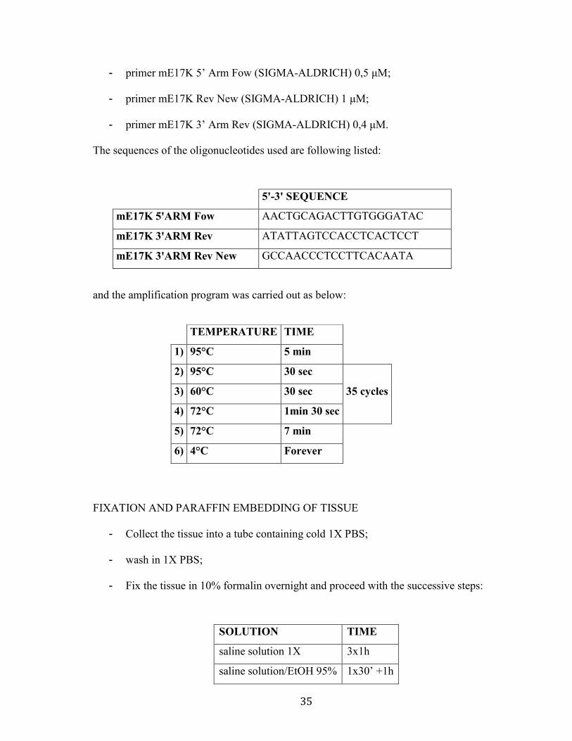
The sequences of the oligonucleotides used are following listed:
5'-3' SEQUENCE
mE17K 5'ARM Fow AACTGCAGACTTGTGGGATAC
mE17K 3'ARM Rev ATATTAGTCCACCTCACTCCT
mE17K 3'ARM Rev New GCCAACCCTCCTTCACAATA
and the amplification program was carried out as below:
FIXATION AND PARAFFIN EMBEDDING OF TISSUE
- Collect the tissue into a tube containing cold 1X PBS;
- wash in 1X PBS;
- Fix the tissue in 10% formalin overnight and proceed with the successive steps:
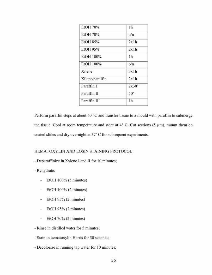
SOLUTION TIME
saline solution 1X 3x1h
saline solution/EtOH 95% 1x30’ +1h
TEMPERATURE TIME
1) 95°C 5 min
2) 95°C 30 sec
35 cycles 3) 60°C 30 sec
4) 72°C 1min 30 sec
5) 72°C 7 min
6) 4°C Forever
36
EtOH 70% 1h
EtOH 70% o/n
EtOH 85% 2x1h
EtOH 95% 2x1h
EtOH 100% 1h
EtOH 100% o/n
Xilene 3x1h
Xilene/paraffin 2x1h
Paraffin I 2x30’
Paraffin II 50’
Paraffin III 1h
Perform paraffin steps at about 60° C and transfer tissue to a mould with paraffin to submerge
the tissue. Cool at room temperature and store at 4° C. Cut sections (5 µm), mount them on
coated slides and dry overnight at 37˚ C for subsequent experiments.
HEMATOXYLIN AND EOSIN STAINING PROTOCOL
- Deparaffinize in Xylene I and II for 10 minutes;
- Rehydrate:
- EtOH 100% (5 minutes)
- EtOH 100% (2 minutes)
- EtOH 95% (2 minutes)
- EtOH 95% (2 minutes)
- EtOH 70% (2 minutes)
- Rinse in distilled water for 5 minutes;
- Stain in hematoxylin Harris for 30 seconds;
- Decolorize in running tap water for 10 minutes;
37
- EtOH 70% for 3 minutes;
- EtOH 95% for 10 minutes;
- Counterstain in alcoholic Eosin for 30 seconds;
- Dehydrate
- EtOH 95 % (5 minutes)
- EtOH 100% (10 minutes)
- Clear in Xylene I and II for10 minutes;
- Mount with Eukitt BIO-OPTICA.
RNA EXTRACTION
To extract RNA from mammary tissue we lysate samples using QIAGEN Tissuelyser for 2
min at 30 Hz by adding 1 ml of TRIzol Reagent (Invitrogen) and following the standard
protocol provided by the manufacturer. The RNA concentration was determined using the
NanoDrop spectrophotometer.
RT-PCR
The reverse transcription reaction is performed using Quantitect Reverse Transcription Kit
(Qiagen). The protocol used requires the following steps:
- Mix 1 μg of total RNA with gDNA Wipeout Buffer 7X in a final volume of 14 μl;
- Incubate at 42° C for 2 minutes;
- Add 1 μl di Quantiscript Reverse Trascriptase, 4 μl di Quantiscript RT Buffer 5X and
1 μl RT Primer Mix;
- Incubate at 42°C for 30 minutes and inactivate reaction at 95°C for 3 minutes;
38
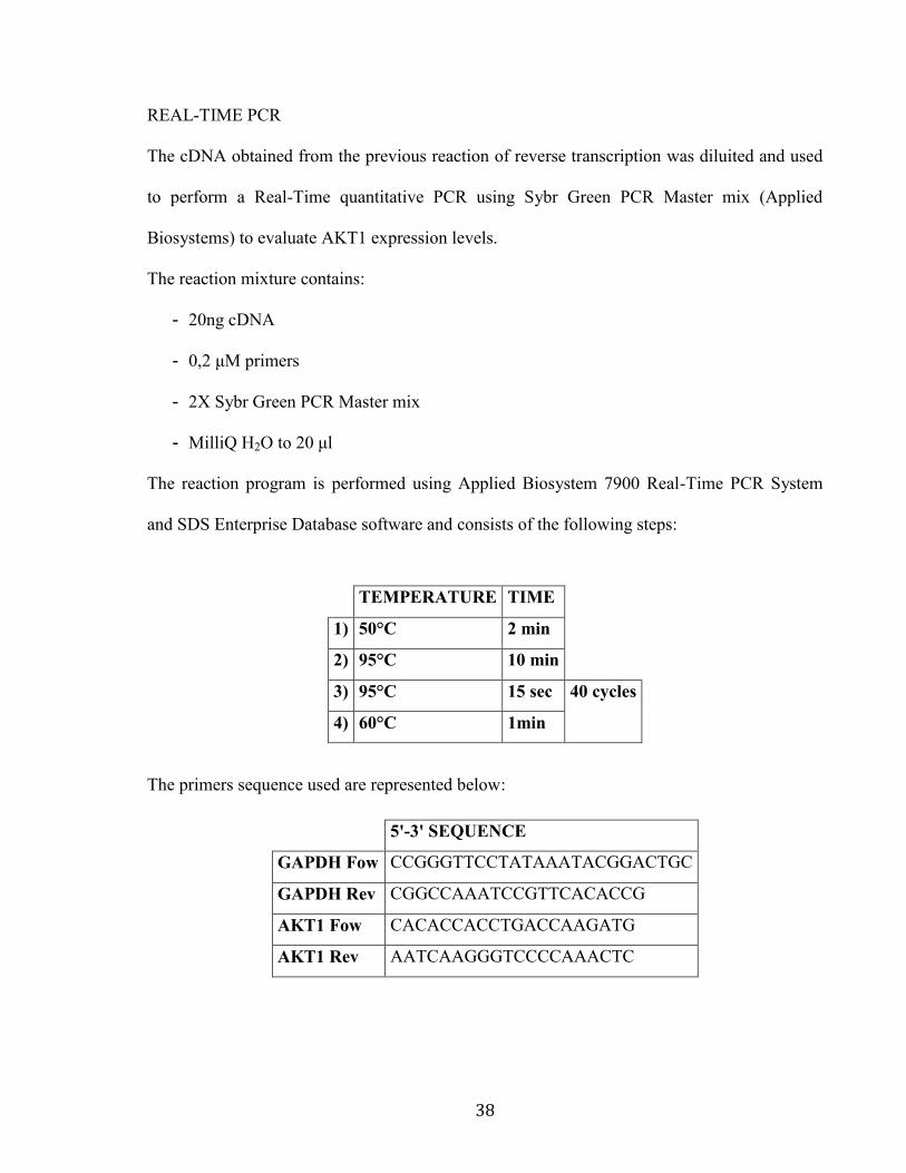
REAL-TIME PCR
The cDNA obtained from the previous reaction of reverse transcription was diluited and used
to perform a Real-Time quantitative PCR using Sybr Green PCR Master mix (Applied
Biosystems) to evaluate AKT1 expression levels.
The reaction mixture contains:
- 20ng cDNA
- 0,2 μM primers
- 2X Sybr Green PCR Master mix
- MilliQ H2O to 20 μl
The reaction program is performed using Applied Biosystem 7900 Real-Time PCR System
and SDS Enterprise Database software and consists of the following steps:
TEMPERATURE TIME
1) 50°C 2 min
2) 95°C 10 min
3) 95°C 15 sec 40 cycles
4) 60°C 1min
The primers sequence used are represented below:
5'-3' SEQUENCE
GAPDH Fow CCGGGTTCCTATAAATACGGACTGC
GAPDH Rev CGGCCAAATCCGTTCACACCG
AKT1 Fow CACACCACCTGACCAAGATG
AKT1 Rev AATCAAGGGTCCCCAAACTC
39
TOTAL PROTEIN EXTRACTION
The mammary tissue protein extraction is performed using a specific lysis buffer that allows to
extract the proteins from the lipid component typical of the breast tissue. Tissue fragments
were lysed using QIAGEN Tissuelyser for 2 min at 30 Hz by adding a volume of lysis buffer
such as to cover the tissue.
The lysis buffer is consists of the following components:
- Urea 7 M;
- Thiourea 2 M;
- CHAPS 2%;
- DTT 50mM;
- Protease inhibitor SIGMAFAST™
1X;
- NaF 1 mM;
- PMSF 1mM;
- Na3VO4 1mM;
- Okadaic Acid 15 nM;
After breaking up with Tissuelyser and short spin to remove tissue debris, the samples were
incubated on ice for 30 minutes and then centrifuged in a refrigerated centrifuge at 4ºC for 30
minutes at 13200 rpm. The supernatant was recovered to a microcentrifuge tube and protein
concentration was determined by a standard Bradford Assay (BioRad) in a Beckman DU 530
spectrophotometer.
40
WESTERN BLOT
This technique was used to analyze the protein levels in different samples.
Polyacrylamide gels were prepared at 10% acrylamide concentrations, according to the
molecular weight of the proteins in study. After complete polymerization of the gel, it is
transferred to a electrophoresis tank that was filled with a running buffer containing 25 mM of
TRIS, 250 mM of Glicine (pH 8.3) and 0.1% of SDS. 50 µg of protein were dispensed in the
wells, and the gels were run at 100 V. When the proteins were separated between them, the
run was stopped and fractionated proteins were transferred from the gel to a nitrocellulose
membrane by Trans-Blot Turbo Transfer System (BIORAD).
After transferring, the membranes were colored with 1X Red Ponceau (ATX Ponceau S Red
staining solution, FLUKA) for 1 minute at room temperature to validate the transferring
homogeneity and quality. Next, the membranes were washed in TTBS (1X TBS and 0,1%
Tween) and were pre-hybridized for 45 minutes in a solution of TTBS containing 5% Nonfat
dried milk (AppliChem) to block the non-specific hybridization sites of the primary antibody.
Subsequently 3 washes of 5 minutes each were made before incubating with the primary
antibodies diluted in red solution (1X TBS, 3% BSA, 0.02% NaAzide, 100 mg Phenol Red),
overnight at 4ºC, with agitation.
The next day, the primary antibody was recovered and the membranes were washed for 5
minutes, 3 times with TTBS. After that, the membranes were incubated with the respective
secondary antibody (rabbit or mouse), diluted at 1:2500 in a solution of TTBS with 5% of
nonfat dried milk, for 1 hour, followed by 3 washes of 5 minutes each with TTBS. Next the
membranes were incubated for 1 minute with an amplified chemiluminescence kit, ECL
(Amersham Inc.), which allows the operator to see the chemiluminescence on High
41
performance chemiluminescence film (Amersham Inc.) due to the reaction between the
substrate present on ECL and the peroxidase covalently bound to the secondary antibody.
The primary antibodies used in this thesis were the following:
- Anti-P-AKT (Ser473) antibody (rabbit), 1:1000 (Cell Signaling
TECHNOLOGY®
, #4058);
- Anti-AKT1 antibody (rabbit), 1:1000 (Cell Signaling TECHNOLOGY®,
#2938);
- Anti-P-FoxO1 (Ser256) antibody (rabbit), 1:1000 (Cell Signaling
TECHNOLOGY®
, #9461);
- Anti-FoxO1 antibody (rabbit), 1:500 (Cell Signaling TECHNOLOGY®,
#2880);
- Anti-P-GSK3α/3β antibody (rabbit), 1:1000 (Cell Signaling TECHNOLOGY®,
#9331);
- Anti-GSK3α antibody (rabbit), 1:1000 (Cell Signaling TECHNOLOGY®,
#9338);
- Anti-GSK3β antibody (rabbit), 1:1000 (Cell Signaling TECHNOLOGY®,
#9315);
- Anti-β-Actin antibody (mouse), 1:10000 (Sigma-Aldrich, A2228)
IHC-PARAFFIN PROTOCOL (IHC-P)
Before proceeding with the staining protocol, the slides must be deparaffinized and
rehydrated. Incomplete removal of paraffin can cause poor staining of the section.
Proceed as follows:
- incubate slides at 60 overnight.
42
- Deparaffinize in Xylene I and II for 10 minutes.
- Rehydrate:
EtOH 100% (5 minutes)
EtOH 100% (2 minutes)
EtOH 95% (2 minutes)
EtOH 95% (2 minutes)
EtOH 70% (2 minutes)
PBS 5 minutes
PBS+Tween20 5 minutes
PBS 5 minutes
- Perform antigen retrieval to unmask the antigenic epitope, using the citrate buffer
Epitope Retrieval 1X pH=6 Novocastra Leica and incubate slides, immersed in this
buffer, in the microwave at 900 WATT for 5 minutes and 300 WATT for 5 minutes, 3
times;
- Cool at 4° C for 30 minutes;
- Air dry the slides and wrap with pap pen;
- Wash in TTBS for 5 minutes, 2 times;
- Incubate sections in 3% hydrogen peroxide in methanol, for blocking of endogenous
peroxidase, for 15 minutes;
- Wash in TTBS for 5 minutes, 2 times;
- Incubate slides with the primary antibody diluted in Bond Primary Antibody Diluent
(Leica AR9352), overnight in humidifying chamber;
- Wash in TTBS for 5 minutes, 2 times.
43
Continue using the kit Novocastra Novolink Polymer Detection Systems (RE7140-K) as
follows:
- Novocastra Post Primary Block (RE7111) for 30 minutes;
- Wash in TTBS for 5 minutes, 2 times;
- Novolink Polymer (RE7112) for 30 minutes;
- Wash in TTBS for 5 minutes, 2 times;
- Develop peroxidase activity with DAB working solution (Ratio 1:20 DAB Chromogen
RE7105/DAB Substrate Buffer RE7106) maximum for 5 minutes;
- Rinse slides in water;
- Counterstain with Hematoxylin RE7107 for 10 minutes;
- Rinse slides in water for 5 minutes;
- Dehydrate (EtOH 70%, 80%, 90%, 100%, each from 5 minutes);
- Clear (xylene 10 minutes, 2 times);
- Mount sections with Eukitt BIO-OPTICA.
ISOLATION OF VIABLE EPITHELIAL CELLS FROM MURINE MAMMARY TISSUE
AND MAMMOSPHERE CULTURES
- mechanically dissociate mammary tissues into small pieces using a surgical blade and
placed in a digestion medium (DMEM/F12) supplemented with 200 U/ml collagenase
(Sigma) and 100 U/ml hyaluronidase (Sigma) for about 5h at 37° C;
- filtering the cell suspensions through 100, 70, 40 and 20 µm meshes;
- Centrifuge at 1200 rpm for 5 minutes;
- Wash the pellet with PBS and centrifuge at 1200 rpm for 5 minutes;
44
Resuspend the pellet in 1-5ml of RBC buffer (NH4Cl 155mM; KHCO3 o NaHCO3
10mM; EDTA 0,1mM) to eliminate the possible presence of red blood cells;
- Incubate at RT for 5 minutes;
- Centrifuge at 1200 rpm for 10 minutes;
- Wash the pellet with PBS and centrifuge at 1200 rpm for 5 minutes;
- Count the cells preparing a 1:1 of the cell suspension using a 0,4% Trypan Blue solution.
Non-viable cells will be blue, viable cells will be unstained.
Resulting cells were plated onto Corning®Ultra-Low Attachment Surface plates at a density of
400,000 viable cell/ml (to obtain primary mammospheres) in a serum-free mammary epithelial basal
medium (MEBM, LONZA), supplemented with 5 µg/ml insulin (Sigma), 0.5 µg/ml hydrocortisone
(Sigma), B27 (Invitrogen), 20 ng/ml EGF and bFGF (PEPROTECH), and 4 µg/ml heparin (Sigma).
Mammospheres were collected after 14 days and dissociated with trypsin, to obtain single
cells derived from mammospheres. For serial passage experiments, 5,000 cells from
disaggregated primary-mammospheres were plated in 24 multiwell plates and, after 14 days,
disaggregated and re-plated at the same density.
EVEROLIMUS TREATMENT
Everolimus (Afinitor®, Novartis), inhibitor of mammalian target of rapamycin (mTOR), was
dissolved in 12,5% DMSO at a concentration of 1,25 mg/ml (5mg/kg). Virgin
Akt1E17K/E17K
;MMTV-Cre female mice, 20-week-old, were treated with two weekly doses of
EVEROLIMUS, for 8 weeks, via oral gavage. Oral gavage consisted of inserting a curved
blunt tipped needle attached to a 1 ml syringe into the mid-throat of a firmly grasped mouse
and injecting 100 μl of a EVEROLIMUS solution. The control group of virgin
Akt1E17K/E17K
;MMTV-Cre female mice was treated with 12,5% DMSO. The mice were
maintained in the absence of males and were checked by palpation for mammary tumor

45
formation. Mice were sacrificed by CO2 inhalation at the end of the period established for the
experiment and mammary tissue was explanted.
46
RESULTS
GENERATION OF A Cre-INDUCIBLE AKT1E17K
MAMMARY MOUSE MODEL
To investigate the contribution of AKT1E17K
mutation in mammary tumorigenesis, we
generated a mouse model that, through a Cre/Lox system, expresses the mutated form of
AKT1 specifically in mammary gland tissue, in time and tissue specific manner. The gene
targeting strategy allowed us to insert the transgene into the mouse Rosa26 locus,
preferentially selected for the knock-in strategies as it has a high recombination frequency and
allows the ubiquitous expression of the inserted gene both in embryonic development and in
adult mice. The pRosa26 plasmid, containing 5 ' and 3’ homology arms for the specific locus,
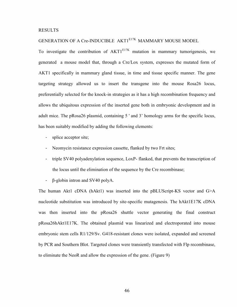
has been suitably modified by adding the following elements:
- splice acceptor site;
- Neomycin resistance expression cassette, flanked by two Frt sites;
- triple SV40 polyadenylation sequence, LoxP- flanked, that prevents the transcription of
the locus until the elimination of the sequence by the Cre recombinase;
- β-globin intron and SV40 polyA.
The human Akt1 cDNA (hAkt1) was inserted into the pBLUScript-KS vector and G>A
nucleotide substitution was introduced by site-specific mutagenesis. The hAkt1E17K cDNA
was then inserted into the pRosa26 shuttle vector generating the final construct
pRosa26hAkt1E17K. The obtained plasmid was linearized and electroporated into mouse
embryonic stem cells R1/129/Sv. G418-resistant clones were isolated, expanded and screened
by PCR and Southern Blot. Targeted clones were transiently transfected with Flp recombinase,
to eliminate the NeoR and allow the expression of the gene. (Figure 9)
47
Figure 9: gene targeting strategy for pRosa26hAkt1E17K construct. A: insertion of the cDNA of interest into the vector
pBLUScript-KS, B: hAkt1E17K insertion of the cDNA into the Rosa26 locus, C: final recombination of the construct with the
genomic locus of the murine Rosa26, D: representation of the genomic locus Rosa26 with the insert of interest; E: excision of
the neomycin resistance cassette by Flp.
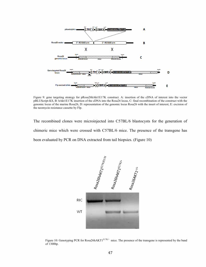
The recombined clones were microinjected into C57BL/6 blastocysts for the generation of
chimeric mice which were crossed with C57BL/6 mice. The presence of the transgene has
been evaluated by PCR on DNA extracted from tail biopsies. (Figure 10)
Figure 10: Genotyping PCR for Rosa26hAKT1E17K/+ mice. The presence of the transgene is represented by the band
of 1300bp.
48
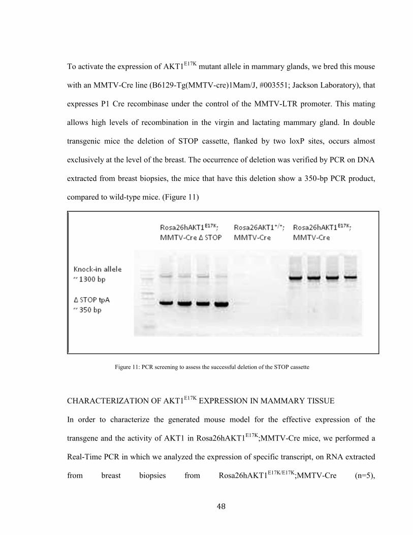
To activate the expression of AKT1E17K
mutant allele in mammary glands, we bred this mouse
with an MMTV-Cre line (B6129-Tg(MMTV-cre)1Mam/J, #003551; Jackson Laboratory), that
expresses P1 Cre recombinase under the control of the MMTV-LTR promoter. This mating
allows high levels of recombination in the virgin and lactating mammary gland. In double
transgenic mice the deletion of STOP cassette, flanked by two loxP sites, occurs almost
exclusively at the level of the breast. The occurrence of deletion was verified by PCR on DNA
extracted from breast biopsies, the mice that have this deletion show a 350-bp PCR product,
compared to wild-type mice. (Figure 11)
Figure 11: PCR screening to assess the successful deletion of the STOP cassette
CHARACTERIZATION OF AKT1E17K
EXPRESSION IN MAMMARY TISSUE
In order to characterize the generated mouse model for the effective expression of the
transgene and the activity of AKT1 in Rosa26hAKT1E17K
;MMTV-Cre mice, we performed a
Real-Time PCR in which we analyzed the expression of specific transcript, on RNA extracted
from breast biopsies from Rosa26hAKT1E17K/E17K
;MMTV-Cre (n=5),
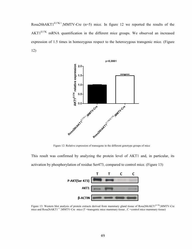
49
Rosa26hAKT1E17K/+
;MMTV-Cre (n=5) mice. In figure 12 we reported the results of the
AKT1E17K
mRNA quantification in the different mice groups. We observed an increased
expression of 1.5 times in homozygous respect to the heterozygous transgenic mice. (Figure
12)
Figure 12: Relative expression of transegene in the different genotype groups of mice
This result was confirmed by analyzing the protein level of AKT1 and, in particular, its
activation by phosphorylation of residue Ser473, compared to control mice. (Figure 13)
Figure 13: Western blot analysis of protein extracts derived from mammary gland tissue of Rosa26hAKT1E17K;MMTV-Cre
mice and Rosa26AKT1+/+;MMTV-Cre mice (T =transgenic mice mammary tissue , C =control mice mammary tissue)
50
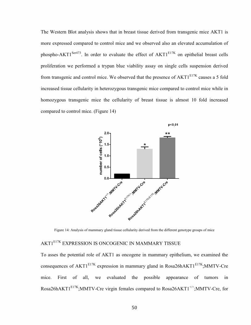
The Western Blot analysis shows that in breast tissue derived from transgenic mice AKT1 is
more expressed compared to control mice and we observed also an elevated accumulation of
phospho-AKT1Ser473
. In order to evaluate the effect of AKT1E17K
on epithelial breast cells
proliferation we performed a trypan blue viability assay on single cells suspension derived
from transgenic and control mice. We observed that the presence of AKT1E17K
causes a 5 fold
increased tissue cellularity in heterozygous transgenic mice compared to control mice while in
homozygous transgenic mice the cellularity of breast tissue is almost 10 fold increased
compared to control mice. (Figure 14)
Figure 14: Analysis of mammary gland tissue cellularity derived from the different genotype groups of mice
AKT1E17K
EXPRESSION IS ONCOGENIC IN MAMMARY TISSUE
To asses the potential role of AKT1 as oncogene in mammary epithelium, we examined the
consequences of AKT1E17K
expression in mammary gland in Rosa26hAKT1E17K
;MMTV-Cre
mice. First of all, we evaluated the possible appearance of tumors in
Rosa26hAKT1E17K
;MMTV-Cre virgin females compared to Rosa26AKT1+/+
;MMTV-Cre, for
51
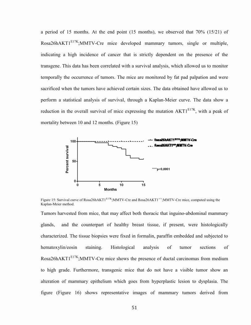
a period of 15 months. At the end point (15 months), we observed that 70% (15/21) of
Rosa26hAKT1E17K
;MMTV-Cre mice developed mammary tumors, single or multiple,
indicating a high incidence of cancer that is strictly dependent on the presence of the
transgene. This data has been correlated with a survival analysis, which allowed us to monitor
temporally the occurrence of tumors. The mice are monitored by fat pad palpation and were
sacrificed when the tumors have achieved certain sizes. The data obtained have allowed us to
perform a statistical analysis of survival, through a Kaplan-Meier curve. The data show a
reduction in the overall survival of mice expressing the mutation AKT1E17K
, with a peak of
mortality between 10 and 12 months. (Figure 15)
Figure 15: Survival curve of Rosa26hAKT1E17K;MMTV-Cre and Rosa26AKT1+/+;MMTV-Cre mice, computed using the
Kaplan-Meier method.
Tumors harvested from mice, that may affect both thoracic that inguino-abdominal mammary
glands, and the counterpart of healthy breast tissue, if present, were histologically
characterized. The tissue biopsies were fixed in formalin, paraffin embedded and subjected to
hematoxylin/eosin staining. Histological analysis of tumor sections of
Rosa26hAKT1E17K
;MMTV-Cre mice shows the presence of ductal carcinomas from medium
to high grade. Furthermore, transgenic mice that do not have a visible tumor show an
alteration of mammary epithelium which goes from hyperplastic lesion to dysplasia. The
figure (Figure 16) shows representative images of mammary tumors derived from
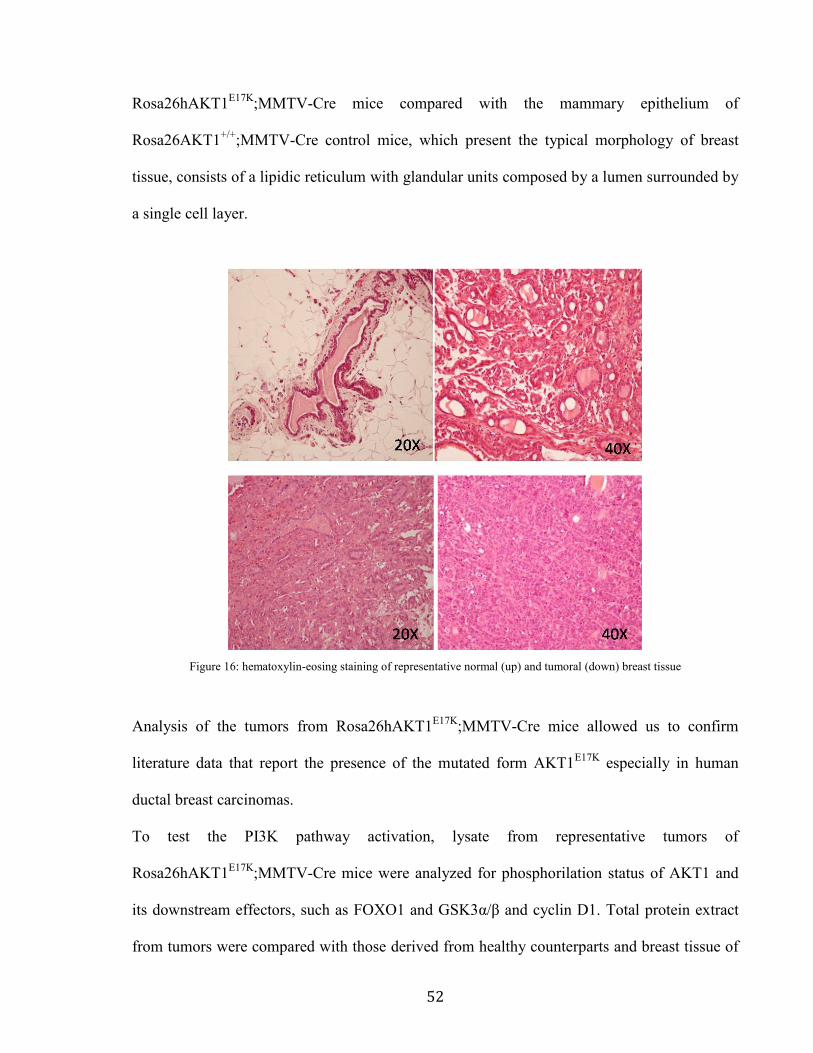
52
Rosa26hAKT1E17K
;MMTV-Cre mice compared with the mammary epithelium of
Rosa26AKT1+/+
;MMTV-Cre control mice, which present the typical morphology of breast
tissue, consists of a lipidic reticulum with glandular units composed by a lumen surrounded by
a single cell layer.
Figure 16: hematoxylin-eosing staining of representative normal (up) and tumoral (down) breast tissue
Analysis of the tumors from Rosa26hAKT1E17K
;MMTV-Cre mice allowed us to confirm
literature data that report the presence of the mutated form AKT1E17K
especially in human
ductal breast carcinomas.
To test the PI3K pathway activation, lysate from representative tumors of
Rosa26hAKT1E17K
;MMTV-Cre mice were analyzed for phosphorilation status of AKT1 and
its downstream effectors, such as FOXO1 and GSK3α/β and cyclin D1. Total protein extract
from tumors were compared with those derived from healthy counterparts and breast tissue of
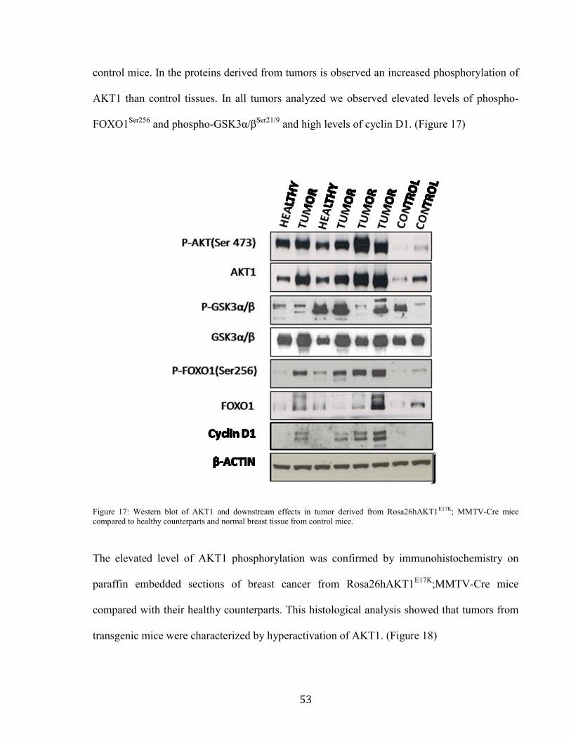
53
control mice. In the proteins derived from tumors is observed an increased phosphorylation of
AKT1 than control tissues. In all tumors analyzed we observed elevated levels of phospho-
FOXO1Ser256
and phospho-GSK3α/βSer21/9
and high levels of cyclin D1. (Figure 17)
Figure 17: Western blot of AKT1 and downstream effects in tumor derived from Rosa26hAKT1E17K; MMTV-Cre mice
compared to healthy counterparts and normal breast tissue from control mice.
The elevated level of AKT1 phosphorylation was confirmed by immunohistochemistry on
paraffin embedded sections of breast cancer from Rosa26hAKT1E17K
;MMTV-Cre mice
compared with their healthy counterparts. This histological analysis showed that tumors from
transgenic mice were characterized by hyperactivation of AKT1. (Figure 18)
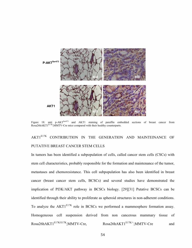
54
Figure 18: anti p-AKTSer473 and AKT1 staining of paraffin embedded sections of breast cancer from
Rosa26hAKT1E17K;MMTV-Cre mice compared with their healthy counterparts.
AKT1E17K
CONTRIBUTION IN THE GENERATION AND MAINTEINANCE OF
PUTATIVE BREAST CANCER STEM CELLS
In tumors has been identified a subpopulation of cells, called cancer stem cells (CSCs) with
stem cell characteristics, probably responsible for the formation and maintenance of the tumor,
metastases and chemoresistance. This cell subpopulation has also been identified in breast
cancer (breast cancer stem cells, BCSCs) and several studies have demonstrated the
implication of PI3K/AKT pathway in BCSCs biology. [29][31] Putative BCSCs can be
identified through their ability to proliferate as spheroid structures in non-adherent conditions.
To analyze the AKT1E17K
role in BCSCs we performed a mammosphere formation assay.
Homogeneous cell suspension derived from non cancerous mammary tissue of
Rosa26hAKT1E17K/E17K
;MMTV-Cre, Rosa26hAKT1E17K/+
;MMTV-Cre and
40X 40X
40X 40X
AKT1
P-AKTSer473
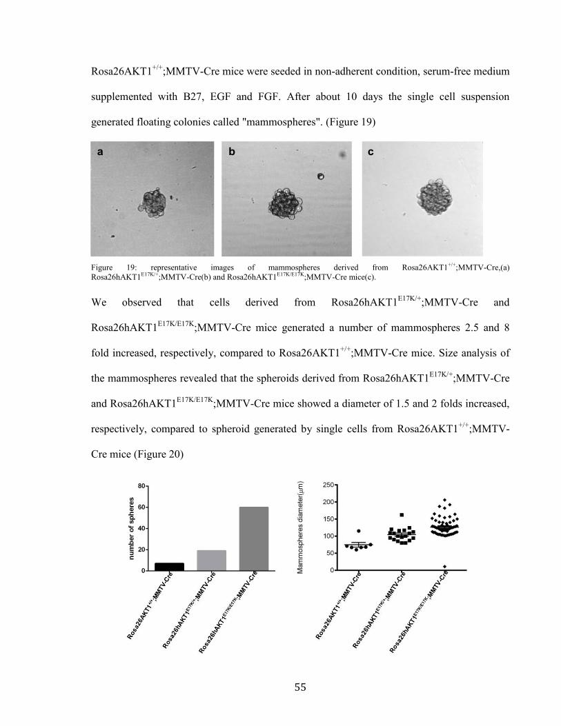
55
Rosa26AKT1+/+
;MMTV-Cre mice were seeded in non-adherent condition, serum-free medium
supplemented with B27, EGF and FGF. After about 10 days the single cell suspension
generated floating colonies called "mammospheres". (Figure 19)
Figure 19: representative images of mammospheres derived from Rosa26AKT1+/+;MMTV-Cre,(a)
Rosa26hAKT1E17K/+;MMTV-Cre(b) and Rosa26hAKT1E17K/E17K;MMTV-Cre mice(c).
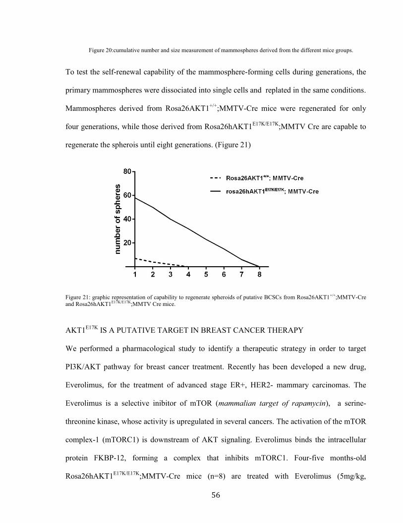
We observed that cells derived from Rosa26hAKT1E17K/+
;MMTV-Cre and
Rosa26hAKT1E17K/E17K
;MMTV-Cre mice generated a number of mammospheres 2.5 and 8
fold increased, respectively, compared to Rosa26AKT1+/+
;MMTV-Cre mice. Size analysis of
the mammospheres revealed that the spheroids derived from Rosa26hAKT1E17K/+
;MMTV-Cre
and Rosa26hAKT1E17K/E17K
;MMTV-Cre mice showed a diameter of 1.5 and 2 folds increased,
respectively, compared to spheroid generated by single cells from Rosa26AKT1+/+
;MMTV-
Cre mice (Figure 20)
b a c
56
Figure 20:cumulative number and size measurement of mammospheres derived from the different mice groups.
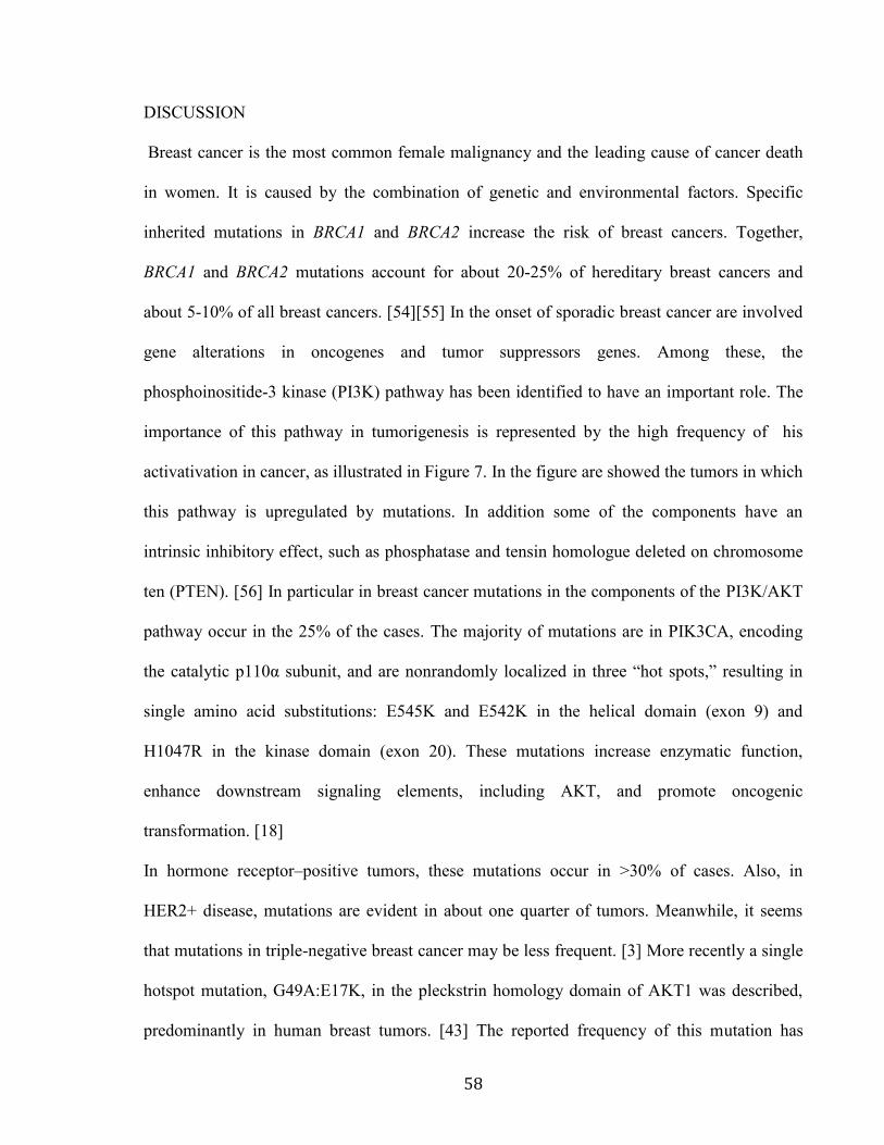
To test the self-renewal capability of the mammosphere-forming cells during generations, the
primary mammospheres were dissociated into single cells and replated in the same conditions.
Mammospheres derived from Rosa26AKT1+/+
;MMTV-Cre mice were regenerated for only
four generations, while those derived from Rosa26hAKT1E17K/E17K
;MMTV Cre are capable to
regenerate the spherois until eight generations. (Figure 21)
Figure 21: graphic representation of capability to regenerate spheroids of putative BCSCs from Rosa26AKT1+/+;MMTV-Cre
and Rosa26hAKT1E17K/E17K;MMTV Cre mice.
AKT1E17K
IS A PUTATIVE TARGET IN BREAST CANCER THERAPY
We performed a pharmacological study to identify a therapeutic strategy in order to target
PI3K/AKT pathway for breast cancer treatment. Recently has been developed a new drug,
Everolimus, for the treatment of advanced stage ER+, HER2- mammary carcinomas. The
Everolimus is a selective inibitor of mTOR (mammalian target of rapamycin), a serine-
threonine kinase, whose activity is upregulated in several cancers. The activation of the mTOR
complex-1 (mTORC1) is downstream of AKT signaling. Everolimus binds the intracellular
protein FKBP-12, forming a complex that inhibits mTORC1. Four-five months-old
Rosa26hAKT1E17K/E17K
;MMTV-Cre mice (n=8) are treated with Everolimus (5mg/kg,

57
gavage), for 8 weeks (2 doses in week), as control we treated a group of mice (n=9) with the
vehicle. At the end of treatment, the breast tissue of both groups was collected and subjected
to histological analysis. We observed that 66.7% (6/9) of the control mice developed tumors
and 11,1% (1/9) severe dysplasia, while only 37.8% (3/8) of treated mice with Everolimus
showed medium-grade dysplasia.
58
DISCUSSION
Breast cancer is the most common female malignancy and the leading cause of cancer death
in women. It is caused by the combination of genetic and environmental factors. Specific
inherited mutations in BRCA1 and BRCA2 increase the risk of breast cancers. Together,
BRCA1 and BRCA2 mutations account for about 20-25% of hereditary breast cancers and
about 5-10% of all breast cancers. [54][55] In the onset of sporadic breast cancer are involved
gene alterations in oncogenes and tumor suppressors genes. Among these, the
phosphoinositide-3 kinase (PI3K) pathway has been identified to have an important role. The
importance of this pathway in tumorigenesis is represented by the high frequency of his
activativation in cancer, as illustrated in Figure 7. In the figure are showed the tumors in which
this pathway is upregulated by mutations. In addition some of the components have an
intrinsic inhibitory effect, such as phosphatase and tensin homologue deleted on chromosome
ten (PTEN). [56] In particular in breast cancer mutations in the components of the PI3K/AKT
pathway occur in the 25% of the cases. The majority of mutations are in PIK3CA, encoding
the catalytic p110α subunit, and are nonrandomly localized in three “hot spots,” resulting in
single amino acid substitutions: E545K and E542K in the helical domain (exon 9) and
H1047R in the kinase domain (exon 20). These mutations increase enzymatic function,
enhance downstream signaling elements, including AKT, and promote oncogenic
transformation. [18]
In hormone receptor–positive tumors, these mutations occur in >30% of cases. Also, in
HER2+ disease, mutations are evident in about one quarter of tumors. Meanwhile, it seems
that mutations in triple-negative breast cancer may be less frequent. [3] More recently a single
hotspot mutation, G49A:E17K, in the pleckstrin homology domain of AKT1 was described,
predominantly in human breast tumors. [43] The reported frequency of this mutation has
59
ranged from 1.8%–8%. [18] [43][44][45] Importantly, mutant AKT1 is able to activate PI3K
signaling pathway and to transform rodent fibroblasts and bone marrow cells . [43] Most
studies have found PTEN, PIK3CA, and AKT1 mutations to be mutually exclusive in
individual tumors [43][44][49], suggesting that mutational activation of the PI3K/AKT
pathway by any one of the components are biologically equivalent.
Until now the role of AKT1 in mammary tumorigenesis represents a secondary event. In vivo
studies have shown that its hyperactivated form, myrAKT1, which results in AKT anchoring
to the plasma membrane and constitutive activation, delay the mammary involution and
increases the incidence of benign lesions. In addition myristoylation signal increases the
susceptibility to epithelial mammary tumor formation after induction by the carcinogen 9,10-
dimethyl-1,2 benzanthracene (DMBA). [52] Somewhat surprisingly, published data
demonstrated that overexpression of AKT1 is able to transform cells in vitro[43], but the
knock-in of mutant AKT1E17K
in MCF10A cells failed to recapitulate this capacity. [53] These
findings are similar to what was observed with knock in of mutant KRAS and may reflect
differences between knock in and transgenic overexpression as well as differences between
cell types in their susceptibility to transformation. However, were not made in vivo studies to
analyze the effect of mutant form of AKT1 in mammary tumorigenesis. For this reason the
aim of this study is the evaluation of the role of AKT1E17K
in the mammary tumorigenesis in a
mouse model that expressed AKT1E17K
specifically in mammary gland tissue. These mice are
derived by crossing Rosa26AKT1E17K
transgenic mice with an MMTV-Cre mouse line. The
resulting mice, Rosa26hAKT1E17K
;MMTV-Cre, drive the expression of AKT1E17K
almost
exclusively in the mammary tissue.
The data from this study showed that the transgene was expressed primarily in the mammary
gland. This is consistent with the findings that increase in MMTV-LTR transcriptional activity
60
is through mammary development and during pregnancy. [57] The hAKT1E17K
specific
transcript was expressed in mammary gland of trangenic mice compared to the control
littermates. In addition, the analysis of the expression of AKT1E17K
in breast tissue derived
from transgenic mice demonstrates that the transgene expression is increased from
heterozygous to homozygous mice. These results are confirmed by the analisys of downstream
pathway activation status, phosphorylation of AKT1 is higher than that wild-type control
mice.
The activation of AKT1 by overexpression or myristoylation signal has been shown to delay
mammary gland involution and induce hyperplasia [51]. This process accelerates mammary
tumorigenesis in MMTV-c-ErbB2 mice [58] but overexpressing or activated AKT1 in
mammary gland alone is not able to induce dysplasia and neoplasia [59][60]. Activation of
AKT1 induces oncogenic transformation upon exposure to carcinogens [52]. Surprisingly, in
our model, we demonstrated that transgenic mice with active AKT1 led to a significant higher
incidence of mammary carcinoma without exposure to the carcinogen insults. In particular
tumor incidence was of about 70%, starting from four months of age, with a peak of mortality
between 10 and 12 months. The histological analysis conducted on the explanted mammary
tumor masses revealed that the tumors are ductal carcinomas from medium to high grade,
confirming the literature data from breast cancer patients showing the presence of the mutant
AKT1E17K
in ductal and lobular carcinomas. [43] The tumor tissues from transgenic mice had
increased phosphorylated AKT1 at Ser473 and the tumors derived from
Rosa26hAKT1E17K
;MMTV-Cre mice showed hyperactivation of the PI3K/Akt pathway.
Increased pAKTSer473
was associated with strong phosphorilation levels of downstream
substrates as FOXO1 and GSK3α/β. Moreover tumor tissues from transgenic mice
overexpress cyclin D1. Cyclin D1 plays role in tumor cell proliferation, migration and was
61
inversely correlated with tumor size in human breast cancer [61] Overexpression of cyclin D1
has been reported between 40% and 90% of cases of invasive breast cancer, while gene
amplification is seen in about 5–20% of tumors [35]. Increase in protein level of cyclin D1
was apparent in hyperplasia and further increased with malignancy. [62][63] Cyclin D1 has
also been shown as an essential oncogenic intermediary for Neu pathway inducing mammary
carcinoma in transgenic mice. [64][65]. In our model, we observed that cyclin D1 expression
was higher in Rosa26hAKT1E17K
;MMTV-Cre mice driven mammary carcinoma, compared to
control mice. The histological analysis revealed also the presence of preneoplastic lesions,
from hyperplasia to dysplasia. These results, associated with the activation of AKT pathway,
correlates the presence of mutant AKT1 to the cell proliferation, in vivo. Mammary gland
derived from 8-12 weeks Rosa26hAKT1E17K
;MMTV-Cre mice showed increased cellularity,
ten fold increased compared to mammary tissue derived from control littermates.
It has been recently suggested that a cellular subpopulation with stem cell (SC)-like features,
known as Cancer Stem Cells (CSCs), is critical for tumor generation and maintenance, and
responsible for breast cancer metastasis. [24] In vitro and in vivo evidences revealed the
importance of PTEN/PI3K/Akt/Wnt/β-catenin pathway in breast cancer stem cells (BCSCs)
biology. Increased AKT Ser473 phosphorylation in suspension culture of putative breast
cancer stem cells (mammospheres) as compared with monolayer cultures, determines
increased levels of GSK3-β phosphorylation and β -catenin activation. β-catenin has been
demonstrated to play an important role in the development of mammary stem cells in mouse
models, suggesting that this pathway may also be active in human mammary stem/progenitor
cells in mammospheres. [31] To assess the role of AKT1E17K
in the context of BCSCs, we
exploited his ability to regulates the growth of cells suspension, derived from
Rosa26hAKT1E17K
;MMTV-Cre an control mammary gland tissue, in spheroid conditions.
62
Performing a mammospheres assay we observed that cells derived from
Rosa26hAKT1E17K
;MMTV-Cre mice generates increased number of mammospheres
compared to those derived from control mice. Furthermore the mammospheres derived from
transgenic mice showed a diameter two fold increased compared to the wild-type mice. The
self-renewal assay revealed that cells derived from Rosa26hAKT1E17K
;MMTV-Cre mice are
capable to renew until 8 generations the spheroid culture. The obtained data allow us to
identify an important role of AKT1E17K
in mammary tumorigenesis and in the maintenance of
the tumor through its involvement in the biology of the BCSCs. We therefore conducted a
pharmacological study in which we evaluated the antitumor activity of the drug Everolimus on
tumor induction and / or progression of breast cancer in Rosa26hAKT1E17K/E17K
;MMTV-Cre
mice. This drug has recently been developed for the treatment of breast cancers in advanced
phase, positive estrogen receptor and HER2 negative. Everolimus is a selective inhibitor of
mTOR (mammalian target of rapamycin) a serine-threonine kinase whose activity is known to
be upregulated in several human cancers. The activation of this complex is downstream of
AKT, which can act in a direct manner on mTOR, through phosphorylation at the residue
Ser2448, and in indirectly, by inactivating phosphorylation at residue Thr246 of PRAS40, a
inhibitory component of the mTORC1 complex and at the level of 5 residues (S939, S981,
S1130, S1132 and T1462) of TSC2, which together with TSC1 constitutes a complex that
inhibits mTOR via RHEB protein. [35] Treatment with this drug has allowed to inhibit, or at
least slow down, the complete transformation of breast tissue in Rosa26hAKT1E17K
;MMTV-
Cre mice.
In summary this project led us to these results: i) the generation of a new mouse model for
breast cancer, AKT1-dependent; ii) the analysis of the mouse models revealed that the
activation of AKT1 in mammary tissue increases the risk of development of mammary tumors
63
iii) the involvement of AKT1E17K
in the generation and maintenance of BCSCs; iiii)
Rosa26hAKT1E17K
;MMTV-Cre transgenic mouse is a valuable resource for understanding the
mechanisms of breast cancer target therapy.
Future experiments will be needed for the molecular characterization of mammary tumors
induced by AKT1E17K
. We will also confirm the stemness of mammospheres derived cells,
testing their positivity to stem marker (such us Sox2, Oct4, Nanog), their symmetric and
asymmetric division and their in vivo tumorigenic capability.
64
BIBLIOGRAPHY
1) Orlando E Silva, Stefano Zurrida: “Breast cancer: a pratical guide”, 3rd edition 2005,
ch 3;
2) Ahmedin Jemal, Freddie Bray, Melissa M. Center, Jacques Ferlay, Elizabeth Ward,
David Forman: “Global Cancer Statistics”, CA CANCER J CLIN 2011;61:69–90;
3) Fattaneh A. Tavassoli and Peter Devilee: “Pathology and Genetics of Tumours of the
Breast and Female Genital organs”, World Health Organization Classification of
Tumours, 4th edition 2003, ISBN 9283224124;
4) American Association for cancer Research, AACR: “Breast Cancer Facts & Figures
2013-2014”; Atlanta American Cancer Society, Inc . 2013;
5) D. Craig Allred: “Ductal Carcinoma In Situ: Terminology, Classification, and Natural
History”, J Natl Cancer Inst Monogr 2010;41:134–138;
6) Yee Vonne Liong, Ga Sze Hong, Jennifer Gek Choo Teo and Geok Hoon Lim: “Breast
ductal carcinoma in situ presenting as recurrent non-puerperal mastitis: case report
and literature review”, World Journal of Surgical Oncology 2013, 11:179;
7) Kilbride KE, Newman LA. Chapter 25: Lobular carcinoma in situ: Clinical
management. In: Harris JR, Lippman ME, Morrow M, Osborne CK, eds. Diseases of
the Breast. 4th edition 2010;
8) www.breastcancer.org;
65
9) Grazia Arpino, Valerie J Bardou, Gary M Clark and Richard M Elledge: “Infiltrating
lobular carcinoma of the breast: tumor characteristics and clinical outcome”, Breast
Cancer Res 2004, 6:R149-R156;
10) S. Eva Singletary, Lina Patel-Parekh and Kirby I. Bland: “Treatment Trends in Early-
Stage Invasive Lobular Carcinoma A Report From the National Cancer Data Base”,
Annals of Surgery, Volume 242, Number 2, August 2005;
11) Wood LD, Parsons DW, Jones S, Lin J, Sjöblom T, Leary RJ, Shen D, Boca SM,
Barber T, Ptak J, Silliman N, Szabo S, Dezso Z, Ustyanksky V, Nikolskaya T,
Nikolsky Y, Karchin R, Wilson PA, Kaminker JS, Zhang Z, Croshaw R, Willis J,
Dawson D, Shipitsin M, Willson JK, Sukumar S, Polyak K, Park BH, Pethiyagoda CL,
Pant PV, Ballinger DG, Sparks AB, Hartigan J, Smith DR, Suh E, Papadopoulos N,
Buckhaults P, Markowitz SD, Parmigiani G, Kinzler KW, Velculescu VE, Vogelstein
B.: “The Genomic Landscapes Of Human Breast And Colorectal Cancers”, Science
2007 Nov 16;318(5853):1108-13;
12) Turner NC, Reis-Filho JS.: “Basal-like breast cancer and the BRCA1 phenotype”,
Oncogene. 2006 Sep 25;25(43):5846-53;
13) Vincent T DeVita, Jr., Theodore S Lawrence, Steven A Rosenberg, Ronald A
DePinho, Robert A Weinberg: “DeVita, Hellman, and Rosenberg's Cancer: Principles
& Practice of Oncology”, 9th edition 2011, ch 105-106;
66
14) Foulkes WD.: “Inherited Susceptibility To Common Cancers”, N Engl J Med. 2008
Nov 13;359(20):2143-53;
15) Stratton MR, Rahman N.:”The Emerging Landscape Of Breast Cancer Susceptibility”.
Nat Genet 2008;40(1):17;
16) Turnbull C, Rahman N.Genetic “Predisposition To Breast Cancer: Past, Present, And
Future”. Annu Rev Genomics Hum Genet 2008;9:321;
17) Kim M. Hirshfield, Timothy R. Rebbeck, and Arnold J. Levine: “Germline Mutations
and Polymorphisms in the Origins of Cancers in Women”, Journal of Oncology
Volume 2010, Article ID 297671;
18) K. Stemke-Hale, A.M. Gonzalez-Angulo, A. Lluch, et al., “An integrative genomic and
proteomic analysis of PIK3CA, PTEN, and AKT mutations in breast cancer” Cancer
Research, vol. 68, no. 15, pp. 6084–6091, 2008;
19) Charles M. Perou and Anne-Lise Børresen-Dale: “Systems Biology and Genomics of
Breast Cancer”, Cold Spring Harb Perspect Biol 2011;3:a003293;
20) M.J. Engstrøm, S. Opdahl, A.I. Hagen, P.R. Romundstad, L.A. Akslen, O.A. Haugen,
L.J. Vatten, A.M. Bofin: “Molecular subtypes, histopathological grade and survival in
a historic cohort of breast cancer patients”, Breast Cancer Res Treat (2013) 140:463–
473;
21) Britta Weigelt, Johannes L. Peterse and Laura J. van’t Veer: “Breast cancer
metastasis: markers and models”, Nature Reviews, Cancer, Volume 5, 2005;
67
22) Linda Vona-Davis, David P. Rose, Vijaya Gadiyaram, Barbara Ducatman, Gerald
Hobbs, Hannah Hazard, Sobha Kurian and Jame Abraham: “Breast Cancer Pathology,
Receptor Status, and Patterns of Metastasis in a Rural Appalachian Population”,
Journal of Cancer Epidemiology,Volume 2014, Article ID 170634;
23) Osama Hussein & Svetlana V. Komarova: “Breast cancer at bone metastatic sites:
recent discoveries and treatment targets”, J. Cell Commun. Signal. (2011) 5:85–99;
24) M Bartucci, R Dattilo, C Moriconi, A Pagliuca, M Mottolese, G Federici, A Di
Benedetto, M Todaro, G Stassi, F Sperati, MI Amabile, E Pilozzi, M Patrizii, M
Biffoni, M Maugeri-Saccà, S Piccolo and R De Maria: “TAZ is required for metastatic
activity and chemoresistance of breast cancer stem cells”, Oncogene (2014), 1 – 10;
25) Marco A. Velasco-Velázquez, Nora Homsi, Marisol De La Fuente, and Richard G.
Pestell: “Breast Cancer Stem Cells”, Int J Biochem Cell Biol. 2012;
26) Robert W. Cho, Xinhao Wang, Maximilian Diehn, Kerby Shedden, Grace Y. Chen,
Gavin Sherlock, Austin Gurney, John Lewicki, Michael F. Clarke: “Isolation and
Molecular Characterization of Cancer Stem Cells in MMTV-Wnt-1 Murine Breast
Tumors” STEM CELLS 2008;26:364 –371;
27) Muhammad Al-Hajj, Max S. Wicha, Adalberto Benito-Hernandez, Sean J. Morrison
and Michael F. Clarke: “Prospective identification of tumorigenic breast cancer
cells”,PNAS May 27, 2003;
28) Kazuharu Kai, Yoshimi Arima, Toshio Kamiya, Hideyuki Saya: “Breast cancer stem
cells”, Breast Cancer April 2010;
68
29) Marco A. Velasco-Velázquez, Vladimir M. Popov, Michael P. Lisanti and Richard G.
Pestell: “The Role of Breast Cancer Stem Cells in Metastasis and Therapeutic
Implications”, The American Journal of Pathology, Vol. 179, No. 1, July 2011;
30) Jay R Harris , Marc E Lippman, C. Kent Osborne, Monica Morrow: “Diseases of the
Breast”, 4th edition 2009;
31) Hasan Korkaya, Amanda Paulson, Emmanuelle Charafe-Jauffret, Christophe Ginestier,
Marty Brown, Julie Dutcher, Shawn G. Clouthier, Max S. Wicha: “Regulation of
Mammary Stem/Progenitor Cells by PTEN/ Akt/β-Catenin Signaling” , PLoS Biology,
2009;
32) Steven P. Zielske, Aaron C. Spalding, Max S. Wicha and Theodore S. Lawrence:
“Ablation of Breast Cancer Stem Cells with Radiation”, Translational Oncology
(2011) 4, 227–233;
33) X Wan, B Harkavy, N Shen, P Grohar and LJ Helman: “Rapamycin induces feedback
activation of Akt signaling through an IGF-1R-dependent mechanism”, Oncogene
(2007) 26, 1932–1940;
34) Viglietto G, Amodio N, Malanga D, Scrima M, De Marco C: “Contribution of
PKB/AKT signaling to thyroid cancer”, Front Biosci (Landmark Ed). 2011 Jan
1;16:1461-87;
35) Liao Y, Hung MC: “Physiological regulation of Akt activity and stability”, Am J
Transl Res. 2010 Jan 1;2(1):19-42;
69
36) Peng Z, Weber JC, Han Z, Shen R, Zhou W, Scott JR, Chan MW, Lin HJ: “Dichotomy
effects of Akt signaling in breast cancer”, Molecular Cancer 2012, 11:61;
37) Michael A. Davies, The University of Texas MD Anderson Cancer Center, Houston,
TX: “Regulation, Role, and Targeting of Akt in Cancer”, Journal Of Clinical
Oncology, Volume 29, Number 35, 2011;
38) James A. Crowell, Vernon E. Steele and Judith R. Fay: “Targeting the AKT protein
kinase for cancer chemoprevention”, Molecular Cancer Therapy 2007;6:2139-2148;
39) Maroulakou IG, Oemler W, Naber SP, Tsichlis PN.: “Akt1 ablation inhibits, whereas
Akt2 ablation accelerates, the development of mammary adenocarcinomas in mouse
mammary tumor virus (MMTV)-ErbB2/neu and MMTV-polyoma middle T transgenic
mice”. Cancer Res 2007;67:167 – 77;
40) Nakatani K, Thompson DA, Barthel A., et al.: “Up-regulation of Akt3 in estrogen
receptor-deficient breast cancers and androgen-independent prostate cancer lines”. J
Biol Chem 1999;274:21528 – 32;
41) Carmen Blanco-Aparicio, Marta Cañamero, Yolanda Cecilia, Belén Pequeño1, Oliver
Renner, Irene Ferrer, Amancio Carnero: “Exploring the Gain of Function Contribution
of AKT to Mammary Tumorigenesis in Mouse Models”, PLoS ONE, February 2010,
Volume 5, Issue 2, e9305;
42) Ambuj Kumar, Rituraj Purohit: “Cancer Associated E17K Mutation Causes Rapid
Conformational Drift in AKT1 Pleckstrin Homology (PH) Domain”, PLOS ONE, May
2013, Volume 8, Issue 5, e64364;
70
43) Carpten JD, Faber AL, Horn C, Donoho GP, Briggs SL, Robbins CM, Hostetter G,
Boguslawski S, Moses TY, Savage S, Uhlik M, Lin A, Du J, Qian YW, Zeckner DJ,
Tucker-Kellogg G, Touchman J, Patel K, Mousses S, Bittner M, Schevitz R, Lai MH,
Blanchard KL, Thomas JE: “A transforming mutation in the pleckstrin homology
domain of AKT1 in cancer”, Nature. 2007 Jul 26;448(7152):439-44;
44) FE Bleeker, L Felicioni, F Buttitta, S Lamba, L Cardone, M Rodolfo, A Scarpa, S
Leenstra, M Frattini, M Barbareschi, M Del Grammastro, MG Sciarrotta, C Zanon, A
Marchetti and A Bardelli: “AKT1E17K
in human solid tumours”, Oncogene (2008) 27,
5648–5650;
45) MS Kim, EG Jeong, NJ Yoo and SH Lee: “Mutational analysis of oncogenic AKT
E17K mutation in common solid cancers and acute leukaemias”, British Journal of
Cancer (2008) 98, 1533 – 1535;
46) Malanga D, Scrima M, De Marco C, Fabiani F, De Rosa N, de Gisi S et al.: “Activating
E17K mutation in the gene encoding the protein kinase AKT in a subset of squamous
cell carcinoma of the lung”, Cell Cycle (2008)5: 665–669;
47) Sjoerd Klarenbeek, Martine H. van Miltenburg, Jos Jonkers: “Genetically engineered
mouse models of PI3K signaling in breast cancer”, Molecular Oncology 2013, 146
e164;
48) W Yuan, E Stawiski, V Janakiraman, E Chan, S Durinck, KA Edgar, NM Kljavin, CS
Rivers, F Gnad, M Roose-Girma, PM Haverty, G Fedorowicz, S Heldens, RH Soriano,
71
Z Zhang, JJ Wallin, L Johnson, M Merchant, Z Modrusan, HM Stern and S Seshagiri:
“Conditional activation of Pik3caH1047R
in a knock-in mouse model promotes mammary
tumorigenesis and emergence of mutations”, Oncogene 2013, 32, 318 – 326;
49) Lao H Saal, Sofia K Gruvberger-Saal, Camilla Persson, Kristina Lövgren, Mervi
Jumppanen, Johan Staaf, Göran Jönsson, Maira M Pires, Matthew Maurer, Karolina
Holm, Susan Koujak, Shivakumar Subramaniyam, Johan Vallon-Christersson, Haökan
Olsson, Tao Su, Lorenzo Memeo, Thomas Ludwig, Stephen P Ethier, Morten Krogh,
Matthias Szabolcs, Vundavalli VVS Murty, Jorma Isola, Hanina Hibshoosh, Ramon
Parsons, and Åke Borg: “Recurrent gross mutations of the PTEN tumor suppressor
gene in breast cancers with deficient DSB repair”, Nature Genetics 2008 January ;
40(1): 102–107;
50) Gang Li, Gertraud W. Robinson, Ralf Lesche, Hilda Martinez-Diaz, Zhaorong Jiang,
Nora Rozengurt, Kay-Uwe Wagner, De-Chang Wu, Timothy F. Lane, Xin Liu, Lothar
Hennighausen and Hong Wu: “Conditional loss of PTEN leads to precocious
development and neoplasia in the mammary gland”, Development 2002, 129, 4159-
4170;
51) Scott Ackler, Shakeel Ahmad, Christopher Tobias, Michael D Johnson and Robert I
Glazer: “Delayed mammary gland involution in MMTV-AKT1 transgenic mice”,
Oncogene 2002, 21, 198-206;
52) Carmen Blanco-Aparicio, Lucìa Pérez-Gallego, Belén Pequeño, Juan F.M.Leal, Oliver
Renner and Amancio Carnero: “Mice expressing myrAKT1 in the mammary gland
72
develop carcinogen-induced ER-positive mammary tumors that mimic human breast
cancer”, Carcinogenesis vol.28 no.3 pp.584–594, 2007;
53) Lauring J, Cosgrove DP, Fontana S, Gustin JP, Konishi H, Abukhdeir AM, Garay
JP, Mohseni M, Wang GM, Higgins MJ, Gorkin D, Reis M, Vogelstein B, Polyak
K, Cowherd M, Buckhaults PJ, Park BH: “Knock in of the AKT1 E17K mutation in
human breast epithelial cells does not recapitulate oncogenic PIK3CA mutations”,
Oncogene. 2010 Apr 22;29(16):2337-45;
54) Easton DF. “How many more breast cancer predisposition genes are there?” Breast
Cancer Research 1999; 1(1):14–17;
55) Campeau PM, Foulkes WD, Tischkowitz MD. Hereditary breast cancer: “New genetic
developments, new therapeutic avenues”. Human Genetics 2008; 124(1):31–42;
56) Torres-Arzayus MI, de Mora JF, Yuan J, Vazquez F, Bronson R, Rue M, Sellers WR,
and Brown M: “High tumor incidence and activation of the PI3K/AKT pathway in
transgenic mice define AIB1 as an oncogene”. Cancer cell 2004, 6:263-274;
57) Torres-Arzayus MI, de Mora JF, Yuan J, Vazquez F, Bronson R, Rue M, Sellers WR,
and Brown M: “High tumor incidence and activation of the PI3K/AKT pathway in
transgenic mice define AIB1 as an oncogene”. Cancer cell 2004, 6:263-274;
58) Young CD. Nolte EC, Lewis A, Serkova NJ, and Anderson SM: “Activated Akt1
accelerates MMTV-c-EebB2 mammary tumorigenesis in mice without activation of
ErbB3”. Breast Cancer Res 2008, 10:R70;
73
59) Sinn E. Muller W, Pattengale P, Tepler I, Wallace R, and Leder P: “Coexpression of
MMTV/v-Ha-ras and MMTV/c-myc genes in transgenic mice: synergistic action of
oncogene in vivo”. Cell 1987, 49: 465-475;
60) Hutchinson J, Jin J, Cardiff RD, Woodgett JR, and Muller WJ: “Activation of Akt
(protein kinase B) in mammary epithelium provides a critical cell survival signal
required for tumor progression”. Mol. Cell Biol 2001, 21:2203-2212;
61) Lehn S, Tobin NP, Berglund P, Nilsson K, Sims AH, Jirstrom K, Harkonen P, Lamb R,
and Landberg G: “Down-regulation of the oncogene cyclin D1 increases migratory
capacity in breast cancer and is linked to unfavorable prognostic features”. Am J
Pathol 2010, 177:2886-2897;
62) Roy PG, Thompson AM: “Cyclin D1 and breast cancer”. Review. The Breast 2006,
15:718–727;
63) Sutherland RL. and Musgrove EA: “Cyclin D1 and mammary carcinoma: new insights
from transgenic mouse models”. Breast Cancer Res 2002, 4:14-17;
64) Hutchinson JN, and Muller WJ: “Transgenic mouse models of human breast cancer”.
Oncogene 2000, 19:6130-6137;
65) Choi YJ, Li X, Hydbring P, Sanda T, Stefano J, Christie AL, Signoretti S, Look AT,
Kung AL, Boehmer HV, and Sicinski P: “The Requirement for Cyclin D Function in
Tumor Maintenance”. Cancer Cell 2012, 22:438–451.

74