Embed Size (px)
Citation preview

Conditional Expression of the Androgen Receptor InducesOncogenic Transformation of the Mouse Prostate*□S
Received for publication, June 9, 2011, and in revised form, July 21, 2011 Published, JBC Papers in Press, July 27, 2011, DOI 10.1074/jbc.M111.269894
Chunfang Zhu‡, Richard Luong§, Ming Zhuo‡, Daniel T. Johnson‡, Jesse K. McKenney¶, Gerald R. Cunha�,and Zijie Sun‡1
From the Departments of ‡Urology and Genetics, §Comparative Medicine, and ¶Pathology, Stanford University School of Medicine,Stanford, California 94305-5328 and the �Department of Urology, School of Medicine, University of California San Francisco,San Francisco, California 94143
Background:The androgen signaling pathwaymediated through the AR is critical in prostate tumorigenesis. However, theprecise role of AR in prostate tumorigenesis still remains largely unknown. Specifically, it is unclear whether overexpressionof AR is sufficient to induce prostate tumor formation in vivo.Results:Conditional expression of the human AR transgene in R26hARloxP:Osr1-Cre� mice induces mouse prostatic intraepi-thelial neoplasia (mPIN) and prostatic adenocarcinoma formation.Conclusion:We demonstrated that conditional expression of transgenic AR induces prostate tumor formation in mice.Significance: This new AR transgenic mouse line mimics the human prostate cancer and can be used for study of prostatetumorigenesis and drug development.
The androgen signaling pathway, mediated through theandrogen receptor (AR), is critical in prostate tumorigenesis.However, the precise role of AR in prostate cancer developmentand progression still remains largely unknown. Specifically, it isunclear whether overexpression of AR is sufficient to induceprostate tumor formation in vivo. Here, we inserted the humanAR transgene with a LoxP-stop-loxP (LSL) cassette into themouse ROSA26 locus, permitting “conditionally” activated ARtransgene expression through Cre recombinase-mediatedremoval of the LSL cassette. By crossing this AR floxed strainwith Osr1-Cre (odd skipped related) mice, in which the Osr1promoter activates at embryonic day 11.5 in urogenital sinusepithelium, we generated a conditional transgenic line,R26hARloxP:Osr1-Cre�. Expression of transgenic AR wasdetected in both prostatic luminal and basal epithelial cells andis resistant to castration. Approximately one-half of the trans-genic mice displayed mouse prostatic intraepithelial neoplasia(mPIN) lesions. Intriguingly, four mice (10%) developed pros-tatic adenocarcinomas, with two demonstrating invasive dis-eases. Positive immunostaining of transgenic AR protein wasobserved in themajority of atypical and tumor cells in themPINand prostatic adenocarcinomas, providing a link between trans-genicARexpression andoncogenic transformation.An increaseinKi67-positive cells appeared in allmPIN and prostatic adeno-carcinoma lesions of the mice. Thus, we demonstrated for thefirst time that conditional activation of transgenic AR expres-sion by Osr1 promoter induces prostate tumor formation inmice. This new AR transgenic mouse line mimics the human
disease and can be used for study of prostate tumorigenesis anddrug development.
Prostate cancer is the most common malignancy amongmales in theWestern world and affects about 2,276,000 men inthe United States. Androgen signaling promotes prostate can-cer development and progression (1, 2). Androgens exert theirbiological effects mainly through the androgen receptor (AR),2amember of the steroid hormone receptor superfamily (3). TheAR is expressed in virtually all primary prostate cancers and inmost castration-resistant prostate cancers (CRPCs) (4, 5). ARproteins containing shorter polyglutamine tracts are moretranscriptionally active and correlate with an increased risk ofdeveloping primary and advanced prostate cancers (6–8).Higher testosterone levels and lower levels of sex steroid-bind-ing globulin, which sequesters androgens, also increases therisk of prostate cancer (9, 10).AR gene amplification appears inalmost one-third of prostate cancers after androgen ablationtherapy (11, 12). Global gene expression profiling shows AR asthe only gene to be consistently up-regulated in CRPCs (13).Mutations within the AR gene and dysregulation of AR co-reg-ulators have also been identified in a significant portion ofCRPCs (14, 15). These multiple lines of evidence elucidate ARaction as a critical determinant of prostate cancer initiation,invasion, and metastasis.In the past decade, significant effort has been devoted to gen-
erating relevant animal models to characterize the biologicalsignificance of the AR-signaling pathway in prostate tumori-genesis. In rat models, administration of testosterone has beenshown to increase the expression of AR in prostate epithelium,and castration causes down-regulation of AR (16). The effects
* This work was supported by Public Health Service Grant CA-070297 fromthe National Cancer Institute.
□S The on-line version of this article (available at http://www.jbc.org) containssupplemental Figs. S1 and S2.
1 To whom correspondence should be addressed: Depts. of Urology andGenetics, S287 Grant Bldg., Stanford University School of Medicine, Stan-ford, CA 94305-5328. Tel.: 650-498-7523; Fax: 650-725-8502; E-mail:[email protected].
2 The abbreviations used are: AR, androgen receptor; mPIN, mouse prostaticintraepithelial neoplasia; LSL, LoxP-stop-loxP; CRPC, castration-resistantprostate cancer.
THE JOURNAL OF BIOLOGICAL CHEMISTRY VOL. 286, NO. 38, pp. 33478 –33488, September 23, 2011© 2011 by The American Society for Biochemistry and Molecular Biology, Inc. Printed in the U.S.A.
33478 JOURNAL OF BIOLOGICAL CHEMISTRY VOLUME 286 • NUMBER 38 • SEPTEMBER 23, 2011
by guest on September 3, 2018
http://ww
w.jbc.org/
Dow
nloaded from
of selectively increasing AR expression in prostate epitheliumhave been assessed using transgenic mice. Mice expressing themouse AR gene driven by the probasin promoter developedfocal areas of prostatic intraepithelial neoplasia (PIN) at old age(17). However, no other proliferative lesions, including overtneoplasia, were observed in this and other similar AR trans-genic mouse models. Additionally, only androgen-regulatedeffects can be evaluated in thesemousemodels because expres-sion of the AR transgene was regulated in a ligand-dependentmanner. Therefore, there is an urgent need for developing bio-logically relevant animal models that can mimic the humandisease and be used for characterizing the molecular mecha-nisms underlying CRPCs and for drug development.In this study, we developed a conditional AR transgenic
mouse strain in which the humanAR transgene was specificallytargeted into the ROSA26 locus (18, 19). Expression of the ARtransgene in thismousemodel can be achieved in a constitutivemanner through the activation of a Cre recombinase. It hasbeen shown that the mouse Osr1 (odd skipped related) pro-moter becomes active at embryonic day 11.5 in urogenital sinusepithelium andmaintains its activity in prostatic epithelial cellsof prostate glands throughout development (20).We generateda conditional AR transgenic mouse line (R26hARloxP:Osr1-Cre�) by intercrossing the AR floxedmice with Osr1-Cre line.The specific expression of the AR transgene was detected inboth luminal and basal epithelial cells of mouse prostaticglands. A total 18 of 40 transgenic mice showed PIN lesionsbetween 6 and 20 months of age. Most intriguingly, four micedeveloped prostatic adenocarcinomas, two of which demon-strated an invasive phenotype. Histological analyses showedspecific expression of the humanAR transgene in both PIN andadenocarcinomas, providing a link between transgenic ARexpression and tumorigenic transformation in the prostate ofthese transgenic mice. Our data demonstrate that this ARtransgenic mouse model is a new and unique strain that can beused to characterize AR action in prostate tumorigenesis anddrug development.
EXPERIMENTAL PROCEDURES
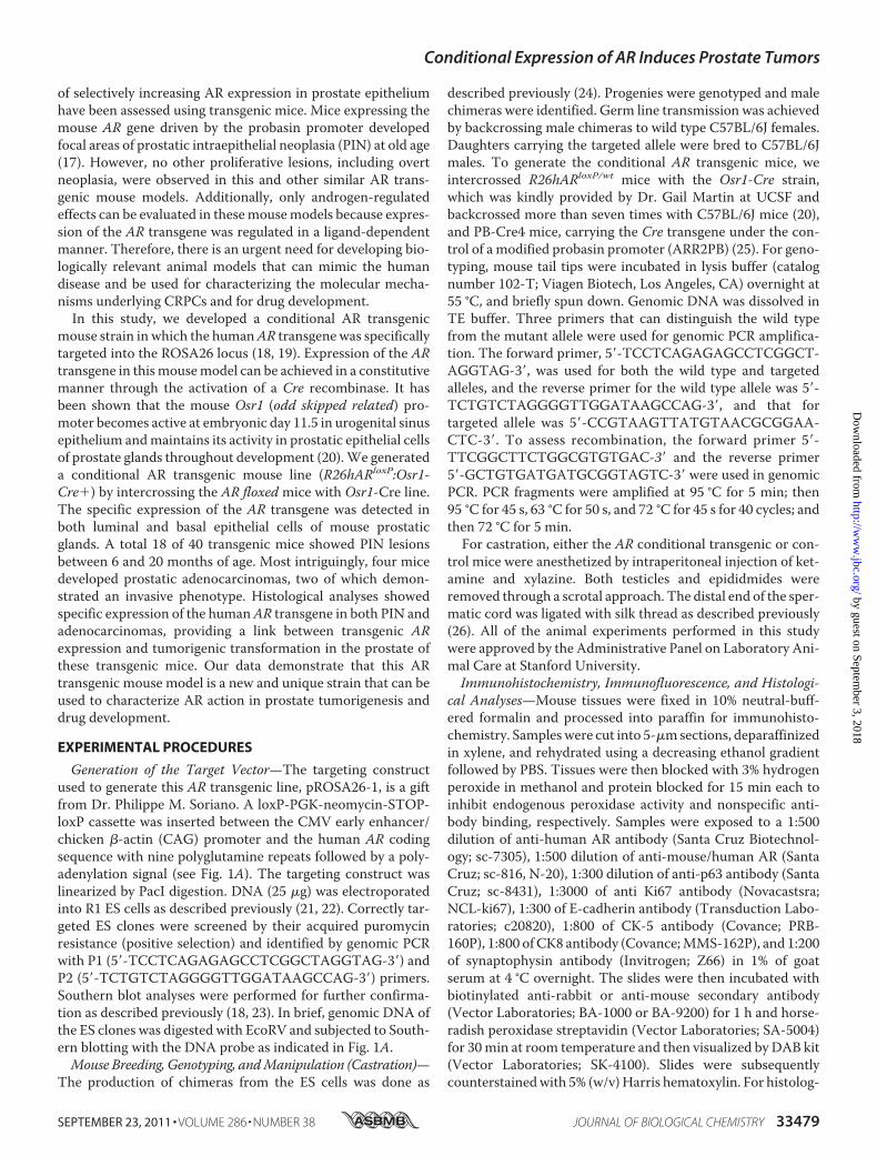
Generation of the Target Vector—The targeting constructused to generate this AR transgenic line, pROSA26-1, is a giftfrom Dr. Philippe M. Soriano. A loxP-PGK-neomycin-STOP-loxP cassette was inserted between the CMV early enhancer/chicken �-actin (CAG) promoter and the human AR codingsequence with nine polyglutamine repeats followed by a poly-adenylation signal (see Fig. 1A). The targeting construct waslinearized by PacI digestion. DNA (25 �g) was electroporatedinto R1 ES cells as described previously (21, 22). Correctly tar-geted ES clones were screened by their acquired puromycinresistance (positive selection) and identified by genomic PCRwith P1 (5�-TCCTCAGAGAGCCTCGGCTAGGTAG-3�) andP2 (5�-TCTGTCTAGGGGTTGGATAAGCCAG-3�) primers.Southern blot analyses were performed for further confirma-tion as described previously (18, 23). In brief, genomic DNA ofthe ES clones was digested with EcoRV and subjected to South-ern blotting with the DNA probe as indicated in Fig. 1A.Mouse Breeding, Genotyping, andManipulation (Castration)—
The production of chimeras from the ES cells was done as
described previously (24). Progenies were genotyped and malechimeras were identified. Germ line transmission was achievedby backcrossing male chimeras to wild type C57BL/6J females.Daughters carrying the targeted allele were bred to C57BL/6Jmales. To generate the conditional AR transgenic mice, weintercrossed R26hARloxP/wt mice with the Osr1-Cre strain,which was kindly provided by Dr. Gail Martin at UCSF andbackcrossed more than seven times with C57BL/6J mice (20),and PB-Cre4 mice, carrying the Cre transgene under the con-trol of a modified probasin promoter (ARR2PB) (25). For geno-typing, mouse tail tips were incubated in lysis buffer (catalognumber 102-T; Viagen Biotech, Los Angeles, CA) overnight at55 °C, and briefly spun down. Genomic DNA was dissolved inTE buffer. Three primers that can distinguish the wild typefrom the mutant allele were used for genomic PCR amplifica-tion. The forward primer, 5�-TCCTCAGAGAGCCTCGGCT-AGGTAG-3�, was used for both the wild type and targetedalleles, and the reverse primer for the wild type allele was 5�-TCTGTCTAGGGGTTGGATAAGCCAG-3�, and that fortargeted allele was 5�-CCGTAAGTTATGTAACGCGGAA-CTC-3�. To assess recombination, the forward primer 5�-TTCGGCTTCTGGCGTGTGAC-3� and the reverse primer5�-GCTGTGATGATGCGGTAGTC-3� were used in genomicPCR. PCR fragments were amplified at 95 °C for 5 min; then95 °C for 45 s, 63 °C for 50 s, and 72 °C for 45 s for 40 cycles; andthen 72 °C for 5 min.For castration, either the AR conditional transgenic or con-
trol mice were anesthetized by intraperitoneal injection of ket-amine and xylazine. Both testicles and epididmides wereremoved through a scrotal approach. The distal end of the sper-matic cord was ligated with silk thread as described previously(26). All of the animal experiments performed in this studywere approved by the Administrative Panel on Laboratory Ani-mal Care at Stanford University.Immunohistochemistry, Immunofluorescence, and Histologi-
cal Analyses—Mouse tissues were fixed in 10% neutral-buff-ered formalin and processed into paraffin for immunohisto-chemistry. Sampleswere cut into 5-�msections, deparaffinizedin xylene, and rehydrated using a decreasing ethanol gradientfollowed by PBS. Tissues were then blocked with 3% hydrogenperoxide in methanol and protein blocked for 15 min each toinhibit endogenous peroxidase activity and nonspecific anti-body binding, respectively. Samples were exposed to a 1:500dilution of anti-human AR antibody (Santa Cruz Biotechnol-ogy; sc-7305), 1:500 dilution of anti-mouse/human AR (SantaCruz; sc-816, N-20), 1:300 dilution of anti-p63 antibody (SantaCruz; sc-8431), 1:3000 of anti Ki67 antibody (Novacastsra;NCL-ki67), 1:300 of E-cadherin antibody (Transduction Labo-ratories; c20820), 1:800 of CK-5 antibody (Covance; PRB-160P), 1:800 of CK8 antibody (Covance;MMS-162P), and 1:200of synaptophysin antibody (Invitrogen; Z66) in 1% of goatserum at 4 °C overnight. The slides were then incubated withbiotinylated anti-rabbit or anti-mouse secondary antibody(Vector Laboratories; BA-1000 or BA-9200) for 1 h and horse-radish peroxidase streptavidin (Vector Laboratories; SA-5004)for 30min at room temperature and then visualized by DAB kit(Vector Laboratories; SK-4100). Slides were subsequentlycounterstainedwith 5% (w/v)Harris hematoxylin. For histolog-
Conditional Expression of AR Induces Prostate Tumors
SEPTEMBER 23, 2011 • VOLUME 286 • NUMBER 38 JOURNAL OF BIOLOGICAL CHEMISTRY 33479
by guest on September 3, 2018
http://ww
w.jbc.org/
Dow
nloaded from

ical analysis, 5-�m serial sections were processed from xyleneto water through a decreasing ethanol gradient, stained withhematoxylin and eosin, and processed back to xylene throughan increasing ethanol gradient. For immunofluorescenceassays, 5-�m sections were boiled in 0.01 M citrate buffer (pH6.0) for 20 min after redehydration from xylene to water, andblocked by 5% goat serum. Tissue sections were then incubatedwith 1:300 dilution of anti-human AR antibody (Santa CruzBiotechnology; sc-7305), 1:500 dilution of anti-mouse/humanAR (Santa Cruz; sc-816), or 1:300 dilution of anti-p63 antibody(Santa Cruz; sc-8343) in 1% of goat serum at 4 °C overnight.Goat anti-mouse Alexa Fluor 594 (Molecular Probes; A21203),or goat anti-rabbit Alexa Fluor 488 (Molecular Probes; A11034)was incubated at 1:1000 dilution for 1 h at room temperature.Sections were mounted by VECTASHIELDmounting mediumwith DAPI (Vector Laboratories; H-1200). Images for all hema-toxylin and eosin and immunohistochemistry experiments inthis study were acquired on a Leica dissecting microscope(model MZ95) using Zeiss Axiovision software. Immunofluo-rescence images were taken using an Olympus BX-52microscope.Immunoprecipitation and Western Blotting—Mouse tissues
were homogenized in ice-cold radioimmune precipitationassay buffer (150 mM sodium chloride, 1% Nonidet P-40, 0.5%sodium deoxycholate, 0.1% SDS, 50 mM Tris, pH 8.0). Proteinconcentrations were measured using a protein assay kit (Bio-Rad; catalog number 500-0006). For immunoprecipitationassays, cell lysates containing 150 �g of total protein werediluted in buffer containing 20 mM HEPES (pH 8.0), 0.5%Nonidet P-40, 100mMNaCl, 1 mM EDTA, 5mMMgCl2, 1 mM
CaCl2, 10 mM ZnCl2, 1 mM DTT, 1 mM PMSF, 5 mg/ml leu-peptin, and 5% glycerol and then incubated with rabbit nor-mal IgG or anti-FLAG antibody conjugated with pre-equili-brated protein A-Sepharose beads at 4 °C with gentlerotation for 7 h. The beads were collected by centrifugationand gently washed three times with the same buffer asdescribed above. Equal amounts of immunoprecipitateswere eluted using 2� sample buffer (125 mM Tris-HCl, pH6.8, 4% SDS, 20% (v/v) glycerol, 0.004% bromphenol blue)and analyzed by Western blot. A 1:500 dilution of a mono-clonal antibody against the human AR (Santa Cruz Biotech-nology; catalog number sc-7305) was used. Protein detectionwas performed using ECL kit according to the manufactur-er’s protocol (Amersham Biosciences).Cell Cultures and Transient Transfections—The monkey
kidney cell line, CV-1, was maintained in DMEM supple-mented with 5% FCS (HyClone, Denver, CO). Transient trans-fections were carried out using a Lipofectamine transfection kit(Invitrogen). Transfection and whole cell collection were per-formed as described previously (27, 28).Whole cell lysates wereprepared from transfected cells and subjected to Western blotanalyses.Statistical Analyses—We presented the data as the
means � S.D. We made comparisons between groups, usinga two-sided Student’s t test. p � 0.05 and p � 0.01 wereconsidered significant.
RESULTS
Generation of the AR Conditional Transgenic Mice—Previ-ous AR transgenic mouse models were developed throughrandom insertion transgenesis in ES cells or pronuclearmicroinjection (17). To achieve high targeting efficiency andcomparable expression of the AR transgene between animals,we developed a “floxed” AR allele in which the humanAR trans-gene containing a short polyglutamine repeat tractwas targetedinto the ROSA26 locus (18, 19). A loxP flanked transcriptionalsilencing element was inserted between the CAG promoter, ahybrid CMV enhancer coupled to a modified chicken �-actinpromoter, and the AR coding sequence in the targeting vector(Fig. 1A). Because the CAG promoter is ubiquitously active inmostmouse tissues in vivo (29),AR transgene expression in thismouse model can be achieved in a constitutive but tissue-spe-cific manner through Cre-recombinase-mediated removal ofthe LSL cassette. Thus, thismousemodel will enable us to char-acterize the specific role of AR in prostate tissues in a ligand-independent manner to avoid the complication observed inprevious mouse models regulated by androgen-induced pro-moters. AR transgene expression was assessed in CV-1 cellsthrough recombinase-mediated removal of the transcriptionalsilencer, the LSL cassette. Expression of FLAG-tagged AR wasobserved in the cells co-transfected with the targeted vectorand CMV-Cre plasmids (Fig. 1B). Genomic DNA samples wereisolated fromES cells and digestedwith EcoRVand subjected toSouthern blot analyses (18, 23). Four positive clones displayed a11.5-kb hybridization band corresponding to the wild typelocus and a 3.8-kb band that represents the targeted ROSA26locus (Fig. 1C). Two independent positive ES cell clones wereinjected into C57BL/6J blastocysts that were then implantedinto pseudopregnant recipients to create chimeric animals.Conditional Expression of the AR Transgene in Mouse Pros-
tatic Epithelium—The Osr1-Cre mouse line is a newly estab-lished tool strain, in which the Osr1 promoter activates atembryonic day 11.5 in urogenital sinus epithelium and retainsits activity in epithelium of the prostate throughout develop-ment (20). Although the activity of the Osr1 promoter is notfully restricted to the prostate gland, its early activation in pros-tatic epithelial cells makes Osr1-Cre a unique tool in assessingAR action in prostate development and tumorigenesis. Wecrossed the floxed AR strain with Osr1-Cre mice to generateboth R26hARloxP/loxP:Osr1-Cre� and R26hARloxP/wt:Osr1-Cre� mice. Using genomic PCR approaches, we examined theactivity of Osr1-Cre in the prostate gland of R26hARloxP/wt:Osr1-Cre� mice at different ages. We observed a 300-bp PCRfragment corresponding to the deletion of the LSL cassettethrough loxP/Cre recombination in four prostatic lobes and thebladder of R26hARloxP/wt:Osr1-Cre� mice at 4, 8, and 24 weeksof age and at a low level in the testis and kidney of 8 and12-week-old mice (supplemental Fig. S1). In contrast, only a1.6-kb nonrecombined fragmentwas observed inmouse tissuesisolated from age matched R26hARloxP/wt:Osr1-Cre� controls.To confirm the expression of transgenic human AR protein inthe mice through LoxP/Cre recombination, we then analyzeddifferent mouse tissues using immunoprecipitation and immu-noblotting. As shown in Fig. 1D, FLAG tagged human AR pro-
Conditional Expression of AR Induces Prostate Tumors
33480 JOURNAL OF BIOLOGICAL CHEMISTRY VOLUME 286 • NUMBER 38 • SEPTEMBER 23, 2011
by guest on September 3, 2018
http://ww
w.jbc.org/
Dow
nloaded from
teins were detected in the prostate gland, bladder, and heart of4-week-old R26hARloxP/wt:Osr1-Cre� mice. Among these tis-sues, the prostate gland showed the highest expression of ARprotein when samples containing equal amounts of total pro-teins from different tissues were used. There is no expression inthe samples isolated from age matched R26hARloxP/wt:Osr1-Cre� control mice. These results demonstrate that the Osr1-Cre transgene can selectively activate expression of the humanAR transgene in the prostate gland and other tissues, which isconsistent with previous reports (20).Next, we performed immunohistochemistry to visualize
transgenic AR expression in R26hARloxP/wt:Osr1-Cre� mice.Using an antibody specifically against the human AR protein(441; Santa Cruz; sc-7305), we surveyed transgenic ARexpression in the transgenic mice. We observed clearnuclear staining of human AR protein in luminal cells of allfour prostate lobes, including anterior, dorsal, lateral, andventral prostate in 8-week-old male R26hARloxP/wt:Osr1-Cre�and R26hARloxP/loxP:Osr1-Cre�mice, but not in age-matched
R26hARloxP/wt:Osr1-Cre� control mice. Representative imagesare shown in Fig. 2. There is no significant difference in inten-sity between heterozygous (R26hARloxP/wt:Osr1-Cre�) andhomozygous (R26hARloxP/loxP:Osr1-Cre�) mice. Notably,staining ofARwas limited to a portion of epithelial cells in someprostate glands. Using immunofluorescence, we further inves-tigated expression of theAR transgene in the prostate tissues ofdifferently aged mice. The immunofluorescence signal of thehuman AR protein appeared consistently in all prostate lobesbetween 4- and 48-week-old R26hARloxP/wt:Osr1-Cre� mice(data not shown).Detection of the AR Transgene Expression in Prostatic Basal
Cells in AR Conditional Transgenic Mice—Observation of thelocal staining pattern of the AR transgene in R26hARloxP/wt:Osr1-Cre� mice is novel and interesting. To confirm trans-genic AR protein expression, we repeated immunohistochem-istry with either an antibody (441; Santa Cruz; sc-7305)specifically against human AR protein or an antibody (N-20;Santa Cruz; sc-816) against both human and mouse AR pro-
FIGURE 1. Generating the AR conditional transgenic mice. A, a scheme of the conditional human AR transgene targeting construct is shown. A PGK-neomycin cassette with flanked loxP sites (LSL cassette) was inserted between the CAG promoter and a FLAG-tagged human AR coding sequence containinga nine-polyglutamine repeat tract. The DNA fragment isolated from the short arm region used as the probe in the Southern blot is marked as a solid line. Theprimers used for genotyping are marked with arrows. B, CV-1 cells were transfected with the targeting vector plasmid in the presence or absence of CMV-Creexpression vector to assess the activation of the AR transgene expression through the loxP/Cre recombination. Western blot was performed on cell lysates usingthe antibody against the human AR or �-tubulin. The expression of FLAG-tagged AR protein was detected in cells co-transfected with CMV-Cre vectors,demonstrating that a loxP/Cre recombination can result in the deletion of the LSL cassette and activation of AR transgene expression. C, Southern blot analysiswas performed to examine ES cells transfected with the targeting vectors. Genomic DNA was digested by EcoRV and hybridized to a 32P-labeled probe(represented in Fig. 1a) located on the short arm. The wild type allele and targeting allele were differentiated by size and labeled. In four clones, two bands ofexpected size, 11.5 and 3.8 kb, were detected, representing the endogenous and targeted ROSA26 locus, respectively. D, whole protein lysates were isolatedfrom different mouse tissues of 16-week-old R26hARloxP/wt:Osr1-Cre� and R26hARloxP/wt:Osr1-Cre� mice. Equal amounts of protein lysates were subjected toimmunoprecipitation (IP) with FLAG antibody and then analyzed by Western blotting with a specific antibody against the human AR to detect the specificexpression of the human AR transgene. IB, immunoblot.
Conditional Expression of AR Induces Prostate Tumors
SEPTEMBER 23, 2011 • VOLUME 286 • NUMBER 38 JOURNAL OF BIOLOGICAL CHEMISTRY 33481
by guest on September 3, 2018
http://ww
w.jbc.org/
Dow
nloaded from

teins. Using these two antibodies allowed us to distinguishexogenous human AR from endogenous mouse AR. UniformAR staining with the N20 antibody appears in the nucleus ofprostatic luminal cells in R26hARloxP/wt:Osr1-Cre� mice andR26hARloxP/wt:Osr1-Cre� controls (Fig. 3, A2 and B2). How-ever, positive staining with the human AR specific antibody(441) was only observed in prostate tissues of R26hARloxP/wt:Osr1-Cre�mice, indicating that expressionof theAR transgene isa result of the LoxP/Cre recombination through activation of Cretransgene (Fig. 3, A1 versus B1). We then used immunofluores-cence toco-localizebothhumanARandendogenousmouseARinthe abovemouse tissues. A uniformnuclear immunofluorescencesignal was observed with the antibody (N20) against human andmouse AR proteins in the prostate of R26hARloxP/wt:Osr1-Cre�mice (Fig. 3C2, green), butonlyaportionofprostatic epithelial cellsshowed a positive immunofluorescence with the human AR anti-body (Fig. 3C1, red). A significant amount of overlaywas observedespecially in luminal epithelial cells with these two antibodies (Fig.3C3,arrows). Interestingly, positive stainingof thehumanARpro-tein also appeared in prostatic basal cells in the AR transgenicmice.
As described above, we observed positive AR immuno-staining in both prostatic luminal and basal epithelial cells ofR26hARloxP/wt:Osr1-Cre� mice (Fig. 3C). It has been suggestedthat prostatic basal cells may contain prostate “stem” or “pro-genitor” cells (30, 31). We then used an antibody against p63, aprostatic basal cell marker, to confirm the expression of theARtransgene in prostatic basal epithelial cells (32). In these exper-iments, we included prostate tissues isolated from 16-week-oldmale R26hARloxP/wt:PB-Cre4� mice in which expression of theAR transgene was limited in prostatic luminal cells through theARR2PB promoter activation. As expected, positive immuno-staining of p63 was observed exclusively in prostatic basal cellsin both R26hARloxP/wt:Osr1-Cre� and R26hARloxP/wt:PB-Cre4� mice (Fig. 3, panels D2 and D5 and panels E2 and E5,respectively). Importantly, as observed previously, positivenuclear stainingwith the humanAR antibody (441) appeared ina portion of prostatic basal cells in the prostate ofR26hARloxP/wt:Osr1-Cre� but not in R26hARloxP/wt:PB-Cre4�mice (Fig. 3, panels D1 andD4 versus panels E1 and E4). Merg-ing of these images showed a significant amount of overlaybetween transgenic human AR and endogenous mouse p63
FIGURE 2. Expression of the AR transgene in R26hARloxP:Osr1-Cre� mice. Immunohistochemistry staining was used to assess expression of the human ARtransgene in the AR transgenic mice. Different paraffin-embedded prostate lobes dissected from 8-week-old male R26hARloxP/wt:Osr1-Cre�, R26hARloxP/loxP:Osr1-Cre�, and R26hARloxP/loxP:Osr1-Cre� mice were stained with an antibody specifically against the human AR protein (Santa Cruz; sc-7305; 1:300 dilution).The sections were also counterstained with hematoxylin. Representative images are shown. Note that in the absence of human AR transgene expression inR26hARloxP/loxP:Osr1-Cre� mice, the human specific AR antibody fails to detect endogenous mouse AR. AP, anterior prostate; DP, dorsal prostate; LP, lateralprostate; VP, ventral prostate.
Conditional Expression of AR Induces Prostate Tumors
33482 JOURNAL OF BIOLOGICAL CHEMISTRY VOLUME 286 • NUMBER 38 • SEPTEMBER 23, 2011
by guest on September 3, 2018
http://ww
w.jbc.org/
Dow
nloaded from

proteins in the basal epithelial cells of the abovemouse prostatetissues (Fig. 3, D3 and D6, blue arrows). The observation thatexpression of transgenic AR protein in prostatic basal cells of
R26hARloxP/wt:Osr1-Cre� mice is novel and interesting, sug-gesting that the mouse model is a new tool to assess AR actionin prostatic epithelial basal cells.Hyperplasia and Intraepithelial Neoplasia in Prostate
Glands of the AR Conditional Transgenic Mice—BothR26hARloxP/loxP:Osr1-Cre� and R26hARloxP/wt:Osr1-Cre�mice were born at the expected Mendelian ratios, suggestingthat there is no significant prenatal lethality associated withgenotypes of these mice. All of the transgenic mice appearednormal and did not show significant difference in appear-ance with age-matched R26hARloxP/loxP:Osr1-Cre� andR26hARloxP/wt:Osr1-Cre� controls as well as wild type litter-mates. In an effort to search for phenotypes of these AR trans-genic mice, we thoroughly examined the mice at 2, 4, 8, 12, and16 weeks and after 16 months of age. We observed atypicalproliferative lesions consistent with mouse prostatic intraepi-thelial neoplasia (mPIN) in both R26hARloxP/loxP:Osr1-Cre�and R26hARloxP/wt:Osr1-Cre� mice as early as 8 weeks. Specif-ically, mPIN lesions appeared mainly as cribriform structuresalong with occasional stratification of cells, papilliferous struc-tures, and tufts of cells. Atypical epithelial cells that appearedirregular, larger than adjacent normal cells and lacking normalpolarity were observed in all prostatic lobes, including anteriorprostate (Fig. 4,A, B,M, P, and S), dorsal prostate (Fig. 4G), andventral prostate (Fig. 4J). The fibromuscular stroma was intact,and the glandular and duct profiles were undisturbed (Fig. 4,Gand J). In Fig. 4 (M,P, and S), the foci of atypical cells partially fillthe lumen of the ducts. Intraluminal glands forming within theoriginal glands in the dysplastic lesions are pronounced in thesecases, which are further characterized by epithelial cell crowd-ing, and enlarged vesicular nuclei that often contained one ormore prominent nucleoli (Fig. 4, N, Q, and T). Using the anti-body against the human AR protein (440), we detected positiveimmunostaining of transgenic AR in almost all atypical cellswithinmPIN lesions (Fig. 4,C, F, I, L,O,R, andU). These resultsprovide a direct link between expression of transgenic AR pro-tein and development of the dysplastic lesions. We observedmPIN lesions in nine of 22 R26hARloxP/wt:Osr1-Cre� mice(40.9%) and in nine of 18 R26hARloxP/loxP:Osr1-Cre� (50%)mice (Table 1). Among those with mPINs, more than halfof themice (11 of 18) developed lesions at less than 12months old.No pathological abnormalities in the prostate glands were ob-served in control littermates and wild type mice. Occur-rence of mPIN lesions in R26hARloxP/wt:Osr1-Cre� andR26hARloxP/loxP:Osr1-Cre�mice is earlier andmore frequent thanin the previous AR transgenic mice (17), suggesting the potentialsignificance of this mousemodel in prostate tumorigenesis.Development of Prostate Adenocarcinoma in the AR Condi-
tional Transgenic Mice—Because there is consensus thatmPINs can progress toward prostate adenocarcinomas, wecontinued examiningmoreAR transgenicmice at progressivelyolder ages. Most intriguingly, we identified prostatic adenocar-cinomas in three R26hARloxP/wt:Osr1-Cre� mice and oneR26hARloxP/loxP:Osr1-Cre� mouse from 8 to 21 months of age.In two of the mice (Fig. 5, A and B), the neoplasms were grosslyevident, being large, extensive tumor masses in the pelvis (Fig.5, A1). The tumors were poorly circumscribed and unencapsu-lated (Fig. 5, A2 and B1) and comprised of haphazard acini and
FIGURE 3. Analysis of the AR transgene expression in the prostate oftheR26hARloxP/wt:Osr1-Cre� mice. A and B, adjacent prostate sections from16-week old male R26hARloxP/wt:Osr1-Cre�(A1 and A2) or R26hARloxP/wt:Osr1-Cre� (B1 and B2) mice were prepared and stained with either the antibodyspecifically against the human AR (A1 and B1) or human and mouse AR (A2and B2) to distinguish exogenous human AR from endogenous mouse AR.Representative images are shown. C, a prostate section isolated from a16-week-old R26hARloxP/wt:Osr1-Cre� mouse was co-stained with the anti-body against the human AR protein (C1) and the antibody against bothhuman and mouse AR proteins (C2). The merged image (C3) shows overlap ofboth exogenous and endogenous AR proteins (arrows). D, the antibodyagainst the human AR protein (red, D1 and D4) and the antibody against thep63 protein (green, D2 and D5) were used to co-stain prostate tissues isolatedfrom R26hARloxP/wt:Osr1-Cre� mice. The merged images show overlay of immu-nostaining with two antibodies (arrows, D3 and D6). E, similar co-stain analyseswere performed using prostate tissues isolated from 16-week-old maleR26hARloxP/wt:PB-Cre� mice, in which there is no overlay of AR and p63co-staining.
Conditional Expression of AR Induces Prostate Tumors
SEPTEMBER 23, 2011 • VOLUME 286 • NUMBER 38 JOURNAL OF BIOLOGICAL CHEMISTRY 33483
by guest on September 3, 2018
http://ww
w.jbc.org/
Dow
nloaded from

lobules of pleomorphic cells (Fig. 5, A4, A5, and B3) with no orlimited amounts of fibrovascular stroma. Tumor necrosis wasalso noted in one of the tumors (Fig. 5B2). In both, malignancy
was evident (in addition to the architectural, cellular, andnuclear features) by obvious vascular invasion by neoplasticcells (Fig. 5, A3 and A6) or local invasion of the tumor beyond
FIGURE 4. Immunohistochemistry analyses of the prostate tissues isolated from R26hARloxP/wt:Osr1-Cre� and R26hARloxP/loxP:Osr1-Cre� mice. Pros-tate tissues isolated from seven R26hARloxP/wt:Osr1-Cre� and R26hARloxP/loxP:Osr1-Cre� male mice between 9 and 33 weeks old were analyzed histologically.Two adjacent sections from each mouse were stained with hematoxylin and eosin or with the antibody against the human AR (Santa Cruz; sc-7305) in whichtissues were also counterstained with hematoxylin. Representative sections of anterior prostate (A, M, P, and S), dorsal prostate (G), and ventral prostate (J) lobesstained with hematoxylin and eosin show several typical dysplastia lesions and mPIN. Corresponding high power images (400�) of hematoxylin and eosinstaining are shown in B, E, H, K, N, Q, and T, accordingly. Immunohistochemical analyses with the human AR specific antibody were used to detect the expressionof the human AR transgene within dysplastic prostatic glands (C, F, I, L, O, R, and U).
Conditional Expression of AR Induces Prostate Tumors
33484 JOURNAL OF BIOLOGICAL CHEMISTRY VOLUME 286 • NUMBER 38 • SEPTEMBER 23, 2011
by guest on September 3, 2018
http://ww
w.jbc.org/
Dow
nloaded from

the basement membrane into surrounding stromal tissues thatshow an early desmoplastic response (Fig. 5B4). In the othertwo mice (Fig. 5, C and D), smaller prostatic adenocarcinomaswere noted by microscopic evaluation only. Specifically, thetumorswerediscrete, circumscribedunencapsulatedmassescom-prised of haphazard solid epithelial sheets (with only rare glandu-
lar formation) of pleomorphic cells with scant fibrovascularstroma. Although neither obvious local nor vascular invasion wasnoted in these tumors, the architectural, cellular, and nuclear fea-turesofneoplasmssuggestmalignantneoplastic transformationofprostatic epithelial cells.Despite extremeattention to thepresenceofmetastases in distant organs in themice showing prostatic ade-nocarcinomas, distant dissemination of neoplastic prostatic epi-thelial cells was not noted in all four mice.Conditional Expression of the Human AR Transgene Induces
Prostatic Cell Proliferation and Contributes to Development ofAdenocarcinomas—A promotional role of AR in cell prolifera-tion has been demonstrated previously (1, 33, 34). To under-stand the cellular effects resulting by conditional expression ofthe AR transgene in the mouse prostate, we assessed for prolif-eration using Ki67 immunohistochemistry in three mice fromeach genotype. A significant increase in Ki67 immunostainingin both mPIN and prostatic adenocarcinoma lesions wasobserved when compared with wild type samples (Fig. 6, A–I).Ki67 immunostaining was quantified by counting a total of1,000 epithelial cells from five high power fields in each sample.
FIGURE 5. Development of prostatic adenocarcinoma was observed in R26hARloxP/wt:Osr1-Cre� and R26hARloxP/loxP:Osr1-Cre�. A, a 20-month-oldR26hARloxP/wt:Osr1-Cre� male mouse was grossly examined, which revealed a 1.8 � 1.3 � 1.3-cm mass at the base of the right seminal vesicle, replacing normaltissues of the right coagulating gland or anterior prostate area (A1). Histologically, the mass was an extensive, expansile, demarcated, unencapsulatedneoplasm that intraluminally expanded a gland of the anterior prostate lobe (A2). The neoplasm consisted of haphazard solid lobules of cells with rare acinarformation (A4 and A5) with small amounts of fibrovascular stroma. The cells were pleomorphic with a large degree of anisocytosis and anisokaryosis, andoccasional mitoses were noted. A tumor embolus was noted within an adjacent thin-walled vessel (A3 and A6). B, histologic analysis of a 19-month-oldR26hARloxP/wt:Osr1-Cre� male mouse revealed a focal, expansile, demarcated neoplasm filling the lumen of a gland of the anterior prostate (B1), with theremaining glandular mucosa demonstrating epithelial stratification consistent with high grade mPIN. The neoplasm consisted of haphazard acini of cells (B3)with small amounts of fibrovascular stroma. A sole, discrete invasive focus of the tumor is noted within the stroma next to the mass (B4), with the stromapresenting with an early desmoplastic response. A focal area of necrosis with abundant acicular (cholesterol) cleft formation is present within the main tumormass itself (B2). C, histologic examination of a 34-week-old R26hARloxP/wt:Osr1-Cre� male mouse revealed a likely circumferential, expansile, demarcatedneoplasm focally expanding the mucosa of the anterior prostate gland (C1), with multifocal areas of the remaining glandular mucosa demonstrating epithelialcribriform changes consistent with high grade mPIN. The neoplasm consisted of haphazard solid sheet of cells with rare acinar formation (C2) with scantfibrovascular stroma. The cells were pleomorphic with a large degree of anisocytosis and anisokaryosis, and rare mitoses were noted. D, histologic assessmentof a 80-week-old R26hARloxP/loxP:Osr1-Cre� male mouse revealed a focal, expansile, demarcated neoplasm expanding the mucosa of the anterior prostategland (D1), with multifocal areas of the remaining glandular mucosa demonstrating epithelial cribriform changes consistent with high grade mPIN. Theneoplasm consisted of haphazard solid sheet of cells with rare acinar formation (D2) with scant fibrovascular stroma. The cells were pleomorphic, with a largedegree of anisocytosis and anisokaryosis, and regular mitoses were noted.
TABLE 1Pathological abnormalities of R26hAR transgenic mice
GenotypeTotal
numberNumberof PIN
Number ofadenocarcinoma
R26hARLoxP/WT:Osr1-Cre� 14�12Mo months 10 0 0�12 months 4 0 0
R26hARLoxP/LoxP:Osr1-Cre� 23�12 months 14 0 0�12 months 9 0 0
R26hARLoxP/WT:Osr1-Cre� 22 9 (40.9%) 3 (13.6%)�12 months 13 6 1�12 months 9 3 2
R26hARLoxP/LoxP:Osr1-Cre� 18 9 (50.0%) 1 (5.6%)�12 months 11 5 0�12 months 7 4 1
Conditional Expression of AR Induces Prostate Tumors
SEPTEMBER 23, 2011 • VOLUME 286 • NUMBER 38 JOURNAL OF BIOLOGICAL CHEMISTRY 33485
by guest on September 3, 2018
http://ww
w.jbc.org/
Dow
nloaded from
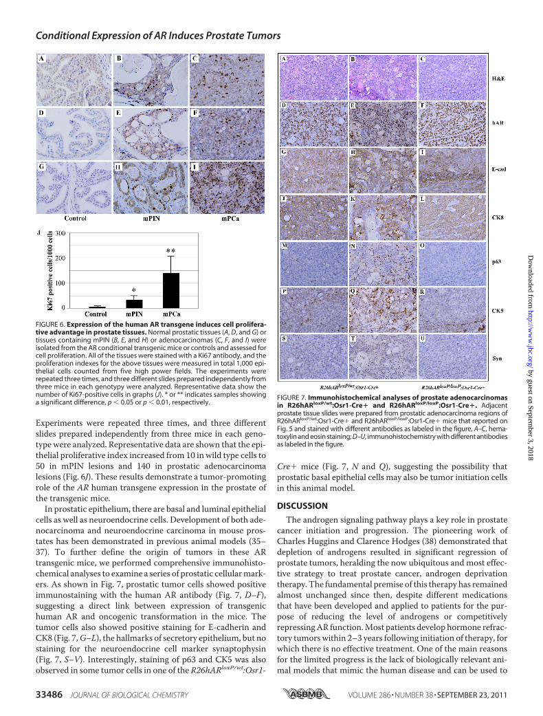
Experiments were repeated three times, and three differentslides prepared independently from three mice in each geno-type were analyzed. Representative data are shown that the epi-thelial proliferative index increased from 10 in wild type cells to50 in mPIN lesions and 140 in prostatic adenocarcinomalesions (Fig. 6J). These results demonstrate a tumor-promotingrole of the AR human transgene expression in the prostate ofthe transgenic mice.In prostatic epithelium, there are basal and luminal epithelial
cells as well as neuroendocrine cells. Development of both ade-nocarcinoma and neuroendocrine carcinoma in mouse pros-tates has been demonstrated in previous animal models (35–37). To further define the origin of tumors in these ARtransgenic mice, we performed comprehensive immunohisto-chemical analyses to examine a series of prostatic cellularmark-ers. As shown in Fig. 7, prostatic tumor cells showed positiveimmunostaining with the human AR antibody (Fig. 7, D–F),suggesting a direct link between expression of transgenichuman AR and oncogenic transformation in the mice. Thetumor cells also showed positive staining for E-cadherin andCK8 (Fig. 7,G–L), the hallmarks of secretory epithelium, but nostaining for the neuroendocrine cell marker synaptophysin(Fig. 7, S–V). Interestingly, staining of p63 and CK5 was alsoobserved in some tumor cells in one of the R26hARloxP/wt:Osr1-
Cre� mice (Fig. 7, N and Q), suggesting the possibility thatprostatic basal epithelial cells may also be tumor initiation cellsin this animal model.
DISCUSSION
The androgen signaling pathway plays a key role in prostatecancer initiation and progression. The pioneering work ofCharles Huggins and Clarence Hodges (38) demonstrated thatdepletion of androgens resulted in significant regression ofprostate tumors, heralding the now ubiquitous and most effec-tive strategy to treat prostate cancer, androgen deprivationtherapy. The fundamental premise of this therapy has remainedalmost unchanged since then, despite different medicationsthat have been developed and applied to patients for the pur-pose of reducing the level of androgens or competitivelyrepressingAR function.Most patients develop hormone refrac-tory tumors within 2–3 years following initiation of therapy, forwhich there is no effective treatment. One of the main reasonsfor the limited progress is the lack of biologically relevant ani-mal models that mimic the human disease and can be used to
FIGURE 6. Expression of the human AR transgene induces cell prolifera-tive advantage in prostate tissues. Normal prostatic tissues (A, D, and G) ortissues containing mPIN (B, E, and H) or adenocarcinomas (C, F, and I) wereisolated from the AR conditional transgenic mice or controls and assessed forcell proliferation. All of the tissues were stained with a Ki67 antibody, and theproliferation indexes for the above tissues were measured in total 1,000 epi-thelial cells counted from five high power fields. The experiments wererepeated three times, and three different slides prepared independently fromthree mice in each genotypy were analyzed. Representative data show thenumber of Ki67-positive cells in graphs (J). * or ** indicates samples showinga significant difference, p � 0.05 or p � 0.01, respectively.
FIGURE 7. Immunohistochemical analyses of prostate adenocarcinomasin R26hARloxP/wt:Osr1-Cre� and R26hARloxP/loxP:Osr1-Cre�. Adjacentprostate tissue slides were prepared from prostatic adenocarcinoma regions ofR26hARloxP/wt:Osr1-Cre� and R26hARloxP/loxP:Osr1-Cre� mice that reported onFig. 5 and stained with different antibodies as labeled in the figure. A–C, hema-toxylin and eosin staining; D–U, immunohistochemistry with different antibodiesas labeled in the figure.
Conditional Expression of AR Induces Prostate Tumors
33486 JOURNAL OF BIOLOGICAL CHEMISTRY VOLUME 286 • NUMBER 38 • SEPTEMBER 23, 2011
by guest on September 3, 2018
http://ww
w.jbc.org/
Dow
nloaded from

investigate AR action in androgen-induced prostate tumor ini-tiation and disease progression.In past years, many genetically modified mouse models have
been established for studying prostate tumorigenesis. Theyinvolve either overexpression of oncogenes or targeted deletionof tumor suppressors in the prostatic epithelium. Transgenic(gain-of-function) models that express SV40 T antigen (i.e. theTRAMP and LADY models) and c-Myc have been developedpreviously (39–41). Knock-out mice with specific deletion ofvarious tumor suppressors in the prostate have also been devel-oped (39, 42–44). One of the best characterized mouse modelsis the conditional pten knock-out (37). Loss of both alleles ofpten results in invasive prostate cancer that metastasizes tolymphnodes and lung in somemice (37). Combining PTEN losswith other genetic abnormalities has led to several additionalmouse models (45–47). Deletion of Nkx3.1 and PTEN resultsin androgen-independent prostate cancer (39, 47). However,despite the progress offered by these genetically modifiedmouse models in analyses of different molecules and signalingpathways in prostate tumorigenesis, there is a great need fornew animal models for characterizing the androgen axis, a keypathway, in prostate development and tumorigenesis.In this study, we report a new AR conditional transgenic
mouse model, R26hARloxP:Osr1-Cre�. In this mouse modelexpression of the human AR transgene is regulated in a consti-tutive but prostate-specific manner by the hybrid CAG pro-moter-coupled CMV enhancer and chicken �-actin promoterthrough loxP/Cre recombination. We used newly establishedOsr1-Cremice to activateAR transgene expression.TheOsr1pro-moter is active at embryonic day 11.5 in urogenital sinus epithe-liumandmaintains its activity in prostatic epithelial cells through-out development (20). We detected transgenic AR expression inboth luminal and basal epithelial cells of prostatic glands of4-week-old R26hARloxP:Osr1-Cre� mice. The robust activity oftheOsr1 promoter is detected in all four prostatic lobes, althoughit is not fully restricted to the prostate as described previously (20).Through loxP/Cre mediated recombination, deletion of the LSLcassette resulted in the activation of the AR transgene expressionin a constitutive manner. These unique and novel features distin-guish our AR transgenic mice from other genetically modifiedmousemodels,which shouldallowus toassess andvalidate theARaction in both prostatic luminal and basal epithelial cells and in aligand-dependent or -independent manner.The majority of human primary prostate cancers are andro-
gen-dependent. Data from previous AR transgenic mousemodels also showed that overexpression of themouseAR trans-gene in prostate luminal epithelial cells promotes proliferationof the epithelium, with the subsequent development of precan-cerous lesions and mouse PIN in aged mice. However, the pre-cise role of AR in promoting prostate cancer development stillremains unclear. In this study, we showed that almost half ofR26hARloxP/loxP:Osr1-Cre� or R26hARloxP/wt:Osr1-Cre� micedeveloped mouse PIN lesions. The onset of mPIN has beenobserved as early as 8 weeks of age. Most importantly, prostaticadenocarcinomas were developed in four of 40 AR transgenicmice between 8 and 20 months of age. It has been well docu-mented that wild type mice have a very low incidence of spon-taneous prostate tumors (please see the review in Ref. 48).
Therefore, identifying prostate adenocarcinomas in these ARconditional transgenicmice directly demonstrates a promotingrole of theAR in prostate cancer development. In this study, wealso examined the role of the human AR transgene inR26hARloxP/wt:PB-Cre� and R26hARloxP/loxed:PB-Cre� mice,inwhich theAR transgene expressionwas selectively targeted inprostatic luminal epithelial cells through ARR2PB promoter(25). We only observed mPIN lesions but no prostatic adeno-carcinomas in those mice. These results are consistent with theprevious studies and implicate that selective activation of theAR transgene expression in different cells of the prostate mayregulate cell proliferation distinctly during the course of mouseprostate cancer development.The promoting role of AR in stimulating prostatic cell
growth has been implicated in human prostate tumorigenesis.However, it is unclear whether increasing AR expression, a ste-roid hormone receptor, is sufficient to induce prostate cancerdevelopment inmice because previousAR transgenicmice onlyshowedmPIN lesions (17). Thus, the finding in this study is veryinteresting and suggests that this mouse model is novel andshould be characterized further. In this mouse model, positiveimmunostaining of transgenic human AR protein appears inthemajority of atypical cells in mPIN lesions and tumor cells ofprostatic adenocarcinomas, providing a direct link betweenARtransgene expression and oncogenic transformation in mouseprostates. In addition, an increase of Ki67-positive cells hasbeen observed in all above prostatic adenocarcinoma and PINsamples. In this study, we also examined cell apoptosis in theabove samples withmPIN and adenocarcinoma lesions and didnot observe any significant changes. These data further supporta promotional role of transgenic AR protein in inducing pros-tatic epithelial proliferation.Using immunohistochemistry, we observed that most tumor
cells in the adenocarcinoma and PIN lesions were E-cadherin-and CK8-positive but synaptophysin-negative. These data sug-gest that tumor cells are immunoreactive to luminal epithelialcellular markers. Interestingly, we also observed some immu-nolabeling with p63 and CK5 antibodies in some tumor cellsfrom one R26hARloxP/wt:Osr1-Cre� mouse. Expression of thehuman AR transgene has been observed in prostatic basal epi-thelial cells in R26hARloxP/loxP:Osr1-Cre� and R26hARloxP/wt:Osr1-Cre�. These data suggest that oncogenic transformationcan be initiated in both basal and luminal epithelial cellsthrough the activation of the androgen signaling pathway. Pre-vious studies showing that both prostatic luminal and basalepithelial cells are competent to function as tumor initiatingcells support this hypothesis (30, 31). In general, most prostaticbasal epithelial cells have no or low expression of the AR.Enforcement of transgenic AR expression in basal cells maydisrupt the normal differentiation pathway and induce onco-genic transformation. Although it is unclear whether activationof theAR transgene throughOsr1-Cre can directly promote andinduce normal basal epithelial cells into “primary” tumor-initi-ating cells, occurrence of prostate adenocarcinomas in this ARtransgenic mouse model provides a direct line of evidence thatdysregulation of the AR signaling pathway can promote pros-tate tumor formation in mice. Interestingly, we only observedprostatic adenocarcinomas in about 10% of the transgenicmice
Conditional Expression of AR Induces Prostate Tumors
SEPTEMBER 23, 2011 • VOLUME 286 • NUMBER 38 JOURNAL OF BIOLOGICAL CHEMISTRY 33487
by guest on September 3, 2018
http://ww
w.jbc.org/
Dow
nloaded from
that have been examined in this study. The low penetrance ofadenocarcinoma in this model further implies that other addi-tional “hits” may be required to enhance AR-mediated onco-genic transformation in prostate tumorigenesis. Thus, the cur-rent AR transgenicmousemodelmimics features of the humandisease and can be used to identify other factors and pathwaysthat promote AR action in inducing prostate cancer initiationand progression.The probasin promoter has been widely used to create pros-
tate genetically modified mouse models in the past. It is acti-vated postnatally in an androgen-inducible manner and is tar-geted selectively to luminal cells (25). One of the mostimportant features for our AR conditional transgenic mousemodel is that AR expression is regulated in a constitutive butprostate-specificmanner by the hybridCAGpromoter throughloxP/Cre recombination (Fig. 1A). In this study, we comparedexpression of the AR transgene between castrated versusuncastrated R26hARloxP/wt:Osr1-Cre� and R26hARloxP/wt:Osr1-Cre� mice. Expression of transgenic human AR proteinappeared unchanged between castrated and intactR26hARloxP/wt:Osr1-Cre� mice (supplemental Fig. S2). Thus,this model allows us to characterize the role of AR in theabsence of androgens. Further studieswith larger cohorts of theAR transgenic mice will allow us to fully assess the effect ofcastration in growth and progression of prostatic adenocarci-nomas in this new mouse model.
REFERENCES1. Kyprianou, N., and Isaacs, J. T. (1988) Endocrinology 122, 552–5622. Abate-Shen, C., and Shen, M. M. (2000) Genes Dev. 14, 2410–24343. Jenster, G. (1999) Semin. Oncol. 26, 407–4214. Culig, Z., Hobisch, A., Bartsch, G., and Klocker, H. (2000) Urol. Res. 28,
211–2195. Koivisto, P., Kolmer,M., Visakorpi, T., andKallioniemi, O. P. (1998)Am. J.
Pathol. 152, 1–96. Salinas, C. A., Austin, M. A., Ostrander, E. O., and Stanford, J. L. (2005)
Prostate 65, 58–657. Palazzolo, I., Gliozzi, A., Rusmini, P., Sau, D., Crippa, V., Simonini, F.,
Onesto, E., Bolzoni, E., and Poletti, A. (2008) J. Steroid Biochem.Mol. Biol.108, 245–253
8. Stanford, J. L., Just, J. J., Gibbs, M., Wicklund, K. G., Neal, C. L., Blumen-stein, B. A., and Ostrander, E. A. (1997) Cancer Res. 57, 1194–1198
9. Gann, P. H., Hennekens, C. H., Ma, J., Longcope, C., and Stampfer, M. J.(1996) J. Natl. Cancer Inst. 88, 1118–1126
10. Shaneyfelt, T., Husein, R., Bubley, G., and Mantzoros, C. S. (2000) J. Clin.Oncol. 18, 847–853
11. Koivisto, P., Kononen, J., Palmberg, C., Tammela, T., Hyytinen, E., Isola, J.,Trapman, J., Cleutjens, K., Noordzij, A., Visakorpi, T., and Kallioniemi,O. P. (1997) Cancer Res. 57, 314–319
12. Ruizeveld de Winter, J. A., Janssen, P. J., Sleddens, H. M., Verleun-Mooi-jman, M. C., Trapman, J., Brinkmann, A. O., Santerse, A. B., Schroder,F. H., and van der Kwast, T. H. (1994) Am. J. Pathol. 144, 735–746
13. Chen, C. D., Welsbie, D. S., Tran, C., Baek, S. H., Chen, R., Vessella, R.,Rosenfeld, M. G., and Sawyers, C. L. (2004) Nat. Med. 10, 33–39
14. Taplin, M. E., Bubley, G. J., Shuster, T. D., Frantz, M. E., Spooner, A. E.,Ogata, G. K., Keer, H. N., and Balk, S. P. (1995) N. Engl. J. Med. 332,1393–1398
15. Gaddipati, J. P., McLeod, D. G., Heidenberg, H. B., Sesterhenn, I. A., Fin-ger,M. J., Moul, J.W., and Srivastava, S. (1994)Cancer Res. 54, 2861–2864
16. Bruchovsky, N., Lesser, B., Van Doorn, E., and Craven, S. (1975) VitamHorm 33, 61–102
17. Stanbrough, M., Leav, I., Kwan, P. W., Bubley, G. J., and Balk, S. P. (2001)Proc. Natl. Acad. Sci. U.S.A. 98, 10823–10828
18. Soriano, P. (1999) Nat. Genet. 21, 70–7119. Srinivas, S., Watanabe, T., Lin, C. S., William, C. M., Tanabe, Y., Jessell,
T. M., and Costantini, F. (2001) BMC Dev. Biol. 1, 420. Grieshammer, U., Agarwal, P., andMartin, G. R. (2008)Genesis 46, 69–7321. Nagy, A., Rossant, J., Nagy, R., Abramow-Newerly, W., and Roder, J. C.
(1993) Proc. Natl. Acad. Sci. U.S.A. 90, 8424–842822. Soriano, P., Montgomery, C., Geske, R., and Bradley, A. (1991) Cell 64,
693–70223. Yanagawa, Y., Kobayashi, T., Ohnishi, M., Kobayashi, T.,Tamura, S., Tsu-
zuki, T., Sanbo,M., Yagi, T., Tashiro, F., andMiyazaki, J. (1999)TransgenicRes. 8, 215–221
24. Robertson, E., Bradley, A., Kuehn, M., and Evans, M. (1986) Nature 323,445–448
25. Wu, X.,Wu, J., Huang, J., Powell,W. C., Zhang, J.,Matusik, R. J., Sangiorgi,F. O., Maxson, R. E., Sucov, H. M., and Roy-Burman, P. (2001)Mech. Dev.101, 61–69
26. Sugimura, Y., Cunha, G. R., and Donjacour, A. A. (1986) Biol. Reprod 34,973–983
27. Huang, C. Y., Beliakoff, J., Li, X., Lee, J., Li, X., Sharma, M., Lim, B., andSun, Z. (2005)Mol. Endocrinol. 19, 2915–2929
28. Li, X., Thyssen, G., Beliakoff, J., and Sun, Z. (2006) J. Biol. Chem. 281,23748–23756
29. Okabe, M., Ikawa, M., Kominami, K., Nakanishi, T., and Nishimune, Y.(1997) FEBS Lett. 407, 313–319
30. Kasper, S. (2008) Stem Cell Rev. 4, 193–20131. Matusik, R. J., Jin, R. J., Sun, Q., Wang, Y., Yu, X., Gupta, A., Nandana, S.,
Case, T. C., Paul, M., Mirosevich, J., Oottamasathien, S., and Thomas, J.(2008) Differentiation 76, 682–698
32. Weinstein, M. H., Signoretti, S., and Loda, M. (2002) Mod. Pathol. 15,1302–1308
33. Culig, Z., Klocker, H., Bartsch, G., Steiner, H., and Hobisch, A. (2003)J. Urol. 170, 1363–1369
34. Gelmann, E. P. (2002) J. Clin. Oncol. 20, 3001–301535. Greenberg, N. M., DeMayo, F., Finegold, M. J., Medina, D., Tilley, W. D.,
Aspinall, J. O., Cunha, G. R., Donjacour, A. A., Matusik, R. J., and Rosen,J. M. (1995) Proc. Natl. Acad. Sci. U.S.A. 92, 3439–3443
36. Garabedian, E. M., Humphrey, P. A., and Gordon, J. I. (1998) Proc. Natl.Acad. Sci. U.S.A. 95, 15382–15387
37. Wang, S., Gao, J., Lei, Q., Rozengurt, N., Pritchard, C., Jiao, J., Thomas,G. V., Li, G., Roy-Burman, P., Nelson, P. S., Liu, X., and Wu, H. (2003)Cancer Cell 4, 209–221
38. Huggins, C., and Hodges, C. V. (2002) J. Urol 168, 9–1239. Abate-Shen, C. (2006) Clin. Cancer Res. 12, 5274–527640. Ellwood-Yen, K., Graeber, T. G., Wongvipat, J., Iruela-Arispe, M. L.,
Zhang, J., Matusik, R., Thomas, G. V., and Sawyers, C. L. (2003) CancerCell 4, 223–238
41. Gingrich, J. R., Barrios, R. J., Kattan, M. W., Nahm, H. S., Finegold, M. J.,and Greenberg, N. M. (1997) Cancer Res. 57, 4687–4691
42. Kerkhofs, S., Denayer, S., Haelens, A., and Claessens, F. (2009) J. Mol.Endocrinol. 42, 11–17
43. Pienta, K. J., Abate-Shen, C., Agus, D. B., Attar, R. M., Chung, L. W.,Greenberg, N. M., Hahn, W. C., Isaacs, J. T., Navone, N. M., Peehl, D. M.,Simons, J. W., Solit, D. B., Soule, H. R., VanDyke, T. A., Weber, M. J., Wu,L., and Vessella, R. L. (2008) Prostate 68, 629–639
44. Winter, S. F., Cooper, A. B., and Greenberg, N. M. (2003) Prostate CancerProstatic Dis. 6, 204–211
45. Chen, Z., Trotman, L. C., Shaffer, D., Lin, H. K., Dotan, Z. A., Niki, M.,Koutcher, J. A., Scher, H. I., Ludwig, T., Gerald, W., Cordon-Cardo, C.,and Pandolfi, P. P. (2005) Nature 436, 725–730
46. Gao, H., Ouyang, X., Banach-Petrosky, W. A., Gerald, W. L., Shen, M.M.,and Abate-Shen, C. (2006) Proc. Natl. Acad. Sci. U.S.A. 103, 14477–14482
47. Gao, H., Ouyang, X., Banach-Petrosky, W. A., Shen, M. M., and Abate-Shen, C. (2006) Cancer Res. 66, 7929–7933
48. Shappell, S. B., Thomas, G. V., Roberts, R. L., Herbert, R., Ittmann, M. M.,Rubin, M. A., Humphrey, P. A., Sundberg, J. P., Rozengurt, N., Barrios, R.,Ward, J. M., and Cardiff, R. D. (2004) Cancer Res. 64, 2270–2305
Conditional Expression of AR Induces Prostate Tumors
33488 JOURNAL OF BIOLOGICAL CHEMISTRY VOLUME 286 • NUMBER 38 • SEPTEMBER 23, 2011
by guest on September 3, 2018
http://ww
w.jbc.org/
Dow
nloaded from

Gerald R. Cunha and Zijie SunChunfang Zhu, Richard Luong, Ming Zhuo, Daniel T. Johnson, Jesse K. McKenney,
Transformation of the Mouse ProstateConditional Expression of the Androgen Receptor Induces Oncogenic
doi: 10.1074/jbc.M111.269894 originally published online July 27, 20112011, 286:33478-33488.J. Biol. Chem.
10.1074/jbc.M111.269894Access the most updated version of this article at doi:
Alerts:
When a correction for this article is posted•
When this article is cited•
to choose from all of JBC's e-mail alertsClick here
Supplemental material:
http://www.jbc.org/content/suppl/2011/07/25/M111.269894.DC1
http://www.jbc.org/content/286/38/33478.full.html#ref-list-1
This article cites 48 references, 18 of which can be accessed free at
by guest on September 3, 2018
http://ww
w.jbc.org/
Dow
nloaded from