Embed Size (px)
Citation preview

Research ArticleConfirming the Effects of Qinghuayin againstChronic Atrophic Gastritis and a Preliminary Observation ofthe Involved Inflammatory Signaling Pathways:An In Vivo Study
Sihan Li,1,2 Minghan Huang,2 Qin Chen,3 Shunan Li,2 XinWang,4 Jianlong Lin,5
Guodong Zhong,5 Ping Lin ,2,6 and Tetsuya Asakawa 6,7
1Basic Medical College of Guangzhou University of Chinese Medicine, No. 232 Waihuandong Road, Guangzhou, 510006, China2Department of Gastroenterology, The Second People’s Hospital Affiliated to Fujian University of Traditional Chinese Medicine,No. 282 Wusibei Road, Fuzhou, 353003, China
3Department of Internal Medicine, Fujian Medical University Union Hospital, Fuzhou, China4Department of Pharmacy, The Second People’s Hospital Affiliated to Fujian University of Traditional Chinese Medicine,Fuzhou, 353003, China
5Department of Pathology, The Second People’s Hospital Affiliated to Fujian University of Traditional Chinese Medicine,Fuzhou, 353003, China
6Research Base of Traditional Chinese Medicine Syndrome, Fujian University of Traditional Chinese Medicine, Fuzhou 350122, China7Department of Neurosurgery, Hamamatsu University School of Medicine, Handayama, Hamamatsu-City, Shizuoka, Japan
Correspondence should be addressed to Ping Lin; [email protected] and Tetsuya Asakawa; [email protected]
Sihan Li and Minghan Huang contributed equally to this work.
Received 15 June 2018; Accepted 5 September 2018; Published 26 September 2018
Academic Editor: Youn C. Kim
Copyright © 2018 Sihan Li et al.This is an open access article distributed under the Creative Commons Attribution License, whichpermits unrestricted use, distribution, and reproduction in any medium, provided the original work is properly cited.
Background. Qinghuayin (QHY) is a Chinese formula that is widely used in the treatment of chronic atrophic gastritis (CAG).Thisstudy was planned with the following objectives: (1) confirming the efficacy of QHY in a rat model of CAG and (2) performing apreliminary observation of the changes in several inflammatory signaling pathways potentially involved in the QHY mechanisms.Methods. A total of 33 rats were used in this study; they were divided into the control (n = 12) and model (n = 21) groups. QHYwas administrated to both the groups. We assessed the pathological manifestations and the serum tumor necrosis factor alpha(TNF-𝛼) level as markers of efficacy. We also performed a preliminary observation of the changes in the protein and messengerribonucleic acid (mRNA) expression of toll-like receptors 4 (TLR4), MyD88, NF-𝜅B, and COX-2.Results.Thepathological changesinduced in the rats by the establishment of the CAGmodels were recovered by low and high doses of QHY.Their serumTNF-𝛼 levelalso reduced following low- and high-dose QHY treatment. Protein and mRNA expressions of TLR4, MyD88, NF-𝜅B, and COX-2 were upregulated by the establishment of CAG models and downregulated by the administration of low- and high-dose QHY.Conclusions. Our data confirm the efficacy of QHY as an adjuvant therapy, based on the theories in traditional Chinese medicine.The preliminary observations indicate that the downregulation of the enhanced inflammatory signaling pathways might be crucialQHY mechanisms that need further verification.
1. Introduction
Chronic atrophic gastritis (CAG) is a common chronic gastricmucosal lesion that plays a crucial role in the development of
gastric carcinoma, a type of cancer with a high mortality ratein China. Development of effective therapies against CAGis extremely important for reducing gastric cancer mortal-ity. The pathogenesis of CAG is complicated, and it is well
HindawiEvidence-Based Complementary and Alternative MedicineVolume 2018, Article ID 4905089, 8 pageshttps://doi.org/10.1155/2018/4905089

2 Evidence-Based Complementary and Alternative Medicine
documented thatHelicobacter pylori (HP) infection is strong-ly associated with CAG and the subsequent development ofgastric cancer [1]. Thus, anti-HP treatment is an importanttherapy for CAG. Further, other therapies, such as gastricmucosa protection, symptomatic pain relief, Vitamin Csupplementation, and proton pump inhibitor administration,have been used in CAG treatment [2]. However, there isno specific treatment for CAG. In China, many therapiesbased on the theories of traditional Chinese medicine (TCM)have been considered. A recent systemic review has suggestedthat a Chinese classical formula, Sijunzi decoction, maybenefit CAG patients [3]. In addition to Sijunzi decoction,Qinghuayin (QHY) is another Chinese formula that is widelyused in the treatment of CAG based on the TCM theories ofclearing heat and resolving dampness. QHY is composed ofherbs, including semen dolichoris album (Baibiandou), Poriacocos (Fuling), coix seeds (Yiyiren), herba artemisia scoparia(Yinchen), herba eupatorii (Peilan), amomum cardamom(Baidoukou), rhizoma coptidis (Huanglian), cortex magnoliaofficinalis (Houpu), and radix paeoniae rubra (Chishao).Several Chinese studies have reported good efficacy/safetyof QHY in CAG treatment [4–6]. However, the underlyingmechanisms remain unclear.
In contrast, HP infection may affect signaling pathways[7]. CAG is also associated with inflammatory signaling path-ways, such as the IL-11/STAT3 signaling pathway [8], leptinreceptor signaling pathway [9, 10], E-cadherin/𝛽-catenin/tcf-4 pathway [11], and PI3k/Akt pathway [12]. Wang et al.reported that Toll-like receptors 4 (TLR4) and TLR9 areregulated by HP and are closely associated to CAG [11]. Wehypothesized that QHYmechanisms are also associated withthe regulation of inflammatory signaling pathways. There-fore, we observed the changes in the biomarkers of severalsignaling pathways in rat CAGmodels treated with QHY.Weattempted to conduct a preliminary examination of the rolesplayed by the signaling pathways in QHYmechanisms.
2. Methods and Materials
2.1. Animals. A total of 33 male Wistar rats (body weight 110g ± 10 g) were involved in the trial. All the rats were carefullytreated as per the National Institute of Health Guidelines forthe Care and Use of Laboratory Animals. All the experimentswere approved and supervised by the Animal Care and UseCommittee of the Fujian University of Traditional ChineseMedicine (approval number: 3100101037). The rats wereraised in a room at 23∘C with normal rhythm (8:00 AM to8:00 PM, light on).
2.2. QHY Preparation. The QHY compound was preparedaccording to the formula approved by the Fujian Provin-cial Food and Drug Administration (approval number:Z05104030). The ingredients of the QHY solution have beenlisted in Table 1. We filtered the decoction and adjusted theconcentration to 2.2 g/mL (raw materials) at 60∘C. The solu-tion was bottled and sterilized for the experiments.
2.3. Experimental Design. The rats were raised normally until1 week before the experiments; thereafter, theywere randomly
divided into the control group (C, n = 12) and the CAGmodel group (M, n = 21). The control group rats were raisedwith normal water and diet. Rats in the model group wereallowed free access to 0.5 g/L ammonia as drinking waterduring the initial 12 weeks. Then, they were gavaged oncedaily with 20 mmol/L sodium deoxycholate solution (2 mLeach time) and gavaged with 60% ethanol two times a week(2 mL each time). At the end of 12 weeks, 3 rats from themodel groupwere selected randomly and deeply anesthetizedwith chloral hydrate (400 mg/kg).The stomach was removedfor pathological examination to confirm the successful estab-lishment of the CAG model.
QHY doses were defined as per the dose in human pa-tients: 1.5 mL/kg was defined as low-dose treatment (LT),while 6.0 mL/kg was considered high-dose treatment (HT).All the agents were diluted with saline to make up a 4-mLsolution for gavage.
The control group rats were randomly divided into thefollowing two groups: Intact group (n = 6) underwent gavagewith 4mL saline; the C +HT group (n = 6) underwent gavagewith high-dose QHY (6.0 mL/kg). The rat CAG models wererandomly divided into the following 3 groups: M group (n= 6) that underwent gavage with 4 mL saline, the M + LTgroup (n = 6) that underwent gavage with low-dose QHY (1.5mL/kg), and theM+HTgroup (n = 6) that underwent gavagewith high-dose QHY (6.0 mL/kg).
Gavage was performed continuously for a period of 30days. On the 31st day, after blood sampling (3 mL) for themeasurement of the serum TNF-𝛼 level, animals were sacri-ficed with an intraperitoneal injection of chloral hydrate (400mg/kg) and their stomachs were removed for pathologicalexamination.
2.4. Serum Tumor Necrosis Factor -𝛼 (TNF-𝛼) Concentrations.The serum TNF-𝛼 level was measured using a standardenzyme-linked immunosorbent assay (ELISA) method. Therat blood samples (3 mL) were subjected to low-temperaturefreezing and centrifuged at 3500 rpm at 4∘C for 15 min; thesupernatant was collected, and measurement steps were con-ducted as per the manual of the ELISA kit (Shanghai WestangBio-Tech. Co. LTD, China).
2.5. Pathological Examination. Each stomach sample was di-vided into twoparts as follows: two blocks of tissues (the samesize as soybeans) were clipped at the gastric antrum and im-mediately placed in liquid nitrogen for real-time polymerasechain reaction (RT-PCR) and western blot analysis; theremaining gastric antrum tissues were used for the hema-toxylin and eosin (HE) staining.
2.5.1. HE Staining. Tissues were fixed with 4% paraformalde-hyde for 24 h; thereafter, the fixed-conventional paraffinembedded tissues were cut into 5 consecutive slices (slicethickness 4𝜇m),HE stainingwas performed, and histopatho-logical changes in the gastric mucosa were observed under anoptical microscope.
2.6. Detection of the mRNA Expression of TLR4, MyD88,NF-𝜅B, and COX-2. A standard RT-PCR was employed for

Evidence-Based Complementary and Alternative Medicine 3
Table 1: Ingredients of the QHY solution.
Ping yin English name Proportion of ingredientsBaibiandou semen dolichoris album 20.408%Fuling Poria cocos 20.408%Yiyiren coix seeds 20.408%Yinchen herba artemisia scoparia 10.204%Peilan herba eupatorii 6.123%Baidoukou amomum cardamom 3.061%Huanglian rhizoma coptidis 3.061%Houpu cortex magnolia officinalis 6.123%Chishao radix paeoniae rubra 10.204%
detecting the mRNA expression of TLR4, MyD88, NF-𝜅B,and COX-2. Briefly, after total RNA extraction, RT-PCRwas performed in the following conditions using a quan-titative real-time RT-PCR machine (Applied Biosystems,7900 Fluorescence-based quantitative PCR system, Carlsbad,USA): predenaturation at 50∘C for 2 min and denaturationat 95∘C for 10 min; 95∘C for 15 s and 60∘C for 1 min, with40 cycles of amplification. The RNA level was measuredwith a ND2000C Ultra-micro spectrophotometer (ThermoScienTific, USA). With the Ct value as the target geneexpression and GAPDH as the internal control, the relativeexpression of target gene was calculated by 2−ΔΔCt.
Primers were as follows: TLR4 sense primer, 5’-GCC-CTCAGTCTTGGAGTGTC-3’; TLR4 antisense primer, 5’-TAACACAGGGCGCCTAAGAG-3’; product length, 93bp;NF-𝜅B sense primer, 5’-AGTTTGACGGTGAGCTGGTA-3’; NF-𝜅B antisense primer, 5’-GCCTCGGCCTGCCGC-AAGCCT-3’; product length, 300bp; COX-2 sense primer,5’ -GGCTGTATATCTGCTCTATATGC-3’; COX-2 anti-sense primer, 5’ -CCGCTTCCTTTGTCCATCAG-3’; prod-uct length, 306bp; MyD88 sense primer, 5’ -GGACTG-CCAGAAATACATACGC-3’; MyD88 antisense primer, 5’ -CTTGTCTGTGGGACACTGCTC-3’; product length, 94bp;GAPDH sense primer, 5’-CAACGGGAAACCCATCACCA-3’; GAPDH antisense primer, 5’ -ACGCCAGTAGACTCC-ACGACAT-3’; product length, 96bp;
2.7. Detection of the Protein Expression of TLR4, MyD88,NF-𝜅B, and COX-2. A standard western blot analysis wasused for detecting the mRNA expressions of TLR4, MyD88,NF-𝜅B, and COX-2. Protein expressions were measuredusing a DCTM protein assay kit (Bio-Rad, USA). Briefly,gastric antrum tissues (150 mg) were lysed for total proteinextraction; protein samples (30 𝜇g) were subjected to SDS-PAGE electrophoresis (Bio-Rad, USA) and then transferredto a nitrocellulose membrane and blocked with TBST for1 h; primary antibodies including TLR4 (1:800), MyD88(1:500), NF-𝜅B (1:1000), COX-2 (1:800), and 𝛽-actin (1:1000)were used for overnight incubation at 4∘C. Subsequently,the membranes were washed thrice with TBST, followedby incubation with second antibodies for 1 h. Enhancedchemical fluorescein imaging and photographing were per-formed. Image analysiswas performed usingGraphic analysissoftware (Ascent software for Multiskan, Thermo ScienTific,USA), and the ratio of the gray values of the target bands tothe internal reference (𝛽-actin) was calculated.
2.8. Statistical Analyses. Datawere analyzed using SPSS 19.0.0software (SPSS Inc., IL, USA).The data are presented asmean± standard deviation values. One-way analysis of variancewas performed followed by a Dunnett post hoc test for multi-ple comparisons. Categorical data were analyzed using therank sum test. P< 0.05was considered statistically significant.
3. Results
3.1. Confirming the Efficacy of QHY
3.1.1. Evidence of Pathological Examination. We confirmedthe pathological changes in both groups. In the controlgroup, we observed normal pathological manifestation in thegastric mucosa. Gastric mucosal epithelial cells and glandswere consistent in shape and size; they were neatly andclosely arranged. The cells were single-layer columnar, andthe boundary between the glandular epithelium and theglandular tube was clear; no hyperplasia was observed. Nodilation or blood stasis was observed in the inherent glandand mucosa, and there was no infiltration of the inflamma-tory cells, intestinal metaplasia, or obvious abnormalities inthe stroma and muscularis mucosa. QHY treatment did notchange the pathological manifestation in the control rats.
In contrast, in the CAG models, we observed thinning ofthe gastric mucosa, reduction of the glands, cystic dilationin the individual glands accompanied by varying degrees ofintestinal epithelial metaplasia and dysplasia, thickened mus-cularis mucosa, and cell infiltration (interstitial lymphocytesand plasma cells) with partial patchy lymphocyte aggregationlesions. The pathological manifestation confirmed successfulestablishment of the CAG rat models.
Treatment with high- and low-dose QHYmay amelioratethese variations. We observed thicker gastric mucosa, morenormal glands, and less cell infiltration (Figure 1).The patho-logical changes also confirmed the efficacy of QHY.
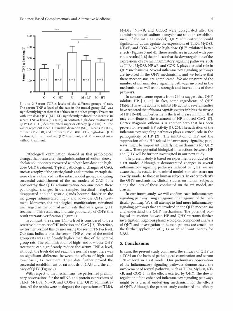
3.1.2. Reduction in the TNF- 𝛼 Level Following QHY Admin-istration. We found that the serum TNF-𝛼 level was signif-icantly enhanced in the CAG model rats (p < 0.001). Treat-ment with low- and high-dose QHY significantly reducedthe serum TNF-𝛼 level. High-dose QHY had a significantlyhigher efficacy than low-dose QHY (p < 0.05). Reductionin the serum inflammatory factors may be a comprehensive

4 Evidence-Based Complementary and Alternative Medicine
Control
Model
Intact HT
Intact LT HT
Figure 1: Representative images of the gastricmucosa in the different groups of rats. In the control group, the ratswhounderwent no treatment(Intact) and those who were administered high-dose QHY treatment (HT) exhibited normal gastric mucosa. Their glands were shapednormally, and there was no atrophy of the gastric glands or intestinal metaplasia. In the model group rats that were not given any treatment(Intact), there was obvious atrophy of the gastric glands and intestinal metaplasia (arrows). However, the group that underwent treatmentwith low-dose QHY (LT) and high-dose QHY (HT) showed improvement in the pathological manifestations. No intestinal metaplasia wasobserved, and the gastric glands were thicker than those of the model group (areas in the white frame). Black bar = 1,000 𝜇m.
result of the inhibition of several inflammatory signalingpathways (Figure 2).
Based on the pathological examinations and the serumTNF-𝛼 level, we confirmed the efficacy ofQHYas an adjuvanttherapy for CAG in a rat model.
3.2. Preliminary Observations of the Effects of QHY on SeveralInflammatory Signaling Pathways. In this study, we conduct-ed a preliminary observation of the mRNA expression andprotein expression of the TLR4, MyD88, NF-𝜅B, and COX-2signaling pathways.
Our results showed that the mRNA expression of theTLR4, MyD88, NF-𝜅B, and COX-2 signaling pathways wasupregulated in the rat models with CAG and can be signifi-cantly downregulated by treatment with high- and low-doseQHY. Furthermore, high-dose QHY exhibited a strongerefficacy for the TLR4, MyD88, and COX-2 pathways (Figures3(a), 3(b), and 3(d)).
The protein expression of the TLR4, MyD88, NF-𝜅B,and COX-2 signaling pathways was enhanced in the ratmodels with CAG. High- and low-dose QHY treatment candownregulate such enhancements in the protein expression
of signaling pathways associated with CAG. However, only inthe TLR4 and MyD88 pathways, high-dose QHY treatmentexhibited a strong efficacy for downregulation (Figures 4(a)and 4(b)).
These results indicate that the downregulation of theenhanced inflammatory signaling pathways play a crucial rolein QHYmechanisms.
4. Discussion
In this study, we confirmed the efficacy of QHYby examiningthe pathological slices and serum TNF-𝛼 level in CAGrat models. We also performed preliminary observationsto determine the effects of QHY on several inflammatorysignaling pathways. Our results suggest that QHY is anefficient treatment for CAG. Our data also indicate thatdownregulation of the enhanced inflammatory signalingpathways plays a crucial role in the QHY mechanisms thatneed further verification. To the best of our knowledge, this isthe first to investigate the efficacy of QHY in CAG treatment,and the results of this study will contribute to CAG treatmentwith alternative therapies, including medicines based onTCM.
Evidence-Based Complementary and Alternative Medicine 5Se
rum
TN
F-
Lev
el (n
g/L)
n = 6 n = 6 n = 6 n = 6 n = 6
∗
∗∗
∗∗∗
C C + HT M M + LT M + HT
500
400
300
0
100
200
Figure 2: Serum TNF-𝛼 levels of the different groups of rats.The serum TNF-𝛼 level of the rats in the model group (M) wassignificantly higher than that of those in the other groups. Treatmentwith low-dose QHY (M + LT) significantly reduced the increase inserum TNF-𝛼 levels (p < 0.05); in contrast, high-dose treatment ofQHY (M + HT) demonstrated superior efficacy (p < 0.01). All thevalues represent mean ± standard deviation (SD); ∗means P < 0.05,∗∗means P < 0.01, and ∗∗∗means P < 0.001. HT = high-dose QHYtreatment, LT = low-dose QHY treatment, and M = model micewithout treatment.
Pathological examination showed us that pathologicalchanges that occur after the administration of sodium deoxy-cholate solutionwere recoveredwith both low-dose and high-dose QHY treatment. Typical pathological changes of CAG,such as atrophy of the gastric glands and intestinalmetaplasia,were clearly observed in the intact model group, indicatingsuccessful establishment of the rat models of CAG. It isnoteworthy that QHY administration can ameliorate thesepathological changes. In our samples, intestinal metaplasiadisappeared and the gastric glands became thicker in therat groups administered high- and low-dose QHY treat-ment. Moreover, the pathological manifestations remainedunchanged in the control group rats that were given QHYtreatment. This result may indicate good safety of QHY; thisresult warrants verification (Figure 1).
In contrast, the serum TNF-𝛼 level is considered to be asensitive biomarker of HP infection and CAG [13].Therefore,we further verified this by measuring the serum TNF-𝛼 level.Our data indicate that the serum TNF-𝛼 level of the modelgroup rats was significantly higher than that of the controlgroup rats. The administration of high- and low-dose QHYtreatment can significantly reduce the serum TNF-𝛼 level,although the levels did not reach the normal range; there wasno significant difference between the effects of high- andlow-dose QHY treatment. These data further proved thesuccessful establishment of rat models of CAG and the effi-cacy of QHY (Figure 2).
With respect to the mechanisms, we performed prelimi-nary observations for the mRNA and protein expressions ofTLR4, MyD88, NF-𝜅B, and COX-2 after QHY administra-tion. All the results were analogous; the expressions of TLR4,
MyD88, NF-𝜅B, and COX-2 were upregulated after theadministration of sodium deoxycholate solution (establish-ment of the rat CAG model). QHY administration couldsignificantly downregulate the expressions of TLR4, MyD88,NF-𝜅B, and COX-2, while high-dose QHY exhibited bettereffects (Figures 3 and 4).These results are in accord with pre-vious results [7, 8] that indicate that the downregulation of theexpressions of several inflammatory signaling pathways, suchas TLR4, MyD88, NF-𝜅B, and COX-2, plays a crucial role inQHYmechanisms. Several inflammatory signaling pathwaysare involved in the QHY mechanisms, and we believe thatthese mechanisms are complicated. We are unaware of thenumber of inflammatory signaling pathways involved in themechanisms as well as the strength and interactions of thesepathways.
In contrast, some reports from China suggest that QHYinhibits HP [14, 15]. In fact, some ingredients of QHY(Table 1) have the ability to inhibit HP activity. Several studieshave reported that rhizoma coptidis extract inhibits the ureaseof HP [16–19]. Epiberberine is the lead urease inhibitor thatmay contribute to the treatment of HP-induced CAG [17].Cortex magnolia officinalis is another herb that has beenproven to have anti-HP activity [18, 20].The activation of theinflammatory signaling pathways plays a crucial role in thepathogenicity of HP [21]. The inhibition of HP and thesuppression of the HP-related inflammatory signaling path-ways might be important underlying mechanisms for QHYefficacy. These potential biological interactions between HPand QHY will be further investigated in our next study.
The present study is based on experiments conducted ona rat model. Although it demonstrated changes in severalinflammatory signaling pathways induced by QHY, we areaware that the results from animal models sometimes are notexactly similar to those in human subjects. In order to clarifythe QHY mechanisms, clinical studies on human subjects,along the lines of those conducted on the rat model, arecrucial.
In our future study, we will confirm each inflammatorysignaling pathway using an agonist or antagonist of that par-ticular pathway. We shall attempt to find more inflammatorysignaling pathways that are involved in the QHYmechanismand understand the QHY mechanisms. The potential bio-logical interaction between HP and QHY warrants furtherinvestigation. Rigorous pharmacological component analysisof QHY and investigation in human patients are crucial forthe further application of QHY as an adjuvant therapy forCAG.
5. Conclusions
In sum, the present study confirmed the efficacy of QHY asa TCM on the basis of pathological examination and serumTNF-𝛼 level in a rat model. Our preliminary observationof the inflammatory signaling pathways demonstrated theinvolvement of several pathways, such as TLR4, MyD88, NF-𝜅B, and COX-2, in the effects exerted by QHY. The down-regulation of the enhanced inflammatory signaling pathwaysmight be a crucial underlying mechanism for the effectsof QHY. Although the present study confirmed the efficacy

6 Evidence-Based Complementary and Alternative Medicine
Rela
tive m
RNA
expr
essi
on o
f TLR
4
n = 6 n = 6 n = 6 n = 6 n = 6
C C + HT M M + LT M + HT
∗
∗∗∗
∗∗∗∗∗∗
5
4
3
2
0
1
(a)
Rela
tive m
RNA
expr
essi
on o
f MyD
88
n = 6 n = 6 n = 6 n = 6 n = 6
C C + HT M M + LT M + HT
∗
∗∗∗
∗∗∗∗∗∗6
4
2
0
(b)
Rela
tive m
RNA
expr
essi
on o
f Nf-
B
n = 6 n = 6 n = 6 n = 6 n = 6
∗∗∗
∗∗∗∗∗∗
C C + HT M M + LT M + HT
8
6
4
0
2
(c)
Rela
tive m
RNA
expr
essi
on o
f CO
X-2
n = 6 n = 6 n = 6 n = 6 n = 6
∗∗
∗∗∗
∗∗∗∗∗∗
C C + HT M M + LT M + HT
4
3
2
0
1
(d)
Figure 3: mRNA expression of TLR4, MyD88, NF-𝜅B, and COX-2 in the different groups of rats. (a) The mRNA expression of the TLR4signaling pathway. The mRNA expression of TLR4 in the model group (M) was significantly higher than that of those in the other groups.Treatment with low-dose QHY (M + LT) significantly downregulated the enhancement of the mRNA expression of TLR4 (p < 0.001); incontrast, treatment with high-dose QHY (M + HT) achieved significantly more reduction (p < 0.001). The high-dose and low-dose exertedsignificantly different effects (p < 0.05). (b) The mRNA expression of the MyD88 signaling pathway. (c) The mRNA expression of the NF-𝜅Bsignaling pathway. (d) The mRNA expression of the COX-2 signaling pathway achieved results similar to those of TLR4 only in the NF-𝜅Bsignaling pathway; there was no significant difference between the high- and low-dose QHY treatments. All the values represent mean ±standard deviation (SD); ∗means P < 0.05, ∗∗means P < 0.01, and ∗∗∗means P < 0.001. HT = high-dose QHY treatment, LT = low-dose QHYtreatment, and M = model mice without treatment.

Evidence-Based Complementary and Alternative Medicine 7
TLR4 95 kD
MyD88 35 kD
NF-B 65 kD
COX-2 68 kD
-actin 41 kD
C C + HT M M + LT M + HT
(a)
Rela
tive P
rote
in ex
pres
sion
of T
LR4
n = 6 n = 6 n = 6 n = 6 n = 6
C C + HT M M + LT M + HT
∗∗∗∗∗∗
∗
∗
1.5
1.0
0.5
0.0
(b)
Rela
tive P
rote
in ex
pres
sion
of M
yD88
n = 6 n = 6 n = 6 n = 6 n = 6
C C + HT M M + LT M + HT
∗∗∗
∗∗∗∗∗∗
∗
1.5
1.0
0.5
0.0
(c)
n = 6 n = 6 n = 6 n = 6 n = 6
Rela
tive P
rote
in ex
pres
sion
of N
f-B
C C + HT M M + LT M + HT
∗∗∗
∗∗∗ ∗∗∗1.5
1.0
0.5
0.0
(d)
Rela
tive P
rote
in ex
pres
sion
of C
OX
-2
n = 6 n = 6 n = 6 n = 6 n = 6
C C + HT M M + LT M + HT
∗∗∗ ∗∗∗
∗∗∗1.0
0.8
0.6
0.4
0.2
0.0
(e)
Figure 4:Theprotein expression of TLR4,MyD88,NF-𝜅B, andCOX-2 in different groups. (a) Representative images ofwestern-blot analyses.(b) The protein expression of the TLR4 signaling pathway. The protein expression of TLR4 in the model group (M) was significantly higherthan those in the other groups. Treatment with low-dose QHY (M + LT) significantly downregulated the enhancement of the proteinexpression of TLR4 (p < 0.05); in contrast, treatment with high-dose QHY (M + HT) achieved significantly more downregulation (p <0.001).The difference between the effects of high-dose and-low dose QHYwas significant (p < 0.05). (c)The protein expression of theMyD88signaling pathway. The protein expression of MyD88 in the model group (M) was significantly higher than that of those in the other groups.Treatmentwith low-dose QHY (M+ LT) significantly reduced the enhancement ofMyD88 protein expression (p < 0.001); however, treatmentwith high-doseQHY (M+HT) achieved amore significant reduction (p< 0.001).The difference between the effect of high-dose and low-dosewas significant (p < 0.05). (d) The protein expression of the NF-𝜅B signaling pathway. The protein expression of NF-𝜅B in the model group(M) was significantly higher than that of those in the other groups. Treatment with low-dose QHY (M + LT) and high-dose QHY (M + HT)caused a significant downregulation in the enhancement of the protein expression of NF-𝜅B (p < 0.001).There was no significant difference inthe effects of high-dose and low-dose treatment. (e)The protein expression of the COX-2 signaling pathway.The protein expression of COX-2 in the model group (M) was significantly higher than those in the other groups. Treatment with low-dose QHY (M + LT) and high-doseQHY (M + HT) significantly downregulated the enhancement COX-2 protein expression (p < 0.001). No significant difference was observedbetween the effects of high-dose and low-dose treatment. All the values represent mean ± standard deviation (SD); 𝛽-actin was used as theinternal control. ∗means P< 0.05, ∗∗means P < 0.01, and ∗∗∗means P< 0.001. HT = high-dose QHY treatment, LT = low-dose QHY treatment,and M = model mice without treatment.

8 Evidence-Based Complementary and Alternative Medicine
of QHY as an adjuvant therapy against CAG, the mechanismsunderlying QHY require further exploration.
Data Availability
Thedata used to support the findings of this study are includ-ed within the article.
Conflicts of Interest
Theauthorsdeclare no conflicts of interest in the present study.
Authors’ Contributions
Sihan Li andMinghan Huang got the original ideas; Sihan Li,Minghan Huang, Qin Chen, Shunan Li, Xin Wang, JianlongLin, Guodong Zhong, Ping Lin, and Tetsuya Asakawa dis-cussed the experimental design; Sihan Li, Minghan Huang,Qin Chen, Shunan Li, XinWang, Jianlong Lin, and GuodongZhong performed the experiments and collected the data;Jianlong Lin and Guodong Zhong checked the pathologicalslices; Sihan Li, Minghan Huang, Qin Chen, Shunan Li, XinWang, Jianlong Lin, Ping Lin, and Tetsuya Asakawa ran thestatistics; SihanLi andTetsuyaAsakawawrote the draft; SihanLi, Minghan Huang, Qin Chen, Shunan Li, Xin Wang, Jian-long Lin, Guodong Zhong, Ping Lin, and Tetsuya Asakawadiscussed and approved the last version. Ping Lin and TetsuyaAsakawa supervised the study. Sihan Li and Minghan Huangare equal contributors.
Acknowledgments
This study was supported by grants from the Japanese Societyfor the Promotion of Science (Grant-in-Aid for Young Sci-entists, Type B, no. 20791025, and Grant-in-Aid for ScientificResearchC, General, nos. 24592157, 15k10358, and 18K08991).
References
[1] P. Dı́az, M. Valenzuela Valderrama, J. Bravo, and A. F. Quest,“Helicobacter pylori and Gastric Cancer: Adaptive CellularMechanisms Involved in Disease Progression,” Frontiers inMicrobiology, vol. 9, 2018.
[2] Z. Li, C. Wu, and L. Li, “Effect of long-term proton pump inhi-bitor administration on gastric mucosal atrophy: A meta-anal-ysis,” Saudi Journal of Gastroenterology, vol. 23, no. 4, pp. 222–228, 2017.
[3] D. Gan, A. Xu, H. Du, and Y. Ye, “Chinese Classical FormulaSijunzi Decoction and Chronic Atrophic Gastritis: Evidencefor Treatment Approach?” Evidence-Based Complementary andAlternativeMedicine, vol. 2017, Article ID 9012929, 9 pages, 2017.
[4] M. Huang, Q. Chen, Y. Gao, S. Li, P. Lin, and H. Huang,“Investigation of efficacy and mechanisms of Qinghuayin totreat chronic atrophic gastritis,” Lishizhen Medicine andMateriaMedica Research, vol. 10, pp. 2444–2446, 2015.
[5] T. Li, H. Li, L. Fei, and Y. Liu, “Treatment of Chronic atrophicgastritis by theory of clining the poison in TCM,” Journal ofSichuan of Traditional Chinese Medicine, vol. 22, no. 1, pp. 17-18,2004.
[6] H. Berndt, “Epidemiological Approaches to the Aetiology ofChronic Atrophic Gastritis,” Digestion, vol. 4, no. 4, pp. 250–254, 2004.
[7] S. Sue, W. Shibata, and S. Maeda, “Helicobacter pylori-InducedSignaling Pathways Contribute to Intestinal Metaplasia andGastric Carcinogenesis,” BioMed Research International, vol.2015, Article ID 737621, 9 pages, 2015.
[8] D. Q. Wang, X. P. Ding, S. Yin, and Y. D. Mao, “Role of the IL-11/STAT3 signaling pathway in human chronic atrophic gastritisand gastric cancer,”Genetics andMolecularResearch, vol. 15, no.2, Article ID gmr.15027358, 2016.
[9] S. Arita, Y. Kinoshita, K. Ushida, A. Enomoto, and K. Inagaki-Ohara, “High-fat diet feeding promotes stemness and precan-cerous changes in murine gastric mucosa mediated by leptinreceptor signaling pathway,” Archives of Biochemistry and Bio-physics, vol. 610, pp. 16–24, 2016.
[10] K. Inagaki-Ohara, S. Okamoto, K. Takagi et al., “Leptin receptorsignaling is required for high-fat diet-induced atrophic gastritisin mice,” Nutrition & Metabolism, vol. 13, no. 1, 2016.
[11] X. Yu, Q. Xu, Y. Xu, Y. Gong, and Y. Yuan, “Expression of theE-cadherin/𝛽-catenin/tcf-4 Pathway in Gastric Diseases withRelation to Helicobacter pylori Infection: Clinical and Patho-logical Implications,”Asian Pacific Journal of Cancer Prevention,vol. 15, no. 1, pp. 215–220, 2014.
[12] Z. Yang, C. Xie,W. Xu et al., “Phosphorylation and inactivationof PTEN at residues Ser380/Thr382/383 induced by Helicobac-ter pylori promotes gastric epithelial cell survival throughPI3K/Akt pathway,” Oncotarget , vol. 6, no. 31, pp. 31916–31926,2015.
[13] N. Hamajima, A. Shibata, N. Katsuda et al., “Subjects withTNF-A-857TT and -1031TT genotypes showed the highestHelicobacter pylori seropositive rate compared with those withother genotypes,” Gastric Cancer, vol. 6, no. 4, pp. 230–236,2003.
[14] Y. Wu, Efficacy of Qinghuayin combined with triple therapy forH.pylori, a clinical investivation.
[15] F. Tang and C. Yang, “Clinical observation of efficacy of Qing-huamieyou capsule to treat Helicobacter pylori,” Journal ofFujian University of Traditional Chinese Medicine, vol. 2, pp. 7-8,2000.
[16] C. Li, J. Xie, X. Chen et al., “Comparison of Helicobacter pyloriUrease Inhibition by Rhizoma Coptidis, Cortex Phellodendriand Berberine: Mechanisms of Interaction with the SulfhydrylGroup,” Planta Medica, vol. 82, no. 4, pp. 305–311, 2015.
[17] L. Tan, C. Li, H. Chen et al., “Epiberberine, a natural protober-berine alkaloid, inhibits urease of Helicobacter pylori and jackbean: Susceptibility andmechanism,” European Journal of Phar-maceutical Sciences, vol. 110, pp. 77–86, 2017.
[18] E.-A. Bae, M. J. Han, N.-J. Kim, and D.-H. Kim, “Anti-Helico-bacter pylori activity of herbalmedicines,”Biological & Pharma-ceutical Bulletin, vol. 21, no. 9, pp. 990–992, 1998.
[19] F. Ma, Y. Chen, J. Li et al., “Screening test for anti-Helicobacterpylori activity of traditional Chinese herbal medicines,” WorldJournal of Gastroenterology, vol. 16, no. 44, pp. 5629–5634, 2010.
[20] D.-H. Shi, Y.-W. Liu,W.-W. Liu, and Z.-F. Gu, “Inhibition of ure-ase by extracts derived from 15 Chinese medicinal herbs,” Phar-maceutical Biology, vol. 49, no. 7, pp. 752–755, 2011.
[21] C. T. Tran, M. Garcia, M. Garnier, C. Burucoa, and C. Bodet,“Inflammatory signaling pathways induced by Helicobacterpylori in primary human gastric epithelial cells,” Journal ofInnate Immunity, vol. 23, no. 2, pp. 165–174, 2017.

Stem Cells International
Hindawiwww.hindawi.com Volume 2018
Hindawiwww.hindawi.com Volume 2018
MEDIATORSINFLAMMATION
of
EndocrinologyInternational Journal of
Hindawiwww.hindawi.com Volume 2018
Hindawiwww.hindawi.com Volume 2018
Disease Markers
Hindawiwww.hindawi.com Volume 2018
BioMed Research International
OncologyJournal of
Hindawiwww.hindawi.com Volume 2013
Hindawiwww.hindawi.com Volume 2018
Oxidative Medicine and Cellular Longevity
Hindawiwww.hindawi.com Volume 2018
PPAR Research
Hindawi Publishing Corporation http://www.hindawi.com Volume 2013Hindawiwww.hindawi.com
The Scientific World Journal
Volume 2018
Immunology ResearchHindawiwww.hindawi.com Volume 2018
Journal of
ObesityJournal of
Hindawiwww.hindawi.com Volume 2018
Hindawiwww.hindawi.com Volume 2018
Computational and Mathematical Methods in Medicine
Hindawiwww.hindawi.com Volume 2018
Behavioural Neurology
OphthalmologyJournal of
Hindawiwww.hindawi.com Volume 2018
Diabetes ResearchJournal of
Hindawiwww.hindawi.com Volume 2018
Hindawiwww.hindawi.com Volume 2018
Research and TreatmentAIDS
Hindawiwww.hindawi.com Volume 2018
Gastroenterology Research and Practice
Hindawiwww.hindawi.com Volume 2018
Parkinson’s Disease
Evidence-Based Complementary andAlternative Medicine
Volume 2018Hindawiwww.hindawi.com
Submit your manuscripts atwww.hindawi.com