Embed Size (px)
Citation preview
Confocal Raman Microspectroscopy on Cornea
Jenkins He
University of Rochester Dr. Andrew J. Berger Biomedical Optics Lab
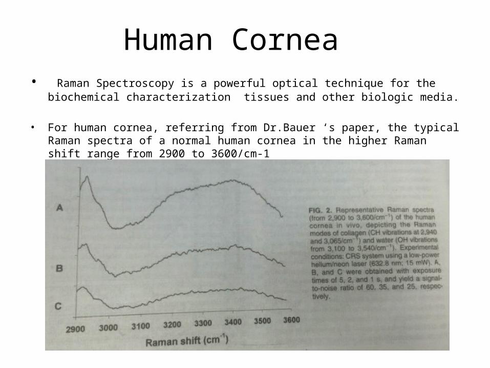
Human Cornea • Raman Spectroscopy is a powerful optical technique for the biochemical characterization
tissues and other biologic media.
• For human cornea, referring from Dr.Bauer ‘s paper, the typical Raman spectra of a normal human cornea in the higher Raman shift range from 2900 to 3600/cm-1
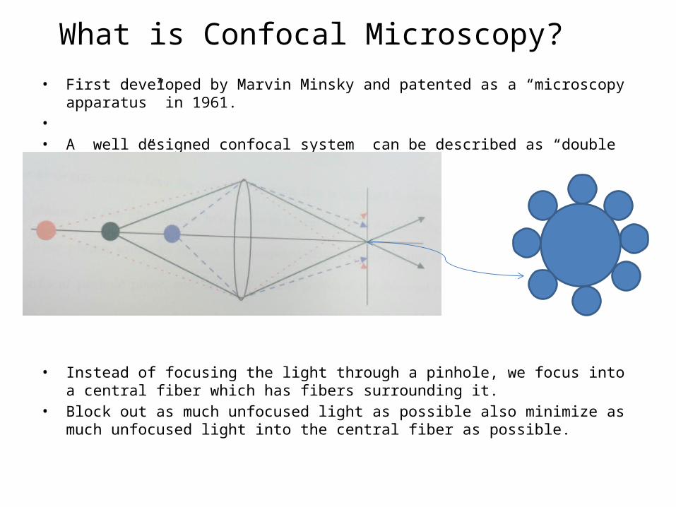
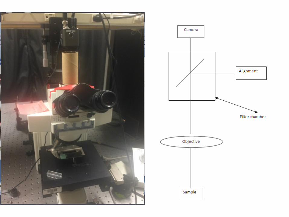
What is Confocal Microscopy?• First developed by Marvin Minsky and patented as a “microscopy apparatus” in 1961.• • A well designed confocal system can be described as “double focusing” system.
• Instead of focusing the light through a pinhole, we focus into a central fiber which has fibers surrounding it.
• Block out as much unfocused light as possible also minimize as much unfocused light into the central fiber as possible.

Our basic alignment
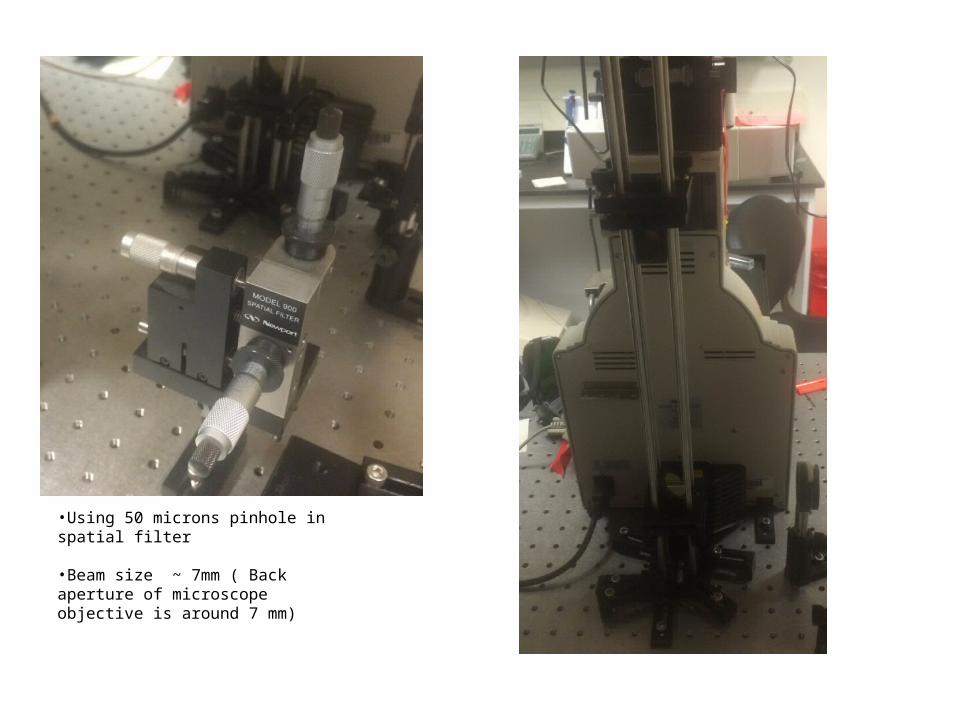
•Using 50 microns pinhole in spatial filter
•Beam size ~ 7mm ( Back aperture of microscope objective is around 7 mm)
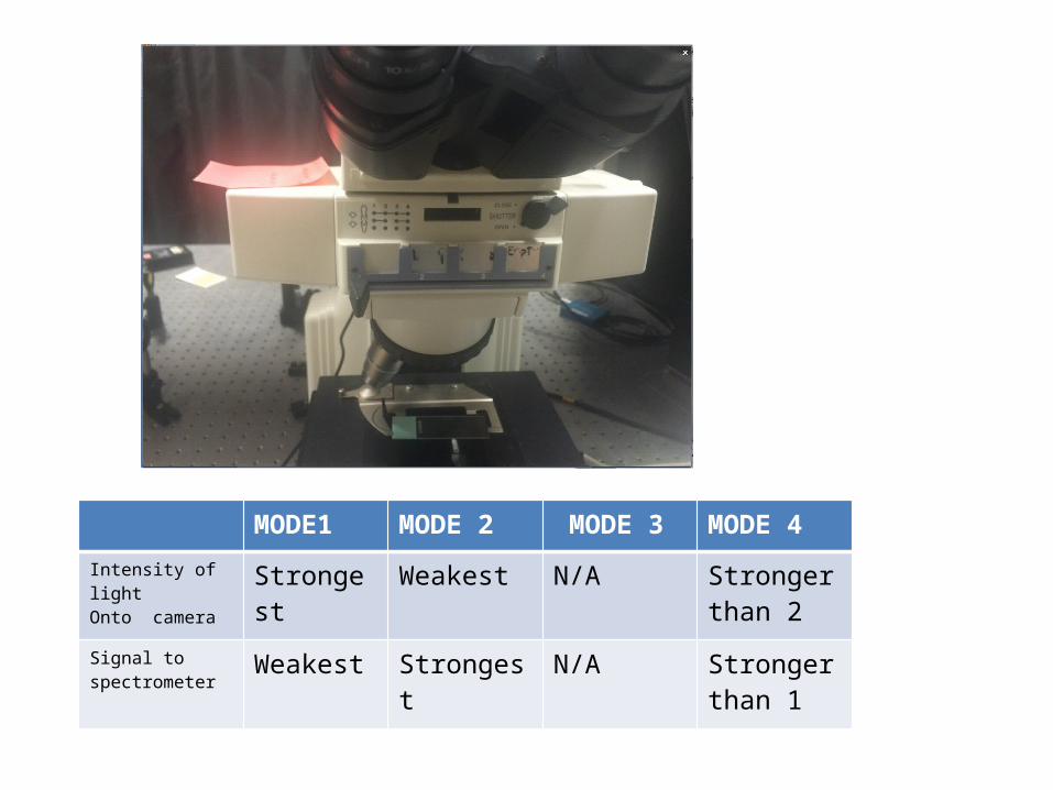
MODE1 MODE 2 MODE 3 MODE 4
Intensity of light Onto camera Strongest Weakest N/A Stronger
than 2
Signal to spectrometer Weakest Strongest N/A Stronger
than 1
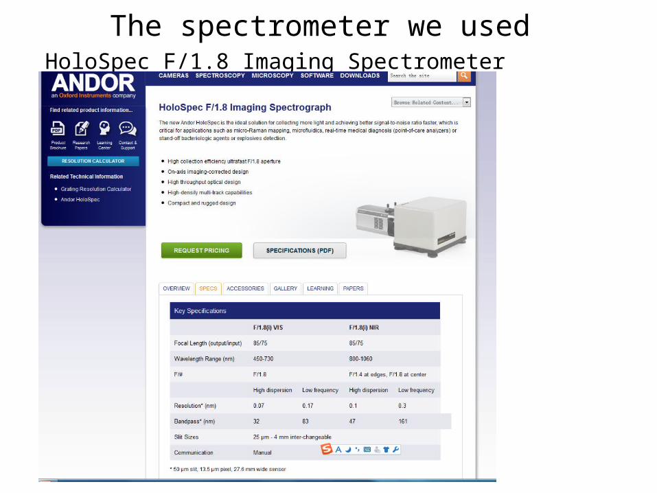
The spectrometer we used HoloSpec F/1.8 Imaging Spectrometer
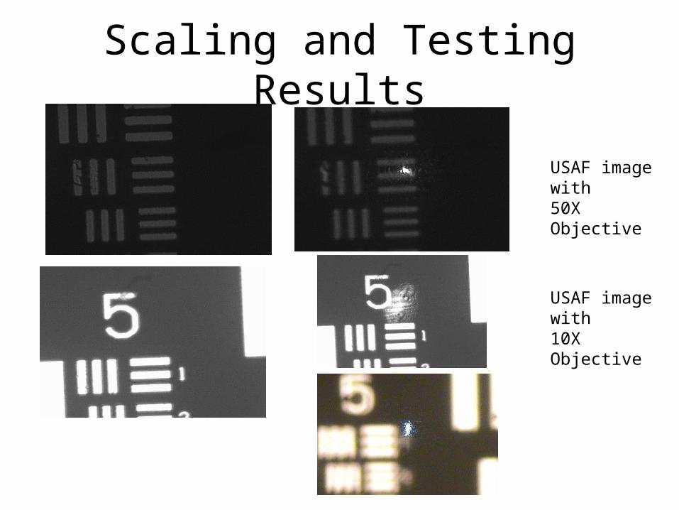
Scaling and Testing Results
USAF image with50X Objective
USAF image with10X Objective

Group Number /Element Number
Line Pairs/ mm Line Width (micros)
Estimated Laser Spot Size (microns)
50X 7/2 143.7 ~3.5 ~ 2
10X 5/1 32 ~15 ~ 10
Scaling and Testing Results
•Tube Length = 200mm with 50X objective gives us the focal length of objective is 4 mm
•The lens focusing beam into fiber has focal length of 200 mm
•So the confocal spot size is magnified by 50X to ~ 100 microns
•Center core fiber has a size of 100 microns

Scaling and Testing Results
Image of fibers for microscope glass slide with 50s exposure time with 50X objective
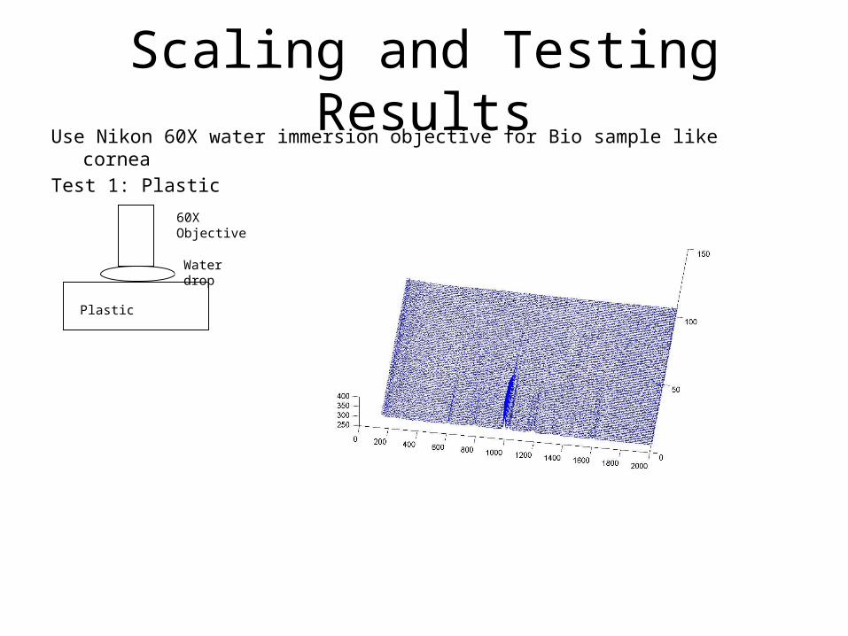
Use Nikon 60X water immersion objective for Bio sample like cornea Test 1: Plastic
Scaling and Testing Results
Plastic
Water drop
60X Objective

Test1: Plastic
FWHM ~ 20 microns
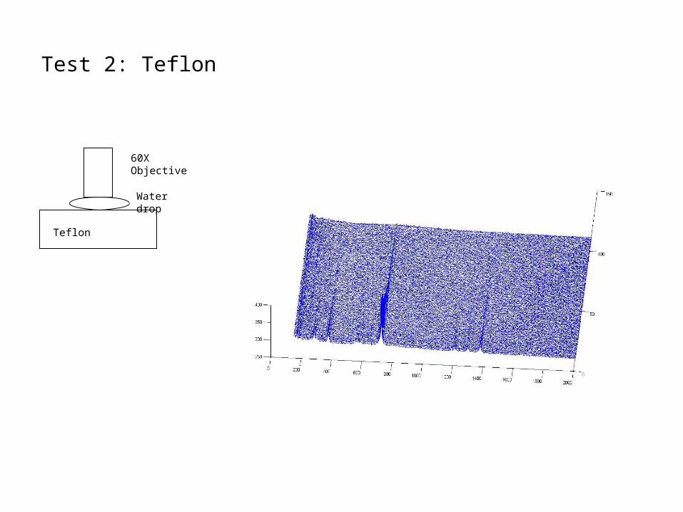
Test 2: Teflon
Teflon
Water drop
60X Objective
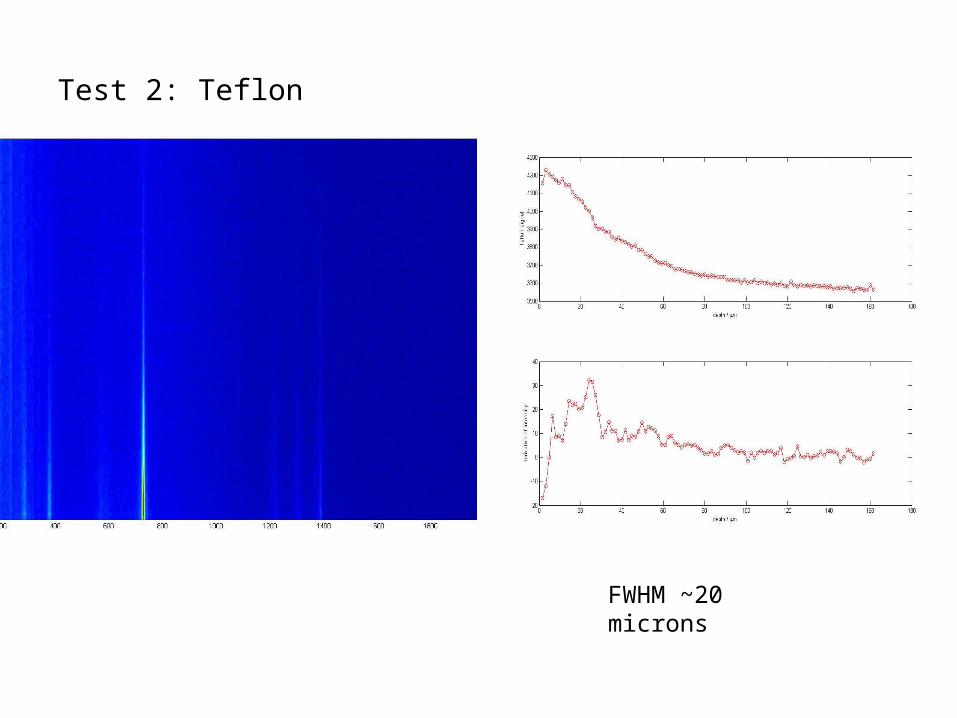
Test 2: Teflon
FWHM ~20 microns
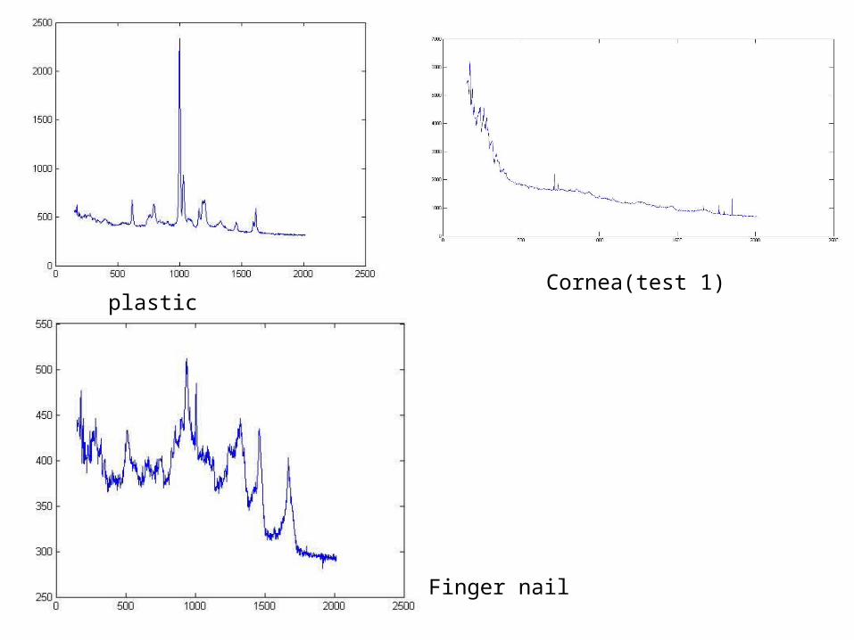
plastic
Finger nail
Cornea(test 1)

Berger’s Rugby League

Thank you for coming!


![In Vivo Confocal Microscopy of the Human Cornea in the ...€¦ · neuropathy, Fabry disease, and HIV associated peripheral neuropathy (Table1) [7–13]. IVCM has also been used to](https://img.pdfslide.net/doc/110x75/60643a427629cd5b4a55fd31/in-vivo-confocal-microscopy-of-the-human-cornea-in-the-neuropathy-fabry-disease.jpg)