Embed Size (px)
Citation preview
306 J . Med. Chem. 1988, 31, 306-312
Conformational Analysis and Structural Comparisons of (1R,3S)-( + )- and (lS,aR)-(-)-Tefludazine, ( S ) - ( +)- and (R)-(-)-Octoclothepin, and (+)-Dexclamol in Relation to Dopamine Receptor Antagonism and Amine-Uptake Inhibition
Tommy Liljefors*t and Klaus P. Bagesai
Department of Organic Chemistry 3, Chemical Center, University of Lund, S-221 00 Lund, Sweden, and Department of Medicinal Chemistry, H. Lundbeck AIS, Ottiliavej 7-9, DK-2500 Copenhagen- Valby, Denmark. Received December 31, 1986
Conformational analysis with molecular mechanics (MM2(85)) and molecular superimposition studies of (lR,3S)-(+)- and (1S,3R)-(-)-4-[3-(4-fluorophenyl)-6-(trifluoromethyl)indan-l-yl]-l-piperazineethanol (tefludazine) and (S)-(+)- and (R)-(-)-octoclothepin have been employed to identify biologically active conformations of these compounds with respect to dopamine receptor antagonism and amine-uptake inhibition. In contrast to what is commonly assumed, these studies indicate that the conformation of (S)-(+I-octoclothepin responsible for the dopamine receptor antagonism is different from the one observed in the crystal. From least-squares molecular superimpositions with the potent and stereoselective dopamine receptor antagonist (lR,3S)-tefludazine, biologically active conformations for the two compounds on the dopamine receptor have been deduced. This analysis also rationalizes the enantioselectivity of octoclothepin on the dopamine receptor. The X-ray structure of (S)-(+)-octoclothepin is shown to correspond structurally to the 1S,3R enantiomer of tefludazine, which is an amine-uptake inhibitor. This correspondence provides a structural basis for the norepinephrine (NE) uptake blocking properties of octoclothepin. It is predicted that the enantioselectivity of the NE-uptake inhibition of octoclothepin should be low with the S-(+) enantiomer as the more active optical isomer. A comparison of the deduced biologically active conformation of (S)-(+)-octoclothepin with (+)-dexclamol is also discussed on the basis of earlier derived superimposition studies with (+)-dexclamol.
4- [3-(4-Fluorophenyl)-6-(trifluoromethyl)indan-l-yl]-l- piperazine ethanol (tefludazine (1)) is a representative of a new class of potent and stereoselective dopamine (DA) receptor antagonists (neuroleptics), the l-piperazino-3- phenylindans.lI2 It has been shown that the neuroleptic activity of 1 exclusively resides in the isomer with a trans c~nfiguration.~,~ Furthermore, it has been found that the neuroleptic properties of 1 are solely confined to the 1R,3S e n a n t i ~ m e r . ~ - ~ The 1S,3R enantiomer shows no appre- ciable DA receptor antagonism, but is instead a moderately strong DA and norepinephrine (NE) uptake i n h i b i t ~ r . ~ Thus, the racemate of 1 contains two active compounds with affinities for different receptor sites.
F 3 C - p OF 9&UF x.3
$ no 5 c.3 HO
lR,3S-l 1S,3R-1
2 3
In an attempt to relate the molecular structures of neuroleptically active thiepins and l-piperazino-3- phenylindans, one of us previously compared the 1S,3R enantiomer of 1 with the structures of (S)-(+)-oxy- prothepin' and (S)-(+)-octoclothepin [ (S)-2],8v9 as observed in the crystalline state, and concluded that a good mo- lecular fit was possib1e.l At that time the absolute con- figuration of the enantiomer of 1, which is active on the DA receptor, was not known. With our present knowledge that the active configuration is 1R,3S, i.e opposite to that used in the molecular superimpositions with the active thiepins described above, this attempt to rationalize the
+University of Lund. * H. Lundbeck AIS.
0022-262318811831-0306$01.50/0
structural basis for DA receptor antagonism of 1 is of course invalid. Howeyer, the active 1R,3S enantiomer shows a very poor correspondence to the structure of (S)-2 as observed in the crystalline state. Since the potent and stereselective compound (S)-2 has often been used as a reference compound for the structural requirements for dopamine receptor antagoni~m,"'~ this structural incom- patibility raises questions about the 3D relationships be- tween compounds l and 2 and, more general, about the geometrical basis for neuroleptic activity. Carnmalm et al., in an attempt to relate the structures of neuroleptically active tetracyclic spiro amines to the X-ray structure of (S)-2, arrived at a similar conclusion. They concluded that the structures of the spiro amines and that of (S)-2 seem to be of opposite conformational types.ll
In addition to its neuroleptic activity, the racemate of 2 is also a potent inhibitor of NE uptakea4 Considering the dual pharmacological activities of both 1 and 2, we recently suggested that the two enantiomers 1R,3S and
(1) Bagesa, K. P. J. Med. Chem. 1983, 26, 935. (2) Svendsen, 0.; Arnt, J.; Boeck, V.; Bagesa, K. P.; Christensen,
A. V.; Hyttel, J.; Larsen, J.-J. Drug. Dev. Res. 1986, 7, 35. (3) Jensen, B. Acta Crystallogr., Sect. c Cryst. Struct. Commun.
1983, C39, 1055. (4) Bagesa, K. P.; Hyttel, J.; Christensen, A. V.; Arnt, J.; Liljefors,
T. In Innovative Approaches in Drug Research; Harms, A. F., Ed.; Pharmacochemistry Library, volume 9; Nauta, W. Th., Rekker, R. F., Eds.; Elsevier: Amsterdam, 1986; p 371.
(5) Jensen, B., unpublished results. (6) Jensen, H. P., unpublished results. (7) Koch, M. H. J.; Eurard, G. Acta Crystallogr., Sect. B: Struct.
CFyStalkgF. Cryst. Chem. 1974, B30, 2929. ( 8 ) Petcher, T. J.; Schmutz, J.; Weber, H. P.; White, T. G. Ez-
perentia 1975, 31, 1390. (9) Jaunin, A,; Petcher, T. J.; Weber, H. P. J. Chem. Soc., Perkin
Trans. 2 1977, 186. (10) Humber. L. G.: Sideridis. N.: Asselin. A. A.: Bruderlein. F. T. ~I
J . Med. Chem: 1978, 21, 1225. (11) Carnmalm, B.; Johansson, L.; Ramsby, S.; Wagner, A. Acta
Pharm. Suec. 1979, 16, 239. (12) Marshall, G. R.; Barry, C. D.; Bosshard, H. E.; Dammkoehler,
R. A.; Dunn, D. A. In Computer Assisted Drug Design; Olson, E. C., Christoffersen, R. E., Eds.; ACS Symposium Series 112; American Chemical Society: Washington, DC, 1979.
(13) Froimowitz, M.; Matthysse, S. Mol. Pharmacol. 1983,24, 243. (14) Tollenaere, J. P.; Moereels, H.; Raymaekers, L. A. In Drug
Design; Ariens, E. J., Ed.; Academic: New York, 1980; Vol. X, p 72.
0 1988 American Chemical Society
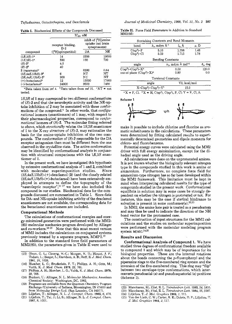
Tefludazines, Octoclothepins, and Dexclamols Journal of Medicinal Chemistry, 1988, Vol. 31, No. 2 307
Table I. Biochemical Effects of the Compounds Discussed ICs09 nM
compound (1R,3S)-lU (1S,3R)-la
(R)-2‘ (S1-26
2 (racemate)U (3S,4aS,13bS)-3b (3R,4aR, 13 bR)-3b (+)-butaclamol’
receptor binding,
( [3H] spiroperidol) D-2
14 590 4.5 40
6 900 12
inhib of [3H]amine uptake (rat brain
synaptosoms) DA NE
2200 15000 130 730
6300 0.64 NT NT NT NT 13000 17000
(-)-butaclamoP 34000 9500 7400 ’Data taken from ref 4. *Data taken from ref 31. “T = not
tested.
1S,3R of 1 may correspond to two different conformations of (S)-2 and that the neuroleptic activity and the NE-up- take inhibition of 2 may be associated with these confor- mations of the c ~ m p o u n d . ~ In other words, that configu- rational isomers (enantiomers) of 1 may, with respect to their pharmacological properties, correspond to confor- mational isomers of (S)-2. The molecular fitting referred to above, which structurally relates the 1S,3R enantiomer of 1 to the X-ray structure of (S) -2 , may rationalize the basis for the amine-uptake inhibition of the two com- pounds. The conformation of (S)-2 responsible for the DA receptor antagonism then must be different from the one observed in the crystalline state. The active conformation may be identified by conformational analysis in conjunc- tion with structural comparisons with the 1R,3S enan- tiomer of 1.
In the present work, we have investigated this hypothesis by extensive conformational analysis of 1 and 2, combined with molecular superimposition studies. Since (3S,4aS,13bS)-(+)-dexclamol (3) (and the closely related (3S,4aS,13bS)-(+)-butaclamol) have been extensively em- ployed in attempts to define the topography of the “neuroleptic recept~r” ,~ l -~’ we have also included this compound in our studies. Biochemical data for the com- pounds discussed are summarized in Table I. Since data for DA- and NE-uptake inhibiting activity of the dexclamol enantiomers are not available, the corresponding data for the butaclamol enantiomers are included. Computational Methods
The calculations of conformational energies and ener- gy-minimized geometries were performed with the MM2- (85) molecular mechanics program developed by Allinger and co-workers.1&21 Note that this most recent version of MM2 includes the calculations on conjugated systems previously treated by a separate program, MMP2.1s
In addition to the standard force field parameters of MM2(85), the parameters given in Table I1 were used to
(15) Olson, G. L.: Chuenn. H.-C.: Morgan, K. D.: Blount. J. F.: Todaro, L.; Berger, L.;Davidson, A.B.; Boff, E. J. Med. Chem. 1981, 24, 1026. Humber, L. G.; Bruderlein, F. T.; Philipp, A. H.; Gotz, M.; Voith, K. J. Med. Chem. 1979, 22, 761. Philipp, A. H.; Humber, L. G.; Voith, K. J. Med. Chem. 1979, 22, 768. Burkert, U.; Allinger, N. L. Molecular Mechanics; American Chemical Society: Washington, DC, 1982. Programs are available from the Quantum Chemistry Program Exchange (University of Indiana, Bloomington, IN 47405) and from Molecular Design Ltd. (San Leandro, CA 94577). Liljefors, T.; Allinger, N. L. J . Comput. Chem. 1985, 6 , 478. Liljefors, T.; Tai, J.; Li, S.; Allinger, N. L. J. Comput. Chem. 1987, 8, 1051.
Table 11. Force Field Parameters in Addition to Standard MM2(85)
Stretching Constants and Bond Moments bond k., mdyn 8,-’ lo, 8, c1, D
C(sp2)-F 5.10 1.356 1.48 C(sp2)-Cl 3.23 1.715 1.78
Bending Constants angle kb, mdyn A rad+ 00, deg
c (spZ)-C (spZ)-XO 0.50 120.0 out-of-plane (C(SP~)-X)~ 0.80 0.0
Torsional Constants angle V2, kcal/mol
_____
X-C(sp2)-C(sp2)-Yb 15.0 “ X = F, C1. b X = H, C(sp3), C(sp2), F, C1; Y = F, C1, S.
Scheme I a 3 e e +- e y+=-- e a a
make it possible to include chlorine and fluorine as aro- matic substituents in the calculations. These parameters were determined by fitting calculated results to experi- mentally determined geometries and dipole moments for chloro- and fluorobenzene.
Potential energy curves were calculated using the MM2 driver with full energy minimization, except for the di- hedral angle used as the driving angle.
All calculations were done on the unprotonated amines. It is not known whether the biologically relevant nitrogen type in the compounds studied in this work is amine or ammonium. Furthermore, no complete force field for ammonium-type nitrogen has so far been developed within the MM2 framework. This limitation must be kept in mind when interpreting calculated results for the type of compounds studied in the present work. Conformational equilibria in solution may in some cases be strongly de- pendent on whether the nitrogen is protonated or not. For instance, this may be the case if sterical hindrance to solvation is present in some conformer(^).^^^^^
In MM2, the amine lone pair is treated as a pseudoatom and may thus be used to indicate the direction of the NH bond vector for the protonated case.
The construction of input structures for the MM2 cal- culations and the studies on molecular superimposition were performed with the molecular modeling program system Results and Discussion
Conformational Analysis of Compound 1. We have studied three degrees of conformational freedom available to compound 1 and which may be of importance for its biological properties. These are the internal rotations about the bonds connecting the p-fluorophenyl and the piperazine rings to the five-membered ring system and the inversion of the five-membered ring. This ring may “flip” between two envelope-type conformations, which inter- converts pseudoaxial (a) and pseudoequatorial (e) positions (Scheme I).
(22) Manoharan, M.; Eliel, E. L. Tetrahedron Lett. 1983,24, 1855. (23) Manoharan, M.; Eliel, E. L. Tetrahedron Lett. 1984,25,3267. (24) Liljefors, T. J. Mol. Graphics 1983, I , 111. (25) Von der Lieth, C. W.; Carter, R. E.; Dolata, D. P.; Liljefors, T.
J . Mol. Graphics 1984, 2, 117.
308 Journal of Medicinal Chemistry, 1988, Vol. 31, No. 2 Liljefors and B~gegeso
A € kca 1 / m o 1
6
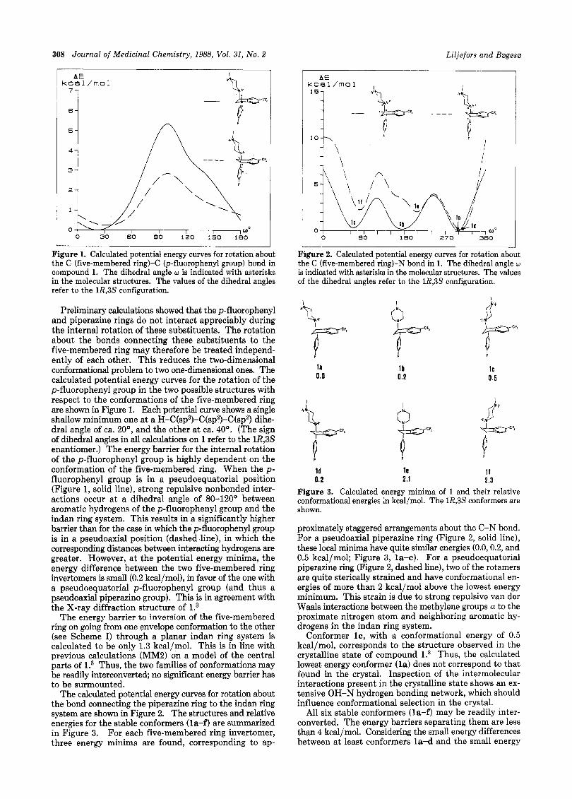
Figure 1. Calculated potential energy curves for rotation about the C (five-membered ring)-(= (p-fluorophenyl group) bond in compound 1. The dihedral angle w is indicated with asterisks in the molecular structures. The values of the dihedral angles refer to the 1R,3S configuration.
Preliminary calculations showed that the p-fluorophenyl and piperazine rings do not interact appreciably during the internal rotation of these substituents. The rotation about the bonds connecting these substituents to the five-membered ring may therefore be treated independ- ently of each other. This reduces the two-dimensional conformational problem to two one-dimensional ones. The calculated potential energy curves for the rotation of the p-fluorophenyl group in the two possible structures with respect to the conformations of the five-membered ring are shown in Figure 1. Each potential curve shows a single shallow minimum one at a H-C(sp3)-C(sp2)-C(sp2) dihe- dral angle of ca. 20°, and the other at ca. 40'. (The sign of dihedral angles in all calculations on 1 refer to the lR,3S enantiomer.) The energy barrier for the internal rotation of the p-fluorophenyl group is highly dependent on the conformation of the five-membered ring. When the p- fluorophenyl group is in a pseudoequatorial position (Figure 1, solid line), strong repulsive nonbonded inter- actions occur a t a dihedral angle of 80-120' between aromatic hydrogens of the p-fluorophenyl group and the indan ring system. This results in a significantly higher barrier than for the case in which the p-fluorophenyl group is in a pseudoaxial position (dashed line), in which the corresponding distances between interacting hydrogens are greater. However, a t the potential energy minima, the energy difference between the two five-membered ring invertomers is small (0.2 kcal/mol), in favor of the one with a pseudoequatorial p-fluorophenyl group (and thus a pseudoaxial piperazino group). This is in agreement with the X-ray diffraction structure of l.3
The energy barrier t o inversion of the five-membered ring on going from one envelope conformation to the other (see Scheme I) through a planar indan ring system is calculated to be only 1.3 kcal/mol. This is in line with previous calculations (MM2) on a model of the central parts of l.3 Thus, the two families of conformations may be readily interconverted; no significant energy barrier has to be surmounted.
The calculated potential energy curves for rotation about the bond connecting the piperazine ring to the indan ring system are shown in Figure 2. The structures and relative energies for the stable conformers (la-f) are summarized in Figure 3. For each five-membered ring invertomer, three energy minima are found, corresponding to ap-
AE kcal/mol I 151 I
5\ b l a
I d IC l b 0
0 90 1 E O 270 360
Figure 2. Calculated potential energy curves for rotation about the C (five-membered ring)-N bond in 1. The dihedral angle w is indicated with asterisks in the molecular structures. The values of the dihedral angles refer to the 1R,3S configuration.
l a 0.0
I *n
I F
l b 0 .2
P
IC 0.5
I fi"'
I d l e I f 0.2 2.1 2.3
Figure 3. Calculated energy minima of 1 and their relative conformational energies in kcal/mol. The 1R,3S conformers are shown.
proximately staggered arrangements about the C-N bond. For a pseudoaxial piperazine ring (Figure 2, solid line), these local minima have quite similar energies (0.0,0.2, and 0.5 kcal/mol; Figure 3, la-c). For a pseudoequatorial piperazine ring (Figure 2, dashed line), two of the rotamers are quite sterically strained and have conformational en- ergies of more than 2 kcal/mol above the lowest energy minimum. This strain is due to strong repulsive van der Waals interactions between the methylene groups a to the proximate nitrogen atom and neighboring aromatic hy- drogens in the indan ring system.
Conformer IC, with a conformational energy of 0.5 kcal/mol, corresponds to the structure observed in the crystalline state of compound l.3 Thus, the calculated lowest energy conformer (la) does not correspond to that found in the crystal. Inspection of the intermolecular interactions present in the crystalline state shows an ex- tensive OH-N hydrogen bonding network, which should influence conformational selection in the crystal.
All six stable conformers (la-f) may be readily inter- converted. The energy barriers separating them are less than 4 kcal/mol. Considering the small energy differences between at least conformers la-d and the small energy
Tefludazines, Octoclothepins, and Dexclarnols Journal of Medicinal Chemistry, 1988, Vol. 31, No. 2 309
I 15-1
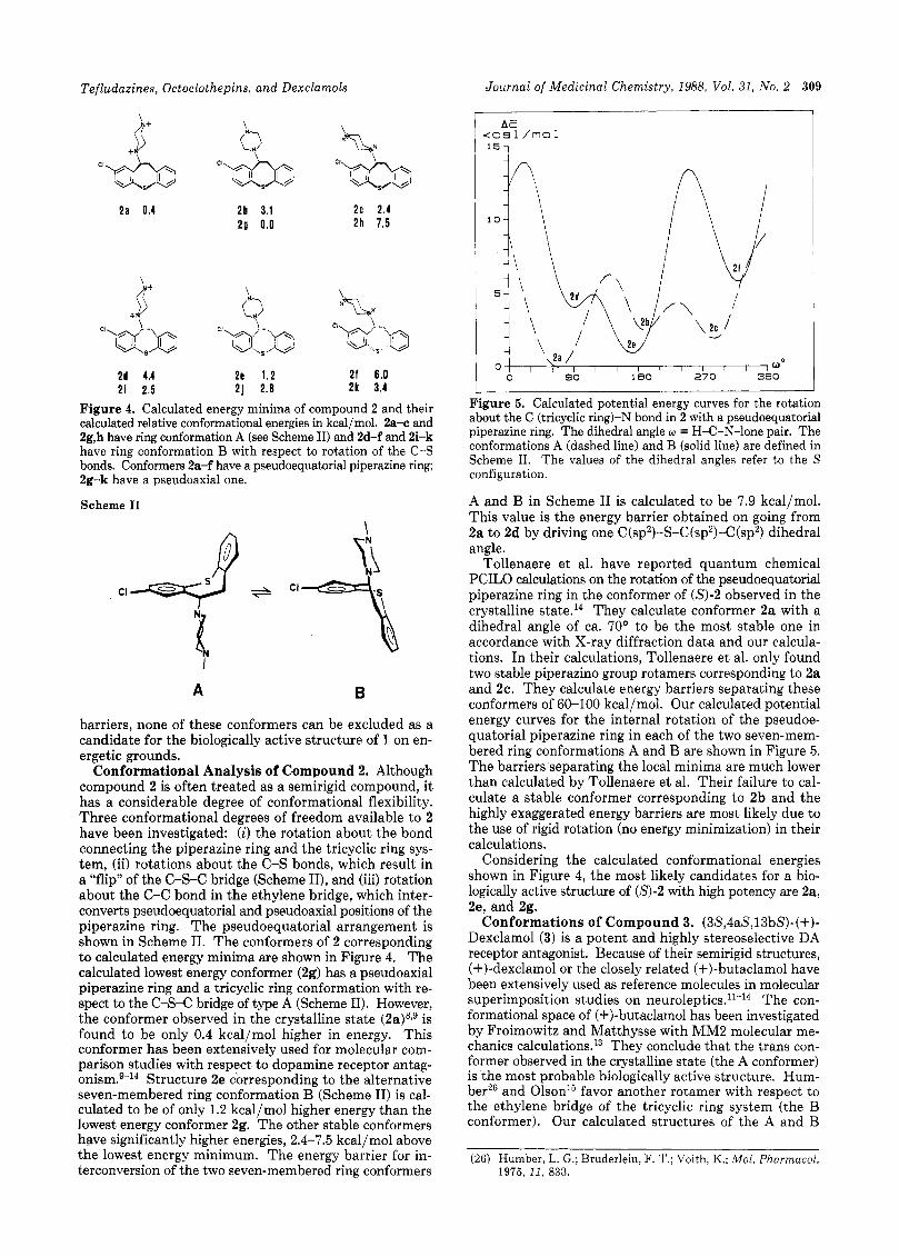
2 a 0.4 2b 3.1 2c 2.4 2 9 0.0 2h 7.5
2d 4.4 2e 1 .2 2 f 6 .0 2 i 2.5 2 j 2 .8 2 k 3.4
Figure 4. Calculated energy minima of compound 2 and their calculated relative conformational energies in kcal/mol. 2a-c and 2g,h have ring conformation A (see Scheme 11) and 2d-f and 2i-k have ring conformation B with respect to rotation of the C-S bonds. Conformers 2a-f have a pseudoequatorial piperazine ring; 2g-k have a pseudoaxial one.
Scheme I1
A B
barriers, none of these conformers can be excluded as a candidate for the biologically active structure of 1 on en- ergetic grounds.
Conformational Analysis of Compound 2. Although compound 2 is often treated as a semirigid compound, it has a considerable degree of conformational flexibility. Three conformational degrees of freedom available to 2 have been investigated: (i) the rotation about the bond connecting the piperazine ring and the tricyclic ring sys- tem, (ii) rotations about the C-S bonds, which result in a "flip" of the C-S-C bridge (Scheme 11), and (iii) rotation about the C-C bond in the ethylene bridge, which inter- converts pseudoequatorial and pseudoaxial positions of the piperazine ring. The pseudoequatorial arrangement is shown in Scheme 11. The conformers of 2 corresponding to calculated energy minima are shown in Figure 4. The calculated lowest energy conformer (2g) has a pseudoaxial piperazine ring and a tricyclic ring conformation with re- spect to the C-s-C bridge of type A (Scheme 11). However, the conformer observed in the crystalline state (2a)s,g is found to be only 0.4 kcal/mol higher in energy. This conformer has been extensively used for molecular com- parison studies with respect to dopamine receptor antag- o n i ~ m . ~ - ~ ~ Structure 2e corresponding to the alternative seven-membered ring conformation B (Scheme 11) is cal- culated to be of only 1.2 kcal/mol higher energy than the lowest energy conformer 2g. The other stable conformers have significantly higher energies, 2.4-7.5 kcal/mol above the lowest energy minimum. The energy barrier for in- terconversion of the two seven-membered ring conformers
1
Figure 5. Calculated potential energy curves for the rotation about the C (tricyclic ring)-N bond in 2 with a pseudoequatorial piperazine ring. The dihedral angle w = H-C-N-lone pair. The conformations A (dashed line) and B (solid line) are defined in Scheme 11. The values of the dihedral angles refer to the S configuration.
A and B in Scheme I1 is calculated to be 7.9 kcal/mol. This value is the energy barrier obtained on going from 2a to 2d by driving one C(sp2)-S-C(sp2)-C(sp2) dihedral angle.
Tollenaere et al. have reported quantum chemical PCILO calculations on the rotation of the pseudoequatorial piperazine ring in the conformer of (S)-2 observed in the crystalline state.14 They calculate conformer 2a with a dihedral angle of ca. 70° to be the most stable one in accordance with X-ray diffraction data and our calcula- tions. In their calculations, Tollenaere et al. only found two stable piperazino group rotmers corresponding to 2a and 2c. They calculate energy barriers separating these conformers of 60-100 kcal/mol. Our calculated potential energy curves for the internal rotation of the pseudoe- quatorial piperazine ring in each of the two seven-mem- bered ring conformations A and B are shown in Figure 5. The barriers separating the local minima are much lower than calculated by Tollenaere et al. Their failure to cal- culate a stable conformer corresponding to 2b and the highly exaggerated energy barriers are most likely due to the use of rigid rotation (no energy minimization) in their calculations.
Considering the calculated conformational energies shown in Figure 4, the most likely candidates for a bio- logically active structure of (S)-2 with high potency are 2a, 2e, and 2g.
Conformations of Compound 3. (3S,4aS,13bS)-(+)- Dexclamol (3) is a potent and highly stereoselective DA receptor antagonist. Because of their semirigid structures, (+)-dexclamol or the closely related (+)-butaclamol have been extensively used as reference molecules in molecular superimposition studies on n e u r o l e p t i c ~ . ~ l - ~ ~ The con- formational space of (+)-butaclamol has been investigated by Froimowitz and Matthysse with MM2 molecular me- chanics calculation^.^^ They conclude that the trans con- former observed in the crystalline state (the A conformer) is the most probable biologically active structure. Hum- ber26 and Olson'5 favor another rotamer with respect to the ethylene bridge of the tricyclic ring system (the B conformer). Our calculated structures of the A and B
(26) Humber, L. G.; Bruderlein, F. T.; Voith, K.; Mol. Pharmacal. 1975, 11, 833.
310 Journal of Medicinal Chemistry, 1988, Vol. 31, No. 2
a
Liljefors and B ~ g e s ~
a
P O b
b Figure 6. Calculated structures of compound 3 showing the A conformer (a) and the B conformer (b).
conformers of (+)-dexclamol are shown in Figure 6. The A conformer is calculated to be of 3.1 kcal/mol lower en- ergy than the B conformer, which is similar to what was calculated by Froimowitz and Matthysse (2.7 kcal/mol).
Structural Comparisons of Compounds ( lR,3S)- l and (5)-2. The lowest energy structure in each family of conformers with respect to the conformation of the five- membered ring of (1R,3S)-1 is shown in Figure 7. In Figure 8 is shown one representative of each of the two ring conformations A and B (Scheme 11) of compound (S)-2. The conformer shown in Figure 8a (2a) corresponds to the one observed in the crystalline state. The structures are oriented so that the “neuroleptic substituents” (CF, and C1) are pointing in the same direction. It has been dem- onstrated that the trifluoromethyl group in (lR,3S)-l oc- cupies a typical “neuroleptic position” similar to the chloro substituent in (S)-2. Variations of the substituents give parallel pharmacological effects in the two classes of DA receptor antagonists.l>* The piperazine ring nitrogens, which are relevant for DA receptor antagonism in (1R,3S)-1 and (S)-2, are indicated by an arrow in Figures 7 and 8. This assignment is based on the observed inactivity of the trans racemate of 4 and the activity of the racemate of 5.4
4 5
If we compare the calculated structures of (1R,3S)-1 (Figure 7) and (S)-2 (Figure 8), it is obvious that the conformation of the tricyclic ring system observed in the crystalline state of (S)-2 (Figure 8a) is not compatible with the structures of (1R,3S)-1 shown in Figure 7a,b. The X-ray structure of (S)-2 clearly has a “wrong” conformation with respect to the chiral sense of the tricyclic ring system. This is also true for the calculated lowest energy structure of 2 (2g, Figure 4). This conformer has the same tricyclic
F
b
Figure 7. Calculated structures of conformers (1R,3S)-la (a) and (lR,aS)-ld (b). The arrow indicates the relevant nitrogen atom for DA receptor antagonism.
a
b
Figure 8. Calculated structures of conformers (S)-2a (a) and (S)-2e (b). The arrow indicates the relevant nitrogen atom for DA receptor antagonism.
ring conformation as the X-ray structure. Note that while the chiral sense of the aromatic rings in (S)-2 depends on the conformation and may thus be changed by a confor-
Tefludazines, Octoclothepins, and Dexclamols
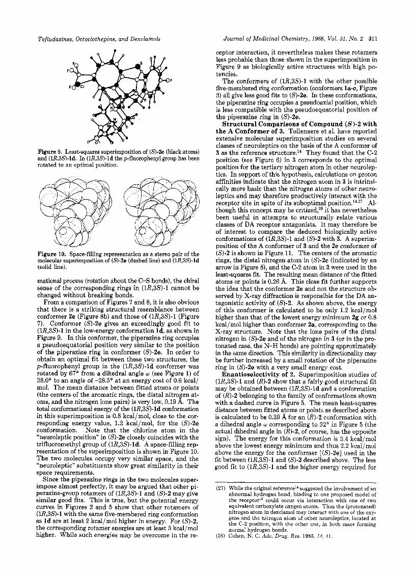
Figure 9. Least-squares superimposition of (S)-2e (black atoms) and (lR,SS)-ld. In (lR,SS)-ld the p-fluorophenyl group has been rotated to an optimal position.
... _.
Figure 10. Space-filling representation as a stereo pair of the molecular superimposition of (S)-2e (dashed line) and (lR,SS)-ld (solid line).
mational process (rotation about the C-S bonds), the chiral sense of the corresponding rings in (1R,3S)-1 cannot be changed without breaking bonds.
From a comparison of Figures 7 and 8, it is also obvious that there is a striking structural resemblance between conformer 2e (Figure 8b) and those of (1R,3S)-1 (Figure 7). Conformer (S)-2e gives an exceedingly good fit to (lR,3S)-l in the low-energy conformation Id, as shown in Figure 9. In this conformer, the piperazine ring occupies a pseudoequatorial position very similar to the position of the piperazine ring in conformer (S)-2e. In order to obtain an optimal fit between these two structures, the p-fluorophenyl group in the (lR,SS)-ld conformer was rotated by 67’ from a dihedral angle w (see Figure 1) of 38.0’ to an angle of -28.5’ a t an energy cost of 0.6 kcal/ mol. The mean distance between fitted atoms or points (the centers of the aromatic rings, the distal nitrogen at- oms, and the nitrogen lone pairs) is very low, 0.19 A. The total conformational energy of the (1R,3S)-ld conformation in this superimposition is 0.8 kcal/mol, close to the cor- responding energy value, 1.2 kcal/mol, for the (S)-2e conformation. Note that the chlorine atom in the “neuroleptic position” in (S)-2e closely coincides with the trifluoromethyl group of (1R,3S)-ld. A space-filling rep- resentation of the superimposition is shown in Figure 10. The two molecules occupy very similar space, and the “neuroleptic” substituents show great similarity in their space requirements.
Since the piperazine rings in the two molecules super- impose almost perfectly, it may be argued that other pi- perazine-group rotamers of (1R,3S)-1 and (S)-2 may give similar good fits. This is true, but the potential energy curves in Figures 2 and 5 show that other rotamers of (lR,3S)-l with the same five-membered ring conformation as Id are a t least 2 kcal/mol higher in energy. For (S)-2, the corresponding rotamer energies are at least 3 kcal/mol higher. While such energies may be overcome in the re-
Journal of Medicinal Chemistry, 1988, Vol. 31, No. 2 311
ceptor interaction, it nevertheless makes these rotamers less probable than those shown in the superimposition in Figure 9 as biologically active structures with high po- tencies.
The conformers of (1R,3S)-1 with the other possible five-membered ring conformation (conformers la-c, Figure 3) all give less good fits to (S)-2e. In these conformations, the piperazine ring occupies a pseudoaxial position, which is less compatible with the pseudoequatorial position of the piperazine ring in (S)-2e.
Structural Comparisons of Compound (S)-2 with the A Conformer of 3. Tollenaere et al. have reported extensive molecular superimposition studies on several classes of neuroleptics on the basis of the A conformer of 3 as the reference structure.14 They found that the C-2 position (see Figure 6) in 3 corresponds to the optimal position for the tertiary nitrogen atom in other neurolep- tics. In support of this hypothesis, calculations on proton affinities indicate that the nitrogen atom in 3 is intrinsi- cally more basic than the nitrogen atoms of other neuro- leptics and may therefore productively interact with the receptor site in spite of its suboptimal p ~ s i t i o n . ~ ~ , ~ ~ Al- though this concept may be critized,2s it has nevertheless been useful in attempts to structurally relate various classes of DA receptor antagonists. I t may therefore be of interest to compare the deduced biologically active conformations of (1R,3S)-1 and (S)-2 with 3. A superim- position of the A conformer of 3 and the 2e conformer of (S)-2 is shown in Figure 11. The centers of the aromatic rings, the distal nitrogen atom in (S)-2e (indicated by an arrow in Figure 8), and the C-2 atom in 3 were used in the least-squares fit. The resulting mean distance of the fitted atoms or points is 0.26 A. This close fit further supports the idea that the conformer 2e and not the structure ob- served by X-ray diffraction is responsible for the DA an- tagonistic activity of (S)-2. As shown above, the energy of this conformer is calculated to be only 1.2 kcal/mol higher than that of the lowest energy minimum 2g or 0.8 kcal/mol higher than conformer 2a, corresponding to the X-ray structure. Note that the lone pairs of the distal nitrogen in (S)-2e and of the nitrogen in 3 (or in the pro- tonated case, the N-H bonds) are pointing approximately in the same direction. This similarity in directionality may be further increased by a small rotation of the piperazine ring in (S)-2e with a very small energy cost.
Enantioselectivity of 2. Superimposition studies of (1R,3S)-1 and (R)-2 show that a fairly good structural fit may be obtained between (1R,3S)-ld and a conformation of (R)-2 belonging to the family of conformations shown with a dashed curve in Figure 5. The mean least-squares distance between fitted atoms or points as described above is calculated to be 0.39 A for an (R)-2 conformation with a dihedral angle w corresponding to 32O in Figure 5 (the actual dihedral angle in (R)-2, of course, has the opposite sign). The energy for this conformation is 3.4 kcal/mol above the lowest energy minimum and thus 2.2 kcal/mol above the energy for the conformer [(S)-2e] used in the fit between (lR,3S)-l and ( 9 - 2 described above. The less good fit to (1R,3S)-1 and the higher energy required for
(27) While the original referenceI4 suggested the involvement of an abnormal hydrogen bond, binding to one proposed model of the receptor15 could occur via interaction with one of two equivalent carboxylate oxygen atoms. Thus the (protonated) nitrogen atom in dexclamol may interact with one of the oxy- gens and the nitrogen atom of other neuroleptics, located a t the C-2 position, with the other one, in both cases forming normal hydrogen bonds.
(28) Cohen, N. C. Adu. Drug. Res. 1985, 14, 41.
312 Journal of Medicinal Chemistry, 1988, Vol. 31, No. 2 Liljefors and Bergeser
?
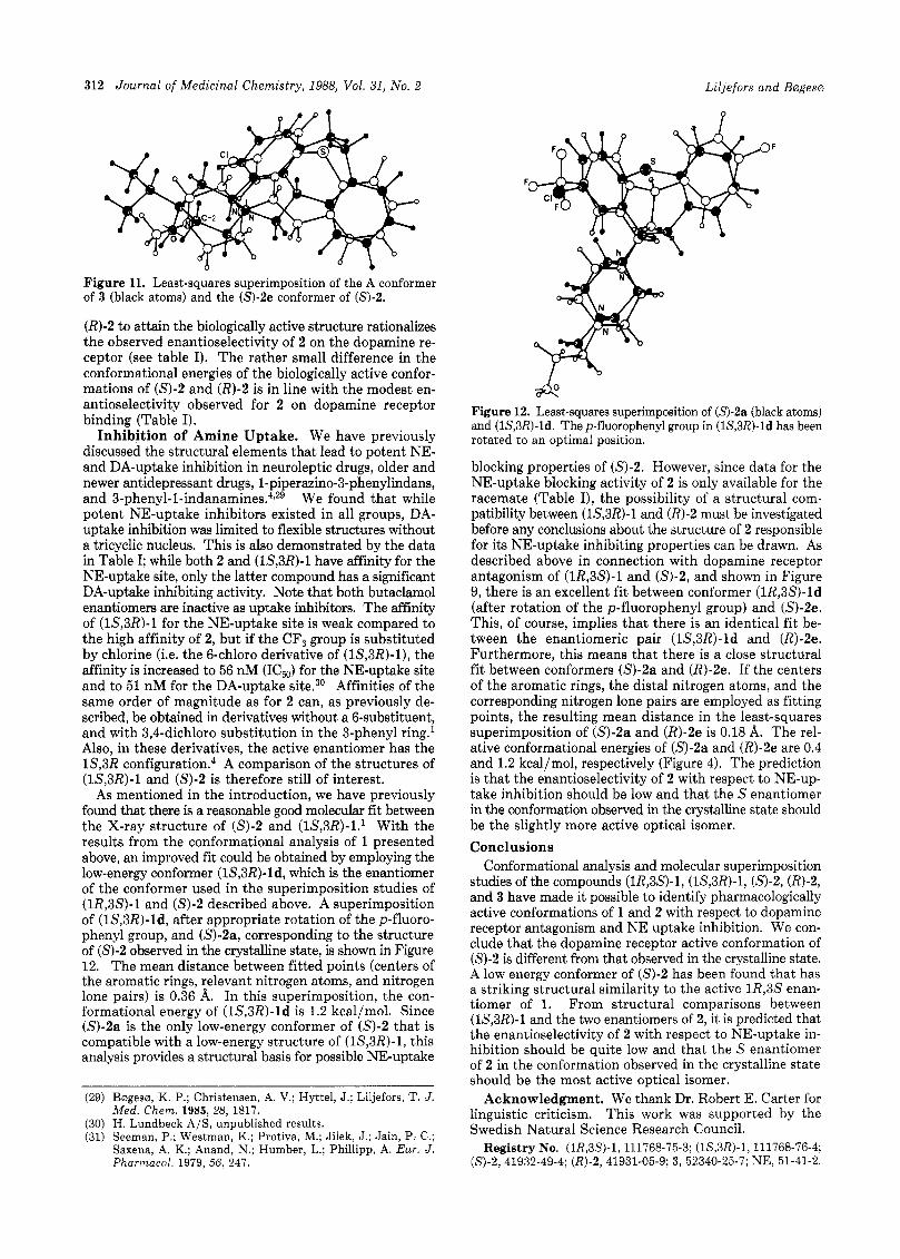
Figure 11. Least-squares superimposition of the A conformer of 3 (black atoms) and the (S)-2e conformer of (S)-2.
(R)-2 to attain the biologically active structure rationalizes the observed enantioselectivity of 2 on the dopamine re- ceptor (see table I). The rather small difference in the conformational energies of the biologically active confor- mations of (S)-2 and (R)-2 is in line with the modest en- antioselectivity observed for 2 on dopamine receptor binding (Table I).
Inhibition of Amine Uptake. We have previously discussed the structural elements that lead to potent NE- and DA-uptake inhibition in neuroleptic drugs, older and newer antidepressant drugs, l-piperazino-3-phenylindans, and 3-phenyl-l-indanamine~.~,~~ We found that while potent NE-uptake inhibitors existed in all groups, DA- uptake inhibition was limited to flexible structures without a tricyclic nucleus. This is also demonstrated by the data in Table I; while both 2 and (lS,3R)-l have affinity for the NE-uptake site, only the latter compound has a significant DA-uptake inhibiting activity. Note that both butaclamol enantiomers are inactive as uptake inhibitors. The affinity of (1S,3R)-l for the NE-uptake site is weak compared to the high affinity of 2, but if the CF, group is substituted by chlorine (Le. the 6-chloro derivative of (lS,3R)-l), the affinity is increased to 56 nM (IC,) for the NE-uptake site and to 51 nM for the DA-uptake site.30 Affinities of the same order of magnitude as for 2 can, as previously de- scribed, be obtained in derivatives without a 6-substituent, and with 3,4-dichloro substitution in the 3-phenyl ring.l Also, in these derivatives, the active enantiomer has the 1S,3R c~nfigurat ion.~ A comparison of the structures of (1S,3R)-1 and (S)-2 is therefore still of interest.
As mentioned in the introduction, we have previously found that there is a reasonable good molecular fit between the X-ray structure of (S)-2 and (lS,3R)-l.l With the results from the conformational analysis of 1 presented above, an improved fit could be obtained by employing the low-energy conformer (lS,3R)-ld, which is the enantiomer of the conformer used in the superimposition studies of (1R,3S)-1 and (S)-2 described above. A superimposition of (lS,3R)-ld, after appropriate rotation of the p-fluoro- phenyl group, and (S)-2a, corresponding to the structure of (S)-2 observed in the crystalline state, is shown in Figure 12. The mean distance between fitted points (centers of the aromatic rings, relevant nitrogen atoms, and nitrogen lone pairs) is 0.36 A. In this superimposition, the con- formational energy of (lS,SR)-ld is 1.2 kcal/mol. Since (S)-2a is the only low-energy conformer of (S)-2 that is compatible with a low-energy structure of (lS,3R)-l, this analysis provides a structural basis for possible NE-uptake
(29) Bergesm, K. P.; Christensen, A. V.; Hyttel, J.; Liljefors, T. J. Med. Chem. 1985, 28, 1817.
(30) H. Lundbeck A/S, unpublished results. (31) Seeman, P.; Westman, K.; Protiva, M.; Jilek, J.; Jain, P. C.;
Saxena, A. K.; Anand, N.; Humber, L.; Phillipp, A. Eur. J . Pharmacol. 1979, 56, 247.
F
F
Figure 12. Least-squares superimposition of (S)-2a (black atoms) and (1S,3R)-ld. The p-fluorophenyl group in (lS,3R)-ld has been rotated to an optimal position.
blocking properties of (S)-2. However, since data for the NE-uptake blocking activity of 2 is only available for the racemate (Table I), the possibility of a structural com- patibility between (1S,3R)-1 and (R)-2 must be investigated before any conclusions about the structure of 2 responsible for its NE-uptake inhibiting properties can be drawn. As described above in connection with dopamine receptor antagonism of (1R,3S)-1 and (S)-2, and shown in Figure 9, there is an excellent fit between conformer (1R,3S)-ld (after rotation of the p-fluorophenyl group) and (S)-2e. This, of course, implies that there is an identical fit be- tween the enantiomeric pair (1S,3R)-ld and (R)-2e. Furthermore, this means that there is a close structural fit between conformers (S)-2a and (R)-2e. If the centers of the aromatic rings, the distal nitrogen atoms, and the corresponding nitrogen lone pairs are employed as fitting points, the resulting mean distance in the least-squares superimposition of (S)-2a and (R)-2e is 0.18 A. The rel- ative conformational energies of (S)-2a and (R)-2e are 0.4 and 1.2 kcal/mol, respectively (Figure 4). The prediction is that the enantioselectivity of 2 with respect to NE-up- take inhibition should be low and that the S enantiomer in the conformation observed in the crystalline state should be the slightly more active optical isomer. Conclusions
Conformational analysis and molecular superimposition studies of the compounds (1R,3S)-1, (1S,3R)-1, (57-2, (R)-2, and 3 have made it possible to identify pharmacologically active conformations of 1 and 2 with respect to dopamine receptor antagonism and NE uptake inhibition. We con- clude that the dopamine receptor active conformation of (S)-2 is different from that observed in the crystalline state. A low energy conformer of (S)-2 has been found that has a striking structural similarity to the active 1R,3S enan- tiomer of 1. From structural comparisons between (1S,3R)-1 and the two enantiomers of 2, it is predicted that the enantioselectivity of 2 with respect to NE-uptake in- hibition should be quite low and that the S enantiomer of 2 in the conformation observed in the crystalline state should be the most active optical isomer.
Acknowledgment. We thank Dr. Robert E. Carter for linguistic criticism. This work was supported by the Swedish Natural Science Research Council.
Registry No. (lR,3S)-l, 111768-75-3; (1S,3R)-1, 111768-76-4; (5)-2,41932-49-4; (R)-2 , 41931-05-9; 3, 52340-25-7; NE, 51-41-2.


![KD-3AS 型] KD-3S 型] KD-3S](https://img.pdfslide.net/doc/110x75/629d5929e245e3147b536a41/kd-3as-kd-3s-kd-3s.jpg)