Embed Size (px)
Citation preview
Biophysical Chemistry, 33 (1989) 39-45 Elsevier
39
BPC 01339
Conformational preferences of sequential fragments of the hinge region of human IgAl immunoglobulin molecule: II *
James Burton a, Stephen G. Wood at Artur Pedyczak b and Ignacy Z. Siemion b a University Hospital, Boston Universi(v, Boston, MA OZILB, U.S.A.
and b Institute of Chemistry, Wrvcknv lJniversi& SO-383 Wrocfaw, Poland
Received 5 September 1988 Accepted 15 November 1988
Immunoglobuiin A,; Hinge region oligopcptide; CD; NMR, 13C-; Enzyme inhibitor
The mean s@ution conformation of tetrapeptide fragments of the hinge region of human IgAl molecule was investigated by CD and “C-t@k methods. Distinct conformational differences for the partial sequences were found. Tetrapeptides with the Tbr-Pro-Ser-Pro sequence were found to show a clear preference For the B-turn conformation. Conformational equilibria of these peptides are only slightly affected by acetylation or pH changes. In the case of Pro-Thr-Pro-Ser tetrapeptides conformational equilibria are dominated
by unordered forms.
1. Introduction
The hinge region of the human IgAl im- munoglobulin molecule contains the sequence
(1, 2):
. ..Pro-Val-Pro-Ser-.~;-Pro-Pro-Thr-Pro-Ser-
Pro-Ser-Thr-Pro-Pro-Thr... 235
which is sensitive to the action of extracellular proteases produced by several human bacterial pathogens. The sequence of the first IgAl pro- tease, isolated from the Gram-negative diplococ- cus Neisseria gonorrhoeae was determined by Pohlner et al. [3].
Continuing our studies [4] on the mean solution conformation of sequential fragments of the hinge
Correspondence address: J. Burton, University Hospital, Bos- ton University, Boston, MA 02118, U.S.A. * Part 1 of Uris work appeared in Biophys. Chem. 31 (1988) 35.
region of human IgAl, we investigated the six following tetrapeptides:
Thr-Pro-Ser-Pro-NH, <I) Ac-Thr-Pro-Ser-Pro (11) Ac-Thr-Pro-Scr-Pro-NH* (III)
Pro-Thr-Pro-Ser-NH, (IV) Ac-Pro-Thr-Pro-Ser (V) Ac-Pro-Thr-Pro-Ser-NH, (VI)
The mean solution conformation of each com- pound was determined by CD and i3C-NMR measurements. The data obtained during our in- vestigation of model tetrapeptides (Ala),Pro and (Ala),Thr [5-71 were used in the interpretation of 13C-NMR spectra. Chemical shifts of the Ca carbon atoms of specific residues (recalculated onto the hydantoin scale [S]) were employed to define, the main conformational characteristics. Clear evidence of conformational differences be- tween peptides I-III and IV-VI was obtained. Peptides I-III show a preference for a p-turn
0301~4622/89/$03.50 8 1989 Elsevier Science Publishers B.V. (Biomedical Division)
40 J. Burton et al. /Conformations of IgA I hinge region fragments
structure, while IV-VI are most probably in an unordered state.
2. Materials and methods
Details of the syntheses of investigated peptides, will be published separately. Each compound was purified by HPLC. Purity and homogeneity of the products were * proved by TLC in three different solvent systems.
CD measurements were carried out using a Jobin-Yvon mark III dichrograph, at room tem- perature with light paths of 1, 2, 5 and 10 mm.
For each compound CD spectra were recorded at acidic (pH - 2), neutral (pH - 7) and alkaline (pH - 10) pH values. The concentration of solutions varied from 0.0358 to 0.1090 g/dm3. Results of the CD measurements are expressed as the molar ellipticity (in degree cm2 dmol-‘).
r3C-NMR spectra were recorded in 2H,0 on a Tesla 567 spectrometer with an operating frequency of 25.142 MHz. Dioxane was used as an internal standard. The chemical shift of dioxane carbon atoms was assumed to be 67.4 ppm on the TMS scale. For each compound measurements were carried out at acidic (pH 2.1-2.6) neutral (pH 6.7-7.4) and alkaline (pH 10.1-10.9) pH val- ues. The concentration of solutions varied from 13.0 to 38.0 g/dm3. The pH of solutions was adjusted with 4 N ,NaOH or 4 N ‘HCl and measured with a Mera-Elwro N-517 pH-meter. Absolute values of 8 angles (angle in the moiety C*-CD-C’-0) were calculated from the equation
[51: 18 1 = 49.7 -I- 19.46,
The coefficients, A,, were determined assuming the following Ca chemical shifts for the corre- sponding hydantoins: CLO, 26.65 ppm; CL, 65.13 ppm; C&, 60.19 ppm.
3. Results and d&w&on
The results of CD measurements are given in table 1. Selected curves which illustrate the ob- served regularities are shown in figs. 1 and 2. The
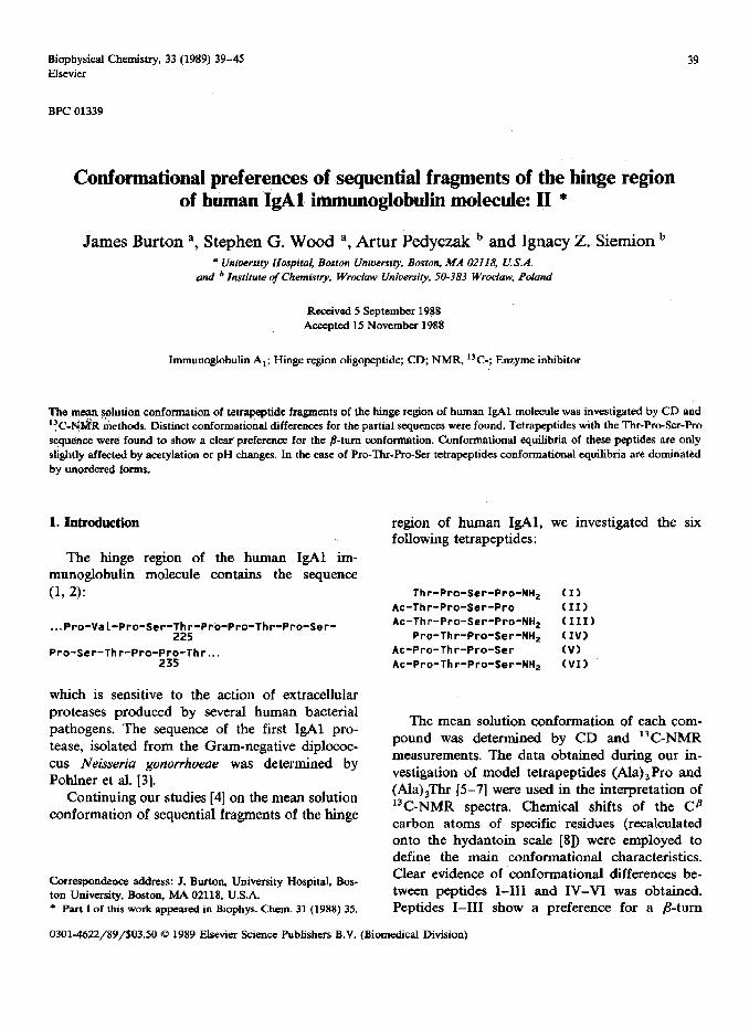
CD spectra of peptides I-III (fig. 1) have the same shape. They exhibit a single minimum at approx. 210 nm, resembling spectra of the P-turn conformation [9,10]. pH changes do not influence the position of the minimum of the CD spectra of peptides I-III (which is the same as the free tetrapeptide Thr-Pro-Ser-Pro [4]). There are slight changes in molar ellipticity with an increase in pH in the spectra of peptides I and III. For peptide II, the influence of pH on ellipticities is significant (-0.68 X lo4 degree cm2 dmol-’ in acidic and -2.35 X lo4 degree cm2 dmol-’ in alkaline medium). The same tendency was observed for the free tetrapeptide Thr-Pro-Ser-Pro [4]. These facts suggest that ellipticity changes are connected with the ionization of the carboxyl group.
In our earlier study [4] we observed that acety- lation of the N-terminal amino group influenced the mean solution conformation of the tetra- peptide Pro-Pro-Thr-Pro. Acetylation destabilized the p-turn structure and shifted the conforma- tional equilibrium toward unordered forms. The phenomenon was confirmed by CD and l3 C-NMR data [4]. In the case of the Tbr-Pro-Ser-Pro tetra- peptide, investigated here, acetylation of the terminal amino group does not produce a similar destabilizing effect. This suggests that the acetyl derivatives (peptides II and III) remain largely in the /Gturn conformation. This is probably due to the amino acid sequence of the peptide, which is highly correlated with B-turn formation, the pref- erence of a Pro residue at position i + 1 for a type I p-turn conformation being well-known. The /%
s I I I I 210 230 250
A (nm)
Fig. 1. CD spectra of tetrapeptides I-III in water at pH 7.
(- ) Thr-Pro-Ser-Pro-NH,; (- . - - - -) AC-Thf-Pm-Ser- Pro; (.-a- 0) Ac-Thr-Pro-k-Pro-NH,.
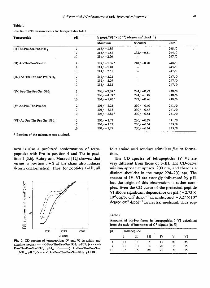
Table 1
.I. Burton et aI./ Conjormations of IgA I hinge region fragments 41
Results of CD measurements for tetrapeptides I-III
Tetrapeptide PH X (nm)/[@) ( x10m4) (degree cm2 dmol-‘)
Minimum Shoulder Zero
(I) Thr-Pro-Ser-Pro-NH* 2 213/ - 1.85 I 212/-1.83
10 211/-2.70
(II) Ac-Thr-Pro-&r-Pro
(III) Ac-Thr-Pro-Ser-Pro-NH,
2 7
10
2
I 10
199/-1.36 ’ 214/-1.48 214/-2X
211/ - 2.22 212/ - 2.29 213/ - 2.52
(IV) Pro-Thr-Pro-Ser-NH1 2 I
10
200/ - 2.09 a 200/-4.39 ’
204/-1.90 s
(V) Ac-Pro-Thr-Pro-Ser 2 7
10
201/ - 2.24
201/-3.18 205/ - 2.86 a
(VI) Ac-Pro-Thr-ProSer-NH1 2 7
10
203/ - 2.13 203/ - 3.21
204/ - 2.27
_ 232/ - 0.41
-
224/ - 0.72 224/ - 1.48
222/ - 0.86
230/ - 0.40
230/ - 0.48 230/ - 0.54
226/ - 0.67 230/ - 0.64 230,’ - 0.64
245/o 244/o 247/O
240/o 243/O 247/o
247;O 247/O 247/O
248/O 240/o 248/O
241/O 241/O 241/O
241/O 243/O 243/O
a Position of the minimum not attained.
turn is also a preferred conformation of tetra- peptides with Pro in position 4 and Thr in posi- tion 1 [5,6]. Aubry and Maraud [12] showed that serine in position i + 2 of the chain also induces p-turn conformation. Thus, for peptides I-III, all
I I I
210 230 250
A (nm)
Pig. 2. CD spectra of tetrapeptides IV and VI in acidic and aIkalinemedia.(--~-~)Pro-TlwPro-Ser-NH,,pH2;(------) ProThr-Pro&r-NH,, pH,,-,; ( -) Ac-Pro-Thr-Pro-Ser-
NH,, PH 2; ( -. . . . a) Ac-Pro-‘Ihr-Pro-Ser-NH,, pH 10.
four amino acid residues stimulate P-turn forma- tion.
The CD spectra of tetrapeptides IV-VI are very different from those of I-III. The CD-curve minima appear at approx. 200 nm, and there is a distinct shoulder in the range 224-230 run. The spectra of IV-VI are strongly influenced by pH, but the origin of this observation is rather com- plex. Even the CD curve of the protected peptide VI shows significant dependence on pH ( - 2.73 x
lo4 degree cm2 dmoll’ in acidic, and - 3.27 X lo4 degree cm2 dmol-’ in neutral medium). This sug-
Table 2
Amounts of &-Pro forms in tetrapeptides I-VI calculated from the ratio of intensities of CB signals (in W)
PI-I Tetrapeptide
I II III IV v VI
2 15 15 15 15 20 25 7 10 10 10 20 15 15
10 15 15 20 25 20 15
42 J. Burton et al. / Conformations of IgA I hinge region fragments
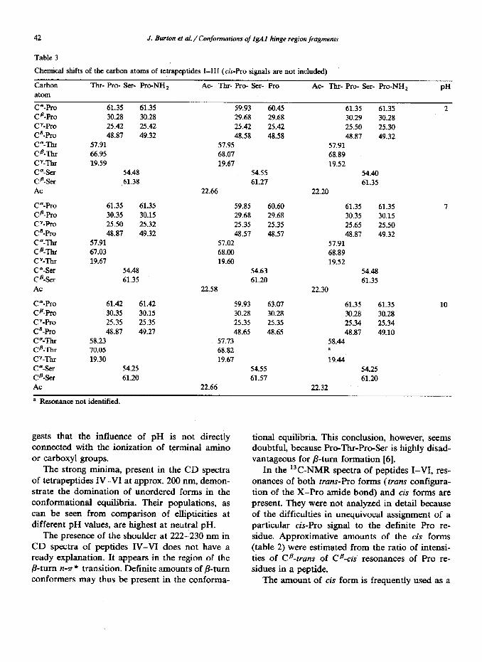
Table 3
Chemical shifts of the carbon atoms of tetrapcptides I-III (&Pro signals are not included)
Carbon Tllr- Pro- ser- Pro-NH, atom
C”-Pro 61.35 61.35 c@-Pro 30.28 30.28 cmFvo 25.42 25.42 P-Pro 48.87 49.32 C”-ThI 57.91 CD-ThI 66.95 C’-ThI 19.59 C”-sex 54.48 c@-ser 61.38 AC
C”-pro 61.35 61.35 CB-Pro 30.35 30.15 CT-Pro 25.50 25.32 @-Pro 48.87 49.32 C”-l-h 57.91 Cfl-TtU 67.03 CY-ThI 19.67
C*-Ser 54.48 CB-Ser 61.35 AC
P-pro 61.42 61.42 c@-FTO 30.35 30.15 CT-pro 25.35 25.35 P-Pro 48.87 49.27 CSThI 58.23 [email protected] 70.05
CY-ThI 19.30 P-SIX 54.25 CE-Ser 61.20 AC
a Resonance not identified.
AC- Tbr- Pro- Ser- Pro AC- Thr- Pro- Ser- Pro-NH,
59.93 60.45 61.35 61.35 29.68 29.68 30.29 30.28 25.42 25.42 25.50 25.30 48.58 48.58 48.81 49.32
57.95 57.91 68.07 68.89 19.67 19.52
54.55 54.40
61.27 61.35 22.66 22.20
59.85 60.60 61.35 61.35 29.68 29.68 30.35 30.15
25.35 25.35 25.65 25.50 48.57 48.57 48.81 49.32
57.02 57.91 68.00 68.89 19.60 19.52
54.63 54.48 61.20 61.35
22.58 22.30
59.93 63.07 61.35 61.35 30.28 30.28 30.28 30.28
25.35 25.35 25.34 25.34 48.65 48.65 48.87 49.10
57.73 58.44 68.82 a
19.67 19.44
54.55 54.25 61.57 61.20
22.66 22.32
PH
2
I
10
gests that the influence of pH is not directly tional equilibria. This conclusion, however, seems connected with the ionization of terminal amino doubtful, because Pro-Thr-Pro-Ser is highly disad- or carboxyl groups. vantageous for B-turn formation [6].
The strong minima, present in the CD spectra of tetrapeptides IV-VI at approx. 200 nm, demon- strate the domination of unordered forms in the conformational equilibria. Their populations, as can be seen from comparison of ellipticities at different pH values, are highest at neutral pH.
The presence of the shoulder at 222-230 nm in CD spectra of peptides IV-VI does not have a ready explanation. It appears in the region of the /?-turn n-r* transition. Definite amounts of P-turn conformers may thus be present in the conforma-
In the 13C-NMR spectra of peptides I-VI, res- onances of both vans-ho forms (trans configura- tion of the X-Pro amide bond) and cis forms are present. They were not analyzed in detail because of the difficulties in unequivocal assignment of a particular cis-Pro signal to the definite Pro re-
sidue. Approximative amounts of the cis forms
(table 2) were estimated from the ratio of intensi- ties of Cadruns of CB-cis resonances of Pro re- sidues in a peptide.
The amount of cis form is frequently used as a
J. Burton et al. / Confotmations of &A 1 hinge region fragments 43
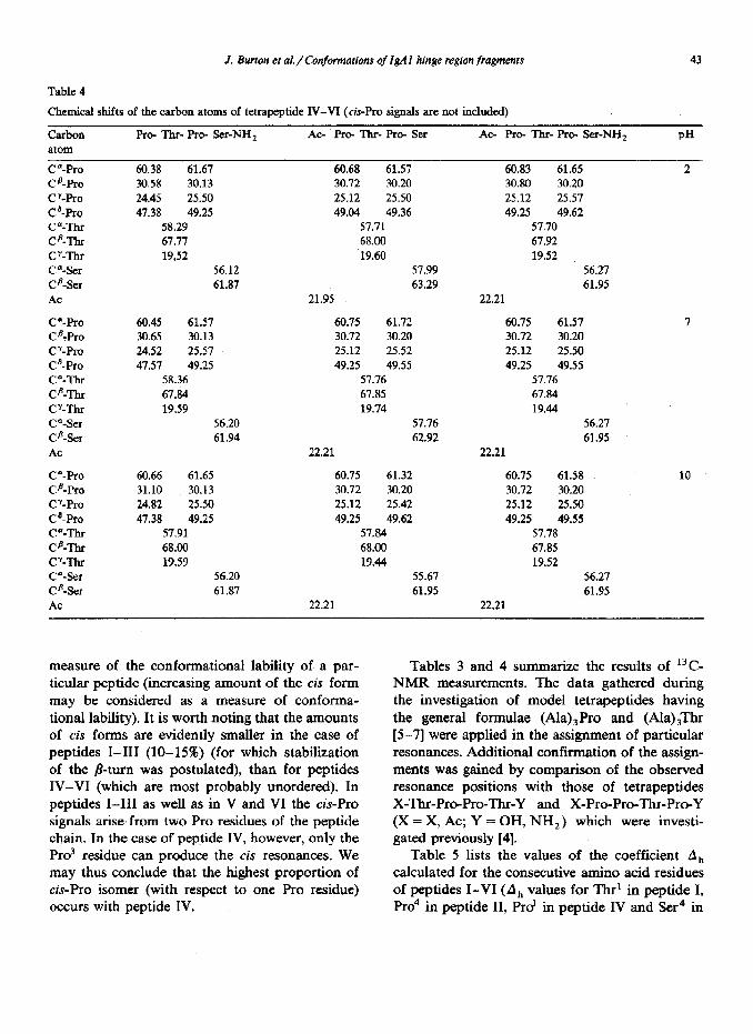
Table 4
Chemical shifts of the carbon atoms of tetrapeptide IV-VI (&Pro signals are not inch&d)
Carbon atom
ca-Pro @-Pro
CT-Pro c6-Pro C”-Thr CB-Thr CY--I% C”-Ser Cfl-Ser AC
ca-Pro CQro
UPro @-Pro C”-Thr Cfl-Thr CY-Thr C “-Ser CB-Ser
AC
C”-Pro CB-Pro cy-Pro C8-PKl C’-Thr CB-Thr C’-Thr C”-Ser Cfi-Ser AC
Pro- -Ii-u- Pro- &r-NH,
60.38 61.67 30.58 30.13
24.45 25.50 47.38 49.25
58.29 67.77 19,52
56.12 61.87
60.45 61.57 30.65 30.13
24.52 25.57 47.57 49.25
58.36 67.84 19.59
56.20 61.94
60.66 61.65 31.10 30.13 24.82 25.50 47.38 49.25
57.91 68.00 19.59
56.20 61.87
AC- Pro- Thr- Pro- Ser AC- Pro- Thr- Pro- W-NH, PH
60.68 61.57 60.83 61.65 2 30.72 30.20 30.80 30.20
25.12 25.50 25.12 25.57 49.04 49.36 49.25 49.62
57.71 57.70 68.00 67.92 19.60 19.52
57.99 56.27 63.29 61.95
21.95 22.21
60.75 61.72 60.75 61.57 30.72 30.20 30.72 30.20
25.12 25.52 25.12 25.50 49.25 49.55 49.25 49.55
57.76 57.76 67.85 67.84 19.74 19.44
57.76 56.27 62.92 61.95
22.21 22.21
60.75 61.32 60.75 61.58 10 30.72 30.20 30.72 30.20 25.12 25.42 25.12 25.50 49.25 49.62 49.25 49.55
57.84 57.78 68.00 67.85 19.44 19.52
55.67 56.27 61.95 61.95
22.21 22.21
measure of the conformational lability of a par- ticular peptide (increasing amount of the cis form may be considered as a measure of conforma- tional lability). It is worth noting that the amounts of cis forms are evidently smaller in the case of peptides I-III (lo-15%) (for which stabilization of the ~-turn was postulated), than for peptides IV-VI (which are most probably unordered). In peptides I-III as well as in V and VI the c&Pro signals arise from two Pro residues of the peptide chain. In the case of peptide IV, however, only the Pro3 residue can produce the cis resonances. We may thus conclude that the highest proportion of &-Pro isomer (with respect to one Pro residue) occurs with peptide IV.
Tables 3 and 4 summarize the results of 13C- NMR measurements. The data gathered during the investigation of model tetrapeptides having the general formulae (Ala),Pro and (Ala)Jhr [5-71 were applied in the assignment of particular resonances. Additional confirmation of the assign- ments was gained by comparison of the observed resonance positions with those of tetrapeptides X-Thr-Pro-Pro-Thr-Y and X-Pro-Pro-Thr-Pro-Y (X = X, AC; Y = OH, NH,) which were investi- gated previously [4].
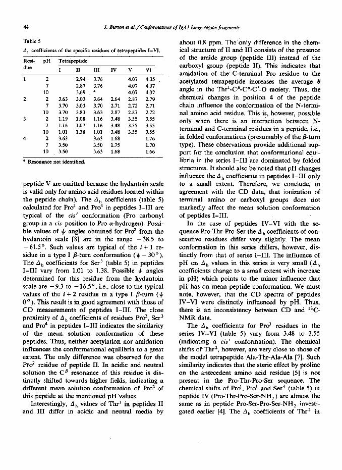
Table 5 lists the values of the coefficient A,, calculated for the consecutive amino acid residues of peptides I-VI (A, values for Thr’ in peptide I, Pro4 in peptide II, Prd in peptide IV and Ser4 in
44 J. Burton et al. /Canjormations of IgAl hinge region fragments
Table 5
A,, coefficients of the specific residues of tetrapeptides I-VI.
Resi- pH Tettipeptide due I II III IV v VI
1 2 2.94 7 2.87
10 3.69 2 2 3.63 3.03
7 3.70 3.03 10 3.70 3.83
3 2 1.19 1.08 7 1.16 1.07
10 1 .Ol 1.38 4 2 3.63
7 3.50 10 3.50
’ Resonance not identified.
3.76 4.07 4.35
3.76 407 4.07 B 4.07 4.07
3.64 2.64 2.87 2.79 3.70 2.11 2.12 2.71 3.63 2.87 2.87 2.12 1.16 3.48 3.55 3.55 1.16 3.48 3.55 3.55 1.01 3.48 3.55 3.55 3.63 1.68 1.76 3.50 1.75 1.70 3.63 1.68 1.66
peptide V are omitted because the hydantoin scale is valid only for amino acid residues located within the peptide chain). The A, coefficients (table 5) calculated for Pro2 and Pro4 in peptides I-III are typical of the cis’ conformation (Pro carbonyl group in a cis position to Pro a-hydrogen). Possi- ble values of II, angles obtained for Pro’ from the hydantoin scale [S] are in the range -38.5 to - 61.5 ‘. Such values are typical of the i + 1 re- sidue in a type I /?-turn conformation ($ - 30”). The A, coefficients for Ser3 (table 5) in peptides I-III vary from 1.01 to 1.38. Possible $ angles determined for this residue from the hydantoin scale are -9.3 to -16S”, i.e., close to the typical values of the i + 2 residue in a type I B-turn ($J 0 O ). This result is in good agreement with those of CD measurements of peptides I-III. The close proximity of A, coefficients of residues Pro*, Ser3 and Pro4 in peptides I-III indicates the similarity of the mean. solution conformation of these peptides. Thus, neither acetylation nor amidation influences the conformational e@ilibria to a great extent. The only difference was observed for the Pro’ residue of peptide II. In acidic and neutral solution the Cfl resonance of this residue is dis- tinctly shifted towards higher fields, indicating a different mean solution conformation of PK? of this peptide at the mentioned pH ‘values.
Interestingly, A,, values of Thr’ in peptides II and III differ in acidic and neutral media by
about 0.8 ppm. The ‘only difference in the chem- ical structure of II and III consists of the presence of the amide group (peptide III) instead of the carboxyl group (peptide II). This indicates that amidation of the C-terminal Pro residue to the acetylated tetrapeptide increases the average B angle in the Thr’-Cs-C”-C’-0 moiety. Thus, the chemical changes in position 4 of the peptide chain influence the conformation of the N-termi- nal amino acid residue. This is, however, possible only when there is an interiction between N- terminal and C-terminal residues in a peptide, i.e., in folded conformations (presumably of the B-turn type). These observations provide additional sup- port for the conclusion that conformational equi- libria in the series I-III are dominated by folded structures. It should also be noted that pH changes influence the A, coefficients in peptides I-III only to a small extent. Therefore, we conclude, in agreement with the CD data, that ionization of terminal amino or carboxyl groups does not markedly affect the mean solution conformation of peptides I-III.
In the case of peptides IV-VI with the se- quence Pro-Thr-Pro-Ser the Ah coefficients of con- secutive residues differ very slightly. The mean conformation in this series differs, however, dis- tinctly from that of series I-III. The influence of pH on A ,, values in this series is very small (A ,, coefficients change to a small extent with increase in pH) which points to the minor influence that pH has on mean peptide conformation. We must note, however, that the CD spectra of peptides IV-VI were distinctly influenced by pH. Thus, there is an inconsistency between CD and 13C- NMR data.
The A,, coefficients for Prd residues in the series IV-VI (table 5) vary from 3.48 to 3.55 (indicating a cis conformation). The chemical shifts of Thr*, however, are very close to those of the model tetrapeptide Ala-Thr-Ala-Ala [7]. Such simihuity indicates that the steric effect by proline on the antecedent amino acid residue [5] is not present in the Pro-Thr-Pro-Ser sequence. The chemical shifts of Prd, Prd and Ser4 (table 5) in peptide IV (Pro-Tbr-Pro-Ser-NH,) are almost the same as in peptide Pro-F&-Pro&r-NH, investi- gated earlier [4]. The A, coefficients of Thr2 in
J. Burton et al./ Conformations of IgAl hinge region fragments 45
Pro-Thr-Pro-Ser-NH, and Ser’ in Pro-Ser-Pro- Ser-NH, differ significantly (2.64-2.87 for Thr* and 1.53 for Ser’) indicating the difference in mean conformation of residue 2 in both peptides.
The comparison of A, coefficients of the Pro2- Sers sequence in the peptides I-III with those of the Thr*-Pro3 sequence in peptides IV-VI shows that the conformation of the peptide chain must be different in both cases. The difference consists of the closing of the 0 angle of residue 2 in the series IV-VI (in comparison with the situation in series I-III) with the simultaneous opening of the 6 angle of residues 3. Both changes act against the p-turn formation.
Acknowledgments
This work was supported by the Polish Academy of Sciences (grant CPBP 01.13) and National Institute of Health (grant DE-07257).
References
1
2
3
4
5
6
I
8
9
10
11
12
Y. Sheng, V. Liu, T.L.K. Low, A. Infante and F.W. Put-
man, Science 193 (1976) 1017. W. Kratzin, P. Altevogt, E. Ruban, A. Kortt, K. StaroScik and N. Hilschmann, Z. Physiol. Chem. 356 (1975) 1337.
J. PohIner, R. Halter, K. Beyreuther and T.F. Meyer,
Nature, 325 (1978) 458. I.Z. Siemion, A. Pedyczak and J. Burton, Biophys. Chem.
31 (1988) 35.
I.Z. Siemion, K. Sobczyk and E. Nawrocka, Int. J. Peptide Protein Res. 19 (1982) 439.
M. Lisowski, 1.2. Siemion and K. Sobczyk, Int. J. Peptide
Protein Res. 21 (1983) 301.
I.Z. Siemion, K. Sobczyk and M. Lisowski, Int. J. Peptide Protein Res. 27 (1986) 127.
I.Z. Siemion, in: Natural products chemistry, eds. RI. Zalewski and J.J. Skohk (Elsevier, Amsterdam, 1985) p.
335. R.W. Woody, in: The peptides, vol. 7 (Academic Press,
New York, 1985) p, 41. G.D. Rose, L.M. Gierash and J.A. Smith, Adv. Protein Chem. 37 (1985) 11.
I.Z. Siemion, M. Lisowski and K. Sobczyk, Ann. N.Y.
Acad. Sci. 419 (1983) 56. A. Aubry and M. Maraud, Biopolymers 22 (1983) 341.