Embed Size (px)
Citation preview

Congenital and Genetic Abnormalities
The etiology of birth defects is not completely understood, malformations may occur from Genetic factors, such as change
in the chromosome number, mutation, or structural abnormalities, or environmental factors such as irradiation,
infection, and drugs. The most frequent malformations occur from interactions between multiple genetics and environmental
factors. Birth defects are common, costly, and critical conditions that affect 1 in every 33 babies born in the United
States each year. Every 4 ½ minutes, a baby is born with a birth defect in the United States. That means nearly 120,000 babies
are affected by birth defects each year.

Abnormalities include but are not limited to:
Congenital cardiac anomalies Congestive heart failure
Cleft palate/lip ( failure of the bone to fuse) Hypospadius ( urethra opens on the undersurface
Epispadius (urethra is on the dorsal surface) Ambiguous genitalia (uncertain male/female)
Craniosynostosis (premature closures) Spina bifida (lack of one or more vertebral arches)
Hydrocephalus (increased CSF/enlarged head) Anencephaly (complete or partial absence of brain)
Microcephaly (smaller head and brain)

Obstructions of the alimentary tract Diaphragmatic hernia
Pyloric stenosis Omphalocele Gastroshisis
Imperforated anus Esophageal atresia
Chromosomal anomalies
Trisomy 13 or d- lifespan <1 year Trisomy 18 or e- lifespan <6 months
Trisomy 21 or down’s syndrome- 1 in 500 births
Musculoskeletal disorders Talipes equinovarus (clubfoot)
Congenital hip dysplasia Scoliosis
Polydactyly (extra digits)

Diaphragmatic Hernia:
Caused by a defect in the development of the diaphragm that allows abdominal organs to
herniate into the thoracic cavity. The majority of cases involve the left leaf of the diaphragm. The mortality rate is high. Stabilization of the infant prior to a surgical repair requires placement of
an ng tube to decompress the abdomen, correction of acidosis, administration of oxygen,
and ventilatory assistance.
If the defect is small, it can be easily repaired. This must be done soon after birth because the
herniated abdominal organs interfere with adequate respiration. If the defect is large, the
mortality is higher. The defect may be repaired by using a graft patch since not enough diaphragm is
there to complete the repair.

Pyloric Stenosis: The incidence of pyloric is 2 to 5 per 1000 live birth.
It usually manifests its symptoms between the second and sixth week by the onset of vomiting that becomes projectile and occurs within 30 minutes after every feeding. The infant loses weight, bowel elimination lessens, highly colored urine becomes scanty, and the symptoms of
dehydration appear. Upon examination, gastric peristalsis is found, and the pyloric “acorn like” tumor may be palpated. Surgery is the treatment of choice. The operation is not usually an
emergency, leaving sufficient time for supportive treatment to correct any dehydration or electrolyte imbalance beforehand. Fluids, electrolytes, and blood replacement may be necessary, depending on the condition of the infant. The surgery
is usually performed laparoscopically. Gastric lavage, from 1 to 2 hours before operation, may be done until returns are clear.
Maintaining body heat before and after surgery is essential.

Omphaloceles Occurs in 1 in 5000 births, in which an amount of abdominal
contents protrudes at the base of the umbilicus. Omphaloceles develop between the 8th and 10th week of fetal life.
The mass is covered with a layer of peritoneum and amion and may rupture at delivery.
Omphaloceles are often seen in conjunction with other cardiac, genitourinary, and extra intestinal anomalies. Treatment requires covering the mass with sterile gauze soaked in saline, ng placement, and immediate total or staged surgical repair. Sepsis is a serious and potential complication.
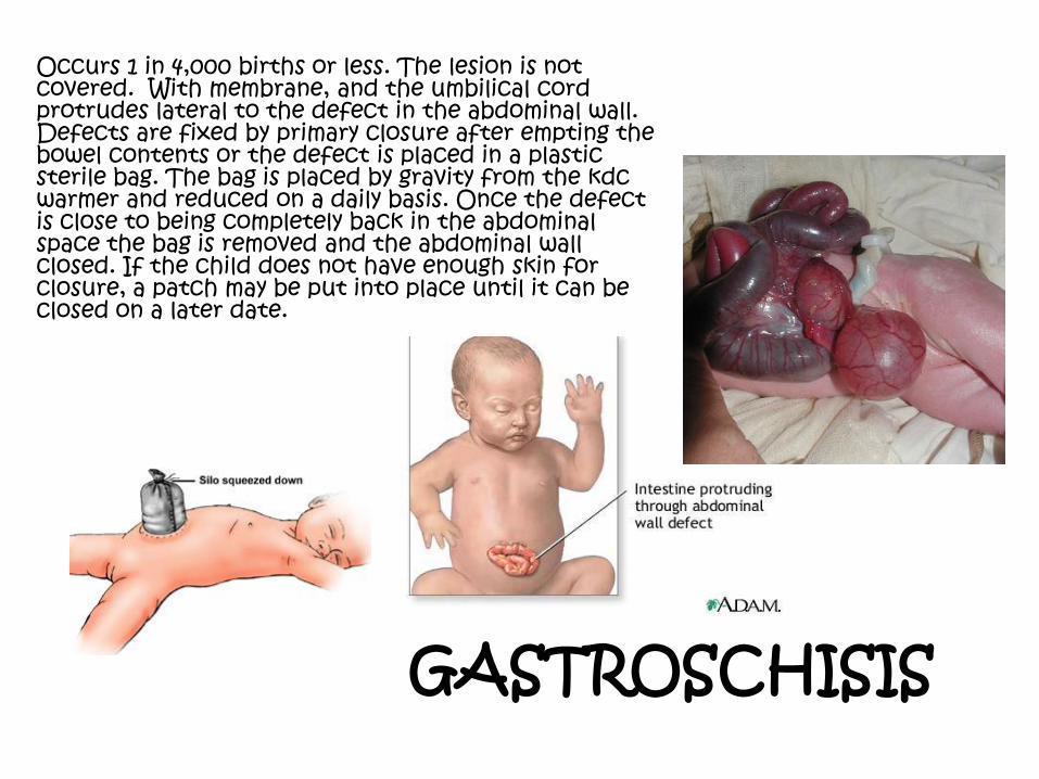
GASTROSCHISIS
Occurs 1 in 4,000 births or less. The lesion is not covered. With membrane, and the umbilical cord protrudes lateral to the defect in the abdominal wall. Defects are fixed by primary closure after empting the bowel contents or the defect is placed in a plastic sterile bag. The bag is placed by gravity from the kdc warmer and reduced on a daily basis. Once the defect is close to being completely back in the abdominal space the bag is removed and the abdominal wall closed. If the child does not have enough skin for closure, a patch may be put into place until it can be closed on a later date.

Talipes Equinosvarus (Club Foot) 1:5,000 births

Hip Dysplasia Checked with Barlow’s and
Ortolani’s maneuvers

Scoliosis Scoliosis is a lateral (toward the side) curvature in the normally straight vertical line of the spine. When viewed from the side, the spine should show a mild roundness in the upper back and shows a degree of swayback (inward curvature) in the lower back. When a person with a normal spine is viewed from the front or back, the spine appears to be straight. When a person with scoliosis is viewed from the front or back, the spine appears to be curved.
Approximately 2% to 3% of Americans at age 16 have scoliosis. Less than 0.1% have spinal curves measuring greater than 40 degrees, which is the point at which surgery becomes a consideration.

Types of Craniosynostosis The types of craniosynostosis depend on what sutures join together early. Sagittal synostosis– The sagittal suture runs along the top of the head, from the baby’s soft spot near the front of the head to the back of the head. When this suture closes too early, the baby’s head will grow long and narrow (scaphocephaly). It is the most common type of craniosynostosis. Coronal synostosis – The right and left coronal sutures run from each ear to the sagittal suture at the top of the head. When one of these sutures closes too early, the baby may have a flattened forehead on the side of the skull that closed early (anterior plagiocephaly). The baby’s eye socket on that side might also be raised up and his or her nose could be pulled toward that side. This is the second most common type of craniosynostosis. Bicoronal synostosis – This type of craniosynostosis occurs when the coronal sutures on both sides of the baby’s head close too early. In this case, the baby’s head will grow broad and short (brachycephaly). Lambdoid synostosis – The lambdoid suture runs along the backside of the head. If this suture closes too early, the baby’s head may be flattened on the back side (posterior plagiocephaly). This is one of the rarest types of craniosynostosis. Metopic synostosis – The metopic suture runs from the baby's nose to the sagittal suture at the top of the head. If this suture closes too early, the top of the baby’s head shape may look triangular, meaning narrow in the front and broad in the back (trigonocephaly). This is one of the rarest types of craniosynostosis.

Congenital Heart Anomalies

Congenital Heart Anomalies
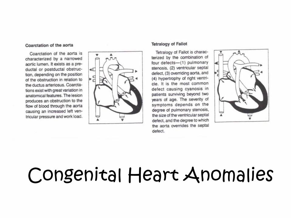
Congenital Heart Anomalies

Congenital Heart
Anomalies
HLHS Norwood
BDG Fontan

Cleft Lip and Palate

Hypospadius, Epispadias, Ambiguous Genitalia

Spina Bifida: Myelomeningocele

Imperforate Anus
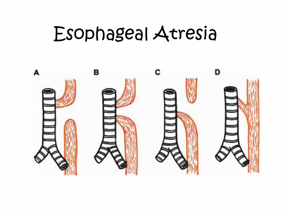
Esophageal Atresia

Questions?