Embed Size (px)
Citation preview
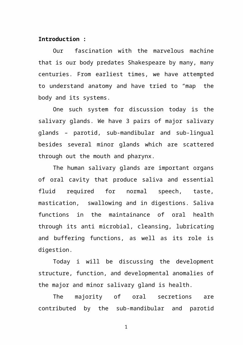
Introduction :
Our fascination with the marvelous machine that is our body predates
Shakespeare by many, many centuries. From earliest times, we have
attempted to understand anatomy and have tried to “map” the body and its
systems.
One such system for discussion today is the salivary glands. We have
3 pairs of major salivary glands – parotid, sub-mandibular and sub-lingual
besides several minor glands which are scattered through out the mouth and
pharynx.
The human salivary glands are important organs of oral cavity that
produce saliva and essential fluid required for normal speech, taste,
mastication, swallowing and in digestions. Saliva functions in the
maintainance of oral health through its anti microbial, cleansing, lubricating
and buffering functions, as well as its role is digestion.
Today i will be discussing the development structure, function, and
developmental anomalies of the major and minor salivary gland is health.
The majority of oral secretions are contributed by the sub-mandibular
and parotid glands which equally provide 80-90% of saliva,reminder is
formed by sub-lingual and minor salivary glands. In a day about 1000 ml –
1500 ml of salivary is produced which contributes to the digestions of food,
to the maintainance of oral hygiene and speech.
The development of these glands are principally induced to begin
their characteristic branching morphogenesis through an interaction of the
cells of the oral epithelium which is derived from the endoderm and the
underlying mensenchyme.
1
DEVELOPMENT OF SALIVARY GLANDS
1) General development process
Epithelial-mesenchymal interactions
Neuro epithelial interactions
2) General development pattern of salivary glands.
3) Stages of development.
1) General development process ;
i) Epithelial-Mesenchymal interactions :
The development of glandular tissue in mammals involves the
interactions of epithelium with the underlying mesenchyme to form
functional part of the gland.
They are also defined as proximate tissue interactions and also
secondary induction – in which the presence of mesenchyme in close
proximity to epithelium is required for normal development of epithelium.
The interactions regulate both initiation and growth of glandular tissue and
cytodifferentiation of cells within salivary glands. The mesenchyme is
therefore required for normal development as well as formation of
supporting part of adult gland.
Lawson studied rat sub-mandibular and parotid epithelium and found
that both types can undergo differntiation with their own (or) reciprocal
gland’s mesenchyme. In those cases when epithelium of one gland was
combined with mesenchyme of other gland (in vitro), growth and
arrangements of acini were found to be governed by mesenchyme but not
the function.
Interaction between salivary epithelium and mesencyme is provided
by recent studies describing the presence of an acid mucopolysaccharide
(MPS) at the epithelio-mesenchymal interface. There is an increasing
concentration of this material at the distal end of growing and branching
lobules. So these authors are suggesting MPS may be involved in
2
morphogenesis. This MPS is located in the basal lamina and appears to be
protein-bound, permits glandular primordial branching when it is retained in
vitro. And if this MPS is removed, the epithelial growth produces only a
spherical, unbranched structure.
In growing of salivary epithelial cords is related to the contractile
ability of intra epithelial microfilaments. Even though microfilaments are
present the budding (clefting) will not occur unless specific salivary
mesenchyme is present.
ii) Mesenchyme : (composed of cells derived from neural crest)
Extracellular matrix (ECM) and basal lamina.
Mesenchyme consists of :
a) Undifferentiated pluripotential connective tissue cells.
b) ECM
a) Undifferentiated pluedripotential C.T cells.
- Fibroblasts
- Macrophages
- Mast cells
b) ECM :
- Glycosaminoglycans
- Proteoglycans.
Glycosaminoglycans :
- Chondroitin sulphate
- Keratan sulphate
- Hyaluronic acid – gives gel like characteristic to ECM.
These glycosamino glucans (1,2) are bound to a core protein to form
proteoglycan sub units.
These sub units are non-covalently bound to hyaluronic acid (another
GAG) – to form the bristle brush like structure of proteoglycan aggregate
found in ECM.
3
Gives gel like characteristic to ECM.
Functions of proteoglycans :
- Forms hydrated ground substance and also function in filtration (eg.
Renal glomerular B.M).
- “Bind signaling molecules (like growth factors to this target cells).
Laminin – Entacin (glycoprotein) – interact with each other and other
components of ECM through receptors, a family of transmembrance linker
proteins – known as Integrins. Eg. Fibronectin receptors.
They allow intra cellular adhesive molecules (ICAMs) facilitate
communication within cell cytoplasm. (ECM to cytoskeleton).
This communication / linked changes – cell shape
- Motility
- Migration
- Proliferation
- Differentiation
All which occurs during salivary gland development.
Influence of ECM on development :
The ECM provides regulatory cues for
- Cell proliferation
- Cell differentiation
- Morphogenesis, the major developmental process required for
the formation of adult salivary gland structure.
Cell proliferation : It is increased in number of cells that occurs during
development as organs enlarge. Cells enter cell cycle, replicate DNA and
undergo cytokinesis to form progeny (daughter cells)
Cell differentiation : It describes process responsible for development of
cell specificity and diversity as observed at the morphologic (or) molecular
level.
It expresses a specific portion of genome that is characteristic of that
particular cell type.
4
Cell morphogensis :
It describes those developmental process that are responsible for
formation of shape and form of organ. Eg. Branding of salivary gland.
Neuro epithelial interactions :
Another study dealing with the events leading to the initiation of
epithelial ingrowth has been under taken by DOZIN. The research
demonstrates formation of both the human and mouse mandibular and
sublingual glands is directly related to “Ganglioneural recess”. Ganglio
neural recess is term descriptive of the developing sub-mandibular ganglion
cells and their location.
The further report demonstrated that both post ganglionic
parasympathetic neurons as well as mesenchyme into which epithelium
invagiantes are composed of ectomesenchyme, thus bringing salivary gland
morphogenesis within the scope of developmental events that involve neural
crest derivative cells.
General development pattern and salivary glands :
All salivary glands follow similar developmental pattern. The
functional glandular tissue (parenchyma) develops as an epithelial out
growth. (Glandular bud) of the buccal epithelium that invades the
underlying mesenchyma. The connective tissue stroma (capsule and septa)
and blood vessels form from the mesenchyme. The mesenchyme is
composed of cells derived from the neural crest. It is essential for normal
differentiation of the salivary glands. ECM components synthesized by
mesenchymal C.T cells provide signals to morphogenesis and
differentiation.
Bud formation and gland origin :
As the epithelial bud forms during development, the portions that are
close to stomodeum (primitive and cavity) differentate into main excretory
5
duct of gland. The distal portions arborize to form the terminal portions of
duct system, secretory end pieces or acini.
The origin of epithelial buds is believed to be ectodermal in the
parotid and minor salivary glands and endodermal in the sub-mandibular
and sub-lingual glands. The break down of oropharyngeal (bucco
pharyngeal) membrane during 4th week of development, permits the
intermingling of stomodeal ectoderm and cranial forgut endoderm, which
complicates identifications of specific germ layer origin of S.glands, parotid
glands originate near corners of stomodeum by 6th week of prenatal life.
Sub-mandibular araise from floor of mouth at the end of 6th or beginning of
7th week of utero. The sub lingual glands form lateral to sub-mandibular
primordial at 8th week. Minor S.glands form from epithelium but don’t
develop until 12th prenatal week.
STAGES OF DEVELOPMENT
Salivary gland development may be divided into 6 stages.
Stage I : Formation : induction of oral epithelium by underlying
mesenchyma.
The mesencyme underlying the buccal epithelium induces
proliferation in the epithelium, which results in the thickening and formation
of epithelial bud. The growing bud is separated from the condensation of
mesenchyme by basal lamina that is secreted by the epithelium.
The process of development of all salivary glands is similar except
the site and time of development.
Stage II : Formation and growth of the epithelial cord :
A solid cord of cells forms the epithelial bud by cell proliferation.
Condensation and proliferation occur in the surrounding mesenchyme that is
closely associated with the epithelial cord. The basal lamina is composed of
GAGs, glycoproteins and collagen.
6
The basal lamina, as well as surrounding mesenchyme influences
morphogenesis and differentiation of salivary glands throughout their
development.
Why mesenchyma induce proliferation there only + how ?
Presence of 1st arch ecto-mesenchyme is essential for full salivary
gland development. Eg. if non-salivary gland mesenchyme is combined with
salivary gland epithelium, the epithelium does not differentiate into
glandular epithelium.
Stage III : Initiation of branching in terminal parts of the epithelial
cord and continuation of glandular differentiation
The epithelial cord proliferates rapidly and branches into terminal
bulbs (presumptive acini). The growth in length of solid epithelial cords and
differentiation of the berry like terminal bulbs are noticed.
Stage IV : Repetative branching of epi-cord and lobule formation :
The branching continues at terminal portions of the cord, forming an
extensive trace like systems of the bulbs. As the branching occurs
connective tissue differetate around the branches, eventually producing
extensive lobulation.
The glandular capsule forms from mesenchyme and surrounds the
entire glandular parenchyma.
Stage V : Canalization of presumptive ducts :
Canalization of epithelial cord, with formation of hallow tube or duct
usually occurs by 6th month in all 3 major salivary glands. Lumen appear 1st
in the proimal (oral, terminal) and distal portions of the main excretory duct
and in the branch ducts, then in the mid portion of main duct and lastly in
the acini, all of which precede the formation of secretory granules. Lumen
development occurs as a result of formation of tight junctions (zonular
occludens) among the cells surrounding what was initially a simpler
7
intercellular space. Extensive branching of duct structure and growth of
connective tissue septa continue at this stage of development.
Stage VI : Cytodifferentiation :
The final morphologic stage of salivary gland development is
cytodifferentation of functional acini and intercalated ducts. During this
period, mitotic activity shifts from the entire epithelial cord to the terminal
bulb portions. Cells of the bulb region are the stem cells that undergo cell
proliferation and subsequent differentiation into acinar cells as well as duct
cells. Myo epithelial cells also arise from epithelial stem cells in the
terminal bulbs of the developing duct system and development in contact
with acinar cytodifferentation. Maturation of acinar cells occur in specific
statges classified according to the morphology of secretory granules and
cellular organells.
Acinor development differs for serous and mucous cells. Therefore,
the parodid, sub-mandibular and sublingual salivary glands show variations
in cuto differentiation patterns. Terminal bulb cells differentiate into
intercalcated duct cells of the acdulct glands and they serve as a stem cell
for acinor, myoepthelial and ductal cells. Secretogogue stimulus-secretion
coupling mechanisms and innervation of the gland continue to mature
following cytodifferentiation.
PATIENT DUCT SYSTEM
In the formation of patient duct system of salivary glands it has been
shown that (a) the mucous membrane at the end of the duct differentiates
insitu into a papilla. (b) ducts are dilated initially by the secretion of water
and of electrolytes by the cells themselves. Further, the prenatal duct cells,
studies invitro, are sensitive to pharmocoliogic agents which tend to
increase or decrease the transport of water and electrolytes.
The later fetal stages of human sub-mandibular salivary gland
differentiations have been studied and they reported that : at 4 months of
8
gestation only undifferentiated cells are present. Mucous like cells can be
seen in striated ducts at 7 months and serous cells are found in the terminal
portions of the gland at gestation.
POSTNATAL DEVELOPMENT
The postnatal developmental changes is the salivary glands was the
work of Jacoby and Leeson with light microscope and electron microscope.
In rat the sub mandibular and sub-lingual glands are more highly
differentiated than the parotid at birth. In rat acinar cells are not present at
birth as they are in man and in pig. Acinar cells are not present until 7 th day
in the rat sub mandibular gland. Terminal ducts do not disappear and acini
do not grow completely until 40th and 120th day. They (Jacoby and Leeson)
described ultra structural changes in the terminal cells as secretory function
is established.
In the mouse, also, the post natal developmental changes include the
opening of the secretory lumina and secretory cell differentiation.
Functional influences on the initiations of salivary secretion is a postnatal
event. Acinar transformation is induced and affected by cellular secretory
activity. Secretion evokes a neural stimulation that effects acinar cyto
differentiation.
The further correlation between function and glandular development
is reported by Redmass and Sreebny. They noted two early postnatal
developmental stages
1. 1st from 1 to 12 days
2. 2nd from 15 to 25 days
In first enzymatic concentration in the cells increase greater than cell
number or size. During 2nd phase cell size in particular is increased. Further
1st phase correlates with the on set of suckling (enzymatic concentration)
and 2nd phase with weaning, leading to suggestion that food substrates and
9
secretory stimuli may have “indictive significance” in salivary gland
differatiation.
PROCESS INVOLVED IN SALIVARY GLAND DEVELOPMENT
1) General
Development of salivary glands is influenced by intrinsic and
extrinsic factors that regulate the process of cells proliferation,
differentation and morphogenesis. The intrinsic factors are defined as the
programmed pattern of gene expression specific for each cell type. This
programmed script, with gene turned-on and turned off at appropriate times,
leads to the normal development and growth of tissues and organs and the
differentiation of cells. Extrinsic factors are signals provided by cell-cell and
cell-matrix interactions, as well as by cytokines, hormones and growth
factors in extracellular milier. The extrinsic factors define boundaries
between groups of cells during development.
Eg. (Fruit fly) Drosaphila has been used as a model of embryonic
development. There is a shift during development between 3 categories of
genes.
Maternal genes are expressed during oogenesis by the mother and act
during oocyte maturation. They define broad regions with in the egg and
regulate expression of the segmentation genes, that determine the number
and / or polarity of segments. Segmentation genes define smaller regions of
embryo. The last group, homeotic genes regulate development of 1 body
part compared to other (same as other side).
Homologous genes to those identified in drosophila are being
identical in mammalian development with remarkable conservation of
structure and regulatory functions.
10
2) Positioning of the glands :
The formation of specialized structures (salivary glands) is regulated
by homeotic genes. These genes contain – homeobox domain i.e. 60-amino
acid, DNA binding domain.
Much of informations is obtained from the studies of drosophila. In
drosophila polarity is established initially along an anterior-posterior axis
(head-tail) that establishes the segmentation of embryo. Further
development within each segment establishes a doral-ventral (back-
abdomen) gradient that is translated into specialized structures in each
segment of the larua and eventually the adult segmented fly.
In drosphila, the gene “SEX COMBS REDUCED” (sex) is a
homeotic gene. It encodes a transcription factor that is responsible for the
location of salivary glands.
Scr is uniformly transcribed in the cells of posterior head segment
where the drosophila suli.gland will develop. The dorso ventral boundaries
of glands are established by genes homologous to mammalian genes, such
as bone morphogenic protein – 4 (BMP-4)
BMP-4 limit the permissibility of Scr expression in drosophilla also
involved in cascode of gene expression that regulates epithelial
mesenchymal interactions involved in branching and other morphogenetic
events.
The development of mammalian salivary glands mrophogenesis
remain unclear. The correct patterning of vertebrate embryo is based upon
expression of HOX genes. The expression and restriction of HOX genes are
responsible for the differentiation of cells along the anterior-posterior axis
of all metazoans. HOX-genes expression occurs in the vertebrate nervous
system and its derivatives, including the neural crest cells.
11
The neural crest is instrumental in the formation of the salivary
glands, teeth and over all craniofacial morphology through formation and
differentiation of bronchial arches.
3) Branching of the epithelial cord :
Branching is the primary morphogenetic process in salivary gland
development. Cleft formation in distal buds initiates branching process that
is followed by epithelial proliferation. Collagen type III accumulates at cleft
points and appear to be critical for branching to occur. Type I and IV
collagen appear to be more ruportant for the maintenance and support of
established branches. Type I : Type III collagen ratio increases at the time of
branching. Type Ileads to stabilization while type III is more involved in
active branching.
More recent studies also indicate that proteoglycan biosynthesis and
deposition are required for branching but not growth of the rudiments.
Chondroitin sulfates are the predominant GAGS in the basal lamina of
actively branching young rudiments and appear to increase during
stabilization.
The independence of epithelial expansion and branding has been
demonstrated by use of tunicamycin (in vitro). Tunicamycin inhibits N-
linked glycosylation resulting in dramatically decreased protein
accumulation and cell proliferation but epithelial branching is unaffected.
After tunicamycin treatment branching accurs and lobules form normally
with inhibited cell proliferations, resulting in smaller rudiment with
miniature lobes. In contrast, the size and number of lobes increase in control
cultures.
Branching and proliferation must be coordinated processes for normal
development. Mitotic activity is normally localized in the most peripheral
regions of the bud. Treatment with hyaluronidase disrupts the basal lamina
interfering with the signal required for cleft formation. Destabilization of
12
basal lamina there fore inhibits cleft development but also effects cell
proliferation. In the absence of normal basal lamina there is an absence of
branching and generalized cell proliferation replaces localized mitotic
activity. So basal lamina is important in initiation and maintenance of
lobular morphology.
The basal lamina regulate morphogenetic changes by selective
filtration or channeling of materials to the epithelium.
For Eg : the regulation of the flow of ions such as Ca++ to epithelium may
alter the function of micro tubules and microfilaments in cellular
proliferation, migrations and arrangement. Synthesis of collagen
(collagenogentic) and selective break down of collagen (collagenolysis)
play a critical role in salivary gland development.
For example collagen synthesis by mesenchyme provides structural
stabilization after branding has occurred. Stabilization appear to be provided
by type I and IV collagen fibres. In addition, collagenolytic activity in the
epithelium and mesenchyme may allow for selective breakdown of basal
lamina and communicate between the epithelium, basal lamina, and
surrounding mesenchyme at key stages of development.
4) Process involved in cyto differentiation :
The interaction of the epithelium and mesenchyme is best stududied
in a culture disb. Where epithelium can be grown in the presence of selected
components of the basal lamina and specific growth factors. Salivary gland
rudiments can branch invitro in the absence of mesenchymal cells, but in
presence of other factors.
A developing salivary gland rudiment have three clefts at the
beginning of culture. The epithelium is grown in serum with the use of an
artificial matrix known as Matrigel, composed of mainly laminin, type IV
collagen, heparan sulfate, entactin, and nidogen. Using this they abserved
that different growth factors appear to regulate distinct parts of
13
morphogenetic process. Fibroblast growth factor has bear shown to alter
stalk elongation and epidermal growth factor (EGF) regulater branching.
The FGF and EGF combination results in morphology similar to in vivo
studies. ECM molecules regulate presentation and the distribution of growth
factors to epithelium at appropriate time during in vivo. There for, the ECM
in concern with specific growth factors appears to regulate the complex
processes involved in branching morphogenesis.
Cyto differentiation is believed as pre-programmed development
occurring in early stages of morphogenesis. A period of in situ epithelial –
mesenchymal contact is required for cyto differentiation. After this contact
has taken place. Exocrine cell differentiation occurs without continued
presence of mesenchyme. There fore it appears there is a partial coupling of
morphogenesis and cyto differentiation. Full differentiation of secretory
components is apparent at birth, but is complete until the onset of a solid
diet and the presence of masticatory stimuli.
This post natal development process includes the maturations of
stimulus – secretion coupling that links secretagogue – membrane receptors
to signal transudation pathways within the cell and control acinar cell
secretion and establishment of neural connections from autonomic nervous
system, the primary regulator of salivary gland function.
Saliva formation : ionic transport :
Saliva is formed in 2 stage.
First stage – is production by acinar cells.
Second stage – is the ducts that change isotonic solution to the hypotonic
solution.
Different systems involved are
1) Na+ K+ - ATpase
2) Na+ K+ Cl- co-trans port system.
3) Bicarbonate secretions – by Na+ / H+ exchanger.
14
4) Chloride secretions – by Na+ / H+ and Cl- / Hco3 exchanger.
5) Ca++ regulated K+ and Cl- channels
6) Osmotic flow of water.
7) K+ / H+ exchangers.
8) Paracellular transport of Na+ and water.
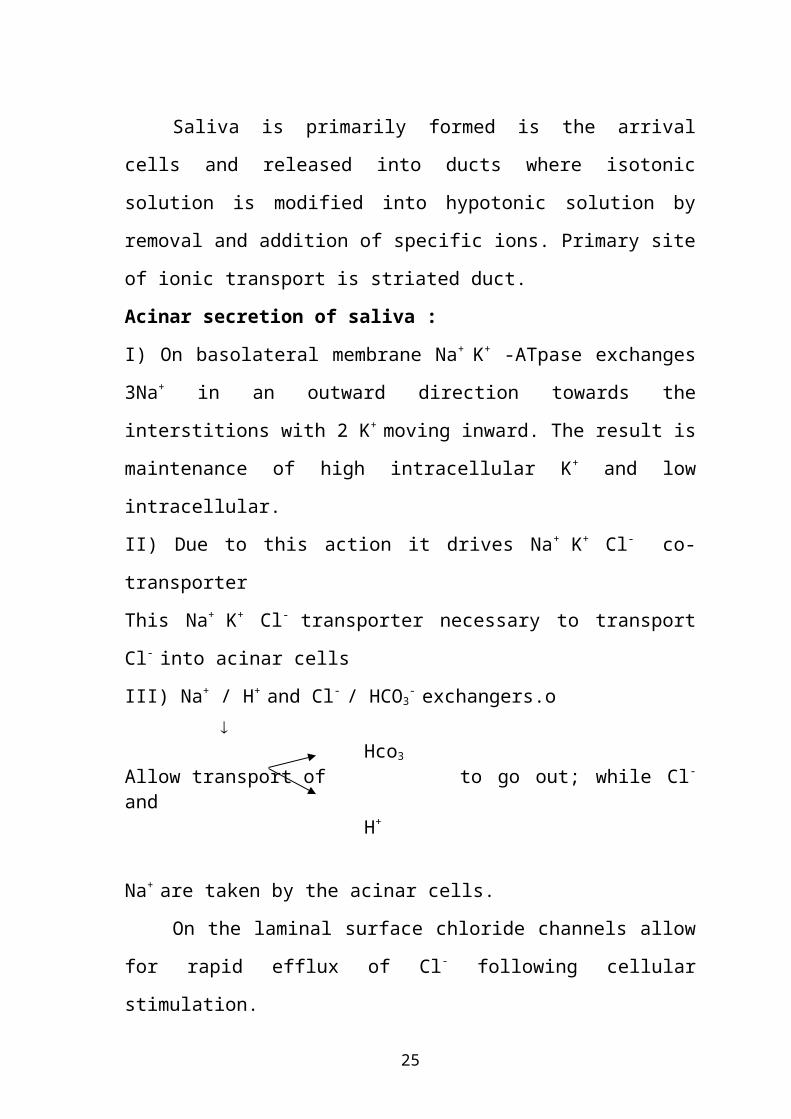
Saliva is primarily formed is the arrival cells and released into ducts
where isotonic solution is modified into hypotonic solution by removal and
addition of specific ions. Primary site of ionic transport is striated duct.
Acinar secretion of saliva :
I) On basolateral membrane Na+ K+ -ATpase exchanges 3Na+ in an outward
direction towards the interstitions with 2 K+ moving inward. The result is
maintenance of high intracellular K+ and low intracellular.
II) Due to this action it drives Na+ K+ Cl- co-transporter
This Na+ K+ Cl- transporter necessary to transport Cl- into acinar cells
III) Na+ / H+ and Cl- / HCO3- exchangers.o
Hco3
Allow transport of to go out; while Cl- and H+
Na+ are taken by the acinar cells.
On the laminal surface chloride channels allow for rapid efflux of Cl -
following cellular stimulation.
Activations of acetylcholine receptors
Following para sympathetic nerve stimulation
Result in increased inter cellular calcium (Ca+)
This (Ca+) drives apical Cl- channer.
And also activate Ca+ activated K+ channel on the
baso lateral surface to preserve the membrane potential by giving the K+ out
into interstitium.
15
II Stage of salivary secretion :
Here the isotonic saliva produced by acinar cells is modified into
hypotonic saliva. The duct cells reobsorb Na+ and Cl- and secrete K+ and
Hco3- with out water reobsorption resulting in hypotonic saliva. The
basolateral membrane of duct cells process high
- Na+ - K+ - ATpase
- Na+ / H+ - exchanger
- Cl- / K+ channels
The luminal surface processes
- Na+ channels
- Cl- channels
- Na+ / H+
- Cl- / HCo3-
- H+ / K+ exchange
A genetic mutation in cystic fibrosis (CF) gene alters Cl- and other
channels in salivary glands and leads to following symptoms.
Seen commonly among Caucasian children is characterized by
general dysfunction of salivary and other exocrine glands and results in
pulmonary, digestive and nutritional difficulties.
Accumulations of glycoproteinaceous material in the acinar cells and
ducts leading to eventually obstructions of the ducts.
During salivary secretion there is a rapid movement of water
following stimulation. Since acinar cells shrink dramatically following
secretions, it appears that most of water moves by osmosis in response to
Na+ in primary saliva. There is also evidence for paracellular and transcellar
movement of water. Aquaporins are membrane proteins that function as
high selective water channels in fluid transporting epithelum. Aqua porins
(AQPS) 1 and 5 are predominant inhuman salivary glands and mainly in
serous acini.
16
CLASSIFICATION OF SALIVARY GLANDS
The glands of the body may be classified into two general types :
1) Exocrine
2) Endocrine
1) Exocrine : Glands are those glands with a duct system to transport
secretion from the glands.
2) Endocrine : Glands are those duct less glands dependent on blood
supply for delivery of their secretary products.
Salivary glands are classified as exocrine glands, but these glands are
associated with a number of biologically active substances (Eg: nerve
growth factor and epidermal growth factor) that may be secreted by an
endocrine mechanism. They are classified as compound tubulo acinar
glands, which indicates the presence of a branched duct system and
secretory units with both tubular and acinar portions.
I) The salivary glands of mammalian species may be divided into
1) Major salivary glands
2) Minor salivary glands.
1) Major salivary glands :
These glands produce major part of salivary secretion most of the (0.5
to 0.75 lit.) saliva produced daily. These glands are located apart from oral
cavity, they communicate with large excretory ducts. There are three pairs
of major salivary glands :
1) Parotid
2) Sub mandibular (formely sub-maxillary)
3) Sub lingual
2) Minor salivary glands :
They are found in the oral cavity and are named according to their
location.
- Buccal
17
- Labial
- Lingual
- Palative
- Glasso palatine
II) Salivary glands may also be classified by types of secretion.
1) Serous
2) Mucous
3) Mixed
1) Serous :
serous secretion contains water, enzymes (primarily salivary amylase
and some maltase), a variety of salts, and organic ions.
i)Parotid gland
ii) Lingual glands – circumvallate papillar (Von Ebner’s glands)
2) Mucous :
Mucous secretion produces mucins, which act as lubricant to aid in
mastication, deglutition and digestin.
i)Glosso palatine –Anterior faucial pillar, and glasso palatine fold.
ii) Palatine glands
iii)posterior lingual glands
3) Mixed glands :
i) Sub-mandibular gland
ii) Sub lingual gland
iii) Labial gland
iv) Buccal glands
v) Anterior lingual glands
18
GROSS ANATOMY OF SALIVARY GLANDS
I. MAJOR SALIVARY GLANDS :
1) Parotid gland :
Parotid gland is the largest of the salivary glands. It weight about
15gm.
Situation :
It is situated below external acoustic meatus, between the ramus of
the mandible and the sternomastoid. A part of this forward extention is
often detached, and is known as the accessory parotid. It lies between the
zygomatic arch and parotid duct.
Parotid capsule :
The investing layer of the deep cervical fascia forms a capsule for the
gland. The fascia splits to enclose the gland. The superficial lamina is thick
and adherent to the gland is attached above to the zygonatic arch. The deep
lamina is thin and is attached to the styloid process, the mandible and
tympanic plate.
A portion of deep lamina extending between styloid and mandible is
thickened to form stylomandibular ligament which separate parotid from
sub-mandibular gland.
External features :
The gland resembles a three sided pyramid with its apex down wards.
The gland has 4 surfaces.
1) Superior (base of the pyramid)
2) Superficial
3) Anteromedial
4) Posteriomedial.
The surfaces are separated by 3 borders :
i) Anterior
ii) Posterior
19
iii) Medial.
Structures within parotid gland :
1) Arteries :
The external carotid artery enters the gland through its postero medial
surface. The maxillary artery leaves the gland through its anteromedial
surface. The superficial temporal vessels emerge at the anterior part of the
superior surface. The posterior auricular artery may araise with in the gland.
2) Veins :
The retromandibular vein is formed with in the gland by the union of
the superficial temporal and maxillary veins. In the lower part of gland the
vein divides into anterior and posterior divisions which emerge at apex.
3) Nerves :
The facial nerve enter the gland through the upper part of its
posteriomedial surface and divides into its terminal branches with in the
gland. The branches leave the gland through anteriomedial surface temporal,
zygomatic, (upper buccal and lower buccal), buccal, mandibular and
cervical branch.
Parotid duct :
It is thick walledand is about 5 cm long emerges form the middle of
the anterior border of the gland. It runs forward and down wards on
masseter. Here its relations are –
Superiorly :
a) Accessory parotid gland
b) Upper buccal branch of facial nerve
c) Transverse facial vesels.
At the anterior border of the masseter it turns medially and pierces
a) Buccal pad of fat
b) Buccal pharyngeal fasia
c) Buccinator.
20
The duct runs forward for a short distance between buccinator and oral
mucosa. Finally the duct turns medially and opens into the vestibule of the
mouth opposite the crown of the upper second molar tooth.
Blood supply :
Parotid gland is supplied by the external carotid artery and its
branches that araise near the gland. The veins drain into the external Jugular
vein.
Nerve supply :
1) Para sympathetic nerves are secretomotor. They reach the gland through
the auriculotemporal nerve. The pre ganglionic fibres begins in the inferior
salivary nucleus, pass through the 9th nerve, its tympanic branch, the
tympanic plexus, and the lesser petrosal nerve and relay in the otic ganglion.
The post ganglionic fibres pass through the auriculotemporal nerve and
reach the gland.
2) Sympathetic nerve supply :
sympathetic nerves are vasomotor and are derived from the plexus
around external carotid artery.
3) Sensory nerve Supply :
Sensory nerve supply to gland comes from the auriculo temporal
nerve, but parotid fascia is innervated by the sensory fibres of the greater
auricular nerve (C2).
Lymphatic drainage :
Lymph drains first to parotid nodes and from there to the upper deep
cervical nodes.
2) Sub – mandibular salivary gland :
This is large salivary gland. It is about size of a walnut. It is roughly
‘J’ shaped.
Situation :
21
It is situated in the anterior part of the digastric triangle. It is roughly
‘J’-shaped being indented by the posterior border of mylohyoid which
divides it into large part superficial to the muscle, and a small part lying
deep to muscle.
Superficial part :
This part extends upwards deep to the mandible upto the mylohyoid
line.
It has a) Inferior
b) Lateral
c) Medial surfaces.
The gland is partially enclosed between two layers of deep cervical
fascia. The superficial layer of fascia covers the inferior surface of the gland
and is attached to the base of the mandible. The deep layer covers the
medial surface of the gland and is attached to mylohyoid line of the
mandible.
Relations :
A) Inferior surface :
Inferior surface is covered by
a) Skin.
b) Platysma
c) Cervical branch of facial nerve
d) Deep fascia
e) Facial vein
f) Sub-mandibular lymph nodes.
B) Lateral surface : Is related to
a) Sub mandibular fossa on the mandible
b) Insertion of medial pterygoid
c) Facial artery
22
C) Medial surface :
This surface is divided into 3 parts.
a) Anterior part
b) Middles part
c) Posterior part
a) Anterior part : Is related to
ii) Mylohyoid muscle
iii) Nerves
iv) Vessels
b) Middle part : Is related to
i) Hyoglossus
ii) Styloglossus
iii) Lingual nerve
iv) Sub mandibular ganglion
v) Hypoglossal nerve
c) Posterior part : Is related to
i) Styloglossus
ii) Stylohyoid ligament
iii) Ninth nerve
iv) Wall of pharynx
And inferiorly it overlaps stylohyoid and the posterior belly of digastric.
Deep part :
This part is small in size. It lies deep to mylohyoid and superficial to
the hyoglossus and the styloglossus. Posteriorly it continues with superficial
part round the posterior border of mylohyoid. Anteriorly it extends upto the
posterior end of sub lingual gland.
Sub mandibular duct :
It is thin walled and is about 5 cm long. It emerges at the anterior end
of the deep part of the gland.
23
Runs between the lingual and hypoglossal nerves.
At the anterior border of hyoglossus the duct is crossed by lingual nerve.
It opens on the floor of the mouth on the summit of the sub lingual papilla at
the side of the francium of the torque.
Blood supply :
It is supplied by the facial artery. The veins drain into the common
facial or lingual vein.
Lymphatic drainage :
Lymph passes to sub mandibular lymph nodes.
Nerve supply :
Supplied by the branches of submandibular ganglion.
These branches convey
a) Secretomotor fibres.
b) Sensory fibres fromlingual nerve
c) Vasomotor sympathetic fibres from the plexus of facial artery.
Secretomotor pathway begins in superior salivarynuclers
Preganglionic fibres pass through the sensory root of facial – nerve,
Geniculate ganglion,
Farial nerve,
Chorda tympani and lingual nerve to reach sub-mandibular ganglion
Then port ganglionic fibres enter the sub mandibular gland.
3) Sub-lingual salivary gland :
24
This is the smallest of the three major salivary glands. It is almond
shaped and weighs about 3-4gm.
Situation :
It lies-above mylohyoid, below mucoso of the floor. Behind is deep
art of sub mandibular gland. Medial to the sub lingual fossa of mandible and
lateral to the genioglosses.
About 15 ducts emerge from the gland. Most of these ducts (ducts of
Rinivus). Open directly into the floor of the mouth on the summit of the
sub-lingual fold. A few of them join sub mandibular duct.
Blood supply :
Blood supply is from lingual and sub mental arteries.
Nerve supply :
It is similar to sub mandibular gland.
II) MINOR SALIVARY GLANDS :
Labial glands :
A large percentage of substance of the lips is glandular tissue. Here
multiple small glands lie between the mucosa of the lip and orbicularies
muscle. They open directly by many small ducts directly onto lip mucosa.
Buccal glands :
In a similar, diffuse fashion, to that labial glands, the buccal glands
are located in the cheek, between the mucosa and the buccinator muscle.
Palatine glands :
The posterior one third of the hard palate is covered with palatine
glands. These glands are sparsely found anterior to the bicuspid region of
the plate. They are also found in the soft palate. They are quite numerous
here.
25
Lingual glands :
There are two groups of lingual glands. Anteriorly, on the ventral
surface of the tongue, is anterior lingual gland (of Blandin and Nuhn). On
the dorsal surface of tongue are the glands surrounding the trough of the
circumvallate papillae. Also, on the dorsal surface are glands around the
lingual crypts at the base of the tongue.
HISTOLOGY OF SALIVARY GLANDS
Serous cells :
The parotid gland is the largest of the salivary glands. The acine of
the gland are serous although mucous cells have occasionally been reported.
The cells have characteristic granular appearance with rouine haemotoxylin
and eosin staining.
Connective tissue septa can be seen sub dividing the secretory
parenchyma into lobes and lobules. The connective tissue contain blood
vessels, nerves and collecting ducts. The lumina of acini are very hallow,
unless distended by the accumulation of secretions.
The prominent nuclei are round and located in the basal third of the
cell which is basophilic (due to presence of ro7ugh endoplasmic reticulum).
In the ultra structural appearance of serous acini, the cells have wedge
shaped outline and surround the central lumen
The basal part of each serous cell is delineated from the surrounding
connective tissue by a basal lamina. This region of cell contains the nucleus
and rough endoplasmic reticulum and capillaries are in close
approximations to this surface. The leminal part of the cell contain dense
round symogen granules. Many narrow canaliculi run between the cells and
join the lumen. Both the canaliculi and lumen are lined by short microvilli.
Adjacent cells membranes contact at desmosomes, gap junctions and
tight junctions.
26
Mucous cells :
In routine microscopy the collections of mucous acini are readily
distinguished in the resting gland because the mucous acini are paler since
their mucinous content does not readily take up routine stains or is lost
during preparation.
The nucli are compressed into the basal part of the cell. small crescent
shaped collections of serous cells may be found in routine sections at the
most distal ends of the mucous acini. These are referred to as serous
demilunes.
Mucous acini can be specifically differentiated from serous cells by
staining with alcian blue (or) PAS (periodoe acid – schiff).
The distentions caused by mucous granules with in each cell results in
a flattering and displacement of the nucleus into the basal cyto plasm.
In the early stages of synthesis of its secretory products, large
amounts of rough endoplasmic reticulum and few mucous droplets are
present. Compared with serous cells, the mucous cells have more golgo
apparatus. As carbohydrates at the later stage of its secretion round pale
granules are exhibited.
Myoepithelial cells :
Myoepithelial cells lie between the basal lamina and the basal
membranes of the acinar secretory cells and intercalated duct cells.
Myoepithelial cells around acini are dendritic cells consisting of a stellate –
shaped body containing the nucleus and a number of tapering processes
radiating from it. Myoepithelial cells in the intercalated ducts are elongated,
run longitudinally along the duct and have few short processes. Around the
acini, the processes lie in gathers on the surface of the secretory cells, so the
out line of the acinus remains smooth. Around the intercalated ducts, the
cells lie more superficially and produce a bulge in the out line of the duct.
Myoepithelial cells contract as a result of activity of both parasympathetic
27
and sympathetic stimulations. Ultrastructurally, the nucleus tends to be
flattened and intra cellular organelles associated with protein synthesis are
not abundant. Cell contains numerous contractile actin microfilaments 4 -
8m in diameter.
Myo epithelial cells have desmoromal attachment with under lying
parenchymal cells, gap junctions and remidesmoromal attachment with the
basal lamina. Myoepithelial cells contain cytokeratin inter mediate filament
14 and contractile acine filaments. The presence of cyto keratin confirms the
epithelial origin of myoepithelial cell. pincytotic vesicles and dense
attachment areas are associated with that part of plasma membrance of the
myoepithelial cell covered by basal lamina.
Functional role of myoepithelial cells in salivary secretions.
1) Accelerate the initial out flow of saliva.
2) Reduce leminal volume
3) Contribute to the secretory pressure.
4) Support the underlying parenchyma and reduce back permeation of
fluid.
5) Help salivary flow to overcome increases in peripheral resisitance –
but of this is excessive of may lead to sialectatic damage of striated
ducts, thereby increasing over all permeability.
And also include assistance for some parenchymal cells to
expel their contents.
HISTOLOGY OF THE SALIVARY DUCTS
Duct :
The duct differs in each of major salivary glands. The duct system has
2 main structural parts: Intra lobular and the interlobular portions. Intra
lobular ducts are of two types :
1) Inter calated ducts
2) Striated ducts.
28
Inter lobular portion ducts are termed the excretory ducts.
Intercaled ducts :
Intercaled ducts are lined by low cuboidal epithelium and drain
secretory end pieces. They contain few secretory granules, rough endo
plasmic reticulum, mitochondria, round / oval centrally placed nucleus.
Striated ducts :
Are next largest ducts located between the excretory and intercalated
ducts. They carry ion-transport functions that occur along the route of the
saliva from the acinar lumen to the oral cavity. They are lined by tall
columnar epithelial cells, with distinct eosnophillic cytoplasm, special,
centrally or ecentrally placed nuclei. The term striated refers to light
microscopic appearance of the basal cytoplasm that has well developed
striations perpendicular to the base of the cells.
Striated duct cells are present around lumen. Sodium reabsorption and
potassium excretion occur within these cells and effect the change of level
of adrenal cortical steroid hormone mainly aldosterone. Sodium
reabsorption changes saliva from an isotonic to a hypotonic osmolarity.
Excretory ducts :
These excretory ducts empty the secretions into the oral cavity. As the
excretory ducts become larger the epithelium lining of these ducts change
from simple columnar to psuedo stratified or stratified columnar epithelium.
At or near the entrance of oral cvity these ducts become lined with stratified
squamous epithelium continued with buccal epithelium.
CONGENITAL ANOMALIES
Salivary gland
Heterotopia : Presence of salivary gland tissue outside the major salivary
glands and the upper aerodigestive tract is called as hypertopia.
Etiology : Embryonic migration of salivary gland.
It is congenital anomaly due to developmental defect.
29
It may be intalymphatic / extralymphatic.
Intra lymphatic hype :
In lymph nodes
Most lymph nodes was to parotid than S.M. (or) upper cervical nodes.
Consist – Inter collated, interlobular ducts, acini.
Extra lymphatic
i) High form – limited to parotid
ii) Low form
- Neoplastic transformation is rare.
Developmental abnormalities / disturbance of salivary glands
2) Aplasia / Agenesis
3) Hyper plasia of palatal glands
4) Atresia
5) Aberrancy
6) Anterior lingual depression.
1) Aplasia / Agenesis : Aplasia is congenital absence of any of the major
salivary gland.
Any of the gland may be missing unilaterally or bilaterally.
Etiology :
Etiology is unknown and is not necessarily associated with othe
rectodermal dysplasias. May be familial (or) hereditary. Two such cases,
accruing in father and son have been reported by smith.
Clinical features :
i) Xerostomia / dry mouth.
ii) Oral mucosa becomes dry and smooth.
iii) Accumulation of debris over oral mucosa is seen.
iv) Cracking of lips
v) Fissuring at corners of mouth
vi) Rarupant caries
30
Treatment :
- Oral hygienes maintenance
- Flouride treatment
2) Hyperplasia of palatal glands :
- It appears as small localized swelling in palatal mucosa has been
described by Giansanti and associates.
- They are 1 cm (or) more in size.
- These are of normal in colour.
Causes : Cause is unknown. The following may result in salivary gland
enlargement. Aging Starvation Adiposity Aglossi-adactyla
syndrome Alcoholism Menopause Hyperthermia Wald enstrom’s
macroglobulinemia Gout Hepatic
disease
Oligomenorrhae Uveo parotid fever
Diabetes Endocranial
disturbances
Parotid swelling Felty’s syndrome
Inflammation Sjogrens
disease
Certain drugs
Benign lympho
epithelial
lesions
Aging process
Clinical features :
- It presents a small localized swelling, measuring from several mm to
1 cm in diameter, usually on hard palate or at the junction of the hard
and soft palates.
- The lesion has intact surface and is firm, sessile and normal in colour.
- It is usually asymptomatic and patient may be un aware of the lesion.
31
Histological features :
Mass appears microscopically as closely packed collections of
normal appearing mucous acini with the usual intermingling of normal
ducts. There is no inflammation, no spillage of mucous, no florosis.
Treatment : They should be excised as these cannot be differentiated from
neoplasm at palatal mucosa. (because malignant palatal neoplasms also
don’t ulcerate). And no further treatment is necessary and the condition is
not reported to recur.
3) Atresia : Congenital occlusion (or) absence of one (or) more of the
major salivary ducts in termed as atresia.
As the salivary duct is absent it may lead to the retention cyst and
xerostomia. Such a case has been reported by forotich and his associates.
4) Aberancy : The situations at which the salivary glands found other than
there usual locations.
Eg. Static Bone Cyst / Stafne’s cyst
It is well circumscribed lesion.
Causes : It is formed due to developmental inclusion of salivary gland
tissues within (or) adjacent to the site, where it is formed.
Radiographic features :
- Ovoid area of radiolucency between mandibular canal and inferior border
of jaw (below mandibular canal).
Differential diagnosis :
- Haemorrhagic bone cyst.
- Traumatic bone cyst.
5) Anterior lingual depression :
- It is poorly circumscribed depression on the anterior part of lingual
aspect of mandible.
- It is present between central incisor and 1st premolar area.
32
This anterior radiolucency also represents a cavity (or)
depression on lingual surface of the mandible.
Langalis and his co-workers examined 12 dried mandibles and
revealed that either anatomic variants related to the digastric (or) sub-
lingual fossa or developmental anomalies caused by impingement of
the sublingual gland.
Cause : It is formed due to inclusion or impingment of salivary gland
tissues.
Complications :
A complications occasionally reported is the development of true
central salivary gland neoplasm from the included salivary gland tissue, but
this is rare.
Clinical considerations
1) Hyper function
Salivary secretion is increased :
Etiology :
- Mentally retarted children
- Underlying neurological disease like cerebral palsy
- Side effect of the neuroleptic drugs like fluphenaz Triflupro mazine
- Ill-fitting dentures
- Optics ulcers
- Rabies
- Heavy metal poisoning
Clinical features –
Patient present with hyper salivation noticed clinically.
- Drooling of saliva in mentally retarted children
- Cerebral palsy
- Mental retardations
- Macerated sores around mouth
33
- Constant soling of cloths and beds
Idiopathic paroxysmal sialorrhea :
- 2 – 5 Minutes
- Cause is unknown
- Episodes of nausea / epigastria pain is noticed.
Treatment :
- If mild treatment is not method
- Increased saliva with gastro esophageal reflux,1st that reflux should be
corrected and saliva decreased.
- Anticholinergic medications – Scopolamine
- Excision of sub mandibular and ligations of parotid duct
- Sectioning of chordac tympanic.
Sometimes relocations of parotid / sub mandibular duct positioned
posteriorly to tonsillarfossa.
2) Hyposalivation :
Decrease in the salivary flow
Etiology :
(i) Iatrogenic – Medicaments
(ii) Neurological – Bells palsy
(iii) Metabolic disease- Mental depression,malnutrition, Vitamin
deficiency dehydrations.
(iv) Hormonal diseases – Diabetes mellitus.
(v) Infections – Bacterial
Viral
HIV
(vi) Auto immune- Sjogren’s syndrome (non specific parotitis).
(vii) Local salivary disease – Sialolithiasis
Tumors
Carcinomas.
34
Porotitis Mumps

(i) Medicaments :
Diuretics :
Ca+ blockers – Neifedepine
Verapmil
Isosorbide dinitrate – Venal dialator
ACE inhibitors
Enalopril
Captorpil
Hydrochlorthiazide
Frusemide
Amiloride
Antihypertensive drugs BETA-adrenergic blockers.
Diuretics
Ca+ channel blockers
ACE inhibitors.
- blockers – phenotalanine
B- Blockers – Atenolol
Propronal
Metaprolol
Alpha beta blockers-labetolol
35
Artery dialators
Mixed dialators
Diuretics
Antidepressant -adrenergic blockers
- Imipramine
- Desipramine
- Doxepin
- Amoxapine
Antiparkinsonisms (cholenergic)
- Leuodopa
- Carbidopa
Antihistamines (cholenergic)
- Promethazine
- Chlorpheniramine
- Cimitidine
- Ranitidine
- Famotidine
- Thioperamide
- Impromidine
Anticholinergic (cholinergic)
- Atropone
- Scopolamine
Antipsychotics -adrenergic
- Chlorparazine
- Thioridazine
Clinical features of hyposalivation / xerostomia :
- Dental caries
- Atrophy of mucosal surfaces
- Increase chance for oral infections
- Burning sensation of tongue
- Difficulty in swallowing
36
H1
H2
H3
Phenothiames
- Altered taste perception
Sjogren’s syndrome :
Autoimmune disease involving salivary and lacrimal glands with
lymphatic infiltration in the ducts.
Etiology :
- Autoimmune
- At the time of viral / bacterial infection it may lead to expression of
foreign molecular species at cell surface of salivary epithelial cells
triggering an immune response.
Treatment :
- Glecerin
- Lemon mouth wash
- Artificial saliva
Sailolithiasis (calculi / stones)
Sialolioths calcified structures that develop within salivary ductal
system.
Araise from the deposition of Ca+ salts around nidus of debris within
duct lesion.
Debris may include
- Bacteria
- Foreign bodies
- Ductal epi cells
Clinical features :
- Mostly sub-mandibular
- Pain, swelling mostly in meal time
- Seviority depends on obstruction / back pressure
- If stone located at terminal portion of duct a hard mass may be
palpated beneath the mucosa.
-
37
Diagnosis :
- Not all stones are visible on standard radiographic examination.
- Terminal portions of ducts – by occlusal radiograph
- In panaromic / periapical x-rays – super imposed on mandible
- Sailography
- CT scan-computerized tomography
- Ultrasound
Treatment :
- Massaging the gland – terminal ductal
- Increase fluid intake
- Sailogogues
Surgical removal including gland
- Salivary gland endoscopy – newer method
i) Introcorporeal lithotripsy to help fragment the stone.
ii) Extracorporeal shock wake lithotripsy have been used successfully
in Europe and Japan
Treatment of xerostomia :
- Artificial saliva
- Continuous water through out the day
- Sugar less candy – stimulate salivary flow
- Biotene
o Lactoferrin
o Lactoperoxidase
o Lysozyme
- Dicontinuation of the medications
- Sailogogue – pilocarpone – parasymptomatic against pilocarpine 5-
10mg – thrice / four times a day.
38
- Cerimeline hydrochloride (acetyl chloride derivative) (recently
approved in USA)
- cevimelius, pilocorpine – avoided in glaucoma.
- Fluoride applications
- Mouth rinses
- Bethanechol 75-200mg /day
- Amifostine – IV before radiotherapy
REFERENCES :
1) Pediatric developmental pathol 2004 May-June 7 (3) 262-267
INTRATHYROIDAL BRANCHIAL CLEFT LIKE CYST WITH
HETEROTOPIC SALIVARY GLAND-TYPE TISSUE
The patient was a 7yr old girl with growing mass in the left lateral neck.
The ultrasonography revealed a cystic lesion in left thyroid.
Histologically, the cyst was lined by squamous / respiratory type epi.
The cyst was intimately associated with hypertropic tissues including
lobules of well differentiated seromucinous salivary glands, mature fat
tissue, and islands of the cartilage.
2) O.S.O.M.O.P.O. Radio Endod 2004 Dec 98 (6) 712-714.
UNILATERAL AGENESIS OF THE PAROTID GLAND.
They reported a case of unilateral agenesis of parotid gland without
involvement of other major glands together with a compensatory
hypertrophy of the contralateral parotid gland.
3) British jour Oral maxillo Fac Surg 2002 Oct 40 (5) : 455.
CONGENITAL ATRESIA OF THE ORIFICE OF THE SAT-
MANDIBULAR DUCT.
39
2 infants presented with unilateral cystic swelling in the floor of the
mouth as a result of imperforate sub-mandibular ducts. This is thought to
result from a congenital failure of canalization of the terminal end of the
duct. Both cases responded to simple incision and decompression of the
fluid-filled duct. Early treatment is important to avoid feeding difficulties
and to prevent later complications such as ranula or sialadenitis.
4) J Oral Pathol Med. 2004 Nov ; 33(10): 634-6. APLASIA OF
SUBMANDIBULAR SALIVARY GLANDS ASSOCIATED WITH
ECTODERMAL DYSPLASIA
CONCLUSION :
The harshness of the process of chewing is mellowed down by the
secretions of salivary glands i.e. and saliva. To put in a nut-shell, all salivary
glands are a collection of secretory acini which empty their secretion into
intercalated ducts. The secretion from several intercalated ducts converges
into larger ducts and the process of convergence continues until finally the
secretion is poured into the mouth.
Thus, the salivary glands and saliva acts as a foundation for the
garden of the oral cavity to maintain the natural form of soft and hard
components.
40