Embed Size (px)
Citation preview
Tiirkisli Neiirosiirgery 8: 105 - 109, 1998 Berkll/oii: DemlOid eysi of Ilie Aiilcnor Foiiloiid
Congenital Dermoid Cyst of the Anterior Fontanel
On Fontanelin Dermoid Kisti
M. ZAFER BERKMAN, SIRZAT BEK, TURGUT DERINKÖK
SSK Okmeydani Hospital Department of Neurosurgery, Istanbul, Turkey
Abstract: Congenital dermoid cysts of the central nervoussystem are ra re lesions. Congenital dermoid cysts locatedover the anterior fontanel are reported in two Turkishinfants. These two cases are examples of such a lesion inan infant of non-African descent.
Key Word s: Anterior fontanel, congenital inclusion cyst,dermoid cyst
INTRODUCTION
Dermoid cysts of the central nervous system arerare lesions, incidence is 0,1- 0,7 % (8,17). First, it was
only reported for black races 0,3-5,7,13-15), the casesfrom otherraces have also beenreported (2,9,11,12,16,18).
Congenital inclusional dermoid andepidermoid cysts develop between 3 to 5 weeks ofintrauterine life (7).
We report two cases of dermoid cysts locatedover the anterior fontanel in two Turkish infants.
CASE REPORT
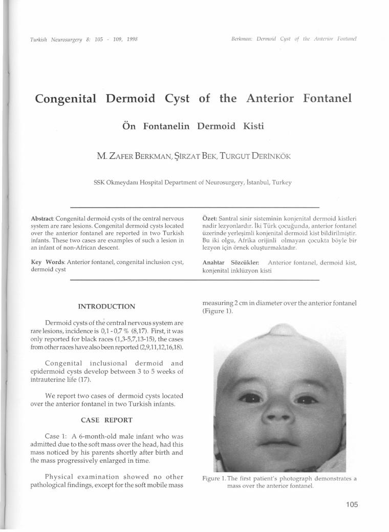
Case 1: A 6-month-old male infant who was
admitted due to the soft mass over the head, had this
mass noticed by his parents shortly af ter birth andthe mass progressively enlarged in time.
Physical examination showed no otherpathological findings, except for the soft mobile mass
Özet: Santral sinir sisteminin konjenital dermoid kistlerinadir lezyonlardir. Iki Türk çocugunda, anterior fontanelüzerinde yerlesimli konjenital dermoid kist bildirilmistir.Bu iki olgu, Afrika orijinli olmayan çocukta böyle birlezyon için örnek olusturmaktadir.
Anahtar Sözcükler: Anterior fontane!, dermoid kist,
konjenital inklüzyon kisti
measuring 2 cm in diameter over the anterior fontanel(Figure 1).
Figure 1.The first patient's photograph demonstrates amass over the anterior fontane!.
105
Tiirkish Neiirosiirgery 8: 105 - 109, 1998
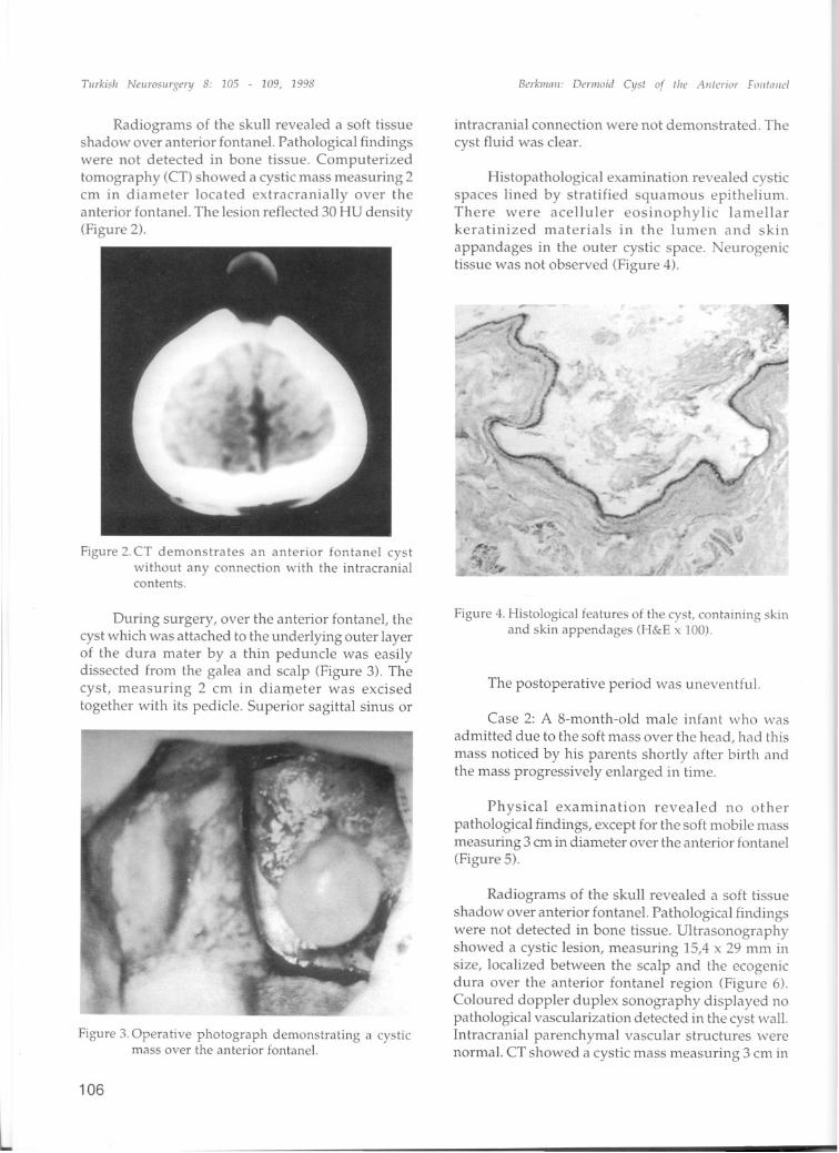
Radiograms of the skull revealed a soft tissueshadow over anterior fontanel. Pathological findingswere not detected in bone tissue. Computerizedtomography (CT) showed a cystic mass measuring 2cm in diameter located extracranial1y over theanterior fontanel. The lesion reflected 30 HU density(Figure 2).
Figure 2. CT demonstrates an anterior fontane! cystwithout any connection with the intracrania!contents.
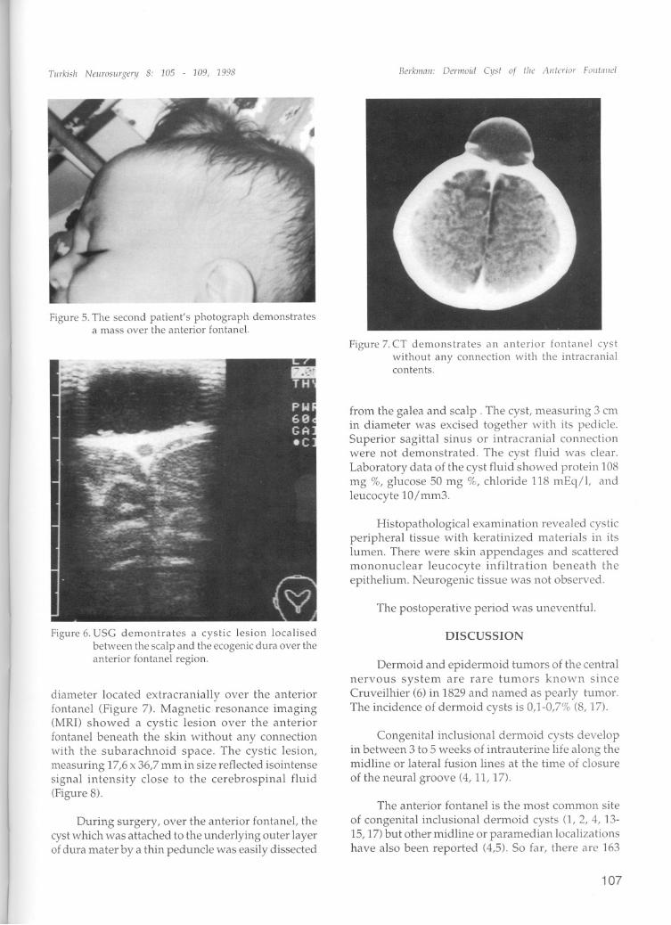
During surgery, over the anterior fontanel, thecyst which was attached to the underlying outer layerof the dura mater by a thin peduncle was easilydissected from the galea and scalp (Figure 3). Thecyst, measuring 2 cm in diameter was excisedtogether with its pedicle. Superior sagittal sinus or
Figure 3. Operative photograph demonstrating a cysticmass over the anterior fontane!.
106
Bak/1i1l1l: Deriiioid eysi of the Aiiterior Fo iiIII iici
intracranial connection were not demonstrated. The
cyst fluid was clear.
Histopathological examination revealed cysticspaces lined by stratified squamous epithelium.There were acel1uler eosinophylic lameiiarkeratinized materials in the lumen and skin
appandages in the outer cystic space. Neurogenictissue was not observed (Figure 4).
Figure 4. Histologica! features of the cyst, containiiig skiiiand skin appendages (H&E x 100).
The postoperative period was uneventful.
Case 2: A 8-month-old male infant who was
admitted due to the soft mass over the head, had this
mass noticed by his parents shortly af ter birth andthe mass progressively enlarged in time.
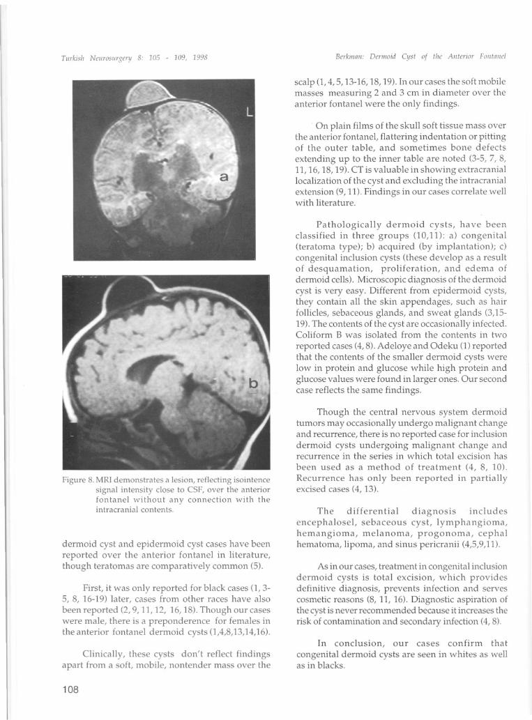
Physical examination revealed no otherpathological findings, except for the soft mobile massmeasuring 3 cm in diameter over the anterior fontanel(Figure 5).
Radiograms of the skull revealed a soft tissue
shadow over anterior fontanel. Pathological findingswere not detected in bone tissue. Ultrasonographyshowed a cystic lesion, measuring 15,4 x 29 mm insize, localized between the scalp and the ecogenicdura over the anterior fontanel region (Figure 6).Coloured doppler duplex sonography displayed nopathological vascularization detected in the cyst wall.Intracranial parenchymal vascular structures werenormaL. CT showed a cystic mass measuring 3 cm in
Tii,.kish Nel/rasl/rge,.y 8: 105 - 109, 1998
Figure 5. The second patient's photograph demonstratesa mass over the anterior fontanel.
Figure 6. use demontrates a eystic lesi on loealisedbetween the sealp and the eeogenie dura over theanterior fontanel region.
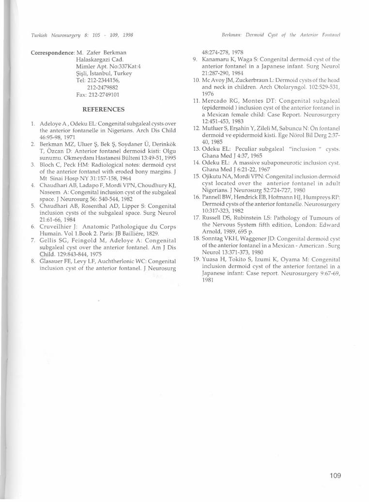
diameter located extracranially over the anteriorfontanel (Figure 7). Magnetic resonance imaging(MRI) showed a cystic lesi on over the anteriorfontanel beneath the skin without any connectionwith the subarachnoid space. The cystic lesion,measuring 17,6x 36,7 mm in size reflected isointensesignal intensity close to the cerebrospinal fluid(Figure 8).
During surgery, over the anterior fontanel, thecyst which was attached to the underlying outer layerof dura mater by a thin pedunde was easily dissected
Be,.kIlI1111: Deriiloiri eyst of the Ante,.io" Fontniid
Figure 7. CT demonstrates an anterior fontanel eystwithout any eonneetion with the intraeranialeontents.
from the galea and scalp . The cyst, measuring 3 cmin diameter was excised together with its pedide.Superior sagittal sinus or intracranial connectionwere not demonstrated. The cyst fluid was dear.Laboratory data of the cyst fluid showed protein 108mg %, glucose 50 mg %, chloride 118 mEq/I, andleucocyte 10/mm3.
Histopathological examination revealed cysticperipheral tissue with keratinized materials in itslumen. There were ski n appendages and scatteredmononuclear leucocyte infiltration beneath theepithelium. Neurogenic tissue was not observed.
The postoperative period was uneventfu!.
DISCUSSION
Dermoid and epidermoid tumors of the centralnervous system are rare tumors known sinceCruveilhier (6) in 1829 and named as pearly tumor.The incidence of dermoid cysts is 0,1-0,7% (8, 17).
Congenital indusional dermoid cysts developin between 3 to 5 weeks of intrauterine life along themidline or lateral fusion lines at the time of c10sure
of the neural groove (4, ll, 17).
The anterior fontanel is the most common site
of congenital inc1usional derm.oid cysts (l, 2, 4, 1315,17) but other midline or paramedian localizationshave alsa been reported (4,5). So far, there are 163
107
Tiirkislr Neiirosiirgery 8: 105 - 109, 1998
Figure 8. MR! demonstrates a lesion, refleeting isointeneesigna! intensity c10se to CSF, over the anteriorfontanel without any connection with theintracrania! contents.
dermoid cyst and epidermoid cyst cases have beenreported over the anterior fontanel in literature,though teratomas are comparatively common (5).
First, it was only reported for black cases O, 35, 8, 16-19) later, cases from other races have also
been reported (2, 9, 11, 12, 16,18). Though our caseswere male, there is a preponderence for females inthe anterior fontanel dermoid cysts 0,4,8,13,14,16).
Clinically, these cysts don't reflect findingsapart from a soft, mobile, nontender mass over the
108
Berkiiinii: Deniioid eyst of tire Aiiterior Foiitaiiel
scalp 0,4,5,13-16,18,19). In our cases the soft mobilemasses measuring 2 and 3 cm in diameter over theanterior fontanel were the only findings.
On plain films of the skull soft tissue mass overthe anterior fontanel, flattering indentation or pittingof the outer table, and sometimes bone defects
extending up to the inner tabi e are noted (3-5, 7, 8,11,16,18,19). CT is valuable in showing extracraniallocalization of the cyst and excluding the intracranialextension (9, 11). Findings in our cases correlate wellwith literature.
Pathologically dermoid cysts, have beenc1assified in three groups (l0,11): a) congenital(teratorna type); b) acquired (by implantation); c)congenital inclusion cysts (these develop as a resultof desquamation, proliferation, and edema ofdermoid cells). Microscopic diagnosis of the dermoidcyst is very easy. Different from epidermoid cysts,they contain all the skin appendages, such as ha irfollicles, sebaceous glands, and sweat glands (3,1519). The contents of the cyst are occasionally infected.Coliform B was isolated from the contents in two
reported cases (4, 8). Adeloye and Odeku O) reportedthat the contents of the smaller dermoid cysts werelow in protein and glucose while high protein andglucose values were found in larger ones. Our secondcase reflects the same findings.
Though the central nervous system dermoidtumors may occasionally undergo malignant changeand recurrence, there is no reported case for inclusiondermoid cysts undergoing malignant change andrecurrence in the series in which total excision has
been used as a method of treatment (4, 8, 10).Recurrence has only be en reported in partiaiiyexcised cases (4, 13).
The differential diagnosis inc1udesencephalosel, sebaceous cyst, lymphangioma,hemangioma, melanoma, progonoma, cephalhematoma, lipoma, and sinus pericranii (4,5,9,11).
As in our cases, treatment in congenital inclusiondermoid cysts is total excision, which providesdefinitive diagnosis, prevents infection and servescosmetic reasons (8, 11, 16). Diagnostic aspiration ofthe cyst is never recommended because it iiKreases therisk of contamination and secondary infection (4, 8).
In conc1usion, our cases confirm that
congenital dermoid cysts are seen in whites as wellas in blacks.
Turkish Neurosiirgenj 8: 105 - 109, 1998
Correspondence: M. Zafer BerkmanHalaskargazi Cad.Mimler Apt. No:337Kat:4Sisli, Istanbul, TurkeyTel: 212-2344156,
212-2479882Fax: 212-2749101
REFERENCES
1. Adeloye A , Odeku EL:Congenital subgaleal cysts overthe anterior fontanelle in Nigerians. Arch Dis Child46:95-98, 1971
2. Ber~man MZ, UIuer S, Bek s, Soydaner Ü, DerinkökT, Ozcan D: Anterior fontanel dermoid kisti: Olgusunumu. Okmeydani Hastanesi Bülteni 13:49-51, 1995
3. Bloch C, Peck HM: Radiological notes: dermoid cystof the anterior fontanel with eroded bony margins. JMt Sinai Hosp NY 31:157-158, 1964
4. Chaudhari AB, Ladapo F, Mordi VPN, Choudhury KI,Naseem A: Congenital inclusion cyst of the subgalealspace. J Neurosurg 56: 540-544, 1982
5. Chaudhari AB, Rosenthal AD, Lipper S: Congenitalinclusion cysts of the subgaleal space. Surg Neurol21:61-66,1984
6. Cruveilhier J: Anatomic Pathologique du CorpsHumain. Vol l.Book 2. Paris: JB Bailliere, 1829.
7. Gellis SG, Feingold M, Adeloye A: Congenitalsubgaleal cyst over the anterior fontane!. Am J DisChild. 129:843-844, 1975
8. Glasauer FE, Levy LF, Auchtherlonic WC: Congenita!inclusion cyst of the anterior fontane!. J Neurosurg
Berkmaii: Demioid eyst of the Aiiterior Foiitaiie!
48:274-278,19789. Kanamaru K, Waga S: Congenital dermoid cyst of the
anterior fontane! in a Japanese infant. Surg Neurol21:287-290, 1984
10. Mc Avoy JM, Zuckerbraun L:Dermoid cystsof the headand neck in children. Arch Otolaryngo!. 102:529-531,1976
11. Mercado RG, Montes DT: Congenita! subgaleal(epidermoid) inclusion cyst of the anterior fontanel ina Mexican female child: Case Report. Neurosurgery12:451-453, 1983
12. MutluerS, Ersahin Y,Zileli M, Sabuncu N: Ön fontaneldermoid ve epidermoid kisti. Ege Nörol Bil Derg 2:3740, 1985
13. Odeku EL: Peculiar subgaleal "inclusion" cysts.Ghana Med J 4:37, 1965
14. Odeku EL: A massive subaponeurotic inclusion cyst.Ghana Med J 6:21-22, 1967
15. Ojikutu NA, Mordi VPN: Congenital inclusion dermoidcyst located over the anterior fontanel in adultNigerians. J Neurosurg 52:724-727, 1980
16. Pannell BW, Hendrick EB, Hofmann HJ, Humpreys RP:Dermoid cysts of the anterior fontanelle. Neurosurgery10:317-323,1982
17. Russell DS, Rubinstein LS: Pathology of Tumours ofthe Nervous System fifth edition, London: EdwardAmold, 1989, 695 p.
18. Sonntag VKH, Waggener JD: Congenital dermoid cystof the anterior fontanel in a Mexican - American. SurgNeuroI13:371-373,1980
19. Yuasa H, Tokito S, lzumi K, Oyama M: Congenitalinclusion dermoid cyst of the anterior fontanel in aJapanese infant: Case report. Neurosurgery 9:67-69,1981
109


![Epidermoid Cyst of the Buccal Mucosa Diagnosed by Magnetic ... › open-access › epidermoid... · and develops into an (epi)dermoid cyst [2]. Epidermoid cysts can occur anywhere](https://img.pdfslide.net/doc/110x75/5f0d012a7e708231d43833de/epidermoid-cyst-of-the-buccal-mucosa-diagnosed-by-magnetic-a-open-access-a.jpg)


![Epidermoid and dermoid cysts of the head and neck region · Sahalok et al. Epidermoid and dermoid cyst removal 348 cyst in the oral cavity, lower lip, or upper lip.[7] Giant epidermoid](https://img.pdfslide.net/doc/110x75/5f0d065f7e708231d4384dcd/epidermoid-and-dermoid-cysts-of-the-head-and-neck-region-sahalok-et-al-epidermoid.jpg)