Embed Size (px)
Citation preview
CONGENITAL DISLOCATION OF THE HIP IN THE NEWBORN
G. B. SMAILL, WELLINGTON, NEW ZEALAND
Though it has long been recognised that the earlier treatment for congenital dislocation
of the hip is started the better is the result, it is only quite recently that serious efforts have
been made to recognise the condition at birth. A clinical method of diagnosing congenital
dislocation of the hip first described by Ortolani (1937) has been used in Northern Italy for
many years, but until von Rosen (1962) published his results only lip service was paid elsewhere
to the diagnosis of hip dislocation in the newborn. Even now most orthopaedic textbooks
emphasise only the classical signs of congenital dislocation of the hip and if it is mentioned
at all Ortolani’s test is usually described incorrectly.
In New Zealand it has been the practice in a few hospitals for all newborn babies to be
examined for hip dislocation as part of a general physical examination. This examination is
carried out by a variety of examiners, some more skilled than others. All babies in this country
are born in maternity hospitals varying in size from about twelve beds to the larger units
attached to general hospitals. Most of these babies are delivered by general practitioners
and there is a wide variation in the number of deliveries each doctor does in a year and in
the thoroughness of the examination of the babies delivered by him.
The present study was made of babies born in the maternity unit attached to Wellington
Hospital. Fourteen per cent of these babies were delivered by members of the obstetric unit,
the remainder by forty-five different doctors. Those deliveries done by the obstetric unit were
either in unbooked cases or where some complication had arisen in pregnancy or labour
necessitating specialist obstetrical management.
From February 1963 to April 1966 the author personally examined the hips of all 6,000
babies born at this hospital. For the first 2,100 babies clinical examination of the hips was
carried out once weekly, so the babies ranged in age from none to seven days. In the first few
months of the survey it was the author’s policy to defer examination when a baby appeared
to be very frail or ill until its condition had improved. As will be seen later, this proved to
be unwise. The later 3,900 babies were seen at examinations conducted twice weekly, so that
the babies ranged in age from none to four days.
METHOD OF EXAMINATION
The examination was carried out with the baby lying on its back in its cot with its nappies
removed. To prevent cross infection the hands were washed between each examination. The
techniques used were those described by Barlow (1962), with slight modification, and Ortolani
(1937). Each hip ofeach baby was checked by the two methods. Though these methods have
been fully described elsewhere, no apology is made here for outlining once again the steps of
the examination.
Barlow’s method-The left hip is examined first with the right hand. The baby’s pelvis is
supported with the thumb of the left hand on the pubis and the sacrum resting in the palm,
the flexed right leg being tethered in the web space between thumb and index finger. The
baby’s left knee and hip are flexed to 90 degrees with the shin resting in the web space between
thumb and index finger of the right hand. The right thumb rests on the medial aspect of the
thigh in the region of the lesser trochanter while the fingers rest on the greater trochanter.
The femur is in fact held firmly between fingers and thumb. Now pressure is exerted backwards
with the thumb and at the same time the hand is slightly pronated. If the hip joint is lax the
524 THE JOURNAL OF BONE AND JOINT SURGERY
CONGENITAL DISLOCATION OF THE HIP IN THE NEWBORN 525
head of the femur is felt to slip out of the acetabulum. By pressing forward with the fingers
and supinating the hand the head of femur is felt to slip back into the acetabulum. The test
is then carried out on the right hip using the left hand to examine the hip while the right hand
supports the pelvis.
This test is a very useful screen for detecting the lax or unstable hip. If the baby is quiet
it is very simple but when the baby is crying muscle spasm sometimes makes it difficult to
displace an unstable hip and more care is needed. It is, therefore, an advantage to examine
babies in their own cots and preferably shortly after they have been fed.
Ortolani’s method-This method is based on the fact that, in babies with dislocation of the
hip, gradual abduction of the hip flexed to a right angle causes the dislocated head of femur
to slip into the acetabulum with a palpable but not audible click. This “ click “ is accentuated
by exerting slight pressure backwards along the line of the femur as it is abducted, thus
displacing the head of the femur more posteriorly. If too much pressure is applied the click
may be missed altogether. Next, as the abducted hip is adducted to the midline the head of
the femur can be felt dislocating again with a palpable click. It is very important that the
hip should not be flexed beyond 90 degrees, or the click may not be felt. The grip used for
this examination is similar to that used for Barlow’s method.
If the hip is very lax the click of dislocation and reduction may be very slight, and if the
examiner is not aware of this laxity as indicated by Barlow’s test it would be quite possible
to miss the diagnosis. The two tests are therefore complementary.
Early in the series the author was confused by a variety of clicks which were not only
palpable but also audible. Most of these arose at the patello-femoral and knee joints, and a
few were from the hips but were not due to instability. The “ click “ of dislocation and
reduction in congenital dislocation is inaudible.
RESULTS
Of the 6,000 babies examined twenty-four were considered to have congenital dislocation
of the hip and twenty-three of these were treated in a von Rosen type of splint. Table I shows
the number of cases diagnosed each year of the survey and the age of the baby in days at the
time of diagnosis.
Sex incidence-As in all other series the incidence of congenital dislocation was far higher
in females than in males (Table II).
Side affected-The left hip was involved more often than the right. Of thirty-four affected
hips twenty-two were left and twelve were right (Table 111).
Race-There was no racial predominance in the incidence of dislocation (Table IV). In China
congenital dislocation of the hip is rare, possibly because of the way babies are carried with
their hips abducted across the mother’s back. Both Chinese babies in this series were full
blooded and unrelated.
Presentation-Six of the twenty-four babies (one boy and five girls) presented by the breech.
According to Mayes (1950) the normal incidence of breech presentation is one in twenty-five.
The higher incidence of hip dislocation in babies presenting by the breech is in accord with
the findings of Barlow (1962).
Family history-Two of the twenty-four babies had a family history of congenital dislocation,
and in one of these it was strong. Several of the babies with dislocation had normal siblings
later but one girl with bilateral dislocation later had a sister who was similarly affected. There
was no previous history of dislocation in this family and the first child born was normal.
There is undoubtedly a hereditary factor in congenital dislocation of the hip but this is
probably not nearly so strong as has been thought in the past.
Prematurity-Premature babies were not more prone to dislocation than those born at full
term.
VOL. 50 B, NO. 3, AUGUST 1968

526 G. B. SMAILL
TABLE I
INCIDENCE OF CONGENITAL DISLOCATION AND AGE AT DIAGNOSIS
Year Number of babies Age of baby in days
1963 4 5,8,5,4
1964 12 4,l,l,3,5,1,3,3,5,1,5,2
1965 6 1, 4, 1, 4, 2, 5
1966 2 2,3
TABLE II
SEX INCIDENCE
Number of babies
YearFemale Male
1963 3
1964 11
1965 4 2
1966 2 0
Total 20 4
TABLE III
SIDE AFFECTED BY CONGENITAL DISLOCATION
Year Bilateral Left , Right
1963 0 3 1
1964 , 5 6 1
1965 3 3 0
1966 2 0 0
TABLE IV
RACIAL INCIDENCE
R Number of babiesace with hip dislocation
British . . 16
Maori . . 3
Chinese. . 2
Polish . . 2
Dutch . 1
THE JOURNAL OF BONE AND JOiNT SURGERY
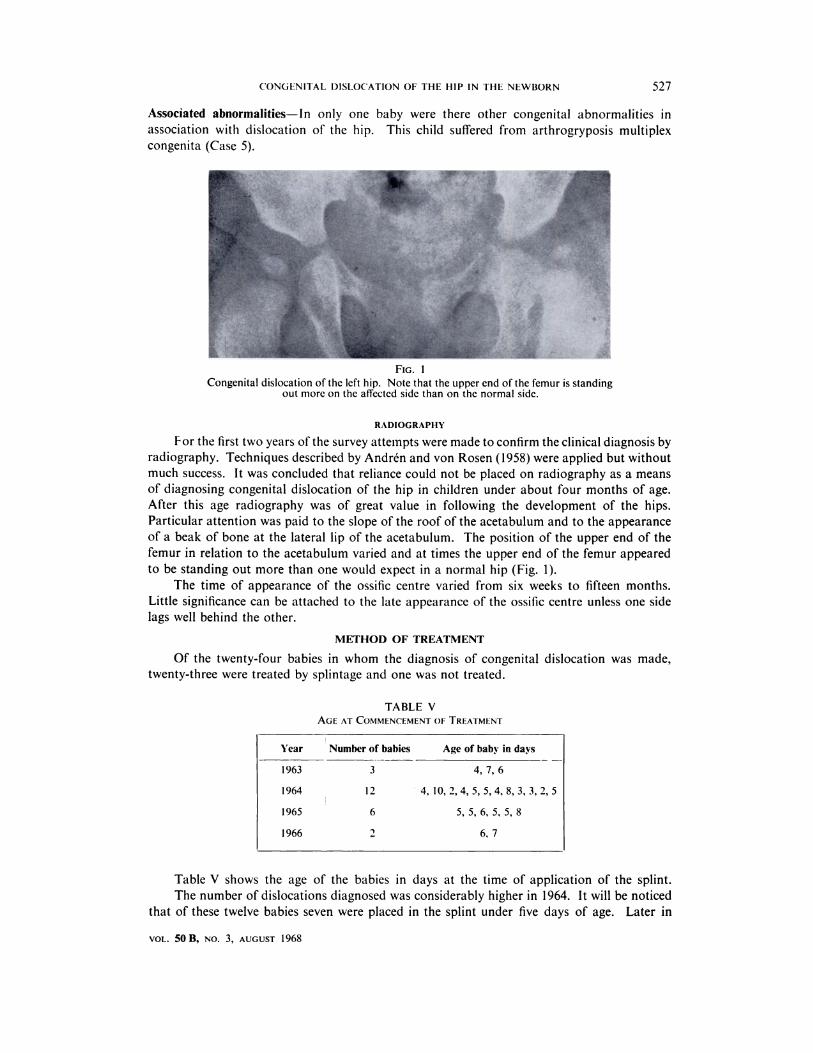
FIG. 1
Year Number of babies Age of baby in days
1963 3 4,7,6
1964 12 4, 10, 2,4, 5, 5,4, 8, 3, 3, 2, 5
1965 6 5, 5, 6, 5, 5, 8
1966 2 6,7
CONGENITAL DISLOCATION OF THE HIP IN THE NEWBORN 527
VOL. 50 B, NO. 3, AUGUST 1968
Associated abnormalities-In only one baby were there other congenital abnormalities in
association with dislocation of the hip. This child suffered from arthrogryposis multiplex
congenita (Case 5).
Congenital dislocation of the left hip. Note that the upper end of the femur is standingout more on the affected side than on the normal side.
RADIOGRAPHY
For the first two years of the survey attempts were made to confirm the clinical diagnosis by
radiography. Techniques described by Andr#{233}n and von Rosen (1958) were applied but without
much success. It was concluded that reliance could not be placed on radiography as a means
of diagnosing congenital dislocation of the hip in children under about four months of age.
After this age radiography was of great value in following the development of the hips.
Particular attention was paid to the slope of the roof of the acetabulum and to the appearance
of a beak of bone at the lateral lip of the acetabulum. The position of the upper end of the
femur in relation to the acetabulum varied and at times the upper end of the femur appeared
to be standing out more than one would expect in a normal hip (Fig. 1).
The time of appearance of the ossific centre varied from six weeks to fifteen months.
Little significance can be attached to the late appearance of the ossific centre unless one side
lags well behind the other.
METHOD OF TREATMENT
Of the twenty-four babies in whom the diagnosis of congenital dislocation was made,
twenty-three were treated by splintage and one was not treated.
TABLE V
AGE AT COMMENCEMENT OF TREATMENT
Table V shows the age of the babies in days at the time of application of the splint.
The number of dislocations diagnosed was considerably higher in 1964. It will be noticed
that of these twelve babies seven were placed in the splint under five days of age. Later in
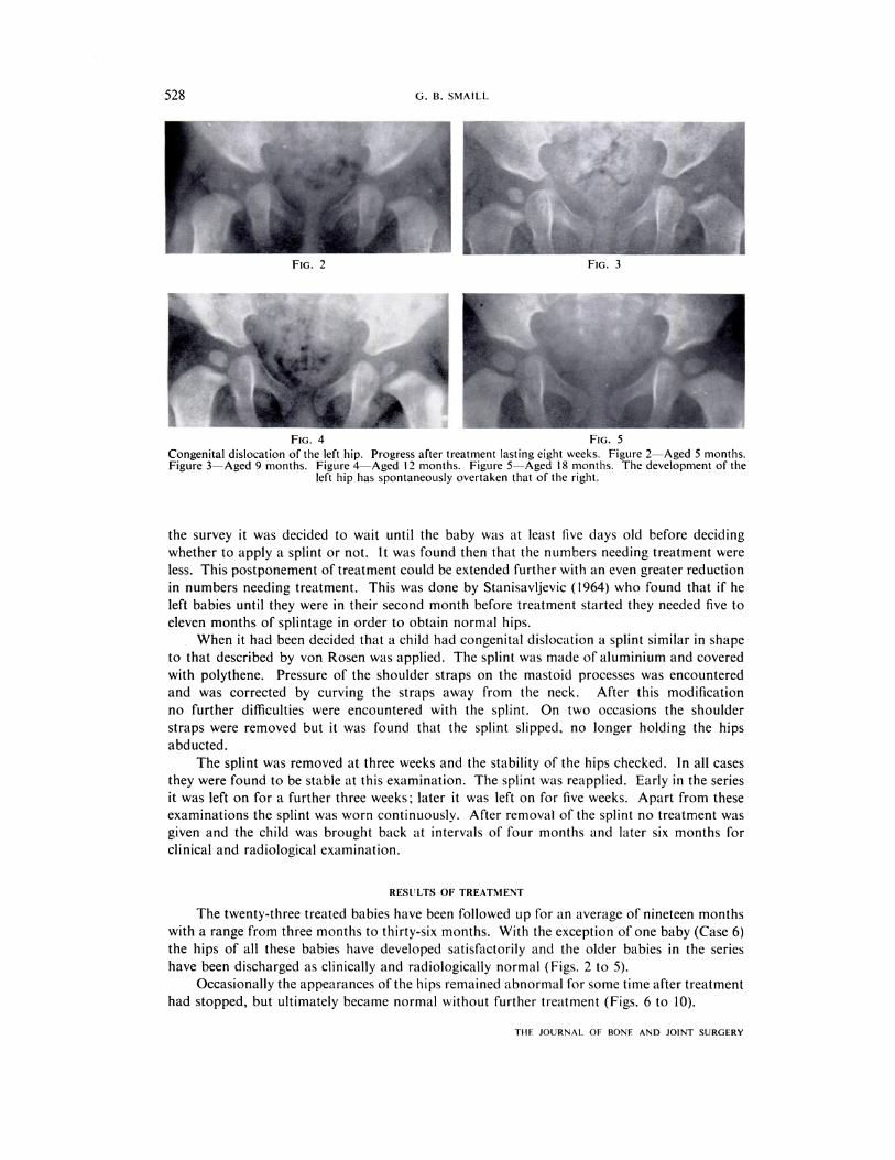
FIG. 2
FIG. 4 FIG. 5
Congenital dislocation of the left hip. Progress after treatment lasting eight weeks. Figure 2-Aged 5 months.Figure 3-Aged 9 months. Figure 4-Aged 12 months. Figure 5-Aged 18 months. The development of the
left hip has spontaneously overtaken that of the right.
528 G. B. SMAILL
TI-IF JOURNAL OF BONE AND JOINT SURGERY
,,,.�. - �.
AFIG. 3
the survey it was decided to wait until the baby was at least five days old before deciding
whether to apply a splint or not. It was found then that the numbers needing treatment were
less. This postponement of treatment could be extended further with an even greater reduction
in numbers needing treatment. This was done by Stanisavljevic (1964) who found that if he
left babies until they were in their second month before treatment started they needed five to
eleven months of splintage in order to obtain normal hips.
When it had been decided that a child had congenital dislocation a splint similar in shape
to that described by von Rosen was applied. The splint was made of aluminium and covered
with polythene. Pressure of the shoulder straps on the mastoid processes was encountered
and was corrected by curving the straps away from the neck. After this modification
no further difficulties were encountered with the splint. On two occasions the shoulder
straps were removed but it was found that the splint slipped, no longer holding the hips
abducted.
The splint was removed at three weeks and the stability of the hips checked. In all cases
they were found to be stable at this examination. The splint was reapplied. Early in the series
it was left on for a further three weeks; later it was left on for five weeks. Apart from these
examinations the splint was worn continuously. After removal of the splint no treatment was
given and the child was brought back at intervals of four months and later six months for
clinical and radiological examination.
RESULTS OF TREATMENT
The twenty-three treated babies have been followed up for an average of nineteen months
with a range from three months to thirty-six months. With the exception of one baby (Case 6)
the hips of all these babies have developed satisfactorily and the older babies in the series
have been discharged as clinically and radiologically normal (Figs. 2 to 5).
Occasionally the appearances of the hips remained abnormal for some time after treatment
had stopped, but ultimately became normal without further treatment (Figs. 6 to 10).
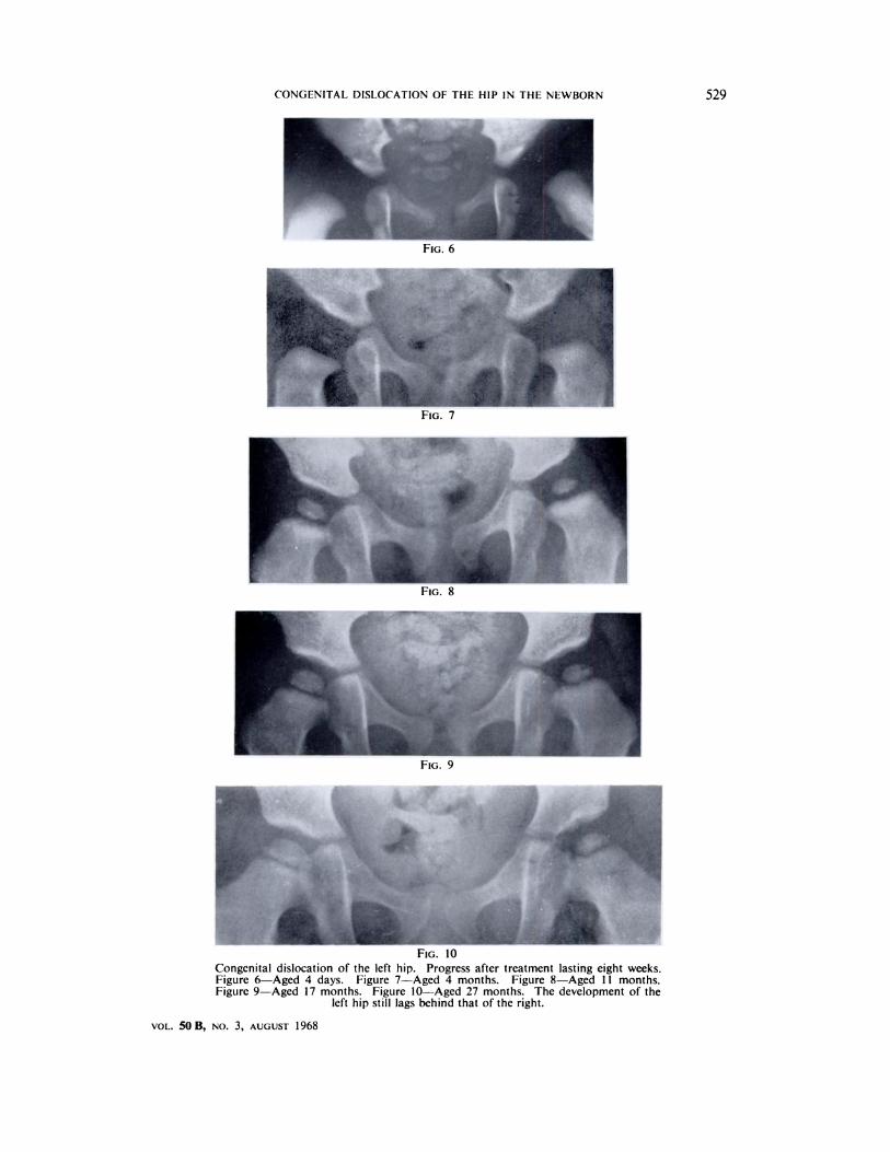
FIG. 10
Congenital dislocation of the left hip. Progress after treatment lasting eight weeks.Figure 6-Aged 4 days. Figure 7-Aged 4 months. Figure 8-Aged 11 months.Figure 9-Aged 17 months. Figure 10-Aged 27 months. The development of the
left hip still lags behind that of the right.
CONGENITAL DISLOCATION OF THE HIP IN THE NEWBORN 529
VOL. 50 B, NO. 3, AUGUST 1968
a,
I �FIG. 6
1
11
‘S
- :
530 G. B. SMAILL
ri ii JOURNAl. OF HONE ANI) JOINT SURGERY
CASES MISSED
During the period of the survey four babies were born who were passed as normal at
birth but who were later found to have abnormal hips.
Case 1-A baby girl, born by breech delivery in the twenty-eighth week of pregnancy, weighed
2 pounds 8 ounces at birth. For the first few weeks there was considerable doubt as to
whether she would survive. In view of this, examination of the hips was deferred until she
was nearly three weeks old and at that time they were passed as normal. At ten months she
was noticed to have asymmetrical skin creases and a clinical diagnosis ofdislocation ofthe left
hip was confirmed by radiography. Even at this age her management was complicated by
repeated episodes of respiratory infection which delayed reduction and splintage.
Two factors were probably responsible for the failure of diagnosis in this child. Firstly,
she was born early in the survey before the author had felt any other newborn babies with
dislocated hips. Secondly, the examination was delayed for three weeks, by which time the
signs may have no longer been present. Examination of the hips causes so slight a disturbance
that it should not be delayed beyond a few days from birth. Owing to the increased difficulty
of examining babies in the incubator special care should be taken with premature infants.
a
Flo.
FIG. 12
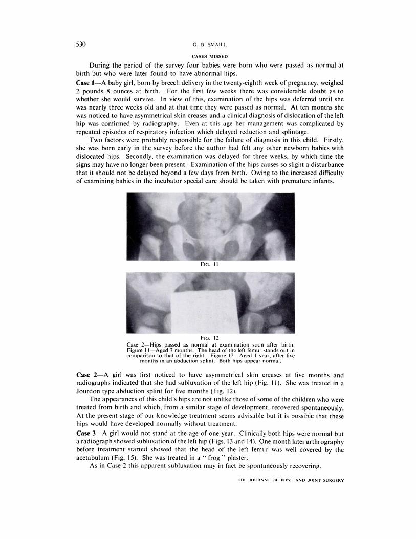
Case 2-Hips passed as normal at examination soon after birth.Figure 11-Aged 7 months. The head of the left femur stands out incomparison to that of the right. Figure 12--Aged 1 year, after five
months in an abduction splint. Both hips appear normal.
Case 2-A girl was first noticed to have asymmetrical skin creases at five months and
radiographs indicated that she had subluxation of the left hip (Fig. I I ). She was treated in a
Jourdon type abduction splint for five months (Fig. 12).
The appearances ofthis child’s hips are not unlike those ofsome ofthe children who were
treated from birth and which, from a similar stage of development, recovered spontaneously.
At the present stage of our knowledge treatment seems advisable but it is possible that these
hips would have developed normally without treatment.
Case 3-A girl would not stand at the age of one year. Clinically both hips were normal but
a radiograph showed subluxation ofthe left hip (Figs. 13 and 14). One month later arthrography
before treatment started showed that the head of the left femur was well covered by the
acetabulum (Fig. 15). She was treated in a “frog” plaster.
As in Case 2 this apparent subluxation may in fact be spontaneously recovering.
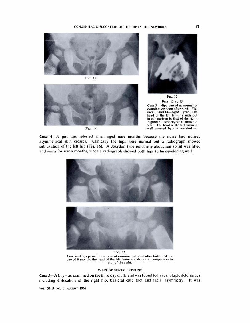
FIG. 14
FIG. 15
FIGs. 13 TO 15Case 3-Hips passed as normal atexamination soon after birth. Fig-ures 13 and 14-Aged 1 year. Thehead of the left femur stands outin comparison to that of the right.Figure 1 5-Arthrograph one monthlater. The head of the left femur iswell covered by the acetabulum.
--
CONGENITAL DISLOCATION OF THE HIP IN THE NEWBORN 531
VOL. 50 B, NO. 3, AUGUST 1968
Case 4-A girl was referred when aged nine months because the nurse had noticed
asymmetrical skin creases. Clinically the hips were normal but a radiograph showed
subluxation of the left hip (Fig. 16). A Jourdon type polythene abduction splint was fitted
and worn for seven months, when a radiograph showed both hips to be developing well.
16
Case 4-Hips passed as normal at examination soon after birth. At theage of 9 months the head of the left femur stands out in comparison to
that of the right.
CASES OF SPECIAL INTEREST
Case 5-A boy was examined on the third day of life and was found to have multiple deformities
including dislocation of the right hip, bilateral club foot and facial asymmetry. It was
TI-IF JOURNAL OF BONE AND JOINT SURGERY
532 G. B. SMAILL
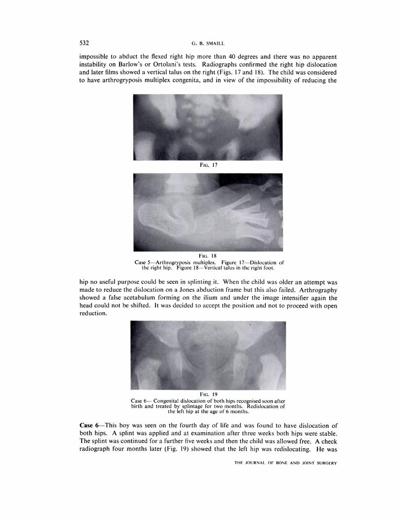
impossible to abduct the flexed right hip more than 40 degrees and there was no apparent
instability on Barlow’s or Ortolani’s tests. Radiographs confirmed the right hip dislocation
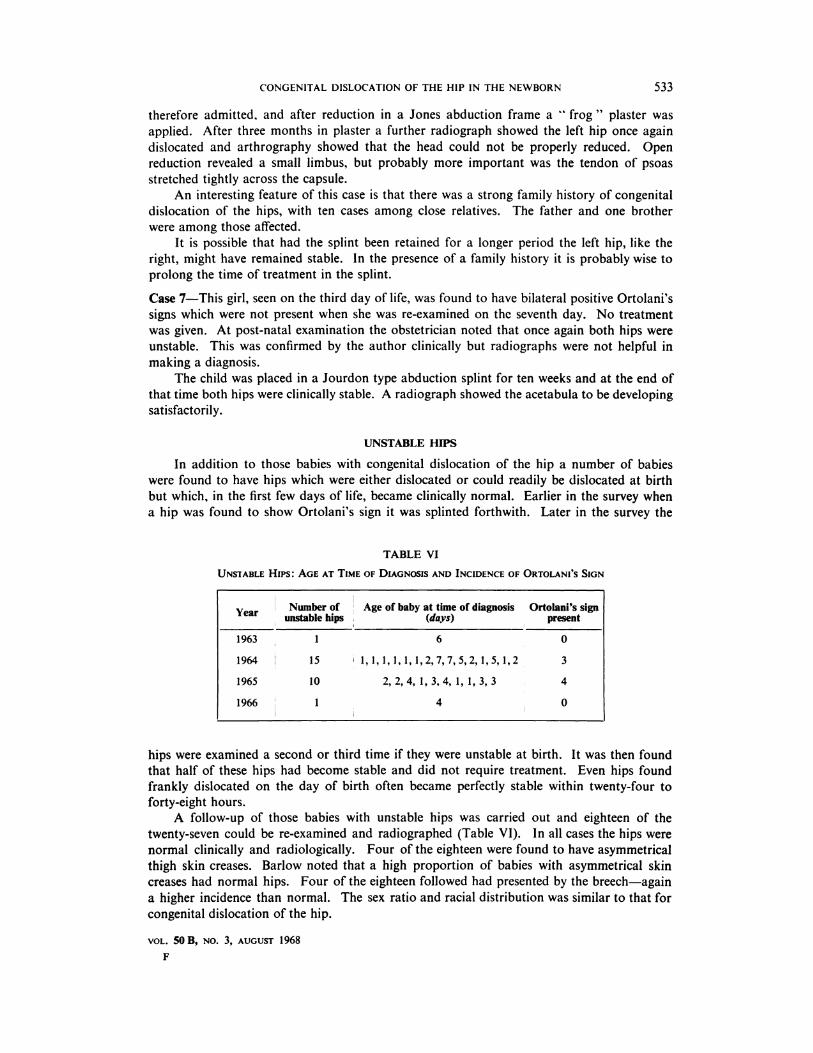
and later films showed a vertical talus on the right (Figs. 17 and 18). The child was considered
to have arthrogryposis multiplex congenita, and in view of the impossibility of reducing the
FIG. 18
Case 5-Arthrogryposis multiplex. Figure 17--Dislocation ofthe right hip. Figure 18-Vertical talus in the right foot.
hip no useful purpose could be seen in splinting it. When the child was older an attempt was
made to reduce the dislocation on a Jones abduction frame but this also failed. Arthrography
showed a false acetabulum forming on the ilium and under the image intensifier again the
head could not be shifted. It was decided to accept the position and not to proceed with open
reduction.
- fr_ -
I
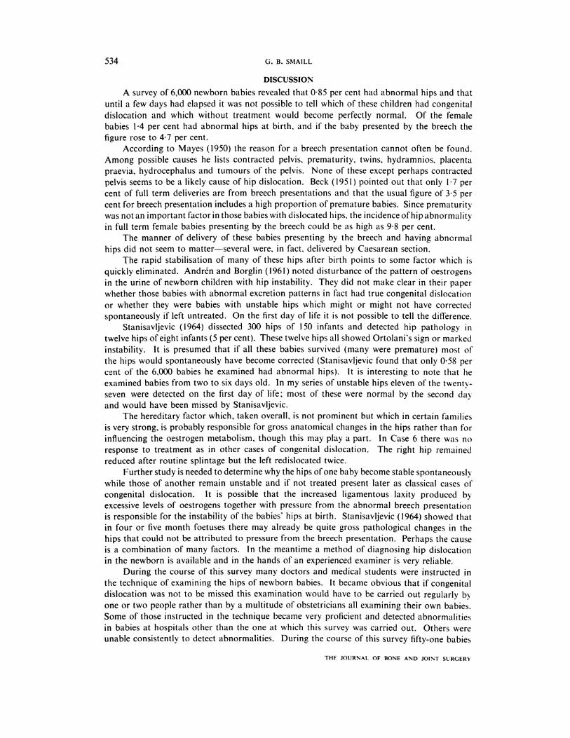
FIG. 19Case 6- Congenital dislocation of both hips recognised soon afterbirth and treated by splintage for two months. Redislocation of
the left hip at the age of 6 months.
Case 6-This boy was seen on the fourth day of life and was found to have dislocation of
both hips. A splint was applied and at examination after three weeks both hips were stable.
The splint was continued for a further five weeks and then the child was allowed free. A check
radiograph four months later (Fig. 19) showed that the left hip was redislocating. He was
CONGENITAL DISLOCATION OF THE HIP IN THE NEWBORN 533
therefore admitted, and after reduction in a Jones abduction frame a “ frog “ plaster was
applied. After three months in plaster a further radiograph showed the left hip once again
dislocated and arthrography showed that the head could not be properly reduced. Open
reduction revealed a small limbus, but probably more important was the tendon of psoas
stretched tightly across the capsule.
An interesting feature of this case is that there was a strong family history of congenital
dislocation of the hips, with ten cases among close relatives. The father and one brother
were among those affected.
It is possible that had the splint been retained for a longer period the left hip, like the
right, might have remained stable. In the presence of a family history it is probably wise to
prolong the time of treatment in the splint.
Case 7-This girl, seen on the third day of life, was found to have bilateral positive Ortolani’s
signs which were not present when she was re-examined on the seventh day. No treatment
was given. At post-natal examination the obstetrician noted that once again both hips were
unstable. This was confirmed by the author clinically but radiographs were not helpful in
making a diagnosis.
The child was placed in a Jourdon type abduction splint for ten weeks and at the end of
that time both hips were clinically stable. A radiograph showed the acetabula to be developing
satisfactorily.
UNSTABLE HIPS
In addition to those babies with congenital dislocation of the hip a number of babies
were found to have hips which were either dislocated or could readily be dislocated at birth
but which, in the first few days of life, became clinically normal. Earlier in the survey when
a hip was found to show Ortolani’s sign it was splinted forthwith. Later in the survey the
TABLE VI
UNSTABLE Hips : AGE AT TIME OF DIAGNOSIS AND INCIDENCE OF ORTOLANI’S SIGN
Y � Number of � Age of baby at time of diagnosis Ortolani’s sign
ear � unstable hips � (days) present
1963 � 1 6 � 0
1964 � 15 � 1, 1,1, 1, 1, 1,2,7,7,5,2,1,5, 1,2 3
1965 10 2, 2, 4, 1, 3, 4, 1, 1, 3, 3 � 4
1966 � 1 4 � 0
hips were examined a second or third time if they were unstable at birth. It was then found
that half of these hips had become stable and did not require treatment. Even hips found
frankly dislocated on the day of birth often became perfectly stable within twenty-four to
forty-eight hours.
A follow-up of those babies with unstable hips was carried out and eighteen of the
twenty-seven could be re-examined and radiographed (Table VI). In all cases the hips were
normal clinically and radiologically. Four of the eighteen were found to have asymmetrical
thigh skin creases. Barlow noted that a high proportion of babies with asymmetrical skin
creases had normal hips. Four of the eighteen followed had presented by the breech-again
a higher incidence than normal. The sex ratio and racial distribution was similar to that for
congenital dislocation of the hip.
VOL. 50 B, NO. 3, AUGUST 1968
F
534 G. B. SMAILL
DISCUSSION
A survey of 6,000 newborn babies revealed that 085 per cent had abnormal hips and that
until a few days had elapsed it was not possible to tell which of these children had congenital
dislocation and which without treatment would become perfectly normal. Of the female
babies 14 per cent had abnormal hips at birth, and if the baby presented by the breech the
figure rose to 4�7 per cent.
According to Mayes (1950) the reason for a breech presentation cannot often be found.
Among possible causes he lists contracted pelvis, prematurity, twins, hydramnios, placenta
praevia, hydrocephalus and tumours of the pelvis. None of these except perhaps contracted
pelvis seems to be a likely cause of hip dislocation. Beck (1951 ) pointed out that only l7 per
cent of full term deliveries are from breech presentations and that the usual figure of 3�5 per
cent for breech presentation includes a high proportion of premature babies. Since prematurity
was not an important factor in those babies with dislocated hips, the incidence ofhip abnormality
in full term female babies presenting by the breech could be as high as 98 per cent.
The manner of delivery of these babies presenting by the breech and having abnormal
hips did not seem to matter-several were, in fact, delivered by Caesarean section.
The rapid stabilisation of many of these hips after birth points to some factor which is
quickly eliminated. Andr#{233}nand Borglin (1961) noted disturbance of the pattern of oestrogens
in the urine of newborn children with hip instability. They did not make clear in their paper
whether those babies with abnormal excretion patterns in fact had true congenital dislocation
or whether they were babies with unstable hips which might or might not have corrected
spontaneously if left untreated. On the first day of life it is not possible to tell the difference.
Stanisavljevic (1964) dissected 300 hips of 150 infants and detected hip pathology in
twelve hips ofeight infants (5 per cent). These twelve hips all showed Ortolani’s sign or marked
instability. lt is presumed that if all these babies survived (many were premature) most of
the hips would spontaneously have become corrected (Stanisavljevic found that only 058 per
cent of the 6,000 babies he examined had abnormal hips). It is interesting to note that he
examined babies from two to six days old. in my series of unstable hips eleven of the twenty-
seven were detected on the first day of life; most of these were normal by the second day
and would have been missed by Stanisavljevic.
The hereditary factor which, taken overall, is not prominent but which in certain families
is very strong, is probably responsible for gross anatomical changes in the hips rather than for
influencing the oestrogen metabolism, though this may play a part. In Case 6 there was no
response to treatment as in other cases of congenital dislocation. The right hip remained
reduced after routine splintage but the left redislocated twice.
Further study is needed to determine why the hips ofone baby become stable spontaneously
while those of another remain unstable and if not treated present later as classical cases of
congenital dislocation. It is possible that the increased ligamentous laxity produced by
excessive levels of oestrogens together with pressure from the abnormal breech presentation
is responsible for the instability ofthe babies’ hips at birth. Stanisavljevic (1964) showed that
in four or five month foetuses there may already be quite gross pathological changes in the
hips that could not be attributed to pressure from the breech presentation. Perhaps the cause
is a combination of many factors. in the meantime a method of diagnosing hip dislocation
in the newborn is available and in the hands of an experienced examiner is very reliable.
During the course of this survey many doctors and medical students were instructed in
the technique of examining the hips of newborn babies. It became obvious that if congenital
dislocation was not to be missed this examination would have to be carried out regularly by
one or two people rather than by a multitude of obstetricians all examining their own babies.
Some of those instructed in the technique became very proficient and detected abnormalities
in babies at hospitals other than the one at which this survey was carried out. Others were
unable consistently to detect abnormalities. During the course of this survey fifty-one babies
THE JOURNAL OF BONE AND JOINT SURGERY
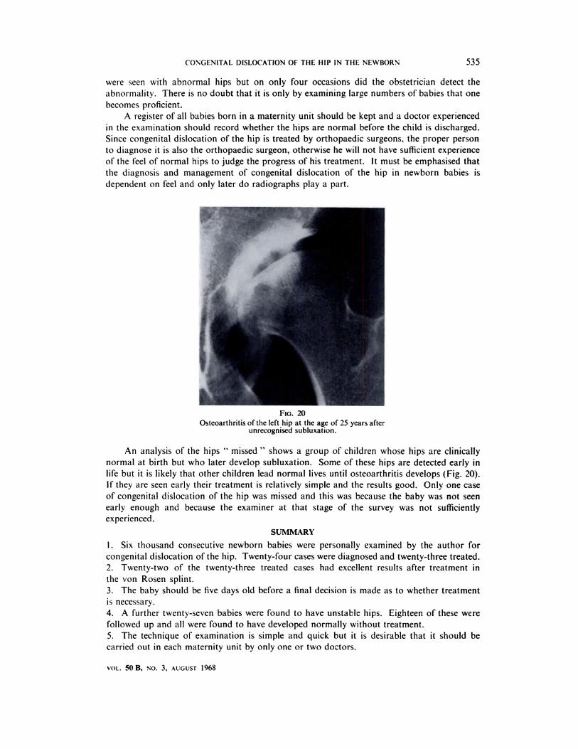
FIG. 20
Osteoarthritis of the left hip at the age of 25 years afterunrecognised subluxation.
CONGENITAL DISLOCATION OF THE HIP IN THE NEWBORN 535
VOL. 50B, NO. 3, AUGUST 1968
were seen with abnormal hips but on only four occasions did the obstetrician detect the
abnormality. There is no doubt that it is only by examining large numbers of babies that one
becomes proficient.
A register of all babies born in a maternity unit should be kept and a doctor experienced
in the examination should record whether the hips are normal before the child is discharged.
Since congenital dislocation of the hip is treated by orthopaedic surgeons, the proper person
to diagnose it is also the orthopaedic surgeon, otherwise he will not have sufficient experience
of the feel of normal hips to judge the progress of his treatment. It must be emphasised that
the diagnosis and management of congenital dislocation of the hip in newborn babies is
dependent on feel and only later do radiographs play a part.
An analysis of the hips “ missed “ shows a group of children whose hips are clinically
normal at birth but who later develop subluxation. Some of these hips are detected early in
life but it is likely that other children lead normal lives until osteoarthritis develops (Fig. 20).
If they are seen early their treatment is relatively simple and the results good. Only one case
of congenital dislocation of the hip was missed and this was because the baby was not seen
early enough and because the examiner at that stage of the survey was not sufficiently
experienced.
SUMMARY
1 . Six thousand consecutive newborn babies were personally examined by the author for
congenital dislocation of the hip. Twenty-four cases were diagnosed and twenty-three treated.
2. Twenty-two of the twenty-three treated cases had excellent results after treatment in
the von Rosen splint.
3. The baby should be five days old before a final decision is made as to whether treatment
is necessary.
4. A further twenty-seven babies were found to have unstable hips. Eighteen of these were
followed up and all were found to have developed normally without treatment.
5. The technique of examination is simple and quick but it is desirable that it should be
carried out in each maternity unit by only one or two doctors.

536 G. B. SMAILL
REFERENCES
ANDREN, L., and BORGLIN, N. E. (1961): Disturbed Urinary Excretion Pattern of Oestrogens in Newbornswith Congenital Dislocation of the Hip. Ada Endocrinologica, 37, 423.
ANDREN, L., and von ROSEN, S. (1958): The Diagnosis ofDislocation of the Hip in Newborns and the Primary
Results of Immediate Treatment. Ada Radiologica, 49, 89.
BARLOW, T. G. (1962): Early Diagnosis and Treatment of Congenital Dislocation of the Hip. Journal of Bone
and Joint Surgery, 44-B, 292.BECK, A. C. (1951): Obstetrical Practice. Baltimore: Williams & Wilkins.
MAYES, B. T. (1950): A Textbook of Obstetrics. Sydney: Australasian Publishing Co.
ORTOLANI, M. (1937): Un segno poco noto e sua importanza per Ia diagnosi precoce di prelussazione congenita
dell’anca. Pediatria, 45, 129.
ROSEN, S. Von (1962): Diagnosis and Treatment of Congenital Dislocation of the Hip Joint in the New-born.Journal of Bone and Joint Surgery, 44-B, 284.
STANISAVLJEVIC, S. (1964): Diagnosis and Treatment of Congenital Hip Pathology in the Newborn. Baltimore:Williams & Wilkins.
THE JOURNAL OF BONE AND JOINT SURGERY