Embed Size (px)
Citation preview
CONICAL FLUKES IN RUMINANTS
Dr. J.H. Vorster, BVSc, MMedVet(Path)Vetdiagnostix Veterinary Pathology Services, PO Box 13624 Cascades, 3202 Tel no: 033 342 5104Cell no: 082 820 5030E-mail: [email protected]
Dr. P.H. Mapham, BVSc (Hon)Veterinary House Hospital,339 Prince Alfred Road, Pietermaritzburg, 3201Tel no: 033 342 4698Cell No: 082 771 3227E-mail: [email protected]
Introduction
Conical fluke is a trematode parasite of the family Paramphistomatidae. This family includes the genera Paramphistomum, Cotylophoron, Ceylonocotyle, Gigantocyle, Gastrothylax Fischoederius, Carmyerius, Gastrodiscus and Pseudodiscus. At least 14 species have been described in domestic ruminants of which the most important flukes are Paramphistomum cervi and P. microbothrium. Others are P. microbothrioides, P. liorchis, P. ichikawai and P. explantum.
Paramphistomes are found worldwide but disease is most common in the warmer regions in Australasia, Africa, and India and is mostly of clinical significance in cattle, goats and sheep. Calicophoron calicophorum, Ceylonocotyle streptocoelium, Calicophoron ijimai, and Cotylophoron cotylophoron have been associated with clinical disease in buffalo and cattle in Asia, and cattle in Australasia and the southern USA. Various other species of Cotyolophoron, Gastrothylax, and Fischoederius have also been associated with disease in these species. In sheep and goats, P.microbothrium, P.ichikawai, P.cervi, P.explanatum, G.crumenifer, Cotylophoron cotylophorum, and F. cobboldi have all been associated with clinicaldisease. Species such as Gastrodiscus have been reported in the small and large intestines of equines in tropical regions. Homologaster has been
1
associated with the large intestine of ruminants in Asia. Gigantocotyle has been reported from the bile ducts in buffalo in the middle and Far East. Human infestations with certain species of paramphistomes have been reported to have medical importance, but from the public health standpoint, the most important species is Gastrodiscoides hominis.
These parasites require an intermediate host in the form of water snails including planorbid snails of the sub – family buliniae. The immature parasites are found in the duodenum from where they migrate towards the rumen and reticulum where the adults are situated. The adult parasite is distinctly visible in the forestomachs of ruminants and veterinary practitioners may frequently need to answer questions from clients regarding these observations. In South Africa P. microbothrium seems to be the most important fluke in domestic ruminants.
Life cycle
The life cycle is fairly typical of the gastrointestinal trematodes. Adult flukes reside in the rumen and reticulum and their un-embryonated eggs are passed in the host’s faeces. After being freed from the faeces the miracidia hatch from the eggs in water within 12 to 16 days and penetrate the intermediate host. Within the snail sporocysts are formed and then a number of generations of rediae may occur resulting in the production of free swimming cercariae which encyst, as metacercariae, on water plants to be ingested by the ruminant concerned.
Intermediate host snails are members of the Gastropoda, Planorbidae, subfamily Bulininae and the genus Bulinus particularly Bulinus tropicus is the most important intermediate host in South Africa.
Miracidia parasitise young snails from birth up to the age of three weeks. Sporocysts are found in the snail within a day and they may be present for up to 11 days. Rediae are observed from 14 days after the snail has been parasitised and daughter rediae are seen from day 20 to day 28. Cercaridae start developing from day 30 and may emerge between day 43 and day 48. Development of second, third and fourth generations rediae, follows a similar pattern as the first generation. Some of these infested snails may continue to shed cercariae for a period of a year. These snails themselves reproduce very rapidly.
2
Cercariae encyst on grass at the water surface and, if they are kept moist and cool, they may survive for two months but metacercariae die off if they become desiccated or completely submerged in water. Metacercariae excyst when in contact with ruminal fluid, HCI, trypsin, an alkaline medium and bile salts. Young flukes leave the metacercaria in the duodenum (usually within the first 3 meters of the duodenum) and attach to the mucous membrane of the intestine with their posterior sucker. Immature worms migrate back to the rumen from day 15 to 57. Adults will start passing eggs after metacercariae have been dosed in calves at day 56, and in sheep at day 70. The entire life cycle, from egg to egg, takes a minimum of 110 days in sheep and 132 days in cattle.
Epidemiology
The infestation of cattle, sheep and goats with flukes is very common. These parasites may survive up to years providing a virtually constant source of infestation for successive generations of snails. The intermediate hosts are extremely adaptable and prolific breeders, which ensure a widespread availability of the snails within infested areas. Asexual multiplication of the parasites in infested snails and the survival of snails for several months in suitable environments may result in the shedding of large numbers of cercariae. Clinical outbreaks of paramphistomosis are usually confined to the drier months due to snail populations becoming concentrated around natural sources of water and, as these areas may provide the only dry season grazing, animals may become heavily infested. Reyneke (1983) reports that cattle develop a strong immunity to P. microbothrium which may be dependent on the presence of adult parasites in the rumen. Sheep were reported to be less resistant after attempts to immunise them by artificial infestation.While cattle, sheep, goats and horses may be the definitive hosts of intestinal or live flukes Conradie (2008) reports infestations of Paramphistomum spp in impala, Amphistome in kudu, Calicophoron and Cotylophoron spp in nyala, Calicophoron in blue wildebeest, Paramphistomum spp in gemsbuck and Cotylophoron and Paramphisomum spp in waterbuck in the Limpopo province. It would appear that game species may be occasional, accidental or even definitive hosts of intestinal flukes.
Planorbid snails are aquatic and adaptable and therefore occupy far more diverse habitats than lymnaeid snails (intermediate hosts for Fasciola spp). Endemic areas for intestinal paramphistomosis and hepatic fasciolosis therefore do not necessarily coincide.
3
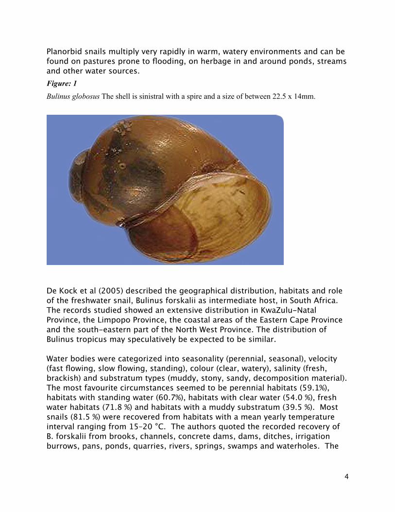
Planorbid snails multiply very rapidly in warm, watery environments and can be found on pastures prone to flooding, on herbage in and around ponds, streams and other water sources. Figure: 1
Bulinus globosus The shell is sinistral with a spire and a size of between 22.5 x 14mm.
De Kock et al (2005) described the geographical distribution, habitats and role of the freshwater snail, Bulinus forskalii as intermediate host, in South Africa. The records studied showed an extensive distribution in KwaZulu-Natal Province, the Limpopo Province, the coastal areas of the Eastern Cape Province and the south-eastern part of the North West Province. The distribution of Bulinus tropicus may speculatively be expected to be similar.
Water bodies were categorized into seasonality (perennial, seasonal), velocity (fast flowing, slow flowing, standing), colour (clear, watery), salinity (fresh, brackish) and substratum types (muddy, stony, sandy, decomposition material). The most favourite circumstances seemed to be perennial habitats (59.1%), habitats with standing water (60.7%), habitats with clear water (54.0 %), fresh water habitats (71.8 %) and habitats with a muddy substratum (39.5 %). Most snails (81.5 %) were recovered from habitats with a mean yearly temperature interval ranging from 15–20 °C. The authors quoted the recorded recovery of B. forskalii from brooks, channels, concrete dams, dams, ditches, irrigation burrows, pans, ponds, quarries, rivers, springs, swamps and waterholes. The
4
highest percentages of snails were recovered from dams (30.4 %) and brooks (28.2 %).
B. forskalii has been recognized to play a role in the natural transmission of the conical fluke, Calicophoron microbothrium, of cattle, goats and sheep in Ethiopia and Niger but its role in South Africa for these host species, has not yet been clarified. This snail has been reported as an intermediate host for the natural transmission of Gastrodiscus aegyptiacus in equids, pigs and warthogs in South Africa, and horses, donkeys and mules elsewhere in Africa.
Reynecke (1983) describes Bulinus tropicus as the intermediate host of Paramphistomum microbothrium, Calicophoron spp and Gastrodiscus aegyptiacus. In Zimbabawe Mavenyengwa et al (2006) reports Bulinus tropicus, as the intermediate host of Calicophoron microbothrium, to have a wide distribution and incriminated it in the majority of outbreaks of paramphistomosis in ruminants in Zimbabwe and the rest of Southern Africa.
Phiri et al (2007), in their studies in Zambia, reported flukes to be present throughout the year with the highest abundance rate (47.8%) found during the post-rainy season (March to May) and the lowest (24.8%) during the cold dry season (June to August). The highest mean egg counts and number of cattle found positive were recorded in the rainy (December to February) and post-rainy seasons (March to May). They found no significant differences in abundance rates between sexes or between ages of cattle.
Tarig et al (2008) studied the prevalence of paramphistomosis in the southern Himalayas and reported it to be 7.3%. Generally, season and age were the factors found to have a significant influence on the risk of paramphistomosis in sheep. The highest infestation was found in the summer season; lower age groups, in males and in the migratory (Bhakarwal) breed. Lower prevalences were reported in winter, adult animals, females and local breeds.
Several investigations have shown that mild temperatures and frequent rainfall throughout the year are suitable conditions for the infestation of cattle with trematode parasites like liver or conical fluke.
There are several reports from overseas claiming that an increased prevalence of Paramphistomum spp infestation is being seen. Mage et al (2002) describes an epidemiological survey in Central France which demonstrated that the prevalence of natural infestations with Paramphistomum sp in cattle increased from 5.2% in 1990 to 44.7% in 1999. This has been partly attributed to the efficacy of fasciolosis control that leaves more intermediate host (Galba
5
truncatula) available for the development of paramphistomes. Other reasons proposed were better quality of diagnosis for the detection of fluke eggs and a lack of an effective treatment against cattle paramphistomosis. Murphy et al (2008) also reported an increase in number of cattle infested with these parasites in Ireland. Very similarly Foster et al (2008) reported an increased number of cases in which these parasites were detected in cattle in England. According to both the latter authors its exact impact is still unknown and infestations may be complicated by concomitant parasitic or nutritional disease or poor husbandry (Ireland) and changing farming practices such as more organic farming, reduced use of herbicides and increasing incidences of flooding (England).Dorny et al (2011) studied the prevalence and seasonal variations of helminth infestations and their association with morbidity parameters in traditionally reared cattle in Cambodia. In their study faecal and blood samples were collected at monthly intervals from 2391 animals over a period of 11 months and body condition scores, faecal consistency, mucous membrane colour (conjunctivae) and packed cell volume recorded. The prevalences of Fasciola and Paramphistomum, estimated by coprological examination, varied between 5-20% and 45-95%, respectively. Pathogenesis
Immature worms attach themselves intimately to the intestinal mucosa and may even penetrate as far as the intestinal muscular layer. A plug of mucosa becomes drawn into their acetabula, with strangulation and necrosis of the mucosa as consequence. The extent of damage is directly related to the number of migrating flukes. Lesions may vary in severity from mild localized enteritis, to areas of villous atrophy to severe destruction of the mucosa. Marked oedema, intestinal discomfort and reduced appetite and food absorption results in anorexia, with a decreased amount of intestinal contents and eventually starvation atrophy as the net result. Plasma albumin is probably directly lost through the eroded and compromised small intestinal mucosa. Plasma calcium levels also drops, most likely due to the loss of the albumin-bound calcium. A fluid, foetid diarrhoea develops due a combination of decreased intake of solid feed, a continuous high water consumption and decomposition of plasma proteins in the intestine. Generalised oedema follows on the low plasma protein concentration. The plasma volume becomes reduced, masking any further protein loss. A reduction in plasma volume leads to reduced blood volume, decreased circulation and hypoxia. Hypoxia causes increased numbers of erythrocytes in the circulation. Pulmonary oedema, exhaustion and starvation finally cause death of the animal.
6
Clinical and production effects are dependent upon the extent of the lesions as some compensation for functional deficiency can take place in the undamaged lower small intestine. The presence of mature flukes in the rumen (figure2) does not usually elicit a significant response but in massive infestations papillae are short and red, becoming fused into aggregations with rumenal contents adhering firmly to the surface.
Clinical findings:
Severe enteritis associated with enormous numbers of migrating flukes in the duodenum seems to be the most significant manifestation of disease. A drop in food consumption occurs from the eighth day after infestation. This drop progresses to complete anorexia by day 21 and water intake may drop to about half the normal level. A characteristic and persistent foetid diarrhoea accompanied by weakness, depression, dehydration and anorexia may be seen. There may also be submaxillary oedema and obvious pallor of the mucosae. Death usually occurs from 5 – 20 days after the first signs appear and the mortality rate in heavily infested animals may be high. Mature flukes in the forestomachs of animals normally cause little harm, although loss of weight, anaemia, a rough dry coat, and a drop in production have been ascribed to heavy infestations.
Clinical Pathology
Plasma proteins start dropping from about day 14 after significant infestation. From day 21 onwards a marked drop, especially in the albumin level, may be seen. A drop in plasma calcium may accomapny the drop in protein. When the protein level drops to below 30 g/l the low osmotic pressure of the plasma may lead to bottle jaw. Ascites, hydrothorax and hydropericard also occur with a resultant drop in the total plasma volume.
Haemoconcentration may be seen with haematocrit levels rising from 0,33 to 0,46 g/l due to a loss of plasma and an increase in the numbers of circulating erythrocytes. The diarrhoea at this stage is usually very severe and with a foetid odour, most likely due to the leakage of proteins into the intestinal lumen and their subsequent putrefaction. Haemorrhage from the rectum may be observed following continuous straining.
7
Mavenyengwa et al (2006) infested one year old oxen with Calicophoron microbothrium metacercariae with a negative control (C) and low (L), medium (M) or high infestation (H) rates. The experimental animals were monitored daily for clinical signs while blood and serum samples were collected every 7 days until day 28 post-infestation, when sample collection was terminated. Moderate to severe diarrhoea developed in the M and H groups at day 21 post-infestation. A significant decrease in total plasma protein, calcium and phosphorus levels coincided with the diarrhoea, particularly in the M group. A significant decrease in the packed cell volume, haemoglobin and red blood cell levels occurred in the M and H groups from day 21 post-infestation, while a significant increase in the circulating eosinophils occurred between 7 and 21 days post-infestation in the LD and the HD groups.
Pathology
Depending on the severity of infestation and duration of the disease affected animals may be in good or cachectic condition. Oedema of the subcutaneous tissues, abomasal folds and mesentery, and fluid accumulation in the body cavities, may be present as consequence of hypoproteinaemia. The proximal small intestine may appear congested externally. Immature paramphistomes penetrated deeply into the intestinal wall, may be visible through the serosa. Flukes may occasionally perforate the intestine and be found free in the abdominal cavity. The duodenal mucosal surface may be oedematous, thickened, corrugated and covered with excessive mucous. Many immature pink or brown flukes which are only a few millimetres in length, may be scattered over the surface and embedded in the mucosa. Most larval paramphistomes are in the first three meters of small intestine but in advanced infestations, some may be present on the abomasal mucosa, or in the forestomachs.
Diagnosis
The clinical diagnosis of paramphistomosis remains challenging as immunological techniques and serum antibody detection is usually not conclusive. Therefore a diagnosis in live animals still depends on faecal detection of eggs, in conjunction with a typical clinical history and clinical signs. In some cases one may not find eggs, or only very few, due to massive infestation with young flukes and relatively long pre-patent period before adult parasites start to produce eggs. Another drawback is that with many standard flotation methods of faecal egg identification it may not be possible to detect conical fluke infestation.
8
A recommended method is to collect approximately 10 g of diarrhoeic faeces and to add 100-200 ml of water to the sample. Allow this mixture to sediment for five minutes, decant any supernatant fluid and repeat this process four of five times. Pour the sediment on a black surface and examine it. Young flukes resemble small white or pink rice grains and the eggs are silver-grey, large and have clear cells, an operculum and a central morula.
In dead animals typical post mortem findings combined with the presence of flukes in the affected intestines would be confirmative. Rieu et al (2007) tested a modified MacMaster method to check its reliability for the diagnosis of bovine paramphistomosis in France. A total number of 148 faecal samples from cows examined post-mortem were analysed. They were able to show a significant relationship between epg counts and parasite burden: more than 100 epg indicated the presence of more than 100 adult paramphistomes in rumen and/or reticulum suggesting a more reliable result.
Dorny et al (2011) investigated the association between morbidity markers and the presence of parasite infestation . A low body condition score was associated with gastrointestinal nematode and liver fluke infestations. Soft faecal consistency was associated with Paramphistomum infestations. Nutritional deficiencies and intercurrent diseases are likely to exacerbate the effects of parasites and should therefore be considered when using these morbidity parameters as indicators of parasitism.
Immunological and serum antibody detection techniques are not considered conclusive as diagnostic techniques due to shared common antigenic epitopes in several trematodes resulting in non specificity and cross-reactions.
In a study by Lofty et al (2010) the authors suggests that PCR-based techniques utilizing ITS2 as molecular marker is useful for species identification of paramphistomes and that it can be used to determine the genetic relatedness of samples within the different taxa of Paramphistomoidea.
Treatment and control
Effective control of most trematode infestations is based on the strategic use of effective chemotherapy. Farm management is critical in limiting the contact between intermediate and final hosts thus preventing or controlling
9
infestations. In addition action may be taken to reduce or eliminate intermediate host populations. The use of any or all of these measures in an integrated strategy should be based on sound economic assessments of the diseases and the relative merits of control options.
A knowledge of the epidemiology and disease prevalence based on the environment, intermediate host prevalence and seasonal weather conditions form the basis of a control program as follows:
(a) Prophylactic treatment of ruminants towards the end of a period of reduced activity of the parasites and the intermediate hosts, for example, during a prolonged dry season, or extreme cold. This is aimed at reducing the pasture contamination of eggs before favourable climatic conditions for larval development and snail activity resume.
(b) Curative treatment about one to two months after the expected peak infestation of the hosts. A curative effect can be achieved by one treatment to remove the residual fluke burden acquired from metacercariae which had survived on the herbage. (c) Additional treatment in highly contaminated areas where seasonal variations do not significantly affect the life cycle of the flukes.These additional treatments may be required occasionally, when the seasonal climatic conditions are favourable for parasite and snail development, or in areas where high metacercariae intake often occurs as a result of restricted grazing of wet areas during dry seasons. If animals are grazing communal areas, it is important to achieve a synchronized reduction in pasture contamination of eggs, if possible. Ideally all animals in the area should receive treatment within a short period of time.The use of molluscicides for the control of snail intermediate hosts is a potential tool for the control of fluke infestations. Copper chloride has been described for the control of intermediate hosts but many habitats are unsuitable for the use of molluscicides and a very careful environmental impact assessment should be done before embarking on such an attempt.Reports from several parts of the world indicate that a number of plants have molluscicidal properties. Planting of these trees and shrubs along streams and irrigation channels can reduce the number of snails in a population. The efficacy of this method for control of flukes has not yet been assessed. Water birds such as ducks may provide a form of biological control. Ducks eat the snails and the fluke species specific to the ducks compete with the fluke
10
species of ruminants in the infestation of snails. It is reported that snails infested with duck flukes will not become infested with flukes of livestock. The important management methods of controlling fluke infestations are: (a) To prevent snail habitats from developing by regular clearing of drainage channels of vegetation which provide suitable sites for snail development. Good drainage and the building of dams at appropriate sites in marshy and low lying areas may reduce the snail problem.
(b) To keep livestock away from pastures contaminated with metacercariae. This may only be possible when the number of animals involved is small. (c) Establish proper watering facilities to prevent animals from drinking from lakes, ponds and streams.
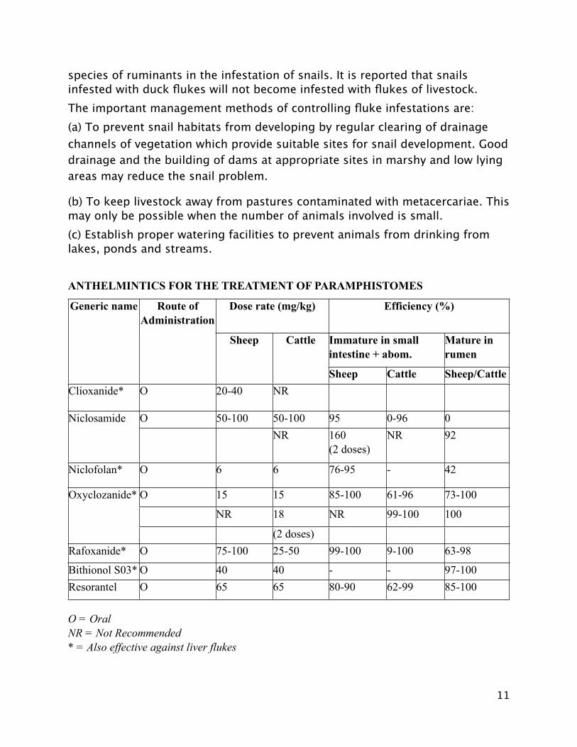
ANTHELMINTICS FOR THE TREATMENT OF PARAMPHISTOMES
Generic name Route of Administration
Dose rate (mg/kg) Dose rate (mg/kg) Efficiency (%)Efficiency (%)Efficiency (%)Generic name Route of Administration
Sheep Cattle Immature in small intestine + abom. Immature in small intestine + abom.
Mature in rumen
Generic name Route of Administration
Sheep Cattle
Sheep Cattle Sheep/Cattle Clioxanide* O 20-40 NR
Niclosamide O 50-100 50-100 95 0-96 0 Niclosamide NR 160
(2 doses) NR 92
Niclofolan* O 6 6 76-95 - 42
Oxyclozanide* O 15 15 85-100 61-96 73-100 Oxyclozanide*
NR 18 NR 99-100 100
Oxyclozanide*
(2 doses) Rafoxanide* O 75-100 25-50 99-100 9-100 63-98
Bithionol S03* O 40 40 - - 97-100 Resorantel O 65 65 80-90 62-99 85-100
O = OralNR = Not Recommended* = Also effective against liver flukes
11
This table is taken from a handbook which was produced by ILRAD with the cooperation of the FAO and ILCA .
References
1- Conradie I. 2008. The prevalence of helminths in warthogs, bushpigs and some antelope species in Limpopo Province, South Africa. Dissertation for Master of Science in Veterinary Tropical Diseases, Faculty of Veterinary Science, University of Pretoria, South Africa.
2- De Kock KN, Wolmarans CT. 2005 Distribution, habitats and role as intermediate host of the freshwater snail, Bulinus forskalii, in South Africa. Onderstepoort Journal of Veterinary Research,165–174.
3- Dorny P, Stoliaroff V, Charlier J, Meas S, Sorn S, Chea B, Holl D, Van Aken D, Vercruysse J. 2011. Infestations with gastrointestinal nematodes, Fasciola and Paramphistomum in cattle in Cambodia and their association with morbidity parameters. Journal of Veterinary Parasitology. Feb 10:175(3-4):293-9.
4- Foster AP, Otter A, O'Sullivan T, Cranwell MP, Twomey DF, Millar MF, Taylor MA. 2008 Rumen fluke (paramphistomosis) in British cattle. Veterinary Record. Apr 19:162(16):528.
5- Infectious Diseases of Livestock 2004. Second edition. Edited by JAW Coetzer and RC Tustin, Oxford University Press Southern Africa, Cape Town.
6- Jubb, Kennedy and Palmer’s Pathology of Domestic Animals 2007. Fifth edition. Edited by MG Maxie. Saunders Elsevier.
7- Lotfy WM, Brant SV, Ashmawy KI, Devkota R, Mkoji GM, Loker ES. 2010 A molecular approach for identification of paramphistomes from Africa and Asia. Veterinary Parasitology. Dec 15:174(3-4):234-40.
8- Mage C, Bourgne H, Toullieu JM, Rondelaud D, Dreyfuss G. 2002. Fasciola hepatica and Paramphistomum daubneyi: changes in prevalences of natural infections in cattle and in Lymnaea truncatula from central France over the past 12 years. Veterinary Research. 2002 Sep-Oct;33(5):439-47.
9- Mavenyengwa M, Mukaratirwa S, Obwolo M, Monrad J 2006. Observations on mass production of Calicophoron microbothrium metacercariae from experimentally and
12
naturally infested Bulinus tropicus. Onderstepoort Journal of Veterinary Research, 73:95–100.
10-Mavenyengwa M, Mukaratirwa S, Monrad J. 2010. Influence of Calicophoron microbothrium amphistomosis on the biochemical and blood cell counts of cattle. Journal of Helminthology. Dec: 84(4): 355-61.
11-Murphy TM, Power EP, Sanchez-Miguel C, Casey MJ, Toolan DP, Fagan JG. 2008. Paramphistomosis in Irish cattle. Veterinary Record. Jun 21;162(25):831.
12-Phiri AM, Chota A, Phiri I K. 2007. Seasonal pattern of bovine amphistomosis in traditionally reared cattle in the Kafue and Zambezi catchment areas of Zambia. Tropical Animal Health Production (2007) 39:97–102.
13-Reinecke RK (1983). Veterinary Helminthology 1983. Butterworths. Durban/Pretoria.
14- Recca A, Bénet JJ, Saana M, Dorchies P, Guillot J. 2007. Reliability of coprological diagnosis of Paramphistomum sp. infection in cows. Veterinary Parasitology. May 31;146(3-4):249-53.
15-Tariq KA, Chishti MZ, Ahmad F, Shawl AS. 2008. The epidemiology of paramphistomosis of sheep (Ovis aries L.) in the north west temperate Himalayan region of India. Veterinary Research Communincation. Jun;32(5):383-91.
16-Veterinary Medicine. A textbook of the diseases of cattle, sheep, pigs, goats and horses. (9th edn). 2000. OM Radostits, CC Gray, Blood DC, Hinchcliff KW. WB Saunders.
17- Veterinary Parasitolgy 1996. Second edition. GM Urquhart, J Armour, Ll Duncan, AM Dunn, FW Jennings. Blackwell Science.
13


![Performance of IBA New Conical Shaped Niobium [18O] Water ... · Vienna sept 2010, poster #9, session P13. Table 2: Results Summary Conical 6 Conical 8 Conical 12 Conical 16 Insert](https://img.pdfslide.net/doc/110x75/5f901a7319a03054823be5c3/performance-of-iba-new-conical-shaped-niobium-18o-water-vienna-sept-2010.jpg)