Embed Size (px)
Citation preview

British Journal of Ophthalmology, 1984, 68, 618-622
Conjunctival myxoma: a case reportJACOB PE'ER, MICHAEL ILSAR, AND AHMED HIDAYAT
From the Department of Ophthalmology, Hadassah University Hospital, Jerusalem, Israel
SUMMARY A rare case of conjunctival myxoma in an 18-year-old female is reported. Clinically itpresented as a painless mass located in the nasal bulbar conjunctiva. It was composed of spindleand stellate shaped cells in a loose mucoid stroma. Some of the cells had intracytoplasmicvacuoles consistent with dilated rough endoplasmic reticulum and/or intranuclear vacuoles ofnuclear membrane invaginations. Mast cells were also seen in the stroma. No recurrence hasbeen reported eight months postoperatively.
Myxoma is an uncommon benign connective tissuetumour that may arise from various parts of thebody.'-2 Myxomas of the ocular adnexa are rare, andonly five cases of conjunctival myxoma have beenreported.-7 The following report describes a well-documented case of this rare neoplasm, a con-junctival myxoma, including ultrastructural study.
Case report
An 18-year-old healthy white female complained ofa small lesion in the nasal bulbar conjunctiva of herright eye which had been present for four months.There was no history of trauma. The lesion waspainless and did not cause any other symptoms, andthe patient sought medical consultation only forcosmetic reasons. Examination of both eyes wasunremarkable except for a pinkish glistening 'cystic'tumour in the nasal bulbar conjunctiva of the righteye. The tumour did not invade the limbus (Fig. 1).A similar though smaller tumour was found at thesame location in the fellow eye. The tumours andthe conjunctiva around them moved freely over thesclera. The patient's complaints, however, werelimited to the right eye, and under local anaesthesiathe tumour was completely removed. After eightmonths there was no scar or recurrence, and thetumour of the fellow eye has not grown in size.
PATHOLOGICAL FINDINGSThe specimen was composed of a well-circumscribedsoft mass measuring 10x4x4 mm. It was composed
Correspondence to Jacob Pe'er, MD, Department of Ophthal-mology, Hadassah University Hospital, PO Box 12000, 91120Jerusalem, Israel.
of a main cyst-like structure, whose cut surfaceappeared myxomatous, and a connective tissue'tail.' The entire specimen was covered on one sidewith a pinkish tissue. A prominent blood vessel,apparently the feeding vessel, was seen entering thetumour (Fig. 2).
Microscopic examination revealed under theslightly acanthotic conjunctival epithelium a mass ofvery loose connective tissue. It was composed ofscattered stellate shaped cells with multiple cyto-plasmic processes and spindle shaped cells withbipolar cytoplasmic processes. Both types of cellshad moderately large hyperchromatic and slightlypleomorphic nuclei, and were embedded in loosemucoid stroma. The stroma contained reticulin
Fig. 1 A shining cystic mass ofthe nasal bulbarconjunctiva ofright eye with prominent blood vessel over it.The mass does not reach the limbus.
618
on 1 February 2019 by guest. P
rotected by copyright.http://bjo.bm
j.com/
Br J O
phthalmol: first published as 10.1136/bjo.68.9.618 on 1 S
eptember 1984. D
ownloaded from

Conjunctival myxoma: a case report
Fig. 2 A well-circumscribed cystic mass with a connectivetissue tail. Note the vessel that enters cyst (x6-5).
fibres and sparse small blood vessels, and collagenfibres that were denser in the periphery (Fig. 3).Some of the tumour cells showed tiny nuclearvacuoles, and some had cytoplasmic vacuoles notimpinging on the nucleus (Fig. 4). The vacuoles didnot stain with oil-red-o, alcian blue, or PAS stains.There was no mitotic activity. A few mast cells werepresent within the myxoid stroma. The loose stromacontained abundant hyaluronidase-sensitive muco-polysaccharides. Bodian stain was negative fornerve fibres.
Electron microscopic examination disclosed apiece of tumour tissue composed of predominantlyfibroblast-like spindle shaped cells that lacked base-ment membranes, with prominent rough endo-plasmic reticulum and very few other cytoplasmicorganelles. Many of the nuclei contained one or two
m ; ,4s.v a .. s >z t w | S ; ' ~~~~~~~~~~~~~~AgeAwi s A= 4it<
.4W^KS ***;
r'{fW a >'@ x ! A A X~~~
Fig. 4 Stellate andspindleshaped cells with intracranialand intracytoplasmic vacuoles embedded in loose mucoidstroma with reticulum and collagenfibres (Movatpentachrome, x250).
Fig. 3 A loose connective tissuethat is denser in its periphery,covered on one side byconjunctival epithelium(Haematoxylin and eosin, x53).
619
on 1 February 2019 by guest. P
rotected by copyright.http://bjo.bm
j.com/
Br J O
phthalmol: first published as 10.1136/bjo.68.9.618 on 1 S
eptember 1984. D
ownloaded from

Jacob Pe'er, Michael Ilsar, andAhmed Hidayat
'.4 '....
T'
i8P ...w-
5.
':.... : ... : .. . : :.. . :: . .. -.; :. ^., ::.' .:. :.i: ' . '
:c :.
:s .. .:l,\
.: ..S
At 0 f e:e,.
.. ,. f .#g\W . ..... -,j .iy Al';Ml'
R
w ;i
Hs Wt: .vr....:i' ':s
*6: & :. ...
: :
'o5
.:: .N.
1 .. .'X't,,,. :...dA.} i . : . .. ffi .X .#.
w:t ::; :. .^. . ., . .. . .. X* i.6' .. :, ..F ... A -
Fig. 5 A cell with infolding ofthe nuclear membrane, forming membrane bounded vacuoles (v). Note two nucleoli insidethe nucleus andprominent rough surfaced endoplasmic reticulum in the cytoplasm (left lower corner) (x12 700). Insert:higher magnification ofa vacuole (x34 650).
620
..Ooo.
*.'.
AO
on 1 February 2019 by guest. P
rotected by copyright.http://bjo.bm
j.com/
Br J O
phthalmol: first published as 10.1136/bjo.68.9.618 on 1 S
eptember 1984. D
ownloaded from
Conjunctival myxoma: a case report
..... ..... --;;.
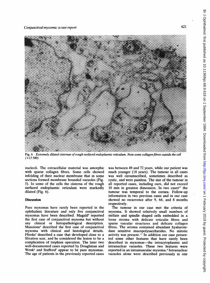
Fig. 6 Extremely dilated cisternae ofrough surfaced endoplasmic reticulum. Note some collagen fibres outside the cell(x13 500).
nucleoli. The extracellular material was amorphicwith sparse collagen fibres. Some cells showedinfolding of their nuclear membrane that in somesections formed membrane bounded vacuoles (Fig.5). In some of the cells the cisterna of the roughsurfaced endoplasmic reticulum were markedlydilated (Fig. 6).
Discussion
Pure myxomas have rarely been reported in theophthalmic literature and only five conjunctivalmyxomas have been described. MagaliP reportedthe first case of conjunctival myxoma but withoutany clinical or histopathological description.Mancione4 described the first case of conjunctivalmyxoma with clinical and histological details.Ffooks5 described a case that developed close to afiltration scar, and he considered the lesion to be acomplication of trephine operation. The later twowell-documented cases reported by Doughman andWenk6 and Stafford7 appear to be pure myxomas.The age of patients in the previously reported cases
was between 49 and 72 years, while our patient wasmuch younger (18 years). The tumour in all caseswas well circumscribed, sometimes described ascystic, and were painless. The size of the tumour inall reported cases, including ours, did not exceed10 mm in greatest dimension. In two cases67 thetumour was temporal to the cornea. Follow-upinformation in two previous cases and in our caseshowed no recurrence after 9, 64, and 8 monthsrespectively.The tumour in our case met the criteria of
myxoma. It showed relatively small numbers ofstellate and spindle shaped cells embedded in aloose stroma with delicate reticulin fibres andsparse vascular structures and delicate collagenfibres. The stroma contained abundant hyaluroni-dase sensitive mucopolysaccharides. No mitoticactivity was present.'2 In addition our case presen-ted some other features that have rarely beendescribed in myxomas-the intracytoplasmic andintranuclear vacuoles. These two features werereported in an intramuscular myxoma.' Intranuclearvacuoles alone were described previously in one
621
on 1 February 2019 by guest. P
rotected by copyright.http://bjo.bm
j.com/
Br J O
phthalmol: first published as 10.1136/bjo.68.9.618 on 1 S
eptember 1984. D
ownloaded from

Jacob Pe'er, Michael Ilsar, andAhmed Hidayat
case of intramuscular myxoma9 and in one figure inStout's study2 without any textual description, whileintracytoplasmic vacuoles alone were described in acase of conjunctival myxoma5 and a case of intra-muscular myxoma.'° The cytoplasmic vacuoles asseen on electron microscopic examination areapparently consistent with markedly dilated roughendoplasmic reticulum, while the intranuclearvacuoles were found to be invaginations of markedlyfolded nuclear membrane.Mast cells, demonstrated by Giemsa stain in our
case, have not been described previously.In the differential diagnosis of pure conjunctival
myxoma we have to consider mixed myxomas suchas fibromyxoma, lipomyxoma, or fibrolipomyxoma,and myxomatous degeneration of tissue or othertumours such as neurofibroma. The degenerativeprocess is sometimes very difficult to prove. It ismore important to consider the possibility ofmalignant tumours such as myxoid liposarcoma andmalignant fibrous histiocytoma.1
References
1 Enzinger FM, Weiss SW. Soft tissue tumors. St Louis: Mosby,1983.
2 Stout AP. Myxoma, the tumor of primitive mesenchyme. AnnSurg 1948; 127: 706-19.
3 Magalif. Myxoma conjunctivae. Klin Monatsbl Augenheilkd1913; 51: 844.
4 Mancione DL. Di una rara forma di tumore epibulbare(mixoma puro) sottocongiuntivale. Arch Ottalmol 1914; 21:300-6.
5 Ffooks 00. Myxoma of the conjunctiva. BrJ Ophthalmol 1962;46: 374-7.
6 Doughman DJ, Wenk RE. Epibulbar myxoma. Am JOphthalmol 1970; 69: 483-5.
7 Stafford WR. Conjunctival myxoma. Arch Ophthalmol 1971;85: 443-4.
8 Glazunov MF, Puckov JG. Uber die sogenannten Muskel-myxome und Myxosarkome des Menschen mit Zellein-schluessen. Z Krebsforsch 1963; 65: 439-45.
9 Feldman PS. A comparative study including ultrastructure, ofintramuscular myxoma and myxoid liposarcoma. Cancer 1979;43: 512-25.
10 Leung TK, Vanzelle JL, Patrirot LM, Lejeune E, Quoneau P.Etude ultrastructurale et cytochinique d'un myxome musculaireassocie a une dysplasie fibrense. Ann Anat Pathol (Paris) 1971;16: 417-28.
622
on 1 February 2019 by guest. P
rotected by copyright.http://bjo.bm
j.com/
Br J O
phthalmol: first published as 10.1136/bjo.68.9.618 on 1 S
eptember 1984. D
ownloaded from
![Mobile left atrial mass-clot or left atrial myxoma....mass includes thrombus, myxoma, lipoma and non-myxomatous neoplasm [7,8]. Among them, cardiac myxoma is the most common benign](https://img.pdfslide.net/doc/110x75/60fedab34ecd6d6c000feba7/mobile-left-atrial-mass-clot-or-left-atrial-mass-includes-thrombus-myxoma.jpg)