Embed Size (px)
Citation preview
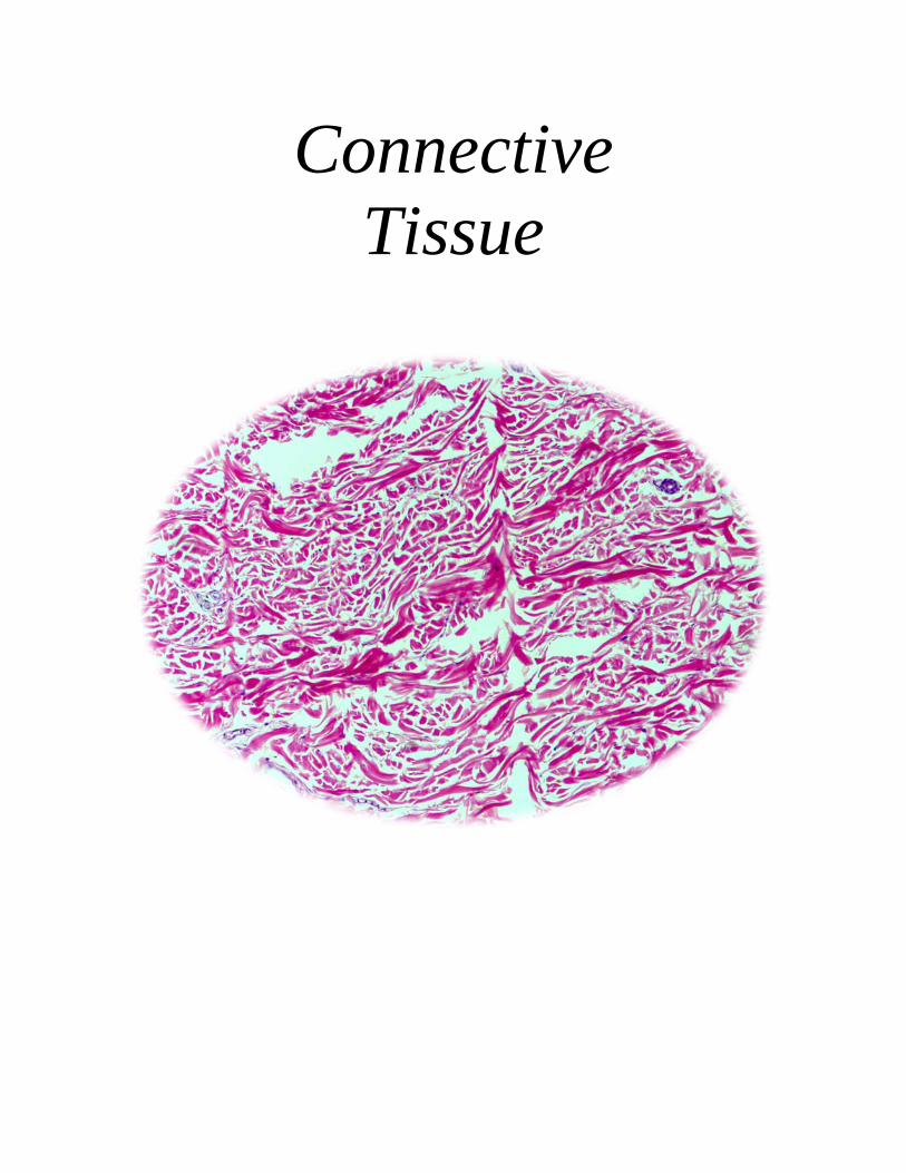
Connective Tissue
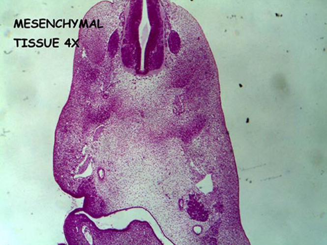
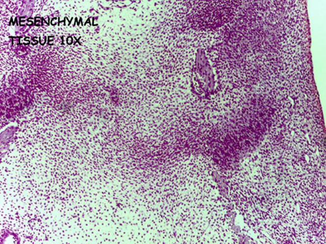
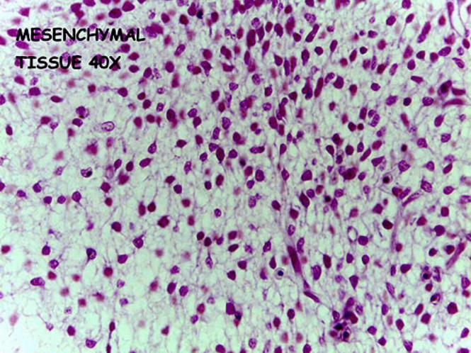
Mesenchymal Connective Tissue
• Cells o Mesenchymal cells
Stellate-shaped fusiform cells Ovoid nucleus
• Fibers/Ground Substance/Other
o Reticular fibers o Amorphous ground substance
• Features
o Undifferentiated loose connective tissue o Cells have stem cell properties o Mesoderm origin
• Function
o Give rise to mature connective tissue
• Location o Embryonic tissues
• Video recording
o Mesenchymal tissue
• Microscope images o 4x o 10x o 40x
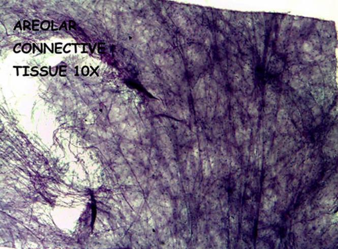
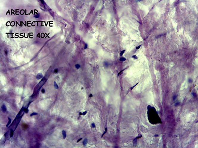
Areolar Connective Tissue
• Cells o Fibroblasts o Adipocytes o Macrophages o Mast cells
• Fibers/Ground Substance/Other
o Collagen fibers o Elastic fibers o Reticular fibers o Blood vessels o Nerves
• Features
o Loose connective tissue o Most commonly encountered connective tissue
• Functions
o Wraps and cushions organs o Immune function o Convey tissue fluid
• Locations
o Fills spaces between organs o Lamina propria of mucous membranes o Adventitia of blood vessels o Parenchyma of glands o Hypodermis
• Video recording
o Areolar connective tissue
• Microscope images o 10x o 40x
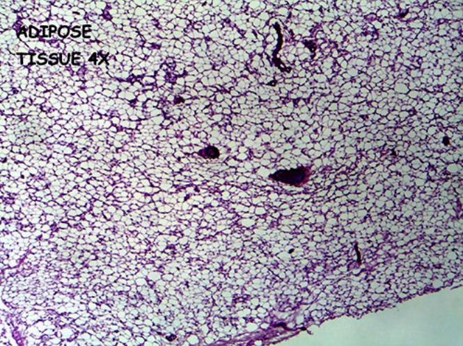
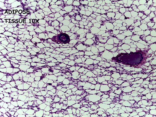
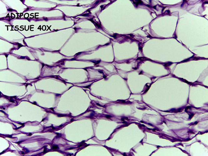
Adipose Tissue (White)
• Cells o Adipocytes (unilocular)
Single triglyceride droplet with nucleus pushed to the periphery (eccentric nucleus)
o Fibroblasts o Macrophages
• Fibers/Ground Substance/Other
o Reticular fibers
• Features o Extensively vascularized
• Functions
o Fuel reserve o Insulation o Support/protect organs
• Locations
o Hypodermis o Surrounding internal organs (visceral fat) o Yellow bone marrow o Breast tissue o Specific locations (adipose depots)
Eyeball, kidneys
• Video recording o Adipose tissue
• Microscope images
o 4x o 10x o 40x
Adipose Tissue (Brown)
• Cells o Adipocytes (multilocular)
Numerous triglyceride droplets with nucleus pushed to the periphery (eccentric nucleus)
o Fibroblasts o Macrophages
• Fibers/Ground Substance/Other
o Reticular fibers
• Features o Extensively vascularized o Brown color (mitochondria, cytochromes) o Lobular organization
• Functions
o Heat production o Fuel reserve o Insulation
• Locations
o Mostly found in infants
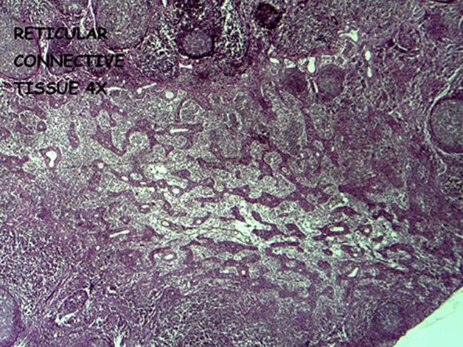
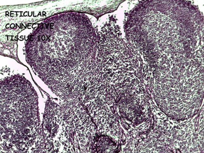
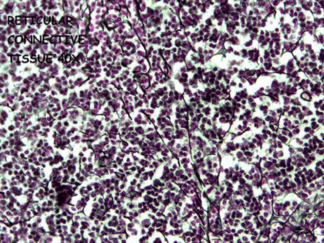
Reticular Connective Tissue
• Cells o Fibroblasts (or reticular cells) o Macrophages o Lymphocytes
• Fibers/Ground Substance/Other
o Reticular fibers (type III collagen)
• Features o Form network-like skeleton
• Functions
o Form an architectural framework to support hematopoietic organs and lymphoid organs
• Locations o Spleen o Lymph nodes o Red bone marrow o Liver sinusoids
• Video recording
o Reticular connective tissue
• Microscope images o 4x o 10x o 40x
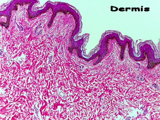
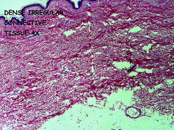
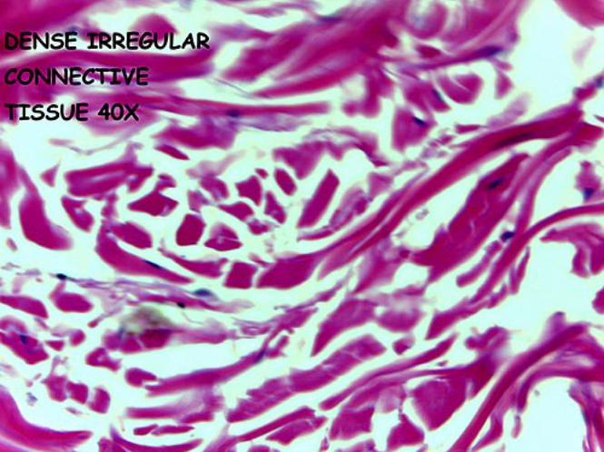
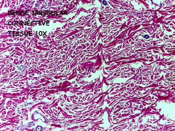
Dense Irregular Connective Tissue
• Cells o Fibroblasts
• Fibers/Ground Substance/Other
o Collagen fibers (type I) Arranged irregularly
o Elastic fibers (few) o Limited ground substance (dense)
• Features
o Fibers arranged irregular as compared to dense regular connective tissue
• Functions o Provide resistance to stress in different directions
• Locations
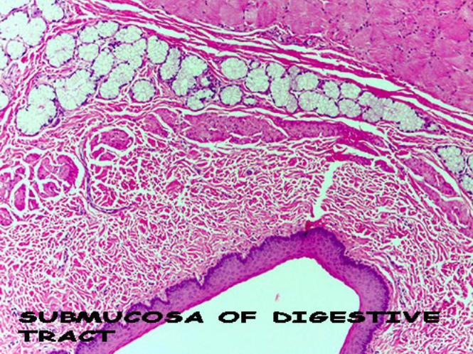
o Dermis (skin) o Sheaths of nerves o Fibrous capsules (spleen, testes, ovary, kidney, lymph node) o Submucosa of digestive tract o Periosteum o Perichondrium
• Video recording
o Dense irregular connective tissue
• Microscope images o 4x o 10x o 40x
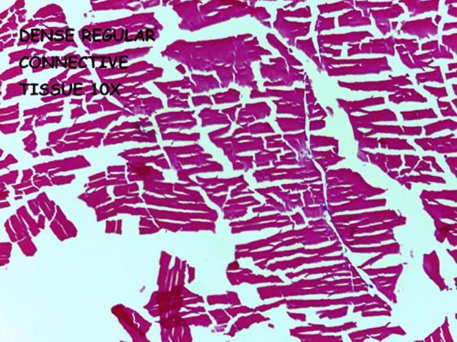
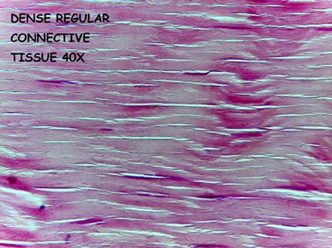
Dense Regular Connective Tissue
• Cells o Fibroblasts
• Fibers/Ground Substance/Other
o Collagen fibers (type I) Densely packed Parallel arrangement
o Elastic fibers (few) o Limited ground substance (dense)
• Features
o Parallel fiber arrangement as compared to dense irregular connective tissue
• Functions o Resistant to stress in one direction (traction forces)
• Locations
o Tendons o Ligaments o Aponeurosis
• Video recording
o Dense regular connective tissue
• Microscope images o 10x o 40x
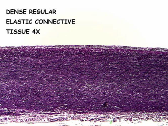
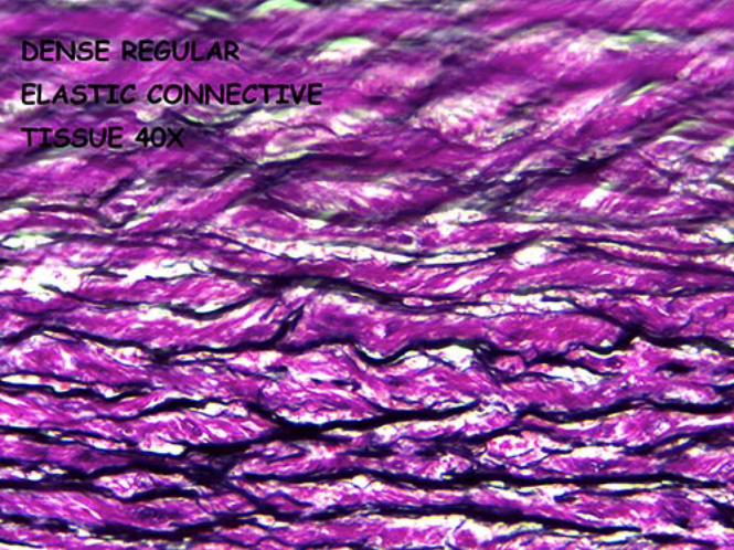
Dense Regular Elastic Connective Tissue
• Cells o Fibroblasts
• Fibers/Ground Substance/Other
o Elastic fibers Arranged in parallel
o Collagen fibers (type I) – few
• Features o Parallel elastic fibers as compared to collagen fibers
• Functions
o Allow recoil of tissue following stretch o Maintain pulsatile flow of blood
• Locations
o Blood vessels (elastic arteries) o Lung tissue (recoil) o Ligamentum flavum o Suspensory ligament (penis)
• Video recording
o Dense regular elastic connective tissue
• Microscope images o 4x o 10x o 40x
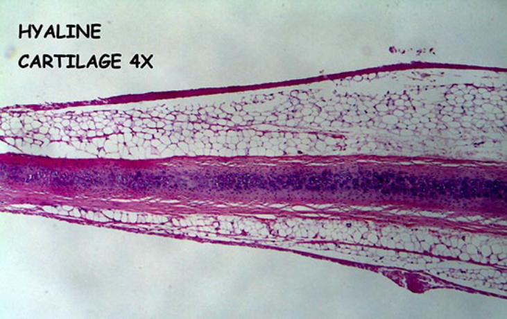
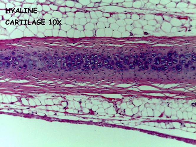
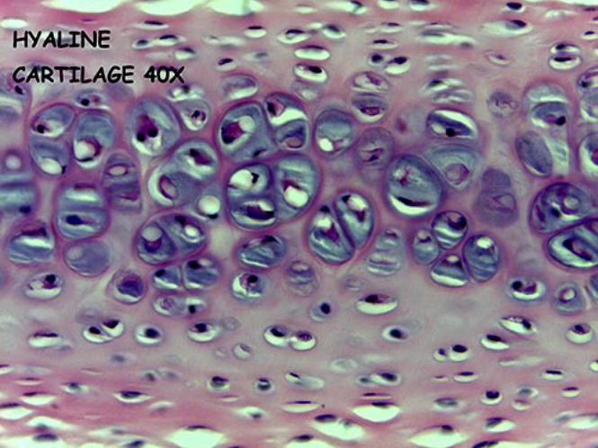
Hyaline Cartilage
• Cells o Chondrocytes within lacunae o Periphery
Ovoid chondroblasts in line with long axis parallel to the surface Appositional growth
o Centrally Chondrocytes rounder Isogenous groups Interstitial growth
• Fibers/Ground Substance/Other
o Collage fibers (type II) Other types also present Not discernable due to refraction
o Matrix Smooth appearance due to refraction Territorial matrix
• Near isogenous groups • Basophilic due to cellular properties
Interterritorial matrix • Space between chondrocytes
• Features
o Perichondrium Outer (fibrous) layer – dense irregular connective tissue
• Contains nerves and blood vessels Inner (chondrogenic) layer – mesenchymal; undifferentiated tissue
• Functions
o Support and reinforcement o Resistance to compressive stresses o Resilient cushioning
• Locations
o Embryonic skeleton, articular cartilage (no perichondrium), costal cartilage, cartilage of nose, trachea, larynx and epiphyseal plate (no perichondrium)
• Video recording o Hyaline cartilage
• Microscope images o 4x o 10x o 40x
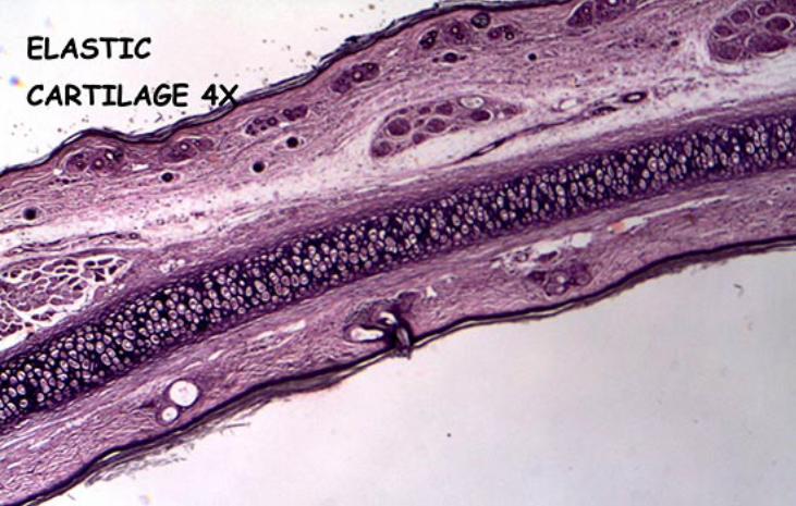
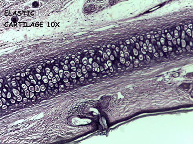
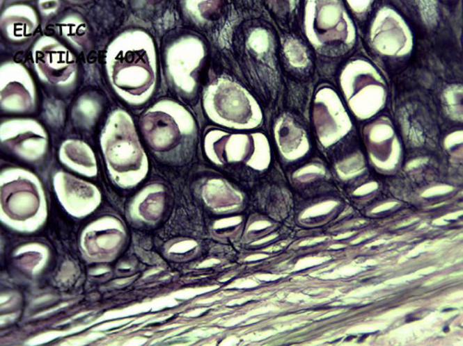
Elastic Cartilage
• Cells o Chondrocytes in lacunae
Larger, more abundant
• Fibers/Ground Substance/Other o Abundant network of elastic fibers o Collagen fibers (type II) o Less matrix
• Features
o Perichondrium Outer (fibrous) layer – dense irregular connective tissue Inner (chondrogenic) layer – mesenchymal; undifferentiated tissue
• Produces elastic fibers Contains nerves and blood vessels
• Function
o Maintain shape of a structure o Allow flexibility (elastic fibers)
• Location
o Pinna of ear o External and internal auditory tubes o Epiglottis o Cuneiform cartilage (larynx)
• Video recording
o Elastic cartilage
• Microscope images o 4x o 10x o 40x
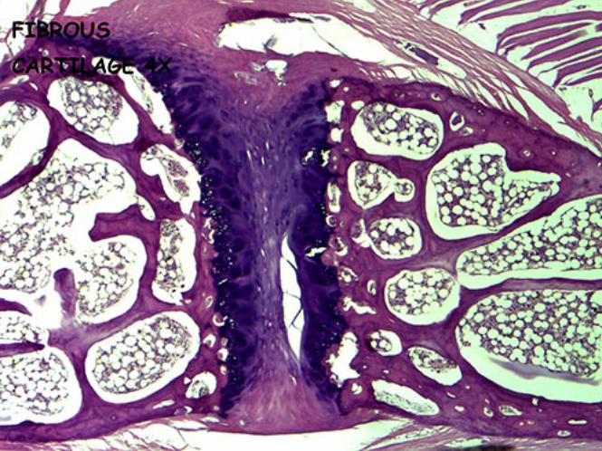
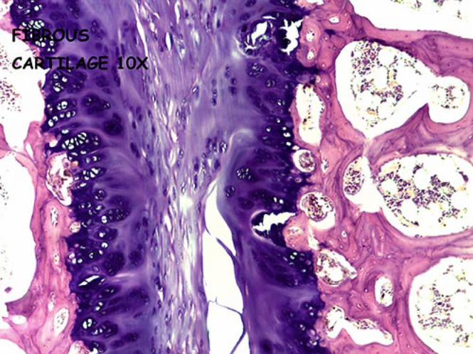
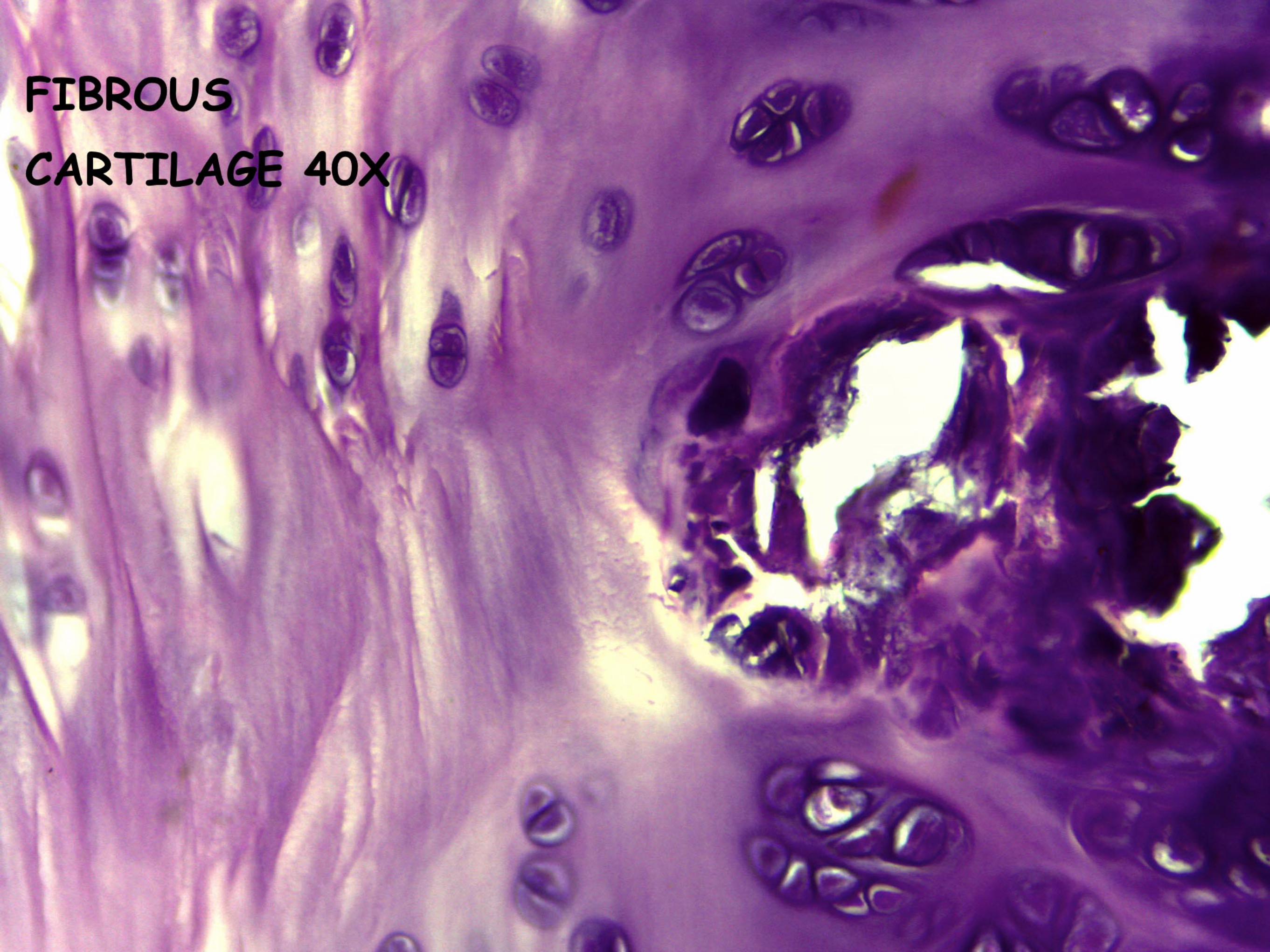
Fibrocartilage
• Cells o Chondrocytes
Line in alternating parallel rows Arise from fibroblasts
• Fibers/Ground Substance/Other
o Collage fibers (type I) Irregular or regular parallel bundles
o Little matrix
• Features o No perichondrium
• Function
o Great tensile strength o Absorb shock
• Location
o Intervertebral disks o Articular disks (i.e. menisci) o Pubic symphysis
• Video recording
o Fibrocartilage
• Microscope images o 4x o 10x o 40x