Embed Size (px)
Citation preview
ORIGINAL RESEARCHpublished: 20 October 2017
doi: 10.3389/fnhum.2017.00507
Frontiers in Human Neuroscience | www.frontiersin.org 1 October 2017 | Volume 11 | Article 507
Edited by:
Xiaolin Zhou,
Peking University, China
Reviewed by:
Michel-Pierre Coll,
University of Oxford, United Kingdom
Jin Fan,
Queens College, CUNY, United States
*Correspondence:
Jamie Ward
Received: 01 June 2017
Accepted: 06 October 2017
Published: 20 October 2017
Citation:
Grice-Jackson T, Critchley HD,
Banissy MJ and Ward J (2017)
Consciously Feeling the Pain of Others
Reflects Atypical Functional
Connectivity between the Pain Matrix
and Frontal-Parietal Regions.
Front. Hum. Neurosci. 11:507.
doi: 10.3389/fnhum.2017.00507
Consciously Feeling the Pain ofOthers Reflects Atypical FunctionalConnectivity between the Pain Matrixand Frontal-Parietal Regions
Thomas Grice-Jackson 1, 2, Hugo D. Critchley 2, 3, Michael J. Banissy 4 and Jamie Ward 1, 2*
1 School of Psychology, University of Sussex, Falmer, United Kingdom, 2 Sackler Centre for Consciousness Science,
University of Sussex, Falmer, United Kingdom, 3 Brighton and Sussex Medical School, University of Sussex, Falmer,
United Kingdom, 4Department of Psychology, Goldsmith’s College, University of London, London, United Kingdom
Around a quarter of the population report “mirror pain” experiences in which bodily
sensations of pain are elicited in response to viewing another person in pain. We have
shown that this population of responders further fractionates into two distinct subsets
(Sensory/localized and Affective/General), which presents an important opportunity to
investigate the neural underpinnings of individual differences in empathic responses. Our
study uses fMRI to determine how regions involved in the perception of pain interact
with regions implicated in empathic regulation in these two groups, relative to controls.
When observing pain in others (minor injuries to the hands and feet), the two responder
groups show activation in both the sensory/discriminative and affective/motivational
components of the pain matrix. The control group only showed activation in the
latter. The two responder groups showed clear differences in functional connectivity.
Notably, Sensory/Localized responders manifest significant coupling between the right
temporo-parietal junction (rTPJ) and bilateral anterior insula. We conclude that conscious
experiences of vicarious pain is supported by specific patterns of functional connectivity
between pain-related and regulatory regions, and not merely increased activity within the
pain matrix itself.
Keywords: social neuroscience, empathy, empathy for pain, vicarious pain, shared representations, rTPJ
INTRODUCTION
For some people, seeing another person in pain, such as having an injection or falling off a bicycle,results in reportable pain-like experiences. These people have been referred to as mirror-sensory ormirror-pain synaesthetes (Fitzgibbon et al., 2012b) or as pain responders (Osborn and Derbyshire,2010). Our recent study found a prevalence rate of mirror-pain of 27%, using a large scale screeningquestionnaire (n = 500+) and a k-means cluster analysis to classify participants (Grice-Jacksonet al., 2017). This has the advantage of being a data driven approach such that groups are determinedbased on the similarity of their vicarious pain experiences rather than arbitrary cut-offs imposedby an experimenter. Grice-Jackson et al. (2017) found two sub-groups of mirror-pain responderswith qualitatively distinct vicarious pain experiences characterized by differences in standardizeddescriptors of physical pain (Melzack, 1987), and the extent to which the evoked pain was localized.
Grice-Jackson et al. Vicarious Pain
One group that we termed Sensory/Localized responders(S/L) reported sensory descriptors (e.g., sharpness) that werelocalized to a specific region of the body (typically the samelocation as the observed pain), and a second group that wetermed Affective/General (A/G) responders reported affectivedescriptors (e.g., nauseating) that were generalized to thewhole body. The validity of these groupings was establishedby showing that the groups dissociate on other measures.When observing pain, the Sensory/Localized responders, showedsignificant differences on a measure of neural synchrony(electroencephalography/EEG suppression of mu and betarhythms) that has previously been linked to somatosensoryprocessing (e.g., Ritter et al., 2009). This pattern was not presentin controls or the Affective-General responders (Grice-Jacksonet al., 2017), raising the possibility that previous results in theliterature were driven by some individuals rather than reflectinga population-level trait. In terms of brain structure, using voxel-based morphometry (VBM), the two responder groups couldbe reliably distinguished from controls but had a similar profileto each other; namely, increased gray matter in somatosensorycortex and anterior insula, reduced gray matter in right temporo-parietal junction, rTPJ (Grice-Jackson et al., 2017).
These earlier observations provide initial evidence thatindividual differences in the phenomenological characteristics ofvicarious pain are meaningful and underpinned by systematicdifferences in brain structure and function. However, ourknowledge of the underlying brain systems is limited. Theonly previous fMRI study of mirror pain classified participantsaccording to whether they had one or more localized painresponses when observing a set of videos/images of pain (Osbornand Derbyshire, 2010). In this study, pain responders werereported to have greater activity when observing pain, relativeto control participants, in regions including anterior insula andsecondary somatosensory cortex. Here, we aimed to extend thisfinding in two important ways. Firstly, we sought to characterizedifferences between our recently discovered subtypes of mirrorpain. Secondly, we aimed to investigate the distinct underlyingfunctional connectivity between brain regions supporting thesetypes of vicarious pain response.
The central processing of pain takes places in a series ofinterconnected neural regions known collectively as the painmatrix (Melzack, 1999). However, it should be noted that regionsinvolved in the perception of pain typically process other kindsof related information too (see Iannetti and Mouraux, 2010). Thepain matrix is often parcellated into two conceptually differentsubdivisions known as the affective-motivation subdivision(processing the affective qualities of emotion preparednessof pain) and the sensory-discriminative subdivision (whichprocesses the sensory aspects of pain) (Peyron et al., 2000).Correspondingly, we predicted that our Sensory/Localized andAffective/General responders will differentially activate thesesub-systems. Moreover, within the Sensory/Localized group, wefurther anticipated a greater somatotopic response to observedpain whereby viewed pain to the hand or foot activateshand or foot regions of somatosensory cortex respectively.In normative populations (i.e., that do not separate out thepresence/absence of mirror pain), there is consistent activation in
the affective/motivation regions of the pain matrix (notably mid-cingulate cortex and anterior insula) when observing others inpain (Lamm et al., 2011). This occurs also when pain is implied,but not directly observed. It is argued that sensory/discriminativeregions of the pain matrix may only be activated when the siteof injury is observed, and not when pain is merely implied(e.g., via facial grimace or a symbolic cue; Lamm et al., 2011).Brain stimulation studies also suggest the “sensory simulation”of the pain of others when directly observing pain (Avenantiet al., 2005; Bufalari et al., 2007). However, these studies did nottake into account the contribution of individual differences invicarious experience. EEG has revealed a greater modulation ofsomatosensory evoked responses when viewing pain in mirror-pain responders compared to controls (Fitzgibbon et al., 2012a).This observation suggests that mirror pain is linked to differencesin neural processing at the level of cortical sensory processing,rather than being merely an enhanced affective response.
Most models of empathy for pain assume not only activity inshared representations of pain (whether affective and/or sensory)but also regions outside of the pain matrix that are involvedin selectively orienting toward self/other either in terms ofbodily location (perspective taking) or in terms of orientingtoward salient social and personal characteristics such as race(Decety and Jackson, 2006; Decety, 2011; Bird and Viding, 2014).For example, in a neurotypical sample, training the ability toregulate self-other representations has been linked with changesin the degree of sensory simulation of the pain of others whendirectly observing pain (de Guzman et al., 2016). These controlmechanisms are needed to dynamically modulate the focus ofattention toward other people (and suppress one’s own feelings)or, conversely, to be able to focus on one’s own feelings andsuppress that of others (i.e., the down-regulation of empathy).One region that has been implicated as acting as a switchbetween self and other is the right temporoparietal junction,TPJ (Bird and Viding, 2014; Lamm et al., 2016). According toWard and Banissy (2015) a disruption of this rTPJ mechanismin mirror pain (and mirror touch synaesthesia) underlies thetendency to experience the pain of others. In effect, for theseindividuals, pain is more likely to be shared rather than selectivelyattributed to self or other. Evidence for a role of this region inmirror pain comes from structural brain imaging studies wherereduced rTPJ gray matter density is observed in people with bothSensory/Localized and General/Affective mirror pain (Grice-Jackson et al., 2017), and in people with the related symptom ofmirror-touch synaesthesia (Holle et al., 2013). The present studyconsiders in more detail the role of this region in our fMRI studyof vicarious pain.
In summary, our hypothesis is that mirror-painphenomenology is linked to increased activity of regionsimplicated in physical pain when observing pain. Morespecifically, we predict that Sensory/Localized responders willhave increased activity in the sensory-discriminative sub-divisionof the pain matrix, whereas Affective/General responders willhave increased activity in the affective-motivational sub-division.Finally, we hypothesize that between-group differences inactivity patterns are mediated by differences in functionalconnectivity between regions of the pain matrix and other
Frontiers in Human Neuroscience | www.frontiersin.org 2 October 2017 | Volume 11 | Article 507
Grice-Jackson et al. Vicarious Pain
regions implicated in empathy and the control of self-otherrepresentations including but not limited to the rTPJ.
METHODS
ParticipantsForty-four healthy participants (18 males, 26 females) agedbetween 18 and 42 years (mean= 23.96, S.E= 1.37) volunteeredto take part in the study. All participants self-reported beingright handed, had normal or corrected vision. Furthermoreparticipants had previously completed the Vicarious PainQuestionnaire (VPQ), an online measure assessing reports andcharacteristics of conscious vicarious pain experiences (Grice-Jackson et al., 2017). It consists of 16 movies depicting injections(N = 8) and sports injuries (N = 8). After each movie,participants report whether it triggered pain on your ownbody (giving a summed score across all movies from 0 to16). Upon giving an affirmative answer they are then askedfollow-up questions namely: to rate the intensity (on a 0–10scale), to select from a series of pain descriptors that describesensory and affective qualities of pain (Melzack, 1987), andto indicate whether the pain is localized or generalized. Thethree groups were derived by performing a two-step clusteranalyses on the larger datatset (n = 573) that included thefMRI participants (as the method requires a sample size ofseveral hundred). The fMRI sample consisted of 21 non-responder controls, 13 Sensory/Localiser responders and 10Affective/General responders. The details of the participants,in relation to their performance on the VPQ, is summarized inTable 1. Participants provided written and informed consent inaccordance with the Declaration of Helsinki. They were paid£15 for their participation in the study. The study’s procedureswere reviewed and approved by the Brighton and Sussex MedicalSchool (BSMS) Research Ethics Committee.
ApparatusA Siemens Avanto 1.5 Tesla MRI scanner was used to collectall images throughout this experiment. A single row four-buttonbutton box was used for tasks 1 and 2 with only the two centralbuttons active so that participants could indicate movements tothe left and right.
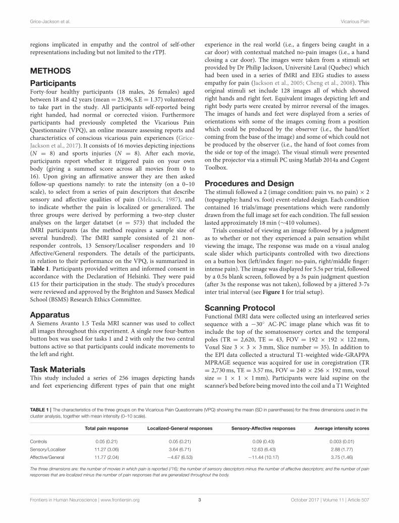
Task MaterialsThis study included a series of 256 images depicting handsand feet experiencing different types of pain that one might
experience in the real world (i.e., a fingers being caught in acar door) with contextual matched no-pain images (i.e., a handclosing a car door). The images were taken from a stimuli setprovided by Dr Philip Jackson, Université Laval (Quebec) whichhad been used in a series of fMRI and EEG studies to assessempathy for pain (Jackson et al., 2005; Cheng et al., 2008). Thisoriginal stimuli set include 128 images all of which showedright hands and right feet. Equivalent images depicting left andright body parts were created by mirror reversal of the images.The images of hands and feet were displayed from a series oforientations with some of the images coming from a positionwhich could be produced by the observer (i.e., the hand/feetcoming from the base of the image) and some of which could notbe produced by the observer (i.e., the hand of foot comes fromthe side or top of the image). The visual stimuli were presentedon the projector via a stimuli PC using Matlab 2014a and CogentToolbox.
Procedures and DesignThe stimuli followed a 2 (image condition: pain vs. no pain) × 2(topography: hand vs. foot) event-related design. Each conditioncontained 16 trials/image presentations which were randomlydrawn from the full image set for each condition. The full sessionlasted approximately 18min (∼410 volumes).
Trials consisted of viewing an image followed by a judgmentas to whether or not they experienced a pain sensation whilstviewing the image, The response was made on a visual analogscale slider which participants controlled with two directionson a button box (left/index finger: no-pain, right/middle finger:intense pain). The image was displayed for 5.5s per trial, followedby a 0.5s blank screen, followed by a 3s pain judgment question(after 3s the response was not taken), followed by a jittered 3-7sinter trial interval (see Figure 1 for trial setup).
Scanning ProtocolFunctional fMRI data were collected using an interleaved seriessequence with a −30◦ AC-PC image plane which was fit toinclude the top of the somatosensory cortex and the temporalpoles (TR = 2,620, TE = 43, FOV = 192 × 192 × 122mm,Voxel Size 3 × 3 × 3mm, Slice number = 35). In addition tothe EPI data collected a structural T1-weighted wide-GRAPPAMPRAGE sequence was acquired for use in coregistration (TR= 2,730ms, TE = 3.57ms, FOV = 240 × 256 × 192mm, voxelsize = 1 × 1 × 1mm). Participants were laid supine on thescanner’s bed before beingmoved into the coil and a T1Weighted
TABLE 1 | The characteristics of the three groups on the Vicarious Pain Questionnaire (VPQ) showing the mean (SD in parentheses) for the three dimensions used in the
cluster analysis, together with mean intensity (0–10 scale).
Total pain response Localized-General responses Sensory-Affective responses Average intensity scores
Controls 0.05 (0.21) 0.05 (0.21) 0.09 (0.43) 0.003 (0.01)
Sensory/Localiser 11.27 (3.06) 3.64 (6.71) 12.63 (6.43) 2.88 (1.77)
Affective/General 11.77 (2.04) −4.67 (6.53) −11.44 (10.17) 3.75 (1.46)
The three dimensions are: the number of movies in which pain is reported (/16); the number of sensory descriptors minus the number of affective descriptors; and the number of pain
responses that are localized minus the number of pain responses that are generalized throughout the body.
Frontiers in Human Neuroscience | www.frontiersin.org 3 October 2017 | Volume 11 | Article 507
Grice-Jackson et al. Vicarious Pain
FIGURE 1 | Stimulus example and trial timings.
structural image was acquired which was then followed by thefMRI data acquisition and task. Each participant’s fMRI sessionwas split between three tasks, of which the main one (alwaysconducted first) is reported here. The other tasks were a cyberballtask of social exclusion, and a pain perception task involving thepresence/absence of facial expressions. The whole session lastedfor approximately 60mins.
fMRI Pre-processingData was processed using Matlab 2014a SPM8 toolbox. Initiallyall images were reoriented relative to the anterior and posteriorcommissure. This was followed by correction of movementduring the task by realigning all of the images to the firstas well as estimation of the movement parameters for eachimage and then by coregistration of realigned images with T1weighted structural images. Raw movement parameters wereused to exclude participants for excessive movement during theexperiment; specifically if a translation regressor exceeded 3mmand a rotational regressor exceeded 5◦. Two controls and oneSensory/Localizer responder were removed because of excessivemovement in the scanner. One more control was removedbecause of an error in recording the trial triggers on the stimulidisplay PC. This resulted in 18 controls, 12 Sensory/localized and10 Affective/generalized responders in the final analysis
EPI images were then normalized to a standardizedanatomical brain (MNI- Montreal Neurological Institute)using SPM’s Dartel normalization function with defaultparameters and finally a Gaussian smoothing kernel (8 × 8 ×
8mm) was applied to the images to increase signal to noise ratio.Homogeneity of variance was used as a check for the warping ofimages during pre-processing.
fMRI AnalysesThe same statistical threshold was applied to all analyses andconsisted of a cluster-level FDR (False Discovery Rate) of p< 0.05and a minimum cluster extent of 40 voxels. Statistical parametricMapping 8 (SPM8, Wellcome trust Center for Neuroimaging)
with the “MarsBar” (Brett et al., 2002) and “anatomy” (Eickhoffet al., 2005) toolboxes was used for the fMRI analysis. Clusterscontaining several peak maxima (separated by at least 8mm) arereported for each cluster when appropriate.
Three sets of first and second level design matrices werecreated for the fMRI analysis. The first focused primarily on theobservations of pain and no pain for the three pain groups. Afirst level design matrix extracted the parameter estimates for allpain and no pain observations (32 trials per condition; InterscanInterval: 2.62s, Microtime Resolution: 16 Microtime onset: 1). Atthis stage, movement regressors were included in the model as acovariate. Six ridged body movement regressors were included inthe first level models throughout the study which included threerotational (roll, pitch, yaw), and three translational regressors (X,Y, Z).For the between group analyses SPM’s contrasts managerwas used to measure the difference between Pain and No-painimages with the t-contrast function in order to estimate theparameters for Pain > No-pain contrasts in order to assess thesize of the difference between condition for each participant. Atthe second level a full factorial design was used to model datawith 2 factors which included: Pain group (3 Levels: Controls,Sensory/Localisers, Affective generals) and Image observations(2 Levels: Pain vs. No Pain). For the between group analysis aOne way ANOVA design was used at the second level using thePain > No Pain parameter estimates with Pain group used as theanalytical factor.
The second set of design matrices was used to modelparameter estimates for the ROI analysis using 7 regions ofthe pain matrix and the rTPJ. The coordinates for the anteriorcingulate cortex (ACC) and anterior insula were based on themeta-analysis of Lamm et al. (2011) namely: ACC, x = 0 y = 12z = 45; left Anterior Insula (AI), x = −40 y = 22 z = 0; andright AI, x = 39 y = 23 z = −4 (in MNI space). The location forthe rTPJ was based on the mid-point between the anterior andposterior rTPJ subdivisions discussed in Krall et al. (2014) meta-analysis of the rTPJ namely x = 54 y =−48 z = 22 (MNI space).For these regions a 10mm spherical binary masks was applied.
Frontiers in Human Neuroscience | www.frontiersin.org 4 October 2017 | Volume 11 | Article 507
Grice-Jackson et al. Vicarious Pain
By contrast, the somatosensory cortex was defined anatomicallyrather than functionally using the masks on SPM’s anatomy toolbox (Eickhoff et al., 2005) for four somatosensory regions (left SI,right SI, left SII, and right SII). Parameter estimates (Betas) wereextracted from these regions using the “MarsBar” tool box (Brettet al., 2002) for Pain>No-pain contrasts. Due to the somatotopicorganization of SI a secondary more detailed ROI analysis wascarried out. Separate hand and foot SI ROIs were selected usinga 10mm spherical masks around the left and right hand areareported by Bingel et al. (2004) for physical pain stimulation (leftMNI: x = 39, y = −30, z = 51; right MNI x = 36, y = −36, z =48, left and right hand areas were compiled into the same ROI)and the left and right foot area (left MNI: x = −9, y = −39, z =57; right MNI x = 9, y=−36, z = 66).
The PPI (Psycho-Physiological Interactions) analysis takesseed regions (in our case, standard regions of the pain matrixidentified via our whole brain analyses) and uses linear regressionto find correlated activity between these seed regions and all otherregions of the brain as a function of a psychological variable(in our case, whether a stimulus depicted pain or not). ThePPI models were produced using the Generalized PPI Toolbox(McLaren et al., 2012) and included all the same event-relatedand nuisance regressors as in the original whole-brain GLM.Additionally, the PPI model included one regressor coding theoverall BOLD time course of the seed region, and “Pain Image”regressor coding the PPI interaction term between Pain andNo-pain image observation. To examine psychophysiologicalinteractions at the group level, we then specified second-levelmodels similar to those used in the whole-brain GLM of BOLDactivations. For each seed region separately, estimates of the PPIinteraction terms relating to the “Pain Image” events regressorswere entered into a 3(Pain group: Controls vs. S/L Responders vs.A/G Responders) × 2 (Pain Image: Pain vs. No Pain) ANOVAmodel.
RESULTS
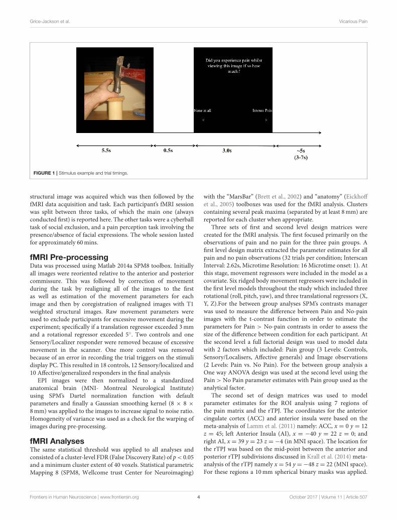
Vicarious Pain RatingsThe visual analog scale was transformed to a 0–100 range witha higher score indicating higher intensity of vicarious pain.The mean scores, shown in Figure 2, were analyzed using a 2(Condition: Pain vs. No-pain stimulus) × 3 (Group) ANOVA.There were significant main effects of stimulus [F(1, 39) = 68.67, p< 0.001, r= 0.64] and group [F(2, 39) = 34.45, p< 0.001, r= 0.65]as well as a significant interaction [F(1, 39) = 15.554, p < 0.001, r= 0.47]. Within group planned comparisons showed that the S/L[t(11) = 5.217, p< 0.001] andA/G groups [t(9) = 3.653, p= 0.011]showed significantly increased scores during Pain relative to No-pain trials but the controls did not [t(17) = 1.655, p = 0.155].In summary, our two responder groups reported increased levelsof vicarious pain for these stimuli during scanning as they hadpreviously done for similar stimuli outside the scanner.
fMRI ResultsWhole Brain Analysis
Initially a whole brain analysis was run on the data. We assessedpain vs. no-pain within groups and contrasted the effects of
FIGURE 2 | Subjective ratings for the pain (filled bars) and no-pain (empty
bars) stimuli. A score of 100 indicates a high perception of pain in the
participant in response to the observation of pain and a score of 0 represents
no perception of pain. Error bars show ±1 SEM.
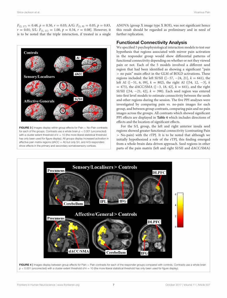
the same stimuli between groups. All of the tests were carriedout using t-contrasts. Table 2 displays all regions which showedsignificantly increased differences in pain vs. no-pain activationfor the three groups (see also Figure 3). This analysis shows thatall groups display effects in regions associated with the affectiveprocessing of pain (anterior insula, and dorsal anterior cingulateextending into the supplementary motor area). However, onlythe S/L and A/G group showed increased activation in thesomatosensory cortices which was confirmed by a subsequentROI analysis.
The differences between groups were explored by assessingPain vs. No-Pain first level t-contrast betas in a second levelone way ANOVA with three groups. Figure 4 and Table 3
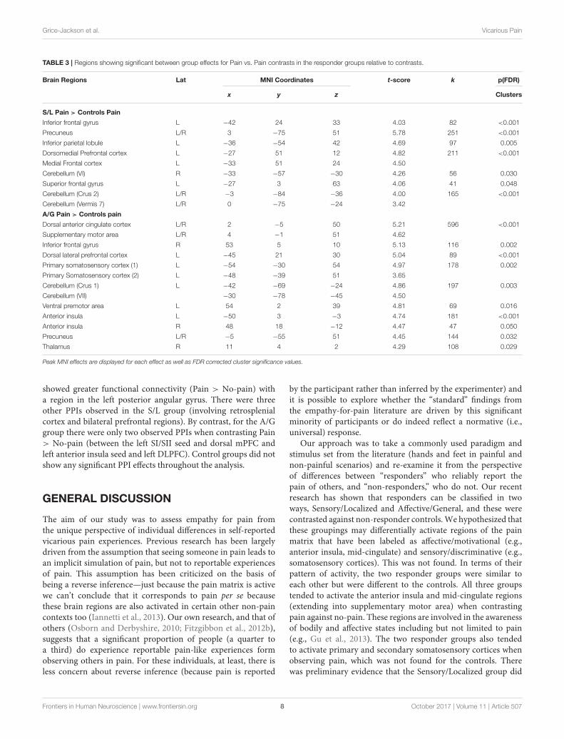
displays regions showing significant effects (contrasts which didnot yield significant effects are not displayed). The respondergroups had greater activity than controls in a variety of regionswhen observing pain, but no effects were observed in theopposite direction (i.e., controls > responders). This includedthe dorsolateral prefrontal cortex and cerebellum for bothsensory/localizer responders and affective/general. No groupdifferences were statistically detected between the two respondergroups suggesting they are broadly similar, at least for the presentlevel of statistical power. Notwithstanding this similarity, wedemonstrate later that the groups differ in their connectivityprofile.
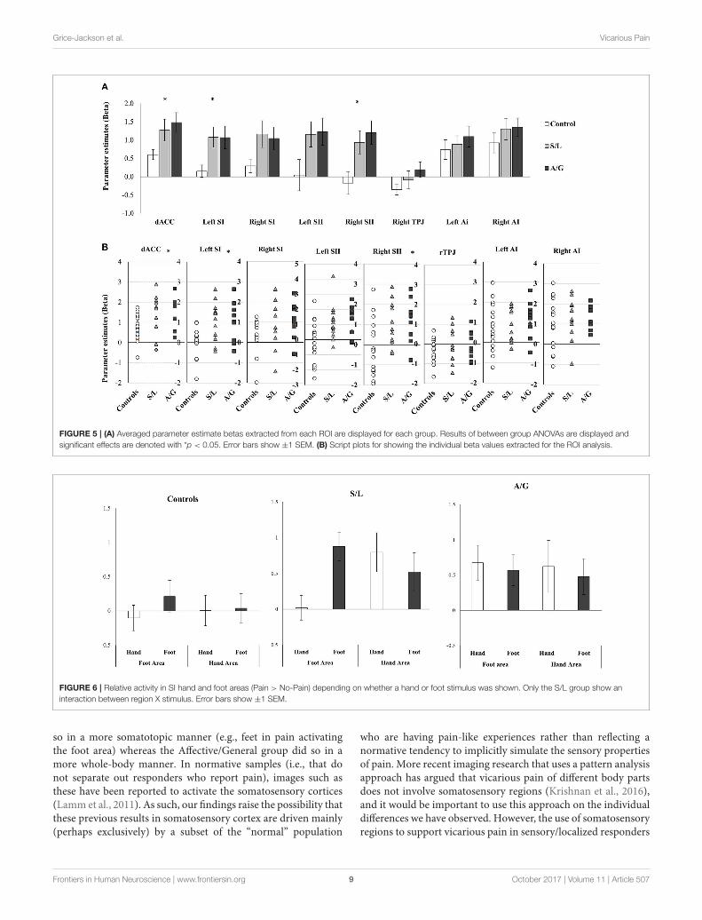
ROI AnalysisParameter estimates were extracted from each ROI for contrastsbetween pain vs. no-pain observations. These parameterestimates show the difference between pain and no painobservations with positive beta values indicating increasedactivation in the region when viewing pain images. A seriesof one way ANOVAs assessing differences between the paingroups was run on each ROI. Given that these analyses werehypothesis-driven, we considered that a type 2 error would bemore detrimental than a type 1 error and therefore opted toreport effect sizes (in addition to p-values), rather than apply a
Frontiers in Human Neuroscience | www.frontiersin.org 5 October 2017 | Volume 11 | Article 507
Grice-Jackson et al. Vicarious Pain
TABLE 2 | Regions showing significant within group effects in Pain > No-pain image contrasts. Peak MNI effects are displayed for each effect as well as FDR corrected
cluster significance values.
Brain Regions Lat MNI Coordinates t-score k P(FDR)
x Y z Cluster
Controls: Pain > No pain
Supplementary motor area L/R 0 3 66 4.89 351 <0.001
Anterior insula L −54 9 −9 4.62 239 <0.001
Inferior frontal gyrus L −57 9 27 4.35
Anterior insula R 57 12 −3 4.38 54 0.045
S/L: Pain > No pain
Cerebellum (VI) R 30 −54 −30 5.48 76 0.011
Primary somatosensory cortex (1/2) L −54 −30 51 5.20 361 <0.001
Secondary somatosensory cortex L −57 −24 21 4.49
Inferior parietal lobule L −50 −33 18 3.92
Dorsal anterior cingulate cortex L/R −3 12 45 5.07 136 0.001
Primary somatosensory cortex (1) R 57 −27 51 4.76 146 0.001
Primary somatosensory cortex (3b) R 54 −18 42 3.79
Anterior insula L −33 12 0 4.51 313 0.001
Parahippocampal gyrus L −21 5 −21 4.47
Anterior insula R 51 3 −3 4.40 105 0.003
Dorsal lateral prefrontal cortex L −30 45 27 4.31 113 0.002
Medial prefrontal cortex L −20 39 39 3.36
Precentral gyrus L −27 0 60 4.27 64 0.017
Caudate nucleus R 15 6 6 3.96 69 0.014
A/G: Pain > No-pain
Supplementary motor area L/R 0 3 66 5.42 780 <0.001
Dorsal anterior cingulate cortex L/R 3 −3 54 4.34
Inferior frontal gyrus (Opercularis) L −57 9 9 4.95 512 <0.001
Temporal pole L −48 12 −9 4.67
Anterior Insula L −45 3 3 4.43
Secondary somatosensory cortex R −60 −21 19 4.81 143 0.027
Ventral striatum R 21 6 −9 4.56 122 0.026
Periaqueductal gray R 12 6 3 4.42
Premotor cortex L 48 −8 56 4.28 137 0.017
Anterior insula R 42 3 3 4.21 100 0.035
Secondary somatosensory cortex L −63 −24 21 4.13 111 0.029
more conservative Bonferroni correction. Four regions displaysignificant differences between the groups, including: dACC[F(2, 39) = 4.714, p = 0.015, r = 0.635], left SI [F(2, 39) = 5.757,p = 0.007, r = 0.741], and right SII [F(2, 39) = 5.441, p =
0.021, r = 0.704], additionally left SII and right SI showed aneffect of borderline significance [Left SII: F(2, 35) = 2.846, p= 0.076, r = 0.589; right SI F(2, 39) = 3.114, p = 0.056, r =
0.491]. For all significant ROIs the two responders groups hadsignificantly higher signal change relative to controls but theydid not differ from each other. Non-significant effects included:the right TPJ [F(2, 39) = 2.0440.634, p = 0.512, r = 0.156],the left AI [F(2, 39) = 0.431, p = 0.653, r = 0.097] and theright AI [F(2, 39) = 0.761, p = 0.474, r = 0.150, see Figure 5].These effects show that both affective and sensory pain matrixregions show differences in activation between the groups withthe two pain responder groups displaying increased differences
between pain and no pain observations relative to controls.These were driven by differences in the pain rather than no-paincondition.
Due to the somatotopic organization of S1 a more detailedROI analysis was carried out. The results are summarized inFigure 6. A 2 (hand vs. foot image) × 2 (hand vs. foot ROI)repeated measures ANOVA was run on the data (one for eachGroup: Controls, S/L, A/G). The S/L group showed a stronginteraction of image type X ROI [F(1, 11) = 20.40, p = 0.001, r= 0.63] such that feet images more strongly activated the footarea and hand images more strongly activated the hand area, Thispattern was absent in the other two groups [Controls: F(2, 17) =0.356, p = 0.559, r = 0.021; A/G: F(1, 9) = 0.01, p = 0.95, r =0.001]. No group showed main effects of image type [Controls:F(1, 17) = 0.07, p = 0.79, r = 0.06; A/G: F(1, 9) = 0.39, p = 0.55, r= 0.04; S/L: F(1, 11) = 1.65, p = 0.22, r = 0.12] or ROI [Controls:
Frontiers in Human Neuroscience | www.frontiersin.org 6 October 2017 | Volume 11 | Article 507
Grice-Jackson et al. Vicarious Pain
F(2, 17) = 0.48, p = 0.50, r = 0.03; A/G: F(1, 9) = 0.05, p = 0.83,r = 0.01; S/L: F(1, 11) = 1.00, p = 0.34, r = 0.08]. However, itis to be noted that the triple interaction, if treated in a single
FIGURE 3 | Images display within group effects for Pain > No-Pain contrasts
for each of the groups. Contrasts use a whole brain p < 0.001 (uncorrected)
with a cluster extent threshold of k = 10 (the more liberal statistical threshold
has only been used for figure display). All groups display increased activation in
affective pain matrix regions (dACC + AI) but only S/L and A/G responders
show effects in the primary and secondary somatosensory cortices.
ANOVA (group X image type X ROI), was not significant hencethis result should be regarded as preliminary and in need offurther replication.
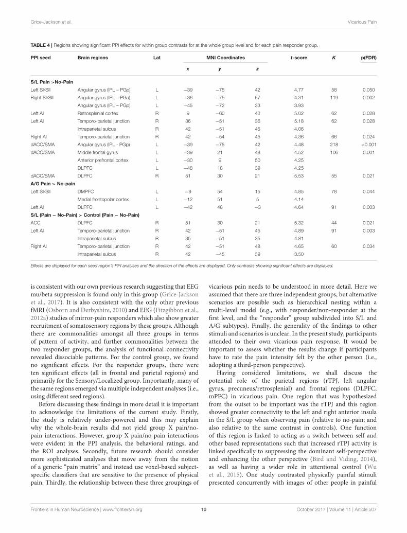
Functional Connectivity AnalysisWe specified 5 psychophysiological interactionmodels to test ourhypothesis that regions associated with mirror pain activationin the responder group would show differential patterns offunctional connectivity depending on whether or not they viewedpain or not. Each of the 5 models involved a different seedregion that had been identified as showing a significant “pain> no pain” main effect in the GLM of BOLD activations. Theseregions included: the left SI/SII ([−57, −24, 21], k = 641), theleft AI ([−51, 6, 09], k = 802), the right AI ([54, 12, −3], k= 475), the dACC/SMA ([−3, 18, 42], k = 641), and the rightSI/SII ([54, −21, 42], k = 390). Each seed region was enteredinto first level models to estimate connectivity between the seedsand other regions during the session. The five PPI analyses wereinvestigated by comparing pain vs. no-pain images for eachgroup, and between group contrasts, comparing pain and no painimages across the groups. All contrasts which showed significantPPI effects are displayed in Table 4 which includes directions ofeffects and the location of significant effects.
For the S/L group, the left and right anterior insula seedregions showed greater functional connectivity (contrasting Pain> No-pain) with the rTPJ. It is to be noted that although weinitially hypothesized a role of the rTPJ, this finding emergedfrom a whole-brain data-driven approach. Seed regions in otherparts of the pain matrix (left and right SI/SII and dACC/SMA)
FIGURE 4 | Images display between group effects for Pain > Pain contrasts for each of the responder groups compared with controls. Contrasts use a whole brain
p < 0.001 (uncorrected) with a cluster extent threshold of k = 10 (the more liberal statistical threshold has only been used for figure display).
Frontiers in Human Neuroscience | www.frontiersin.org 7 October 2017 | Volume 11 | Article 507
Grice-Jackson et al. Vicarious Pain
TABLE 3 | Regions showing significant between group effects for Pain vs. Pain contrasts in the responder groups relative to contrasts.
Brain Regions Lat MNI Coordinates t-score k p(FDR)
x y z Clusters
S/L Pain > Controls Pain
Inferior frontal gyrus L −42 24 33 4.03 82 <0.001
Precuneus L/R 3 −75 51 5.78 251 <0.001
Inferior parietal lobule L −36 −54 42 4.69 97 0.005
Dorsomedial Prefrontal cortex L −27 51 12 4.82 211 <0.001
Medial Frontal cortex L −33 51 24 4.50
Cerebellum (VI) R −33 −57 −30 4.26 56 0.030
Superior frontal gyrus L −27 3 63 4.06 41 0.048
Cerebellum (Crus 2) L/R −3 −84 −36 4.00 165 <0.001
Cerebellum (Vermis 7) L/R 0 −75 −24 3.42
A/G Pain > Controls pain
Dorsal anterior cingulate cortex L/R 2 −5 50 5.21 596 <0.001
Supplementary motor area L/R 4 −1 51 4.62
Inferior frontal gyrus R 53 5 10 5.13 116 0.002
Dorsal lateral prefrontal cortex L −45 21 30 5.04 89 <0.001
Primary somatosensory cortex (1) L −54 −30 54 4.97 178 0.002
Primary Somatosensory cortex (2) L −48 −39 51 3.65
Cerebellum (Crus 1) L −42 −69 −24 4.86 197 0.003
Cerebellum (VII) −30 −78 −45 4.50
Ventral premotor area L 54 2 39 4.81 69 0.016
Anterior insula L −50 3 −3 4.74 181 <0.001
Anterior insula R 48 18 −12 4.47 47 0.050
Precuneus L/R −5 −55 51 4.45 144 0.032
Thalamus R 11 4 2 4.29 108 0.029
Peak MNI effects are displayed for each effect as well as FDR corrected cluster significance values.
showed greater functional connectivity (Pain > No-pain) witha region in the left posterior angular gyrus. There were threeother PPIs observed in the S/L group (involving retrosplenialcortex and bilateral prefrontal regions). By contrast, for the A/Ggroup there were only two observed PPIs when contrasting Pain> No-pain (between the left SI/SII seed and dorsal mPFC andleft anterior insula seed and left DLPFC). Control groups did notshow any significant PPI effects throughout the analysis.
GENERAL DISCUSSION
The aim of our study was to assess empathy for pain fromthe unique perspective of individual differences in self-reportedvicarious pain experiences. Previous research has been largelydriven from the assumption that seeing someone in pain leads toan implicit simulation of pain, but not to reportable experiencesof pain. This assumption has been criticized on the basis ofbeing a reverse inference—just because the pain matrix is activewe can’t conclude that it corresponds to pain per se becausethese brain regions are also activated in certain other non-paincontexts too (Iannetti et al., 2013). Our own research, and that ofothers (Osborn and Derbyshire, 2010; Fitzgibbon et al., 2012b),suggests that a significant proportion of people (a quarter toa third) do experience reportable pain-like experiences formobserving others in pain. For these individuals, at least, there isless concern about reverse inference (because pain is reported
by the participant rather than inferred by the experimenter) andit is possible to explore whether the “standard” findings fromthe empathy-for-pain literature are driven by this significantminority of participants or do indeed reflect a normative (i.e.,universal) response.
Our approach was to take a commonly used paradigm andstimulus set from the literature (hands and feet in painful andnon-painful scenarios) and re-examine it from the perspectiveof differences between “responders” who reliably report thepain of others, and “non-responders,” who do not. Our recentresearch has shown that responders can be classified in twoways, Sensory/Localized and Affective/General, and these werecontrasted against non-responder controls.We hypothesized thatthese groupings may differentially activate regions of the painmatrix that have been labeled as affective/motivational (e.g.,anterior insula, mid-cingulate) and sensory/discriminative (e.g.,somatosensory cortices). This was not found. In terms of theirpattern of activity, the two responder groups were similar toeach other but were different to the controls. All three groupstended to activate the anterior insula and mid-cingulate regions(extending into supplementary motor area) when contrastingpain against no-pain. These regions are involved in the awarenessof bodily and affective states including but not limited to pain(e.g., Gu et al., 2013). The two responder groups also tendedto activate primary and secondary somatosensory cortices whenobserving pain, which was not found for the controls. Therewas preliminary evidence that the Sensory/Localized group did
Frontiers in Human Neuroscience | www.frontiersin.org 8 October 2017 | Volume 11 | Article 507
Grice-Jackson et al. Vicarious Pain
FIGURE 5 | (A) Averaged parameter estimate betas extracted from each ROI are displayed for each group. Results of between group ANOVAs are displayed and
significant effects are denoted with *p < 0.05. Error bars show ±1 SEM. (B) Script plots for showing the individual beta values extracted for the ROI analysis.
FIGURE 6 | Relative activity in SI hand and foot areas (Pain > No-Pain) depending on whether a hand or foot stimulus was shown. Only the S/L group show an
interaction between region X stimulus. Error bars show ±1 SEM.
so in a more somatotopic manner (e.g., feet in pain activatingthe foot area) whereas the Affective/General group did so in amore whole-body manner. In normative samples (i.e., that donot separate out responders who report pain), images such asthese have been reported to activate the somatosensory cortices(Lamm et al., 2011). As such, our findings raise the possibility thatthese previous results in somatosensory cortex are driven mainly(perhaps exclusively) by a subset of the “normal” population
who are having pain-like experiences rather than reflecting anormative tendency to implicitly simulate the sensory propertiesof pain. More recent imaging research that uses a pattern analysisapproach has argued that vicarious pain of different body partsdoes not involve somatosensory regions (Krishnan et al., 2016),and it would be important to use this approach on the individualdifferences we have observed. However, the use of somatosensoryregions to support vicarious pain in sensory/localized responders
Frontiers in Human Neuroscience | www.frontiersin.org 9 October 2017 | Volume 11 | Article 507
Grice-Jackson et al. Vicarious Pain
TABLE 4 | Regions showing significant PPI effects for within group contrasts for at the whole group level and for each pain responder group.
PPI seed Brain regions Lat MNI Coordinates t-score K p(FDR)
x y z
S/L Pain >No-Pain
Left SI/SII Angular gyrus (IPL – PGp) L −39 −75 42 4.77 58 0.050
Right SI/SII Angular gyrus (IPL – PGa) L −36 −75 57 4.31 119 0.002
Angular gyrus (IPL – PGp) L −45 −72 33 3.93
Left AI Retrosplenial cortex R 9 −60 42 5.02 62 0.028
Left AI Temporo-parietal junction R 36 −51 36 5.18 62 0.028
Intraparietal sulcus R 42 −51 45 4.06
Right AI Temporo-parietal junction R 42 −54 45 4.36 66 0.024
dACC/SMA Angular gyrus (IPL - PGp) L −39 −75 42 4.48 218 <0.001
dACC/SMA Middle frontal gyrus L −39 21 48 4.52 106 0.001
Anterior prefrontal cortex L −30 9 50 4.25
DLPFC L −48 18 39 4.25
dACC/SMA DLPFC R 51 30 21 5.53 55 0.021
A/G Pain > No-pain
Left SI/SII DMPFC L −9 54 15 4.85 78 0.044
Medial frontopolar cortex L −12 51 5 4.14
Left AI DLPFC L −42 48 −3 4.64 91 0.003
S/L (Pain − No-Pain) > Control (Pain − No-Pain)
ACC DLPFC R 51 30 21 5.32 44 0.021
Left AI Temporo-parietal junction R 42 −51 45 4.89 91 0.003
Intraparietal sulcus R 35 −51 35 4.81
Right AI Temporo-parietal junction R 42 −51 48 4.65 60 0.034
Intraparietal sulcus R 42 −45 39 3.50
Effects are displayed for each seed region’s PPI analyses and the direction of the effects are displayed. Only contrasts showing significant effects are displayed.
is consistent with our own previous research suggesting that EEGmu/beta suppression is found only in this group (Grice-Jacksonet al., 2017). It is also consistent with the only other previousfMRI (Osborn and Derbyshire, 2010) and EEG (Fitzgibbon et al.,2012a) studies of mirror-pain responders which also show greaterrecruitment of somatosensory regions by these groups. Althoughthere are commonalities amongst all three groups in termsof pattern of activity, and further commonalities between thetwo responder groups, the analysis of functional connectivityrevealed dissociable patterns. For the control group, we foundno significant effects. For the responder groups, there wereten significant effects (all in frontal and parietal regions) andprimarily for the Sensory/Localized group. Importantly, many ofthe same regions emerged via multiple independent analyses (i.e.,using different seed regions).
Before discussing these findings in more detail it is importantto acknowledge the limitations of the current study. Firstly,the study is relatively under-powered and this may explainwhy the whole-brain results did not yield group X pain/no-pain interactions. However, group X pain/no-pain interactionswere evident in the PPI analysis, the behavioral ratings, andthe ROI analyses. Secondly, future research should considermore sophisticated analyses that move away from the notionof a generic “pain matrix” and instead use voxel-based subject-specific classifiers that are sensitive to the presence of physicalpain. Thirdly, the relationship between these three groupings of
vicarious pain needs to be understood in more detail. Here weassumed that there are three independent groups, but alternativescenarios are possible such as hierarchical nesting within amulti-level model (e.g., with responder/non-responder at thefirst level, and the “responder” group subdivided into S/L andA/G subtypes). Finally, the generality of the findings to otherstimuli and scenarios is unclear. In the present study, participantsattended to their own vicarious pain response. It would beimportant to assess whether the results change if participantshave to rate the pain intensity felt by the other person (i.e.,adopting a third-person perspective).
Having considered limitations, we shall discuss thepotential role of the parietal regions (rTPJ, left angulargyrus, precuneus/retrosplenial) and frontal regions (DLPFC,mPFC) in vicarious pain. One region that was hypothesizedfrom the outset to be important was the rTPJ and this regionshowed greater connectivity to the left and right anterior insulain the S/L group when observing pain (relative to no-pain; andalso relative to the same contrast in controls). One functionof this region is linked to acting as a switch between self andother based representations such that increased rTPJ activity islinked specifically to suppressing the dominant self-perspectiveand enhancing the other perspective (Bird and Viding, 2014),as well as having a wider role in attentional control (Wuet al., 2015). One study contrasted physically painful stimulipresented concurrently with images of other people in painful
Frontiers in Human Neuroscience | www.frontiersin.org 10 October 2017 | Volume 11 | Article 507
Grice-Jackson et al. Vicarious Pain
or neutral situations (Godinho et al., 2012). Seeing someoneelse in pain increases the self-reported intensity of physical pain(a normal form of self-other confusion) and this was linkedto the same posterior region of the rTPJ that we observed(and not to increased activity in the pain matrix). The patternanalysis study of Krishnan et al. (2016) found that the rTPJ(and other regions of the mentalizing network, but not thepain matrix) were involved in vicarious pain, although in thisstudy participants performed perspective taking (imaginingsomeone else’s experiences on their own body). Our explanationof the Sensory/Localized group is that they systematically fail toattribute shared bodily representations to others and this, at leastin part, reflects structural and functional differences within therTPJ coupled with other differences (e.g., in left parietal cortex).
Aside from the rTPJ, other regions were highlighted by theconnectivity analysis for the S/L group. A region in the leftangular gyrus showed greater connectivity (when observing Pain> No-pain) to three seed regions (left and right somatosensorycortices, and the cingulate/SMA region). This parietal regionis not the left hemispheric homolog of the rTPJ but is severalcentimeters posterior to it. This region has been found tobe important in several studies relating to agency and bodyownership. Long-term gamers who habitually use a certain avataractivate this region when thinking about (Ganesh et al., 2012)or observing (Lemenager et al., 2014) their avatar. Patientswith left parietal lobe damage are more inclined to claimagency to third-person perspective hand movements executed byothers (Sirigu et al., 1999), and TMS over this region in healthycontrols disrupts agency attribution (Chambon et al., 2015).The precise function(s) of this region is uncertain, but onetheory is that it computes a mental simulation of the self intoalternate spatial scenes and perspectives (Buckner and Carroll,2007). The precuneus/retrosplenial area was also implicated bymultiple independent analyses (Pain > No-pain in both S/Land A/G groups, and PPI connectivity to left anterior insulain the S/L group). While this region may also serve a generalrole in mental simulation/imagery (Cavanna and Trimble, 2006),it has also been hypothesized to have a more specific rolein pain. Stimulation of this region in rats has an analgesiceffect (Rossaneis et al., 2011) and, in humans, patients withfibromyalgia have higher resting levels of activity in this regionwhich may arguably reflect an analgesic function (Wik et al.,2003).
With regards to the frontal lobe, two regions (medial PFCand dorso-lateral PFC) are noteworthy. The medial PFC regionwas implicated in the functional connectivity analysis for theAffective/General group. It has been linked specifically to theself-concept (e.g., thinking about one’s own characteristics) ratherthan bodily self (e.g., Mitchell et al., 2005). This particular resultneeds to be treated with caution given that the region was notimplicated by any other analysis. The DLPFC has widespreadeffects on cognitive control (Duncan, 2010) including empathy(Moriguchi et al., 2007) and emotion regulation (Ochsner et al.,2012), so it would be unwise to infer a specific role for mirrorpain. The region (both left and right) was implicated acrossmultiple analyses. It is important to explore the role of thisregion, alongside the parietal regions previously discussed, using
methods such as non-invasive brain stimulation (NIBS) andcombinedNIBS-fMRI to examine its causal role on vicarious painperception.
To conclude, our research has important theoreticalimplications for research on empathy for pain (and other sharedstates). It suggests that greater attention should be paid toindividual differences in reportable experiences. These havethe potential to distort what is assumed to be a normativeresponse. In particular, activity within the somatosensorycortex when observing others in pain may be primarily (andpossibly exclusively) linked to those individuals who reportpain when seeing others in pain. Patients with congenital paininsensitivity activate some regions of the “pain matrix,” notablyinsula and mid-cingulate, when viewing others in pain butnotably not the somatosensory cortex (Danziger et al., 2009).Contrary to our initial predictions, the amount of activity insomatosensory cortices does not seem to strongly reflect thedistinction between Sensory/Localized and Affective/General,but they may nonetheless show differences in how these regionsare activated (body-part vs. whole body respectively).
Increased activity within pain matrix regions might beproposed to be both necessary and sufficient for consciouslyexperienced vicarious pain. We previously referred to this asThreshold Theory (Ward and Banissy, 2015) and is based onthe notion that all individuals activate, to varying degrees,the pain matrix on seeing pain but only those that do soabove a threshold for awareness have reportable pain-likeexperiences. We do not doubt that this is part of the explanation,however, we question whether it is sufficient. In particular,we argue that it is interactions between the pain matrix andvarious fronto-parietal regions that give rise to these reportablevicarious pain experiences. This is more clearly the case forthe Sensory/Localized group for whom we observed enhancedfunctional connectivity between pain matrix regions and therTPJ and left angular gyrus, both of which are implicated indiscriminating self from other and bodily perspective taking.The explanation for the Affective/General group is presentlylacking and, in some respects, appears to be intermediate betweenthe Sensory/Localized and the non-responder groups. Onepossibility is that this group reflects differences on autonomicmeasures (for a model incorporating this see Giummarraand Fitzgibbon, 2015). In summary, our research providesfresh evidence that these individual differences are importantto consider methodologically (as they can skew results) andtheoretically, as they provide important test cases for currentmodels.
AUTHOR CONTRIBUTIONS
TG collected and analyzed the data. TG, MB, HC, and JW allcontributed to the design of the study, interpretation of data, andwriting of the manuscript.
FUNDING
This research is funded by the Sackler Center for ConsciousnessScience.
Frontiers in Human Neuroscience | www.frontiersin.org 11 October 2017 | Volume 11 | Article 507
Grice-Jackson et al. Vicarious Pain
REFERENCES
Avenanti, A., Bueti, D., Galati, G., and Aglioti, S. M. (2005). Transcranial magnetic
stimulation highlights the sensorimotor side of empathy for pain.Nat. Neurosci.
8, 955–960. doi: 10.1038/nn1481
Bingel, U., Lorenz, J., Glauche, V., Knab, R., Gläscher, J., Weiller, C.,
et al. (2004). Somatotopic organization of human somatosensory
cortices for pain: a single trial fMRI study. Neuroimage 23, 224–232.
doi: 10.1016/j.neuroimage.2004.05.021
Bird, G., and Viding, E. (2014). The self to other model of empathy:
providing a new framework for understanding empathy impairments in
psychopathy, autism, and alexithymia. Neurosci. Biobehav. Rev. 47, 520–532.
doi: 10.1016/j.neubiorev.2014.09.021
Brett, M., Valabregue, R., and Poline, J. (2002). “Region of interest analysis using
an SPM toolbox,” in 8th International Conference on Functional Mapping of the
Human Brain, Vol. 16 (Sendai).
Buckner, R. L., and Carroll, D. C. (2007). Self-projection and the brain. Trends
Cogn. Sci. 11, 49–57. doi: 10.1016/j.tics.2006.11.004
Bufalari, I., Aprile, T., Avenanti, A., Di Russo, F., and Aglioti, S. M. (2007).
Empathy for pain and touch in the human somatosensory cortex.Cereb. Cortex.
17, 2553–2561. doi: 10.1093/cercor/bhl161
Cavanna, A. E., and Trimble, M. R. (2006). The precuneus: a review of
its functional anatomy and behavioural correlates. Brain 129, 564–583.
doi: 10.1093/brain/awl004
Chambon, V., Moore, J. W., and Haggard, P. (2015). TMS stimulation over the
inferior parietal cortex disrupts prospective sense of agency. Brain Struct. Funct.
220, 3627–3639. doi: 10.1007/s00429-014-0878-6
Cheng, Y. W., Lee, P. L., Yang, C. Y., Lin, C. P., Hung, D., and Decety, J. (2008).
Gender differences in the Mu Rhythm of the human mirror-neuron system.
PLoS ONE 3:e2113. doi: 10.1371/journal.pone.0002113
Danziger, N., Faillenot, I., and Peyron, R. (2009). Can we share a pain
we never felt? neural correlates of empathy in patients with congenital
insensitivity to pain. Neuron 61, 203–212. doi: 10.1016/j.neuron.2008.
11.023
de Guzman, M., Bird, G., Banissy, M. J., and Catmur, C. (2016). Self-other control
processes in social cognition: from imitation to empathy. Philos. Trans. R. Soc.
B Biol. Sci. 371:20150079. doi: 10.1098/rstb.2015.0079
Decety, J. (2011). Dissecting the neural mechanisms mediating empathy. Emot.
Rev. 3, 92–108. doi: 10.1177/1754073910374662
Decety, J., and Jackson, P. L. (2006). A social-neuroscience perspective on empathy.
Curr. Dir. Psychol. Sci. 15, 54–58. doi: 10.1111/j.0963-7214.2006.00406.x
Duncan, J. (2010). The multiple-demand (MD) system of the primate brain:
mental programs for intelligent behaviour. Trends Cogn. Sci. 14, 172–179.
doi: 10.1016/j.tics.2010.01.004
Eickhoff, S., Stephan, K. E., Mohlberg, H., Grefkes, C., Fink, G. R., Amunts,
K., et al. (2005). A new SPM toolbox for combining probabilistic
cytoarchitectonic maps and functional imaging data. Neuroimage 25,
1325–1335. doi: 10.1016/j.neuroimage.2004.12.034
Fitzgibbon, B. M., Enticott, P. G., Giummarra, M. J., Thomson, R. H., Georgiou-
Karistianis, N., and Bradshaw, J. L. (2012a). Atypical electrphysiological activity
during pain observation in amputees who experience synaesthetic pain. Soc.
Cogn. Affect. Neurosci. 7, 357–368. doi: 10.1093/scan/nsr016
Fitzgibbon, B. M., Enticott, P. G., Rich, A. N., Giummarra, M. J., Georgiou-
Karistianis, N., and Bradshaw, J. L. (2012b). Mirror-sensory synaesthesia:
exploring ’shared’ sensory experiences as synaesthesia. Neurosci. Biobehav. Rev.
36, 645–657. doi: 10.1016/j.neubiorev.2011.09.006
Ganesh, S., van Schie, H. T., de Lange, F. P., Thompson, E., and Wigboldus, D.
H. J. (2012). How the human brain goes virtual: distinct cortical regions of
the person-processing network are involved in self-identification with virtual
agents. Cereb. Cortex 22, 1577–1585. doi: 10.1093/cercor/bhr227
Giummarra, M. J., and Fitzgibbon, B. M. (2015). Into the looking glass: broadening
models to explain the spectrum of sensory and affective vicarious experiences.
Cogn. Neurosci. 6, 135–137. doi: 10.1080/17588928.2015.1053853
Godinho, F., Faillenot, I., Perchet, C., Frot, M., Magnin, M., and Garcia-
Larrea, L. (2012). How the pain of others enhances our pain: searching the
cerebral correlates of “compassional hyperalgesia”. Eur. J. Pain 16, 748–759.
doi: 10.1002/j.1532-2149.2011.00039.x
Grice-Jackson, T., Critchley, H. D., Banissy, M. J., and Ward, J. (2017).
Common and distinct neural mechanisms associated with the conscious
experience of vicarious pain. Cortex 94, 152–163. doi: 10.1016/j.cortex.2017.
06.015
Gu, X. S., Hof, P. R., Friston, K. J., and Fan, J. (2013). Anterior insular
cortex and emotional awareness. J. Comparat. Neurol. 521, 3371–3388.
doi: 10.1002/cne.23368
Holle, H., Banissy, M. J., and Ward, J. (2013). Functional and structural
brain correlates of mirror-touch synaesthesia. Neuroimage 83, 1041–1050.
doi: 10.1016/j.neuroimage.2013.07.073
Iannetti, G. D., and Mouraux, A. (2010). From the neuromatrix to the pain
matrix (and back). Exp. Brain Res. 205, 1–12. doi: 10.1007/s00221-010-
2340-1
Iannetti, G. D., Salomons, T. V., Moayedi, M., Mouraux, A., and Davis,
K. D. (2013). Beyond metaphor: contrasting mechanisms of social and
physical pain. Trends Cogn. Sci. 17, 371–378. doi: 10.1016/j.tics.2013.
06.002
Jackson, P. L., Meltzoff, A. N., andDecety, J. (2005). How dowe perceive the pain of
others? A window into the neural processes involved in empathy. Neuroimage
24, 771–779. doi: 10.1016/j.neuroimage.2004.09.006
Krall, S. C., Rottschy, C., Oberwelland, E., Bzdok, D., Fox, P. T., Eickhoff, S. B.,
et al. (2014). The role of the right temporoparietal junction in attention and
social interaction as revealed by ALE meta-analysis. Brain Struc. Funct. 220,
587–604. doi: 10.1007/s00429-014-0803-z
Krishnan, A., Woo, C. W., Chang, L. J., Ruzic, L., Gu, X. S., Lopez-Sola, M., et al.
(2016). Somatic and vicarious pain are represented by dissociable multivariate
brain patterns. Elife 5:e15166. doi: 10.7554/eLife.15166
Lamm, C., Bukowski, H., and Silani, G. (2016). From shared to distinct self-
other representations in empathy: evidence from neurotypical function and
socio-cognitive disorders. Philos. Trans. R. Soc. B Biol. Sci. 371:20150083.
doi: 10.1098/rstb.2015.0083
Lamm, C., Decety, J., and Singer, T. (2011). Meta-analytic evidence
for common and distinct neural networks associated with directly
experienced pain and empathy for pain. Neuroimage 54, 2492–2502.
doi: 10.1016/j.neuroimage.2010.10.014
Lemenager, T., Dieter, J., Hill, H., Koopmann, A., Reinhard, I., Sell, M.,
et al. (2014). Neurobiological correlates of physical self-concept and self-
identification with avatars in addicted players of Massively Multiplayer
Online Role-Playing Games (MMORPGs). Addict. Behav. 39, 1789–1797.
doi: 10.1016/j.addbeh.2014.07.017
McLaren, D. G., Ries, M. L. Xu, G., and Johnson, S. C. (2012). A
generalized form of context-dependent psychophysiological interactions
(gPPI): a comparison to standard approaches. NeuroImage 61, 1277–1286.
doi: 10.1016/j.neuroimage.2012.03.068
Melzack, R. (1987). The short-form McGill pain questionnaire. Pain 30, 191–197.
doi: 10.1016/0304-3959(87)91074-8
Melzack, R. (1999). From the gate to the neuromatrix. Pain 82, 121–126.
doi: 10.1016/S0304-3959(99)00145-1
Mitchell, J. P., Banaji, M. R., and Macrae, C. N. (2005). The link between social
cognition and self-referential thought in the medial prefrontal cortex. J. Cogn.
Neurosci. 17, 1306–1315. doi: 10.1162/0898929055002418
Moriguchi, Y., Decety, J., Ohnishi, T., Maeda, M., Mori, T., Nemoto, K., et al.
(2007). Empathy and judging other’s pain: an fMRI study of alexithymia. Cereb.
Cortex 17, 2223–2234. doi: 10.1093/cercor/bhl130
Ochsner, K. N., Silvers, J. A., and Buhle, J. T. (2012). Functional imaging
studies of emotion regulation: a synthetic review and evolving model of
the cognitive control of emotion. Year Cogn. Neurosci. 1251, E1–E24.
doi: 10.1111/j.1749-6632.2012.06751.x
Osborn, J., and Derbyshire, S. W. G. (2010). Pain sensation evoked by observing
injury in others. Pain 148, 268–274. doi: 10.1016/j.pain.2009.11.007
Peyron, R., Laurent, B., and Garcia-Larrea, L. (2000). Functional imaging of
brain responses to pain. A review and meta-analysis. Neurophysiol. Clin. Clin.
Neurophysiol. 30, 263–288. doi: 10.1016/S0987-7053(00)00227-6
Ritter, P., Moosmann, M., and Villringer, A. (2009). Rolandic alpha and beta
EEG rhythms’ strengths are inversely related to fMRI-BOLD signal in
primary somatosensory and motor cortex. Hum. Brain Mapp. 30, 1168–1187.
doi: 10.1002/hbm.20585
Frontiers in Human Neuroscience | www.frontiersin.org 12 October 2017 | Volume 11 | Article 507
Grice-Jackson et al. Vicarious Pain
Rossaneis, A. C., Reis, G. M., and Prado, W. A. (2011). Stimulation of the occipital
or retrosplenial cortex reduces incision pain in rats. Pharmacol. Biochem. Behav.
100, 220–227. doi: 10.1016/j.pbb.2011.08.024
Sirigu, A., Daprati, E., Pradat-Diehl, P., Franck, N., and Jeannerod, M. (1999).
Perception of self-generated movement following left parietal lesion. Brain 122,
1867–1874. doi: 10.1093/brain/122.10.1867
Ward, J., and Banissy, M. J. (2015). Explaining mirror-touch synesthesia. Cogn.
Neurosci. 6, 118–133. doi: 10.1080/17588928.2015.1042444
Wik, G., Fischer, H., Bragee, B., Kristianson, M., and Fredrikson, M. (2003).
Retrosplenial cortical activation in the fibromyalgia syndrome. Neuroreport 14,
619–621. doi: 10.1097/00001756-200303240-00019
Wu, Q., Chang, C. F., Xi, S. S., Huang, I. W., Liu, Z. X., Juan, C. H., et al. (2015).
A critical role of temporoparietal junction in the integration of top-down and
bottom-up attentional control. Human Brain Mapping, 36(11), 4317–4333.
doi: 10.1002/hbm.22919
Conflict of Interest Statement: The authors declare that the research was
conducted in the absence of any commercial or financial relationships that could
be construed as a potential conflict of interest.
Copyright © 2017 Grice-Jackson, Critchley, Banissy andWard. This is an open-access
article distributed under the terms of the Creative Commons Attribution License (CC
BY). The use, distribution or reproduction in other forums is permitted, provided the
original author(s) or licensor are credited and that the original publication in this
journal is cited, in accordance with accepted academic practice. No use, distribution
or reproduction is permitted which does not comply with these terms.
Frontiers in Human Neuroscience | www.frontiersin.org 13 October 2017 | Volume 11 | Article 507