Embed Size (px)
Citation preview
Dow
nloadedfrom
https://journals.lww.com
/fpmrsby
BhDMf5ePH
Kav1zEoum1tQ
fN4a+kJLhEZgbsIH
o4XMi0hC
ywCX1AW
nYQp/IlQ
rHD3i3D
0OdR
yi7TvSFl4Cf3VC
1y0abggQZXdtw
nfKZBYtws=
on12/28/2020
Downloadedfromhttps://journals.lww.com/fpmrsbyBhDMf5ePHKav1zEoum1tQfN4a+kJLhEZgbsIHo4XMi0hCywCX1AWnYQp/IlQrHD3i3D0OdRyi7TvSFl4Cf3VC1y0abggQZXdtwnfKZBYtws=on12/28/2020
Consensus Definitions and Interpretation Templates forFluoroscopic Imaging of Defecatory Pelvic Floor Disorders
Proceedings of the Consensus Meeting of the Pelvic Floor Consortium of theAmerican Society of Colon and Rectal Surgeons, the Society of Abdominal
Radiology, the International Continence Society, the American UrogynecologicSociety, the International Urogynecological Association, and the Society of
Gynecologic Surgeons
Ian Paquette, MD,* David Rosman, MD,† Rania El Sayed, MD,‡ Tracy Hull, MD,§ Ervin Kocjancic, MD,||Lieschen Quiroz, MD,} Susan Palmer, MD,** Abbas Shobeiri, MD, MBA,†† Milena Weinstein, MD,‡‡
Gaurav Khatri, MD,§§ Liliana Bordeianou, MD, MPH,||||and Members of the Expert Workgroup on Fluoroscopic Imaging of Pelvic Floor Disorders
Key Words: defecography, dynamic defecogram, fluorodefecography,pelvic floor proctogram
(Female Pelvic Med Reconstr Surg 2021;27: e1–e12)
T he Pelvic Floor Disorders Consortium (PFDC) is a multidisci-plinary organization of colorectal surgeons, urogynecologists,
urologists, gynecologists, gastroenterologists, radiologists,physiotherapists, and other advanced care practitioners. Because
all these specialists are dedicated to the care of patients withpelvic floor disorders, but sometimes approach evaluationand treatment of patients with pelvic floor complaints withdiffering perspectives, the PFDC was formed to arrange col-laboration between these specialties. The PFDC’s goal is tocollaborate to develop and evaluate educational programs,create clinical guidelines and algorithms, and promote overallquality of care in this unique population. The following rec-ommendations arising from this effort represent the workproduct of the PFDC Working Group on Fluoroscopic Imag-ing of Pelvic Floor Disorders. The objective was to generateinclusive, rather than prescriptive, guidance for all practi-tioners, irrespective of discipline, in the care and treatment of pa-tients with pelvic floor disorders. This process was intended toclarify which domains of fluoroscopic defecography have con-sensus among multidisciplinary experts, and which areas de-serve further dedicated research.
STATEMENT OF THE PROBLEMFluoroscopic defecography (FD) is a critical tool long used
in the evaluation of defecatory disorders. Like most imaging stud-ies, such examinations are ordered bymultiple different specialtieseach with their own needs from the examination and differentmeans of interpretation. Fluoroscopic defecography providesfunctional evaluation during defecation and demonstrates the in-terplay of small bowel, distal colon, rectum and the pelvic organsduring evacuation. There are many excellent articles written onFD from radiological,1 colorectal,2 or urogynecologic perspec-tives.3 However, different definitions of pathology, different proto-cols, and contradicting interpretations of these tests are oftendescribed. This lack of consensus leads to a significant variationin performance, use, and applicability to clinical practice amonghealth care providers and institutions and even within institu-tions.4 As a result, research efforts or publications that use radio-logical images to quantify or define studied pathology cannot bepulled together into meaningful meta-analyses, and data cannotbe easily compared from study to study. Furthermore, imagingmay need to be repeated when the studies are performed at out-side institutions due to variations in technique or interpretation.
From the *Department Colorectal Surgery, University of Cincinnati, Cincinnati,OH; †Department of Radiology, Pelvic FloorDisorders Center at theMassachusettsGeneral Hospital, Harvard Medical School, Boston, MA; ‡Department ofRadiology, Cairo University Pelvic Floor Centre of Excellency and ResearchLab at Cairo University Faculty of Medicine and Teaching Hospitals, Cairo,Egypt; §Department of Colorectal Surgery, Cleveland Clinic Hospitals,Cleveland, OH; ||Department of Urology, University of Illinois, Chicago, IL;}Department of Obstetrics & Gynecology, University of Oklahoma, OklahomaCity, OK; **Department of Radiology, Keck Medical Center of USC, LosAngeles, CA; ††Department of Obstetrics & Gynecology, University ofVirginia, INOVAWomen’s Hospital, Falls Church, VA; ‡‡Department of Ob-stetrics & Gynecology, Massachusetts General Hospital Pelvic Floor DisordersCenter, HarvardMedical School, Boston,MA; §§Department of Radiology, UTSouthwestern Medical Center, Dallas, TX; and ||||Section of Colorectal Surgery,Massachusetts General Hospital Pelvic Floor Disorders Center, Harvard Medi-cal School, Boston, MA.Funding/Support: None reported.Financial Disclosures: None reported.Plenary presentation at the 2020 annual meeting of The American Society of
Colon and Rectal Surgeons, June 2020 (virtual).Ian Paquette andDavid Rosman contributed equally to thiswork (first co-authors).Liliana Bordeianou and Gaurav Khatri contributed equally to this work (senior
co-authors).This article is being published concurrently inDiseases of the Colon & Rectum,
Female Pelvic Medicine and Reconstructive Surgery, and Techniques inColoproctology. The articles are identical except for minor stylistic andspelling differences in keeping with each journal’s style. Citation from anyof the 3 journals can be used when citing this article.
Correspondence: Liliana Bordeianou, MD. E-mail: [email protected] digital content is available for this article. Direct URL citations appear
in the printed text, and links to the digital files are provided in the HTML andPDF versions of this article on the journal’s Web site (www.dcrjournal.com).
Copyright © 2020 The American Society of Colon and Rectal Surgeons andAmerican Urogynecologic Society. All rights reserved.
DOI: 10.1097/SPV.0000000000000956
AUGS JOINT PUBLICATION
Female Pelvic Medicine & Reconstructive Surgery • Volume 27, Number 1, January 2021 www.fpmrs.net e1
Copyright © 2020 American Urogynecologic Society. Unauthorized reproduction of this article is prohibited.
Discordant findings on such studies may contribute to patientand physician confusion, particularly because patients often de-velop perceptions regarding the severity of their pathology basedon radiological reports.5,6
Thus, this effort was undertaken to address some of these in-consistencies by initiating a consensus process that included repre-sentatives from colon and rectal surgery, female pelvic medicineand reconstructive surgery, female urology, gastroenterology, phys-iotherapy, radiology, urology, and their respective advanced practicepractitioners, thus allowing for all voices to be heard in discussionto reach unity, which, via an a priory decision by the group, was de-fined as a 70% consensus. Participants agreed, a priori, that a deci-sion reaching a 70% consensus would be adopted unanimously bythe group for the sake of promoting multidisciplinary collaborationand cohesiveness as the minimum suggested baseline.With this un-derstanding, the group convened to review the relevant literature,discuss the current radiological protocols used to perform FD, andprovide each other with input on the clinical significance of the var-ious possible radiological observations and measurements. Thegoalwas to create a template for FD technique that is clinically rel-evant, radiologically feasible, and ultimately useful in efforts tostandardize the care of patients with pelvic floor conditions.
Of note, this is not meant to be an exhaustive description orpictorial essay of all disease processes found on FD. Rather, thisis an effort to identify areas of consensus across disciplines so thata common language can be utilized to achieve the shared goal ofcaring for patients with defecatory pelvic floor disorders. Areaswhere consensus cannot be achieved will become topics for furtherresearch to help further standardize best practices in the future.
METHODOLOGYThis document was created at the initiative of the Pelvic
Floor Disorders Consortium (PFDC) Working Group on Fluoro-scopic Imaging. The PFDC is composed of clinicians with dem-onstrated expertise in the care and treatment of pelvic floorconditions. The Working Group was created by enlisting a subsetof Pelvic Floor Consortium members by invitation (Table 1). Invi-tation criteria included leadership in the field of pelvic floor disor-ders with academic scholarship and history of cross-disciplinarycollaboration. Members of the working group participated in atleast 2 group preliminary phone calls and researched an assignedtopic. Each topic had at least 2 members assigned, always fromdifferent specialties. Each group identified the literature on a rele-vant topic or controversy and performed a careful review of the liter-ature using a specified format to address these points systematicallyby using a standardized literature review format.
These reviews involved an organized search of MEDLINE,PubMed, EMBASE, and the Cochrane Database of Collected Re-views performedwith an end date of April 1, 2019. Retrieved pub-lications were limited to the English language, but no limits onyear of publication were applied. The search terms included “fecalincontinence, urinary incontinence, constipation, lower urinarytract symptoms in men and women, and pelvic floor disorders inmen and women.” The search strategies used “defecography,”“proctography,” “defecogram,” fluorodefecography,” “fluoroscopic,”“dynamic,” “enterocoele,” “omentocele,” “rectocele,” “intussus-ception,” “contrast,” “pubococcygeal line,” “constipation,” “pel-vic floor,” “rectal prolapse,” “perineal descent,” “radiologicaldefinition,” and “radiological management” as primary searchterms. Directed searches of the embedded references from the pri-mary articles were also performed. Criteria for inclusion of thereferences included articles that described technical componentsof radiological measurements discussed during the meeting, orclinically relevant literature describing use of radiological imaging
in clinical practice. The working groups then presented their pre-liminary research to the consortium at large for further discussion.
Pelvic Floor Consortium Expert MeetingThe PFDC Expert Meeting convened on June 2, 2019 in
Cleveland, Ohio. It included 126 in-person or online participantsfrom the United States, Europe, Asia, England, and Canada. Theseexperts belonged to several subspecialties (colorectal surgery, gas-troenterology, urogynecology, urology, physiotherapy, and radiology).There were also members of numerous professional societies in-volved in the diagnosing and treating of pelvic floor disorders.The event was also audited by formal representatives from theAmerican Society of Colon & Rectal Surgeons (ASCRS), the So-ciety of Abdominal Radiology (SAR), the International Conti-nence Society, the American Urogynecologic Society, theInternational Urodynamics Association, and the Society Gyneco-logic Surgeons. The meeting was funded by the ASCRS, who gra-ciously helped host the PFDC Expert meeting the day before theASCRS annual meeting.
The participants at the expert consensus meeting analyzed allof the proposed radiological definitions measuring or identifyingeach of the conditions reviewed in this statement, ultimatelyrecommending a synoptic reporting template that included the rec-ommended steps for a thorough and clinically relevant examination,
TABLE 1. Members of the Expert Panel (in Alphabetical Order)
Author Institution
Bordeianou, Liliana Massachusetts General HospitalPaquette, Ian M University of Cincinnati College
of MedicineRosman, David Mass General Massachusetts
General HospitalAtkinson, Sarah J University of WashingtonAyscue, Jennifer Medstar Washington HospitalBasilio, Pedro Clinica de Saúde Intestinal
-Rio de Janeiro, BrazilColorectal Surgeon Institute D’Orde Oncologia - Clinica São Vicente
Bhullar, Jasneet UPMC WilliamsportEl Sayad, Rania Farouk Cairo University HospitalsHuang, Emily Ohio State UniversityHull, Tracy Cleveland ClinicKhatri, Guarav UT SouthwesternKrishnamurty, Devi Mukkai Creighton UniversityMimura, Toshiki Jichi Medical UniversityOgilve, James W Jr Michigan State UniversityPalmer, Suzanne L Keck School of Medicine,
University of Southern CaliforniaParlade, Albert J Cleveland Clinic FloridaRatto, Carlo Catholic University, Foundation
University HospitalSchizas, Alexis Guy’s and St. Thomas
Hospital, LondonSnyder, Michael McGovern School of MedicineSperanza, Jenny University of RochesterTyler, Kelly University of Massachusetts BaystateWexner, Steven D Cleveland Clinic FloridaYamana, Tetsuo Tokyo Yamate Medical CenterZutshi, Massarat Cleveland Clinic
Paquette et al Female Pelvic Medicine & Reconstructive Surgery • Volume 27, Number 1, January 2021
e2 www.fpmrs.net © 2020 American Urogynecologic Society. All rights reserved.
Copyright © 2020 American Urogynecologic Society. Unauthorized reproduction of this article is prohibited.
as well as the clinically relevant radiological definitions for com-mon defecatory pelvic floor disorders seen on FD. They labeledthis final template as the “Fluoroscopic Interpretation Templatefor the Initial Measurement of Patient Reported Defecatory Pel-vic FloorComplaints” or Fluoro-IMPACT (Table 2). For a recom-mendation tomake it into the Fluoro-IMPACT template, an expertconsensus was required. Consensus was defined as at least 70%agreement or more from the in-person or remote voting partici-pants at the PFDC Meeting. When consensus was not reached,the workgroups performed additional research and literature re-views to clarify additional questions raised. A subsequent commit-teemeetingwas held to conduct final voting on the recommendations
and definitions listed in the Fluoro-IMPACT document, whilekeeping the directives of the expert consensus panel discussionsin mind.
Final ReviewOnce the document was finalized, the proposed recommen-
dations were reviewed by the ASCRS Pelvic Floor DisordersSteering Committee. This steering committee develops clinicalpractice recommendations for colorectal pelvic floor disordersbased on best available evidence. The ASCRS Steering Commit-tee edited the document and sent it to the ASCRS Executive
TABLE 2. The Clinically Relevant Interpretation Synoptic Template Based on These Consensus Recommendations
TEMPLATEThe participants at the expert consensus meeting analyzed all of the proposed radiological definitions measuring or identifying each of theconditions reviewed in this statement, ultimately recommending a synoptic reporting template that included the recommended steps for athorough and clinically relevant examination, as well as the clinically relevant radiological definitions for common defecatory pelvic floordisorders seen on fluoroscopic defecography. They labeled this final template as the Fluoroscopic Interpretation Template for the InitialMeasurement of Patient Reported Pelvic Floor Complaints (Fluoro-IMPACT)
TECHNIQUEThe patient was informed of the nature of the procedure. An external radiopaque marker [was/was not] placed on the perineum. Fluoroscopyand spot images were obtained in the lateral projection with patient in sitting position while at rest and during defecation.
Scout anteroposterior radiograph of lower abdomen and pelvis [discuss findings].Digital rectal examination: [not performed/ mention presence or absence of masses, sphincter tone, etc]Contrast used:Rectal: […] cc of barium paste inserted into rectumVaginal: [None/ if yes, give type and amount]Small bowel: [None/ if yes, give type and amount]Bladder: [None/ if yes, give type and amount]
FINDINGSEvacuation: Patient made [good/moderate/poor efforts to evacuate/had fecal incontinence and could not be evaluated duringevacuation during the test].
Perineal descent: Excessive descent [present/absent] (assess location of anorectal junction at maximal defecation relative to rest ormeasure relative to PCL).
Anterior compartment: There [is/is no significant] mass effect on the anterior vaginal wall due to bladder descent. Findings [are/are not]consistent with cystocele.
Middle compartment: There [is/is no significant prolapse] of the vaginal apex.Cul-de-sac hernia (if visible): [None/sigmoidocele, enterocele, peritoneocele (describe and if quantifying measure relative to PCL andvagina)] [extending into the rectovaginal septum to the level of the upper one-third of the vaginal wall/middle two-thirds of the vaginalwall/all the way to the pelvic floor] and extending […] cm below the PCL.
Posterior compartment: There was [complete/incomplete] emptying of the rectum with [no rectal contrast evacuated/one-third of thebaseline rectal contrast evacuated/two-thirds of the rectal contrast evacuated/ all rectal contrast still evacuated] at the end of theexamination. [Contrast was retained in the entire rectum] [Contrast was retained in the rectocele only].
Anorectal angle:There is [expected widening/paradoxical narrowing] of the anorectal angle during defecation or attempted evacuation.Anorectal angle at rest:Anorectal angle at defecation/attempted evacuation:The findings [are/are not] consistent with pelvic floor dyssynergia.
Rectocele: [Present/absent] (report size/retention of contrast)Patient manipulation for defecation: The patient [did use/did not use manipulation to assist emptying [of rectum] [of rectocele] [of both therectum and rectocele].
Type and effectiveness of manipulation: [Describe/N/A]Intussusception/prolapse: [Present/absent (report intrarectal, intra-anal, external)]Other findings: [Both imaging and clinical observation (ie, does the patient have persistent symptoms despite an empty rectum?)]Radiation exposure:Fluoroscopy time: XX min; Dose: XX mGy; Dose Area Product: XX Gy�cm2; Number of Spot films: XX
IMPRESSION[…]
Abbreviations: N/A, not available; PCL, pubococcygeal line.
Female Pelvic Medicine & Reconstructive Surgery • Volume 27, Number 1, January 2021 Fluoro-Defecography Consensus
© 2020 American Urogynecologic Society. All rights reserved. www.fpmrs.net e3
Copyright © 2020 American Urogynecologic Society. Unauthorized reproduction of this article is prohibited.
Committee for final approval for publication. Similar reviews andendorsements were also given by the American UrogynecologicSociety Publication Committee, the SAR Board of Directors,the SARDisease Focus Panel on Pelvic Floor Dysfunction, the In-ternational Continence Society Board of Directors, and supportedby the Board of Directors of the Society of Gynecologic Surgeons.In accordance with their policy, the International UrogynecologicalAssociation Board of Directors distributed the document for reviewby its entire membership and subsequently endorsed the docu-ment as well.
RECOMMENDATIONS
General Considerations1. Findings on fluoroscopic defecography report are highly de-pendent on patient effort, and the quality of defecatory effortshould be reported as “good,” “moderate,” or “poor” to pro-vide clinical context (Degree of consensus: 100%).
Fluoroscopic defecography is performed following carefulpatient counseling to assure their understanding of the goal ofthe study, its benefits, and its limitations. The study generates sig-nificant patient anxiety, and care must be taken to assure that theradiology team is caring, professional, and understanding of thechallenges faced by patients when asked to evacuate in public.7
The patient should be carefully coached to empty fully andpush/bear down completely during the test to assure maximum vi-sualization of pathology, without having toworry about spillage ofcontrast or “accidents.”Radiologists should be aware that many ofpatients experiencing evacuatory dysfunction may have had a his-tory of prior sexual abuse, and utmost care and kindness is neededto coax patients through this experience.8
It is also important to stress that these fluoroscopic imagesmust be obtained in the sitting position on a radiolucent commode(or at the edge of the fluoroscopy table), so as to best mimic phys-iologic position. When this is not possible, consortium expertsagreed (degree of consensus: 81%) that a supine imaging exami-nation (such as MRI) could be acquired, but that the interpretationof pathology in these cases may be difficult, especially if the test is“normal” and its findings do not match clinical impression.
Coaching and support should continue throughout the pro-cess of image acquisition to assure patients push hard and try toexpel the contrast despite their embarrassment. “Good effort” toempty the contrast should be defined as either complete rectal
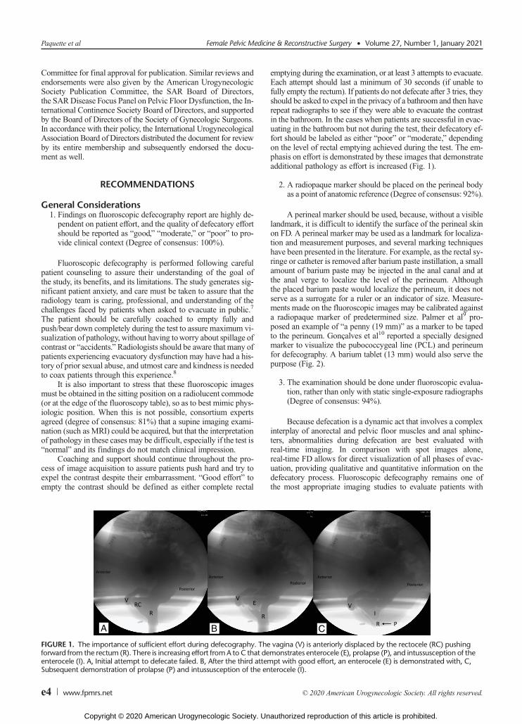
emptying during the examination, or at least 3 attempts to evacuate.Each attempt should last a minimum of 30 seconds (if unable tofully empty the rectum). If patients do not defecate after 3 tries, theyshould be asked to expel in the privacy of a bathroom and then haverepeat radiographs to see if they were able to evacuate the contrastin the bathroom. In the cases when patients are successful in evac-uating in the bathroom but not during the test, their defecatory ef-fort should be labeled as either “poor” or “moderate,” dependingon the level of rectal emptying achieved during the test. The em-phasis on effort is demonstrated by these images that demonstrateadditional pathology as effort is increased (Fig. 1).
2. A radiopaque marker should be placed on the perineal bodyas a point of anatomic reference (Degree of consensus: 92%).
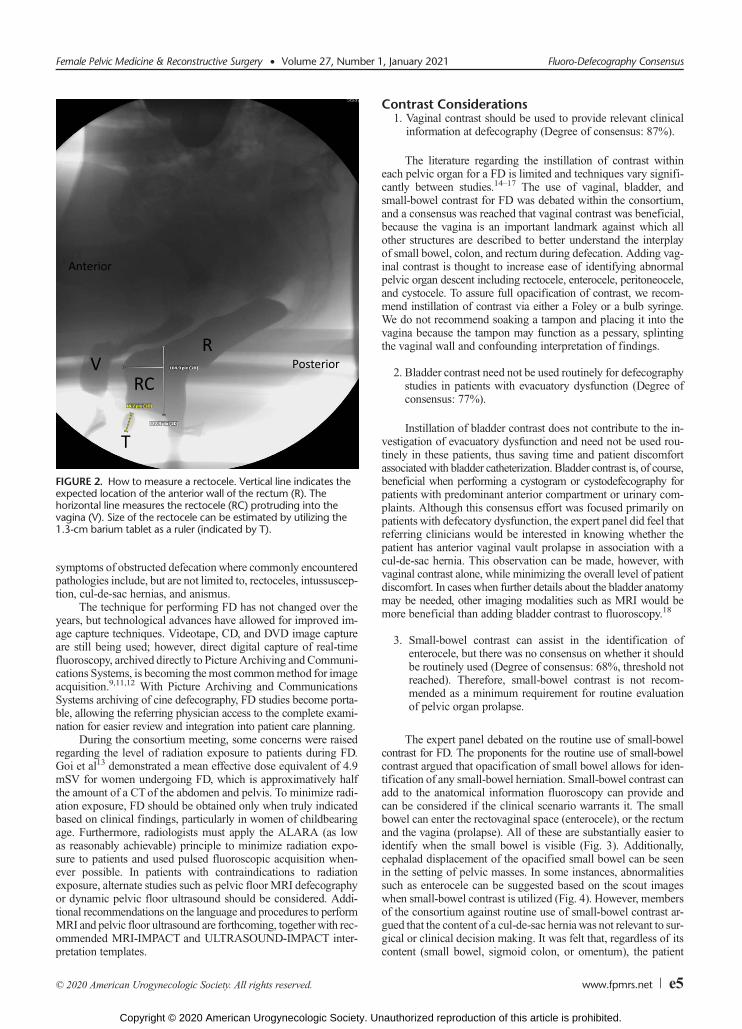
A perineal marker should be used, because, without a visiblelandmark, it is difficult to identify the surface of the perineal skinon FD. A perineal marker may be used as a landmark for localiza-tion and measurement purposes, and several marking techniqueshave been presented in the literature. For example, as the rectal sy-ringe or catheter is removed after barium paste instillation, a smallamount of barium paste may be injected in the anal canal and atthe anal verge to localize the level of the perineum. Althoughthe placed barium paste would localize the perineum, it does notserve as a surrogate for a ruler or an indicator of size. Measure-ments made on the fluoroscopic images may be calibrated againsta radiopaque marker of predetermined size. Palmer et al9 pro-posed an example of “a penny (19 mm)” as a marker to be tapedto the perineum. Gonçalves et al10 reported a specially designedmarker to visualize the pubococcygeal line (PCL) and perineumfor defecography. A barium tablet (13 mm) would also serve thepurpose (Fig. 2).
3. The examination should be done under fluoroscopic evalua-tion, rather than only with static single-exposure radiographs(Degree of consensus: 94%).
Because defecation is a dynamic act that involves a complexinterplay of anorectal and pelvic floor muscles and anal sphinc-ters, abnormalities during defecation are best evaluated withreal-time imaging. In comparison with spot images alone,real-time FD allows for direct visualization of all phases of evac-uation, providing qualitative and quantitative information on thedefecatory process. Fluoroscopic defecography remains one ofthe most appropriate imaging studies to evaluate patients with
FIGURE 1. The importance of sufficient effort during defecography. The vagina (V) is anteriorly displaced by the rectocele (RC) pushingforward from the rectum (R). There is increasing effort fromA to C that demonstrates enterocele (E), prolapse (P), and intussusception of theenterocele (I). A, Initial attempt to defecate failed. B, After the third attempt with good effort, an enterocele (E) is demonstrated with, C,Subsequent demonstration of prolapse (P) and intussusception of the enterocele (I).
Paquette et al Female Pelvic Medicine & Reconstructive Surgery • Volume 27, Number 1, January 2021
e4 www.fpmrs.net © 2020 American Urogynecologic Society. All rights reserved.
Copyright © 2020 American Urogynecologic Society. Unauthorized reproduction of this article is prohibited.
symptoms of obstructed defecation where commonly encounteredpathologies include, but are not limited to, rectoceles, intussuscep-tion, cul-de-sac hernias, and anismus.
The technique for performing FD has not changed over theyears, but technological advances have allowed for improved im-age capture techniques. Videotape, CD, and DVD image captureare still being used; however, direct digital capture of real-timefluoroscopy, archived directly to Picture Archiving and Communi-cations Systems, is becoming themost commonmethod for imageacquisition.9,11,12 With Picture Archiving and CommunicationsSystems archiving of cine defecography, FD studies become porta-ble, allowing the referring physician access to the complete exami-nation for easier review and integration into patient care planning.
During the consortium meeting, some concerns were raisedregarding the level of radiation exposure to patients during FD.Goi et al13 demonstrated a mean effective dose equivalent of 4.9mSV for women undergoing FD, which is approximatively halfthe amount of a CTof the abdomen and pelvis. To minimize radi-ation exposure, FD should be obtained only when truly indicatedbased on clinical findings, particularly in women of childbearingage. Furthermore, radiologists must apply the ALARA (as lowas reasonably achievable) principle to minimize radiation expo-sure to patients and used pulsed fluoroscopic acquisition when-ever possible. In patients with contraindications to radiationexposure, alternate studies such as pelvic floor MRI defecographyor dynamic pelvic floor ultrasound should be considered. Addi-tional recommendations on the language and procedures to performMRI and pelvic floor ultrasound are forthcoming, together with rec-ommended MRI-IMPACT and ULTRASOUND-IMPACT inter-pretation templates.
Contrast Considerations1. Vaginal contrast should be used to provide relevant clinical
information at defecography (Degree of consensus: 87%).
The literature regarding the instillation of contrast withineach pelvic organ for a FD is limited and techniques vary signifi-cantly between studies.14–17 The use of vaginal, bladder, andsmall-bowel contrast for FD was debated within the consortium,and a consensus was reached that vaginal contrast was beneficial,because the vagina is an important landmark against which allother structures are described to better understand the interplayof small bowel, colon, and rectum during defecation. Adding vag-inal contrast is thought to increase ease of identifying abnormalpelvic organ descent including rectocele, enterocele, peritoneocele,and cystocele. To assure full opacification of contrast, we recom-mend instillation of contrast via either a Foley or a bulb syringe.We do not recommend soaking a tampon and placing it into thevagina because the tampon may function as a pessary, splintingthe vaginal wall and confounding interpretation of findings.
2. Bladder contrast need not be used routinely for defecographystudies in patients with evacuatory dysfunction (Degree ofconsensus: 77%).
Instillation of bladder contrast does not contribute to the in-vestigation of evacuatory dysfunction and need not be used rou-tinely in these patients, thus saving time and patient discomfortassociated with bladder catheterization. Bladder contrast is, of course,beneficial when performing a cystogram or cystodefecography forpatients with predominant anterior compartment or urinary com-plaints. Although this consensus effort was focused primarily onpatients with defecatory dysfunction, the expert panel did feel thatreferring clinicians would be interested in knowing whether thepatient has anterior vaginal vault prolapse in association with acul-de-sac hernia. This observation can be made, however, withvaginal contrast alone, while minimizing the overall level of patientdiscomfort. In cases when further details about the bladder anatomymay be needed, other imaging modalities such as MRI would bemore beneficial than adding bladder contrast to fluoroscopy.18
3. Small-bowel contrast can assist in the identification ofenterocele, but there was no consensus on whether it shouldbe routinely used (Degree of consensus: 68%, threshold notreached). Therefore, small-bowel contrast is not recom-mended as a minimum requirement for routine evaluationof pelvic organ prolapse.
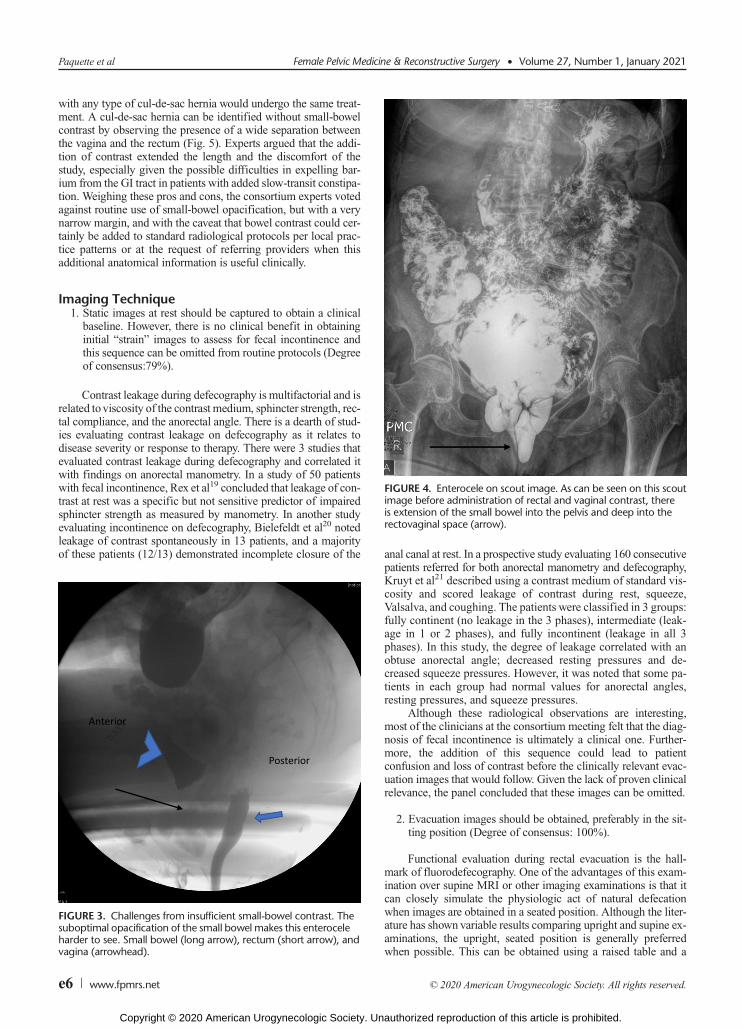
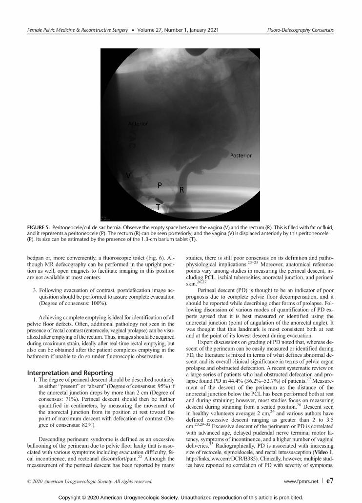
The expert panel debated on the routine use of small-bowelcontrast for FD. The proponents for the routine use of small-bowelcontrast argued that opacification of small bowel allows for iden-tification of any small-bowel herniation. Small-bowel contrast canadd to the anatomical information fluoroscopy can provide andcan be considered if the clinical scenario warrants it. The smallbowel can enter the rectovaginal space (enterocele), or the rectumand the vagina (prolapse). All of these are substantially easier toidentify when the small bowel is visible (Fig. 3). Additionally,cephalad displacement of the opacified small bowel can be seenin the setting of pelvic masses. In some instances, abnormalitiessuch as enterocele can be suggested based on the scout imageswhen small-bowel contrast is utilized (Fig. 4). However, membersof the consortium against routine use of small-bowel contrast ar-gued that the content of a cul-de-sac herniawas not relevant to sur-gical or clinical decision making. It was felt that, regardless of itscontent (small bowel, sigmoid colon, or omentum), the patient
FIGURE 2. How to measure a rectocele. Vertical line indicates theexpected location of the anterior wall of the rectum (R). Thehorizontal line measures the rectocele (RC) protruding into thevagina (V). Size of the rectocele can be estimated by utilizing the1.3-cm barium tablet as a ruler (indicated by T).
Female Pelvic Medicine & Reconstructive Surgery • Volume 27, Number 1, January 2021 Fluoro-Defecography Consensus
© 2020 American Urogynecologic Society. All rights reserved. www.fpmrs.net e5
Copyright © 2020 American Urogynecologic Society. Unauthorized reproduction of this article is prohibited.
with any type of cul-de-sac hernia would undergo the same treat-ment. A cul-de-sac hernia can be identified without small-bowelcontrast by observing the presence of a wide separation betweenthe vagina and the rectum (Fig. 5). Experts argued that the addi-tion of contrast extended the length and the discomfort of thestudy, especially given the possible difficulties in expelling bar-ium from the GI tract in patients with added slow-transit constipa-tion. Weighing these pros and cons, the consortium experts votedagainst routine use of small-bowel opacification, but with a verynarrow margin, and with the caveat that bowel contrast could cer-tainly be added to standard radiological protocols per local prac-tice patterns or at the request of referring providers when thisadditional anatomical information is useful clinically.
Imaging Technique1. Static images at rest should be captured to obtain a clinical
baseline. However, there is no clinical benefit in obtaininginitial “strain” images to assess for fecal incontinence andthis sequence can be omitted from routine protocols (Degreeof consensus:79%).
Contrast leakage during defecography is multifactorial and isrelated toviscosity of the contrast medium, sphincter strength, rec-tal compliance, and the anorectal angle. There is a dearth of stud-ies evaluating contrast leakage on defecography as it relates todisease severity or response to therapy. There were 3 studies thatevaluated contrast leakage during defecography and correlated itwith findings on anorectal manometry. In a study of 50 patientswith fecal incontinence, Rex et al19 concluded that leakage of con-trast at rest was a specific but not sensitive predictor of impairedsphincter strength as measured by manometry. In another studyevaluating incontinence on defecography, Bielefeldt et al20 notedleakage of contrast spontaneously in 13 patients, and a majorityof these patients (12/13) demonstrated incomplete closure of the anal canal at rest. In a prospective study evaluating 160 consecutive
patients referred for both anorectal manometry and defecography,Kruyt et al21 described using a contrast medium of standard vis-cosity and scored leakage of contrast during rest, squeeze,Valsalva, and coughing. The patients were classified in 3 groups:fully continent (no leakage in the 3 phases), intermediate (leak-age in 1 or 2 phases), and fully incontinent (leakage in all 3phases). In this study, the degree of leakage correlated with anobtuse anorectal angle; decreased resting pressures and de-creased squeeze pressures. However, it was noted that some pa-tients in each group had normal values for anorectal angles,resting pressures, and squeeze pressures.
Although these radiological observations are interesting,most of the clinicians at the consortium meeting felt that the diag-nosis of fecal incontinence is ultimately a clinical one. Further-more, the addition of this sequence could lead to patientconfusion and loss of contrast before the clinically relevant evac-uation images that would follow. Given the lack of proven clinicalrelevance, the panel concluded that these images can be omitted.
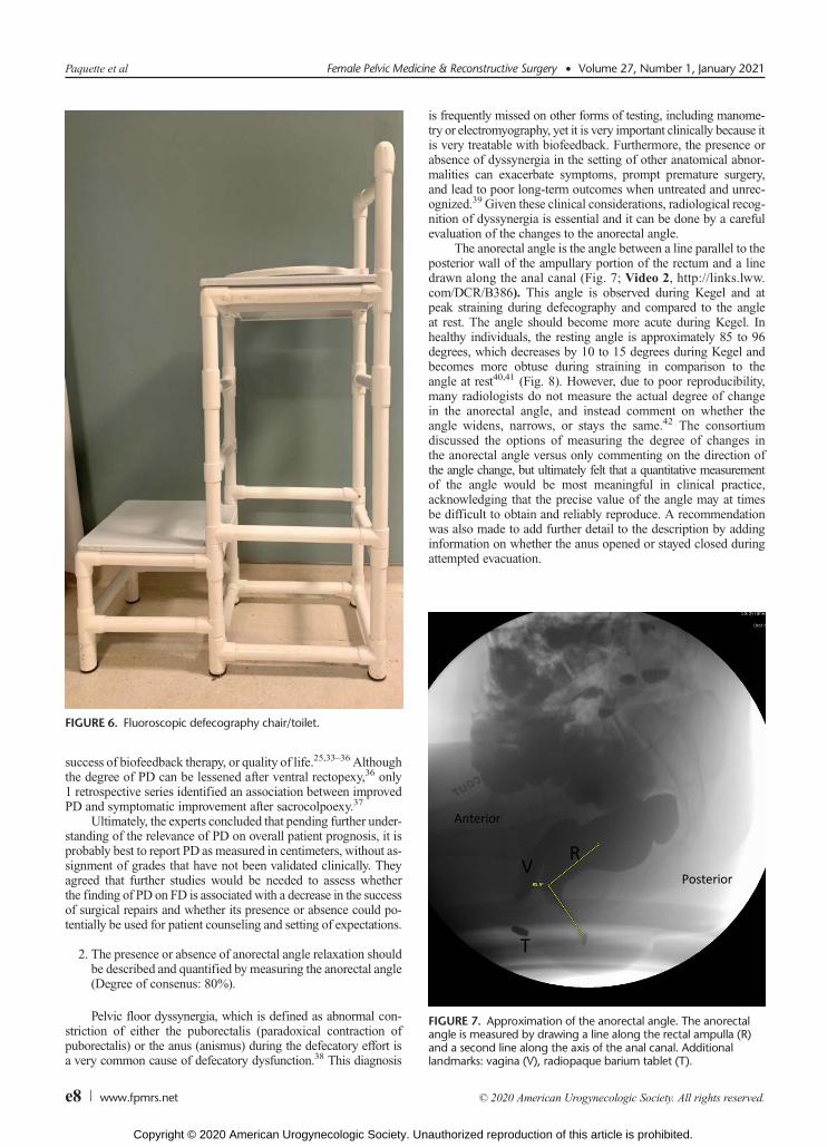
2. Evacuation images should be obtained, preferably in the sit-ting position (Degree of consensus: 100%).
Functional evaluation during rectal evacuation is the hall-mark of fluorodefecography. One of the advantages of this exam-ination over supine MRI or other imaging examinations is that itcan closely simulate the physiologic act of natural defecationwhen images are obtained in a seated position. Although the liter-ature has shown variable results comparing upright and supine ex-aminations, the upright, seated position is generally preferredwhen possible. This can be obtained using a raised table and a
FIGURE 3. Challenges from insufficient small-bowel contrast. Thesuboptimal opacification of the small bowel makes this enteroceleharder to see. Small bowel (long arrow), rectum (short arrow), andvagina (arrowhead).
FIGURE 4. Enterocele on scout image. As can be seen on this scoutimage before administration of rectal and vaginal contrast, thereis extension of the small bowel into the pelvis and deep into therectovaginal space (arrow).
Paquette et al Female Pelvic Medicine & Reconstructive Surgery • Volume 27, Number 1, January 2021
e6 www.fpmrs.net © 2020 American Urogynecologic Society. All rights reserved.
Copyright © 2020 American Urogynecologic Society. Unauthorized reproduction of this article is prohibited.
bedpan or, more conveniently, a fluoroscopic toilet (Fig. 6). Al-though MR defecography can be performed in the upright posi-tion as well, open magnets to facilitate imaging in this positionare not available at most centers.
3. Following evacuation of contrast, postdefecation image ac-quisition should be performed to assure complete evacuation(Degree of consensus: 100%).
Achieving complete emptying is ideal for identification of allpelvic floor defects. Often, additional pathology not seen in thepresence of rectal contrast (enterocele, vaginal prolapse) can be visu-alized after emptying of the rectum. Thus, images should be acquiredduring maximum strain, ideally after real-time rectal emptying, butalso can be obtained after the patient completes emptying in thebathroom if unable to do so under fluoroscopic observation.
Interpretation and Reporting1. The degree of perineal descent should be described routinelyas either “present” or “absent” (Degree of consensus: 95%) ifthe anorectal junction drops by more than 2 cm (Degree ofconsensus: 71%). Perineal descent should then be furtherquantified in centimeters, by measuring the movement ofthe anorectal junction from its position at rest toward thepoint of maximum descent with defecation of contrast (De-gree of consensus: 82%).
Descending perineum syndrome is defined as an excessiveballooning of the perineum due to pelvic floor laxity that is asso-ciated with various symptoms including evacuation difficulty, fe-cal incontinence, and rectoanal discomfort/pain.22 Although themeasurement of the perineal descent has been reported by many
studies, there is still poor consensus on its definition and patho-physiological implications.23–25 Moreover, anatomical referencepoints vary among studies in measuring the perineal descent, in-cluding PCL, ischial tuberosities, anorectal junction, and perinealskin.26,27
Perineal descent (PD) is thought to be an indicator of poorprognosis due to complete pelvic floor decompensation, and itshould be reported while describing other forms of prolapse. Fol-lowing discussion of various modes of quantification of PD ex-perts agreed that it is best measured or identified using theanorectal junction (point of angulation of the anorectal angle). Itwas thought that this landmark is most consistent both at restand at the point of its lowest descent during evacuation.
Expert discussions on grading of PD noted that, whereas de-scent of the perineum can be easily measured or identified duringFD, the literature is mixed in terms of what defines abnormal de-scent and its overall clinical significance in terms of pelvic organprolapse and obstructed defecation. A recent systematic review ona large series of patients who had obstructed defecation and pro-lapse found PD in 44.4% (36.2%–52.7%) of patients.27 Measure-ment of the descent of the perineum as the distance of theanorectal junction below the PCL has been performed both at restand during straining; however, most studies focus on measuringdescent during straining from a seated position.28 Descent seenin healthy volunteers averages 2 cm,26 and various authors havedefined excessive descent ranging as greater than 2 to 3.5cm.23,29–32 Excessive descent of the perineum or PD is correlatedwith advanced age, delayed pudendal nerve terminal motor la-tency, symptoms of incontinence, and a higher number of vaginaldeliveries.33 Radiographically, PD is associated with increasingsize of rectocele, sigmoidocele, and rectal intussusception (Video 1,http://links.lww.com/DCR/B385). Clinically, however, multiple stud-ies have reported no correlation of PD with severity of symptoms,
FIGURE 5. Peritoneocele/cul-de-sac hernia. Observe the empty space between the vagina (V) and the rectum (R). This is filledwith fat or fluid,and it represents a peritoneocele (P). The rectum (R) can be seen posteriorly, and the vagina (V) is displaced anteriorly by this peritoneocele(P). Its size can be estimated by the presence of the 1.3-cm barium tablet (T).
Female Pelvic Medicine & Reconstructive Surgery • Volume 27, Number 1, January 2021 Fluoro-Defecography Consensus
© 2020 American Urogynecologic Society. All rights reserved. www.fpmrs.net e7
Copyright © 2020 American Urogynecologic Society. Unauthorized reproduction of this article is prohibited.
success of biofeedback therapy, or quality of life.25,33–36 Althoughthe degree of PD can be lessened after ventral rectopexy,36 only1 retrospective series identified an association between improvedPD and symptomatic improvement after sacrocolpoexy.37
Ultimately, the experts concluded that pending further under-standing of the relevance of PD on overall patient prognosis, it isprobably best to report PD as measured in centimeters, without as-signment of grades that have not been validated clinically. Theyagreed that further studies would be needed to assess whetherthe finding of PD on FD is associated with a decrease in the successof surgical repairs and whether its presence or absence could po-tentially be used for patient counseling and setting of expectations.
2. The presence or absence of anorectal angle relaxation shouldbe described and quantified by measuring the anorectal angle(Degree of consenus: 80%).
Pelvic floor dyssynergia, which is defined as abnormal con-striction of either the puborectalis (paradoxical contraction ofpuborectalis) or the anus (anismus) during the defecatory effort isa very common cause of defecatory dysfunction.38 This diagnosis
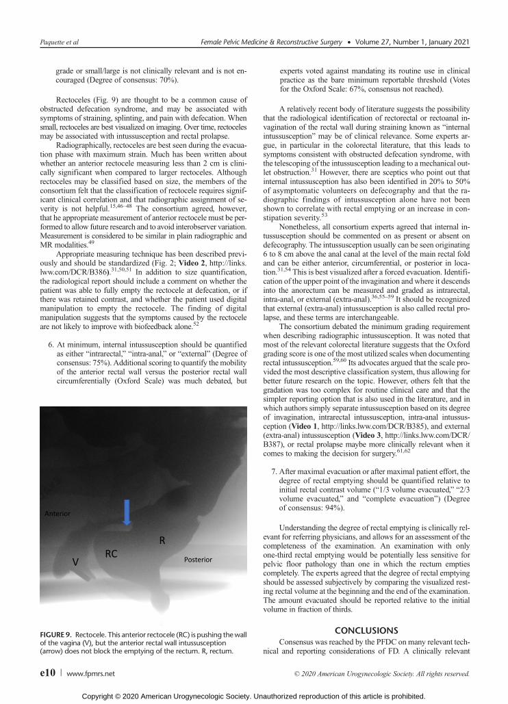
is frequently missed on other forms of testing, including manome-try or electromyography, yet it is very important clinically because itis very treatable with biofeedback. Furthermore, the presence orabsence of dyssynergia in the setting of other anatomical abnor-malities can exacerbate symptoms, prompt premature surgery,and lead to poor long-term outcomes when untreated and unrec-ognized.39 Given these clinical considerations, radiological recog-nition of dyssynergia is essential and it can be done by a carefulevaluation of the changes to the anorectal angle.
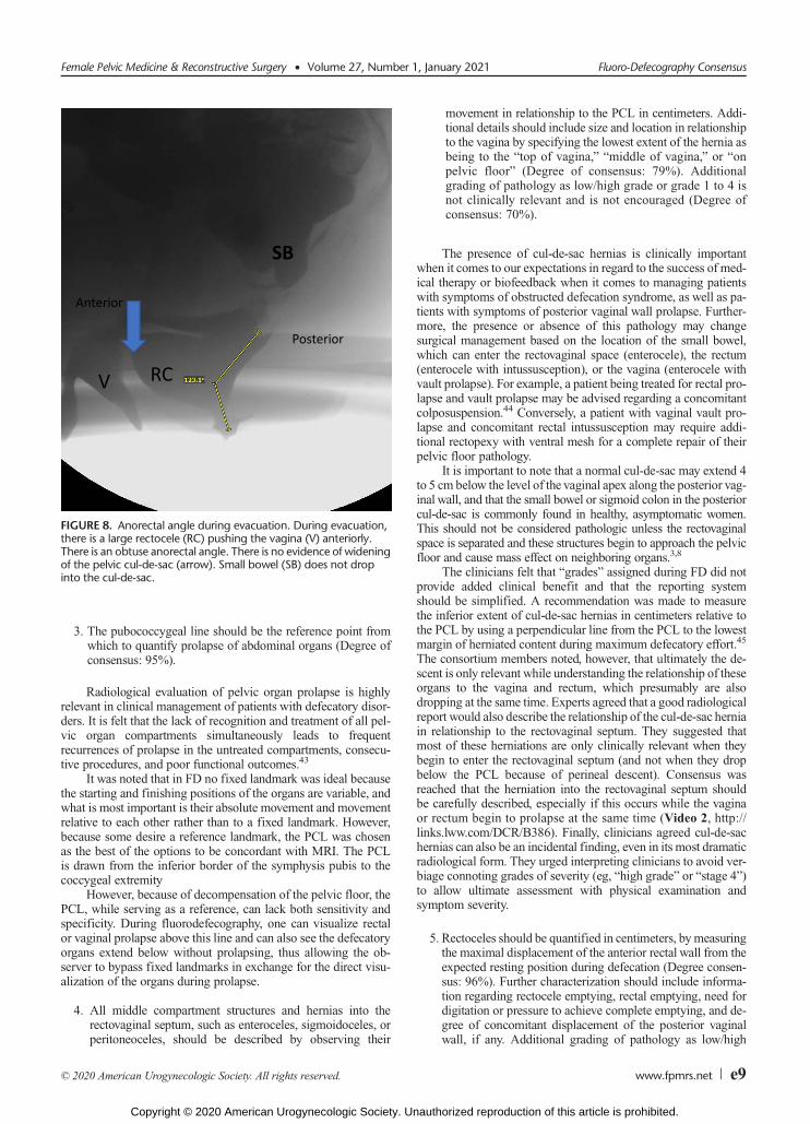
The anorectal angle is the angle between a line parallel to theposterior wall of the ampullary portion of the rectum and a linedrawn along the anal canal (Fig. 7; Video 2, http://links.lww.com/DCR/B386). This angle is observed during Kegel and atpeak straining during defecography and compared to the angleat rest. The angle should become more acute during Kegel. Inhealthy individuals, the resting angle is approximately 85 to 96degrees, which decreases by 10 to 15 degrees during Kegel andbecomes more obtuse during straining in comparison to theangle at rest40,41 (Fig. 8). However, due to poor reproducibility,many radiologists do not measure the actual degree of changein the anorectal angle, and instead comment on whether theangle widens, narrows, or stays the same.42 The consortiumdiscussed the options of measuring the degree of changes inthe anorectal angle versus only commenting on the direction ofthe angle change, but ultimately felt that a quantitative measurementof the angle would be most meaningful in clinical practice,acknowledging that the precise value of the angle may at timesbe difficult to obtain and reliably reproduce. A recommendationwas also made to add further detail to the description by addinginformation on whether the anus opened or stayed closed duringattempted evacuation.
FIGURE 6. Fluoroscopic defecography chair/toilet.
FIGURE 7. Approximation of the anorectal angle. The anorectalangle is measured by drawing a line along the rectal ampulla (R)and a second line along the axis of the anal canal. Additionallandmarks: vagina (V), radiopaque barium tablet (T).
Paquette et al Female Pelvic Medicine & Reconstructive Surgery • Volume 27, Number 1, January 2021
e8 www.fpmrs.net © 2020 American Urogynecologic Society. All rights reserved.
Copyright © 2020 American Urogynecologic Society. Unauthorized reproduction of this article is prohibited.
3. The pubococcygeal line should be the reference point fromwhich to quantify prolapse of abdominal organs (Degree ofconsensus: 95%).
Radiological evaluation of pelvic organ prolapse is highlyrelevant in clinical management of patients with defecatory disor-ders. It is felt that the lack of recognition and treatment of all pel-vic organ compartments simultaneously leads to frequentrecurrences of prolapse in the untreated compartments, consecu-tive procedures, and poor functional outcomes.43
It was noted that in FD no fixed landmark was ideal becausethe starting and finishing positions of the organs are variable, andwhat is most important is their absolute movement and movementrelative to each other rather than to a fixed landmark. However,because some desire a reference landmark, the PCL was chosenas the best of the options to be concordant with MRI. The PCLis drawn from the inferior border of the symphysis pubis to thecoccygeal extremity
However, because of decompensation of the pelvic floor, thePCL, while serving as a reference, can lack both sensitivity andspecificity. During fluorodefecography, one can visualize rectalor vaginal prolapse above this line and can also see the defecatoryorgans extend below without prolapsing, thus allowing the ob-server to bypass fixed landmarks in exchange for the direct visu-alization of the organs during prolapse.
4. All middle compartment structures and hernias into therectovaginal septum, such as enteroceles, sigmoidoceles, orperitoneoceles, should be described by observing their
movement in relationship to the PCL in centimeters. Addi-tional details should include size and location in relationshipto the vagina by specifying the lowest extent of the hernia asbeing to the “top of vagina,” “middle of vagina,” or “onpelvic floor” (Degree of consensus: 79%). Additionalgrading of pathology as low/high grade or grade 1 to 4 isnot clinically relevant and is not encouraged (Degree ofconsensus: 70%).
The presence of cul-de-sac hernias is clinically importantwhen it comes to our expectations in regard to the success of med-ical therapy or biofeedback when it comes to managing patientswith symptoms of obstructed defecation syndrome, as well as pa-tients with symptoms of posterior vaginal wall prolapse. Further-more, the presence or absence of this pathology may changesurgical management based on the location of the small bowel,which can enter the rectovaginal space (enterocele), the rectum(enterocele with intussusception), or the vagina (enterocele withvault prolapse). For example, a patient being treated for rectal pro-lapse and vault prolapse may be advised regarding a concomitantcolposuspension.44 Conversely, a patient with vaginal vault pro-lapse and concomitant rectal intussusception may require addi-tional rectopexy with ventral mesh for a complete repair of theirpelvic floor pathology.
It is important to note that a normal cul-de-sac may extend 4to 5 cm below the level of the vaginal apex along the posterior vag-inal wall, and that the small bowel or sigmoid colon in the posteriorcul-de-sac is commonly found in healthy, asymptomatic women.This should not be considered pathologic unless the rectovaginalspace is separated and these structures begin to approach the pelvicfloor and cause mass effect on neighboring organs.3,8
The clinicians felt that “grades” assigned during FD did notprovide added clinical benefit and that the reporting systemshould be simplified. A recommendation was made to measurethe inferior extent of cul-de-sac hernias in centimeters relative tothe PCL by using a perpendicular line from the PCL to the lowestmargin of herniated content during maximum defecatory effort.45
The consortium members noted, however, that ultimately the de-scent is only relevant while understanding the relationship of theseorgans to the vagina and rectum, which presumably are alsodropping at the same time. Experts agreed that a good radiologicalreport would also describe the relationship of the cul-de-sac herniain relationship to the rectovaginal septum. They suggested thatmost of these herniations are only clinically relevant when theybegin to enter the rectovaginal septum (and not when they dropbelow the PCL because of perineal descent). Consensus wasreached that the herniation into the rectovaginal septum shouldbe carefully described, especially if this occurs while the vaginaor rectum begin to prolapse at the same time (Video 2, http://links.lww.com/DCR/B386). Finally, clinicians agreed cul-de-sachernias can also be an incidental finding, even in its most dramaticradiological form. They urged interpreting clinicians to avoid ver-biage connoting grades of severity (eg, “high grade” or “stage 4”)to allow ultimate assessment with physical examination andsymptom severity.
5. Rectoceles should be quantified in centimeters, bymeasuringthe maximal displacement of the anterior rectal wall from theexpected resting position during defecation (Degree consen-sus: 96%). Further characterization should include informa-tion regarding rectocele emptying, rectal emptying, need fordigitation or pressure to achieve complete emptying, and de-gree of concomitant displacement of the posterior vaginalwall, if any. Additional grading of pathology as low/high
FIGURE 8. Anorectal angle during evacuation. During evacuation,there is a large rectocele (RC) pushing the vagina (V) anteriorly.There is an obtuse anorectal angle. There is no evidence of wideningof the pelvic cul-de-sac (arrow). Small bowel (SB) does not dropinto the cul-de-sac.
Female Pelvic Medicine & Reconstructive Surgery • Volume 27, Number 1, January 2021 Fluoro-Defecography Consensus
© 2020 American Urogynecologic Society. All rights reserved. www.fpmrs.net e9
Copyright © 2020 American Urogynecologic Society. Unauthorized reproduction of this article is prohibited.
grade or small/large is not clinically relevant and is not en-couraged (Degree of consensus: 70%).
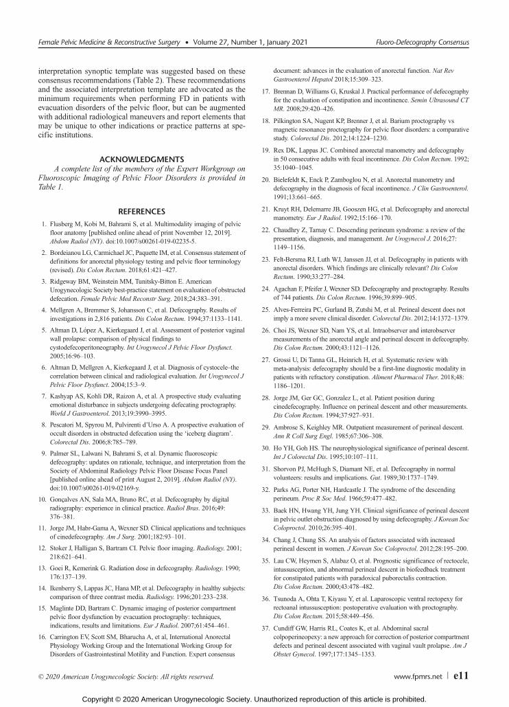
Rectoceles (Fig. 9) are thought to be a common cause ofobstructed defecation syndrome, and may be associated withsymptoms of straining, splinting, and pain with defecation. Whensmall, rectoceles are best visualized on imaging. Over time, rectocelesmay be associated with intussusception and rectal prolapse.
Radiographically, rectoceles are best seen during the evacua-tion phase with maximum strain. Much has been written aboutwhether an anterior rectocele measuring less than 2 cm is clini-cally significant when compared to larger rectoceles. Althoughrectoceles may be classified based on size, the members of theconsortium felt that the classification of rectocele requires signif-icant clinical correlation and that radiographic assignment of se-verity is not helpful.15,46–48 The consortium agreed, however,that he appropriate measurement of anterior rectocele must be per-formed to allow future research and to avoid interobserver variation.Measurement is considered to be similar in plain radiographic andMR modalities.49
Appropriate measuring technique has been described previ-ously and should be standardized (Fig. 2; Video 2, http://links.lww.com/DCR/B386).31,50,51 In addition to size quantification,the radiological report should include a comment on whether thepatient was able to fully empty the rectocele at defecation, or ifthere was retained contrast, and whether the patient used digitalmanipulation to empty the rectocele. The finding of digitalmanipulation suggests that the symptoms caused by the rectoceleare not likely to improve with biofeedback alone.52
6. At minimum, internal intussusception should be quantifiedas either “intrarectal,” “intra-anal,” or “external” (Degree ofconsensus: 75%). Additional scoring to quantify the mobilityof the anterior rectal wall versus the posterior rectal wallcircumferentially (Oxford Scale) was much debated, but
experts voted against mandating its routine use in clinicalpractice as the bare minimum reportable threshold (Votesfor the Oxford Scale: 67%, consensus not reached).
A relatively recent body of literature suggests the possibilitythat the radiological identification of rectorectal or rectoanal in-vagination of the rectal wall during straining known as “internalintussusception” may be of clinical relevance. Some experts ar-gue, in particular in the colorectal literature, that this leads tosymptoms consistent with obstructed defecation syndrome, withthe telescoping of the intussusception leading to a mechanical out-let obstruction.31 However, there are sceptics who point out thatinternal intussusception has also been identified in 20% to 50%of asymptomatic volunteers on defecography and that the ra-diographic findings of intussusception alone have not beenshown to correlate with rectal emptying or an increase in con-stipation severity.53
Nonetheless, all consortium experts agreed that internal in-tussusception should be commented on as present or absent ondefecography. The intussusception usually can be seen originating6 to 8 cm above the anal canal at the level of the main rectal foldand can be either anterior, circumferential, or posterior in loca-tion.31,54 This is best visualized after a forced evacuation. Identifi-cation of the upper point of the invagination andwhere it descendsinto the anorectum can be measured and graded as intrarectal,intra-anal, or external (extra-anal).36,55–59 It should be recognizedthat external (extra-anal) intussusception is also called rectal pro-lapse, and these terms are interchangeable.
The consortium debated the minimum grading requirementwhen describing radiographic intussusception. It was noted thatmost of the relevant colorectal literature suggests that the Oxfordgrading score is one of the most utilized scales when documentingrectal intussusception.59,60 Its advocates argued that the scale pro-vided the most descriptive classification system, thus allowing forbetter future research on the topic. However, others felt that thegradation was too complex for routine clinical care and that thesimpler reporting option that is also used in the literature, and inwhich authors simply separate intussusception based on its degreeof invagination, intrarectal intussusception, intra-anal intussus-ception (Video 1, http://links.lww.com/DCR/B385), and external(extra-anal) intussusception (Video 3, http://links.lww.com/DCR/B387), or rectal prolapse maybe more clinically relevant when itcomes to making the decision for surgery.61,62
7. After maximal evacuation or after maximal patient effort, thedegree of rectal emptying should be quantified relative toinitial rectal contrast volume (“1/3 volume evacuated,” “2/3volume evacuated,” and “complete evacuation”) (Degreeof consensus: 94%).
Understanding the degree of rectal emptying is clinically rel-evant for referring physicians, and allows for an assessment of thecompleteness of the examination. An examination with onlyone-third rectal emptying would be potentially less sensitive forpelvic floor pathology than one in which the rectum emptiescompletely. The experts agreed that the degree of rectal emptyingshould be assessed subjectively by comparing the visualized rest-ing rectal volume at the beginning and the end of the examination.The amount evacuated should be reported relative to the initialvolume in fraction of thirds.
CONCLUSIONSConsensus was reached by the PFDC on many relevant tech-
nical and reporting considerations of FD. A clinically relevant
FIGURE9. Rectocele. This anterior rectocele (RC) is pushing thewallof the vagina (V), but the anterior rectal wall intussusception(arrow) does not block the emptying of the rectum. R, rectum.
Paquette et al Female Pelvic Medicine & Reconstructive Surgery • Volume 27, Number 1, January 2021
e10 www.fpmrs.net © 2020 American Urogynecologic Society. All rights reserved.
Copyright © 2020 American Urogynecologic Society. Unauthorized reproduction of this article is prohibited.
interpretation synoptic template was suggested based on theseconsensus recommendations (Table 2). These recommendationsand the associated interpretation template are advocated as theminimum requirements when performing FD in patients withevacuation disorders of the pelvic floor, but can be augmentedwith additional radiological maneuvers and report elements thatmay be unique to other indications or practice patterns at spe-cific institutions.
ACKNOWLEDGMENTSA complete list of the members of the Expert Workgroup on
Fluoroscopic Imaging of Pelvic Floor Disorders is provided inTable 1.
REFERENCES1. Flusberg M, Kobi M, Bahrami S, et al. Multimodality imaging of pelvic
floor anatomy [published online ahead of print November 12, 2019].Abdom Radiol (NY). doi:10.1007/s00261-019-02235-5.
2. Bordeianou LG, Carmichael JC, Paquette IM, et al. Consensus statement ofdefinitions for anorectal physiology testing and pelvic floor terminology(revised). Dis Colon Rectum. 2018;61:421–427.
3. Ridgeway BM, Weinstein MM, Tunitsky-Bitton E. AmericanUrogynecologic Society best-practice statement on evaluation of obstructeddefecation. Female Pelvic Med Reconstr Surg. 2018;24:383–391.
4. Mellgren A, Bremmer S, Johansson C, et al. Defecography. Results ofinvestigations in 2,816 patients. Dis Colon Rectum. 1994;37:1133–1141.
5. Altman D, López A, Kierkegaard J, et al. Assessment of posterior vaginalwall prolapse: comparison of physical findings tocystodefecoperitoneography. Int Urogynecol J Pelvic Floor Dysfunct.2005;16:96–103.
6. Altman D, Mellgren A, Kierkegaard J, et al. Diagnosis of cystocele–thecorrelation between clinical and radiological evaluation. Int Urogynecol JPelvic Floor Dysfunct. 2004;15:3–9.
7. Kashyap AS, Kohli DR, Raizon A, et al. A prospective study evaluatingemotional disturbance in subjects undergoing defecating proctography.World J Gastroenterol. 2013;19:3990–3995.
8. Pescatori M, Spyrou M, Pulvirenti d’Urso A. A prospective evaluation ofoccult disorders in obstructed defecation using the ‘iceberg diagram’.Colorectal Dis. 2006;8:785–789.
9. Palmer SL, Lalwani N, Bahrami S, et al. Dynamic fluoroscopicdefecography: updates on rationale, technique, and interpretation from theSociety of Abdominal Radiology Pelvic Floor Disease Focus Panel[published online ahead of print August 2, 2019]. Abdom Radiol (NY).doi:10.1007/s00261-019-02169-y.
10. Gonçalves AN, Sala MA, Bruno RC, et al. Defecography by digitalradiography: experience in clinical practice. Radiol Bras. 2016;49:376–381.
11. Jorge JM, Habr-Gama A, Wexner SD. Clinical applications and techniquesof cinedefecography. Am J Surg. 2001;182:93–101.
12. Stoker J, Halligan S, Bartram CI. Pelvic floor imaging. Radiology. 2001;218:621–641.
13. Goei R, Kemerink G. Radiation dose in defecography. Radiology. 1990;176:137–139.
14. Ikenberry S, Lappas JC, Hana MP, et al. Defecography in healthy subjects:comparison of three contrast media. Radiology. 1996;201:233–238.
15. Maglinte DD, Bartram C. Dynamic imaging of posterior compartmentpelvic floor dysfunction by evacuation proctography: techniques,indications, results and limitations. Eur J Radiol. 2007;61:454–461.
16. Carrington EV, Scott SM, Bharucha A, et al, International AnorectalPhysiology Working Group and the International Working Group forDisorders of Gastrointestinal Motility and Function. Expert consensus
document: advances in the evaluation of anorectal function. Nat RevGastroenterol Hepatol 2018;15:309–323.
17. Brennan D, Williams G, Kruskal J. Practical performance of defecographyfor the evaluation of constipation and incontinence. Semin Ultrasound CTMR. 2008;29:420–426.
18. Pilkington SA, Nugent KP, Brenner J, et al. Barium proctography vsmagnetic resonance proctography for pelvic floor disorders: a comparativestudy. Colorectal Dis. 2012;14:1224–1230.
19. Rex DK, Lappas JC. Combined anorectal manometry and defecographyin 50 consecutive adults with fecal incontinence. Dis Colon Rectum. 1992;35:1040–1045.
20. Bielefeldt K, Enck P, Zamboglou N, et al. Anorectal manometry anddefecography in the diagnosis of fecal incontinence. J Clin Gastroenterol.1991;13:661–665.
21. Kruyt RH, Delemarre JB, Gooszen HG, et al. Defecography and anorectalmanometry. Eur J Radiol. 1992;15:166–170.
22. Chaudhry Z, Tarnay C. Descending perineum syndrome: a review of thepresentation, diagnosis, and management. Int Urogynecol J. 2016;27:1149–1156.
23. Felt-Bersma RJ, Luth WJ, Janssen JJ, et al. Defecography in patients withanorectal disorders. Which findings are clinically relevant? Dis ColonRectum. 1990;33:277–284.
24. Agachan F, Pfeifer J, Wexner SD. Defecography and proctography. Resultsof 744 patients. Dis Colon Rectum. 1996;39:899–905.
25. Alves-Ferreira PC, Gurland B, Zutshi M, et al. Perineal descent does notimply a more severe clinical disorder. Colorectal Dis. 2012;14:1372–1379.
26. Choi JS, Wexner SD, Nam YS, et al. Intraobserver and interobservermeasurements of the anorectal angle and perineal descent in defecography.Dis Colon Rectum. 2000;43:1121–1126.
27. Grossi U, Di Tanna GL, Heinrich H, et al. Systematic review withmeta-analysis: defecography should be a first-line diagnostic modality inpatients with refractory constipation. Aliment Pharmacol Ther. 2018;48:1186–1201.
28. Jorge JM, Ger GC, Gonzalez L, et al. Patient position duringcinedefecography. Influence on perineal descent and other measurements.Dis Colon Rectum. 1994;37:927–931.
29. Ambrose S, Keighley MR. Outpatient measurement of perineal descent.Ann R Coll Surg Engl. 1985;67:306–308.
30. Ho YH, Goh HS. The neurophysiological significance of perineal descent.Int J Colorectal Dis. 1995;10:107–111.
31. Shorvon PJ, McHugh S, Diamant NE, et al. Defecography in normalvolunteers: results and implications. Gut. 1989;30:1737–1749.
32. Parks AG, Porter NH, Hardcastle J. The syndrome of the descendingperineum. Proc R Soc Med. 1966;59:477–482.
33. Baek HN, Hwang YH, Jung YH. Clinical significance of perineal descentin pelvic outlet obstruction diagnosed by using defecography. J Korean SocColoproctol. 2010;26:395–401.
34. Chang J, Chung SS. An analysis of factors associated with increasedperineal descent in women. J Korean Soc Coloproctol. 2012;28:195–200.
35. Lau CW, Heymen S, Alabaz O, et al. Prognostic significance of rectocele,intussusception, and abnormal perineal descent in biofeedback treatmentfor constipated patients with paradoxical puborectalis contraction.Dis Colon Rectum. 2000;43:478–482.
36. Tsunoda A, Ohta T, Kiyasu Y, et al. Laparoscopic ventral rectopexy forrectoanal intussusception: postoperative evaluation with proctography.Dis Colon Rectum. 2015;58:449–456.
37. Cundiff GW, Harris RL, Coates K, et al. Abdominal sacralcolpoperineopexy: a new approach for correction of posterior compartmentdefects and perineal descent associated with vaginal vault prolapse. Am JObstet Gynecol. 1997;177:1345–1353.
Female Pelvic Medicine & Reconstructive Surgery • Volume 27, Number 1, January 2021 Fluoro-Defecography Consensus
© 2020 American Urogynecologic Society. All rights reserved. www.fpmrs.net e11
Copyright © 2020 American Urogynecologic Society. Unauthorized reproduction of this article is prohibited.
38. Bordeianou L, Savitt L, Dursun A. Measurements of pelvic floordyssynergia: which test result matters?Dis Colon Rectum. 2011;54:60–65.
39. Seong MK, Kim TW. Significance of defecographic parameters indiagnosing pelvic floor dyssynergia. J Korean Surg Soc. 2013;84:225–230.
40. Karasick S, Karasick D, Karasick SR. Functional disorders of the anus andrectum: findings on defecography. AJR Am J Roentgenol. 1993;160:777–782.
41. Maccioni F. Functional disorders of the ano-rectal compartment of thepelvic floor: clinical and diagnostic value of dynamic MRI. AbdomImaging. 2013;38:930–951.
42. Bartram CI, Turnbull GK, Lennard-Jones JE. Evacuation proctography: aninvestigation of rectal expulsion in 20 subjects without defecatorydisturbance. Gastrointest Radiol. 1988;13:72–80.
43. Bordeianou L, Hicks CW, Olariu A, et al. Effect of coexisting pelvic floordisorders on fecal incontinence quality of life scores: a prospective,survey-based study. Dis Colon Rectum. 2015;58:1091–1097.
44. Watadani Y, Vogler SA, Warshaw JS, et al. Sacrocolpopexy with rectopexyfor pelvic floor prolapse improves bowel function and quality of life.Dis Colon Rectum. 2013;56:1415–1422.
45. Hainsworth AJ, Solanki D, Hamad A, et al. Integrated total pelvic floorultrasound in pelvic floor defaecatory dysfunction. Colorectal Dis. 2017;19:O54–O65.
46. Carter D, Gabel MB. Rectocele–does the size matter? Int J Colorectal Dis.2012;27:975–980.
47. Delemarre JB, Kruyt RH, Doornbos J, et al. Anterior rectocele: assessmentwith radiographic defecography, dynamic magnetic resonance imaging,and physical examination. Dis Colon Rectum. 1994;37:249–259.
48. Maglinte DD, Bartram CI, Hale DA, et al. Functional imaging of the pelvicfloor. Radiology. 2011;258:23–39.
49. Poncelet E, Rock A, Quinton JF, et al. Dynamic MR defecography of theposterior compartment: comparison with conventional x-ray defecography.Diagn Interv Imaging. 2017;98:327–332.
50. Liu J, Zhai LD, Li YS, et al. Measuring the space between vagina andrectum as it relates to rectocele.World J Gastroenterol. 2009;15:3051–3054.
51. Colaiacomo MC, Masselli G, Polettini E, et al. Dynamic MR imaging ofthe pelvic floor: a pictorial review. Radiographics. 2009;29:e35.
52. Hicks CW, Weinstein M, Wakamatsu M, et al. In patients with rectocelesand obstructed defecation syndrome, surgery should be the option of lastresort. Surgery. 2014;155:659–667.
53. Palit S, Bhan C, Lunniss PJ, et al. Evacuation proctography: a reappraisal ofnormal variability. Colorectal Dis. 2014;16:538–546.
54. Cariou de Vergie L, Venara A, Duchalais E, et al. Internal rectalprolapse: definition, assessment and management in 2016. J Visc Surg.2017;154:21–28.
55. Cavallaro PM, Staller K, Savitt LR, et al. The contributions of internalintussusception, irritable bowel syndrome, and pelvic floor dyssynergia toobstructed defecation syndrome. Dis Colon Rectum. 2019;62:56–62.
56. Kim NY, Kim DH, Pickhardt PJ, et al. Defecography: an overview oftechnique, interpretation, and impact on patient care. Gastroenterol ClinNorth Am 2018;47:553–568.
57. Tsunoda A, Takahashi T, Ohta T, et al. Anterior intussusception descentduring defecation is correlated with the severity of fecal incontinence inpatients with rectoanal intussusception. Tech Coloproctol. 2016;20:171–176.
58. Hawkins AT, Olariu AG, Savitt LR, et al. Impact of rising grades of internalrectal intussusception on fecal continence and symptoms of constipation.Dis Colon Rectum. 2016;59:54–61.
59. Wijffels NA, Jones OM, Cunningham C, et al. What are the symptoms ofinternal rectal prolapse? Colorectal Dis. 2013;15:368–373.
60. National Institute for Health and Care Excellence. Interventional ReviewProgramme. In: Interventional procedure overview of laparoscopic ventralmesh rectopexy for internal prolapse. 2018.
61. Morandi C, Martellucci J, Talento P, et al. Role of enterocele in theobstructed defecation syndrome (ODS): a new radiological point of view.Colorectal Dis. 2010;12:810–816.
62. Jorge JM, Yang YK, Wexner SD. Incidence and clinical significance ofsigmoidoceles as determined by a new classification system. Dis ColonRectum. 1994;37:1112–1117.
Paquette et al Female Pelvic Medicine & Reconstructive Surgery • Volume 27, Number 1, January 2021
e12 www.fpmrs.net © 2020 American Urogynecologic Society. All rights reserved.
Copyright © 2020 American Urogynecologic Society. Unauthorized reproduction of this article is prohibited.