Embed Size (px)
Citation preview
Consequences of Motor Copy Numberon the Intracellular Transport ofKinesin-1-Driven Lipid DropletsGeorge T. Shubeita,1,4,6 Susan L. Tran,2,3,5,6 Jing Xu,1 Michael Vershinin,1 Silvia Cermelli,1 Sean L. Cotton,3
Michael A. Welte,2,3,5,7,* and Steven P. Gross1,7,*1Department of Developmental and Cell Biology, University of California Irvine, Irvine, CA 92697, USA2Department of Biology, University of Rochester, Rochester, NY 14627, USA3Department of Biology and Rosenstiel Medical Research Center, Brandeis University, Waltham, MA 02454, USA4Present address: Center for Nonlinear Dynamics and Department of Physics, University of Texas at Austin, Austin, TX 78712, USA5Present address: Department of Biology, University of Rochester, Rochester, NY 14627, USA6These authors contributed equally to this work7These authors contributed equally to this work
*Correspondence: [email protected] (M.A.W.), [email protected] (S.P.G.)
DOI 10.1016/j.cell.2008.10.021
SUMMARY
The microtubule motor kinesin-1 plays central roles inintracellular transport. It has been widely assumedthat many cellular cargos are moved by multiple kine-sins and that cargos with more motors move fasterand for longer distances;concrete evidence, however,is sparse. Here we rigorously test these notions usinglipid droplets in Drosophila embryos. We first employantibody inhibition, genetics, biochemistry, and parti-cle tracking to demonstrate that kinesin-1 mediatesplus-end droplet motion. We then measure how varia-tion in kinesin-1 expression affects the forces drivingindividual droplets and estimate the number of kine-sins actively engaged per droplet. Unlike in vitro, in-creased motor number results in neither longer traveldistances nor higher velocities. Our data suggest thatcargos in vivo can simultaneously engage multiplekinesins and that transport properties are largely unaf-fected by variation in motor number. Apparently,higher-order regulatory mechanisms rather than mo-tor number per se dominate cargo transport in vivo.
INTRODUCTION
Intracellular transport along microtubules is powered by molec-
ular motors of the kinesin and cytoplasmic dynein families. Most
kinesins travel toward the plus ends of microtubules, whereas
dynein travels toward minus ends. Frequently, kinesins and
dyneins are attached to the same cargo, but engage with the
microtubule alternatively, resulting in constant back-and-forth
cargo motion. Such bidirectional transport is widespread and
has been observed for mitochondria, melanosomes, various
vesicles, neurofilaments, ribonucleoprotein granules, and vi-
ruses (reviewed in Welte, 2004). Because distinct bidirectional
cargos exhibit very similar motion characteristics, the regulatory
1098 Cell 135, 1098–1107, December 12, 2008 ª2008 Elsevier Inc.
mechanisms to control such transport might be quite general
(reviewed in Gross, 2004).
Cells modulate net directionality and speed of bidirectional
transport in response to extrinsic and intrinsic cues. Because
net transport is determined by whether—on average—plus-
end or minus-end travel distances are longer, the key to under-
standing transport regulation is to determine how cargo travel
distances are controlled. Although molecules important for travel
length control are being identified, it remains unclear how they
affect the activities of their ultimate targets, the motors.
Studies in vitro suggest one possible mechanism: these regula-
tors might modulate the number of motors active per cargo.
In vitro, single molecules of kinesin-1 or dynein can travel
�1 mm along microtubules, either on their own or when attached
to glass or plastic beads. Beads moved by multiple copies of such
motors travel much further (Mallik et al., 2005; Vershinin et al.,
2007).
Although important in vitro, the relevance of motor copy num-
ber for transport in vivo remains unclear, in large part because
measuring the number of motors active per cargo is challenging.
It is not sufficient to determine bulk levels of motors cofractionat-
ing with particular cargos, because motor number might vary
from cargo to cargo or even on a single cargo over time. Further-
more, not every motor physically present on a cargo contributes
to motion, either because it might not contact the track or
because its activity is regulated. The crucial parameter to deter-
mine is the number of engaged motors.
Because motors in vitro slow down if opposed by a significant
load, some studies infer motor number from the velocity that car-
gos display in vivo: faster cargos are assumed to have more mo-
tors engaged (Hill et al., 2004; Kural et al., 2005; Levi et al., 2006).
However, the relationship between velocity and motor number is
likely complex: the loads that cargos experience in vivo might not
be high enough to explain observed velocity variations, and it is
unresolved whether velocities are also modulated by regulatory
factors (see Supplemental Data available online). It therefore re-
mains unclear to what extent variation in the number of engaged
motors controls either velocity or travel distances in vivo. It is not

even conclusively established that a cellular cargo can indeed
employ more than one motor at a time.
In this article, we estimate the number of engaged motors
using a direct measure, the force needed to stall a moving cargo.
Theoretical considerations predict that such stall forces are pro-
portional to the number of engaged motors (Kunwar et al., 2008),
a notion confirmed in vitro for both kinesin-1 (Vershinin et al.,
2007) and cytoplasmic dynein (Mallik et al., 2005). To implement
this strategy in vivo, we analyzed lipid droplets in Drosophila
embryos. Lipid droplets move bidirectionally along microtu-
bules, and stall forces for individual droplets can be determined
using optical tweezers (G.T.S. et al., unpublished data).
Plus-end droplet transport is developmentally regulated:
during embryogenesis, plus-end travel distances vary, whereas
minus-end travel lengths remain fixed (Gross et al., 2000; Welte
et al., 1998). Thus, the plus-end motor appears to be the best
candidate to explore a possible link between regulation of travel
distance and motor copy number. However, only the minus-end
droplet motor, cytoplasmic dynein (Gross et al., 2000), has been
identified, whereas the plus-end motor is unknown.
Here we employ multiple independent approaches to show
that droplet plus-end motion is powered by kinesin-1. We then
manipulate kinesin-1 expression and determine how droplet stall
forces are affected. These studies allow us to show, for the first
time, that cargos in vivo can engage more than one copy of kine-
sin. We further find that an increase in motor number does not
lead to an increase in droplet travel distance and that develop-
mental regulation of transport is not accomplished by changes
in motor copy number.
RESULTS
Kinesin-1 Is Required for Net Droplet TransportIn the early Drosophila embryo, lipid droplets move along radially
arranged microtubules, which are oriented with the plus ends to-
ward the center of the embryo and the minus ends toward the
periphery. Because lipid droplets are large organelles that scat-
ter light, transport-induced changes in droplet distribution
dramatically alter the transparency of the embryo (Figure 1A
and Movie S1). The peripheral cytoplasm is initially full of drop-
lets and appears brown and hazy (phase I); although droplets
are moving constantly, there is no net transport. In response to
developmental signals (phase II, cycle 14 of embryogenesis),
droplets undergo net inward (plus-end) transport, causing the
periphery to turn transparent (a process called ‘‘clearing’’). An
hour later (phase III), net outward (minus-end) droplet transport
results in darkening of the periphery (‘‘clouding’’). Mutations
that specifically disrupt transport of droplets demonstrate that
clearing and clouding are indeed due to altered droplet distribu-
tion (Gross et al., 2003; Welte et al., 1998).
Circumstantial evidence suggested that the motor responsible
for plus-end droplet motion might be kinesin-1. The plus-end
droplet motor is maternally provided to embryos (Gross et al.,
2003) and works in concert with cytoplasmic dynein (Gross
et al., 2000). Similarly, kinesin-1 is maternally provided to the
embryo (Brendza et al., 2000) and, in Drosophila, cooperates
with cytoplasmic dynein in many cellular processes (e.g., Ling
et al., 2004; Mische et al., 2007; Pilling et al., 2006).
Kinesin-1 is a heterotetramer, composed of two copies each
of the kinesin heavy chain (Khc) and the kinesin light chain
(Klc). To test whether kinesin-1 is required for droplet motion,
we generated embryos that lack Khc. Because animals without
any Khc die, we employed germ-line clone (GLC) technology
with the protein null allele Khc27 to remove kinesin-1 function se-
lectively during oogenesis (Serbus et al., 2005). Embryos laid by
GLC mothers lack Khc (Figure 1B). Many of these embryos were
morphologically abnormal, but a fraction (5%–10%) developed
to phase II and exhibited a very opaque periphery, a phenotype
indicating that lipid droplets failed to undergo inward (net plus-
end) transport (Figure 1C). When Khc27 GLCs were generated
in the presence of a transgene (P{Khc+}) that provides wild-
type Khc (Figure 1B), embryo development was rescued and
inward droplet transport in phase II was restored (Figure 1C).
Thus, aberrant droplet distribution in the Khc27 GLCs is due to
lack of kinesin-1.
To detect lipid droplets specifically, we stained for the droplet-
associated Klarsicht protein (Klar) (Guo et al., 2005). Klar accu-
mulated basally in wild-type embryos, but was present through-
out the periphery in Khc27 GLC embryos (Figure 1D). To validate
that Klar marks droplets even when kinesin-1 is missing, we
centrifuged embryos to separate lipid droplets from other organ-
elles by density; the droplet-layer that forms at one side of the
embryo is highly enriched in droplet proteins, including Klar
(Guo et al., 2005). Klar is still droplet-associated in Khc27 GLC
embryos (Figure 1E). Thus, both Klar staining and embryo opac-
ity indicate that Khc27 GLC embryos do not support net plus-end
droplet transport.
We also generated germ-line clones for a null allele of the other
kinesin-1 subunit, Klc. Embryos that reached phase II failed to
display net plus-end accumulation of lipid droplets, as deter-
mined by embryo opacity (Figure 1C) and Klar distribution (Fig-
ure 1D). Unlike certain light-chain independent cargos (Glater
et al., 2006; Ling et al., 2004; Palacios and St Johnston, 2002),
droplets apparently require both Khc and Klc for proper trans-
port, similar to many membrane-bound vesicles.
Kinesin-1 Is Required for the Motionof Individual DropletsFailure of net plus-end transport can be due to subtle changes in
the relative run lengths of plus- and minus-end motion (Gross
et al., 2003). We therefore analyzed the motion of individual drop-
lets in the Khc27 GLC embryos. Droplet transport was dramati-
cally reduced (Figure 2E): most droplets appeared to be station-
ary and only occasionally was there evidence for directed, yet
still extremely limited, motion.
To quantify this impairment, we used a measure of average
distance a droplet travels as a function of time, the mean square
displacement (MSD) (Snider et al., 2004). For a droplet moving
directionally, distance traveled is proportional to time, and so
the MSD increases quadratically with time. For a diffusing parti-
cle, the MSD increases linearly. For droplet motion in wild-type
embryos, the MSD was approximately quadratic; in the GLC
embryos, it was greatly reduced and roughly linear, reminiscent
of diffusion (Figure 2F). Because the MSD is insensitive to
the direction of motion, droplets in the GLC embryos display
Cell 135, 1098–1107, December 12, 2008 ª2008 Elsevier Inc. 1099
Figure 1. Net Transport of Lipid Droplets
Requires Kinesin-1
(A) Lipid-droplet distribution in wild-type embryos, as
revealed by transparency of the embryo periphery in
transmitted light. During phases I (top) and III (bottom),
the periphery is opaque because lipid droplets are
present throughout. In phase II (middle), droplets
move inward, and as a result the periphery is transpar-
ent (‘‘clearing’’).
(B) Khc levels in embryos of various genotypes. Top:
full description of genotypes; bottom: summary how
many copies of the endogenous Khc gene (#EG) and
the Khc transgene P{Khc+} (#TG) are present. Proteins
were extracted from embryos laid by mothers of the
indicated genotypes, and Khc was detected by west-
ern analysis. Coomassie blue staining of membranes
demonstrated equal protein loading across lanes
(not shown).
(C) Kinesin-1 function is required for clearing of the
embryonic periphery in phase II. Embryos that either
completely lack one of the kinesin-1 subunits (Khc27
GLC and Klc8ex94 GLC) or contain only mutant kinesin
(Khc23 GLC and Khc17 GLC) fail to undergo clearing
during phase II. Expression of wild-type Khc (from the
P{Khc+} transgene) restores clearing in the absence of
endogenous Khc (Khc27 GLCs) or when all endoge-
nous Khc is mutant (embryos from mothers homozy-
gous for Khc23, rescued to viability by P{Khc+}).
(D) Kinesin-1 function is required for net inward trans-
port of lipid droplets in phase II. Embryos of various
genotypes were stained for DNA (blue) and the drop-
let-marker Klar (green). In the wild-type, Klar accumu-
lates basally, away from the peripheral nuclei (blue) in
phase II, due to net plus-end droplet transport. When
kinesin-1 is impaired (Khc17 GLC), Klar is uniformly
distributed in the peripheral cytoplasm below the
nuclei. When either of the kinesin-1 subunits is entirely
absent (Khc27 or Klc8ex94 GLC), Klar is present not only
throughout the peripheral cytoplasm, but also apical
to nuclei and deep within the central yolk (where
microtubules are not present). This severe mislocali-
zation suggests that droplets are no longer attached
to microtubules and diffuse throughout the embryo.
(E) Droplet association of Klar does not require kine-
sin-1. When embryos are centrifuged, the low-density
lipid droplets accumulate in a distinct top layer, just
above the nuclei (blue). Klar (green) is highly enriched
in the droplet layer of both wild-type and Khc27 GLC
embryos.
only minimal active motion in both the plus- and the minus-end
direction.
Lack of motion might indicate absence of tracks. Immunos-
taining indicates that microtubules in Khc27 GLC embryos are
less organized and extend farther internally in some embryonic
regions, but overall microtubules are abundant, intact, and still
radially arranged (Figure 2A). Embryos have some morphological
defects (not shown), but microtubule integrity is sufficient to sup-
port cargo transport because yolk vesicles in Khc27 GLC em-
bryos accumulate in the center (Figure S1); this process requires
microtubules (Foe et al., 1993). We conclude that lack of droplet
motion is not due to lack of microtubule tracks. Similarly, lack of
tracks does not explain aberrant droplet transport in Klc GLC
embryos because embryos that reached phase II had abundant
1100 Cell 135, 1098–1107, December 12, 2008 ª2008 Elsevier Inc.
microtubules (Figure 2A) and supported inward accumulation of
yolk vesicles (not shown).
Partial Impairment of Kinesin-1 Function Is Sufficientto Alter Droplet TransportThe lack of droplet motion in the absence of kinesin-1 might
indicate that during oogenesis kinesin-1 helps assemble the
droplet transport machinery, consisting of some unknown
plus-end motor and cytoplasmic dynein. Alternatively, kinesin-1
might directly power droplet motion, but—as observed in other
instances of bidirectional transport—the activities of plus- and
minus-end motors might be highly interdependent, so that im-
pairment of one motor disrupts motion in both directions (Ling
et al., 2004; Mische et al., 2007; Pilling et al., 2006). We found
Figure 2. Kinesin-1 Is Directly Responsible
for Droplet Motion
(A) Microtubule tracks are present in embryos with
disrupted kinesin-1 function. Microtubules and
nuclei were visualized by staining for b tubulin
(green) and DNA (blue). Radially arranged microtu-
bules are present in the embryo periphery of all ge-
notypes shown, though their detailed arrangement
is altered in the mutants: microtubules display less
apical bundling (around the nuclei) and reach
deeper into the embryos; sometimes regional
patches are devoid of microtubules, possibly
because nuclei failed to migrate to those regions
earlier in development (not shown).
(B) Both subunits of kinesin-1 (Khc and Klc) are
present on purified lipid droplets. Equal amounts
of protein from total embryo lysate (EL) and
biochemically purified lipid droplets (LD) were an-
alyzed by western blotting. Droplets were purified
as described previously (Cermelli et al., 2006).
(C) Khc alleles dominantly slow net droplet motion.
Embryos from Khc17 or Khc23 heterozygous
mothers (i.e., carrying both a mutant and a wild-
type Khc allele) often display less efficient periph-
eral clearing in phase II than embryos from wild-
type (top) mothers. Embryos shown are at very
similar developmental stages (see Movie S1 for
a time-lapse comparison). This clearing defect
was variable in penetrance and severity; we
estimate that at least half of embryos from either
genotype were distinguishable from the wild-type.
(D) Acute inhibition of kinesin-1 promotes net mi-
nus-end droplet transport. Wild-type mid-phase
II embryos were injected (on the right) with anti-
bodies at concentrations of �2–3 mg/ml. Embryos
injected with a function-blocking antibody against
Khc (top) rapidly turned opaque near the injection
site, indicating outward motion of lipid droplets.
Injection of a control antibody (bottom) had no
effect.
(E) In the absence of Khc, droplet motion is severely impaired. Lipid droplets were tracked in phase II embryos; three sample traces are shown per genotype.
Wild-type (WT) and Khc27/+ embryos show similar traces with bidirectional transport; Khc27 GLCs display almost no directed motion.
(F) Droplets in Khc27 GLC embryos exhibit minimal directed motion, as assessed by the MSD as a function of time. The quadratic shape for wild-type (WT)
indicates directed transport; the reduced, linear MSD for the Khc27 GLC embryos is reminiscent of diffusion. Error bars are standard errors of the mean.
that kinesin-1 is expressed during early embryogenesis when
droplet transport occurs (Figures 1B and S2) and that it is present
on biochemically purified lipid droplets (Figure 2B), supporting
a direct role in droplet transport.
To disentangle direct kinesin-1 roles from roles in assembly of
the transport machinery, we examined droplet motion when
kinesin-1 is present but impaired. Khc23 and Khc17 encode full-
length mutant kinesins that display reduced velocity of microtu-
bule gliding in vitro (Brendza et al., 1999) and drive slower ooplas-
mic streaming in vivo (Serbus et al., 2005). GLC embryos for these
alleles expressed abundant Khc (Figure S2E), had fewer morpho-
logical defects, and generally supported embryonic development
better than the null allele (see also Serbus et al., 2005). In these
embryos, microtubules were present (Figure 2A) and able to
support inward transport of yolk vesicles (Figure S1) as well as
membrane growth during cellularization (not shown).
In Khc17 GLC embryos, kinesin-1 is largely functional as the
vast majority of such embryos develops into viable adults (Ser-
bus et al., 2005) and lipid droplets displayed vigorous bidirec-
tional motion (not shown). However, net plus-end droplet trans-
port fails, because phase II embryos remain opaque (Figure 1C),
and Klar is present throughout the periphery (Figure 1D). In addi-
tion, both travel distances (data not shown) and travel velocities
(Figure S5) of plus-end motion were significantly reduced. These
defects were much milder than when Khc is entirely lacking, both
at the level of individual droplets (slower motion versus no mo-
tion) and of net transport (droplets were confined to the region
between nuclei and yolk rather than displaced throughout the
embryo; Figure 1D).
We observed an intermediate effect on droplet motion in Khc23
GLC embryos: droplets moved bidirectionally, but travel veloci-
ties were lower than for Khc17 (Figure S5). This is consistent with
past reports (Serbus et al., 2005) that Khc23 impairs kinesin-1
activity more than Khc17. These allele-specific effects on droplet
motion are consistent with kinesin-1 directly moving the drop-
lets. If kinesin-1 were simply delivering another motor to drop-
lets, there would be no reason to expect allele-specific effects
on velocity of transport.
Cell 135, 1098–1107, December 12, 2008 ª2008 Elsevier Inc. 1101
Supporting evidence for a direct role of kinesin-1 comes from
analysis of embryos that express both wild-type and mutant Khc
(Khc17/+ and Khc23/+). In a subset of such embryos, clearing was
less efficient than in the wild-type (Figure 2C and Movie S1),
suggesting that the slower mutant kinesins dominantly interfere
with droplet transport. We noticed no such impaired clearing in
Khc27/+ embryos (Movie S1) that express wild-type Khc at
reduced levels (Figure 1B).
Acute Inhibition of Kinesin-1 Induces NetMinus-End TransportIf kinesin-1 powers droplet motion, acute kinesin-1 inhibition
should alter transport. We employed a function-blocking anti-
body that recognizes Khc but not other kinesins (see Supple-
mental Data, antibody injections). This antibody was injected
into wild-type embryos in mid-phase II, which had a functional
transport machinery because droplets had already undergone
net plus-end transport. Within 2 min or less after injection, the
embryonic periphery in the vicinity of the injection site turned
opaque, indicating net outward transport of droplets (Figure 2D,
top). To achieve such a global redistribution requires significant
net movement of the droplets within these 2 min, indicating that
the actual change in droplet motion occurs much more rapidly,
likely in seconds. Yolk vesicles remained in the interior. This
effect was seen in essentially every injected embryo (n = 50–60
embryos in each of two experimental trials) and was specific: in-
jection of a generic rabbit anti-chicken antibody failed to induce
such transparency changes (Figure 2D, bottom). The rapidity of
the net outward transport strongly suggests that kinesin-1 acts
directly on lipid droplets and represents a core component of
the transport machinery. Injection of the antibody apparently
directly targets kinesin-1 and does not displace the rest of the
motor machinery such as cytoplasmic dynein because the
injected embryos display net minus-end droplet transport.
In summary, we conclude that kinesin-1 directly powers plus-
end motion of lipid droplets. Although our data do not exclude
that other plus-end motors might contribute to droplet transport,
kinesin-1 clearly plays a dominant role because there is no plus-
end droplet motion in Khc null embryos, specific acute inhibition
of Khc is sufficient to induce net minus-end transport, and im-
paired Khc significantly reduces travel velocities.
Are Droplets Moved by One or Multiple Copiesof Kinesin-1?In vitro, the number of kinesins active per glass bead profoundly
affects transport, including distances traveled and velocities un-
der load (Vershinin et al., 2007). To understand kinesin-1-driven
transport in vivo, it is thus crucial to know the number of active
kinesins per cargo. The hypothesis that cargos in vivo employ
multiple copies of the same motor (Gross et al., 2007) has
been challenging to test directly (see Introduction).
In vitro motor stall forces are additive (Mallik et al., 2005; Ver-
shinin et al., 2007), so measuring the forces generated by cargos
could provide a read-out of how many motors are actively
engaged. Stalls are measured at zero cargo velocity, so vis-
cous/cytosolic drag does not affect such measurements. We
previously employed optical tweezers to estimate droplet stall
forces (Gross et al., 2000; Welte et al., 1998) and found that
1102 Cell 135, 1098–1107, December 12, 2008 ª2008 Elsevier Inc.
they vary in a quantized manner during embryogenesis. We
therefore hypothesized that droplets are transported by multiple
motors and that motor copy number changes developmentally.
Although suggestive, other explanations could not be excluded;
for example, the force generated by a single motor might be
upregulated or downregulated in a stepwise manner.
The identification of kinesin-1 as the plus-end droplet motor
allowed us to test the multiple-motor hypothesis. We determined
whether reducing the overall amount of kinesin-1 alters droplet
stall forces. If droplets normally carry a single kinesin-1 mole-
cule, such a reduction should yield two droplet populations—
some droplets that fail to move, and some that move with stall
forces identical to the wild-type. But, if droplets are normally
moved by multiple kinesins, this decrease should result in drop-
lets driven by fewer kinesins and thus exhibiting reduced stall
forces.
Droplets Can Engage Multiple Copies of Kinesin-1We first developed an improved method to measure stall forces
in vivo. Previously, we determined what fraction of lipid droplets
escape from an optical trap at a set laser power (Welte et al.,
1998). By varying laser power (and thus applied force), we
extrapolated a mean stall force required to stop motion. This
population measure cannot determine the force required to stall
a particular cargo or correct for variations in cargo size. Here we
precisely measure the force needed to stall individual lipid drop-
lets (G.T.S. et al., unpublished data; see also Supplemental Data,
force measurements). Typical stalls of a plus-end or minus-end
moving droplet are shown in Figures 3A and 3B.
This new method reveals, for the first time, the distribution of
stall forces for droplets. In vitro, multiple motors have a stall-
force distribution with multiple peaks (Mallik et al., 2005; Vershi-
nin et al., 2007). For plus-end moving droplets in vivo (Figure 3C),
we observe two peaks, one at �2.6 pN and one at �5.2 pN. Al-
though this distribution can be legitimately modeled as a bimodal
distribution (c2 = 1.15952, degrees of freedom [df] = 3; p < 0.25),
it is not well described by a unimodal distribution (c2 = 22.86,
df = 6; p > 0.999). Because in vitro stall forces for kinesin-1 are
approximately additive (Vershinin et al., 2007), we propose that
these two peaks reflect the activity of one and two motors,
respectively.
For minus-end moving droplets (Figure 3D), a peak at�2.4 pN
presumably represents the activity of a single motor. There is no
well-resolved peak at 4.8 pN, but the increased weight in the
histogram at forces larger than 4 pN (Figure 3D, arrows) is
suggestive of the action of two motors. Investigating the details
of minus-end stall forces is beyond the scope of this paper;
however, the similarity in average stall force for minus- and
plus-end moving droplets (4.0 ± 0.5 pN versus 3.9 ± 0.2 pN; Fig-
ures 4B and 4C) further suggests that future studies with more
extensive statistics might reveal a second peak also for minus-
end travel.
Force measurements on the same droplet over time provide
independent evidence that changes in overall force production
can be attributed to quick changes in the number of actively en-
gaged motors. In Figure 3A, for example, a plus-end moving
droplet stalls at 2.7 pN when the trap is switched on. After
a few stalls and detachments (when the droplet falls back to
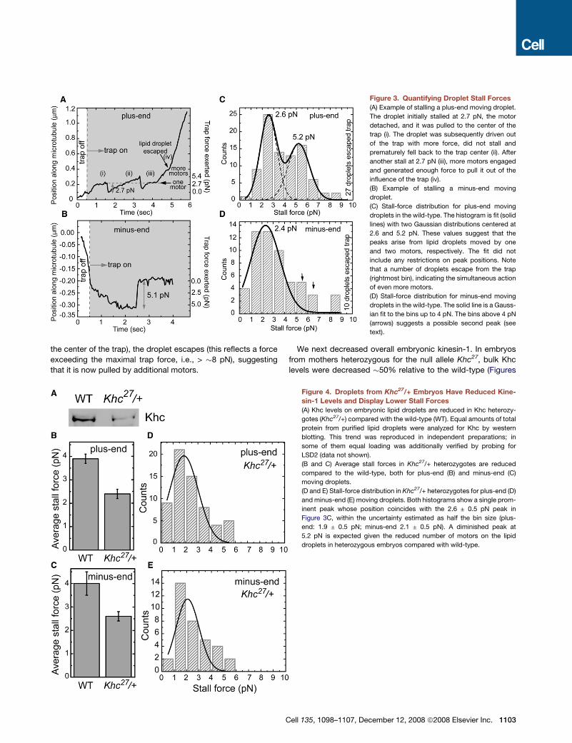
the center of the trap), the droplet escapes (this reflects a force
exceeding the maximal trap force, i.e., > �8 pN), suggesting
that it is now pulled by additional motors.
We next decreased overall embryonic kinesin-1. In embryos
from mothers heterozygous for the null allele Khc27, bulk Khc
levels were decreased �50% relative to the wild-type (Figures
Figure 4. Droplets from Khc27/+ Embryos Have Reduced Kine-
sin-1 Levels and Display Lower Stall Forces
(A) Khc levels on embryonic lipid droplets are reduced in Khc heterozy-
gotes (Khc27/+) compared with the wild-type (WT). Equal amounts of total
protein from purified lipid droplets were analyzed for Khc by western
blotting. This trend was reproduced in independent preparations; in
some of them equal loading was additionally verified by probing for
LSD2 (data not shown).
(B and C) Average stall forces in Khc27/+ heterozygotes are reduced
compared to the wild-type, both for plus-end (B) and minus-end (C)
moving droplets.
(D and E) Stall-force distribution in Khc27/+ heterozygotes for plus-end (D)
and minus-end (E) moving droplets. Both histograms show a single prom-
inent peak whose position coincides with the 2.6 ± 0.5 pN peak in
Figure 3C, within the uncertainty estimated as half the bin size (plus-
end: 1.9 ± 0.5 pN; minus-end 2.1 ± 0.5 pN). A diminished peak at
5.2 pN is expected given the reduced number of motors on the lipid
droplets in heterozygous embryos compared with wild-type.
Figure 3. Quantifying Droplet Stall Forces
(A) Example of stalling a plus-end moving droplet.
The droplet initially stalled at 2.7 pN, the motor
detached, and it was pulled to the center of the
trap (i). The droplet was subsequently driven out
of the trap with more force, did not stall and
prematurely fell back to the trap center (ii). After
another stall at 2.7 pN (iii), more motors engaged
and generated enough force to pull it out of the
influence of the trap (iv).
(B) Example of stalling a minus-end moving
droplet.
(C) Stall-force distribution for plus-end moving
droplets in the wild-type. The histogram is fit (solid
lines) with two Gaussian distributions centered at
2.6 and 5.2 pN. These values suggest that the
peaks arise from lipid droplets moved by one
and two motors, respectively. The fit did not
include any restrictions on peak positions. Note
that a number of droplets escape from the trap
(rightmost bin), indicating the simultaneous action
of even more motors.
(D) Stall-force distribution for minus-end moving
droplets in the wild-type. The solid line is a Gauss-
ian fit to the bins up to 4 pN. The bins above 4 pN
(arrows) suggests a possible second peak (see
text).
Cell 135, 1098–1107, December 12, 2008 ª2008 Elsevier Inc. 1103
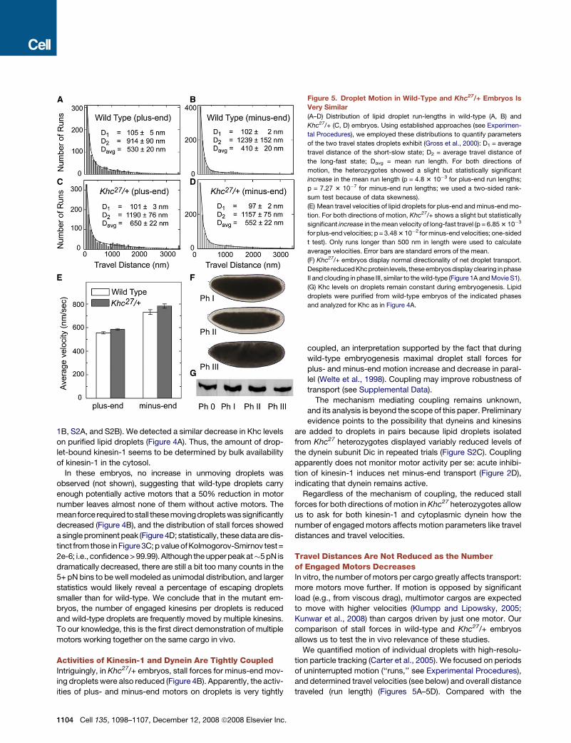
1B, S2A, and S2B). We detected a similar decrease in Khc levels
on purified lipid droplets (Figure 4A). Thus, the amount of drop-
let-bound kinesin-1 seems to be determined by bulk availability
of kinesin-1 in the cytosol.
In these embryos, no increase in unmoving droplets was
observed (not shown), suggesting that wild-type droplets carry
enough potentially active motors that a 50% reduction in motor
number leaves almost none of them without active motors. The
meanforce required to stall these movingdroplets wassignificantly
decreased (Figure 4B), and the distribution of stall forces showed
a single prominent peak (Figure 4D; statistically, these data are dis-
tinct from those in Figure3C; p value of Kolmogorov-Smirnov test=
2e-6; i.e., confidence > 99.99). Although the upper peak at�5 pN is
dramatically decreased, there are still a bit too many counts in the
5+ pN bins to be well modeled as unimodal distribution, and larger
statistics would likely reveal a percentage of escaping droplets
smaller than for wild-type. We conclude that in the mutant em-
bryos, the number of engaged kinesins per droplets is reduced
and wild-type droplets are frequently moved by multiple kinesins.
To our knowledge, this is the first direct demonstration of multiple
motors working together on the same cargo in vivo.
Activities of Kinesin-1 and Dynein Are Tightly CoupledIntriguingly, in Khc27/+ embryos, stall forces for minus-end mov-
ing droplets were also reduced (Figure 4B). Apparently, the activ-
ities of plus- and minus-end motors on droplets is very tightly
Figure 5. Droplet Motion in Wild-Type and Khc27/+ Embryos Is
Very Similar
(A–D) Distribution of lipid droplet run-lengths in wild-type (A, B) and
Khc27/+ (C, D) embryos. Using established approaches (see Experimen-
tal Procedures), we employed these distributions to quantify parameters
of the two travel states droplets exhibit (Gross et al., 2000): D1 = average
travel distance of the short-slow state; D2 = average travel distance of
the long-fast state; Davg = mean run length. For both directions of
motion, the heterozygotes showed a slight but statistically significant
increase in the mean run length (p = 4.8 3 10�3 for plus-end run lengths;
p = 7.27 3 10�7 for minus-end run lengths; we used a two-sided rank-
sum test because of data skewness).
(E) Mean travel velocities of lipid droplets for plus-end and minus-end mo-
tion. For both directions of motion, Khc27/+ shows a slight but statistically
significant increase in the mean velocity of long-fast travel (p = 6.85 3 10�3
for plus-end velocities; p = 3.48 3 10�2 for minus-end velocities; one-sided
t test). Only runs longer than 500 nm in length were used to calculate
average velocities. Error bars are standard errors of the mean.
(F) Khc27/+ embryos display normal directionality of net droplet transport.
Despite reduced Khcprotein levels, these embryosdisplay clearing inphase
II and clouding in phase III, similar to the wild-type (Figure 1A and Movie S1).
(G) Khc levels on droplets remain constant during embryogenesis. Lipid
droplets were purified from wild-type embryos of the indicated phases
and analyzed for Khc as in Figure 4A.
coupled, an interpretation supported by the fact that during
wild-type embryogenesis maximal droplet stall forces for
plus- and minus-end motion increase and decrease in paral-
lel (Welte et al., 1998). Coupling may improve robustness of
transport (see Supplemental Data).
The mechanism mediating coupling remains unknown,
and its analysis is beyond the scope of this paper. Preliminary
evidence points to the possibility that dyneins and kinesins
are added to droplets in pairs because lipid droplets isolated
from Khc27 heterozygotes displayed variably reduced levels of
the dynein subunit Dic in repeated trials (Figure S2C). Coupling
apparently does not monitor motor activity per se: acute inhibi-
tion of kinesin-1 induces net minus-end transport (Figure 2D),
indicating that dynein remains active.
Regardless of the mechanism of coupling, the reduced stall
forces for both directions of motion in Khc27 heterozygotes allow
us to ask for both kinesin-1 and cytoplasmic dynein how the
number of engaged motors affects motion parameters like travel
distances and travel velocities.
Travel Distances Are Not Reduced as the Numberof Engaged Motors DecreasesIn vitro, the number of motors per cargo greatly affects transport:
more motors move further. If motion is opposed by significant
load (e.g., from viscous drag), multimotor cargos are expected
to move with higher velocities (Klumpp and Lipowsky, 2005;
Kunwar et al., 2008) than cargos driven by just one motor. Our
comparison of stall forces in wild-type and Khc27/+ embryos
allows us to test the in vivo relevance of these studies.
We quantified motion of individual droplets with high-resolu-
tion particle tracking (Carter et al., 2005). We focused on periods
of uninterrupted motion (‘‘runs,’’ see Experimental Procedures),
and determined travel velocities (see below) and overall distance
traveled (run length) (Figures 5A–5D). Compared with the
1104 Cell 135, 1098–1107, December 12, 2008 ª2008 Elsevier Inc.
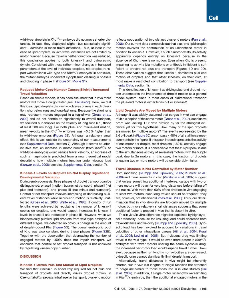
wild-type, droplets in Khc27/+ embryos did not move shorter dis-
tances. In fact, they displayed slight—but statistically signifi-
cant—increases in mean travel distances. Thus, at least in the
case of lipid droplets, in vivo travel distances are not limited by
motor number. Because travel in neither direction was reduced,
this conclusion applies to both kinesin-1 and cytoplasmic
dynein. Consistent with these rather minor changes in transport
parameters at the level of individual droplets, net droplet trans-
port was similar in wild-type and Khc27/+ embryos; in particular,
the mutant embryos underwent cytoplasmic clearing in phase II
and clouding in phase III (Figure 5F, Movie S1).
Reduced Motor Copy Number Causes Slightly IncreasedTravel VelocitiesBased on simple models, it has been assumed that in vivo more
motors will move a cargo faster (see Discussion). Here, we test
this idea. Lipid droplets display two classes of runs in each direc-
tion: short-slow runs and long-fast runs. Because the short runs
may represent motors engaged in a tug-of-war (Gross et al.,
2000) and do not contribute significantly to overall transport,
we focused our analysis on the velocity of long runs (i.e., those
at least 500 nm long). For both plus- and minus-end motion,
mean velocity in the Khc27/+ embryos was �5.5% higher than
in wild-type embryos (Figure 5E). Although a relatively small
effect, this is well outside the uncertainty of our measurements
(see Supplemental Data, section 7). Although it seems counter-
intuitive that an increase in motor number (from Khc27/+ to
wild-type embryos) would reduce travel velocity, an increase of
such a magnitude is predicted from a new theoretical model
describing how multiple motors function under viscous load
(Kunwar et al., 2008; see also Supplemental Data, section 7).
Kinesin-1 Levels on Droplets Do Not Display SignificantDevelopmental VariationDuring embryogenesis, three phases of droplet transport can be
distinguished: phase I (motion, but no net transport), phase II (net
plus-end transport), and phase III (net minus-end transport).
Control of net transport involves increasing or decreasing plus-
end travel distances while minus-end motion is relatively unaf-
fected (Gross et al., 2000; Welte et al., 1998). If control of run
lengths were achieved by regulating the number of kinesin-1
copies on droplets, one would expect increases in kinesin-1
levels in phase II and reduction in phase III. However, when we
biochemically purified lipid droplets from wild-type embryos of
different stages, we detected no obvious change in the amount
of droplet-bound Khc (Figure 5G). The overall embryonic pool
of Khc was also constant during these phases (Figure S2B).
Together with the observation that decreasing the number of
engaged motors by �50% does not impair transport, we
conclude that control of net droplet transport is not achieved
by regulating kinesin copy number.
DISCUSSION
Kinesin-1 Drives Plus-End Motion of Lipid DropletsWe find that kinesin-1 is absolutely required for net plus-end
transport of droplets and directly drives droplet motion. In
Caenorhabditis elegans intraflagellar transport, plus-end motion
reflects cooperation of two distinct plus-end motors (Pan et al.,
2006). Our current data cannot rule out that plus-end lipid droplet
motion involves the contribution of an unidentified motor in
addition to kinesin-1. However, if such a motor exists, its activity
apparently depends entirely on kinesin-1 because in the
absence of Khc there is no motion. Even when Khc is present,
impairing its activity (via mutations or antibody inhibition) is suf-
ficient to prevent net plus-end transport (Figures 1D and 2D).
These observations suggest that kinesin-1 dominates plus-end
motion of droplets and that other kinesins, on their own, at
most make a restricted contribution to transport (see Supple-
mental Data, section 1).
This identification of kinesin-1 as driving plus-end droplet mo-
tion underscores the importance of droplet motion as a general
model system, since in most cases of bidirectional transport
the plus-end motor is either kinesin-1 or kinesin-2.
Lipid Droplets Are Moved by Multiple MotorsAlthough it was widely assumed that cargos in vivo can engage
multiple copies of the same motor (Gross et al., 2007), conclusive
proof was lacking. Our data provide by far the strongest evi-
dence yet for this hypothesis. How many of the lipid droplets
are moved by multiple motors? The events represented by the
2.6 pN peak in Figure 3C encompass�40% of all stall force mea-
surements in the figure. If this peak indeed represents the activity
of one motor per droplet, most droplets (�60%) actively engage
two motors or more. It is conceivable that the 2.6 pN peak is due
to the simultaneous activity of several (n) motors, with the 5.2 pN
peak due to 2n motors. In this case, the fraction of droplets
engaging two or more motors will be considerably higher.
Travel Distance Is Not Controlled by Motor NumberBoth modeling (Klumpp and Lipowsky, 2005; Kunwar et al.,
2008) and measurements in vitro (Vershinin et al., 2007) suggest
that unless something additional interferes, cargos with two or
more motors will travel for very long distances before falling off
the tracks. With more than 60% of the droplets in vivo engaging
at least two motors, such long travels should be frequent; they
are, however, not observed (Gross et al., 2000). Thus, our deter-
mination that in vivo droplets are typically moved by multiple
motors but move relatively short distances suggests that some
additional factor is present in vivo that is absent in vitro.
This in vivo/in vitro difference might be explained by high cyto-
solic viscosity, because the resulting load could decrease both
travel distance and velocity (Klumpp and Lipowsky, 2005). Cyto-
solic load has been invoked to account for variations in travel
velocities of other intracellular cargos (Hill et al., 2004; Kural
et al., 2005; Levi et al., 2006). But if viscous drag was limiting
travel in the wild-type, it would be even more potent in Khc27/+
embryos: with fewer motors sharing the same cytosolic drag,
the increased per-motor load would impede travel further. How-
ever, because neither run lengths nor velocities are decreased,
cytosolic drag cannot significantly limit droplet transport.
Alternatively, travel distances in vivo might be inherently
shorter. But in vivo run lengths of single Kinesins not attached
to cargo are similar to those measured in in vitro studies (Cai
et al., 2007). In addition, if single-motor run lengths were limiting
in Khc27/+ embryos, then the additional engaged motors in the
Cell 135, 1098–1107, December 12, 2008 ª2008 Elsevier Inc. 1105
wild-type would allow longer travels, a prediction not borne out
by our experiments.
Droplet travel distances, therefore, appear not to be limited by
inherent motor properties, but by distinct, higher-level mecha-
nisms. We previously proposed the existence of a ‘‘switch’’
that actively terminates runs and thus cuts short the long travels
expected from multiple engaged motors (Gross et al., 2000). Our
new data reveal that the switch mechanism is rather insensitive
to motor number: over a 2-fold range of engaged motors, param-
eters of motion change minimally. Although we currently favor
the hypothesis that the switch mechanism reflects the activity
of a complex that regulates motor activity and coordinates mo-
tors, a recent theoretical model (Muller et al., 2008) suggests
that under some circumstances, an unregulated competition
between opposite motors could lead to the switching behavior
we observe. Our identification of the plus-end droplet motor
and its in vivo properties will provide the basis for future studies
to distinguish between these models.
Forces Produced by Single Motors In VivoOur previous population stall-force measurements (Welte et al.,
1998) suggested that the force required to stall a single droplet
motor (for either direction) was�1.1 pN. The new data presented
here lead us to conclude that this force is actually �2.6 pN
(Figure 3C). We envision two possible explanations for this differ-
ence in estimates. One possibility is our improved ability to iden-
tify truly stalled droplets. As droplets move against the load
applied by the optical trap, they can prematurely detach from
the microtubule, even before the motors experience maximal
load (e.g., Figure 3A, feature (ii), and Figure S3); this occurs quite
frequently. Our old measurements scored droplets as ‘‘trapped’’
if they failed to escape from the trap and thus included premature
detachments; because this criterion overestimates the fraction
of stalled droplets, this approach underestimates the stall force.
In our new measurements, we monitor the position of the droplet
relative to the trap center and only count events in which the
droplet indeed stalls out; that is, the droplet slows down gradu-
ally and remains at a fixed position away from the trap center for
a period. Because this criterion excludes premature detach-
ments, these events do not inappropriately depress the calcu-
lated stall force. A second possibility is that in wild-type phase
II embryos the single-motor state is rare and the 2.6 pN peak
in Figure 3C represents the activity of two or more motors. If
so, a single-motor force peak might become apparent if Khc
expression can be reduced even further than in the Khc27/+
embryos or when different phases of transport are examined,
because stall forces vary developmentally (Welte et al., 1998).
The stall force for single kinesin-1 molecules in vitro is between
4 to 8 pN; recombinant Drosophila kinesin-1, in particular, gener-
ates�5 pN (Carter and Cross, 2005). Our measurements put the
effective stall force of a single kinesin-1 in vivo at �2.6 pN (or
even less). We believe that this difference accurately reflects dis-
tinct properties of kinesin-1-mediated transport in vivo and
in vitro because our own in vitro kinesin-1 stall force measure-
ments are�5 pN (Vershinin et al., 2007), and we have extensively
checked our calibration procedures (see Supplemental Data,
force measurement). We speculate that cofactors present
in vivo, but not in vitro, modulate the motor’s force output.
1106 Cell 135, 1098–1107, December 12, 2008 ª2008 Elsevier Inc.
Regulation of Multiple Motors In VivoIn vivo, the distances motors travel are highly regulated: for
bidirectional transport, such regulation determines net direction
of transport. However, the mechanisms controlling run length
remain unknown. Our analysis of droplet motion indicates that
whereas cargos in vivo can simultaneously engage multiple
motors, travel lengths do not dramatically increase as more mo-
tors are engaged, contrary to unregulated in vitro systems. Thus,
alternative mechanisms can dominate run termination in vivo.
Whether regulation of motor copy number tunes transport of
any cargo remains an open question.
The mechanisms that terminate runs are an area of active
investigation. For lipid droplets, four proteins are implicated:
the dynein cofactor dynactin (Gross et al., 2002) and the novel
protein Klar (Welte et al., 1998) might coordinate dynein and
kinesin-1 activity (i.e., turn one motor off when the opposing mo-
tor is active). The transacting signal Halo (Gross et al., 2003) and
the droplet-associated ‘‘conductor’’ LSD2 (Welte et al., 2005)
mediate how travel distances change during development. Mo-
lecular dissection of these regulators and identification of their
binding partners should reveal the mechanisms of run length
control. Because these molecules and their orthologs are impor-
tant for many other transport processes, these findings will likely
illuminate the regulation of microtubule motors in general.
EXPERIMENTAL PROCEDURES
Fly Strains, Antibody Injections, and Western Blot Analysis
The wild-type stock was Oregon R; Khc and Klc germ-line clones were gener-
ated as previously described (Serbus et al., 2005; Palacios and St Johnston,
2002; see also Supplemental Data). For embryo injections, anti-Khc antibody
was processed as previously described (Serbus et al., 2005) and injected using
standard procedures (e.g., Gross et al., 2003). Antibody specificity and details
for western blot analysis are described in Supplemental Experimental Proce-
dures.
Immunolocalization and Microscopy
Yolk vesicles, Klar, microtubules, and DNA were detected as previously
described (Gross et al., 2003; Guo et al., 2005; Sisson et al., 2000), using
yolk autofluorescence, Klar-M or b-tubulin immunostaining, and Hoechst
33258, respectively. Embryo centrifugation was performed as described pre-
viously (Guo et al., 2005). To compare clearing progress between embryos of
different genotypes, image sequences from time-lapse movies were aligned
such that the frames representing the midpoint of cellularization (when mem-
branes reach the basal tip of the nuclei) were synchronized.
Lipid-Droplet Tracking and Track Analysis
Quantification of droplet motion was performed in phase II during the clearing
process, where droplets are on average moving toward the embryo center,
and was done as previously described (Gross et al., 2000; see Supplemental
Experimental Procedures for details). Run-length distributions and velocities
were calculated from at least 8 embryos per genotype (an average of 30 ± 5
droplets tracked per embryo). Run-length distributions were fit by two expo-
nential decays as detailed previously (Gross et al., 2000). The two decay
lengths (D1 and D2) characterize the short and long run lengths, respectively.
Both decay lengths as well as the average run length (Davg) are shown in
Figures 5A–5D.
Force Measurements
Experimental details of the stall-force measurements on individual lipid drop-
lets will be published elsewhere (G.T.S. et al., unpublished data). Briefly, an
optical trap setup was built atop an inverted optical microscope. For fast
and precise alignment of the trap with the position of the lipid droplet, a
computer-controlled piezoelectric stage-mounted mirror was used in conjunc-
tion with a fast single-particle tracking program capable of real-time tracking of
particles at rates exceeding 30 frames/sec (Carter et al., 2005). At the click of
the mouse on the CCD camera image of a moving lipid droplet, the program
determines the droplet’s position with an accuracy of a few nanometers, trig-
gers the piezo-driven mirror to move to that position and opens the shutter to
trap the droplet. A cargo was scored as stalled if it remained stationary out of
the center of the trap for R 0.35 sec. More details, including trap calibration,
are provided in the Supplemental Data.
SUPPLEMENTAL DATA
Supplemental Data include Supplemental Experimental Procedures, five
figures, and one movie and are available with this article online at http://
www.cell.com/supplemental/S0092-8674(08)01313-5.
ACKNOWLEDGMENTS
We thank the Bloomington Stock Center, B. Saxton, and I. Palacios for fly
stocks, B. Theurkauf and B. Cha for advice on antibody microinjections,
N. Rizzo for help with western analysis, and A. Muller and D. Lambert for com-
ments on the manuscript. This work was supported by NIGMS grant GM 64624
(to S.P.G.) and NIGMS grant GM64687 and start-up support from the Univer-
sity of Rochester (to M.A.W). G.T.S. was a Paul Sigler/Agouron Fellow of the
Helen Hay Whitney Foundation.
Received: April 14, 2008
Revised: August 8, 2008
Accepted: October 6, 2008
Published: December 11, 2008
REFERENCES
Brendza, K.M., Rose, D.J., Gilbert, S.P., and Saxton, W.M. (1999). Lethal kine-
sin mutations reveal amino acids important for ATPase activation and struc-
tural coupling. J. Biol. Chem. 274, 31506–31514.
Brendza, R.P., Serbus, L.R., Duffy, J.B., and Saxton, W.M. (2000). A function
for kinesin I in the posterior transport of oskar mRNA and Staufen protein.
Science 289, 2120–2122.
Cai, D., Verhey, K.J., and Meyhofer, E. (2007). Tracking single kinesin mole-
cules in the cytoplasm of mammalian cells. Biophys. J. 92, 4137–4144.
Carter, B.C., Shubeita, G.T., and Gross, S.P. (2005). Tracking single particles:
a user-friendly quantitative evaluation. Phys. Biol. 2, 60–72.
Carter, N.J., and Cross, R.A. (2005). Mechanics of the kinesin step. Nature 435,
308–312.
Cermelli, S., Guo, Y., Gross, S.P., and Welte, M.A. (2006). The lipid-droplet pro-
teomereveals thatdropletsare aprotein-storagedepot.Curr. Biol.16, 1783–1795.
Foe, V.E., Odell, G.M., and Edgar, B.A. (1993). Mitosis and morphogenesis in
the Drosophila embryo. In The Development of Drosophila melanogaster, M.
Bate and A. Martinez-Arias, eds. (Cold Spring Harbor, NY: Cold Spring Harbor
Laboratory Press), pp. 149–300.
Glater, E.E., Megeath, L.J., Stowers, R.S., and Schwarz, T.L. (2006). Axonal
transport of mitochondria requires milton to recruit kinesin heavy chain and
is light chain independent. J. Cell Biol. 173, 545–557.
Gross, S.P. (2004). Hither and yon: a review of bi-directional microtubule-
based transport. Phys. Biol. 1, R1–R11.
Gross, S.P., Guo, Y., Martinez, J.E., and Welte, M.A. (2003). A determinant for
directionality of organelle transport in Drosophila embryos. Curr. Biol. 13,
1660–1668.
Gross, S.P., Vershinin, M., and Shubeita, G.T. (2007). Cargo transport: two
motors are sometimes better than one. Curr. Biol. 17, R478–R486.
Gross, S.P., Welte, M.A., Block, S.M., and Wieschaus, E.F. (2000). Dynein-me-
diated cargo transport in vivo: a switch controls travel distance. J. Cell Biol.
148, 945–956.
Gross, S.P., Welte, M.A., Block, S.M., and Wieschaus, E.F. (2002). Coordina-
tion of opposite-polarity microtubule motors. J. Cell Biol. 156, 715–724.
Guo, Y., Jangi, S., and Welte, M.A. (2005). Organelle-specific control of intra-
cellular transport: distinctly targeted isoforms of the regulator Klar. Mol. Biol.
Cell 16, 1406–1416.
Hill, D.B., Plaza, M.J., Bonin, K., and Holzwarth, G. (2004). Fast vesicle trans-
port in PC12 neurites: velocities and forces. Eur. Biophys. J. 33, 623–632.
Klumpp, S., and Lipowsky, R. (2005). Cooperative cargo transport by several
molecular motors. Proc. Natl. Acad. Sci. USA 102, 17284–17289.
Kunwar, A., Vershinin, M., Xu, J., and Gross, S.P. (2008). Stepping, strain gat-
ing, and an unexpected force-velocity curve for multiple-motor based trans-
port. Curr. Biol. 18, 1173–1183.
Kural, C., Kim, H., Syed, S., Goshima, G., Gelfand, V.I., and Selvin, P.R. (2005).
Kinesin and dynein move a peroxisome in vivo: a tug-of-war or coordinated
movement? Science 308, 1469–1472.
Levi, V., Serpinskaya, A.S., Gratton, E., and Gelfand, V. (2006). Organelle
transport along microtubules in Xenopus melanophores: evidence for cooper-
ation between multiple motors. Biophys. J. 90, 318–327.
Ling, S.C., Fahrner, P.S., Greenough, W.T., and Gelfand, V.I. (2004). Transport
of Drosophila fragile X mental retardation protein-containing ribonucleoprotein
granules by kinesin-1 and cytoplasmic dynein. Proc. Natl. Acad. Sci. USA 101,
17428–17433.
Mallik, R., Petrov, D., Lex, S.A., King, S.J., and Gross, S.P. (2005). Building
complexity: an in vitro study of cytoplasmic dynein with in vivo implications.
Curr. Biol. 15, 2075–2085.
Mische, S., Li, M., Serr, M., and Hays, T.S. (2007). Direct observation of regu-
lated ribonucleoprotein transport across the nurse cell/oocyte boundary. Mol.
Biol. Cell 18, 2254–2263.
Muller, M.J., Klumpp, S., and Lipowsky, R. (2008). Tug-of-war as a cooperative
mechanism for bidirectional cargo transport by molecular motors. Proc. Natl.
Acad. Sci. USA 105, 4609–4614.
Palacios, I.M., and St Johnston, D. (2002). Kinesin light chain-independent
function of the Kinesin heavy chain in cytoplasmic streaming and posterior
localisation in the Drosophila oocyte. Development 129, 5473–5485.
Pan, X., Ou, G., Civelekoglu-Scholey, G., Blacque, O.E., Endres, N.F., Tao, L.,
Mogilner, A., Leroux, M.R., Vale, R.D., and Scholey, J.M. (2006). Mechanism of
transport of IFT particles in C. elegans cilia by the concerted action of kinesin-II
and OSM-3 motors. J. Cell Biol. 174, 1035–1045.
Pilling, A.D., Horiuchi, D., Lively, C.M., and Saxton, W.M. (2006). Kinesin-1 and
dynein are the primary motors for fast transport of mitochondria in Drosophila
motor axons. Mol. Biol. Cell 17, 2057–2068.
Serbus, L.R., Cha, B.J., Theurkauf, W.E., and Saxton, W.M. (2005). Dynein and
the actin cytoskeleton control kinesin-driven cytoplasmic streaming in Dro-
sophila oocytes. Development 132, 3743–3752.
Sisson, J.C., Field, C., Ventura, R., Royou, A., and Sullivan, W. (2000). Lava
lamp, a novel peripheral golgi protein, is required for Drosophila melanogaster
cellularization. J. Cell Biol. 151, 905–918.
Snider, J., Lin, F., Zahedi, N., Rodionov, V., Yu, C.C., and Gross, S.P. (2004).
Intracellular actin-based transport: how far you go depends on how often you
switch. Proc. Natl. Acad. Sci. USA 101, 13204–13209.
Vershinin, M., Carter, B.C., Razafsky, D.S., King, S.J., and Gross, S.P. (2007).
Multiple-motor based transport and its regulation by Tau. Proc. Natl. Acad.
Sci. USA 104, 87–92.
Welte, M.A. (2004). Bidirectional transport along microtubules. Curr. Biol. 14,
R525–R537.
Welte, M.A., Cermelli, S., Griner, J., Viera, A., Guo, Y., Kim, D.H., Gindhart,
J.G., and Gross, S.P. (2005). Regulation of lipid-droplet transport by the peril-
ipin homolog LSD2. Curr. Biol. 15, 1266–1275.
Welte, M.A., Gross, S.P., Postner, M., Block, S.M., and Wieschaus, E.F. (1998).
Developmental regulation of vesicle transport in Drosophila embryos: forces
and kinetics. Cell 92, 547–557.
Cell 135, 1098–1107, December 12, 2008 ª2008 Elsevier Inc. 1107