Embed Size (px)
Citation preview
CONSERVATION OF 19TH AND EARLY 20TH
CENTURY OIL PAINTINGS: STUDIES OF
PIGMENT DISCOLOURATION BY SCANNING
ELECTRON MICROSCOPY
ByRachel Elizabeth White
SUBMITTED IN FULFILLMENT OF THE REQUIREMENTS FOR THE DEGREE OF
DOCTOR OF PHILOSOPHY AT THE
UNIVERSITY OF TECHNOLOGY, SYDNEY AUSTRALIA
2007

Certificate
I certify that the work in this thesis has not previously been submitted for a degree nor has it been submitted as part of requirements for a degree except as fully acknowledged within the text.
I also certify that the thesis has been written by me. Any help that I have received in rny research work and the preparation of the thesis itself has been acknowledged. In addition, I certify that all information sources and literature used are indicated in the thesis.
Signature of Author
1

For Lawson
11

Table of Contents
List of Figures vi
List of Tables xvi
Nomenclature xvii
Abstract xviii
Acknowledgements xix
1 Introduction 11.1 Pigment interaction in historic oil paintings .......................................... 21.2 Motivation................................................................................................... 31.3 Thesis structure.......................................................................................... 51.4 Publications................................................................................................ 6
1.4.1 Proceedings and prizes................................................................. 6
2 Discolouring interactions of historic oil paint pigments 82.1 Discolouration of artistic pigments........................................................... 92.2 Oil painting................................................................................................ 102.3 Discolouration of oil paintings................................................................. 152.4 Chemistry of pigment interaction........................................................... 22
2.4.1 Copper sulfide production........................................................... 23
3 Scanning Electron Microscopy: Instrumentation and Techniques 273.1 Scanning Electron Microscopy.................................................................. 28
3.1.1 SEM instrumentation.................................................................... 293.1.2 X-ray microanalysis....................................................................... 313.1.3 X-ray mapping and scatter diagrams........................................ 33
iii

3.2 Environmental Scanning Electron Microscopy....................................... 353.2.1 ESEM instrumentation and features............................................. 353.2.2 Hydration techniques.................................................................... 383.2.3 Use in conservation studies........................................................... 41
4 Experimental Techniques 434.1 Sample preparation................................................................................... 44
4.1.1 Pigment samples.......................................................................... 444.1.2 Paint samples................................................................................ 454.1.3 Interaction samples....................................................................... 484.1.4 Solvent action on paints.............................................................. 49
4.2 Optical microscopy................................................................................... 514.2.1 Time resolved microscopy........................................................... 51
4.3 Characterisation techniques.................................................................... 524.3.1 XRD................................................................................................ 524.3.2 Thermal Analysis.......................................................................... 534.3.3 SEM EDS...................................................................................... 53
4.4 X-ray Mapping.......................................................................................... 554.5 ESEM......................................................................................................... 56
4.5.1 ESEM instrumentation................................................................. 564.5.2 ESEM techniques.......................................................................... 56
5 Observation of pigment interaction 635.1 Interactions between paint layers ........................................................... 645.2 Cadmium yellow and malachite interaction........................................... 70
5.2.1 Aqueous media interaction........................................................... 705.2.2 Oil paint interaction.................................................................... 775.2.3 Solvent activation of interaction................................................. 83
5.3 Interaction of cadmium yellow with other copper compounds.............. 855.3.1 Time resolved interactions........................................................... 85
5.4 Summation ................................................................................................ 88
6 Characterisation of cadmium yellow and malachite interaction 896.1 XRD............................................................................................................ 90
6.1.1 Pigment characterisation.............................................................. 906.1.2 Characterisation of interaction products by XRD.................. 946.1.3 Effect of progressive discolouration........................................... 96
6.2 Thermal Analyses....................................................................................... 1036.3 SEM EDS................................................................................................... 112
IV
6.3.1 Pigment characterisation.............................................................. 1126.3.2 Characterisation of interaction products ................................... 117
6.4 Summation................................................................................................ 128
7 X-ray Mapping 1297.1 X-ray maps and scatter diagram analyses.............................................. 130
7.1.1 Unreacted pigment mixtures........................................................ 1307.1.2 Pigment mixtures reacted in water........................................... 142
7.2 Artifacts...................................................................................................... 1647.3 Summation................................................................................................ 168
8 Dynamic ESEM studies of pigment interaction 1698.1 Hydration experiment procedures........................................................... 170
8.1.1 Hydration of dry samples.............................................................. 1708.1.2 Maintaining hydrated samples.................................................... 178
8.2 Summation................................................................................................ 180
9 Discussion of key results 182
10 Conclusions 19110.1 Interaction between cadmium yellow
and malachite pigments .......................................................................... 19210.2 Suitability of SEM as a pigment analysis
technique ................................................................................................... 19310.3 Significance for painting conservators..................................................... 194
Bibliography 196
v
List of Figures
2.1 Summer by Phillips Fox, 1912. Art Gallery of New South Wales, Sydney, Australia (reproduced with permission)........................................... 18
2.2 Optical micrograph of the cross-section of a paint fragment taken fromthe lower region of Phillip Fox’s Summer (1912), showing the darkened interface between cadmium yellow and emerald green paints (reproduced with permission)............................................................................... 19
2.3 Phase diagram for the system Cu-S, from Roseboom-.Jr (1966)............ 26
3.1 Schematic diagram showing the ESEM vacuum system. The vacuumsystem consists of five stages of increasing vacuum level. The stages are the specimen chamber, first environmental chamber (EC1), second environmental chamber (EC2), electron column and electron gun. The column and chamber regions are separated by two pressure limiting apertures (PLAs). The PL As are placed close together to minimize primary electron scattering (adapted from Philips Electron Optics, 1996). IP=ion pump, DP=diffusion pump, RT=rotary pump. (Morgan 2005). 36
3.2 Vapour pressure diagram for maintaining relative desired humidity inthe ESEM (adapted from Philips Electron Optics (1996)).................... 40
vi
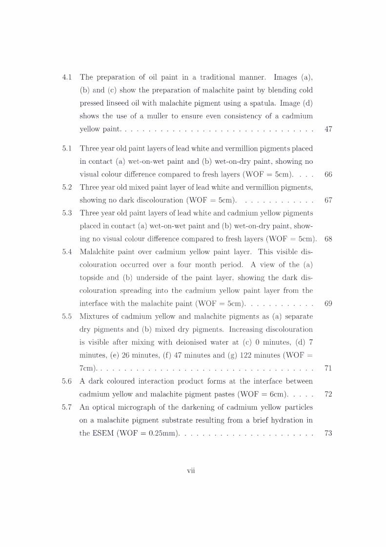
4.1 The preparation of oil paint in a traditional manner. Images (a),(b) and (c) show the preparation of malachite paint by blending cold pressed linseed oil with malachite pigment using a spatula. Image (d) shows the use of a muller to ensure even consistency of a cadmium yellow paint.................................................................................................. 47
5.1 Three year old paint layers of lead white and vermillion pigments placedin contact (a) wet-on-wet paint and (b) wet-on-dry paint, showing no visual colour difference compared to fresh layers (WOF = 5cm). ... 66
5.2 Three year old mixed paint layer of lead white and vermillion pigments,showing no dark discolouration (WOF = 5cm)....................................... 67
5.3 Three year old paint layers of lead white and cadmium yellow pigmentsplaced in contact (a) wet-on-wet paint and (b) wet-on-dry paint, showing no visual colour difference compared to fresh layers (WOF = 5cm). 68
5.4 Malalchite paint over cadmium yellow paint layer. This visible discolouration occurred over a four month period. A view of the (a) topside and (b) underside of the paint layer, showing the dark discolouration spreading into the cadmium yellow paint layer from the interface with the malachite paint (WOF = 5cm).................................. 69
5.5 Mixtures of cadmium yellow and malachite pigments as (a) separate dry pigments and (b) mixed dry pigments. Increasing discolouration is visible after mixing with deionised water at (c) 0 minutes, (d) 7 minutes, (e) 26 minutes, (f) 47 minutes and (g) 122 minutes (WOF =7cm)............................................................................................................... 71
5.6 A dark coloured interaction product forms at the interface betweencadmium yellow and malachite pigment pastes (WOF = 6cm)............. 72
5.7 An optical micrograph of the darkening of cadmium yellow particles on a malachite pigment substrate resulting from a brief hydration inthe ESEM (WOF = 0.25mm).................................................................... 73
vii
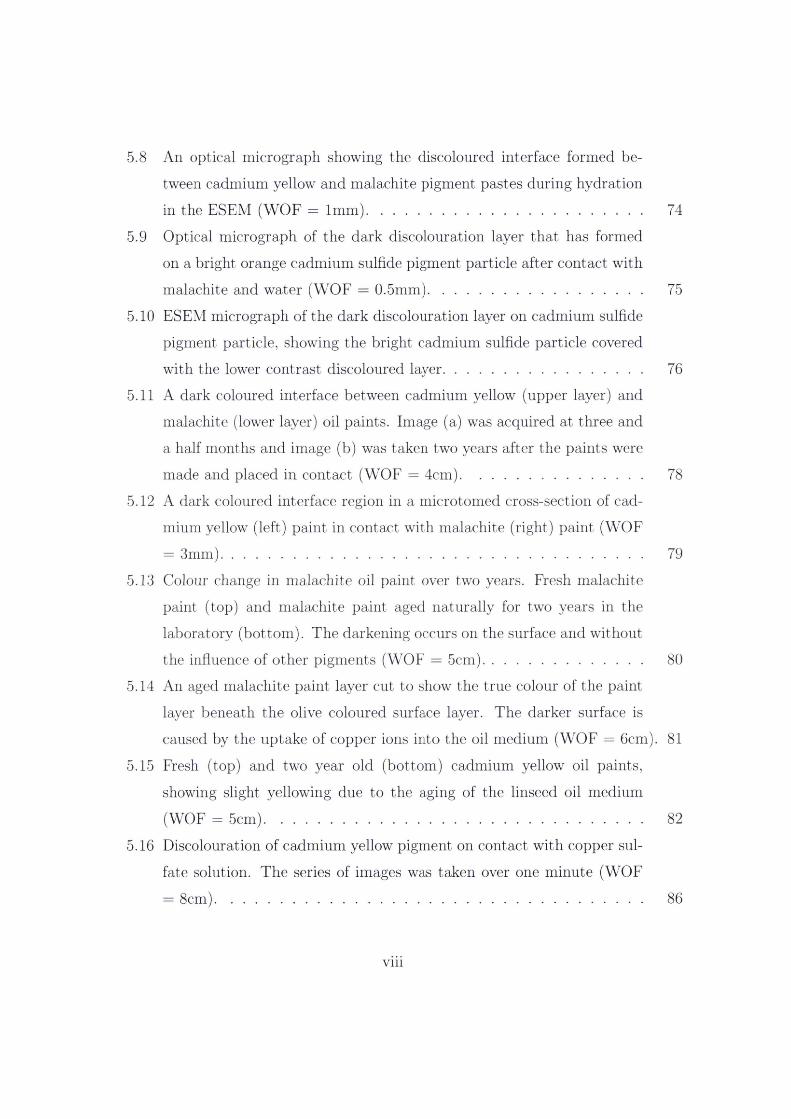
5.8 An optical micrograph showing the discoloured interface formed between cadmium yellow and malachite pigment pastes during hydrationin the ESEM (WOF = 1mm)..................................................................... 74
5.9 Optical micrograph of the dark discolouration layer that has formedon a bright orange cadmium sulfide pigment particle after contact with malachite and water (WOF = 0.5mm)..................................................... 75
5.10 ESEM micrograph of the dark discolouration layer on cadmium sulfidepigment particle, showing the bright cadmium sulfide particle covered with the lower contrast discoloured layer................................................. 76
5.11 A dark coloured interface between cadmium yellow (upper layer) andmalachite (lower layer) oil paints. Image (a) was acquired at three and a half months and image (b) was taken two years after the paints were made and placed in contact (WOF = 4cm)............................................. 78
5.12 A dark coloured interface region in a microtomed cross-section of cadmium yellow (left) paint in contact with malachite (right) paint (WOF= 3mm)......................................................................................................... 79
5.13 Colour change in malachite oil paint over two years. Fresh malachite paint (top) and malachite paint aged naturally for two years in the laboratory (bottom). The darkening occurs on the surface and withoutthe influence of other pigments (WOF = 5cm)........................................ 80
5.14 An aged malachite paint layer cut to show the true colour of the paint layer beneath the olive coloured surface layer. The darker surface is caused by the uptake of copper ions into the oil medium (WOF = 6cm). 81
5.15 Fresh (top) and two year old (bottom) cadmium yellow oil paints,showing slight yellowing due to the aging of the linseed oil medium (WOF = 5cm).............................................................................................. 82
5.16 Discolouration of cadmium yellow pigment on contact with copper sulfate solution. The series of images was taken over one minute (WOF = 8cm)
viii
86
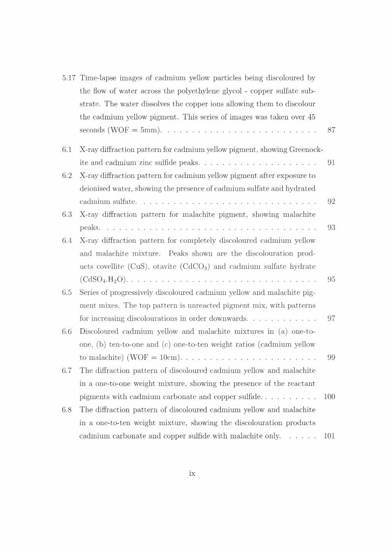
5.17 Time-lapse images of cadmium yellow particles being discoloured by the flow of water across the polyethylene glycol - copper sulfate substrate. The water dissolves the copper ions allowing them to discolour the cadmium yellow pigment. This series of images was taken over 45 seconds (WOF = 5mm).............................................................................. 87
6.1 X-ray diffraction pattern for cadmium yellow pigment, showing Greenock-ite and cadmium zinc sulfide peaks........................................................... 91
6.2 X-ray diffraction pattern for cadmium yellow pigment after exposure todeionised water, showing the presence of cadmium sulfate and hydrated cadmium sulfate........................................................................................... 92
6.3 X-ray diffraction pattern for malachite pigment, showing malachitepeaks.............................................................................................................. 93
6.4 X-ray diffraction pattern for completely discoloured cadmium yellowand malachite mixture. Peaks shown are the discolouration products covellite (CuS), otavite (CdCCb) and cadmium sulfate hydrate
(CdS04.H20)................................................................................................ 956.5 Series of progressively discoloured cadmium yellow and malachite pig
ment mixes. The top pattern is unreacted pigment mix, with patternsfor increasing discolourations in order downwards.................................. 97
6.6 Discoloured cadmium yellow and malachite mixtures in (a) one-to- one, (b) ten-to-one and (c) one-to-ten weight ratios (cadmium yellowrto malachite) (WOF = 10cm).................................................................... 99
6.7 The diffraction pattern of discoloured cadmium yellow and malachitein a one-to-one weight mixture, showing the presence of the reactant pigments with cadmium carbonate and copper sulfide............................... 100
6.8 The diffraction pattern of discoloured cadmium yellow and malachitein a one-to-ten weight mixture, showing the discolouration products cadmium carbonate and copper sulfide with malachite only..................101
IX
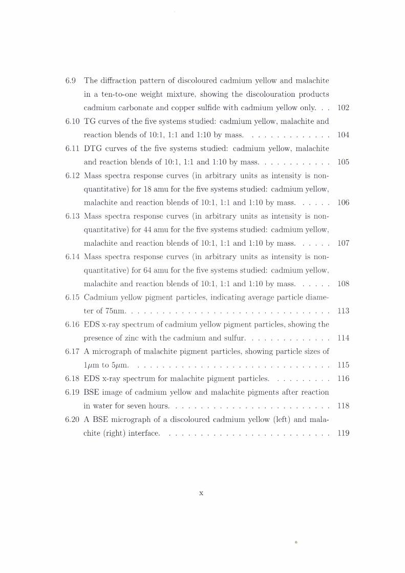
6.9 The diffraction pattern of discoloured cadmium yellow and malachitein a ten-to-one weight mixture, showing the discolouration products cadmium carbonate and copper sulfide with cadmium yellow only. . . 102
6.10 TG curves of the five systems studied: cadmium yellow, malachite andreaction blends of 10:1, 1:1 and 1:10 by mass.......................................... 104
6.11 DTG curves of the five systems studied: cadmium yellow, malachiteand reaction blends of 10:1, 1:1 and 1:10 by mass.................................. 105
6.12 Mass spectra response curves (in arbitrary units as intensity is non-quantitative) for 18 amu for the five systems studied: cadmium yellow, malachite and reaction blends of 10:1, 1:1 and 1:10 by mass................ 106
6.13 Mass spectra response curves (in arbitrary units as intensity is non-quantitative) for 44 amu for the five systems studied: cadmium yellow, malachite and reaction blends of 10:1, 1:1 and 1:10 by mass................ 107
6.14 Mass spectra response curves (in arbitrary units as intensity is non-quantitative) for 64 amu for the five systems studied: cadmium yellow, malachite and reaction blends of 10:1, 1:1 and 1:10 by mass................ 108
6.15 Cadmium yellow pigment particles, indicating average particle diameter of 75nm................................................................................................... 113
6.16 EDS x-ray spectrum of cadmium yellow pigment particles, showing thepresence of zinc with the cadmium and sulfur......................................... 114
6.17 A micrograph of malachite pigment particles, showing particle sizes oflpm to 5pm.................................................................................................. 115
6.18 EDS x-ray spectrum for malachite pigment particles............................. 1166.19 BSE image of cadmium yellow and malachite pigments after reaction
in water for seven hours............................................................................... 1186.20 A BSE micrograph of a discoloured cadmium yellow (left) and mala
chite (right) interface................................................................................... 119
x
6.21 A micrograph of the interface region from Figure 6.20, cadmium yellow (left) and malachite (right), showing plate-like cadmium sulfate crystals on the surface of the malachite region........................................ 120
6.22 A micrograph of the malachite region near the interface from Figure5.15, showing micron length needle-like protrusions from the malachite particles......................................................................................................... 121
6.23 Cadmium yellow particles partially discoloured with copper sulfate solution, showing darker regions at the particle edges.................................. 124
6.24 Cadmium sulfide substrate (lower bright area) with copper sulfide coating (upper darker area)................................................................................ 125
6.25 EDS spectrum at 20 kV of copper sulfide layer from Figure 6.24. Copper and sulfur are detected with a small amount of cadmium from the substrate........................................................................................................ 126
6.26 EDS spectrum at 20 kV of cadmium sulfide substrate from Figure 6.24.Cadmium and sulfur are detected with a small amount of copper from some residual copper sulfide layer as visible in the micrograph (Figure 6.24)............................................................................................................... 127
7.1 X-ray maps for an unreacted cadmium yellow and malachite pigmentmix. Image (a) secondary electron image, and x-ray maps for (b) cadmium, (c) copper and (d) sulfur (20 kV, WOF = 43^m)................ 131
7.2 Scatter diagrams for copper and sulfur (left) and for cadmium andsulfur (right) from unreacted cadmium yellow and malachite pigment mixture x-ray maps (Figure 7.1)............................................................... 133
7.3 X-ray maps at 15 kV for a pressed pellet of unreacted cadmium yellow and malachite pigment mix. Image (a) secondary electron image, and x-ray maps for (b) cadmium, (c) copper and (d) sulfur (15 kV, WOF= 47/rm)........................................................................................................ 135
xi

7.4 Scatter diagrams for copper and sulfur (left) and for cadmium and sulfur (right) from the pressed unreacted cadmium yellow and malachite pigment mixture x-ray maps (Figure 7.3)................................................ 136
7.5 Scatter diagram for copper and sulfur (left) showing the mid regionbetween the copper and sulfur nodes. The visual representation of that region on the secondary electron image (right) (15 kV, WOF =
47/xm)............................................................................................................ 1377.6 Scatter diagram for copper and sulfur (left) showing the copper node.
The visual representation of the copper region on the secondary electron image (right) (15 kV, WOF = 47/mi).............................................. 139
7.7 Scatter diagrams for cadmium-sulfur and copper-sulfur (top) showing the selection of the sulfur node. The visual representation of the cadmium sulfide region on the secondary electron image (bottom) (15 kV,WOF - 47/im)............................................................................................. 140
7.8 Pseudo-coloured image of the pressed unreacted pigment mixture. The blue colour indicates the copper-containing regions; the yellow shows the cadmium sulfide phase; and the lack of a light, blue colour indicatesno common copper-sulfur phase. (15 kV, WOF = 47/mi)..................... 141
7.9 X-ray maps for a cadmium yellow and malachite pigment mix reactedin water for one hour. Image (a) secondary electron image, and x-ray maps for (b) cadmium, (c) copper and (d) sulfur (WOF = 31.5/mi). . 143
7.10 Scatter diagrams for copper and sulfur (left) and for cadmium andsulfur (right) from x-ray maps of a cadmium yellow and malachite pigment mixture reacted for one hour (Figure 7.9)................................ 144
7.11 X-ray maps for a completely discoloured cadmium yellow and malachite pigment mix. Image (a) secondary electron image, and x-ray maps for(b) cadmium, (c) copper and (d) sulfur (20 kV, WOF = 47/mi). . . . 146
xii
7.12 Scatter diagrams for copper and sulfur (left) and for cadmium andsulfur (right) from x-ray maps of a completely discoloured cadmium yellow and malachite pigment mixture (Figure 7.11)............................. 147
7.13 X-ray maps for a pressed sample of completely discoloured cadmium yellow and malachite pigment mix. Image (a) secondary electron image, and x-ray maps for (b) cadmium, (c) copper and (d) sulfur (15kV, WOF = 47/iin)..................................................................................... 149
7.14 Scatter diagrams for copper and sulfur (left) and for cadmium andsulfur (right) from x-ray maps of a completely discoloured and pressed cadmium yellow and malachite pigment mixture (Figure 7.13)............ 150
7.15 Scatter diagrams for copper and sulfur (left) and for cadmium andsulfur (right) with copper sulfide, cadmium carbonate and a previously unidentified high-copper copper sulfide phase nodes indicated............. 152
7.16 Copper-sulfur scatter diagrams of the pressed completely reacted pigment mixture (left) with the selected area visualised on the secondary electron image (right) (15 kV, WOF = 47/mi)........................................ 153
7.17 Scatter diagrams for copper and sulfur with the copper region selectedand overlaid on the SE image. This area is high-copper copper sulfide species not identified by XRD (15 kV, WOF = 47/mi).......................... 155
7.18 Scatter diagrams (copper-sulfur on the left and cadmium-sulfur on the right) of the pressed completely reacted pigment mixture with the cadmium containing phase selected and visualised on the SE image (15 kV,WOF = 47/irn)............................................................................................. 157
7.19 Scatter diagrams (copper-sulfur on the left and cadmium-sulfur on the right) of the pressed completely reacted pigment mixture with the area between the phase nodes selected and visualised on the SE image (15kV, WOF = 47/rni)..................................................................................... 158
xiii
7.20 A pseudo-coloured image of the pressed completely reacted pigmentmixture. Purple represents the copper- and sulfur- containing phase; blue the copper regions; green the cadmium-containing phase. The absence of yellow region indicates there is no cadmium sulfide phase present (15 kV, WOF = 47/un)................................................................. 159
7.21 Scatter diagrams for copper and sulfur for unpressed samples of (a) unreacted pigment mix, (b) pigment mix reacted for one hour and (c) completely discoloured pigment mixture. These show that the association between copper and sulfur increases with the extent of reaction. 161
7.22 Copper-sulfur scatter diagrams for unreacted pigment mix (left) andthe completely discoloured pigment mixture (right), showing the increase of copper-sulfur phase association after discolouration............... 162
7.23 Cadmium-sulfur scatter diagrams for unreacted pigment mix (left) andthe completely discoloured pigment mixture (right), showing the breakdown of the common cadmium-sulfur phase after discolouration. . . . 163
7.24 Scatter diagram of cadmium and sulfur for pure cadmium yellow pigment. 1657.25 Scatter diagram of copper and sulfur at 5 kV and 15 kV, showing the
lack of resolution at 5 kV........................................................................... 167
8.1 Gaseous secondary electron image of water droplets forming on a mixedcadmium yellow and malachite pigment sample during hydration in the ESEM......................................................................................................... 171
8.2 ESEM images, (a) and (b), showing the surface crust of reaction products of a cadmium yellow and malachite pigment mixture after hydration in the ESEM for four hours............................................................... 172
8.3 Back-scattered electron images of (a) a dry malachite (left) and cadmium yellow (right) pigment interface, (b) the interface after 1 hour of hydration and (c) after 2 hours of hydration, showing the darkeningin the boxed region...................................................................................... 174
xiv
8.4 Series of back-scattered electron images showing cadmium yellow (brightregions) and malachite pigment interface. Image (a) is before hydration, (b) the interface during hydration and (c) the interface after dehydration, showing the development of a mid-brightness region. ... 176
8.5 Series of back-scattered electron images showing mid-brightness region formed between malachite (left darker region) and cadmium yellow (right bright region) during hydration at accelerating voltages of (a)25 kV, (b) 15 kV and (c) 10 kV................................................................ 177
8.6 BSE images of an interface between hydrated cadmium yellow andmalachite pigments. Image (a) is the first image taken of the hydrated sample after pump-down sequence. Image (b) is the dehydrated interface after 80 minutes of maintained hydration. Image (c) is an optical micrograph of the interface after dehydration (WOF = 1mm)............. 179
9.1 Interaction model for the discolouration of cadmium yellow pigmentby malachite pigment.................................................................................. 187
xv

List of Tables
1 List of symbols and abbreviations................................................................xvii
2.1 Oil absorption values................................................................................. 11
4.1 Pigments used............................................................................................. 444.2 Paint layers prepared................................................................................. 464.3 Solvent action on paints.......................................................................... 504.4 Back-scattered contrast coefficients at 25 kV......................................... 604.5 Contrast visible between interaction products ....................................... 62
5.1 Paint layer discolourations....................................................................... 655.2 Solvent action on paints.......................................................................... 84
xvi

Nomenclature
Table 1: List of symbols and abbreviations
amu atomic mass units
BSE backscattered electrons
DTG Differential Thermogravimetric Analyses
E0 Electron beam energy
ESEM Environmental Scanning Electron Microscope
nA nanoamps
nm nanometre
SEM Scanning Electron Microscope
T Torr
TG Thermogravimetric Analyses
WOF width of field
XRD X-ray Diffraction
xvn
Abstract
The discolouration of artistic oil paintings due to pigment interaction has been a
concern for artists and painting conservators since the early 1800s. Since then there
has been considerable speculation on the origin and mode of this discolouration. This
project sought to determine what discolouring interactions between pigments exist
in historic oil paintings and to understand the mechanisms involved. The discolour
ing pigment system was studied using x-ray diffraction, thermal analysis, x-ray mi
croanalysis techniques and hydration experiments using an Environmental Scanning
Electron Microscope.
A discolouring chemical interaction between cadmium yellow (a cadmium sulfide
pigment) and malachite (basic copper carbonate) was identified. The darkening reac
tion between copper containing pigments and the range of cadmium sulfide pigments
was established to be the only discolouring that occurs between artistic pigments in
vestigated in this work. This interaction occurs due to copper ions being mobile in the
drying oils used for oil painting. The copper ions are taken up by the oil medium and
transported throughout the oil layer to adjoining paint layers. Any cadmium sulfide
present in the oil painting will undergo ion exchange at its surface with the copper
ions in the medium to produce copper sulfide. The copper-cadmium ion exchange
was found to continue until the cadmium sulfide is completely converted to copper
sulfide. For the combination of cadmium yellow and malachite it was established that
the discolouring copper sulfide was covellite, CuS.

Acknowledgements
I acknowledge my gratitude for the guidance, knowledge and assistance of my principal
supervisor Associate Professor Matthew Phillips and my great appreciation of the
assistance and support of my co-supervisors Dr Paul Thomas and Dr Richard Wuhrer.
Special thanks are due to Katie McBean arid Mark Berkahn for their invaluable
help and support over the course of this project. Jean Paul Guerbois, Dr Norman
Booth and Anthea Harris are acknowledged for their extensive technical assistance.
I would like to express my gratitude to Paula Dredge from the Art Gallery of New
South Wales for providing samples and images along with her ongoing assistance with
all facets of painting conservation. Thanks also to the friendly and helpful staff at
Parkers - Sydney Fine Art Supplies in the Rocks.
The friendship, support and assistance I received throughout from Kin Friolo,
Katie McBean, Dr Brian Reedy, Dr Scott Morgan and Victor Lo has been greatly
appreciated. Thank you to my proof readers, Dr Ross White, Dr John Miles and
Lawson Goulter for their valuable input.
Finally, I’d like to recognise my family and thank them for their continued love
and support .
xix