Embed Size (px)
Citation preview
36 Journal of Nepalese Association of Pediatric Dentistry : Vol. 2, No. 1, Jan-Dec, 2021 37Journal of Nepalese Association of Pediatric Dentistry : Vol. 2, No. 1, Jan-Dec, 2021
Case Report
INTRODUCTION
Submandibular space is a potential space below the
floor of mouth comprising of two spaces, sublingual
and submaxillary, separated by the mylohyoid muscle.1
Origin of most of the submandibular space infections
are odontogenic in nature.2 Other causes include
submandibular gland sialadenitis, lymphadenitis,
peritonsillar/para-pharyngeal abscess, trauma, or surgery.1,3
In children, infections can spread rapidly, producing
significant symptoms including fever, dehydration,
and airway compromise. Thus, early recognition and
management becomes crucial.4 This case reports the
timely management of submandibular space infection in
a child via incision and drainage followed by endodontic
treatment of the offending carious tooth and placement of
stainless-steel crown.
Conservative Management of Submandibular Space Infection in a 5-Year-Old Child: A Case Report
Sanchita Khadka,1 Bandana Koirala,2 Mehul Rajesh Jaisani,3 Siddhartha Rai4
1,4Junior Resident, 2Professor, 3Additional Professor
1,2Department of Pedodontics and Preventive Dentistry, 3,4Department of Oral and Maxillofacial Surgery,
College of Dental Surgery, B.P. Koirala Institute of Health Sciences, Dharan, Nepal.
J Nepal Assoc Pediatr Dent. 2021;2(1):36-40
Correspondence
Dr. Sanchita KhadkaJunior Resident,Department of Pedodontics and Preventive Dentistry, B.P. Koirala Institute of Health Sciences, Dharan, Nepal.E-mail: [email protected]
Citation
Khadka S, Koirala B, Jaisani M R, Rai S. Conservative Management
of Submandibular Space Infection in a 5-Year-Old Child: A Case
Report. J Nepal Assoc Pediatr Dent. 2021;2(1):36-40.
ABSTRACT
Submandibular space infection is a potentially fatal infection that could arise as a result of odontogenic or non-odontogenic infections.
The management should be prompt as the infection can spread rapidly leading to airway obstruction. A 5-year-old child reported
with a complaint of swelling on the left side of the face for four days. On examination, patient had a diffuse swelling involving the
left submandibular region with decreased mouth opening secondary to carious 75. The patient was administered intravenous (i.v)
fluids, antibiotics and analgesics. Incision and drainage of the abscess was done extraorally under i.v sedation using midazolam with
local anaesthesia followed by rubber drain placement. Patient responded to the treatment with progressive decrease in the swelling.
Pulpectomy of 75 was performed followed by stainless-steel crown placement. This case highlights the importance of prompt
appropriate treatment supplemented by salvage treatment to overcome the associated morbidity at this very young age.
Keywords: Incision and drainage; ludwig’s angina; orofacial fascial; submandibular space infection.
CASE REPORT
A 5-year-old male child was brought to the pediatric
emergency of B.P. Koirala Institute of Health Sciences,
Dharan, Nepal with a complaint of progressive swelling
on the left side of the face for four days with difficulty in
mouth opening, eating and speaking. General examination
showed an afebrile, non-toxic appearing patient. Extraoral
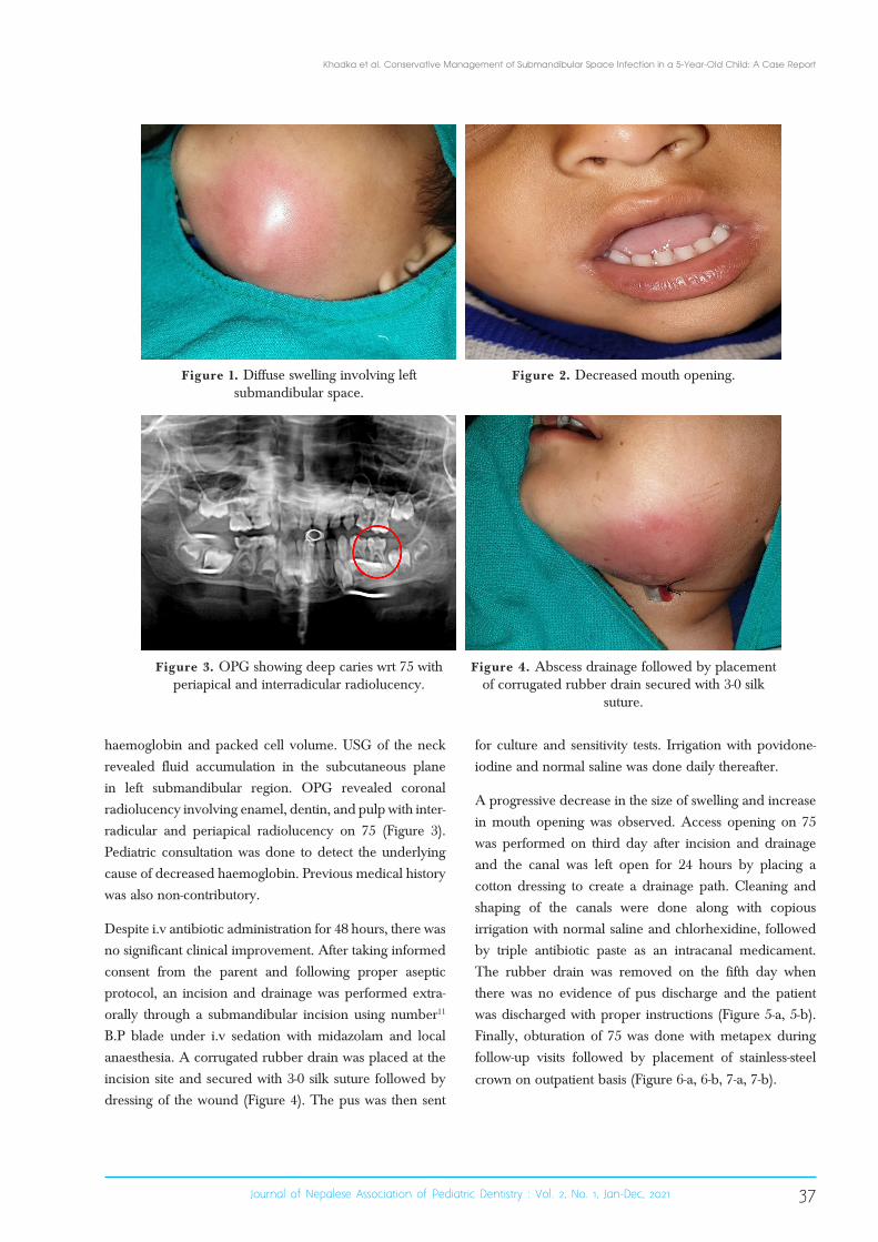
examination revealed diffuse swelling of approximately 4
× 3 cm2 on the left side of the face involving submandibular
space with obvious facial asymmetry and reduced mouth
opening (Figure 1, 2). The swelling was soft to firm in
consistency. The overlying skin was erythematous and
shiny with a local rise in temperature and tender on
palpation. Submandibular and submental lymph nodes
on the left side were palpable and tender. Intraoral
examination revealed deep caries on 75 with obliteration
of the vestibule.
The patient was immediately administered intravenous
fluids considering the decreased oral intake, and
administered intravenous antibiotics (amoxicillin/
clavulanate and metronidazole) and analgesics
(paracetamol). Routine blood investigations, neck
ultrasonography (USG), and orthopantomograph (OPG)
were advised. Blood investigation revealed decreased
36 Journal of Nepalese Association of Pediatric Dentistry : Vol. 2, No. 1, Jan-Dec, 2021 37Journal of Nepalese Association of Pediatric Dentistry : Vol. 2, No. 1, Jan-Dec, 2021
Figure 1. Diffuse swelling involving left submandibular space.
Figure 3. OPG showing deep caries wrt 75 with periapical and interradicular radiolucency.
Figure 2. Decreased mouth opening.
Figure 4. Abscess drainage followed by placement of corrugated rubber drain secured with 3-0 silk
suture.
haemoglobin and packed cell volume. USG of the neck
revealed fluid accumulation in the subcutaneous plane
in left submandibular region. OPG revealed coronal
radiolucency involving enamel, dentin, and pulp with inter-
radicular and periapical radiolucency on 75 (Figure 3).
Pediatric consultation was done to detect the underlying
cause of decreased haemoglobin. Previous medical history
was also non-contributory.
Despite i.v antibiotic administration for 48 hours, there was
no significant clinical improvement. After taking informed
consent from the parent and following proper aseptic
protocol, an incision and drainage was performed extra-
orally through a submandibular incision using number11
B.P blade under i.v sedation with midazolam and local
anaesthesia. A corrugated rubber drain was placed at the
incision site and secured with 3-0 silk suture followed by
dressing of the wound (Figure 4). The pus was then sent
for culture and sensitivity tests. Irrigation with povidone-
iodine and normal saline was done daily thereafter.
A progressive decrease in the size of swelling and increase
in mouth opening was observed. Access opening on 75
was performed on third day after incision and drainage
and the canal was left open for 24 hours by placing a
cotton dressing to create a drainage path. Cleaning and
shaping of the canals were done along with copious
irrigation with normal saline and chlorhexidine, followed
by triple antibiotic paste as an intracanal medicament.
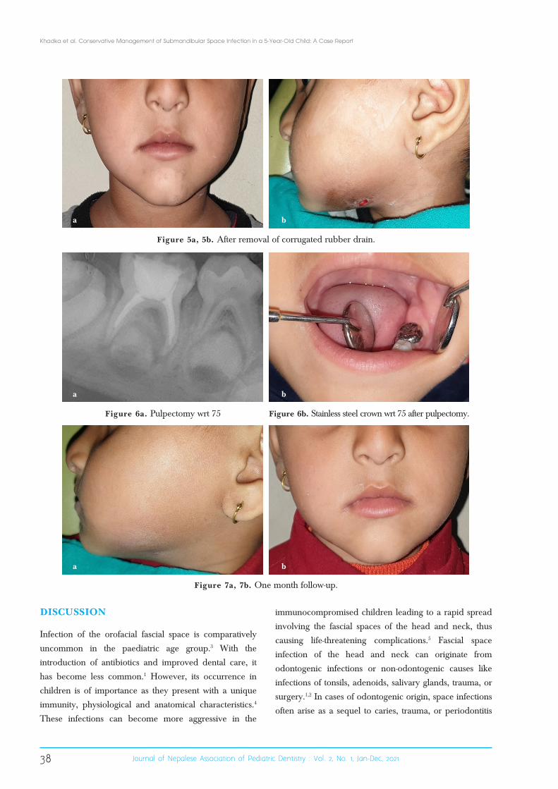
The rubber drain was removed on the fifth day when
there was no evidence of pus discharge and the patient
was discharged with proper instructions (Figure 5-a, 5-b).
Finally, obturation of 75 was done with metapex during
follow-up visits followed by placement of stainless-steel
crown on outpatient basis (Figure 6-a, 6-b, 7-a, 7-b).
Khadka et al. Conservative Management of Submandibular Space Infection in a 5-Year-Old Child: A Case Report
38 Journal of Nepalese Association of Pediatric Dentistry : Vol. 2, No. 1, Jan-Dec, 2021 39Journal of Nepalese Association of Pediatric Dentistry : Vol. 2, No. 1, Jan-Dec, 2021
DISCUSSION
Infection of the orofacial fascial space is comparatively
uncommon in the paediatric age group.3 With the
introduction of antibiotics and improved dental care, it
has become less common.1 However, its occurrence in
children is of importance as they present with a unique
immunity, physiological and anatomical characteristics.4
These infections can become more aggressive in the
immunocompromised children leading to a rapid spread
involving the fascial spaces of the head and neck, thus
causing life-threatening complications.5 Fascial space
infection of the head and neck can originate from
odontogenic infections or non-odontogenic causes like
infections of tonsils, adenoids, salivary glands, trauma, or
surgery.1,2 In cases of odontogenic origin, space infections
often arise as a sequel to caries, trauma, or periodontitis
Figure 5a, 5b. After removal of corrugated rubber drain.
Figure 6a. Pulpectomy wrt 75 Figure 6b. Stainless steel crown wrt 75 after pulpectomy.
Figure 7a, 7b. One month follow-up.
a
a
a
b
b
b
Khadka et al. Conservative Management of Submandibular Space Infection in a 5-Year-Old Child: A Case Report
38 Journal of Nepalese Association of Pediatric Dentistry : Vol. 2, No. 1, Jan-Dec, 2021 39Journal of Nepalese Association of Pediatric Dentistry : Vol. 2, No. 1, Jan-Dec, 2021
which spreads beyond the alveolar bone to involve fascial
space around the face and oral cavity. These infections
tend to pass along the path of least resistance. It usually
begins on the buccal and lingual sides of the maxilla and
mandible, respectively where the alveolar bone is the
weakest to involve the primary fascial spaces and progress
to involve the secondary spaces and even extra-facial
regions.6
Unlike the adult population where these infections are
often odontogenic in nature, the cause may be uncertain
in very young children and infants. There is a need to rule
out the immune system compromise when these infections
occur in very young children.4 However, in the present
case, the cause of infection was purely odontogenic.
The submandibular space is located below the floor of
the mouth and composed of two spaces, sublingual or
superior space and the submaxillary or inferior space
separated anteriorly by the mylohyoid muscle. These
spaces communicate freely around the posterior border of
the mylohyoid muscle where the infection in submaxillary
space ascends to involve the sublingual space. Ludwig’s
angina is a potentially lethal bilateral diffuse gangrenous
cellulitis of the submandibular, sublingual as well as the
submental spaces that was first described in 1836 by
Wilhelm Frederick von Ludwig, a German physician.1
Rapid swelling of the bilateral submandibular tissues can
induce airway blockage due to an elevation of the tongue
against the roof of the mouth and posterior pharyngeal
wall, or as a result of anterior visceral space involvement
with laryngeal oedema. Therefore, maintenance of a safe
and secure airway is mandatory in submandibular space
infections.1
The microbiology of these infections is usually
polymicrobial with mixed aerobic and anaerobic
organisms. Thus, the combination of penicillin and
beta-lactamase inhibitor (amoxicillin/clavulanate,
ticarcillin/clavulanate, piperacillin/tazobactam), cefoxitin,
carbapenem, or clindamycin are considered the most
effective antimicrobial agents. In patients with penicillin
allergy, macrolides or ketolides combined with
metronidazole should be considered.1 Antimicrobial
treatment should be started as soon as possible after a
diagnosis and before the surgery to shorten the infection
cycle and reduce the chances of bacteremia.3 Empirical
antibiotics are to be initiated before the culture results, with
adjustments followed once culture reports are available.7
In the present case, intravenous amoxicillin/clavulanate
and metronidazole were used and the culture report came
to be sterile which meant that the given antibiotics were
effective.
The signs and symptoms of infection differ based on
the stage and severity of the same. This may include
toothache, localized abscess, and rapidly developing
inflammation affecting several fascial planes ranging from
the submandibular, sublingual and submental spaces
to high-risk areas such as the pterygomandibular and
pharyngeal spaces.8 There may be persistent fever or
signs of fluid loss, as well as signs of the central nervous
system (CNS) involvement like decreased level of
consciousness, headache, or abnormal eye signs such as
proptosis, pupillary dilation, diplopia, or papilloedema.9
Trismus may be present when the infection involves
the masticatory spaces.10 Patient may present with ill-
looking appearance and may rapidly develop dysphagia
and respiratory distress. The overlying skin may have
draining sinus or can be necrosed in severe cases.9 With
no immediate intervention, the infection can spread to the
carotid space, cranial fossa, mediastinum and ultimately
cause septicaemia and death.4 This case presented with
diffuse swelling involving the submandibular space along
with trismus. There was no draining sinus and the patient
had a non-toxic appearance, was afebrile with no signs of
dysphagia or respiratory distress.
Early diagnosis is crucial while treating the paediatric
patients since the symptoms can progress rapidly causing
various systemic symptoms like fever, dehydration,
and airway obstruction as well as long term adverse
consequences. Aggressive management is critical for quick
infection resolution and to decrease the morbidity.3 The
diagnosis is made based on a thorough medical history
and clinical evaluation, as well as the radiographic and
haematological investigations.
The mainstay of treatment of submandibular space
infections are airway management, broad-spectrum
antibiotics, adequate hydration, and if necessary, surgical
drainage along with the removal of source of infection
which includes extraction of offending teeth.1,3 Improper
use of antibiotics, steroids, and nonsteroidal anti-
Khadka et al. Conservative Management of Submandibular Space Infection in a 5-Year-Old Child: A Case Report

40 Journal of Nepalese Association of Pediatric Dentistry : Vol. 2, No. 1, Jan-Dec, 2021 41Journal of Nepalese Association of Pediatric Dentistry : Vol. 2, No. 1, Jan-Dec, 2021
inflammatory drugs can obscure the signs/symptoms of
infection and alter the clinical presentation, rendering it
more difficult to diagnose as well as cause a slow course
of disease, slower healing, and development of further
complications.1 In the present case, incision and drainage
of the submandibular abscess was done extra-orally as
there was no response to antibiotic therapy alone within
48 hours. The swelling subsequently decreased thereafter,
and there was a progressive increase in mouth opening.
A conservative management of the offending carious
tooth was opted over extraction, where pulpectomy was
performed followed by stainless-steel crown placement
in the subsequent follow-up visits which ensured the
successful outcome.
CONCLUSIONS
Submandibular space infection can lead to potentially fatal
complications if not treated on time. The use of broad-
spectrum antibiotics and timely surgical incision and
drainage along with endodontic treatment of the offending
carious tooth ensured a successful outcome in this case.
Conflict of Interest: None
JNAPD
REFERENCES1. Boscolo-Rizzo P, Da Mosto MC. Submandibular space infection: a potentially lethal infection. Int J Infect Dis. 2009 May;13(3):327-33. [PubMed | DOI]
2. Heimdahl A, von Konow L, Satoh T, Nord CE. Clinical appearance of orofacial infections of odontogenic origin in relation to microbiological findings. J Clin Microbiol. 1985 Aug;22(2):299-302. [PubMed | Full Text]
3. Parkins G. Padiatric Oro-facial Fascial Space Infections. J West Afr Coll Surg. 2018 Oct-Dec;8(4):x-xiv. [PubMed | Full Text]
4. Al-Malik M, Al-Sarheed M. Pattern of management of oro-facial infection in children: A retrospective. Saudi J Biol Sci. 2017 Sep;24(6):1375-1379. [PubMed | Full Text | DOI]
5. Yellon RF. Infections of the fascial spaces of the head and neck in children. Seminars in Pediatric Infectious Diseases. WB Saunders. 1998 Jan 1;9(1):60-69. Full Text [DOI]
6. Okoje VN, Omeje KU, Okafor E, Adeyemo YI, Abubaccar J, Roberts C, Samateh AL. ORO-FACIAL FASCIAL SPACE INFECTION IN A PAEDIATRIC GAMBIAN POPULATION: A REVIEW OF 93 CASES. J West Afr Coll Surg. 2018 Oct-Dec;8(4):1-23. [PubMed | Full Text]
7. Raghani MJ, Raghani N. Bilateral deep neck space infection in pediatric patients: review of literature and report of a case. J Indian Soc Pedod Prev Dent. 2015 Jan-Mar;33(1):61-5. [PubMed | DOI]
8. Brotherton H, Templeton K, Rowney DA, Montague ML. Ludwig’s Angina: Paediatric Case Report and Literature Review. Intern Med. 2014;4(174):2. [Full Text | DOI]
9. Blankson PK, Parkins G, Boamah MO, Abdulai AE, Ahmed AM, Bondorin S, Nuamah I. Severe odontogenic infections: a 5-year review of a major referral hospital in Ghana. Pan Afr Med J. 2019 Feb 12;32:71. [PubMed | Full Text | DOI]
10. Bridgeman A, Wiesenfeld D, Hellyar A, Sheldon W. Major maxillofacial infections. An evaluation of 107 cases. Aust Dent J. 1995 Oct;40(5):281-8. [PubMed | DOI]
Khadka et al. Conservative Management of Submandibular Space Infection in a 5-Year-Old Child: A Case Report