Embed Size (px)
Citation preview
Mol Gen Genet (1989) 219:%16
O Springer-Verlag 1989
Conserved function in Nicotiana tabacum of a single Drosophila hsp70 promoter heat shock element when fused to a minimal T-DNA promoter David Wing*, Csaba Koncz, and Jeff Schell Max-Planck-Institut fur Zuchtungsforschung, Egelspfad, D-5000 Koln 30, Federal Republic of Germany
Summary. To demonstrate the extent of evolutionary con- servation in the mechanism of induction of heat shock genes between plants and animals, the minimal sequence from the Drosophila hsp70 promoter sufficient to confer heat shock inducible transcription in tobacco was determined. Segments of the hsp70 promoter were fused to a minimal promoter of the T-DNA indole-3-acetamide hydrolase (iaaH) gene, in a chimaeric gene fusion to a neomycin phosphotransferase (NPT II) reporter gene. Sequences bearing one or more heat shock elements (HSEs) rendered the minimal promoter heat shock inducible, with a 37 bp fragment containing a single complete HSE sufficing. The induced NPT I1 mRNA peaked during the heat shock peri- od, but the maximal level of NPT I1 activity was not ob- served until 4 h later in the recovery phase, showing that the translation of the NPT II mRNA was shifted from the heat shock period of the recovery phase. That similar se- quences containing a single HSE of the Drosophila hsp70 promoter could function in both flies and tobacco indicates the high degree of homology between the two heat shock gene induction systems.
Key words: Heat shock - Plant gene transfer vector - Dro- sophila hsp70 - Tobacco - Minimal promoter
Introduction
A sudden increase in temperature, or heat shock, induces a rapid change in gene expression of all organisms so far examined (Schlesinger et al. 1982). During the initial stages of the heat shock response the rate of transcription of a set of genes, termed the heat shock genes, is increased dra- matically (Ashburner and Bonner 1979). These new mRNAs are preferentially translated during the heat shock period (McKenzie et al. 1975; Lindquist 1981) and encode the heat shock proteins which are thought to protect the cell from the thermal stress (Schlesinger et al. 1982).
Biochemical and genetic evidence illustrates the critical 'role played by a 14bp consensus sequence CTnGAAnnTTCnAG, termed the heat shock element (HSE), to mediate the transcriptional activation of the heat shock genes. It is observed, often in multiple copies, in the 5' upstream regions of the heat shock genes of all eukar- yotes (Pelham 1985). A heat shock transcription factor
* Present address: National Institute of Agrobiological Resources, 2-1-2, Kannondai, Tsukuba, Ibaraki, 305 Japan
Offprint requests to: C . Koncz
(HSTF) isolated from Drosophila or yeast cells can bind specifically in vitro to HSEs of hsp70 promoters of either organism (Wiederrecht et al. 1987). Probing of the chroma- tin structure of the Drosophila hsp70 gene after heat shock shows that the HSEs are complexed with protein, presum- ably with HSTF (Wu 1984). DNA binding and in vivo heat shock activation experiments using the cloned yeast HSTF gene confirm its role in heat shock gene regulation, which is to bind to the HSE and mediate the heat shock induced increase in transcription of the heat shock gene (Sorger and Pelham 1988; Wiederrecht et al. 1988).
Promoter deletion analysis of the Drosophila hsp70 gene assayed in stably transformed Drosophila cells has demon- strated that a single HSE is required for partial heat shock inducible transcription, although promoters with two HSEs more closely approached the level of induction of an intact promoter (Dudler and Travers 1984; Xiao and Lis 1988). Furthermore, in DNA transfection experiments, a single synthetic HSE oligonucleotide placed 5' of a thymidine ki- nase gene promoter of herpes virus is heat shock inducible in COS monkey cells (Pelham and Bienz 1982). As a demon- stration of how well conserved is the function of the HSE throughout the animal kingdom, two constructions using the Drosophila hsp70 promoter, one carrying two HSEs on the sequence from - 108 to - 37 added to a truncated Xeno- pus hsp70 promoter (Bienz and Pelham 1986) and a second leaving only one HSE on the 5' deletion to -67, are both heat shock inducible in monkey COS cells (Mirault et al. 1982; Pelham 1982).
The structure and function of the HSE have been found to be conserved in the plant kingdom as well. HSEs have been observed in the promoter regions of heat shock genes of soybean and maize (Schoffl et al. 1984; Czarnecka et al. 1985; Rochester et al. 1986). A further indication of how well the function of the HSE is maintained throughout all eukaryotes is shown by the ability of a 5' deletion to -250 of the Drosophila hsp70 promoter to confer heat shock regu- lation to a reporter gene in tobacco (Spena et al. 1985). Promoter deletion analysis of the soybean gene has demon- strated that an overlapping dimer of the HSEs, a structure not present in the Drosophila hsp70 promoter, functions in a regulated manner in tobacco (Baumann et al. 1987) and that this overlapping dimer also confers heat shock inducibility to a truncated CaMV 35s RNA promoter (Strittmatter and Chua 1987).
In order to determine whether and to what degree the function of the HSE is conserved between'the animal and plant kingdoms, the Drosophila hsp70 promoter was ana-
lysed in tobacco callus to delimit the minimal sequence re- quired for heat shock inducible gene activation. Using a pair of vectors constructed for assaying positively regulated enhancers, segments of the hsp70 promoter were linked to a heterologous minimal promoter derived from the TL- DNA indole-3-acetamide hydrolase (iaaH) gene and trans- ferred on a plant reporter gene binary vector by Agrobacter- ium-mediated DNA'transfer to tobacco cells. A fragment bearing a single complete HSE from the Drosophila hsp70 promoter was sufficient for heat shock induction in tobacco cells.
Materials and methods
Bacterial strains and media. Escherichia coli strain DHI (Hanahan 1983) was transformed with plasmid DNA con- structions (Maniatis et al. 1982) and served as recipient for back-mating with Agrobacterium tumefaciens (Koncz and Schell 1986). E. coli BMH71-18 (Yanisch-Perron et al. 1985) was transformed with IacZ-HPT fusion construc- tions. E. coli S17-1 (Simon et al. 1983) was transformed with the binary vectors, which were subsequently transfered into A. twnefaciens pGV3101 (pMP90RK) by bacterial con- jugation (Koncz and Schell1986). E. coli and A. twnefaciens cultures were grown respectively in liquid or on agar-con- taining LB and YEB medium (Miller 1972). The antibiotic concentrations used for selection of E. coli were 50 pg/ml ampicillin or 10 pg/ml gentamycin and of A. tumefaciens, 100 pg/ml rifampicin, 100 pg/ml carbinicillin or 25 pg/ml gentamycin.
Transformation and maintenance of plant tissues. Nicotiana tabacum W38 leaf-discs were transformed by A. tumefa- ciens-mediated DNA transfer (Horsch et al. 1985). For iso- lation and growth of transformed tobacco calli, the inocu- lated leaf-discs were placed on callus induction medium [LS medium supplemented with 1 mg/l naphthyl-1-acetic acid (NAA) and 0.2 mg/l 6-benzyl aminopurine (BAP), Linsmaier and Skoog 19651. To regenerate shoots, the in- oculated leaf-discs were placed on LS medium supple- mented with 0.1 mg/l NAA and 0.5 mg/l BAP. Shoots were rooted and propagated on MS medium without growth fac- tors (Murashige and Skoog 1962). Claforan (500 pg/ml) was present in all media except during the inoculation of leaf-discs. Hygromycin at 15 pg/ml was used to select for transformed tissues.
In vitro DNA recombination. Standard recombinant DNA techniques were used for DNA isolation, restriction and modification, DNA transformation of DH 1 and S17-1 cells and identification of bacterial colonies containing the cor- rect DNA plasmids (Maniatis et al. 1982).
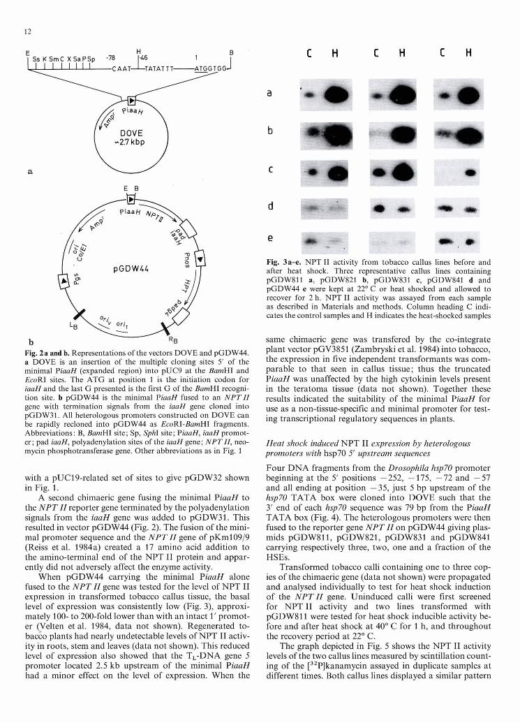
Construction of DOVE. A BamHI fragment from pGVOl53 (De Vos et al. 1981) containing the iaaH gene of the octo- pine Ti plasmid pTiAch5 was digested at the Tag1 (-417) and HaeIII sites (+8, the A of the initiation codon of the iaaH gene is referred to as position 1) and subcloned into pBR322 which had filled-in BamHI and AccI (position 2246) ends. This resulted in plasmid p6M3. The iaaH pro- moter (PiaaH) was then isolated from p6M3 DNA as a BamHI - HincII fragment and inserted into pUC9 DNA (Vieira and Messing 1982) with BamHI and filled-in EcoRI ends. The SalI, PstI and HindIII sites of pUC9 3' of PiaaH were removed by restriction endonuclease digestion fol-
lowed by S1 nuclease treatment, resulting in plasmid pUC9- pro/b.
In the final step, a modified pUC19 (Yanisch-Perron et al. 1985) polylinker sequence, containing a BamHI site filled-in and religated to generate a ClaI site (plasmid pUC19*), was placed 5' of the PiaaH sequence. pUC19* DNA was digested with HindIII and the ends were filled-in. The DNA was then partially digested with PvuI to yield a fragment with the 5' half of the j3-lactamase gene and the polylinker. This was ligated to pUC9-pro/b DNA, di- gested with EcoRI, filled-in and digested with PvuI leaving the 3' half of the j3-lactamase gene. Figure 2a shows the resulting plasmid, DOVE (Domain VEctor), which served as the vehicle for cloning positively regulated enhancers situated 5' of a minimal PiaaH.
Mutagenesis of the hygromycin phosphotransferase (HPT) gene. A BamHI fragment bearing the HPT gene (Gritz and Davies 1983) from plasmid pVUlOll (kindly provided by P. Van den Elzen) was subcloned into the BamHI site of an EcoRI site deficient pUC19 vector to create a lacZ-HPT fusion (plasmid pUC-hyg) conferring hygromycin resis- tance to E. coli strain BMH71-18. Thc cells were treated with hydroxylamine (Miller 1972) and EcoRI site deficient plasmids were enriched for by four repetitions of EcoRI digestion of reisolated pUC-hyg DNA followed by retrans- formation of E. coli BMH71-18 with the mixed population of linear and supercoiled DNAs (Vieira and Messing 1982). Sequencing of an isolated EcoRI site deficient pUC-hyg (plasmid pGDW11) revealed an A to C transversion (GAATTC to GCATTC), resulting in a glutamic acid to alanine substitution in the amino acid sequence of the HPT protein. Enzyme kinetics of HPT in crude bacterial extracts showed no change in the KM of the mutated enzyme for hygromycin (Gritz and Davies 1983).
Construction of a plant gene transfer vector carrying the EcoRI site deficient HPT gene. The EcoRI site deficient HPT gene was cloned as a BamHI fragment into the BclI site of pCV5013 DNA (C. Koncz, unpublished result), be- tween the nopaline synthase gene promoter and the polya- denylation sites from T-DNA gene 4. This was moved to binary vector pPCV002 (Koncz and Schell 1986) as a Hin- dIII-SphI fragment resulting in plasmid pGDW31. To create pGDW32, the TL-DNA gene 5 promoter near the left border was deleted and the multiple cloning sites were replaced with those of pUC19* from the EcoRI through the SalI sites.
Construction of the DOVE promoter-reporter gene fusion vectors. pGDW44 was based on the binary vector pGDW31. The polyadenylation signal from the iaaH gene was added 3' of the neomycin phosphotransferase (NPT ZI) gene on pKM109/9 (Reiss et al. 1984a) by inserting into the Aha11 and SmaI sites, a CIaI (+1307) and filled-in HindIII (666 bp downstream of the stop codon) digested fragment from pGV0153 DNA resulting in plasmid pKM109/9-pAd. To this the minimal PiaaH from DOVE was added as an EcoRI + BamHI digested fragment. The PiaaH-NPT ZI gene with the iaaH gene polyadenylation site was then cloned onto pGDW31 as an EcoRI-SalI fragment resulting in plasmid pGDW44 shown in Fig. 2.
Construction of the hsp70-PiaaH heterologous promoters. pHs-neo (Spena et al. 1985), a subclone of the Drosophila
hsp70 promoter provided the hsp70 sequences. The follow- ing fragments were cloned into DOVE to give the heterolo- gous promoters listed: pD811 is the EcoRI to HpaI frag- ment in the EcoRI and ClaI sites; pD821 is the HaeIII to HpaI fragment in the SmaI and ClaI sites; pD831 is the Tag1 (position -72) to HpaI fragment in the ClaI site; pD841 is the Tag1 (position -58) to HpaI fragment in the ClaI site.
DNA sequencing. DNA sequences were determined by the dideoxynucleotide chain termination method (Sanger et al. 1977). One guanidine was lost from the ligation junction between the HaeIII end of the iaaH sequence and the filled- in BamHI site of pBR322.
NPT II assay. 50 to 100 mg of callus tissue was ground in 100 p1 of extraction buffer 0.0625 M TRIS-HCl, pH 6.8, 10% glycerol, 5% 8-mercaptoethanol, 0.01 % SDS, 0.01 % bromphenol blue). 50 pg of protein, determined by the Bradford (1 976) assay, was loaded onto polyacrylamide gels for native protein separation. An in situ assay was used to detect the NPT I1 activity (Reiss et al. 198413).
RNA isolation. RNA was isolated from frozen pulverized calli by grinding the tissue in a guanidium thiocyanate solu- tion followed by centrifugation of the resulting homogenate over a 5.7 M CsCl cushion (Chirgwin et al. 1979). RNA pellets were redissolved in water. Polyadenylated RNA was purified by two passages through oligo(dT) cellulose (Aviv and Leder 1972). RNA concentrations were determined by absorbance at 260 nm.
DNA isolation. Total plant cell DNA was isolated as a mini- preparation (Dellaporta 1983) and further purified by diges- tion with RNase and then proteinase K, extraction with phenol, phenol :chloroform and chloroform, precipitation with isopropanol and washing with 70% EtOH. DNA con- centrations were determined by absorbance at 260 nm.
Nucleicacid transfer to and detection on nitrocellulosefilters. Transfer of DNA or RNA after size resolution on agarose gels was done according to standard procedures. DNA hy- bridization was done in 6 x SSC solution at 65" C . RNA hybridization was done in 50% formamide solution at 42" C using an NPT II probe and at 39" C using the soy- bean actin probe (kindly provided by R. Meahger) (Mania- tis 1982). Radioactive DNA probes were generated by "ran- dom primer" synthesis with [32P]dCTP to specific activities of at least 5 x 10' cpm/pg DNA (Feinberg and Vogelstein 1984). Autoradiographs were taken using Kodak X-OMAT films.
Results
Characteristics of the minimal PiaaH applied towar& assaying for positively regulated enhancers using vectors DOVE and pGD W44
DOVE is a 2.7 kb vector containing multiple cloning sites located 5' of a truncated PiaaH (Fig. 2). The vector is half of a system designed to facilitate the testing of putative enhancer sequences placed upstream of a heterologous min- imal promoter. The basis of the minimal promoter is a
truncated PiaaH from the octopine Ti plasmid pTiAch5 (Gielen et al. 1984). The full promoter drives a very low level of mRNA transcription, representing about 0.00005% to 0.0001% of the total mRNA population in T-DNA transformed cells (Willmitzer et al. 1981). This reduced level of transcriptional activity was chosen to provide a low basal level of expression, thereby increasing the likelihood of de- tecting positively regulated enhancers. Figure 2 shows the minimal PiaaH consisting of the sequences begining at the "CCAAT" box, through the "TATA" box of the PiaaH and ending at position +8, preserving the 5' untranslated leader sequence and the ATG of the iaaH gene. Suspected positively regulated enhancer sequences can be simply cloned into one of the multiple cloning sites placed 5' of the minimal PiaaH, and then shuttled as an heterologous promoter into the EcoRI and BamHI sites on the heterolo- gous promoter-plant reporter gene transfer vector pGDW44 shown in Fig. 2.
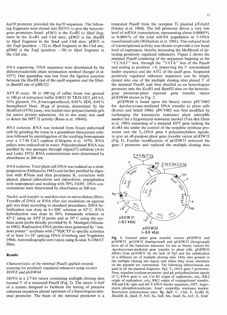
pGDW44 is based upon the binary vector pPCV002 for Agrobacterium-mediated DNA transfer to plant cells (Koncz and Schell 1986). pPCV002 was first modified by exchanging the kanamycin resistance plant selectable marker for a hygromycin resistance marker (Van den Elzen et al. 1985) consisting of a mutated HPT gene lacking the EcoRI site under the control of the nopaline synthase pro- moter and the TL-DNA gene 4 polyadenylation signals, to give an all-purpose plant gene transfer vector pGDW31 (Fig. 1). Further modification of pGDW31 removed the gene 5 promoter and replaced the multiple cloning sites
E H X B g P S a v
Fig. 1. General plant gene transfer vectors pGDW31 and pGDW32. pGDW31 (background) and pGDW32 (foreground) have all of the functions necessary for use as binary vectors for Agrobacteriwn-mediated gene transfer to plant cells. pGDW32 differs from pGDW31 by the lack of Pg5 and the substitution of a different set of multiple cloning sites. Only sites present in the multiple cloning site region and where they occur elsewhere on the plasmid are represented. The following abbreviations are used in all the plasrnid diagrams. Pg5, TL-DNA gene 5 promoter; Pnos, nopaline synthase promoter; pad g4, polyadenylation signals of TL-DNA gene 4; ori, Col El origin of replication; ori,, RK2 origin of replication; oriT, RK2 origin of conjugational transfer; RB and LB, right and left T-DNA border sequences; HPT, hygro- rnycin phosphotransferase; Amp', ampicillin resistance marker. Restriction endonuclease sites: Bg, Bgn; C, ClaI; E, EcoRI H, HindIII; K, Q n I ; P, PstI; Sa, Sun; Sm, SmaI; Ss, SstI; X, XbaI
1' DOVE
Fig. 2a and b. Representations of the vectors DOVE and pGDW44. a DOVE is an insertion of the multiple cloning sites 5' of the minimal PiaaH (expanded region) into pUC9 at the BmnHI and EcoRI sites. The ATG at position 1 is the initiation d o n for iaaH and the last G presented is the first G of the BamHI recogni- tion site. b pGDW44 is the minimal PiaaH fused to an NPTZZ gene with termination signals from the iaaH gene cloned into pGDW31. All heterologous promoters constructed on DOVE can be rapidly recloned into pGDW44 as EcoRI-BanzHI fragments. Abbreviations: B, BmHI site; Sp, SphI site; P W , iaaH promot- er; pad iaaH, polyadenylation sites of the iaaH gene; NPTZZ, neo- mycin phosphotransferase gene. Other abbreviations as in Fig. 1
with a pUC19-related set of sites to give pGDW32 shown in Fig. 1.
A second chimaeric gene fusing the minimal PiaaH to the NPT N reporter gene terminated by the polyadenylation signals from the iaaH gene was added to pGDW31. This resulted in vector pGDW44 (Fig. 2). The fusion of the mini- mal promoter sequence and the NPT II gene of pKm109/9 (Reiss et al. 1984a) created a 17 amino acid addition to the amino-terminal end of the NPT I1 protein and appar- ently did not adversely affect the enzyme activity.
When pGDW44 carrying the minimal PiaaH alone fused to the NPT N gene was tested for the level of NPT I1 expression in transformed tobacco callus tissue, the basal level of expression was consistently low (Fig. 3), approxi- mately 100- to 200-fold lower than with an intact 1' promot- er pelten et al. 1984, data not shown). Regenerated to- ba rn plants had nearly undetectable levels of NPT I1 activ- ity in roots, stem and leaves (data not shown). This reduced level of expression also showed that the TL-DNA gene 5 promoter located 2.5 kb upstream of the minimal PiaaH had a minor effect on the level of expression. When the
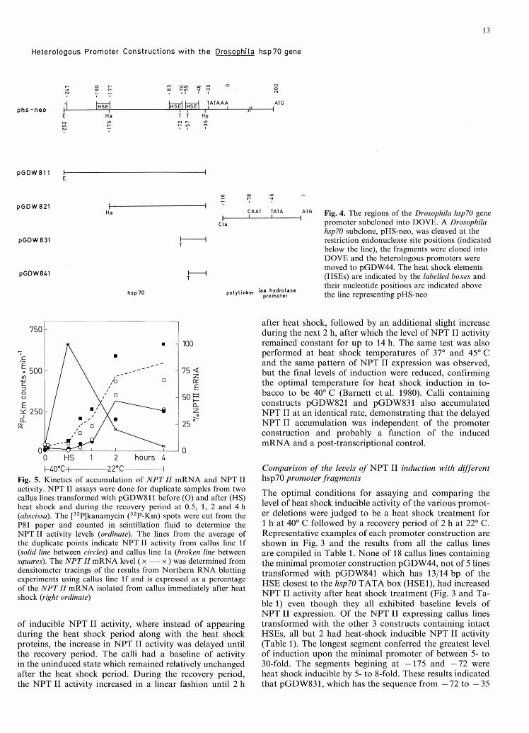
Fig. 3- NPT I1 activity from tobacco callus lines before and aRer heat shock. Three representative callus lines containing pGDW811 a, pGDW821 b, pGDW831 o, pGDW841 d and pGDW44 e were kept at 22O C or heat shocked and allowed to rewver for 2 h. WPT I1 activity was assayed from each sample as described in Materials and methods. Column heading C indi- cates the control samples and H indicates the heat-shocked samples
same chimaeric gene was transfer4 by the co-integrate plant vector pGV3851 (Zambryski et al. 1984) into tobacco, the expression in five independent transformants was com- parable to that seen in callus tissue; thus the truncated PiaaH was unaffected by the high cytokinin levels present in the teratoma tissue (data not shown). Together these results indicated the suitability of the minimal PiaaN for use as a non-tissue-spedic and minimal promoter for test- ing transcriptional regulatory sequences in plants.
Heat shock induced NPT I1 expression by heterologous promoters witk hsp70 5' upstream sequences
Four DNA fragments from the Drosophila hspFt? promoter beginning at the 5' positions -252, -175, -72 and -57 and all ending at position -35, just 5 bp upstream of the hsp70 TATA box were cloned into DOVE such that the 3' end of each hsp70 sequence was 79 bp from the PiaaH TATA box (Fig. 4). The heterologous promoters were then fused to the reporter gene NPT II on pGDW44 giving plas- mids pGDW811, pGDW821, pGDW831 and pGDW841 carrying respectively three, two, one and a fraction of the HSEs.
Transformed tobacco calli containing one to three cop- ies of the chimaeric gene (data not shown) were propagated and analysed individually to test for heat shock induction of the NPTIZ gene. Uninduced calli were first screened for NPT I1 activity and two lines transformed with pGDW811 were tested for heat shock inducible activity be- fore and after heat shock at 40' C for 1 h, and throughout the recovery period at 22" C.
The graph depicted in Fig. 5 shows the NPT I1 activity levels of the two callus lines measured by scintillation count- ing of the [3zP]kanamycin assayed in duplicate samples at different times. Both callus lines displayed a similar pattern
Heterologous Promoter Constructions with the Drosophila hsp70 gene
IHSEI IHSE~ i n s ~ l TATAA4 ATG phs-neo 1 /H
E Ha T T Hp
polllinker iaa hydrolase promoter
S a) cD 7
C -, 7
I I ' Ha , CAAT , TATA , ,"lG Fig. 4. The regions of the Drosophila hsp70 gene
Cia promoter subcloned into DOVE. A Drosophila hsp70 subclone, pHs-neo, was cleaved at the restriction endonuclease site positions (indicated below the line), the fragments were cloned into DOVE and the heterologous promoters were moved to pGDW44. The heat shock elements (HSEs) are indicated by the labelled boxes and their nucleotide positions are indicated above the line representing pHs-neo
- 0 HS 1 2 hours 4 t - 4 0 ° C ~ 2 2 0 C +
Fig. 5. Kinetics of accumulation of NPTZZ mRNA and NPT I1 activity. NPT I1 assays were done for duplicate samples from two callus lines transformed with pGDW811 before ( 0 ) and after (HS) heat shock and during the recovery period at 0.5, 1, 2 and 4 h (abscissa). The [32P]kanamycin (32P-Km) spots were cut from the P81 paper and counted in scintillation fluid to determine the NPT I1 activity levels (ordinate). The lines from the average of the duplicate points indicate NPT I1 activity from callus line If (solid line between circles) and callus line l a (broken line between squares). The N P T II mRNA level ( x - x ) was determined from densitometer tracings of the results from Northern RNA blotting experiments using callus line If and is expressed as a percentage of the N P T II mRNA isolated from callus immediately after heat shock (right ordinate)
of inducible NPT I1 activity, where instead of appearing during the heat shock period along with the heat shock proteins, the increase in NPT I1 activity was delayed until the recovery period. The calli had a baseline of activity in the uninduced state which remained relatively unchanged after the heat shock period. During the recovery period, the NPT I1 activity increased in a linear fashion until 2 h
after heat shock, followed by an additional slight increase during the next 2 h, after which the level of NPT I1 activity remained constant for up to 14 h. The same test was also performed at heat shock temperatures of 37" and 45" C and the same pattern of NPT I1 expression was observed, but the final levels of induction were reduced, confirming the optimal temperature for heat shock induction in to- bacco to be 40" C (Barnett et al. 1980). Calli containing constructs pGDW821 and pGDW831 also accumulated NPT I1 at an identical rate, demonstrating that the delayed NPT I1 accumulation was independent of the promoter construction and probably a function of the induced mRNA and a post-transcriptional control.
Comparison of the levels of NPT I1 induction with dijJerent hsp70 promoter fragments
The optimal conditions for assaying and comparing the level of heat shock inducible activity of the various promot- er deletions were judged to be a heat shock treatment for 1 h at 40" C followed by a recovery period of 2 h at 22" C . Representative examples of each promoter construction are shown in Fig. 3 and the results from all the callus lines are compiled in Table 1. None of 18 callus lines containing the minimal promoter construction pGDW44, not of 5 lines transformed with pGDW841 which has 13/14 bp of the HSE closest to the hsp70 TATA box (HSEl), had increased NPT I1 activity after heat shock treatment (Fig. 3 and Ta- ble 1) even though they all exhibited baseline levels of NPT I1 expression. Of the NPT I1 expressing callus lines transformed with the other 3 constructs containing intact HSEs, all but 2 had heat-shock inducible NPT I1 activity (Table 1). The longest segment conferred the greatest level of induction upon the minimal promoter of between5 to 30-fold. The segments begining at -175 and -72 were heat shock inducible by 5- to 8-fold. These results indicated that pGDW831, which has the sequence from -72 to -35
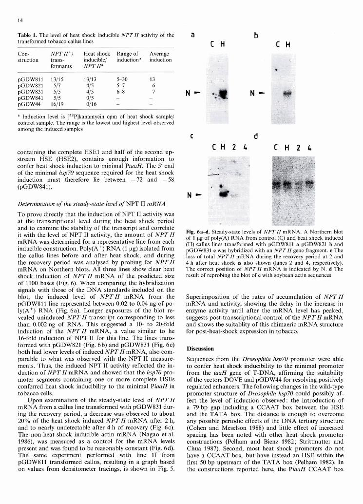
Table 1. The level of heat shock inducible NPT II activity of the transformed tobacco callus lines
Con- struction
NPT II+/ Heat shock Range of Average trans- inducible/ inductiona induction formants NPT II"
" Induction level is [32P]kanamycin cpm of heat shock sample/ control sample. The range is the lowest and highest level observed among the induced samples
containing the complete HSEl and half of the second up- stream HSE (HSEZ), contains enough information to confer heat shock induction to minimal PiaaH. The 5' end of the minimal hsp70 sequence required for the heat shock induction must therefore lie between -72 and -58 (pGDW841).
Determination of the steady-state level of NPT I1 mRNA
To prove directly that the induction of NPT I1 activity was at the transcriptional level during the heat shock period and to examine the stability of the transcript and correlate it with the level of NPT I1 activity, the amount of NPT ZZ mRNA was determined for a representative line from each inducible construction. Poly(A+) RNA (1 pg) isolated from the callus lines before and after heat shock, and during the recovery period was analysed by probing for NPT ZZ mRNA on Northern blots. All three lines show clear heat shock induction of NPTZZ mRNA of the predicted size of 1100 bases (Fig. 6). When comparing the hybridization signals with those of the DNA standards included on the blot, the induced level of NPTZZ mRNA from the pGDW811 line represented between 0.02 to 0.04 ng of po- ly(A+) RNA (Fig. 6a). Longer exposures of the blot re- vealed uninduced NPT ZZ transcript corresponding to less than 0.002 ng of RNA. This suggested a 10- to 20-fold induction of the NPTZZ mRNA, a value similar to he 16-fold induction of NPT I1 for this line. The lines trans- formed with pGDW821 (Fig. 6b) and pGDW831 (Fig. 6c) both had lower levels of induced NPT ZZ mRNA, also com- parable to what was observed with the NPT I1 measure- ments. Thus, the induced NPT I1 activity reflected the in- duction of NPT II mRNA and showed that the hsp70 pro- moter segments containing one or more complete HSEs conferred heat shock inducibility to the minimal PiaaH in tobacco cells.
Upon examination of the steady-state level of NPT ZZ mRNA from a callus line transformed with pGDW831 dur- ing the recovery period, a decrease was observed to about 20% of the heat shock induced NPT ZZ mRNA after 2 h, and to nearly undetectable after 4 h of recovery (Fig. 6c). The non-heat-shock inducible actin mRNA (Nagao et al. 1986), was measured as a control for the mRNA levels present and was found to be reasonably constant (Fig. 6d). The same experiment performed with line If from pGDW811 transformed callus, resulting in a graph based on values from densitometer tracings, is shown in Fig. 5.
Fig. 6a-d. Steady-state levels of NPT 11 mRNA. A Northern blot of 1 pg of poly(A) RNA from control (C) and heat shock induced (H) callus lines transformed with pGDW811 a pGDW821 b and pGDW831 c was hybridized with an NPT 11 gene fragment. c The loss of total NPT 11 mRNA during the recovery period at 2 and 4 h after heat shock is also shown (lanes 2 and 4, respectively). The correct position of NPT II mRNA is indicated by N. d The result of reprobing the blot of c with soybean actin sequences
Superimposition of the rates of accumulation of NPT ZZ mRNA and activity, showing the delay in the increase in enzyme activity until after the mRNA level has peaked, suggests post-transcriptional control of the NPT ZZ mRNA and shows the suitablity of this chimaeric mRNA structure for post-heat-shock expression in tobacco.
Discussion
Sequences from the Drosophila hsp70 promoter were able to confer heat shock inducibility to the minimal promoter from the iaaH gene of T-DNA, affirming the suitability of the vectors DOVE and pGDW44 for resolving positively regulated enhancers. The following changes in the wild-type promoter structure of Drosophila hsp70 could possibly af- fect the level of induction observed: the introduction of a 79 bp gap including a CCAAT box between the HSE and the TATA box. The distance is enough to overcome any possible periodic effects of the DNA tertiary structure (Cohen and Meselson 1988) and little effect of increased spacing has been noted with other heat shock promoter constructions (Pelham and Bienz 1982; Strittrnatter and Chua 1987). Second, most heat shock promoters do not have a CCAAT box, but have instead an HSE within the first 50 bp upstream of the TATA box (Pelham 1982). In the constructions reported here, the PiaaH CCAAT box
did not prevent the heat shock response, consistent with an example from the Xenopus hsp70 gene which contains a CCAAT box found necessary for heat shock inducible transcription (Bienz and Pelham 1986).
The profile of induction of NPT II mRNA and enzyme activity presented a clear demonstration of the role of post- transcriptional regulation in gene expression. The NPT 11 mRNA, while properly induced by heat shock, was very inefficiently translated, if at all, during the heat shock phase, as little or no corresponding increase in the level of NPT I1 could be detected then. It is unlikely that the NPT I1 is synthesized and then rapidly inactivated or de- graded in tobacco, since callus lines expressing the same NPT II fusion constitutively had no change in levels of NPT I1 after heat shock (unpublished results). Thus the translation of the chimaeric mRNA is most probably re- pressed.
This is observed for many of the mRNAs translated at normal growth temperatures in Drosophila (McKenzie et al. 1975) and plant cells (Key et al. 1981 ; Barnett et al. 1980), where heat shock represses the translation of the mRNAs and upon recovery the cell resumes synthesizing the proteins from pre-existing mRNAs (Storti et al. 1980; Lindquist 1981; Key et al. 1981). In Drosophila cells, the first 95 nucleotides of the 5' untranslated region of the hsp70 mRNA can shift the translation of a non-heat-shock mRNA to the heat shock period, demonstrating that a heat shock 5' mRNA leader has a particular structure which allows heat shock mRNAs to be translated during heat stress (Klemenz et al. 1985). Thus plant heat shock mRNAs may also require a specific leader sequence to bestow trans- latability on an mRNA during heat shock. These sequences are probably different from those of Drosophila hsp70, be- cause the NPT II mRNA with the Drosophila 5' leader is also poorly translated during heat shock (Spena et al. 1985). This translational control represents one divergent feature of heat shock regulation in plants and animals where a similar function is now seemingly directed by a different structure.
The 37 bp hsp70 sequence containing a complete HSE1, as similarly observed in stably transformed Drosophila cells (Dudler and Travers 1984; Xiao and Lis 1988), confers heat shock inducibility to a gene. Examination of a number of synthetic promoters structured after the hsp70 promoter showing the importance of positions bordering the HSE 14 bp dyad, led to a proposed expanded consensus sequence which is a dimer of a 10 bp unit centred over the original HSE (Xiao and Lis 1988). As evidence for the 10-mer basic unit, a synthesized sequence containing one and a half 14 bp HSEs, or in other terms a trimer of the 10 bp unit, is par- tially heat shock inducible. That sequence corresponds in structure to the 37 bp segment used in pGDW831 which contains the 3' half of HSE2 and the complete HSEl and is similarly partially heat shock inducible. In comparison, the two overlapping HSEs in the 36 bp sequence of the soybean hs6871 gene promoter which can-render genes heat shock inducible in tobacco (Baumann et al. 1987; Strittmat- ter and Chua 1987) also represent one and a half HSEs. However, when viewed in terms of a 10 bp element, as sug- gested on observing overlapping HSE structures (Xiao and Lis 1988), the 36 bp soybean sequence is a trimer of that element. The structural similarity between the Drosophila and soybean sequences which are able to confer partial heat shock inducibility to a gene is better described as a
trimer of the 10 bp basic unit and thus, as in Drosophila, the dimer of the 10 bp unit may better describe an HSE in tobacco.
Unlike the situation in stably transformed Drosophila though, where two complete HSEs can confer full heat shock inducibility to a gene (Dudler and Travers 1984; Xiao and Lis 1988), the 120 bp sequence with both HSEl and HSE2 on pGDW821 results in no further increase in the level of induction in tobacco. Models for activation of the hsp70 promoter based on the genetic evidence and the stronger affinity of HSTF for HSEl than for HSE2, hy- pothesize that the two HSEs work cooperatively to mediate full heat shock induction (Topol et al. 1985). This discrep- ancy suggests possible nuances in the structure, interaction and roles of the HSE and HSTF in the mechanism of heat shock induced gene transcription of tobacco and Drosophi- la. Indeed, the analysis of the soybean heat shock promoter points out the need for an upstream non-HSE enhancer-like element for maximal heat shock induction in tobacco (Bau- mann et al. 1987).
The ability of the one and a half HSE sequence from the Drosophila hsp70 promoter to confer partial heat shock inducible transcription to a gene in both Drosophila and tobacco, corroborates the high degree of conservation of function of the HSE between the plant and animal kingdoms. This conservation of structure and function most likely extends to the HSTF as well, since the Drosophila HSE is presumably acting as the binding site for the tobacco HSTF. The eventual isolation of the tobacco HSTF should further demonstrate the extent of the conservation and clar- ify possible differences between Drosophila and tobacco heat shock induced gene transcription.
Acknowledgements. The authors would like to thank A. Spena for invaluable material and advice needed for the completion of the project. I. Czaja gave excellent technical assistance and undergrad- uate students D. Priifer, C. Schipmann and C. Reichel made fine contributions. D.W. was supported by a Max-Planck Society fel- lowship.
References
Ashburner M, Bonner JJ (1979) The induction of gene activity in Drosophila by heat shock. Cell 17:241-254
Aviv H, Leder P (1972) Purification of biologically active globin messenger RNA by chromatography on oligothymidylic acid- cellulose. Proc Natl Acad Sci USA 69: 1408-1412
Barnett T, Altschuler M, McDaniel CN, Mascarenhas J (1980) Heat shock induced proteins in plant cells. Dev Genet 1 : 331-340
Baumann G, Raschke E, Bevan M, Schoffl F (1987) Functional analysis of sequences required for transcriptional activation of a soybean heat shock gene in transgenic tobacco plants. EMBO J 6:1161-1166
Bienz M, Pelham HRB (1986) Heat shock regulatory elements function as an inducible enhancer in the Xenopus hsp70 gene and when linked to a heterologous promoter. Cell 45: 753760
Bradford M (1976) A rapid and sensitive method for quantification of microgram quantities of protein utilizing the principle of protein-dye binding. Anal Biochem 72 : 248-254
Chirgwin JM, Przybyla AE, MacDonald RJ, Rutter WJ (1979) Isolation of biologically active ribonucleic acid from sources enriched in ribonuclease. Biochemistry 18 : 5294-5299
Cohen RS, Meselson M (1988) Periodic interactions of heat shock transcriptional elements. Nature 332: 856858
Czarnecka E, Gurley WB, Nagao RT, Mosquera LA, Key JL
(1985) DNA sequence and transcript mapping of a soybean gene encoding a small heat shock protein. Proc Natl Acad Sci USA 82: 37263730
Dellaporta SL, Wood J, Hicks JB (1983) A plant DNA miniprepar- ation: version 11. Plant Mol Biol Rep 1 : 19-21
De Vos G, De Beuckeleer M, Van Montagu M, Schell J (1981) Restriction endonuclease mapping of the octopine tumor-in- ducing plasmid pTiAch5 of Agrobacterium tumefaciens. Plasmid 6: 249-253
Dudler R, Travers AA (1984) Upstream elements necessary for optimal function of the hsp70 in transformed flies. Cell 38:391-398
Feinberg AP, Vogelstein B (1984) A technique for radiolabeling DNA restriction endonculease fragments to high specific activi- ty. Anal Biochem 137: 266267
Gielen J, De Beuckeleer M, Seurinck J, Deboeck F, DeGreve H, Lemmers M, Van Montagu M, Schell J (1984) The complete nucleotide sequence of the TL-DNA of the Agrobacterium tu- mefaciens plasmid pTiAch5. EMBO J 3: 835-846
Gritz L, Davies J (1983) Plasmid-encoded hygromycin B resistance: the sequence of hygromycin B phosphotransferase gene and its expression in Escherichia coli and Saccharomyces cerevisiae. Gene 25: 179-188
Hanahan D (1983) Studies on transformations of Escherichia coli with plasmids. J Mol Biol 166: 557-580
Horsch RB, Fry JE, Hoffmann NL, Eichholtz D, Rogers SG, Fra- ley RT (1985) A simple and general method for transferring genes into plants. Science 227: 1229-1231
Key JL, Lin CY, Chen YM (1981) Heat shock proteins of higher plants. Proc Natl Acad Sci USA 78:35263530
Klemenz R, Hultmark D, Gehring WJ (1985) Selective translation of heat shock mRNA in'Drosophila melanogaster depends on sequence information in the leader. EMBO J 4:2053-2060
Koncz Cs, Schell J (1986) The promoter of TL-DNA gene 5 con- trols the tissue-specific expression of chimaeric genes carried by a novel type of Agrobacterium binary vector. Mol Gen Genet 204: 383-396
Lindquist S (1981) Regulation of protein synthesis during heat shock. Nature 293 : 31 1-314
Linsmaier EM, Skoog F (1965) Organic growth factor require- ments of tobacco tissue cultures. Physiol Plantarum 18: 100-127
Maniatis T, Fritsch EF, Sambrook J (1982) Molecular cloning: a laboratory manual. Cold Spring Harbor Laboratory, Cold Spring Harbor, NY
McKenzie SL, Henikoff S, Meselson M (1975) Localization of RNA from heat-induced polysomes at puff sites in Drosophila melanogaster. Proc Natl Acad Sci USA 72: 11 17-1 121
Miller J (1972) Experiments in molecular genetics. Cold Spring Harbor Laboratory, Cold Spring Harbor, NY
Mirault M-E, Southgate R, Delwart E (1982) Regulation of heat- shock genes: a DNA sequence upstream of Drosophila hsp70 genes is essential for their induction in monkey cells. EMBO J 1:1279-1285
Murashige T, Skoog F (1962) A revised medium for rapid growth and bio-assays with tobacco tissue cultures. Physiol Plantarum 15 : 473497
Nagao RT, Kimpel JA, Vierling E, Key JL (1986) The heat shock response: a comparative analysis. Oxford SUN Plant Mol Cell Biol 3 : 384-438
Pelham H (1982) A regulatory upstream promoter element in the Drosophila hsp7O heat-shock gene. Cell 30: 517-528
Pelham H (1985) Activation of heat-shock genes in eukaryotes. Trends Genet 3 : 31-35
Pelham H, Bienz M (1982) A synthetic heat-shock promoter ele- ment confers heat-inducibility on the herpes simplex virus thy- midine kinase gene. EMBO J 1 : 1473-1477
Reiss B, Sprengel R, Schaller H (1984a) Protein fusions with the kanamycin resistance gene from transposon Tn5. EMBO J 3 : 331 7-3322
Reiss B, Sprengel R, Will H, Schaller H (1984b) A new sensitive
method for qualitative and quantitative assay of neomycin phosphotransferase in crude cell extracts. Gene 30 : 21 1-21 8
Rochester DE, Winer JA, Shah DM (1986) The structure and ex- pression of maize genes encoding the major heat shock protein, hsp70. EMBO J 5:451-458
Sanger F, Nicklen S, Coulson AR (1977) DNA sequencing with chain terminating inhibitors. Proc Natl Acad Sci USA 74 : 5463-5467
Schlesinger MJ, Ashburner M, Tissieres A (1982) Heat shock from bacteria to man. Cold Spring Harbor Laboratory, Cold Spring Harbor, NY
Schoffl F, Raschke E, Nagao R (1984) The DNA sequence analysis of soybean heat-shock genes and identification of possible regu- latory promoter elements. EMBO J 3:2491-2497
Simon R, Priefer U, Piihler A (1983) A broad host range mobiliza- tion system for in vitro genetic engineering; transposon muta- genesis in Gram-negative bacteria. Bio/technology 1 : 784-791
Sorger PK, Pelham HRB (1988) Yeast heat shock factor is an essential DNA-binding protein that exhibits temperature-de- pendent phosphorylation. Cell 54:85>864
Spena A, Hain R, Ziewogel U, Saedler H, Schell J (1985) Construc- tion of a heat-inducible gene for plants. Demonstration of heat- inducible activity of the Drosophila hsp70 promoter in plants. EMBO J 4: 2739-2743
Storti RV, Scott MP, Rich A, Pardue ML (1980) Translational control of protein synthesis in response to heat shock in D. melanogaster cells. Cell 22: 825834
Strittmatter G, Chua N-H (1987) Artifical combination of two cis-regulatory elements generates a unique pattern of expression in transgenic plants. Proc Natl Acad Sci USA 84: 89868990
Topol J, Ruden DM, Parker CS (1985) Sequences required for in vitro transcriptional activation of a Drosophila hsp70 gene. Cell 42: 527-537
Van den Elzen PJM, Townsend J, Lee KY, Bedbrook JR (1985) A chimaeric hygromycin resistance gene as a selectable marker in plant cells. Plant Mol Biol 5 : 299-302
Velten J, Velten L, Hain R, Schell J (1984) Isolation of a dual plant promoter fragment from the Ti plasmid of Agrobacterium twnefaciens. EMBO J 3 : 2723-2730
Vieira J, Messing J (1982) The pUC plasmids, an M13mp7-derived system for insertion mutagenesis and sequencing with synthetic universal primers. Gene 19 : 259-268
Wiederrecht G, Shuey DJ, Kibbe WA, Parker CS (1987) The Sac- charomyces and Drosophila heat shock transcription factors are identical in size and DNA binding properties. Cell 48:507-515
Wiederrecht G, Seto D, Parker SS (1988) Isolation of the gene encoding the S. cerevisiae heat shock transcription factor. Cell 54:841-853
Willmitzer L, Otten L, Simons G, Schmalenbach W, Schroder J, Schroder G, van Montagu M, de Vos G, Schell J (1981) Nuclear and polysomal transcripts of T-DNA in octopine crown gall suspension and callus cultures. Mol Gen Genet 182:255-262
Wu C (1984) Two protein-binding sites in chromatin implicated in the activation of heat-shock genes. Nature 309: 229-234
Xiao H, Lis JT (1988) Germline transformation used to define key features of heat-shock response elements. Science 239:1139-1142
Yanisch-Perron C, Vieira J, Messing J (1985) Improved MI3 clon- ing vectors and hot strains: nucleotide sequences of the M13mp18 and pUC 19 vectors. Gene 33 : 103-1 19
Zambryski P, Herrera-Estrella L, Block M, Van Montagu M, Schell J (1984) In: Hollaender A, Setlow J (eds) Genetic eng- ineering, principles and methods, vol 6. Plenum, New York, pp 253-278
Communicated by H. Saedler
Received March 31, 1989