Embed Size (px)
Citation preview

University of Groningen
Constitutional chromothripsis rearrangements involve clustered double-stranded DNA breaksand nonhomologous repair mechanismsKloosterman, Wigard P; Tavakoli-Yaraki, Masoumeh; van Roosmalen, Markus J; vanBinsbergen, Ellen; Renkens, Ivo; Duran, Karen; Ballarati, Lucia; Vergult, Sarah; Giardino,Daniela; Hansson, KerstinPublished in:Cell reports
DOI:10.1016/j.celrep.2012.05.009
IMPORTANT NOTE: You are advised to consult the publisher's version (publisher's PDF) if you wish to cite fromit. Please check the document version below.
Document VersionPublisher's PDF, also known as Version of record
Publication date:2012
Link to publication in University of Groningen/UMCG research database
Citation for published version (APA):Kloosterman, W. P., Tavakoli-Yaraki, M., van Roosmalen, M. J., van Binsbergen, E., Renkens, I., Duran,K., ... Cuppen, E. (2012). Constitutional chromothripsis rearrangements involve clustered double-strandedDNA breaks and nonhomologous repair mechanisms. Cell reports, 1(6), 648-655.https://doi.org/10.1016/j.celrep.2012.05.009
CopyrightOther than for strictly personal use, it is not permitted to download or to forward/distribute the text or part of it without the consent of theauthor(s) and/or copyright holder(s), unless the work is under an open content license (like Creative Commons).
Take-down policyIf you believe that this document breaches copyright please contact us providing details, and we will remove access to the work immediatelyand investigate your claim.
Downloaded from the University of Groningen/UMCG research database (Pure): http://www.rug.nl/research/portal. For technical reasons thenumber of authors shown on this cover page is limited to 10 maximum.
Download date: 28-05-2020

Cell Reports
Report
Constitutional Chromothripsis RearrangementsInvolve Clustered Double-Stranded DNA Breaksand Nonhomologous Repair MechanismsWigard P. Kloosterman,1,* Masoumeh Tavakoli-Yaraki,1 Markus J. van Roosmalen,1 Ellen van Binsbergen,1 Ivo Renkens,1
Karen Duran,1 Lucia Ballarati,2 Sarah Vergult,3 Daniela Giardino,2 Kerstin Hansson,4 Claudia A.L. Ruivenkamp,4
Myrthe Jager,1 Arie van Haeringen,4 Elly F. Ippel,1 Thomas Haaf,5 Eberhard Passarge,6 Ron Hochstenbach,1
Bjorn Menten,3 Lidia Larizza,2,7 Victor Guryev,8 Martin Poot,1 and Edwin Cuppen1,8,*1Department of Medical Genetics, University Medical Center Utrecht, Universiteitsweg 100, 3584 CG Utrecht, The Netherlands2Laboratorio di CitogeneticaMedica eGeneticaMolecolare, Centro di Ricerche e Tecnologie Biomediche, IRCCS-Istituto Auxologico Italiano,Via Ariosto 13, 20145 Milano, Italy3Center for Medical Genetics, Ghent University Hospital, De Pintelaan 185, B-9000 Ghent, Belgium4Leiden University Medical Center, Department of Clinical Genetics, Albinusdreef 2, 2333 ZA Leiden, The Netherlands5Institute of Human Genetics, Wurzburg University, 97074 Wurzburg, Germany6Institut fur Humangenetik, Universitatsklinikum Essen, Hufelandstr. 55, D-45122 Essen, Germany7University of Milan, San Paolo School of Medicine, Via A. di Rudinı 8, 20142, Milano, Italy8Hubrecht Institute and University Medical Center Utrecht, Uppsalalaan 8, 3584 CT Utrecht, The Netherlands*Correspondence: [email protected] (W.P.K.), [email protected] (E.C.)
DOI 10.1016/j.celrep.2012.05.009
SUMMARY
Chromothripsis represents a novel phenomenonin the structural variation landscape of cancergenomes. Here, we analyze the genomes of tenpatients with congenital disease who were prese-lected to carry complex chromosomal rearrange-ments with more than two breakpoints. The rear-rangements displayed unanticipated complexityresembling chromothripsis. We find that eightof them contain hallmarks of multiple clustereddouble-stranded DNA breaks (DSBs) on one ormore chromosomes. In addition, nucleotide resolu-tion analysis of 98 breakpoint junctions indicatesthat break repair involves nonhomologous or micro-homology-mediated end joining. We observed thatthese eight rearrangements are balanced or containsporadic deletions ranging in size between a fewhundred base pairs and several megabases. Thetwo remaining complex rearrangements did notdisplay signs of DSBs and contain duplications,indicative of rearrangement processes involvingtemplate switching. Our work provides detailedinsight into the characteristics of chromothripsisand supports a role for clustered DSBs drivingsome constitutional chromothripsis rearrangements.
INTRODUCTION
Recently, a new genome rearrangement phenomenon has
emerged from the study of highly complex rearrangements in
cancer genomes involving frequent oscillations between only
648 Cell Reports 1, 648–655, June 28, 2012 ª2012 The Authors
two copy number states (Stephens et al., 2011). This phenom-
enon has been termed chromothripsis and may have resulted
from localized shattering of one or several chromosomes and
subsequent assembly of resulting chromosomal pieces by
nonhomologous end joining (NHEJ). Further studies have
identified several instances of somatic chromothripsis rear-
rangements in a wide variety of cancer specimens (Magrangeas
et al., 2011; Kloosterman et al., 2011b; Molenaar et al., 2012;
Rausch et al., 2012; Maher and Wilson, 2012). We and others
have shown that de novo complex balanced translocations
in patients with developmental defects arose from a similar
chromosome shattering mechanism (Kloosterman et al.,
2011a; Chiang et al., 2012).
A replication-based model (MMBIR, microhomology-
mediated break-induced replication) has been proposed for
complex rearrangements in the germline that involve copy
losses and gains and contain microhomologous sequences at
breakpoint junctions (Hastings et al., 2009a, 2009b). Recently,
complex genomic rearrangements involving multiple and
region-focused copy number changes on one chromosome,
were also ascribed toMMBIR and it was speculated that MMBIR
could be an explanation for chromothripsis in cancer cells (Liu
et al., 2011). In support of this, Crasta et al. proposed that
chromosome pulverization could occur due to defects in DNA
replication and repair in micronuclei resulting from errors in
mitosis (Crasta et al., 2012).
Given the considerable controversy regarding the mecha-
nisms driving complex rearrangements in cancer and develop-
ment disorders, we here investigated the genetic architecture
of ten constitutional complex chromosomal rearrangements
(CCRs) using genome-wide long mate-pair sequencing, copy
number profiling, and breakpoint-junction sequencing.We found
that in eight cases the rearrangements are characterized by
multiple clustered double-stranded DNA breaks (DSBs) and
break repair was driven by end-joining mechanisms. These

findings suggest that these rearrangements involve chromo-
some shattering similarly as originally proposed for chromothrip-
sis in cancer (Stephens et al., 2011). The other two rearrange-
ments display distinct molecular characteristics. They do not
show signs of DSBs and contain complex copy number changes
including copy gains, resembling the chromothripsis-like chro-
mosome catastrophes caused by template switching (Hastings
et al., 2009a; Liu et al., 2011). Our findings suggest that chro-
mosome shattering by DSBs results in a distinct type of chro-
mothripsis rearrangements and exists next to processes
involving template switching.
RESULTS
Genomic Analysis of Ten Constitutional CCRsTo gain insight in the characteristics of complex constitutional
rearrangements, we here analyzed all breakpoints of ten con-
stitutional CCRs by mate-pair sequencing, array analysis, and
fusion-point sequencing (Tables S1, S2, and S3; Extended
Experimental Procedures). The CCRs were solely selected
based on prior evidence indicating more than two breakpoints
(de Pater et al., 2002; Giardino et al., 2006; Ballarati et al.,
2009; Poot et al., 2009) (Table S1). Hundreds of CCRs with
similar karyotypes as the ones selected here have been
described in literature (Astbury et al., 2004; Borg et al., 2005;
Gribble et al., 2005; De Gregori et al., 2007; Pellestor et al.,
2011). Eight of the CCRs had occurred de novo and two
CCRs were transmitted from a mother to her child (Table S1).
Based on mate-pair sequencing, we identified between 3 and
24 inter- and intrachromosomal breakpoint junctions per CCR.
The breakpoint junctions were independently validated by
PCR and capillary sequencing. Where possible, parents were
included in the validation assays (Table S3). For eight of the
CCRs (patients 1–8), plotting the breakpoint junctions onto the
reference genome revealed that closely spaced junction points
(i.e., the exact genomic coordinates on the reference genome
where the junctions occurred) form pairs on the reference
genome (Figures 1A and S1). We considered two junction points
as a pair when their relative distance was below 1 kb. Further-
more, one junction point in such a pair is connected at its tail
(T) side and the other junction point in a pair is connected at its
head (H) side. For clarity, we have schematically depicted the
concepts of breakpoint junction, orientation, and junction-point
pair in Figure 1B. The observation of a junction-point pair is most
simply explained by the repair of both free DNA ends resulting
from a DSB. We observed that the free DNA ends are repaired
by fusion to other (remote) chromosome fragments (which also
resulted from DSBs as suggested by the presence of junction-
point pairs), leading to complex inter- and intrachromosomal
rearrangements. Chromosomal breaks were occurring on one
(patients 5 and 8) or several chromosomes (five in patient 7)
and showed significantly more clustering along the chromosome
than expected by chance (p < 0.001, Figure S2). The derivative
chromosomes of patient 1 and patient 2 were reconstructed
based on the breakpoint junctions identified by the mate-pair
sequencing (Figures 1B and 2). The derivative chromosomes
were matching the karyotypes as previously established based
on cytogenetic analysis (Figure 2A) (Ballarati et al., 2009).
We performed copy number analysis of these eight CCRs to
determine whether losses or gains had occurred as a result of
the many inter- and intrachromosomal breakpoint junctions
(Figures 1A and S1; Table S1). We only observed deletions, but
no copy gains. Deletions are flanked by both inter- or intrachro-
mosomal breakpoint junctions (Figure 1, patients 1, 6, and 8).
This deletion architecture is different from a ‘‘classic deletion,’’
where both flanks of the deletion have been fused to each other.
Notably, junction-point pairs were not identified at deletion
borders (Figure 1; Figure S1, black arrows). The rearrangement
of patient 7 was copy neutral, despite a massive amount of
24 breakpoint junctions. For four de novo CCRs, we tested the
parental origin of the rearrangements by SNP-array analysis of
deletion intervals in the father, mother, and patient. This demon-
strated that these rearrangements had occurred on paternal
chromosomes (Table S4). The parents had normal karyotypes
and the age of these four fathers ranged between 29 and 41.
Our data suggest that multiple clustered double-stranded
DNA breaks triggered the eight CCRs described here. This led
to copy balanced rearrangements or rearranged chromosomes
with sporadic deletions.
Breakpoint Characteristics of Eight ConstitutionalChromothripsis RearrangementsTo further characterize the process of break repair in the eight
constitutional chromothripsis CCRs, we analyzed the sequences
of 98 breakpoint junctions by PCR-based capillary sequencing
(Figure 3A, Figure S3). Most junctions displayed homology of
1–7 nucleotides (45%) or absence of homology (29%). For
26% of junctions, we observed short insertions of 1–97 nontem-
plated nucleotides. For patient 8, we observed several long
nontemplated insertions of up to 97 bp at the breakpoint
junctions (Table S3). These inserted fragments did not match
with any sequence in the human reference genome and were
substantially longer than the nontemplated insertions for the
breakpoint junctions in the other patients. In addition, we did
observe multiple losses for patient 8 (Table S5), which is in line
with prior observations (Simsek and Jasin, 2010).
Based on the capillary sequencing of breakpoint junctions, we
could also derive the precise characteristics of 56 junction-point
pairs (Table S5; Figure 1B). Junction-point pairs are considered
to indicate the occurrence of DSBs. At some positions of DSBs,
gains and losses of one or several nucleotides occurred,
possibly following repair of staggered cuts or exonuclease
digestion of free DNA ends, respectively (Figures 3B and 3C)
(Gajecka et al., 2008; Lieber, 2008; Simsek and Jasin, 2010).
Together, our data suggest that break repair in the eight chro-
mothripsis CCRs involved canonical or noncanonical NHEJ (ex-
plaining the blunt fusions) or MMEJ (explaining the fusions with
microhomology) (McVey and Lee, 2008; Lieber, 2010; Simsek
and Jasin, 2010). However, we should note that break repair
mechanism cannot be precisely inferred from junction
sequences alone.
Based on the spacing between adjacent junction points on
the reference genome, we could infer the occurrence of
deletions of several kilobases in size that were too small to be
unequivocally detected by sequence coverage depth analysis
or array analysis (see Figure 1B for a schematic example). For
Cell Reports 1, 648–655, June 28, 2012 ª2012 The Authors 649

Figure 1. Overview of Breakpoint Junctions Involved in Constitutional CCRs
(A) Visualization of breakpoints junctions of complex rearrangements in patients 1, 6, 7, and 8 using circos plots. The colored lines indicate breakpoint junctions.
These are based on the mate-pair data and can be in either of four orientations: HH (head-head), red lines; TT (tail-tail), yellow lines; TH (tail-head), blue lines; HT
(head-tail), green lines (low coordinate to high coordinate). The outer circle displays the chromosome ideogram and the inner circle displays the copy number
profile based on SNP array data (patient 1 and 7) or depth of coverage analysis (patient 6 and 8). Red arrowheads indicate examples of junction-point pairs and
black arrowheads indicate examples of single junction-points flanking deletions.
(B) Schematic drawing indicating the terminology used in this paper. A breakpoint junction can occur in each of four different orientations between intra- or
interchromosomal genomic fragments: HH, TT, TH, and HT. The positions on the reference genome (i.e., genomic coordinate) where the breakpoint-junction
starts and ends are called junction points. Junction points form a pair if they are in close proximity on the reference genome (with a distance of <1,000 bp). In
addition, one junction point in a pair should have its T side connected to another fragment, while the other junction point should have its H side connected to
another fragment. Junction point (1) and junction point (2) form a pair. A pair of junction points is the result of a repaired DSB, because a DSB generates two free
DNA ends. Each of these ends can be fused to other chromosomal fragments, resulting in a junction-point pair. Break repair may result in erosion of the ends by
650 Cell Reports 1, 648–655, June 28, 2012 ª2012 The Authors

A
B
1
243412608
146558384
148132374
174865844
178122101
127121662
128505748
128904203
128906395
129010017
143596595
144357834
145496715
173101766
148132740
174865044
173636118
146556447
145628493
145600136
145627873
128907892
128911334
128506785
128903965
27746706
102531392
1
124075186
243412915
247932508
145581427
145497032
178960096
178703965
178966636
243199373
1
27746403
173635702
173101937
247933718
249250621
Chromosome15
der(1)
der(2)
der(15)
21
1
147801010
116898981
115819103
116899526
146364022
1
47912432
47918838
48084526
47918709
47912597
87670525
115818679
148534948
147801050
148535258
153605077
160123210
153621846
160123294
163141373
166014559
166011208
166016921
191897220
194718106
243199373
1
90127819
93369965
90127935
106187365
93604139
87503991
87669639
87503861
84218139
48086057
84217369
106187688
108216229
111879588
108223657
108221866
108222608
111881628
115169878
Chromosome13der(2)
der(8)
der(13)
82
Figure 2. Reconstruction of Derivative Chromosomes for Patients 1 and 2 Based on Mate-Pair Data
(A) Reconstructed derivative chromosomes for patient 1. The derivative chromosomes match the previous FISH and karyotyping analysis of this patient (Ballarati
et al., 2009).
(B) Reconstructed derivative chromosomes for patient 2. The reconstructed derivative chromosomes are in line with the karyotype as based on cytogenetic
analysis (Table S1). Derivative chromosomes were reconstructed based on the de novo breakpoint junctions that we identified in these patients, similarly as we
have demonstrated before (Kloosterman et al., 2011a). The chromosomal coordinates of junction points are based on themate-pair data as provided in Table S3.
Chromosomes are not drawn to scale.
example, in patient 1 we identified adjacent junction points that
suggested the occurrence of two deletions of 6,041 and
18,231 bp in size, respectively (Figure 3D). Quantitative (qPCR)
analysis showed that these two regions were indeed heterozy-
gously deleted in patient 1, but not in both parents (Figure 3D).
For all predicted deletion intervals we calculated normalized
sequence coverage depth relative to a common reference
sample (Table S5), which confirmed the deletions. Deletions
could arise from erosion of the free DNA ends resulting from
a DSB or be the result of two adjacent DSBs. Based on our
breakpoint data, we could not distinguish between these
two possibilities. All together, the dynamic size range of the
deletion spectrum (1 bp to 8.8 Mb) associated with DSBs adds
another level of complexity to constitutional chromothripsis
rearrangements.
Two Complex Rearrangements Involving ReplicativeProcessesFor two of the ten CCRs described here (patients 9 and 10), we
observed different breakpoint characteristics as compared to
the eight chromothripsis rearrangements described above.
One of these cases contained an inherited complex rearrange-
ment on chromosome X and the other involved four de novo
duplications on chromosomes 4, 8, and 14 (patients 9 and 10,
Table S1). Most notably, we found that the junction points for
these two rearrangements do not form pairs on the reference
exonuclease digestion or addition of extra nucleotides. See Figure 3B for a deta
junction point (4) do not form a pair, because they are not in close physical proxim
deleted, because it is not connected to other fragments at its H and T sides. H,
See also Figures S1 and S2 and Tables S1, S2, S3, and S4.
genome (Figure 4). Therefore, we regard it unlikely that a chromo-
some-shattering event as a result of simultaneous DSBs trig-
gered these two rearrangements. For patient 9, we identified
three unique breakpoint junctions that align with the deletion
and the two duplications (Figure 4A). For patient 10, we found
breakpoint junctions for three of the four de novo duplications.
Together with coverage depth analysis, these breakpoint junc-
tions showed that three tandem duplications had occurred
in patient 10. For the fourth duplication on chromosome 14, we
only found evidence based on depth of coverage analysis
using both parents as a reference (Figure 4B). The findings for
patients 9 and 10 can be explained by template-switching
events during replication, which is in line with the MMBIR
mechanism as previously reported (Hastings et al., 2009a; Liu
et al., 2011).
DISCUSSION
Here, we provide a comprehensive analysis of the breakpoints
of ten constitutional CCRs. We found that eight CCRs exhibited
hallmarks of multiple simultaneous double-stranded DNA
breaks on one or several chromosomes. The other two complex
rearrangements were likely caused by replication errors
involving template switching. The two classes of rearrange-
ments that we observed here likely represent distinct instances
of chromothripsis rearrangements as has been previously
iled overview of losses and gains at breakpoint regions. Junction point (3) and
ity. The genomic segment in between junction point (3) and junction point (4) is
head; T, tail.
Cell Reports 1, 648–655, June 28, 2012 ª2012 The Authors 651

AATCTCTGTATTATGGAGTGACATTGCACCACAAAATTCTGCCCATCCTAGAAGCAATGACACCCCAGTAGCAACAAACACAGCTA
AGCAATTACAAACATGTGTATCTCCTTGTTCTGTCAGCTGAGAGGGCCTAGAAGCAATGACACCCCAGTAGCAACAAACACAGCTA
AATCTCTGTATTATGGAGTGACATTGCACCACAAAATTCTGCCCATAAAAGGTGAGTCAAAAATCTTCAAAAGAAGCCGCACCTTC
breakpoint junction 2
chr2 (+)chr2 (-)
2:163141421(+) -> 2: 166014625(-) (breakpoint junction 53): blunt
AGCAATTACAAACATGTGTATCTCCTTGTTCTGTCAGCTGAGAGGGCCTAGTGCTCCTCATAGGCCATGGCCACCACAGCCAGGAT
AGCAATTACAAACATGTGTATCTCCTTGTTCTGTCAGCTGAGAGGGCCTAGAAGCAATGACACCCCAGTAGCAACAAACACAGCTT TGCTGAAATTCGGCCTCTTTTTGTTCTGCTTCTTCCAAGGTGGCCTGATTCTGCTCCTCATAGGCCATGGCCACCACAGCCAGGAT
breakpoint junction 1
chr2 (-)chr2 (+)
2:166014621(-) -> 2: 166011053(+) (breakpoint junction 59): blunt
*chr2 (-) AGCAATTACAAACATGTGTATCTCCTTGTTCTGTCAGCTGAGAGGGCCTAGAAGCAATGACACCCCAGTAGCAACAAACACAGCTA5 bp duplicated
A
>5
num
ber
of ju
nctio
ns
blunthomology (nt)insertion (nt)
1 2 3 4 5 >5 1 2 3 4 5
B
C
D
reference chr2 HT
reference chr2 HT
145581576 145599808
178960434 178966476
6041 bp
18231 bp
norm
aliz
ed c
opy
num
ber
0.0
0.2
0.4
0.6
0.8
1.0
1.2
F
deletion 1
deletion 2
M C F M C F M C F M C
deletion 1 deletion 2
amplicon 1
1:247933064 (T) 2:145628344 (H)
2:178703789 (H)2:145496803 (H)
amplicon 2 amplicon 1 amplicon 2
0
5
10
15
20
25
30
0 10 20 30 40 50
12
510
2050
100
200
breakpoint (index)
base
-pai
rs d
elet
ed (
red)
or
dupl
icat
ed (
gree
n)
Figure 3. Characteristics of Eight Constitutional Chromothripsis Rearrangements
(A) Bar diagram showing the numbers of bluntly ligated breakpoint junctions (blue bar), breakpoint junctions with one or more nucleotides microhomology (red
bars) and breakpoint junctions containing nontemplated insertions (green bars).
(B) Bar diagram showing the loss (red) and gain (red) of nucleotides caused by repair of 56 DSBs. The breakpoint erosion was inferred from capillary sequencing
reads across breakpoint junctions. Black bars indicate breaks with no losses or gains.
(C) Sequence composition of two breakpoint junctions that together create a junction-point pair on reference chromosome 2 (patient 2). The breakpoint-junction
sequencing reads were aligned to the reference genome. Sequence homology to normal genomic sequences is indicated in light blue and red. The genomic
sequence from chromosome 2 marked with an asterisk (*) shares homology with breakpoint junction 1 (green box) and breakpoint junction 2 (pink box). Both
stretches of homology overlap in the middle, showing that these 5 bp ended up in both breakpoint-junction sequences and are duplicated. Chromosomal
coordinates indicate the junction point for each of the two genomic sequences that form a breakpoint junction. Both coordinates are indicated in the genomic
sequence by a yellow box. The red coordinate for breakpoint junction 1 and the blue coordinate for breakpoint junction 2 together form a pair on reference
chromosome 2. The breakpoint-junction numbers correspond to the numbering in Table S3.
(D) qPCR analysis of two small de novo deletions (6,041 bp and 18,231 bp) on chr 2 in patient 2 that were predicted based on the mate-pair analysis. The bar
diagramdisplays the relative copy number for the amplified fragments within the deletion intervals in patient 2 and the father andmother. Error bars represent SDs
based on 2^(-DDCt) values from triplicate experiments. Red arrows indicate primer pairs. Breakpoint junctions flanking the deleted fragments are indicated by
genomic coordinates.
See also Figure S3 and Table S5.
proposed (Chen et al., 2011; Liu et al., 2012; Maher and Wilson,
2012).
By careful analysis of breakpoint junctions, we derived
precise molecular characteristics for the eight chromothripsis
rearrangements that were caused by multiple DSBs and
we conclude that (1) rearrangements can be confined to a
single chromosome (patients 5 and 8) or multiple chromo-
652 Cell Reports 1, 648–655, June 28, 2012 ª2012 The Authors
somes (e.g., five in patient 7), (2) rearrangements may lead to
sporadic deletions (including small deletions of only a few
hundred base pairs), but can also explain copy neutral rear-
rangements (patient 7), (3) DSBs are clustered, (4) break repair
involves nonhomologous or microhomology-mediated repair
mechanisms, and (5) rearrangements occur on paternal
chromosomes.
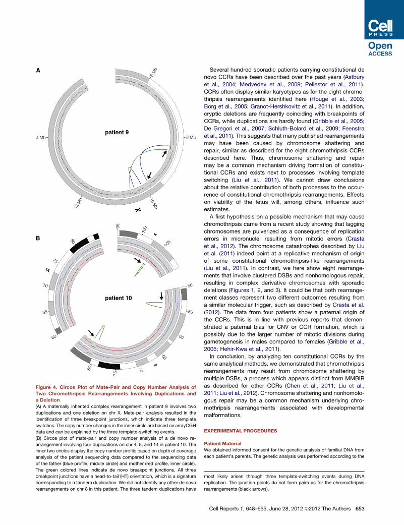
Figure 4. Circos Plot of Mate-Pair and Copy Number Analysis of
Two Chromothripsis Rearrangements Involving Duplications and
a Deletion
(A) A maternally inherited complex rearrangement in patient 9 involves two
duplications and one deletion on chr X. Mate-pair analysis resulted in the
identification of three breakpoint junctions, which indicate three template
switches. The copy number changes in the inner circle are based on arrayCGH
data and can be explained by the three template-switching events.
(B) Circos plot of mate-pair and copy number analysis of a de novo re-
arrangement involving four duplications on chr 4, 8, and 14 in patient 10. The
inner two circles display the copy number profile based on depth of coverage
analysis of the patient sequencing data compared to the sequencing data
of the father (blue profile, middle circle) and mother (red profile, inner circle).
The green colored lines indicate de novo breakpoint junctions. All three
breakpoint junctions have a head-to-tail (HT) orientation, which is a signature
corresponding to a tandem duplication. We did not identify any other de novo
rearrangements on chr 8 in this patient. The three tandem duplications have
Several hundred sporadic patients carrying constitutional de
novo CCRs have been described over the past years (Astbury
et al., 2004; Medvedev et al., 2009; Pellestor et al., 2011).
CCRs often display similar karyotypes as for the eight chromo-
thripsis rearrangements identified here (Houge et al., 2003;
Borg et al., 2005; Granot-Hershkovitz et al., 2011). In addition,
cryptic deletions are frequently coinciding with breakpoints of
CCRs, while duplications are hardly found (Gribble et al., 2005;
De Gregori et al., 2007; Schluth-Bolard et al., 2009; Feenstra
et al., 2011). This suggests that many published rearrangements
may have been caused by chromosome shattering and
repair, similar as described for the eight chromothripsis CCRs
described here. Thus, chromosome shattering and repair
may be a common mechanism driving formation of constitu-
tional CCRs and exists next to processes involving template
switching (Liu et al., 2011). We cannot draw conclusions
about the relative contribution of both processes to the occur-
rence of constitutional chromothripsis rearrangements. Effects
on viability of the fetus will, among others, influence such
estimates.
A first hypothesis on a possible mechanism that may cause
chromothripsis came from a recent study showing that lagging
chromosomes are pulverized as a consequence of replication
errors in micronuclei resulting from mitotic errors (Crasta
et al., 2012). The chromosome catastrophes described by Liu
et al. (2011) indeed point at a replicative mechanism of origin
of some constitutional chromothripsis-like rearrangements
(Liu et al., 2011). In contrast, we here show eight rearrange-
ments that involve clustered DSBs and nonhomologous repair,
resulting in complex derivative chromosomes with sporadic
deletions (Figures 1, 2, and 3). It could be that both rearrange-
ment classes represent two different outcomes resulting from
a similar molecular trigger, such as described by Crasta et al.
(2012). The data from four patients show a paternal origin of
the CCRs. This is in line with previous reports that demon-
strated a paternal bias for CNV or CCR formation, which is
possibly due to the larger number of mitotic divisions during
gametogenesis in males compared to females (Gribble et al.,
2005; Hehir-Kwa et al., 2011).
In conclusion, by analyzing ten constitutional CCRs by the
same analytical methods, we demonstrated that chromothripsis
rearrangements may result from chromosome shattering by
multiple DSBs, a process which appears distinct from MMBIR
as described for other CCRs (Chen et al., 2011; Liu et al.,
2011; Liu et al., 2012). Chromosome shattering and nonhomolo-
gous repair may be a common mechanism underlying chro-
mothripsis rearrangements associated with developmental
malformations.
EXPERIMENTAL PROCEDURES
Patient Material
We obtained informed consent for the genetic analysis of familial DNA from
each patient’s parents. The genetic analysis was performed according to the
most likely arisen through three template-switching events during DNA
replication. The junction points do not form pairs as for the chromothripsis
rearrangements (black arrows).
Cell Reports 1, 648–655, June 28, 2012 ª2012 The Authors 653

guidelines of the Medical Ethics Committee of the University Medical Center
Utrecht. See also Extended Experimental Procedures.
Preparation of Mate-Pair Libraries and SOLiD Sequencing
Mate-paired libraries were prepared and sequenced as described previously
(Kloosterman et al., 2011a, 2011b). See also Extended Experimental
Procedures.
Bioinformatic Analysis of Mate-Pair Reads
Bioinformatic analysis of mate-pair reads was performed as described previ-
ously (Kloosterman et al., 2011a). All custom developed tools are available
upon request from the authors. See also Extended Experimental Procedures.
Depth of Coverage Calculations
Depth of sequence coverage was analyzed by an in-house developed
Dynamic Window Approach for CNV detection (DWAC-seq: http://fedor21.
hubrecht.eu/dwac-seq). Briefly, BAM files for F3 and R3 tags were merged
using samtools and the tag densities of cases (test set) were compared to
the tag densities of controls (reference set, e.g., parent or unrelated individual).
For targeted depth of coverage calculations we generated a common
reference based on the concordant mate-pair tags from Patient1_mother,
Patient1_father, Patient10_mother, and Patient10_father. The normalized tag
count for predicted deletion intervals in patient genomes was measured
relative to the normalized tag count for the same intervals in the common
reference data set.
Capillary Sequencing of Breakpoints and Analysis
of Sequence Reads
Primers for sequencing the breakpoint junctions of structural variants were
designed using primer3 software. Where possible, we avoided repetitive
elements and designed the primers according to the orientations that were
indicated by the mate-pair tags. PCR was performed using Taq polymerase
(Invitrogen) with an elongation times of 1–2 min. PCR products were purified
from gel when needed. Sanger sequencing reads were aligned to the human
reference genome (GRCh37/hg19) using BLAST and BLAT software (http://
www.ensembl.org). Hits were analyzed manually to define the exact break-
point and breakpoint characteristics. PCR breakpoint assays were also used
to distinguish de novo junctions from inherited junctions.
qPCR Analysis of Copy Number Changes
For two deletions (18,231 bp and 6,041 bp) in patient 1 we designed primers for
qPCR analysis according to the Applied Biosystems HT7900 Real-Time PCR
system manual. We performed qPCR reactions using Sybr Green PCR master
mix (Applied Biosystems). For normalization, we used primers for GDF7 and
PRKD3. Ct values were normalized relative to Ct values for these two control
genes and relative to the Ct values of the father. We plotted 2^(-DDCt) values todisplay relative copy number changes. All reactions were performed in
triplicate.
SNP-Array Analysis
SNP-array analysis was performed using Infinium HumanHap300, Hu-
manCNV370-QuadV3 and HumanHap300v1 Genotyping BeadChips (Illumina,
Inc., San Diego, CA) and used according to the protocol of the manufacturer.
Data Visualization
Mate-pair and array data were visualized using Circos software and custom
R scripts (Krzywinski et al., 2009). We used the chr1, s1, e1, chr2, s2, e2,
and orientation(count) values from Table S3 for the colored links in the circos
plots. For orientation HH (or hh), we used chr1, s1, chr2, and s2 coordinates;
for orientation TT (or tt), we used chr1, e1, chr2, and e2 coordinates; for orien-
tation TH (or th), we used chr1, e1, chr2, and s2 coordinates; for orientation HT
(or ht), we used chr1, s1, chr2, and e2 coordinates.
Simulation of Random Breakpoints on Chromosomes 2 and 9
We simulated thousand sets of random double-stranded breaks for chromo-
somes 2 and 9. For chromosome 2, we used 16 random breaks per set, and
for chromosome 9 we used 12 random breaks per set. The average length
654 Cell Reports 1, 648–655, June 28, 2012 ª2012 The Authors
of the chromosomal fragments resulting from the simulated breaks was
calculated for each of the simulated sets. The first and last chromosomal
fragments (from the chromosome start to the first break and from the last
break to the chromosome end) were not included in the calculations.
ACCESSION NUMBERS
The ENA SRA accession number for the next-generation mate-pair se-
quencing is ERP001035. The NCBI GEO accession number for the Illumina
SNP array data is GSE37906.
SUPPLEMENTAL INFORMATION
Supplemental Information includes Extended Experimental Procedures, three
figures, and five tables and can be found with this article online at doi:10.1016/
j.celrep.2012.05.009.
LICENSING INFORMATION
This is an open-access article distributed under the terms of the Creative
Commons Attribution 3.0 Unported License (CC-BY; http://creativecommons.
org/licenses/by/3.0/legalcode).
ACKNOWLEDGMENTS
We thank the patients and their parents for participation in this study.We thank
Gijs van Haaften for critically reading the manuscript. The study was made
possible by primary funding of the Department of Medical Genetics of the
University Medical Center Utrecht to E.C.
Received: October 7, 2011
Revised: February 9, 2012
Accepted: May 14, 2012
Published online: June 14, 2012
REFERENCES
Astbury, C., Christ, L.A., Aughton, D.J., Cassidy, S.B., Fujimoto, A., Pletcher,
B.A., Schafer, I.A., and Schwartz, S. (2004). Delineation of complex chromo-
somal rearrangements: evidence for increased complexity. Hum. Genet.
114, 448–457.
Ballarati, L., Recalcati, M.P., Bedeschi, M.F., Lalatta, F., Valtorta, C., Bellini,
M., Finelli, P., Larizza, L., and Giardino, D. (2009). Cytogenetic, FISH and
array-CGH characterization of a complex chromosomal rearrangement
carried by a mentally and language impaired patient. Eur. J. Med. Genet. 52,
218–223.
Borg, K., Stankiewicz, P., Bocian, E., Kruczek, A., Obersztyn, E., Lupski, J.R.,
and Mazurczak, T. (2005). Molecular analysis of a constitutional complex
genome rearrangement with 11 breakpoints involving chromosomes 3, 11,
12, and 21 and a approximately 0.5-Mb submicroscopic deletion in a patient
with mild mental retardation. Hum. Genet. 118, 267–275.
Chen, J.-M., Ferec, C., and Cooper, D.N. (2011). Transient hypermutability,
chromothripsis and replication-based mechanisms in the generation of
concurrent clustered mutations. Mutat. Res. 750, 52–59.
Chiang, C., Jacobsen, J.C., Ernst, C., Hanscom, C., Heilbut, A., Blumenthal, I.,
Mills, R.E., Kirby, A., Lindgren, A.M., Rudiger, S.R., et al. (2012). Complex reor-
ganization and predominant non-homologous repair following chromosomal
breakage in karyotypically balanced germline rearrangements and transgenic
integration. Nat. Genet. 44, 390–397, S1.
Crasta, K., Ganem, N.J., Dagher, R., Lantermann, A.B., Ivanova, E.V., Pan, Y.,
Nezi, L., Protopopov, A., Chowdhury, D., and Pellman, D. (2012). DNA breaks
and chromosome pulverization from errors in mitosis. Nature 482, 53–58.
De Gregori, M., Ciccone, R., Magini, P., Pramparo, T., Gimelli, S., Messa, J.,
Novara, F., Vetro, A., Rossi, E., Maraschio, P., et al. (2007). Cryptic deletions

are a common finding in ‘‘balanced’’ reciprocal and complex chromosome re-
arrangements: a study of 59 patients. J. Med. Genet. 44, 750–762.
de Pater, J.M., Ippel, P.F., van Dam, W.M., Loneus, W.H., and Engelen, J.J.M.
(2002). Characterization of partial trisomy 9p due to insertional translocation
by chromosomal (micro)FISH. Clin. Genet. 62, 482–487.
Feenstra, I., Hanemaaijer, N., Sikkema-Raddatz, B., Yntema, H., Dijkhuizen,
T., Lugtenberg, D., Verheij, J., Green, A., Hordijk, R., Reardon, W., et al.
(2011). Balanced into array: genome-wide array analysis in 54 patients with
an apparently balanced de novo chromosome rearrangement and a meta-
analysis. Eur. J. Hum. Genet. 19, 1152–1160.
Gajecka, M., Gentles, A.J., Tsai, A., Chitayat, D., Mackay, K.L., Glotzbach,
C.D., Lieber, M.R., and Shaffer, L.G. (2008). Unexpected complexity at break-
point junctions in phenotypically normal individuals and mechanisms involved
in generating balanced translocations t(1;22)(p36;q13). Genome Res. 18,
1733–1742.
Giardino, D., Corti, C., Ballarati, L., Finelli, P., Valtorta, C., Botta, G., Giudici,
M., Grosso, E., and Larizza, L. (2006). Prenatal diagnosis of a de novo complex
chromosome rearrangement (CCR) mediated by six breakpoints, and a review
of 20 prenatally ascertained CCRs. Prenat. Diagn. 26, 565–570.
Granot-Hershkovitz, E., Raas-Rothschild, A., Frumkin, A., Granot, D., Silver-
stein, S., and Abeliovich, D. (2011). Complex chromosomal rearrangement in
a girl with psychomotor-retardation and a de novo inversion: inv(2)(p15;q24.2).
Am. J. Med. Genet. A. 155A, 1825–1832.
Gribble, S.M., Prigmore, E., Burford, D.C., Porter, K.M., Ng, B.L., Douglas,
E.J., Fiegler, H., Carr, P., Kalaitzopoulos, D., Clegg, S., et al. (2005). The
complex nature of constitutional de novo apparently balanced translocations
in patients presenting with abnormal phenotypes. J. Med. Genet. 42, 8–16.
Hastings, P.J., Ira, G., and Lupski, J.R. (2009a). A microhomology-mediated
break-induced replication model for the origin of human copy number varia-
tion. PLoS Genet. 5, e1000327.
Hastings, P.J., Lupski, J.R., Rosenberg, S.M., and Ira, G. (2009b). Mecha-
nisms of change in gene copy number. Nat. Rev. Genet. 10, 551–564.
Hehir-Kwa, J.Y., Rodrıguez-Santiago, B., Vissers, L.E., de Leeuw, N., Pfundt,
R., Buitelaar, J.K., Perez-Jurado, L.A., and Veltman, J.A. (2011). De novo copy
number variants associated with intellectual disability have a paternal origin
and age bias. J. Med. Genet. 48, 776–778.
Houge, G., Liehr, T., Schoumans, J., Ness, G.O., Solland, K., Starke, H., Claus-
sen, U., Strømme, P., Akre, B., and Vermeulen, S. (2003). Ten years follow
up of a boy with a complex chromosomal rearrangement: going from a > 5
to 15-breakpoint CCR. Am. J. Med. Genet. A. 118A, 235–240.
Kloosterman, W.P., Guryev, V., van Roosmalen, M., Duran, K.J., de Bruijn, E.,
Bakker, S.C.M., Letteboer, T., van Nesselrooij, B., Hochstenbach, R., Poot,
M., and Cuppen, E. (2011a). Chromothripsis as a mechanism driving complex
de novo structural rearrangements in the germline. Hum. Mol. Genet. 20,
1916–1924.
Kloosterman, W.P., Hoogstraat, M., Paling, O., Tavakoli-Yaraki, M., Renkens,
I., Vermaat, J.S., van Roosmalen, M.J., van Lieshout, S., Nijman, I.J., Roes-
singh, W., et al. (2011b). Chromothripsis is a common mechanism driving
genomic rearrangements in primary and metastatic colorectal cancer.
Genome Biol. 12, R103.
Krzywinski, M., Schein, J., Birol, I., Connors, J., Gascoyne, R., Horsman, D.,
Jones, S.J., and Marra, M.A. (2009). Circos: an information aesthetic for
comparative genomics. Genome Res. 19, 1639–1645.
Lieber, M.R. (2008). The mechanism of human nonhomologous DNA end
joining. J. Biol. Chem. 283, 1–5.
Lieber, M.R. (2010). The mechanism of double-strand DNA break repair by
the nonhomologous DNA end-joining pathway. Annu. Rev. Biochem. 79,
181–211.
Liu, P., Erez, A., Nagamani, S.C.S., Dhar, S.U., Ko1odziejska, K.E., Dharmad-
hikari, A.V., Cooper, M.L., Wiszniewska, J., Zhang, F., Withers, M.A., et al.
(2011). Chromosome catastrophes involve replication mechanisms generating
complex genomic rearrangements. Cell 146, 889–903.
Liu, P., Carvalho, C.M., Hastings, P., and Lupski, J.R. (2012). Mechanisms for
recurrent and complex human genomic rearrangements. Curr. Opin. Genet.
Dev. 22, 1–10.
Magrangeas, F., Avet-Loiseau, H., Munshi, N.C., and Minvielle, S. (2011).
Chromothripsis identifies a rare and aggressive entity among newly diagnosed
multiple myeloma patients. Blood 118, 675–678.
Maher, C.A., and Wilson, R.K. (2012). Chromothripsis and human disease:
piecing together the shattering process. Cell 148, 29–32.
McVey, M., and Lee, S.E. (2008). MMEJ repair of double-strand breaks
(director’s cut): deleted sequences and alternative endings. Trends Genet.
24, 529–538.
Medvedev, P., Stanciu, M., and Brudno,M. (2009). Computational methods for
discovering structural variation with next-generation sequencing. Nat.
Methods 6(11, Suppl), S13–S20.
Molenaar, J.J., Koster, J., Zwijnenburg, D.A., van Sluis, P., Valentijn, L.J., van
der Ploeg, I., Hamdi, M., van Nes, J., Westerman, B.A., van Arkel, J., et al.
(2012). Sequencing of neuroblastoma identifies chromothripsis and defects
in neuritogenesis genes. Nature 483, 589–593.
Pellestor, F., Anahory, T., Lefort, G., Puechberty, J., Liehr, T., Hedon, B., and
Sarda, P. (2011). Complex chromosomal rearrangements: origin and meiotic
behavior. Hum. Reprod. Update 17, 476–494.
Poot, M., van’t Slot, R., Leupert, R., Beyer, V., Passarge, E., and Haaf, T.
(2009). Three de novo losses and one insertion within a pericentric inversion
of chromosome 6 in a patient with complete absence of expressive speech
and reduced pain perception. Eur. J. Med. Genet. 52, 27–30.
Rausch, T., Jones, D.T.W., Zapatka, M., Stutz, A.M., Zichner, T., Weischen-
feldt, J., Jager, N., Remke, M., Shih, D., Northcott, P.A., et al. (2012). Genome
sequencing of pediatric medulloblastoma links catastrophic DNA rearrange-
ments with TP53 mutations. Cell 148, 59–71.
Schluth-Bolard, C., Delobel, B., Sanlaville, D., Boute, O., Cuisset, J.-M.,
Sukno, S., Labalme, A., Duban-Bedu, B., Plessis, G., Jaillard, S., et al.
(2009). Cryptic genomic imbalances in de novo and inherited apparently
balanced chromosomal rearrangements: array CGH study of 47 unrelated
cases. Eur. J. Med. Genet. 52, 291–296.
Simsek, D., and Jasin, M. (2010). Alternative end-joining is suppressed by the
canonical NHEJ component Xrcc4-ligase IV during chromosomal transloca-
tion formation. Nat. Struct. Mol. Biol. 17, 410–416.
Stephens, P.J., Greenman, C.D., Fu, B., Yang, F., Bignell, G.R., Mudie, L.J.,
Pleasance, E.D., Lau, K.W., Beare, D., Stebbings, L.A., et al. (2011). Massive
genomic rearrangement acquired in a single catastrophic event during cancer
development. Cell 144, 27–40.
Cell Reports 1, 648–655, June 28, 2012 ª2012 The Authors 655

![[3,3]-Sigmatropic rearrangements - Massey Universitygjrowlan/stereo2/lecture11.pdf · 123.702 Organic Chemistry Claisen rearrangements • One of the most useful sigmatropic rearrangements](https://img.pdfslide.net/doc/110x75/5adcada77f8b9a213e8bd8b0/33-sigmatropic-rearrangements-massey-gjrowlanstereo2lecture11pdf123702.jpg)


![Ruthenium-Catalyzed [3,3]-Sigmatropic Rearrangements …d-scholarship.pitt.edu/7918/1/JessiePenichMSThesis6_7_2011.pdf · Ruthenium-Catalyzed [3,3]-Sigmatropic Rearrangements of](https://img.pdfslide.net/doc/110x75/5b77f3947f8b9a47518e2fcb/ruthenium-catalyzed-33-sigmatropic-rearrangements-d-ruthenium-catalyzed.jpg)


![35 [2,3]-sigmatropic rearrangements](https://img.pdfslide.net/doc/110x75/55504042b4c905b2788b48e9/35-23-sigmatropic-rearrangements.jpg)


![36 [1,n]-sigmatropic rearrangements](https://img.pdfslide.net/doc/110x75/55504a55b4c9058f768b5083/36-1n-sigmatropic-rearrangements.jpg)

![34 [3,3]-sigmatropic rearrangements](https://img.pdfslide.net/doc/110x75/55503fb4b4c9058f768b4911/34-33-sigmatropic-rearrangements.jpg)