Embed Size (px)
Citation preview
Article
Johannes G.M
0022-2836/$ - see front m
Constitutive Nuclear Localization of anAlternatively Spliced Sirtuin-2 Isoform
. Rack, Magali R. VanLinde
n, Timo Lutter,Rein Aasland and Mathias ZieglerDepartment of Molecular Biology, University of Bergen, Postbox 7803, 5020 Bergen, Norway
Correspondence to Mathias Ziegler: [email protected]://dx.doi.org/10.1016/j.jmb.2013.10.027Edited by S. Khorasanizadeh
Abstract
Sirtuin-2 (SIRT2), the cytoplasmic member of the sirtuin family, has been implicated in the deacetylation ofnuclear proteins. Although the enzyme has been reported to be located to the nucleus during G2/M phase, itsspectrum of targets suggests functions in the nucleus throughout the cell cycle. While a nucleocytoplasmicshuttling mechanism has been proposed for SIRT2, recent studies have indicated the presence of aconstitutively nuclear isoform. Here we report the identification of a novel splice variant (isoform 5) of SIRT2that lacks a nuclear export signal and encodes a predominantly nuclear isoform. This novel isoform 5 fails toshow deacetylase activity using several assays, both in vitro and in vivo, and we are led to conclude that thisisoform is catalytically inactive. Nevertheless, it retains the ability to interact with p300, a known interactionpartner. Moreover, changes in intrinsic tryptophan fluorescence upon denaturation indicate that the protein isproperly folded. These data, together with computational analyses, confirm the structural integrity of thecatalytic domain. Our results suggest an activity-independent nuclear function of the novel isoform.
© 2013 Elsevier Ltd. All rights reserved.
Introduction
Members of the sirtuin family of NAD+-dependentdeacetylases are modulators of various cellularprocesses including metabolic homeostasis, cellcycle control, development and chromatin organiza-tion [1–6]. Consequently, the regulation of sirtuinactivity, for example, by alteration of expression levels,posttranslational modifications or subcellular compart-mentation [7–12], is a key issue for normal cellularfunction. A total of seven human sirtuin homologues(SIRT1–SIRT7) with distinct cellular locations havebeen identified.SIRT1, SIRT6 and SIRT7 are primarily localized
to the nucleus, while SIRT3, SIRT4 and SIRT5 aremitochondrial proteins, and SIRT2 has a predominantcytosolic localization [13–15]. Moreover, severalreports suggest that the presence of some sirtuins isnot confined to a single subcellular compartment.Localization of SIRT1 was found to be tissue specificin mice with either nuclear, cytoplasmic or nucleocy-toplasmic distribution [16,17]. SIRT3 contains amitochondrial targeting signal and, hence, localizes
atter © 2013 Elsevier Ltd. All rights reserve
predominantly to the mitochondria but also displays apartial nuclear localization [18,19]. Similarly, SIRT5does not only localize to the mitochondria but can beretained in the cytoplasm [9], while a cell cycle-dependent shuttling from the cytosol to the nucleushas been reported for SIRT2 [20].The molecular mechanisms and functional conse-
quences underlying the different cellular distributionsof sirtuins are so far poorly understood. It has beenshown that the activation of the phosphatidylinositol3-kinase pathway in myoblast cells leads to alterationin the subcellular localization of SIRT1 [16,17]. This isachieved by control of the two nuclear localizationsignals (NLSs) and two nuclear export signals (NESs)in the primary structure of SIRT1 via posttranslationalmodifications [17]. For SIRT5, subcellular localizationis regulated by alternative splicing, affecting the aminoacid composition at the C-terminal end of the protein.One of the SIRT5 splice variants contains a GPCGmotif that is required for retaining the protein in thecytoplasm, whereas the C-terminus of the other mayanchor the protein in the outer mitochondrial mem-brane [9].
d. J. Mol. Biol. (2014) 426, 1677–1691
1678 Nuclear Localization of a Sirtuin-2 Isoform
For SIRT2, four different human splice variants aredeposited in the GenBank sequence database.However, only transcript variants 1 and 2 haveconfirmed protein products of physiological relevance.A leucine-rich NES within the N-terminal region ofthese two isoforms was identified. Since deletion ofthe NES led to nucleocytoplasmic distribution, it wassuggested to mediate their cytosolic localization [21].Cytosolic functions of SIRT2 include the regulation ofmicrotubule acetylation, control of myelination in the
1 2 3 4 5 6 7 8 16
748 bp701 bp538 bp
isoform 1isoform 2isoform 3
9 1011
iso 1iso 2iso 3
1000
500
1000
500
cntr
NIH-3
T3
brain
iso 3
1 2
758 bp711 bp548 bp
U87
1000
500
1000500
genomic DNA
1 2 3 4 5 6 7 8 9 10 11 12 13 1isoform 1
isoform 2
isoform 5
1 3 4 5 6 7 8 9 10 11 12 13 1
1 5 6 7 8 9 10 11 12 13 1
1
AUG (AUG)
AUG
AUG
1 2 3 4 5
293
cntr
50
37
ORF V5•His5’-UTR
isoform 1
AUG AUG?
ORF V5•His5’-UTR
isoform 1
AAG AUG
ORF V5•His
AUG
ORF V5•His5’-UTR
isoform 5
AUG
iso 1
iso 1r.343u>a
iso 2
iso 5
vehiclecntr
5’-UTRisoform 2
(a) (b)
(c)
(d)
central and peripheral nervous system and gluconeo-genesis [15,22–24]. There is growing evidence foradditional functions of SIRT2 in the nucleus. Duringthe G2/M transition, nuclear SIRT2 is responsible forglobal deacetylation of H4K16, facilitating H4K20methylation and subsequent chromatin compaction[3,20,25]. This process links SIRT2 to maintenance ofgenomic integrity and may thus explain, at least inpart, why SIRT2-deficientmice show elevated rates oftumorigenesis. In response to DNA damage, SIRT2
3 4 5 6 7 8 16
isoform 1isoform 2isoform 5
HeLa S
3
143B
Hep G
2
A431MCF-7
cntr
iso 1iso 2iso 5
iso 5
164 15
164 15
164 15
2109
UGA
UGA
UGA
etc. until exon 16
iso 1 iso
1
r.343
u>a
iso 2
iso 5
iso 1
incr
ease
d load
SIRT2 (V5)
1679Nuclear Localization of a Sirtuin-2 Isoform
was also found to deacetylate H3K56 in vivo [26].Finally, SIRT2 negatively regulates the acetyltrans-ferase activity of the transcriptional co-activator p300via deacetylation of an automodification loop within itscatalytic domain [26–28].The characterization of SIRT2 isoforms as cyto-
solic proteins is in apparent contradiction with thereported nuclear functions. Earlier studies, whichindicated the nuclear accumulation of SIRT2 afterCRM1 inhibition of protein export from the nucleus,suggested the presence of a nucleocytoplasmicshuttling mechanism for SIRT2 [21,29]. However, afunctional NLS has not been detected in the SIRT2primary structure [21]. Additionally, the NES ispresent in both known isoforms, which indicates apassive form of nuclear accumulation. This raisesthe question as to how the nuclear presence ofSIRT2 is brought about. In mice, another Sirt2 splicevariant was demonstrated [24,30]. Interestingly, thecorresponding protein would lack the amino acidsthat constitute the NES in the human isoforms.On the basis of these findings, we asked whether
additional isoforms for human SIRT2 that result fromalternative splicing of the primary transcript exist. Wehere demonstrate the identification of an alternative-ly spliced transcript, which results from skipping ofexons 2–4 and encodes a novel SIRT2 isoform thatwe have designated as isoform 5. The new isoform ispredominantly localized in the nucleus and doesnot exhibit any detectable deacetylase activity asdemonstrated in several assays. However, theprotein appears to be properly folded and retainsthe ability to bind p300. Unless isoform 5 requiresother, as yet unidentified, cofactors for activity, our
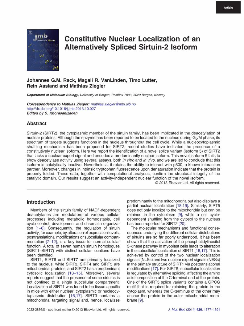
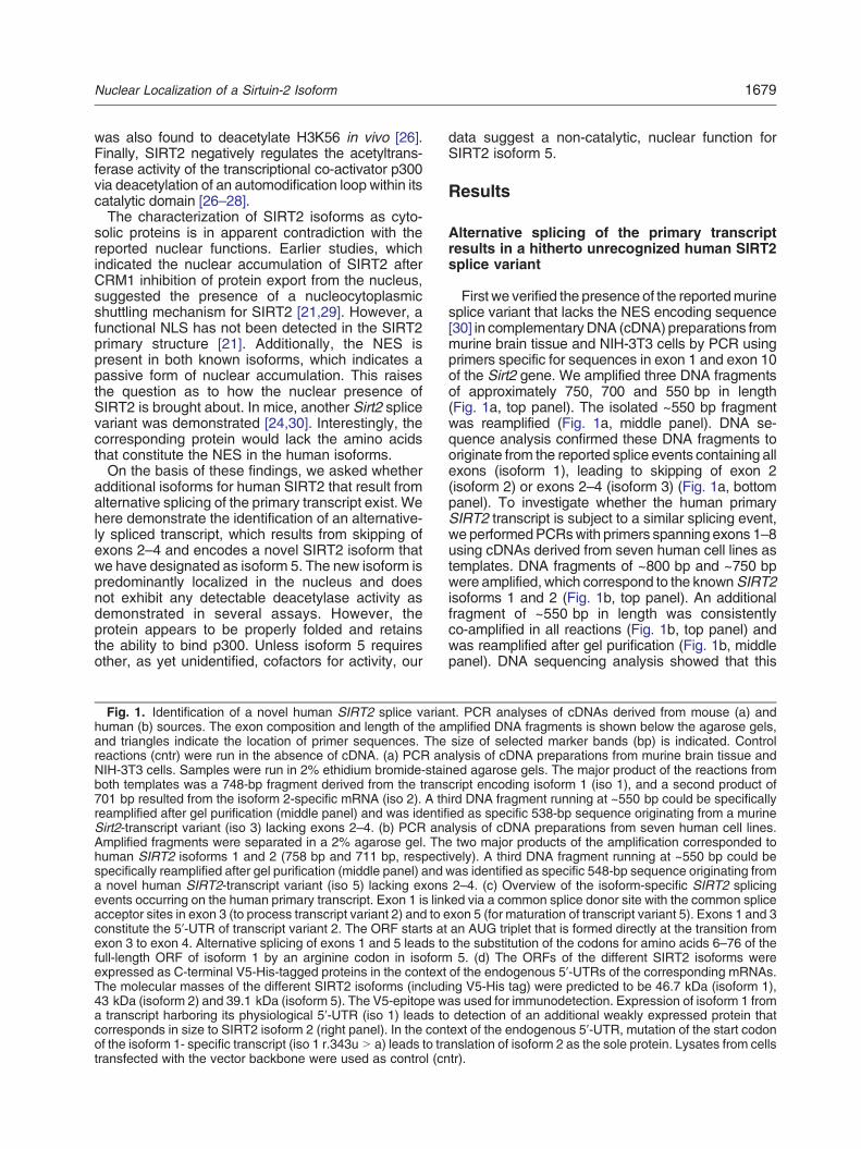
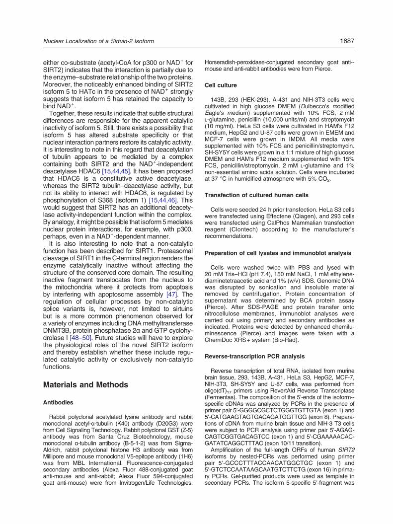
Fig. 1. Identification of a novel human SIRT2 splice variahuman (b) sources. The exon composition and length of the aand triangles indicate the location of primer sequences. Thereactions (cntr) were run in the absence of cDNA. (a) PCR anNIH-3T3 cells. Samples were run in 2% ethidium bromide-staiboth templates was a 748-bp fragment derived from the trans701 bp resulted from the isoform 2-specific mRNA (iso 2). A threamplified after gel purification (middle panel) and was identifSirt2-transcript variant (iso 3) lacking exons 2–4. (b) PCR anAmplified fragments were separated in a 2% agarose gel. Thhuman SIRT2 isoforms 1 and 2 (758 bp and 711 bp, respectspecifically reamplified after gel purification (middle panel) anda novel human SIRT2-transcript variant (iso 5) lacking exonsevents occurring on the human primary transcript. Exon 1 is linkacceptor sites in exon 3 (to process transcript variant 2) and to econstitute the 5′-UTR of transcript variant 2. The ORF starts atexon 3 to exon 4. Alternative splicing of exons 1 and 5 leads tofull-length ORF of isoform 1 by an arginine codon in isoformexpressed as C-terminal V5-His-tagged proteins in the contextThe molecular masses of the different SIRT2 isoforms (includ43 kDa (isoform 2) and 39.1 kDa (isoform 5). The V5-epitope wa transcript harboring its physiological 5′-UTR (iso 1) leads tocorresponds in size to SIRT2 isoform 2 (right panel). In the conof the isoform 1- specific transcript (iso 1 r.343u N a) leads to tratransfected with the vector backbone were used as control (cn
data suggest a non-catalytic, nuclear function forSIRT2 isoform 5.
Results
Alternative splicing of the primary transcriptresults in a hitherto unrecognized human SIRT2splice variant
First we verified the presence of the reportedmurinesplice variant that lacks the NES encoding sequence[30] in complementaryDNA (cDNA) preparations frommurine brain tissue and NIH-3T3 cells by PCR usingprimers specific for sequences in exon 1 and exon 10of the Sirt2 gene. We amplified three DNA fragmentsof approximately 750, 700 and 550 bp in length(Fig. 1a, top panel). The isolated ~550 bp fragmentwas reamplified (Fig. 1a, middle panel). DNA se-quence analysis confirmed these DNA fragments tooriginate from the reported splice events containing allexons (isoform 1), leading to skipping of exon 2(isoform 2) or exons 2–4 (isoform 3) (Fig. 1a, bottompanel). To investigate whether the human primarySIRT2 transcript is subject to a similar splicing event,we performedPCRswith primers spanning exons 1–8using cDNAs derived from seven human cell lines astemplates. DNA fragments of ~800 bp and ~750 bpwere amplified, which correspond to the knownSIRT2isoforms 1 and 2 (Fig. 1b, top panel). An additionalfragment of ~550 bp in length was consistentlyco-amplified in all reactions (Fig. 1b, top panel) andwas reamplified after gel purification (Fig. 1b, middlepanel). DNA sequencing analysis showed that this
nt. PCR analyses of cDNAs derived from mouse (a) andmplified DNA fragments is shown below the agarose gels,size of selected marker bands (bp) is indicated. Controlalysis of cDNA preparations from murine brain tissue andned agarose gels. The major product of the reactions fromcript encoding isoform 1 (iso 1), and a second product ofird DNA fragment running at ~550 bp could be specificallyied as specific 538-bp sequence originating from a murinealysis of cDNA preparations from seven human cell lines.e two major products of the amplification corresponded toively). A third DNA fragment running at ~550 bp could bewas identified as specific 548-bp sequence originating from2–4. (c) Overview of the isoform-specific SIRT2 splicinged via a common splice donor site with the common splicexon 5 (for maturation of transcript variant 5). Exons 1 and 3an AUG triplet that is formed directly at the transition fromthe substitution of the codons for amino acids 6–76 of the5. (d) The ORFs of the different SIRT2 isoforms were
of the endogenous 5′-UTRs of the corresponding mRNAs.ing V5-His tag) were predicted to be 46.7 kDa (isoform 1),as used for immunodetection. Expression of isoform 1 fromdetection of an additional weakly expressed protein that
text of the endogenous 5′-UTR, mutation of the start codonnslation of isoform 2 as the sole protein. Lysates from cellstr).
1680 Nuclear Localization of a Sirtuin-2 Isoform
third PCRproduct originated from a splicing event thatlinks exon 1 to exon 5 (Fig. 1b, bottom panel).† Thepresence of this splicing event was further supportedby analyses of expressed sequence tags (GenBank)from human and Sumatran orangutan tissues.We then asked whether there are further alternative
splicing events downstream of exon 5 in the identifiedtranscript. Nested PCRs were performed in order toamplify the full-length messenger RNA (mRNA) of thenovel transcript variant. cDNA preparations from 293and SH-SY5Y cells were subjected to primary PCRsusing primers spanning exons 1–16. Three secondary
LRNLFSQTLSL
1 2 3 4 5 6 7
isoform 1
secondary structure
conservation
isoform 2
isoform 5
corresponding exons
11
0
α0
SIRT2 (V5)
α-Tubulin
iso 1
iso 1
MA
iso 2
iso 5
cntr iso 1
iso 1
iso 2
iso 5
SIRT2 (V5)
iso 1 iso 1 MA i
input
input
input
cyto
cyto
nucnuc
α-Tubulin
histone H3
(a)
(b) (c)
(d)
PCRswere carried out using (i) a primer pair spanningthe 5′-untranslated region (UTR) to the exon 1/5junction, (ii) a primer pair spanning the exon 1/5junction to exon 16 and (iii) a primer pair spanningexons 1–16 (Fig. S1, bottom panel). All reactions ledto amplification of specific products. DNA sequenceanalysis revealed no further alternative splicingevents downstream of exon 5 of the novel humanSIRT2 splice variant. Thus, the novel human SIRT2transcript is composed of exon 1 and exons 5–16(Fig. S1). The open reading frame (ORF) starts fromthe same AUG triplet in exon 1 that is used to initiate
8 9 10 11 12 13 14 15 16
20 aa
α15
DAPI V5 merge
DAPI V5 merge
DAPI V5 merge
DAPI V5 merge
MA
so 2 iso 5
input
cyto
cyto
nucnuc
1681Nuclear Localization of a Sirtuin-2 Isoform
translation of SIRT2 isoform 1 and terminates in exon16 at the common stop codon for all (known) SIRT2isoforms (Fig. 1c). As transcripts variants 3 and 4 arealready deposited in GenBank database,‡ the novelhuman SIRT2 isoform encoded by the alternativesplicing events reported here is designated isoform 5.Since we could not identify sequences correspondingto transcript variants 3 and 4, all subsequentexperimentswere carried out using the three identifiedtranscript variants 1, 2 and 5.Next, we analyzed whether the composition of the
5′-UTRs of the three humanSIRT2mRNAsmay affectthe translation into proteins. To this end, we expressedC-terminally V5-His-tagged SIRT2 isoforms 1, 2 and 5in the context of their endogenous 5′-UTRs in 293 cells.Immunoblot analyses showed expression of C-termi-nally V5-tagged proteins of expected size (Fig. 1d).Wealso observed a weak band corresponding in size toisoform2 in cells transfectedwith the plasmid encodingisoform 1 (Fig. 1d, right panel). When the start codon ofisoform 1 was mutated (r.343u N a), we observe aband pattern identical with the one seen for wild-type(wt) isoform 2 (Fig. 1d, r.343u N a). This observationsuggests that the second AUG triplet within the ORFof isoform 1 may be used as alternative start codonleading to leaky co-translation of isoform 2. Thus,co-translation of SIRT2 isoform 2 from the transcriptencoding isoform 1 may be of relevance in vivo.
Predominant nuclear localization of humanSIRT2 isoform 5
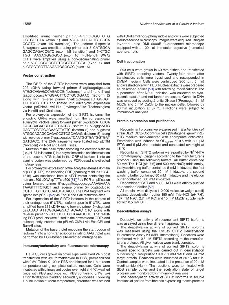
Compared to isoform 1, alternative splicing events ofthe SIRT2 primary transcript result in N-terminaltruncation (residues 1–37) of isoform 2 and, for isoform5, in the substitution of residues 6–76 by an arginineresidue (Fig. 2a and Fig. S2). As a consequence, thecatalytic domain (defined here as residues 64–336 bymultiple sequence alignment and structure analysis) isslightly truncated. However, a conservation analysis
Fig. 2. SIRT2 isoform 5 predominantly localizes to the nucleleads to three distinct proteins. The ORF of isoform 2 startsthereby shortening isoform 2 by the first 37 amino acids relatisoform 1 are exchanged by an arginine leading to the removalis given with gray rectangles representing α-helices and orangewas calculated from multiple sequence alignment (gaps matchfrom 1 to 10 reflecting conservation of biophysical propertieconservation. The catalytic domain is indicated in green; flankinco-substrate coordination and catalysis, in gray; zinc-coordinreported structures (PDB IDs: 3ZGO and 3ZGV) [42]. (b) V5expressing SIRT2 isoforms. Overexpression of SIRT2 isofortranslational start from the second in frame AUG triplet. MutatioSirtT2 isoform 1 leads to sole detection of isoform 1. (c) The diffcell lysates subjected to V5 immunocytochemistry. SIRT2 isofoisoform 5 is predominantly found in the nucleus. The bar repreoverexpressing SIRT2 isoforms. Isoforms 1 (wt and MA) and 2predominantly detected in the nuclear fraction. The purity of thwith α-tubulin and histone H3 antibodies.
using the phylogenetic dataset described by Frye[31] indicated that neither highly conserved residuesnor amino acids involved in co-substrate bindingwould be affected by the changes in the primarystructure (Fig. 2a). Importantly, N-terminal aminoacids that are absent from isoform 5 represent thereported NES found in the other isoforms. Therefore,isoform 5 may display a different cellular distributionthanSIRT2 isoforms 1 and 2. To address this question,we investigated subcellular localization of C-terminallyV5-His-tagged SIRT2 isoforms in HeLa S3 and 293cells. To avoid expression of isoform 2 from theconstruct encoding isoform 1, we included a mutantform of isoform 1 (M38A) to eliminate the alternativetranslational start site for isoform 2 (Fig. 2b). Asexpected, V5 immunocytochemistry showed predom-inant cytoplasmic localizations of isoforms 1 (wt andM38A) and 2. In contrast, isoform 5 localized primarilyto the nucleus (Fig. 2c). This finding was substantiatedby immunoblot analyses of subcellular fractions from293 cells (Fig. 2d).
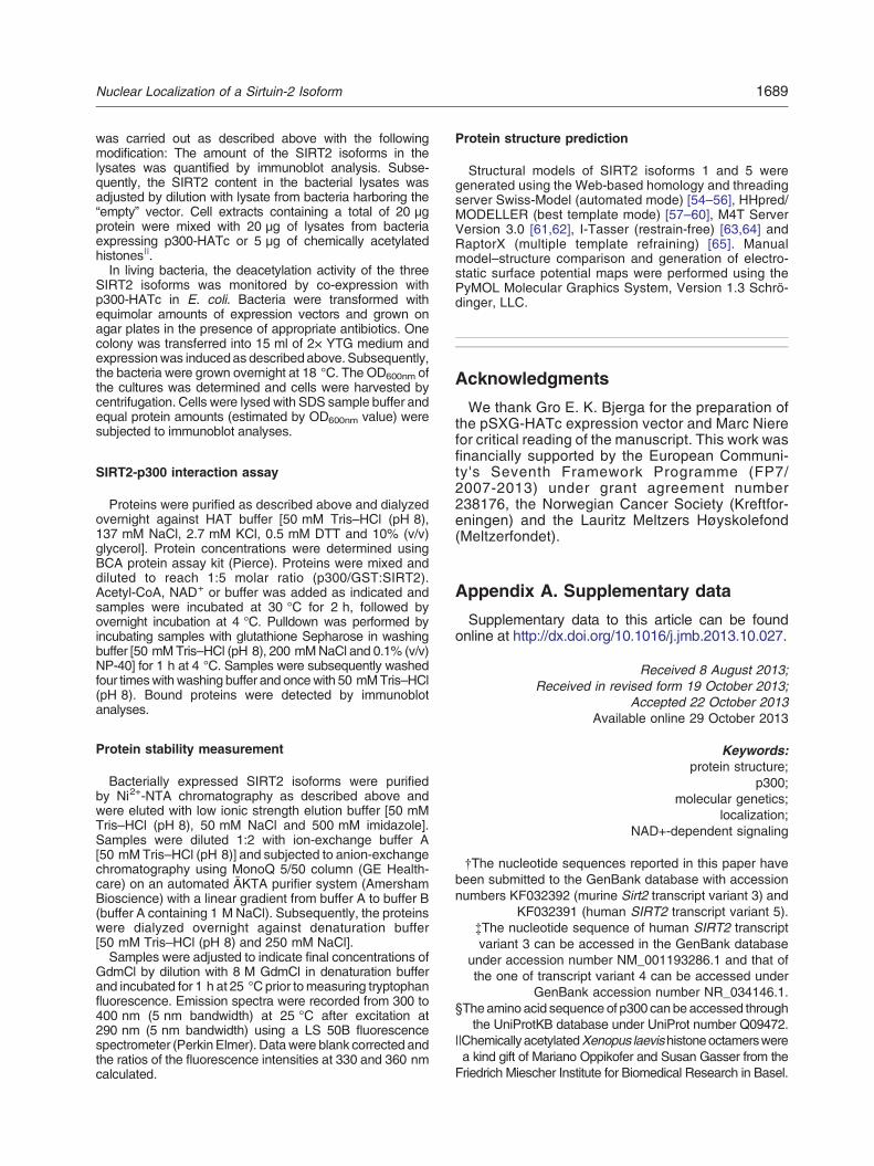
SIRT2 isoform 5 is catalytically inactive in vitroand in vivo
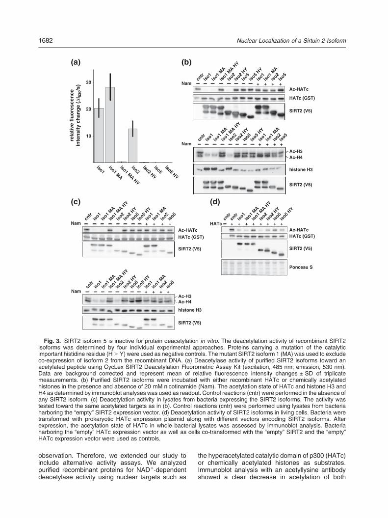
Next, we asked whether the lack of amino acidsencoded by exons 2–4 in isoform 5 might change thecatalytic properties of this protein. The deacetylaseactivities of purified SIRT2 isoforms were measuredusing the CycLex SIRT2 fluorometric activity kit.Isoform 5 showed no detectable activity, whereasisoforms 1 (wt and M38A) and 2 showed a robustdeacetylase activity toward the fluoro-peptide(Fig. 3a). Lack of activity when measuring mutantproteins (H N Y) in which the essential proton acceptorhistidine 187 (isoform 1) was replaced by tyrosineconfirmed the specificity of the reactions.Given that isoform 5 contains all residues predict-
ed to be required for catalysis, the lack of deacetyla-tion activity of this isoform was a rather unexpected
us. (a) Alternative splicing of the human SIRT2 pre-mRNAat a methionine codon formed at the exon 3/4 transition,ive to isoform 1. In SIRT2 isoform 5, amino acids 6–76 ofof the NES (indicated by the red box). Secondary structurearrows representing β-sheets. The degree of conservationing non-SIRT2 residue were removed) with a score ranges of residues and a maximum score of 11 for absoluteg N- and C-terminal regions, in brown; residues involved inating residues, in white. All annotation correspond to the-immunoblot analysis of lysates from 293 cells transientlym 1 results in co-expression of isoform 2 by alternativen of methionine at position 38 to alanine (MA) in the ORF oferent SIRT2 isoforms were expressed in HeLa S3 cells andrms 1 (wt andMA) and 2 localize to the cytoplasm, whereassents 10 μm. (d) Cell fractionation of 293 cells transientlyare detected in cytoplasmic fractions, whereas isoform 5 ise cellular fractions was tested by probing the membranes
HATc + + + + + + + +cn
trcn
triso
1iso
1 MA
iso1 M
A HY
iso2iso
2 HY
iso5iso
5 HY
Ac-HATc
SIRT2 (V5)
HATc (GST)
Ponceau S
cntr
iso1
iso1 M
A
iso1 M
A HY
iso2iso
2 HY
iso5iso
5 HY
iso1iso
1 MA
iso2
iso5
Nam + + + +
SIRT2 (V5)
Ac-HATc
HATc (GST)
cntr
iso1
iso1 M
A
iso1 M
A HY
iso2iso
2 HY
iso5
iso5 H
Y
iso1iso
1 MA
iso2
iso5
Nam + + + +
SIRT2 (V5)
Ac-HATc
HATc (GST)
cntr
iso1
iso1 M
A
iso1 M
A HY
iso2
iso2 H
Y
iso5iso
5 HY
iso1iso
1 MA
iso2iso
5
Nam + + + +
SIRT2 (V5)
histone H3
Ac-H3Ac-H4
cntr
iso1
iso1 M
A
iso1 M
A HY
iso2iso
2 HY
iso5
iso5 H
Y
iso1iso
1 MA
iso2iso
5
Nam + + + +
SIRT2 (V5)
histone H3
Ac-H3Ac-H4
30
20
10
rela
tive
flu
ore
scen
cein
ten
sity
ch
ang
e (Δ
I 530
/s)
iso1iso1 MA
iso1 MA HY
iso2iso2 HY
iso5iso5 HY
(a) (b)
(c) (d)
Fig. 3. SIRT2 isoform 5 is inactive for protein deacetylation in vitro. The deacetylation activity of recombinant SIRT2isoforms was determined by four individual experimental approaches. Proteins carrying a mutation of the catalyticimportant histidine residue (H N Y) were used as negative controls. The mutant SIRT2 isoform 1 (MA) was used to excludeco-expression of isoform 2 from the recombinant DNA. (a) Deacetylase activity of purified SIRT2 isoforms toward anacetylated peptide using CycLex SIRT2 Deacetylation Fluorometric Assay Kit (excitation, 485 nm; emission, 530 nm).Data are background corrected and represent mean of relative fluorescence intensity changes ± SD of triplicatemeasurements. (b) Purified SIRT2 isoforms were incubated with either recombinant HATc or chemically acetylatedhistones in the presence and absence of 20 mM nicotinamide (Nam). The acetylation state of HATc and histone H3 andH4 as determined by immunoblot analyses was used as readout. Control reactions (cntr) were performed in the absence ofany SIRT2 isoform. (c) Deacetylation activity in lysates from bacteria expressing the SIRT2 isoforms. The activity wastested toward the same acetylated targets as in (b). Control reactions (cntr) were performed using lysates from bacteriaharboring the “empty” SIRT2 expression vector. (d) Deacetylation activity of SIRT2 isoforms in living cells. Bacteria weretransformed with prokaryotic HATc expression plasmid along with different vectors encoding SIRT2 isoforms. Afterexpression, the acetylation state of HATc in whole bacterial lysates was assessed by immunoblot analysis. Bacteriaharboring the “empty” HATc expression vector as well as cells co-transformed with the “empty” SIRT2 and the “empty”HATc expression vector were used as controls.
1682 Nuclear Localization of a Sirtuin-2 Isoform
observation. Therefore, we extended our study toinclude alternative activity assays. We analyzedpurified recombinant proteins for NAD+-dependentdeacetylase activity using nuclear targets such as
the hyperacetylated catalytic domain of p300 (HATc)or chemically acetylated histones as substrates.Immunoblot analysis with an acetyllysine antibodyshowed a clear decrease in acetylation of both
1683Nuclear Localization of a Sirtuin-2 Isoform
targets when using isoform 1 (wt and M38A) or 2 inthe reactions. In contrast, no deacetylation activitywas detected for isoform 5 (Fig. 3b).To rule out inactivation of isoform 5 by the
purification procedure, we performed deacetylationassays using lysates from bacteria expressing thedifferent SIRT2 isoforms. Similar to the experimentswith isolated proteins, bacterial lysates containingisoforms 1 (wt and M38A) or 2 deacetylated HATcand histones, whereas no activity was observed inlysates from bacteria expressing recombinant iso-form 5 (Fig. 3c).HATc is hyperacetylatedwhen expressed in bacteria
[32]. Since NAD+ is also freely available in prokaryoticcells, co-expression with SIRT2 may reduce thedegree of HATc acetylation [33]. As shown in Fig. 3d,co-expression with SIRT2 isoforms 1 (wt and M38A)and 2 substantially reduced HATc self-acetylation. Incontrast, co-expressed SIRT2 isoform 5 had no effecton the acetylation state of HATc (Fig. 3d).Together, these data suggest that SIRT2 isoform 5 is
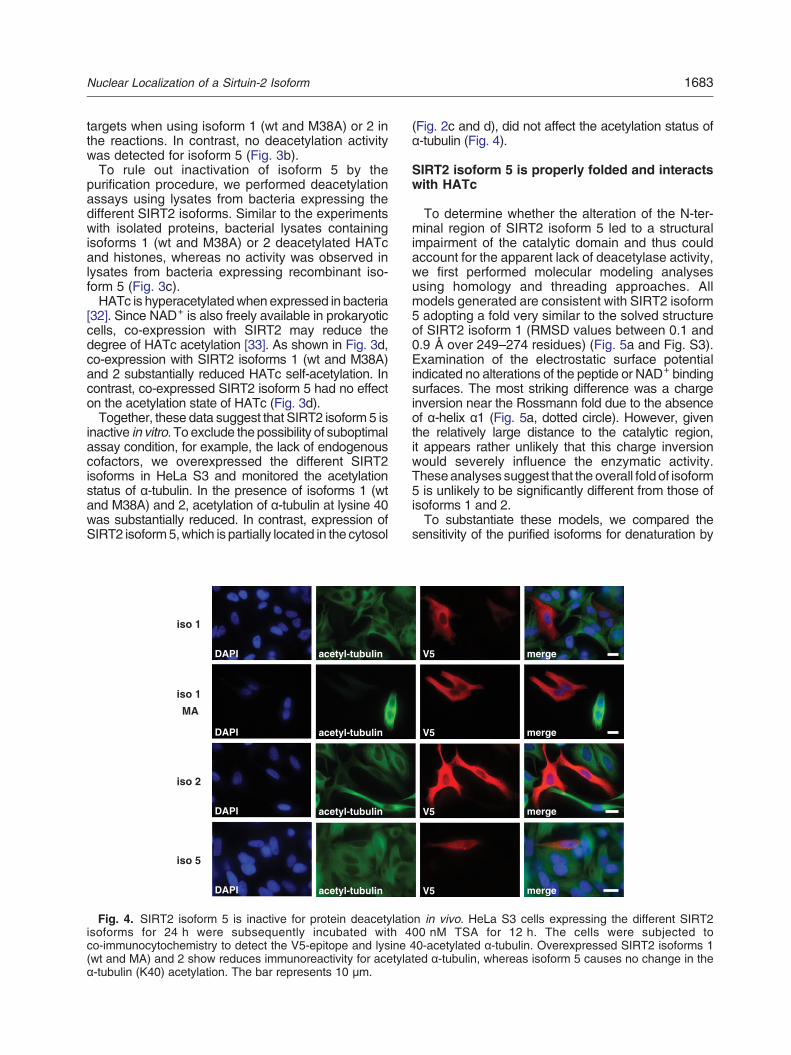
inactive in vitro. To exclude the possibility of suboptimalassay condition, for example, the lack of endogenouscofactors, we overexpressed the different SIRT2isoforms in HeLa S3 and monitored the acetylationstatus of α-tubulin. In the presence of isoforms 1 (wtand M38A) and 2, acetylation of α-tubulin at lysine 40was substantially reduced. In contrast, expression ofSIRT2 isoform5,which is partially located in the cytosol
DAPI
DAPI
DAPI
DAPI
acetyl-tubulin
acetyl-tubulin
acetyl-tubulin
acetyl-tubulin
iso 1
iso 1
MA
iso 2
iso 5
Fig. 4. SIRT2 isoform 5 is inactive for protein deacetylatioisoforms for 24 h were subsequently incubated with 4co-immunocytochemistry to detect the V5-epitope and lysine(wt and MA) and 2 show reduces immunoreactivity for acetylaα-tubulin (K40) acetylation. The bar represents 10 μm.
(Fig. 2c and d), did not affect the acetylation status ofα-tubulin (Fig. 4).
SIRT2 isoform 5 is properly folded and interactswith HATc
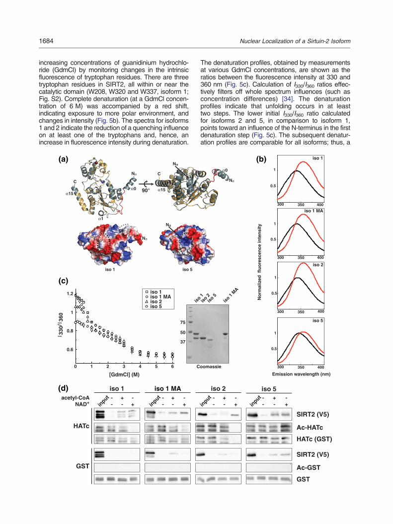
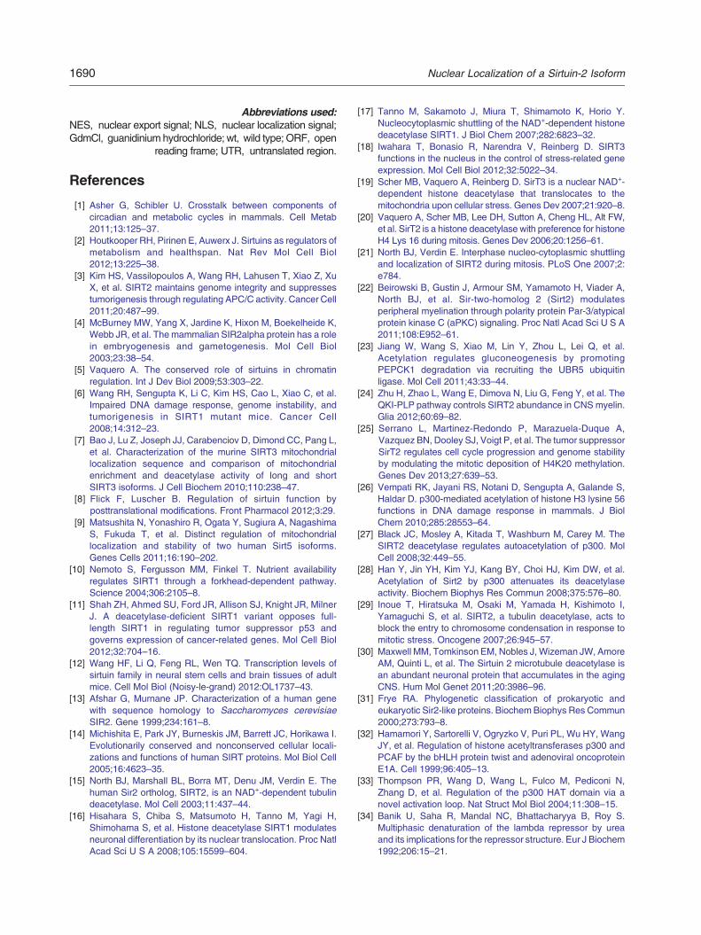
To determine whether the alteration of the N-ter-minal region of SIRT2 isoform 5 led to a structuralimpairment of the catalytic domain and thus couldaccount for the apparent lack of deacetylase activity,we first performed molecular modeling analysesusing homology and threading approaches. Allmodels generated are consistent with SIRT2 isoform5 adopting a fold very similar to the solved structureof SIRT2 isoform 1 (RMSD values between 0.1 and0.9 Å over 249–274 residues) (Fig. 5a and Fig. S3).Examination of the electrostatic surface potentialindicated no alterations of the peptide or NAD+ bindingsurfaces. The most striking difference was a chargeinversion near the Rossmann fold due to the absenceof α-helix α1 (Fig. 5a, dotted circle). However, giventhe relatively large distance to the catalytic region,it appears rather unlikely that this charge inversionwould severely influence the enzymatic activity.Theseanalyses suggest that the overall fold of isoform5 is unlikely to be significantly different from those ofisoforms 1 and 2.To substantiate these models, we compared the
sensitivity of the purified isoforms for denaturation by
V5
V5
V5
V5
merge
merge
merge
merge
n in vivo. HeLa S3 cells expressing the different SIRT200 nM TSA for 12 h. The cells were subjected to40-acetylated α-tubulin. Overexpressed SIRT2 isoforms 1ted α-tubulin, whereas isoform 5 causes no change in the
1684 Nuclear Localization of a Sirtuin-2 Isoform
increasing concentrations of guanidinium hydrochlo-ride (GdmCl) by monitoring changes in the intrinsicfluorescence of tryptophan residues. There are threetryptophan residues in SIRT2, all within or near thecatalytic domain (W208, W320 and W337, isoform 1;Fig. S2). Complete denaturation (at a GdmCl concen-tration of 6 M) was accompanied by a red shift,indicating exposure to more polar environment, andchanges in intensity (Fig. 5b). The spectra for isoforms1 and 2 indicate the reduction of a quenching influenceon at least one of the tryptophans and, hence, anincrease in fluorescence intensity during denaturation.
90°
C
CNi1
Ni5
iso 1 iso 5
Ni1
Ni5
330
/36
0
[GdmCl] (M)
1 2 3 4 5 60
0.8
0.6
1
1.2
75
50
37
is
iso 1iso 1 MAiso 2iso 5
C
GST
input -
-+-
-+
acetyl-CoANAD+
HATc
iso 1
input -
--+
iso 1 MA+-
(a)
(c)
(d)
ΙΙ
The denaturation profiles, obtained by measurementsat various GdmCl concentrations, are shown as theratios between the fluorescence intensity at 330 and360 nm (Fig. 5c). Calculation of I330/I360 ratios effec-tively filters off whole spectrum influences (such asconcentration differences) [34]. The denaturationprofiles indicate that unfolding occurs in at leasttwo steps. The lower initial I330/I360 ratio calculatedfor isoforms 2 and 5, in comparison to isoform 1,points toward an influence of the N-terminus in the firstdenaturation step (Fig. 5c). The subsequent denatur-ation profiles are comparable for all isoforms; thus, aiso 1
300 400350
0.5
1
iso 1 MA
300 400350
0.5
1
iso 2
300 400350
0.5
1
Emission wavelength (nm)
No
rmal
ized
flu
ore
scen
ce in
ten
sity
iso 5
300 400350
0.5
1
Ni1
o 1iso
2iso
5iso
1 MA
oomassie
input -
-+-
-+
Ac-HATc
SIRT2 (V5)
HATc (GST)
iso 5
Ac-GST
SIRT2 (V5)
GST
input -
-+-
-+
iso 2
(b)
1685Nuclear Localization of a Sirtuin-2 Isoform
similar ordered structure of all isoforms can beassumed (Fig. 5c). The increase in fluorescenceintensity, and hence the release of the quenchingeffect, coincides with the first denaturation event (datanot shown). The observed quenching may be due toelectron transfer or static quenching of the tryptophanswithin the structure (W208 and W337) or a result of aninteraction between the C- or the N-terminal regionswith the surface tryptophan W330, or both.These observations support the results of the
modeling approaches suggesting that the overallstructure of isoform 5 is not severely perturbed.Therefore, we considered the possibility that isoform5 might still possess the ability to interact with SIRT2binding partners. A previous study demonstrated aninteraction of SIRT2 isoform 1 with the C-terminal partof p300 including the HAT domain [28]. We investi-gated whether the SIRT2 isoforms 2 and 5 retainedthis property. Indeed, as shown in Fig. 5d, all SIRT2isoforms specifically interacted with the core catalyticdomain of p300 in vitro. We occasionally observedweak binding of SIRT2 isoforms to GST (glutathioneS-transferase). However, the specific interaction withp300 was invariably far stronger for all isoforms.Interestingly, an increase in the interaction wasobserved in the presence of co-substrates (eitheracetyl-CoA or NAD+). Since p300 is a substrate forSIRT2 and vice versa [27,28], stronger binding in thepresence of co-substrate may indicate that theinteraction partially results from an enzyme–substraterelationship. Noteworthy, the interaction of HATc andisoform5 is also strengthened by the addition of NAD+
indicating that isoform 5 retained the structural featuresto bind this nucleotide.
Discussion
In the present study, we have identified the novelisoform 5 of human SIRT2. Our analyses show that it isanevolutionarily conserved isoform that corresponds to
Fig. 5. SIRT2 isoform 5 is structurally intact and can interact w5 structure predictions (upper panel). Models were generatedM4T: isoform 5, blue gray). Model of isoform 1 differs from the spresence of a connection loop (red) between α-helices α0 and α1resolved in chains A and B of the asymmetric unit of 3ZGO. Theand the catalytic histidine (H187, isoform 1) is given in stick realteration in the electrostatic surface potential at the lower site opatches (lower panel). In the surface potentialmap, blue represenwhite represents neutral areas. The black dotted circle indicatebeginning of the modeled loop. The structures were preparedisoforms in the absence (black) or presence (red) of 6 M Gdmmaximal emission of the folded protein. Spectra represent the aGdmCl denaturation of SIRT2 isoforms as monitored by intrinsSIRT2 isoforms in response to increased GdmCl concentration360 nm (excitation at 290 nm) (left panel). Data represent meanCoomassie-stained gel of SIRT2 isoforms used in the denaturaticatalytic domain of p300 (HATc) and SIRT2 isoforms. Prior topresence of co- substrates (acetyl-CoA or NAD+). The molar ratHATc was monitored by acetyllysine and HATc immunoblot ana
apreviously reported splice variant, designated isoform3, in mouse. As isoform 5 lacks the NES, it localizespredominantly to the nucleus. Despite the presence ofan apparently intact SIRT2 catalytic domain, isoform 5does not deacetylate known targets of SIRT2 isoforms1 and 2. Nevertheless, it retains the overall protein foldand the ability to interact with p300. These resultssuggest a non-enzymatic function for this isoform,which remains to be elucidated. Alternatively, the novelisoform 5 might have a substrate specificity that isdifferent from that of the knownSIRT2 isoforms 1 and 2or may require a hitherto unidentified cofactor.
Generation of SIRT2 isoforms by alternativesplicing and translation
The alternatively spliced transcript encoding iso-form 5 has been detected in all human cell linesanalyzed. Additionally, the existence of a similar splicevariant in mice has been reported recently [24,30] andwas confirmed in this study. The discovery of thissplice variant in primates and rodents suggests thatisoform 5 is the result of an evolutionary selection andnot a coincidental “slippage” of the splicingmachinery.In addition to alternative splicing, our results
point toward utilization of an internal start codonas a second mechanism to regulate the presence ofisoforms 1 and 2. Earlier, it was shown that isoform2 is expressed from plasmid carrying isoform 1 in thepresence of an artificial 5′-UTR [35]. Our experimentsutilizing the endogenous 5′-UTR of SIRT2 showedexpression of isoform 2 from the plasmid encodingisoform 1. Moreover, mutation of the isoform 1 startcodon showed efficient expression of isoform 2. In thecontext of the endogenous 5′-UTR, isoform 1 has amoderately strong Kozak sequence (position −3:cytosine; position +4: guanine), whereas the startcodon of isoform 2 is surrounded by a strong Kozaksequence (positions −3 and +4: guanine) [36].Consequently, the expression of isoform 2 in the
ith p300-HATc. (a) Comparison of the SIRT2 isoforms 1 andusing homology modeling (Swiss-Model: isoform 1, yellow;olved structure (PDB ID: 3ZGO, RMSD: 0.069 Å) only in the. Both α0 and α1 are absent in isoform 5. Note that α0 is onlyC- and N-termini (Ni1: isoform 1, Ni5: isoform 5) are indicated,presentation. Absence of α1 from the structure leads to anf the Rossmann fold, but not to an exposure of hydrophobicts positively charged, red represents negatively chargedands the position of α1, and the gray dotted line indicates thewith PyMOL. (b) Fluorescence emission spectra for SIRT2Cl (excitation at 290 nm). Values were normalized to theverage of four measurements and are buffer corrected. (c)ic tryptophan fluorescence. The denaturation profile of theis shown as the ratios of fluorescence intensities at 330 ands ± SD of quadruple measurements. The right panel showson shift assay. (d) GST-pulldown assay using the GST-fusedthe pulldown, samples were incubated in the absence or
io of proteins (HATc/GST: SIRT2) was 1:5. SIRT2 binding tolyses. Recombinant GST was used as negative control.
1686 Nuclear Localization of a Sirtuin-2 Isoform
context of isoform 1 could be a result of leaky ribosomescanning. That is, isoform 2may be generated from theisoform 1 transcript in vivo and thus might contribute tothe differences in isoform expression observed inmurine tissues [30]. However, the favored expressionof isoform 1 from its endogenous 5′-UTR indicates thatisoform 2 originates predominantly from an isoform-specific alternatively spliced mRNA. Since the isoform5 was detectable when overexpressed in context of itsendogenous 5′-UTR, it is more than likely that it is alsoreadily translated from its endogenous mRNA.The observed nuclear SIRT2 activity must be a
result of a catalytically active isoform. In light of ourstudy, two mechanisms to promote nuclear SIRT2localization appear most plausible: Absence of theNES (Ref. [21] and this study) or conditions ofCRM1-dependent export inhibition [21,29]. Our anal-yses of the NES encoding region indicate that isoform5 is the only splice variant that excludes the NESfrom the primary structure. On the other hand, theessentially exclusive cytoplasmic localization ofisoforms 1 and 2 indicates highly active CRM1-dependent nuclear export. Consequently, it wouldappear that the nuclear SIRT2 activity is likely a resultof low-level abundance of SIRT2 isoform(s) broughtabout either by constant shuttling or by masking of theNES, for example, in a nuclear complex.
Isoform 5 shows nuclear enrichment,however, is catalytically inactive
The absence of the NES from isoform 5 immediatelysuggested the possibility that itmight be localized to thenucleus. Indeed, using both immunocytochemistry andcell fractionation, we observed a predominant nuclearlocalization of SIRT2 isoform 5. This pattern is distinctfrom isoforms 1 and 2, which are solely cytoplasmic.Considering the molecular size of SIRT2 isoform 5, anactive transport mechanism either by a non-canonicalNLS or “piggy-backing” in complex with other proteinsseems likely. Computational NLS prediction usingcNLS mapper [37] indicated the presence of a weakbipartite NLS containing an extended linker region(residues 342–375 in isoform 1). The weak score (2.8)is in accordance with the observed partial cytoplasmiclocalization of isoform 5 and, thus, may be a result oflow NLS activity.Given that the known SIRT2 isoforms 1 and 2
localized to the cytoplasm (Refs. [13–15] and thisstudy), it appeared likely that the new nuclear isoformcould account for the SIRT2-dependent deacetylationof nuclear proteins. Surprisingly, in contrast to isoforms1 and 2, isoform 5 displayed no catalytic activity in ourexperiments. The absence of detectable activity inmultiple approaches and experimental systems pro-vides compelling evidence that the novel SIRT2isoform has no deacetylase activity, at least towardknown SIRT2 targets (Refs. [27,38–40] and confirmedhere): When using isolated, recombinant SIRT2
isoform 5, it neither deacetylated the fluorogenicpeptide nor histones or HATc of p300. Moreover,when expressed and directly analyzed in bacterial orhuman cells, isoform 5 exhibited no deacetylaseactivity, whereas isoforms 1 and 2 were active. Thus,even though isoform 5 provided an excellent candidateto account for nuclear SIRT2 activity, our results seemto rule out such a function.
The fold of the catalytic domain of SIRT2 isoform5 is not substantially impaired
An obvious explanation for the catalytic inactivityof isoform 5 would have been the lack of the mostN-terminal part of the catalytic domain (includingα-helix α1). The absence of these 12 residues couldeither cause structural impairment or involve cata-lytic residues. Several lines of evidence suggest,however, that this is not the case. For example,isolation of recombinant isoform 5 after expression inbacteria resulted in a readily soluble protein. Moreover,analyses of the tryptophan fluorescence duringGdmCl-dependent denaturation further indicated thatisolated isoform 5 is properly folded. These experi-ments were also in line with our structural model thatpredicts the catalytic core of isoform 5 to resemble thatof isoforms 1 and 2. All residues known to be essentialfor catalysis [41,42] are still present in isoform 5.However, comparison with the structure of SIRT2isoform 1 revealed that α-helix α1 is missing in isoform5. Sinceα1 is part of theNAD+-bindingRossmann fold,the structural integrity of isoform 5 could be affected.However, the modeling using homology and threadingapproaches indicates that the general structure couldstill be maintained. This is supported by a crystallo-graphic study of cobB, a bacterial sirtuin homologue[43]. The cobB protein used in that study lackedthe equivalent α-helix in the Rossmann fold but wascatalytically active.Examination of the denaturation curves suggests
that, at least, one of the tryptophans of isoform 1 is inlabile environment created by the presence of theN-terminal region. Shortening of the N-terminus(isoform 2) or its removal (isoform 5) leads to anincreased solvent exposure as indicated by the drop inthe I330/I360 ratio. Considering the positioning of thetryptophans within the structure, it is likely that eitherthe N-terminus folds back onto the catalytic domainshielding the surface tryptophan W320 (isoform 1) orhelix α15 separates from the core structure exposingW337 (isoform 1). Both changes could have anindirect influence on the substrate binding capabilityof SIRT2 without altering the major fold. An additionalindication that the observed inactivity of isoform 5 islikely to be a result of more subtle conformationaldifferences is its preserved ability to interact with thecatalytic domain of p300. The latter feature is sharedby isoforms 1 and 2. The strengthened interactionobserved for all three isoforms in the presence of
1687Nuclear Localization of a Sirtuin-2 Isoform
either co-substrate (acetyl-CoA for p300 or NAD+ forSIRT2) indicates that the interaction is partially due tothe enzyme–substrate relationship of the two proteins.Moreover, the noticeably enhanced binding of SIRT2isoform 5 to HATc in the presence of NAD+ stronglysuggests that isoform 5 has retained the capacity tobind NAD+.Together, these results indicate that subtle structural
differences are responsible for the apparent catalyticinactivity of isoform 5. Still, there exists a possibility thatisoform 5 has altered substrate specificity or thatnuclear interaction partners restore its catalytic activity.It is interesting to note in this regard that deacetylationof tubulin appears to be mediated by a complexcontaining both SIRT2 and the NAD+-independentdeacetylase HDAC6 [15,44,45]. It has been proposedthat HDAC6 is a constitutive active deacetylase,whereas the SIRT2 tubulin–deacetylase activity, butnot its ability to interact with HDAC6, is regulated byphosphorylation of S368 (isoform 1) [15,44,46]. Thiswould suggest that SIRT2 has an additional deacety-lase activity-independent function within the complex.By analogy, itmight bepossible that isoform5mediatesnuclear protein interactions, for example, with p300,perhaps, even in a NAD+-dependent manner.It is also interesting to note that a non-catalytic
function has been described for SIRT1. Proteasomalcleavage of SIRT1 in the C-terminal region renders theenzyme catalytically inactive without affecting thestructure of the conserved core domain. The resultinginactive fragment translocates from the nucleus tothe mitochondria where it protects from apoptosisby interfering with apoptosome assembly [47]. Theregulation of cellular processes by non-catalyticsplice variants is, however, not limited to sirtuinsbut is a more common phenomenon observed fora variety of enzymes including DNAmethyltransferaseDNMT3B, protein phosphatase 2α and GTP cyclohy-drolase I [48–50]. Future studies will have to explorethe physiological roles of the novel SIRT2 isoformand thereby establish whether these include regu-lated catalytic activity or exclusively non-catalyticfunctions.
Materials and Methods
Antibodies
Rabbit polyclonal acetylated lysine antibody and rabbitmonoclonal acetyl-α-tubulin (K40) antibody (D20G3) werefrom Cell Signaling Technology. Rabbit polyclonal GST (Z-5)antibody was from Santa Cruz Biotechnology, mousemonoclonal α-tubulin antibody (B-5-1-2) was from Sigma-Aldrich, rabbit polyclonal histone H3 antibody was fromMillipore and mouse monoclonal V5-epitope antibody (1H6)was from MBL International. Fluorescence-conjugatedsecondary antibodies (Alexa Fluor 488-conjugated goatanti-mouse and anti-rabbit; Alexa Fluor 594-conjugatedgoat anti-mouse) were from Invitrogen/Life Technologies.
Horseradish-peroxidase-conjugated secondary goat anti--mouse and anti-rabbit antibodies were from Pierce.
Cell culture
143B, 293 (HEK-293), A-431 and NIH-3T3 cells werecultivated in high glucose DMEM (Dulbecco's modifiedEagle's medium) supplemented with 10% FCS, 2 mML-glutamine, penicillin (10,000 units/ml) and streptomycin(10 mg/ml). HeLa S3 cells were cultivated in HAM's F12medium, HepG2 and U-87 cells were grown in EMEM andMCF-7 cells were grown in IMDM. All media weresupplemented with 10% FCS and penicillin/streptomycin.SH-SY5Y cells were grown in a 1:1 mixture of high glucoseDMEM and HAM's F12 medium supplemented with 15%FCS, penicillin/streptomycin, 2 mM L-glutamine and 1%non-essential amino acids solution. Cells were incubatedat 37 °C in humidified atmosphere with 5% CO2.
Transfection of cultured human cells
Cells were seeded 24 h prior transfection. HeLa S3 cellswere transfected using Effectene (Qiagen), and 293 cellswere transfected using CalPhos Mammalian transfectionreagent (Clontech) according to the manufacturer'srecommendations.
Preparation of cell lysates and immunoblot analysis
Cells were washed twice with PBS and lysed with20 mM Tris–HCl (pH 7.4), 150 mM NaCl, 1 mM ethylene-diaminetetraacetic acid and 1% (w/v) SDS. Genomic DNAwas disrupted by sonication and insoluble materialremoved by centrifugation. Protein concentration ofsupernatant was determined by BCA protein assay(Pierce). After SDS-PAGE and protein transfer ontonitrocellulose membranes, immunoblot analyses werecarried out using primary and secondary antibodies asindicated. Proteins were detected by enhanced chemilu-minescence (Pierce) and images were taken with aChemiDoc XRS+ system (Bio-Rad).
Reverse-transcription PCR analysis
Reverse transcription of total RNA, isolated from murinebrain tissue, 293, 143B, A-431, HeLa S3, HepG2, MCF-7,NIH-3T3, SH-SY5Y and U-87 cells, was performed fromoligo(dT)17 primers using RevertAid Reverse Transcriptase(Fermentas). The composition of the 5′-ends of the isoform--specific cDNAs was analyzed by PCRs in the presence ofprimer pair 5′-GGGGCGCTCTGGGTGTTGTA (exon 1) and5′-CATGAAGTAGTGACAGATGGTTGG (exon 8). Prepara-tions of cDNA from murine brain tissue and NIH-3 T3 cellswere subject to PCR analysis using primer pair 5′-AGAG-CAGTCGGTGACAGTCC (exon 1) and 5′-CGAAAAACAC-GATATCAGGCTTTAC (exon 10/11 transition).Amplification of the full-length ORFs of human SIRT2
isoforms by nested-PCRs was performed using primerpair 5′-GCCCTTTACCAACATGGCTGC (exon 1) and5′-GTCTCCAATAAGCAATGTCTTCTG (exon 16) in prima-ry PCRs. Gel-purified products were used as template insecondary PCRs. The isoform 5-specific 5′-fragment was
1688 Nuclear Localization of a Sirtuin-2 Isoform
amplified using primer pair 5 ′-GGGGCGCTCTGGGTGTTGTA (exon 1) and 5′-CAGATGACTCTGCCACGGTC (exon 1/5 transition). The isoform 5-specific3′-fragment was amplified using primer pair 5′-CATGGCAGAGCCAGACCGTC (exon 1/5 transition) and 5′-CTGCTGGTTAAGAGGGGGCC (exon 16). Full-length SIRT2ORFs were amplified using a non-discriminating primerpair 5′-GGGGCGCTCTGGGTGTTGTA (exon 1) and5′-CTGCTGGTTAAGAGGGGGCC (exon 16).
Vector construction
The ORFs of the SIRT2 isoforms were amplified from293 cDNA using forward primer 5′-agttaagcttgccaccATGGCAGAGCCAGACCG (isoforms 1 and 5) and 5′-agttaagcttgccaccATGGACTTCCTGCGGAAC (isoform 2)along with reverse primer 5′-atcgtctagaacaCTGGGGTTTCTCCCTCTC and ligated into eukaryotic expressionvector pcDNA3.1/V5-His (Invitrogen/Life Technologies)via HindIII and XbaI sites.For prokaryotic expression of the SIRT2 isoforms, the
encoding ORFs were amplified from the correspondingeukaryotic vectors using forward primer 5′-gcatccATGGCAGAGCCAGACCCCTCTCACCC (isoform 1), 5′-cgtgccATGGACTTCCTGCGGAACTTATTC (isoform 2) and 5′-gcatccATGGCAGAGCCAGACCGTCGCAGAG (isoform 5) alongwith reverseprimer 5′-ggacggatccTCAATGGTGATGGTGATGATGACC. The DNA fragments were ligated into pET9d(Novagen) via NcoI and BamHI sites.Mutation of the base triplet encoding the catalytic histidine
(i.e., H187 in isoform1) into a tyrosine codonand themutationof the second ATG triplet in the ORF of isoform 1 into analanine codon was performed by PCR-based site-directedmutagenesis.For prokaryotic expression of the acetyltransferase domain
of p300 (HATc), the encodingORF (spanning residues1284–1845) was subcloned from a pT7T vector containing thehuman p300 cDNA (pT7Ts-p300 [51])§ by PCR amplificationusing forward primer 5′-gcgaattcagcggccgcAAAGAAAATAAGTTTTCTGCT and reverse primer 5′- gcgtacgtcgacCCTGTTGCTGCCCAACCACACC. The DNA fragment wasligated into pSXG [52] via EcoRI and SalI restriction sites.For expression of the SIRT2 isoforms in the context of
their endogenous 5′-UTRs, isoform-specific 5′-UTRs wereamplified from 293 cDNA using forward primer 5′-ctcgtttagtgaaAGAGTATTCGGGAGGACTACAACTCTC along withreverse primer 5′-GCGCGGTGCTGAAGCCC. The result-ing PCR products were fused to the downstream ORFs andsubsequently inserted into pFLAG-CMV4 via EcoICRI andBamHI sites.Mutation of the base triplet encoding the start codon of
isoform 1 into a non-transcription initiating AAG triplet wasperformed by PCR-based site-directed mutagenesis.
Immunocytochemistry and fluorescence microscopy
HeLa S3 cells grown on cover slips were fixed 24 h posttransfection with 4% formaldehyde in PBS, permeabilizedwith 0.5% Triton X-100 in PBS and blocked for 1 h at roomtemperature using complete culture medium. Cells wereincubatedwith primary antibodies overnight at 4 °C, washedtwice with PBS and once with PBS containing 0.1% (v/v)Triton X-100 prior to adding secondary antibodies. Following1 h incubation at room temperature, chromatin was stained
with 4′,6-diamidino-2-phenylindole and cells were subjectedto fluorescencemicroscopy. Imageswere acquired using aninverted Leica DMI 6000B fluorescence microscopeequipped with a 100× oil immersion objective (numericalaperture, 1.4).
Cell fractionation
293 cells were grown in 60 mm dishes and transfectedwith SIRT2 encoding vectors. Twenty-four hours aftertransfection, cells were trypsinized and resuspended inDMEM medium. Cells were centrifuged (900 rpm, 5 min)andwashed oncewithPBS.Nuclear extractswere preparedas described earlier [53] with following modifications: Thesupernatant, after NP-40 addition, was collected as cyto-plasmic fraction and not further processed. Genomic DNAwas removed by adding 2 units DNase I (Promega), 5 mMMgCl2 and 5 mM CaCl2 to the nuclear pellet followed by20 min incubation at 37 °C. Fractions were subject toimmunoblot analyses.
Protein expression and purification
Recombinant proteins were expressed inEscherichia colistrain BL21(DE3)-CodonPlus cells (Stratagene) grown in 2×YTG medium supplemented with appropriate antibiotics.Expression was induced at OD600nm of 0.6 with 300 μMIPTG and 5 μM zinc acetate and conducted overnight at18 °C.Recombinant SIRT2 isoforms were purified by Ni2+-NTA
chromatography (Qiagen) according to the manufacturer'sprotocol using the following buffers: All buffer contained50 mM Tris–HCl (pH 7.6) and 500 mM NaCl; additionally,the lysis/binding buffer contained 5 mM imidazole, the firstwashing buffer contained 20 mM imidazole, the secondwashing buffer contained 50 mM imidazole and the elutionbuffer contained 500 mM imidazole.Recombinant GST and p300-HATs were affinity purified
as described earlier [52].All proteins were dialyzed (10,000 molecular weight cutoff)
against deacetylation buffer [50 mM Tris–HCl (pH 7.6),137 mM NaCl, 2.7 mM KCl and 10 mM MgCl2] supplement-ed with 0.5 mM DTT.
Deacetylation assays
Deacetylation activity of recombinant SIRT2 isoformswas assayed using four different approaches.The deacetylation activity of purified SIRT2 isoforms
was measured using the CycLex SIRT2 DeacetylationFluorometric Assay Kit (MBL International). Reactions wereperformed with 0.8 μM SIRT2 according to the manufac-turer's protocol. All given values were blank corrected.The deacetylation activity of purified SIRT2 isoforms
toward specific targets was carried out in deacetylationbuffer using 1 mM purified SIRT2, 1 mMNAD+ and 0.4 mMtarget protein. Reactions were incubated at 30 °C for 2 h.Control samples were incubated in the presence of 20 mMnicotinamide (Nam). The reactions were terminated withSDS sample buffer and the acetylation state of targetproteins was monitored by immunoblot analyses.The deacetylation activity of SIRT2 isoforms in soluble
fractions of lysates from bacteria expressing theses proteins
1689Nuclear Localization of a Sirtuin-2 Isoform
was carried out as described above with the followingmodification: The amount of the SIRT2 isoforms in thelysates was quantified by immunoblot analysis. Subse-quently, the SIRT2 content in the bacterial lysates wasadjusted by dilution with lysate from bacteria harboring the“empty” vector. Cell extracts containing a total of 20 μgprotein were mixed with 20 μg of lysates from bacteriaexpressing p300-HATc or 5 μg of chemically acetylatedhistones||.In living bacteria, the deacetylation activity of the three
SIRT2 isoforms was monitored by co-expression withp300-HATc in E. coli. Bacteria were transformed withequimolar amounts of expression vectors and grown onagar plates in the presence of appropriate antibiotics. Onecolony was transferred into 15 ml of 2× YTG medium andexpressionwas inducedas describedabove. Subsequently,the bacteria were grown overnight at 18 °C. The OD600nm ofthe cultures was determined and cells were harvested bycentrifugation. Cells were lysed with SDS sample buffer andequal protein amounts (estimated by OD600nm value) weresubjected to immunoblot analyses.
SIRT2-p300 interaction assay
Proteins were purified as described above and dialyzedovernight against HAT buffer [50 mM Tris–HCl (pH 8),137 mM NaCl, 2.7 mM KCl, 0.5 mM DTT and 10% (v/v)glycerol]. Protein concentrations were determined usingBCA protein assay kit (Pierce). Proteins were mixed anddiluted to reach 1:5 molar ratio (p300/GST:SIRT2).Acetyl-CoA, NAD+ or buffer was added as indicated andsamples were incubated at 30 °C for 2 h, followed byovernight incubation at 4 °C. Pulldown was performed byincubating samples with glutathione Sepharose in washingbuffer [50 mMTris–HCl (pH 8), 200 mMNaCl and 0.1% (v/v)NP-40] for 1 h at 4 °C. Samples were subsequently washedfour timeswith washing buffer and oncewith 50 mMTris–HCl(pH 8). Bound proteins were detected by immunoblotanalyses.
Protein stability measurement
Bacterially expressed SIRT2 isoforms were purifiedby Ni2+-NTA chromatography as described above andwere eluted with low ionic strength elution buffer [50 mMTris–HCl (pH 8), 50 mM NaCl and 500 mM imidazole].Samples were diluted 1:2 with ion-exchange buffer A[50 mMTris–HCl (pH 8)] and subjected to anion-exchangechromatography using MonoQ 5/50 column (GE Health-care) on an automated ÄKTA purifier system (AmershamBioscience) with a linear gradient from buffer A to buffer B(buffer A containing 1 M NaCl). Subsequently, the proteinswere dialyzed overnight against denaturation buffer[50 mM Tris–HCl (pH 8) and 250 mM NaCl].Samples were adjusted to indicate final concentrations of
GdmCl by dilution with 8 M GdmCl in denaturation bufferand incubated for 1 h at 25 °C prior tomeasuring tryptophanfluorescence. Emission spectra were recorded from 300 to400 nm (5 nm bandwidth) at 25 °C after excitation at290 nm (5 nm bandwidth) using a LS 50B fluorescencespectrometer (PerkinElmer). Datawere blank corrected andthe ratios of the fluorescence intensities at 330 and 360 nmcalculated.
Protein structure prediction
Structural models of SIRT2 isoforms 1 and 5 weregenerated using the Web-based homology and threadingserver Swiss-Model (automated mode) [54–56], HHpred/MODELLER (best template mode) [57–60], M4T ServerVersion 3.0 [61,62], I-Tasser (restrain-free) [63,64] andRaptorX (multiple template refraining) [65]. Manualmodel–structure comparison and generation of electro-static surface potential maps were performed using thePyMOL Molecular Graphics System, Version 1.3 Schrö-dinger, LLC.
Acknowledgments
We thank Gro E. K. Bjerga for the preparation ofthe pSXG-HATc expression vector and Marc Nierefor critical reading of the manuscript. This work wasfinancially supported by the European Communi-ty's Seventh Framework Programme (FP7/2007-2013) under grant agreement number238176, the Norwegian Cancer Society (Kreftfor-eningen) and the Lauritz Meltzers Høyskolefond(Meltzerfondet).
Appendix A. Supplementary data
Supplementary data to this article can be foundonline at http://dx.doi.org/10.1016/j.jmb.2013.10.027.
Received 8 August 2013;Received in revised form 19 October 2013;
Accepted 22 October 2013Available online 29 October 2013
Keywords:protein structure;
p300;molecular genetics;
localization;NAD+-dependent signaling
†The nucleotide sequences reported in this paper havebeen submitted to the GenBank database with accessionnumbers KF032392 (murine Sirt2 transcript variant 3) and
KF032391 (human SIRT2 transcript variant 5).‡The nucleotide sequence of human SIRT2 transcriptvariant 3 can be accessed in the GenBank database
under accession number NM_001193286.1 and that ofthe one of transcript variant 4 can be accessed under
GenBank accession number NR_034146.1.§Theamino acid sequence of p300 canbeaccessed through
the UniProtKB database under UniProt number Q09472.||Chemically acetylatedXenopus laevishistoneoctamerswerea kind gift of Mariano Oppikofer and Susan Gasser from the
Friedrich Miescher Institute for Biomedical Research in Basel.
1690 Nuclear Localization of a Sirtuin-2 Isoform
Abbreviations used:NES, nuclear export signal; NLS, nuclear localization signal;GdmCl, guanidinium hydrochloride; wt, wild type; ORF, open
reading frame; UTR, untranslated region.
References
[1] Asher G, Schibler U. Crosstalk between components ofcircadian and metabolic cycles in mammals. Cell Metab2011;13:125–37.
[2] Houtkooper RH, Pirinen E, Auwerx J. Sirtuins as regulators ofmetabolism and healthspan. Nat Rev Mol Cell Biol2012;13:225–38.
[3] Kim HS, Vassilopoulos A, Wang RH, Lahusen T, Xiao Z, XuX, et al. SIRT2 maintains genome integrity and suppressestumorigenesis through regulating APC/C activity. Cancer Cell2011;20:487–99.
[4] McBurney MW, Yang X, Jardine K, Hixon M, Boekelheide K,Webb JR, et al. The mammalian SIR2alpha protein has a rolein embryogenesis and gametogenesis. Mol Cell Biol2003;23:38–54.
[5] Vaquero A. The conserved role of sirtuins in chromatinregulation. Int J Dev Biol 2009;53:303–22.
[6] Wang RH, Sengupta K, Li C, Kim HS, Cao L, Xiao C, et al.Impaired DNA damage response, genome instability, andtumorigenesis in SIRT1 mutant mice. Cancer Cell2008;14:312–23.
[7] Bao J, Lu Z, Joseph JJ, Carabenciov D, Dimond CC, Pang L,et al. Characterization of the murine SIRT3 mitochondriallocalization sequence and comparison of mitochondrialenrichment and deacetylase activity of long and shortSIRT3 isoforms. J Cell Biochem 2010;110:238–47.
[8] Flick F, Luscher B. Regulation of sirtuin function byposttranslational modifications. Front Pharmacol 2012;3:29.
[9] Matsushita N, Yonashiro R, Ogata Y, Sugiura A, NagashimaS, Fukuda T, et al. Distinct regulation of mitochondriallocalization and stability of two human Sirt5 isoforms.Genes Cells 2011;16:190–202.
[10] Nemoto S, Fergusson MM, Finkel T. Nutrient availabilityregulates SIRT1 through a forkhead-dependent pathway.Science 2004;306:2105–8.
[11] Shah ZH, Ahmed SU, Ford JR, Allison SJ, Knight JR, MilnerJ. A deacetylase-deficient SIRT1 variant opposes full-length SIRT1 in regulating tumor suppressor p53 andgoverns expression of cancer-related genes. Mol Cell Biol2012;32:704–16.
[12] Wang HF, Li Q, Feng RL, Wen TQ. Transcription levels ofsirtuin family in neural stem cells and brain tissues of adultmice. Cell Mol Biol (Noisy-le-grand) 2012:OL1737–43.
[13] Afshar G, Murnane JP. Characterization of a human genewith sequence homology to Saccharomyces cerevisiaeSIR2. Gene 1999;234:161–8.
[14] Michishita E, Park JY, Burneskis JM, Barrett JC, Horikawa I.Evolutionarily conserved and nonconserved cellular locali-zations and functions of human SIRT proteins. Mol Biol Cell2005;16:4623–35.
[15] North BJ, Marshall BL, Borra MT, Denu JM, Verdin E. Thehuman Sir2 ortholog, SIRT2, is an NAD+-dependent tubulindeacetylase. Mol Cell 2003;11:437–44.
[16] Hisahara S, Chiba S, Matsumoto H, Tanno M, Yagi H,Shimohama S, et al. Histone deacetylase SIRT1 modulatesneuronal differentiation by its nuclear translocation. Proc NatlAcad Sci U S A 2008;105:15599–604.
[17] Tanno M, Sakamoto J, Miura T, Shimamoto K, Horio Y.Nucleocytoplasmic shuttling of the NAD+-dependent histonedeacetylase SIRT1. J Biol Chem 2007;282:6823–32.
[18] Iwahara T, Bonasio R, Narendra V, Reinberg D. SIRT3functions in the nucleus in the control of stress-related geneexpression. Mol Cell Biol 2012;32:5022–34.
[19] Scher MB, Vaquero A, Reinberg D. SirT3 is a nuclear NAD+-dependent histone deacetylase that translocates to themitochondria upon cellular stress. Genes Dev 2007;21:920–8.
[20] Vaquero A, Scher MB, Lee DH, Sutton A, Cheng HL, Alt FW,et al. SirT2 is a histone deacetylase with preference for histoneH4 Lys 16 during mitosis. Genes Dev 2006;20:1256–61.
[21] North BJ, Verdin E. Interphase nucleo-cytoplasmic shuttlingand localization of SIRT2 during mitosis. PLoS One 2007;2:e784.
[22] Beirowski B, Gustin J, Armour SM, Yamamoto H, Viader A,North BJ, et al. Sir-two-homolog 2 (Sirt2) modulatesperipheral myelination through polarity protein Par-3/atypicalprotein kinase C (aPKC) signaling. Proc Natl Acad Sci U S A2011;108:E952–61.
[23] Jiang W, Wang S, Xiao M, Lin Y, Zhou L, Lei Q, et al.Acetylation regulates gluconeogenesis by promotingPEPCK1 degradation via recruiting the UBR5 ubiquitinligase. Mol Cell 2011;43:33–44.
[24] Zhu H, Zhao L, Wang E, Dimova N, Liu G, Feng Y, et al. TheQKI-PLP pathway controls SIRT2 abundance in CNS myelin.Glia 2012;60:69–82.
[25] Serrano L, Martinez-Redondo P, Marazuela-Duque A,Vazquez BN, Dooley SJ, Voigt P, et al. The tumor suppressorSirT2 regulates cell cycle progression and genome stabilityby modulating the mitotic deposition of H4K20 methylation.Genes Dev 2013;27:639–53.
[26] Vempati RK, Jayani RS, Notani D, Sengupta A, Galande S,Haldar D. p300-mediated acetylation of histone H3 lysine 56functions in DNA damage response in mammals. J BiolChem 2010;285:28553–64.
[27] Black JC, Mosley A, Kitada T, Washburn M, Carey M. TheSIRT2 deacetylase regulates autoacetylation of p300. MolCell 2008;32:449–55.
[28] Han Y, Jin YH, Kim YJ, Kang BY, Choi HJ, Kim DW, et al.Acetylation of Sirt2 by p300 attenuates its deacetylaseactivity. Biochem Biophys Res Commun 2008;375:576–80.
[29] Inoue T, Hiratsuka M, Osaki M, Yamada H, Kishimoto I,Yamaguchi S, et al. SIRT2, a tubulin deacetylase, acts toblock the entry to chromosome condensation in response tomitotic stress. Oncogene 2007;26:945–57.
[30] Maxwell MM, Tomkinson EM, Nobles J, Wizeman JW, AmoreAM, Quinti L, et al. The Sirtuin 2 microtubule deacetylase isan abundant neuronal protein that accumulates in the agingCNS. Hum Mol Genet 2011;20:3986–96.
[31] Frye RA. Phylogenetic classification of prokaryotic andeukaryotic Sir2-like proteins. BiochemBiophys Res Commun2000;273:793–8.
[32] Hamamori Y, Sartorelli V, Ogryzko V, Puri PL, Wu HY, WangJY, et al. Regulation of histone acetyltransferases p300 andPCAF by the bHLH protein twist and adenoviral oncoproteinE1A. Cell 1999;96:405–13.
[33] Thompson PR, Wang D, Wang L, Fulco M, Pediconi N,Zhang D, et al. Regulation of the p300 HAT domain via anovel activation loop. Nat Struct Mol Biol 2004;11:308–15.
[34] Banik U, Saha R, Mandal NC, Bhattacharyya B, Roy S.Multiphasic denaturation of the lambda repressor by ureaand its implications for the repressor structure. Eur J Biochem1992;206:15–21.
1691Nuclear Localization of a Sirtuin-2 Isoform
[35] North BJ, Verdin E. Mitotic regulation of SIRT2 by cyclin-dependent kinase 1-dependent phosphorylation. J Biol Chem2007;282:19546–55.
[36] Kozak M. Point mutations define a sequence flanking theAUG initiator codon that modulates translation by eukaryoticribosomes. Cell 1986;44:283–92.
[37] Kosugi S, Hasebe M, Tomita M, Yanagawa H. Systematicidentification of cell cycle-dependent yeast nucleocytoplas-mic shuttling proteins by prediction of composite motifs. ProcNatl Acad Sci U S A 2009;106:10171–6.
[38] Borra MT, O'Neill FJ, Jackson MD, Marshall B, Verdin E,Foltz KR, et al. Conserved enzymatic production andbiological effect of O-acetyl-ADP-ribose by silent informationregulator 2-like NAD+-dependent deacetylases. J Biol Chem2002;277:12632–41.
[39] Dan L, Klimenkova O, Klimiankou M, Klusman JH, van denHeuvel-Eibrink MM, Reinhardt D, et al. The role of sirtuin 2activation by nicotinamide phosphoribosyltransferase in theaberrant proliferation and survival of myeloid leukemia cells.Haematologica 2012;97:551–9.
[40] Dryden SC, Nahhas FA, Nowak JE, Goustin AS, TainskyMA.Role for human SIRT2 NAD-dependent deacetylase activityin control of mitotic exit in the cell cycle. Mol Cell Biol2003;23:3173–85.
[41] Finnin MS, Donigian JR, Pavletich NP. Structure of thehistone deacetylase SIRT2. Nat Struct Biol 2001;8:621–5.
[42] Moniot S, Schutkowski M, Steegborn C. Crystal structureanalysis of human Sirt2 and its ADP-ribose complex. J StructBiol 2013;182:136–43.
[43] Zhao K, Chai X, Marmorstein R. Structure and substratebinding properties of cobB, a Sir2 homolog protein deacety-lase from Escherichia coli. J Mol Biol 2004;337:731–41.
[44] Nahhas F, Dryden SC, Abrams J, Tainsky MA. Mutations inSIRT2 deacetylase which regulate enzymatic activity but notits interaction with HDAC6 and tubulin. Mol Cell Biochem2007;303:221–30.
[45] Rual JF, Venkatesan K, Hao T, Hirozane-Kishikawa T, DricotA, Li N, et al. Towards a proteome-scale map of the humanprotein–protein interaction network. Nature 2005;437:1173–8.
[46] Pandithage R, Lilischkis R, Harting K, Wolf A, Jedamzik B,Luscher-Firzlaff J, et al. The regulation of SIRT2 function bycyclin-dependent kinases affects cell motility. J Cell Biol2008;180:915–29.
[47] Oppenheimer H, Gabay O, Meir H, Haze A, Kandel L,Liebergall M, et al. 75-kd sirtuin 1 blocks tumor necrosisfactor alpha-mediated apoptosis in human osteoarthriticchondrocytes. Arthritis Rheum 2012;64:718–28.
[48] Gordon CA, Hartono SR, Chedin F. Inactive DNMT3B splicevariants modulate de novo DNA methylation. PLoS One2013;8:e69486.
[49] Migueleti DL, Smetana JH, Nunes HF, Kobarg J, Zanchin NI.Identification and characterization of an alternatively spliced
isoform of the human protein phosphatase 2Aalpha catalyticsubunit. J Biol Chem 2012;287:4853–62.
[50] Pandya MJ, Golderer G, Werner ER, Werner-Felmayer G.Interaction of human GTP cyclohydrolase I with its splicevariants. Biochem J 2006;400:75–80.
[51] Li Q, Herrler M, Landsberger N, Kaludov N, Ogryzko VV,Nakatani Y, et al. Xenopus NF-Y pre-sets chromatin topotentiate p300 and acetylation-responsive transcriptionfrom the Xenopus hsp70 promoter in vivo. EMBO J1998;17:6300–15.
[52] Ragvin A, Valvatne H, Erdal S, Arskog V, Tufteland KR,Breen K, et al. Nucleosome binding by the bromodomain andPHD finger of the transcriptional cofactor p300. J Mol Biol2004;337:773–88.
[53] Schreiber E, Matthias P, Muller MM, Schaffner W. Rapiddetection of octamer binding proteins with “mini-extracts”,prepared from a small number of cells. Nucleic Acids Res1989;17:6419.
[54] Arnold K, Bordoli L, Kopp J, Schwede T. The SWISS-MODELworkspace: a Web-based environment for protein structurehomology modelling. Bioinformatics 2006;22:195–201.
[55] Kiefer F, Arnold K, Kunzli M, Bordoli L, Schwede T. TheSWISS-MODEL repository and associated resources.Nucleic Acids Res 2009;37:D387–92.
[56] Peitsch MC. Protein modeling by e-mail. Bio-Technology1995;13:658–60.
[57] Eswar N, Webb B, Marti-Renom MA, Madhusudhan MS,Eramian D, Shen MY, et al. Comparative protein structuremodeling using Modeller. Curr Protoc Bioinformatics 2006[Chapter 5, Unit 5 6].
[58] Sali A, Blundell TL. Comparative protein modelling bysatisfaction of spatial restraints. J Mol Biol 1993;234:779–815.
[59] Soding J. Protein homology detection by HMM-HMMcomparison. Bioinformatics 2005;21:951–60.
[60] Soding J, Biegert A, Lupas AN. The HHpred interactiveserver for protein homology detection and structure predic-tion. Nucleic Acids Res 2005;33:W244–8.
[61] Fernandez-Fuentes N, Madrid-Aliste CJ, Rai BK, Fajardo JE,Fiser A. M4T: a comparative protein structure modelingserver. Nucleic Acids Res 2007;35:W363–8.
[62] Fernandez-Fuentes N, Rai BK, Madrid-Aliste CJ, Fajardo JE,Fiser A. Comparative protein structure modeling by combin-ing multiple templates and optimizing sequence-to-structurealignments. Bioinformatics 2007;23:2558–65.
[63] Roy A, Kucukural A, Zhang Y. I-TASSER: a unified platformfor automated protein structure and function prediction. NatProtoc 2010;5:725–38.
[64] Zhang Y. I-TASSER server for protein 3D structure predic-tion. BMC Bioinformatics 2008;9:40.
[65] Kallberg M, Wang H, Wang S, Peng J, Wang Z, Lu H, et al.Template-based protein structure modeling using the RaptorXWeb server. Nat Protoc 2012;7:1511–22.