Embed Size (px)
Citation preview
Virology 435 (2013) 453–462
Contents lists available at SciVerse ScienceDirect
Virology
0042-68
http://d
n Corr
E-m1 Cu
sions, F
journal homepage: www.elsevier.com/locate/yviro
Construction and biological activities of the first infectious cDNA clonesof the genus Foveavirus
Baozhong Meng a,n, Srividhya Venkataraman a, Caihong Li a, Weizhou Wang a,1,Cathy Dayan-Glick b, Munir Mawassi b
a Department of Molecular and Cellular Biology, University of Guelph, 50 Stone Road, Guelph, Ontario, Canada N1G2W1b The Plant Pathology Department—The Virology Unit, Plant Protection Institute, Agricultural Research Organization, The Volcani Center, Bet-Dagan 50250, Israel
a r t i c l e i n f o
Article history:
Received 7 August 2012
Returned to author for revisions
6 September 2012
Accepted 30 September 2012Available online 23 October 2012
Keywords:
Fluorescence microscopy
Immuno electron microscopy
RT-PCR
Virions
Nicotiana benthamiana
Green fluorescent protein
Western blotting
Agro-infiltration
22/$ - see front matter & 2012 Elsevier Inc. A
x.doi.org/10.1016/j.virol.2012.09.045
esponding author. Fax: þ1 519 837 1802.
ail address: [email protected] (B. Meng).
rrent adrress: Molecular Diagnostics Progra
erris State University, Big Rapids, Michigan, U
a b s t r a c t
Grapevine rupestris stem pitting-associated virus (GRSPaV, genus Foveavirus, family Betaflexiviridae) is one of
the most prevalent viruses in grapevines and is associated with three distinct diseases: rupestris stem pitting,
vein necrosis and Syrah decline. Little is known about the biology and pathological properties of GRSPaV. In
this work, we engineered a full-length infectious cDNA clone for GRSPaV and a GFP-tagged variant, both
under the transcriptional control of Cauliflower mosaic virus 35 S promoter. We demonstrated that these
cDNA clones were infectious in grapevines and Nicotiana benthamiana through fluorescence microscopy,
RT-PCR, Western blotting and immuno electron microscopy. Interestingly, GRSPaV does not cause systemic
infection in four of the most commonly used herbaceous plants, even in the presence of the movement
proteins of two other viruses which are known to complement numerous movement-defective viruses. These
infectious clones are the first of members of Foveavirus which would allow further investigations into
mechanisms governing different aspects of replication for GRSPaV and perhaps related viruses.
& 2012 Elsevier Inc. All rights reserved.
Introduction
Grapevine rupestris stem pitting-associated virus (GRSPaV) is amember of the genus Foveavirus (family Betaflexiviridae, orderTymovirales) (King et al., 2011). GRSPaV has a positive sense, single-stranded RNA (Martelli and Jelkmann, 1998) genome of 8725nucleotides that contains five ORFs, encoding the replicase polypro-tein, triple gene block (TGB) movement proteins, and the capsidprotein (Fig. 1, Meng et al., 1998; Zhang et al., 1998; Meng andGonsalves, 2007). Betaflexiviridae was a newly established family andcontains viruses of the following genera: Foveavirus, Carlavirus,Vitivirus, Capillovirus, Trichovirus, and Citrivirus (King et al., 2011). Acommon feature for this family of viruses, except some members ofCarlavirus, is that they naturally infect woody perennials. As withviruses infecting woody plants, grapevine viruses are much under-studied compared to those infecting herbaceous plants due todifficulties in working with woody plants. First, woody plants takea long time to propagate. Second, it is more difficult to inoculatewoody plants, with grafting and vector-mediated inoculation as themajor means of virus inoculation. Third, virus titer is generally low
ll rights reserved.
m, College of Health Profes-
SA.
and viruses are often unevenly distributed in an infected plant.Fourth, purification of viruses and viral components is more challen-ging due to the presence of inhibitory substances (such as polyphe-nols and polysaccharides) that abound in woody plants (Newburyand Possingham, 1997; Rowhani et al., 1997). Lastly, a single vine isoften infected with multiple viruses and viral strains.
The translation product of GRSPaV ORF1 contains signaturedomains indicative of a typical replicase polyprotein of members ofthe alphavirus-like supergroup (Koonin and Dolja, 1993). Theseinclude a methyl-transferase (MTR), RNA helicase (HEL), and RNA-dependent RNA polymerase (POL) domain. An interesting featurethat distinguish GRSPaV (and a few other related viruses) from themajority of plant RNA viruses is that the replicase polyprotein alsocontains two cysteine protease domains (the papain-like proteaseand the ovarian tumor protease) and an alkylation B (AlkB)domain. The AlkB domain has been recently identified throughbioinformatics and is encoded by only a limited number of virusesinfecting woody plants (Martelli et al., 2007). Although its biolo-gical function in these viruses is unknown, the AlkB domain isbelieved to be involved in safeguarding viral RNAs throughrestoration of nucleic acids methylated by RNA silencing machin-ery (Bratlie and Drablos, 2005). Clearly, GRSPaV represents a groupof unique viruses that warrants further investigation.
GRSPaV is among the most prevalent viruses of grapevines (Menget al., 2006; Nolasco et al., 2006; Terlizzi et al., 2010; Alabi et al., 2010).
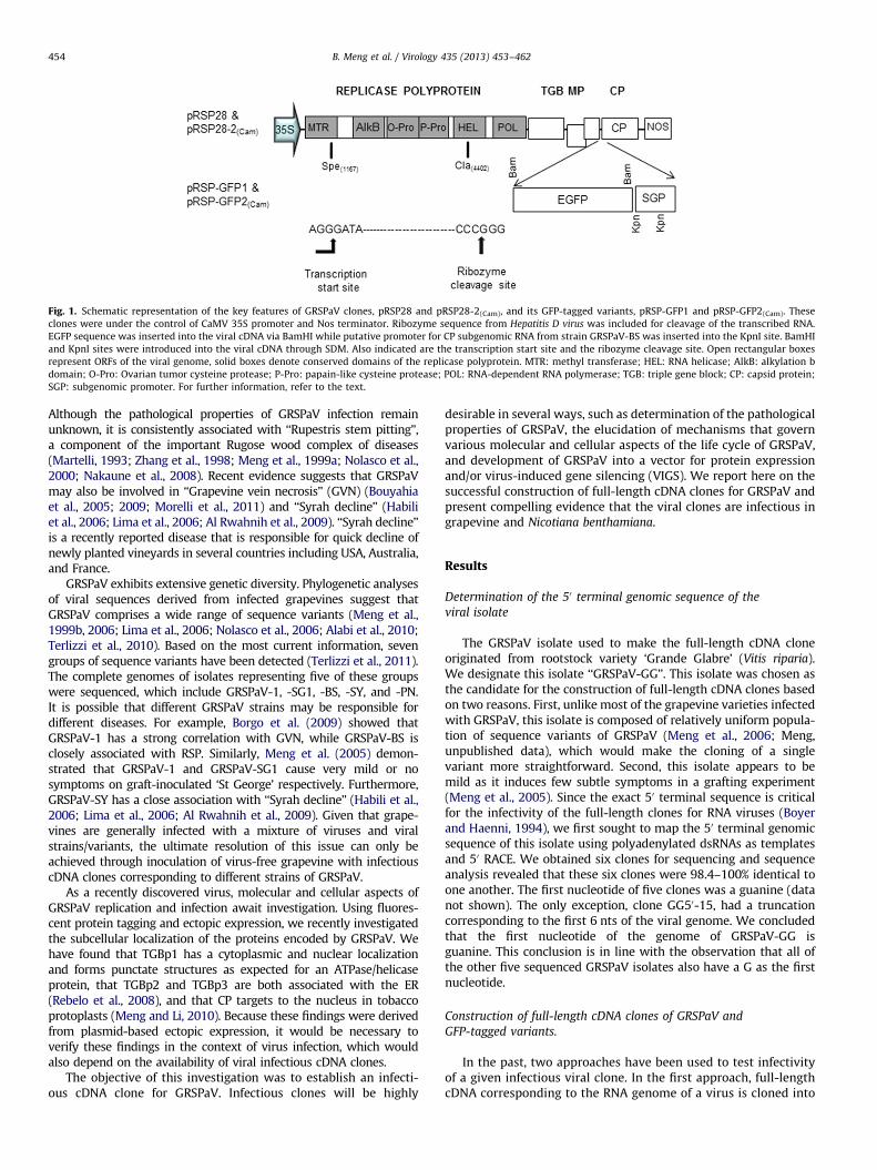
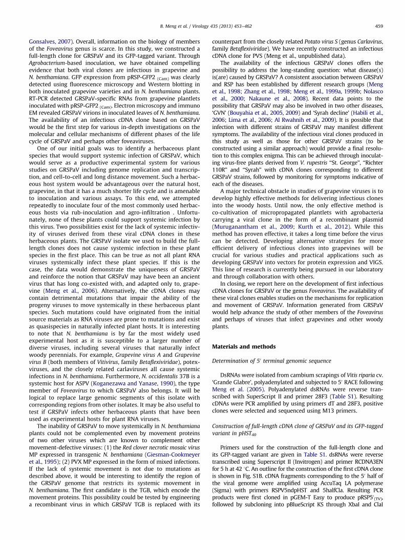
Fig. 1. Schematic representation of the key features of GRSPaV clones, pRSP28 and pRSP28-2(Cam), and its GFP-tagged variants, pRSP-GFP1 and pRSP-GFP2(Cam). These
clones were under the control of CaMV 35S promoter and Nos terminator. Ribozyme sequence from Hepatitis D virus was included for cleavage of the transcribed RNA.
EGFP sequence was inserted into the viral cDNA via BamHI while putative promoter for CP subgenomic RNA from strain GRSPaV-BS was inserted into the KpnI site. BamHI
and KpnI sites were introduced into the viral cDNA through SDM. Also indicated are the transcription start site and the ribozyme cleavage site. Open rectangular boxes
represent ORFs of the viral genome, solid boxes denote conserved domains of the replicase polyprotein. MTR: methyl transferase; HEL: RNA helicase; AlkB: alkylation b
domain; O-Pro: Ovarian tumor cysteine protease; P-Pro: papain-like cysteine protease; POL: RNA-dependent RNA polymerase; TGB: triple gene block; CP: capsid protein;
SGP: subgenomic promoter. For further information, refer to the text.
B. Meng et al. / Virology 435 (2013) 453–462454
Although the pathological properties of GRSPaV infection remainunknown, it is consistently associated with ‘‘Rupestris stem pitting’’,a component of the important Rugose wood complex of diseases(Martelli, 1993; Zhang et al., 1998; Meng et al., 1999a; Nolasco et al.,2000; Nakaune et al., 2008). Recent evidence suggests that GRSPaVmay also be involved in ‘‘Grapevine vein necrosis’’ (GVN) (Bouyahiaet al., 2005; 2009; Morelli et al., 2011) and ‘‘Syrah decline’’ (Habiliet al., 2006; Lima et al., 2006; Al Rwahnih et al., 2009). ‘‘Syrah decline’’is a recently reported disease that is responsible for quick decline ofnewly planted vineyards in several countries including USA, Australia,and France.
GRSPaV exhibits extensive genetic diversity. Phylogenetic analysesof viral sequences derived from infected grapevines suggest thatGRSPaV comprises a wide range of sequence variants (Meng et al.,1999b, 2006; Lima et al., 2006; Nolasco et al., 2006; Alabi et al., 2010;Terlizzi et al., 2010). Based on the most current information, sevengroups of sequence variants have been detected (Terlizzi et al., 2011).The complete genomes of isolates representing five of these groupswere sequenced, which include GRSPaV-1, -SG1, -BS, -SY, and -PN.It is possible that different GRSPaV strains may be responsible fordifferent diseases. For example, Borgo et al. (2009) showed thatGRSPaV-1 has a strong correlation with GVN, while GRSPaV-BS isclosely associated with RSP. Similarly, Meng et al. (2005) demon-strated that GRSPaV-1 and GRSPaV-SG1 cause very mild or nosymptoms on graft-inoculated ‘St George’ respectively. Furthermore,GRSPaV-SY has a close association with ‘‘Syrah decline’’ (Habili et al.,2006; Lima et al., 2006; Al Rwahnih et al., 2009). Given that grape-vines are generally infected with a mixture of viruses and viralstrains/variants, the ultimate resolution of this issue can only beachieved through inoculation of virus-free grapevine with infectiouscDNA clones corresponding to different strains of GRSPaV.
As a recently discovered virus, molecular and cellular aspects ofGRSPaV replication and infection await investigation. Using fluores-cent protein tagging and ectopic expression, we recently investigatedthe subcellular localization of the proteins encoded by GRSPaV. Wehave found that TGBp1 has a cytoplasmic and nuclear localizationand forms punctate structures as expected for an ATPase/helicaseprotein, that TGBp2 and TGBp3 are both associated with the ER(Rebelo et al., 2008), and that CP targets to the nucleus in tobaccoprotoplasts (Meng and Li, 2010). Because these findings were derivedfrom plasmid-based ectopic expression, it would be necessary toverify these findings in the context of virus infection, which wouldalso depend on the availability of viral infectious cDNA clones.
The objective of this investigation was to establish an infecti-ous cDNA clone for GRSPaV. Infectious clones will be highly
desirable in several ways, such as determination of the pathologicalproperties of GRSPaV, the elucidation of mechanisms that governvarious molecular and cellular aspects of the life cycle of GRSPaV,and development of GRSPaV into a vector for protein expressionand/or virus-induced gene silencing (VIGS). We report here on thesuccessful construction of full-length cDNA clones for GRSPaV andpresent compelling evidence that the viral clones are infectious ingrapevine and Nicotiana benthamiana.
Results
Determination of the 50 terminal genomic sequence of the
viral isolate
The GRSPaV isolate used to make the full-length cDNA cloneoriginated from rootstock variety ‘Grande Glabre’ (Vitis riparia).We designate this isolate ‘‘GRSPaV-GG’’. This isolate was chosen asthe candidate for the construction of full-length cDNA clones basedon two reasons. First, unlike most of the grapevine varieties infectedwith GRSPaV, this isolate is composed of relatively uniform popula-tion of sequence variants of GRSPaV (Meng et al., 2006; Meng,unpublished data), which would make the cloning of a singlevariant more straightforward. Second, this isolate appears to bemild as it induces few subtle symptoms in a grafting experiment(Meng et al., 2005). Since the exact 50 terminal sequence is criticalfor the infectivity of the full-length clones for RNA viruses (Boyerand Haenni, 1994), we first sought to map the 50 terminal genomicsequence of this isolate using polyadenylated dsRNAs as templatesand 50 RACE. We obtained six clones for sequencing and sequenceanalysis revealed that these six clones were 98.4–100% identical toone another. The first nucleotide of five clones was a guanine (datanot shown). The only exception, clone GG50-15, had a truncationcorresponding to the first 6 nts of the viral genome. We concludedthat the first nucleotide of the genome of GRSPaV-GG isguanine. This conclusion is in line with the observation that all ofthe other five sequenced GRSPaV isolates also have a G as the firstnucleotide.
Construction of full-length cDNA clones of GRSPaV and
GFP-tagged variants.
In the past, two approaches have been used to test infectivityof a given infectious viral clone. In the first approach, full-lengthcDNA corresponding to the RNA genome of a virus is cloned into
B. Meng et al. / Virology 435 (2013) 453–462 455
a vector under the transcriptional control of a bacteriophagepromoter. Transcripts corresponding to the viral genome areproduced using an in vitro transcription system and used as theinoculum to inoculate a plant host. In the second approach, a full-length viral cDNA is cloned in a vector so that it is flanked byCaMV 35S promoter and a transcription terminator. Plasmidscontaining the viral cDNA can be delivered into plants throughrub-inoculation, biolistic bombardment, or indirectly throughagro-infiltration . The 35S promoter would be recognized by theplant transcription machinery, producing transcripts that areequivalent to the viral genomic RNAs. Viral replication wouldhence commence.
In this study, we took the second approach to construct full-length cDNA clones for GRSPaV. Using a seven step strategy, the firstfull-length clone, pRSP28, was created (Figs. 1 and S1). The cDNAcopy of the viral genome was inserted in pHST40 in such a way thatits transcription was controlled by the 35S promoter and the nopa-line synthase terminator. The in vivo transcripts would undergo selfcleavage to produce the exact viral genomic RNA, due to the activityof the ribozyme sequence that was included in pHST40 (Scholthof,1999). The second construct, pRSP-GFP1, was made so that the EGFPsequence was inserted into the viral cDNA in a position upstream ofthe CP ORF and therefore its transcription would be driven by thenative promoter for the CP subgenomic RNA (Fig. 1). To ensuretranscription of the CP mRNA and to avoid instability of the progenyrecombinant virus, a putative subgenomic promoter sequence wasobtained through PCR from a distinct isolate, GRSPaV-BS (Meng et al.,2005). The regions that contain the putative promoter sequence inthese two isolates are 24% different (data not shown). Upon restric-tion digestion, the PCR products were inserted into the cDNAconstruct upstream of the CP ORF using the KpnI site that wasearlier introduced via site-directed mutagenesis (Fig. 1). Thus, arecombinant viral RNA genome tagged with the GFP sequence wouldbe transcribed in plant cells in a way similar to that of pRSP28.Both pRSP28 and pRSP-GFP1 were introduced into various plantsthrough rub-inoculation . To enable more efficient delivery of viralconstructs into plant tissue, we subcloned inserts in both constructsinto a binary vector, pCambia1390, resulting in pRSP28-2 (Cam) andpRSP-GFP2 (Cam) (Fig. 1. And see Materials and Methods). These newrecombinant plasmids were then used to inoculate plants throughagro-infiltration .
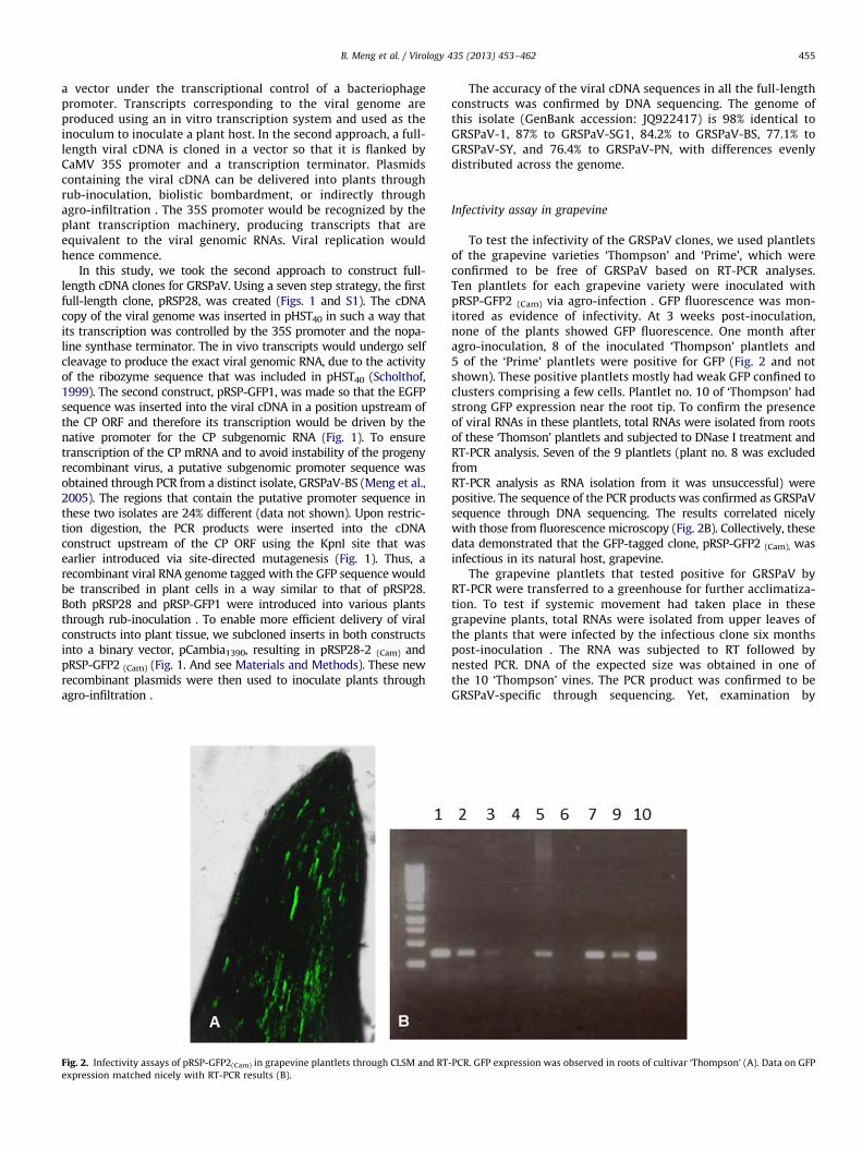
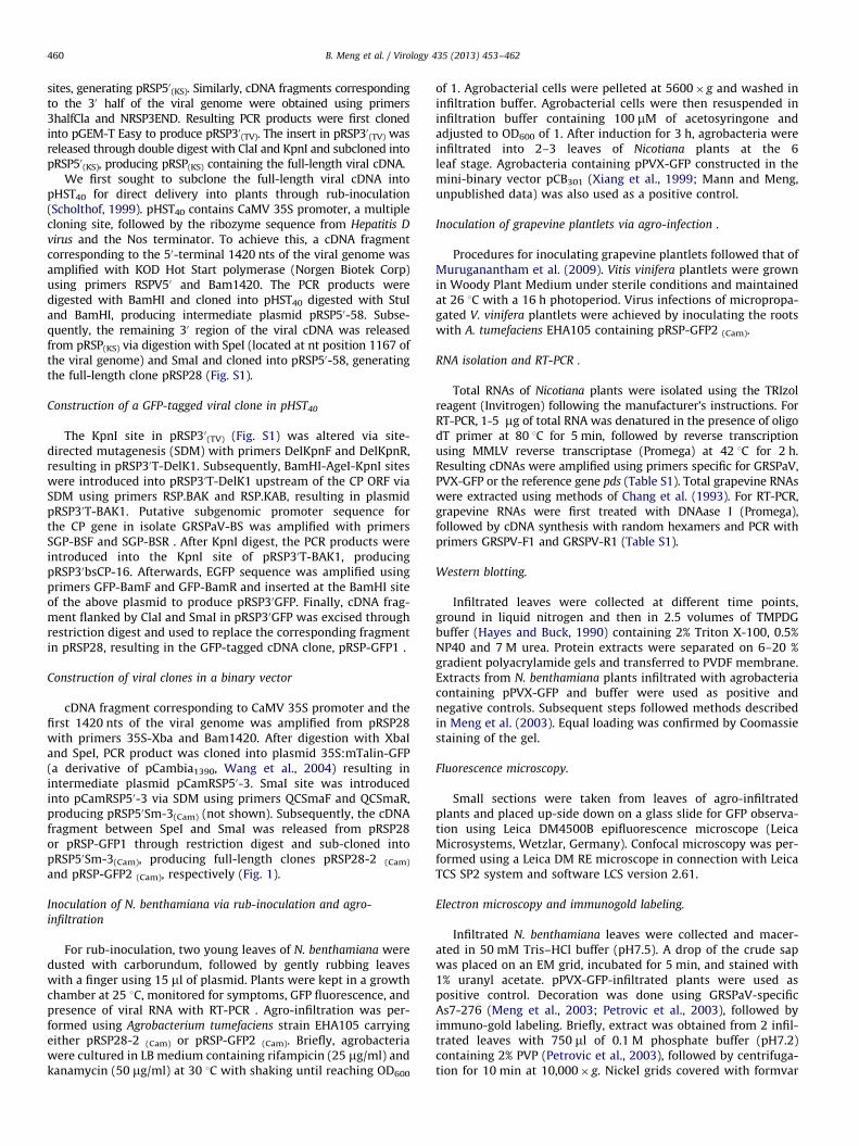
Fig. 2. Infectivity assays of pRSP-GFP2(Cam) in grapevine plantlets through CLSM and RT
expression matched nicely with RT-PCR results (B).
The accuracy of the viral cDNA sequences in all the full-lengthconstructs was confirmed by DNA sequencing. The genome ofthis isolate (GenBank accession: JQ922417) is 98% identical toGRSPaV-1, 87% to GRSPaV-SG1, 84.2% to GRSPaV-BS, 77.1% toGRSPaV-SY, and 76.4% to GRSPaV-PN, with differences evenlydistributed across the genome.
Infectivity assay in grapevine
To test the infectivity of the GRSPaV clones, we used plantletsof the grapevine varieties ‘Thompson’ and ‘Prime’, which wereconfirmed to be free of GRSPaV based on RT-PCR analyses.Ten plantlets for each grapevine variety were inoculated withpRSP-GFP2 (Cam) via agro-infection . GFP fluorescence was mon-itored as evidence of infectivity. At 3 weeks post-inoculation,none of the plants showed GFP fluorescence. One month afteragro-inoculation, 8 of the inoculated ‘Thompson’ plantlets and5 of the ‘Prime’ plantlets were positive for GFP (Fig. 2 and notshown). These positive plantlets mostly had weak GFP confined toclusters comprising a few cells. Plantlet no. 10 of ‘Thompson’ hadstrong GFP expression near the root tip. To confirm the presenceof viral RNAs in these plantlets, total RNAs were isolated from rootsof these ‘Thomson’ plantlets and subjected to DNase I treatment andRT-PCR analysis. Seven of the 9 plantlets (plant no. 8 was excludedfromRT-PCR analysis as RNA isolation from it was unsuccessful) werepositive. The sequence of the PCR products was confirmed as GRSPaVsequence through DNA sequencing. The results correlated nicelywith those from fluorescence microscopy (Fig. 2B). Collectively, thesedata demonstrated that the GFP-tagged clone, pRSP-GFP2 (Cam), wasinfectious in its natural host, grapevine.
The grapevine plantlets that tested positive for GRSPaV byRT-PCR were transferred to a greenhouse for further acclimatiza-tion. To test if systemic movement had taken place in thesegrapevine plants, total RNAs were isolated from upper leaves ofthe plants that were infected by the infectious clone six monthspost-inoculation . The RNA was subjected to RT followed bynested PCR. DNA of the expected size was obtained in one ofthe 10 ‘Thompson’ vines. The PCR product was confirmed to beGRSPaV-specific through sequencing. Yet, examination by
-PCR. GFP expression was observed in roots of cultivar ‘Thompson’ (A). Data on GFP
B. Meng et al. / Virology 435 (2013) 453–462456
confocal fluorescence microscopy of this GRSPaV-positive plantreviewed no fluorescence due to GFP, suggesting extremely lowvirus titer or loss of the GFP sequence.
Infectivity assay in N. benthamiana through agro-infiltration
Because N. benthamiana is by far the most commonly usedherbaceous host for many plant viruses (Goodin et al., 2008), we firstattempted to infect N. benthamiana plants through rub-inoculation .However, repeated attempts were unsuccessful as judged by lack ofsymptoms and green fluorescence, as well as negative RT-PCR results(Meng et al., 2009 and data not shown). These data suggestedthat rub-inoculation was not suitable for initiating infection inN. benthamiana using cDNA clones of GRSPaV. To increase efficiencyof inoculation, we then attempted inoculation through agro-infiltration . This method could achieve high levels of inoculationefficiency as agrobacteria can deliver T-DNA containing viral cDNAinto a majority of plant cells. Again, upon agro-infiltration withpRSP28-2 (Cam), no symptoms were observed on either the infiltrated
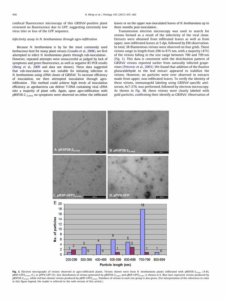
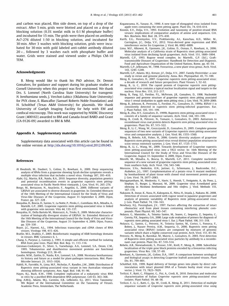
Fig. 3. Electron micrographs of virions observed in agro-infiltrated plants. Virions
pRSP-GFP2(Cam) (C), or pPVX-GFP (D). Size distribution of virions generated by pRSP28-
pRSP28-2(Cam), while red bars denote virions produced by pRSP-GFP2(Cam). Numbers of v
in this figure legend, the reader is referred to the web version of this article.)
leaves or on the upper non-inoculated leaves of N. benthamiana up tothree months post-inoculation .
Transmission electron microscopy was used to search forvirions formed as a result of the infectivity of the viral clone.Extracts were obtained from infiltrated leaves as well as fromupper, non-infiltrated leaves at 5 dpi, followed by EM observation.In total, 38 filamentous virions were observed on four grids. Thesevirions range in length from 296 to 875 nm, with a majority (47%)of the virions falling in the size range between 700 and 799 nm(Fig. 3). This data is consistent with the distribution pattern ofGRSPaV virions reported earlier from naturally infected grape-vines (Petrovic et al., 2003). We found that addition of the fixativeglutaraldehyde to the leaf extract appeared to stabilize thevirions. However, no particles were ever observed in extractsmade from upper, non-infiltrated leaves. To verify the identity ofthese virions, immunogold labeling using GRSPaV-specific anti-serum, As7-276, was performed, followed by electron microscopy.As shown in Fig. 3B, these virions were clearly labeled withgold particles, confirming their identify as GRSPaV. Observation of
shown were from N. benthamiana plants infiltrated with pRSP28-2(Cam) (A-B),
2(Cam) and pRSP-GFP2(Cam) is shown in E. Blue bars represent virions produced by
irions in each size group is also given. (For interpretation of the references to color
B. Meng et al. / Virology 435 (2013) 453–462 457
GRSPaV virions demonstrates that this viral clone was indeedinfectious, at least in the infiltrated leaves of N. benthamiana.
Electron microscopy of extracts from leaves infiltrated withpRSP-GFP2 (Cam) at 5 dpi also revealed the presence of virionsindistinguishable from those produced by pRSP28-2 (Cam)
(Fig. 3C). These data suggest that this GFP-tagged viral clone isalso infectious in N. benthamiana leaves upon agro-infiltration .However, the number of virions produced by pRSP-GFP2 (Cam) wasmuch lower than that from pRSP28-2 (Cam)-infiltrated leaves.
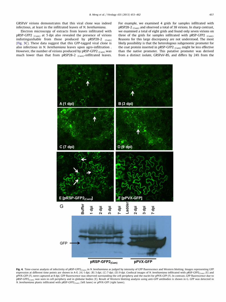
Fig. 4. Time-course analysis of infectivity of pRSP-GFP2(Cam) in N. benthamiana as judge
expression at different time points are shown in A-E. (A) 1 dpi; (B) 3 dpi; (C) 7 dpi; (D)
pPVX-GFP (F), were captured at 8 dpi. GFP fluorescence was observed surrounding the c
pRSP-GFP2(Cam) was seen in cell periphery and in globular bodies (E). Result of Wester
N. benthamiana plants infiltrated with pRSP-GFP2(Cam) (left lanes) or pPVX-GFP (right l
For example, we examined 4 grids for samples infiltrated withpRSP28-2 (Cam) and observed a total of 38 virions. In sharp contrast,we examined a total of eight grids and found only seven virions onthree of the grids for samples infiltrated with pRSP-GFP2 (Cam).Reasons for this large discrepancy are not understood. The mostlikely possibility is that the heterologous subgenomic promoter forthe coat protein inserted in pRSP-GFP2 (Cam) might be less effectivethan the native promoter. This putative promoter was derivedfrom a distinct isolate, GRSPaV-BS, and differs by 24% from the
d by intensity of GFP fluorescence and Western blotting. Images representing GFP
9 dpi. Confocal images of N. benthamiana infiltrated with pRSP-GFP2(Cam) (E) and
ell periphery and the nuclei for pPVX-GFP (F). In contrast, GFP fluorescence due to
n blotting analysis using anti-GFP antibodies is shown in G. GFP was detected in
anes).
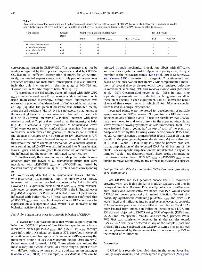
Table 1Agro-infiltration of four commonly used herbaceous plant species for viral cDNA clones of GRSPaV. For each plant, 2 leaves (1 partially expanded
and 1 fully expanded) were infiltrated with buffer or agrobacterial suspension containing either pRSP28-2(Cam) or pRSP-GFP2(Cam).
Plant species Growth
stage
Number of plants inoculated with RT-PCR result
Buffer pRSP28-2(Cam) pRSP-GFP2(Cam) PDS GRSPaV
Nicotiana benthamiana 6 1 4 4 þ �
N. benthamiana-MP 6 1 4 4 þ �
N. occidentalis 37B 6–7 1 4 4 þ �
N. clevelandii 8–9 1 4 4 þ �
B. Meng et al. / Virology 435 (2013) 453–462458
corresponding region in GRSPaV-GG . This sequence may not bereadily recognized by the replicase enzymes encoded by GRSPaV-GG, leading to inefficient transcription of mRNA for CP. Alterna-tively, the inserted sequence may contain only part of the promotersequence required for maximum transcription. It is also interest-ingly that only 1 virion fell in the size range of 700–799 and3 virions fell in the size range of 600–699 (Fig. 3E).
To corroborate the EM results, plants infiltrated with pRSP-GFP2
(Cam) were monitored for GFP expression at different time pointsusing fluorescence microscopy. Strong green fluorescence wasobserved in patches of epidermal cells of infiltrated leaves startingat 1 dpi (Fig. 4A). The green fluorescence was distributed mainlyalong the cell periphery (Fig. 4A–D ). It is noteworthy that numerousfluorescent globular structures were also observed in these cells(Fig. 4A–D , arrows). Intensity of GFP signal increased with time,reached a peak at 7 dpi, and remained at similar intensity at 9 dpi(Fig. 4). To achieve a higher resolution, N. benthamiana leaves7 dpi were observed under confocal laser scanning fluorescencemicroscope, which revealed the general GFP fluorescence as well asthe globular structures (Fig. 4E). Similar to EM observations, GFPfluorescence was never observed in upper, non-infiltrated leavesthroughout the entire course of observation. As a control, agrobac-teria containing pPVX-GFP was also infiltrated into N. benthamiana
leaves. Typical and uniform green fluorescence was observed in boththe infiltrated and the upper non-infiltrated leaves (Fig. 4F).
To further verify the above findings, crude protein extracts wereobtained from the leaves of N. benthamiana plants that wereinoculated with pRSP-GFP2 (Cam) or pPVX-GFP and tested byWestern blotting. As shown in Fig. 4G, protein bands correspondingtoGFP were clearly detected in N. benthamiana leaves infiltratedwith pRSP-GFP2 (Cam) as early as 1 dpi. The intensity of GFP slowlyincreased with time and reached a maximum by 7 dpi (Fig. 4G).However, GFP expression levels of pRSP-GFP2 (Cam) were consider-ably lower compared to those of pPVX-GFP in the infiltrated leaves(Fig. 4G). As expected, GFP was not detected in control plants mock-infiltrated with buffer (Fig. 4G). These data further confirmed thatpRSP-GFP2 (Cam) was capable of replication as GFP could only beexpressed on a subgenomic RNA, which is an indicator of thebiological activity of the viral clone.
Search for a herbaceous Host for systemic infection of GRSPaV
To search for a herbaceous host that would support systemicinfection of GRSPaV, plants of four Nicotiana species were inocu-lated with clones pRSP28-2 (Cam) and pRSP-GFP2 (Cam) throughagro-infiltration: Nicotiana occidentalis 37B, Nicotiana clevelandii,N. benthamiana, and transgenic N. benthamiana-MP expressing themovement protein of Red clover necrotic mosaic virus (RCNMV)(Vaewhongs and Lommel, 1995). These plants are among themost susceptible systemic hosts for a wide range of plant viruseswith different origin, genome structure and expression strategies(Goodin et al., 2008). For example, N. occidentalis 37B can be
infected through mechanical inoculation, albeit with difficulty,and serves as a systemic host for Apple stem pitting virus, the typemember of the Foveavirus genus (King et al., 2011; Koganezawaand Yanase, 1990). Inclusion of transgenic N. benthamiana wasbased on the observation that RCNMV MP complemented move-ment of several diverse viruses which were rendered defectivein movement, including PVX and Tobacco mosaic virus (Morozovet al., 1997; Giesman-Cookmeyer et al., 1995). In total, nineseparate experiments were conducted involving some or all ofthese plant species in each experiment. Table 1 shows the resultof one of these experiments in which all four Nicotiana specieswere tested in a single experiment.
Inoculated plants were monitored for development of possiblesymptoms and for GFP expression. No symptoms or GFP were everdetected on any of these plants. To test the possibility that GRSPaVmay have moved to, and were present in, the upper non-inoculatedleaves without showing symptoms or GFP fluorescence, total RNAswere isolated from a young leaf on top of each of the plants at24 dpi and tested by RT-PCR using virus-specific primers RSP21 andRSP22. As internal control, primers PDS853F and PDS1352R that arespecific to the plant gene phytoene desaturase (pds) were also usedin RT-PCR . While RT-PCR using PDS-specific primers producedstrong amplification of the expected DNA for all but one of theplants, GRSPaV-specific amplification was not detected in any of thesamples ( Table 1 and data not shown). These results demonstratedthat viruses derived from pRSP28-2 (Cam) or pRSP-GFP2 (Cam) wereunable to move systemically in any of these four Nicotiana species.
Co-infection with PVX does not enable GRSPaV to move systemically
in N. benthamiana
Both GRSPaV and PVX genomes encode the TGB movementproteins, which are highly similar in modular structure and thus inbiological function. Because PVX readily infects N. benthamiana
both locally and systemically, we hoped that PVX would enableGRSPaV to move systemically in mixed infection. To test thispossibility, agrobacteria containing pRSP28-2 (Cam) and pPVX-GFPwere mixed, and infiltrated into N. benthamiana leaves. As controls,N. benthamiana plants were also infiltrated with buffer. Total RNAswere isolated from upper, non-infiltrated leaves at 9, 14, 27, and150 dpi and subjected to RT-PCR using GRSPaV-specific (RSP21 andRSP22) and PVX-specific (PVX6440 and PVX6972) primers. WhilePVX RNA was consistently detected in all the samples tested,GRSPaV RNA was never detected in any of the samples (data notshown). This data suggested that GRSPaV systemic movement wasnot complemented by the movement function encoded by PVX inthe context of co-infection .
Discussion
GRSPaV is a recently identified virus in the genus Foveavirus
(family Betaflexiviridae) and is widespread in grapevines (Meng and
B. Meng et al. / Virology 435 (2013) 453–462 459
Gonsalves, 2007). Overall, information on the biology of membersof the Foveavirus genus is scarce. In this study, we constructed afull-length clone for GRSPaV and its GFP-tagged variant. ThroughAgrobacterium-based inoculation, we have obtained compellingevidence that both viral clones are infectious in grapevine andN. benthamiana. GFP expression from pRSP-GFP2 (Cam) was clearlydetected using fluorescence microscopy and Western blotting inboth inoculated grapevine varieties and in N. benthamiana plants.RT-PCR detected GRSPaV-specific RNAs from grapevine plantletsinoculated with pRSP-GFP2 (Cam). Electron microscopy and immunoEM revealed GRSPaV virions in inoculated leaves of N. benthamiana.The availability of an infectious cDNA clone based on GRSPaVwould be the first step for various in-depth investigations on themolecular and cellular mechanisms of different phases of the lifecycle of GRSPaV and perhaps other foveaviruses.
One of our initial goals was to identify a herbaceous plantspecies that would support systemic infection of GRSPaV, whichwould serve as a productive experimental system for variousstudies on GRSPaV including genome replication and transcrip-tion, and cell-to-cell and long distance movement. Such a herbac-eous host system would be advantageous over the natural host,grapevine, in that it has a much shorter life cycle and is amenableto inoculation and various assays. To this end, we attemptedrepeatedly to inoculate four of the most commonly used herbac-eous hosts via rub-inoculation and agro-infiltration . Unfortu-nately, none of these plants could support systemic infection bythis virus. Two possibilities exist for the lack of systemic infectiv-ity of viruses derived from these viral cDNA clones in theseherbaceous plants. The GRSPaV isolate we used to build the full-length clones does not cause systemic infection in these plantspecies in the first place. This can be true as not all plant RNAviruses systemically infect these plant species. If this is thecase, the data would demonstrate the uniqueness of GRSPaVand reinforce the notion that GRSPaV may have been an ancientvirus that has long co-existed with, and adapted only to, grape-vine (Meng et al., 2006). Alternatively, the cDNA clones maycontain detrimental mutations that impair the ability of theprogeny viruses to move systemically in these herbaceous plantspecies. Such mutations could have originated from the initialsource materials as RNA viruses are prone to mutations and existas quasispecies in naturally infected plant hosts. It is interestingto note that N. benthamiana is by far the most widely usedexperimental host as it is susceptible to a larger number ofdiverse viruses, including several viruses that naturally infectwoody perennials. For example, Grapevine virus A and Grapevine
virus B (both members of Vitivirus, family Betaflexiviridae), potex-viruses, and the closely related carlaviruses all cause systemicinfections in N. benthamiana. Furthermore, N. occidentalis 37B is asystemic host for ASPV (Koganezawa and Yanase, 1990), the typemember of Foveavirus to which GRSPaV also belongs. It will belogical to replace large genomic segments of this isolate withcorresponding regions from other isolates. It may be also useful totest if GRSPaV infects other herbaceous plants that have beenused as experimental hosts for plant RNA viruses.
The inability of GRSPaV to move systemically in N. benthamiana
plants could not be complemented even by movement proteinsof two other viruses which are known to complement othermovement-defective viruses: (1) the Red clover necrotic mosaic virus
MP expressed in transgenic N. benthamiana (Giesman-Cookmeyeret al., 1995); (2) PVX MP expressed in the form of mixed infections.If the lack of systemic movement is not due to mutations asdescribed above, it would be interesting to identify the region ofthe GRSPaV genome that restricts its systemic movement inN. benthamiana. The first candidate is the TGB, which encode themovement proteins. This possibility could be tested by engineeringa recombinant virus in which GRSPaV TGB is replaced with its
counterpart from the closely related Potato virus S (genus Carlavirus,family Betaflexiviridae). We have recently constructed an infectiouscDNA clone for PVS (Meng et al., unpublished data).
The availability of the infectious GRSPaV clones offers thepossibility to address the long-standing question: what disease(s)is(are) caused by GRSPaV? A consistent association between GRSPaVand RSP has been established by different research groups (Menget al., 1998; Zhang et al., 1998; Meng et al., 1999a, 1999b; Nolascoet al., 2000; Nakaune et al., 2008). Recent data points to thepossibility that GRSPaV may also be involved in two other diseases,‘GVN’ (Bouyahia et al., 2005, 2009) and ‘Syrah decline’ (Habili et al.,2006; Lima et al., 2006; Al Rwahnih et al., 2009). It is possible thatinfection with different strains of GRSPaV may manifest differentsymptoms. The availability of the infectious viral clones produced inthis study as well as those for other GRSPaV strains (to beconstructed using a similar approach) would provide a final resolu-tion to this complex enigma. This can be achieved through inoculat-ing virus-free plants derived from V. rupestris ‘‘St. George’’, ‘‘Richter110R’’ and ‘‘Syrah’’ with cDNA clones corresponding to differentGRSPaV strains, followed by monitoring for symptoms indicative ofeach of the diseases.
A major technical obstacle in studies of grapevine viruses is todevelop highly effective methods for delivering infectious clonesinto the woody hosts. Until now, the only effective method isco-cultivation of micropropagated plantlets with agrobacteriacarrying a viral clone in the form of a recombinant plasmid(Muruganantham et al., 2009; Kurth et al., 2012). While thismethod has proven effective, it takes a long time before the viruscan be detected. Developing alternative strategies for moreefficient delivery of infectious clones into grapevines will becrucial for various studies and practical applications such asdeveloping GRSPaV into vectors for protein expression and VIGS.This line of research is currently being pursued in our laboratoryand through collaboration with others.
In closing, we report here on the development of first infectiouscDNA clones for GRSPaV or the genus Foveavirus. The availability ofthese viral clones enables studies on the mechanisms for replicationand movement of GRSPaV. Information generated from GRSPaVwould help advance the study of other members of the Foveavirus
and perhaps of viruses that infect grapevines and other woodyplants.
Materials and methods
Determination of 50 terminal genomic sequence
DsRNAs were isolated from cambium scrapings of Vitis riparia cv.‘Grande Glabre’, polyadenylated and subjected to 50 RACE followingMeng et al. (2005). Polyadenylated dsRNAs were reverse tran-scribed with SuperScript II and primer 28F3 (Table S1). ResultingcDNAs were PCR amplified by using primers dT and 28F3, positiveclones were selected and sequenced using M13 primers.
Construction of full-length cDNA clone of GRSPaV and its GFP-tagged
variant in pHST40
Primers used for the construction of the full-length clone andits GFP-tagged variant are given in Table S1. dsRNAs were reversetranscribed using Superscript II (Invitrogen) and primer RCDNA3ENfor 5 h at 42 1C. An outline for the construction of the first cDNA cloneis shown in Fig. S1B. cDNA fragments corresponding to the 50 half ofthe viral genome were amplified using AccuTaq LA polymerase(Sigma) with primers RSPV5ndpHST and 5halfCla. Resulting PCRproducts were first cloned in pGEM-T Easy to produce pRSP50(TV),followed by subcloning into pBlueScript KS through XbaI and ClaI
B. Meng et al. / Virology 435 (2013) 453–462460
sites, generating pRSP50(KS). Similarly, cDNA fragments correspondingto the 30 half of the viral genome were obtained using primers3halfCla and NRSP3END. Resulting PCR products were first clonedinto pGEM-T Easy to produce pRSP30(TV). The insert in pRSP30(TV) wasreleased through double digest with ClaI and KpnI and subcloned intopRSP50(KS), producing pRSP(KS) containing the full-length viral cDNA.
We first sought to subclone the full-length viral cDNA intopHST40 for direct delivery into plants through rub-inoculation(Scholthof, 1999). pHST40 contains CaMV 35S promoter, a multiplecloning site, followed by the ribozyme sequence from Hepatitis D
virus and the Nos terminator. To achieve this, a cDNA fragmentcorresponding to the 50-terminal 1420 nts of the viral genome wasamplified with KOD Hot Start polymerase (Norgen Biotek Corp)using primers RSPV50 and Bam1420. The PCR products weredigested with BamHI and cloned into pHST40 digested with StuIand BamHI, producing intermediate plasmid pRSP50-58. Subse-quently, the remaining 30 region of the viral cDNA was releasedfrom pRSP(KS) via digestion with SpeI (located at nt position 1167 ofthe viral genome) and SmaI and cloned into pRSP50-58, generatingthe full-length clone pRSP28 (Fig. S1).
Construction of a GFP-tagged viral clone in pHST40
The KpnI site in pRSP30(TV) (Fig. S1) was altered via site-directed mutagenesis (SDM) with primers DelKpnF and DelKpnR,resulting in pRSP30T-DelK1. Subsequently, BamHI-AgeI-KpnI siteswere introduced into pRSP30T-DelK1 upstream of the CP ORF viaSDM using primers RSP.BAK and RSP.KAB, resulting in plasmidpRSP30T-BAK1. Putative subgenomic promoter sequence forthe CP gene in isolate GRSPaV-BS was amplified with primersSGP-BSF and SGP-BSR . After KpnI digest, the PCR products wereintroduced into the KpnI site of pRSP30T-BAK1, producingpRSP30bsCP-16. Afterwards, EGFP sequence was amplified usingprimers GFP-BamF and GFP-BamR and inserted at the BamHI siteof the above plasmid to produce pRSP30GFP. Finally, cDNA frag-ment flanked by ClaI and SmaI in pRSP30GFP was excised throughrestriction digest and used to replace the corresponding fragmentin pRSP28, resulting in the GFP-tagged cDNA clone, pRSP-GFP1 .
Construction of viral clones in a binary vector
cDNA fragment corresponding to CaMV 35S promoter and thefirst 1420 nts of the viral genome was amplified from pRSP28with primers 35S-Xba and Bam1420. After digestion with XbaIand SpeI, PCR product was cloned into plasmid 35S:mTalin-GFP(a derivative of pCambia1390, Wang et al., 2004) resulting inintermediate plasmid pCamRSP50-3. SmaI site was introducedinto pCamRSP50-3 via SDM using primers QCSmaF and QCSmaR,producing pRSP50Sm-3(Cam) (not shown). Subsequently, the cDNAfragment between SpeI and SmaI was released from pRSP28or pRSP-GFP1 through restriction digest and sub-cloned intopRSP50Sm-3(Cam), producing full-length clones pRSP28-2 (Cam)
and pRSP-GFP2 (Cam), respectively (Fig. 1).
Inoculation of N. benthamiana via rub-inoculation and agro-
infiltration
For rub-inoculation, two young leaves of N. benthamiana weredusted with carborundum, followed by gently rubbing leaveswith a finger using 15 ml of plasmid. Plants were kept in a growthchamber at 25 1C, monitored for symptoms, GFP fluorescence, andpresence of viral RNA with RT-PCR . Agro-infiltration was per-formed using Agrobacterium tumefaciens strain EHA105 carryingeither pRSP28-2 (Cam) or pRSP-GFP2 (Cam). Briefly, agrobacteriawere cultured in LB medium containing rifampicin (25 mg/ml) andkanamycin (50 mg/ml) at 30 1C with shaking until reaching OD600
of 1. Agrobacterial cells were pelleted at 5600� g and washed ininfiltration buffer. Agrobacterial cells were then resuspended ininfiltration buffer containing 100 mM of acetosyringone andadjusted to OD600 of 1. After induction for 3 h, agrobacteria wereinfiltrated into 2–3 leaves of Nicotiana plants at the 6leaf stage. Agrobacteria containing pPVX-GFP constructed in themini-binary vector pCB301 (Xiang et al., 1999; Mann and Meng,unpublished data) was also used as a positive control.
Inoculation of grapevine plantlets via agro-infection .
Procedures for inoculating grapevine plantlets followed that ofMuruganantham et al. (2009). Vitis vinifera plantlets were grownin Woody Plant Medium under sterile conditions and maintainedat 26 1C with a 16 h photoperiod. Virus infections of micropropa-gated V. vinifera plantlets were achieved by inoculating the rootswith A. tumefaciens EHA105 containing pRSP-GFP2 (Cam).
RNA isolation and RT-PCR .
Total RNAs of Nicotiana plants were isolated using the TRIzolreagent (Invitrogen) following the manufacturer’s instructions. ForRT-PCR, 1-5 mg of total RNA was denatured in the presence of oligodT primer at 80 1C for 5 min, followed by reverse transcriptionusing MMLV reverse transcriptase (Promega) at 42 1C for 2 h.Resulting cDNAs were amplified using primers specific for GRSPaV,PVX-GFP or the reference gene pds (Table S1). Total grapevine RNAswere extracted using methods of Chang et al. (1993). For RT-PCR,grapevine RNAs were first treated with DNAase I (Promega),followed by cDNA synthesis with random hexamers and PCR withprimers GRSPV-F1 and GRSPV-R1 (Table S1).
Western blotting.
Infiltrated leaves were collected at different time points,ground in liquid nitrogen and then in 2.5 volumes of TMPDGbuffer (Hayes and Buck, 1990) containing 2% Triton X-100, 0.5%NP40 and 7 M urea. Protein extracts were separated on 6–20 %gradient polyacrylamide gels and transferred to PVDF membrane.Extracts from N. benthamiana plants infiltrated with agrobacteriacontaining pPVX-GFP and buffer were used as positive andnegative controls. Subsequent steps followed methods describedin Meng et al. (2003). Equal loading was confirmed by Coomassiestaining of the gel.
Fluorescence microscopy.
Small sections were taken from leaves of agro-infiltratedplants and placed up-side down on a glass slide for GFP observa-tion using Leica DM4500B epifluorescence microscope (LeicaMicrosystems, Wetzlar, Germany). Confocal microscopy was per-formed using a Leica DM RE microscope in connection with LeicaTCS SP2 system and software LCS version 2.61.
Electron microscopy and immunogold labeling.
Infiltrated N. benthamiana leaves were collected and macer-ated in 50 mM Tris–HCl buffer (pH7.5). A drop of the crude sapwas placed on an EM grid, incubated for 5 min, and stained with1% uranyl acetate. pPVX-GFP-infiltrated plants were used aspositive control. Decoration was done using GRSPaV-specificAs7-276 (Meng et al., 2003; Petrovic et al., 2003), followed byimmuno-gold labeling. Briefly, extract was obtained from 2 infil-trated leaves with 750 ml of 0.1 M phosphate buffer (pH7.2)containing 2% PVP (Petrovic et al., 2003), followed by centrifuga-tion for 10 min at 10,000� g. Nickel grids covered with formvar
B. Meng et al. / Virology 435 (2013) 453–462 461
and carbon was placed, film side down, on top of a drop of theextract. After 5 min, grids were blotted and placed on a drop ofblocking solution (0.3% nonfat milk in 0.1 M phosphate buffer)and incubated for 15 min. The grids were then placed on antibodyAs7-276 diluted 1:50 in blocking solution, and incubated for30 min. After 3 washes with blocking solution, grids were incu-bated for 30 min with gold labeled anti-rabbit antibody diluted20� , followed by 3 washes each with phosphate buffer andwater. Grids were stained and viewed under a Philips CM-10TEM.
Acknowledgments
B. Meng would like to thank his PhD advisor, Dr. DennisGonsalves, for guidance and support during his graduate studies atCornell University when this project was first envisioned. We thankDrs. S. Lommel (North Carolina State University) for transgenicN. benthamiana seeds, J. Verchot-Lubicz (Oklahoma State University)for PVX clone, E. Blancaflor (Samuel Roberts Noble Foundation) andH. Scholthof (Texas A&M University) for plasmids. We thankUniversity of Guelph Imaging Facility for help with EM andconfocal microscopy. This work was supported by NSERC DiscoveryGrant (400163) awarded to BM and Canada-Israel BARD and Grant(CA-9126-09) awarded to BM & MM.
Appendix A. Supplementary materials
Supplementary data associated with this article can be found inthe online version at http://dx.doi.org/10.1016/j.virol.2012.09.045.
References
Al Rwahnih, M., Daubert, S., Golino, D., Rowhani, A., 2009. Deep sequencinganalysis of RNAs from a grapevine showing Syrah decline symptoms reveals amultiple vı́rus infection that includes a novel vı́rus. Virology 387, 395–410.
Alabi, O.J., Martin, R.R., Naidu, R.A., 2010. Sequence diversity, population geneticsand potential recombination events in grapevine rupestris stem pitting-associated vı́rus in Pacific North-West vineyards. J. Gen. Virol. 91, 265–276.
Borgo, M., Bertazzon, N., Anaclerio, F., Angelini, E., 2009. Different variants ofGRSPaV are associated to diverse diseases in grapevine. In: Extended Abstractsof the 16th Meeting of the International Council for the Study of Virus andVirus-like Diseases of the Grapevine, August 31–September 4, 2009. Dijon,France, pp. 327–328 .
Bouyahia, H., Boscia, D., Savino, V., La Notte, P., Pirolo, C., Castellano, M.A., Minafra, A.,Martelli, G.P., 2005. Grapevine rupestris stem pitting-associated virus is linkedwith grapevine vein necrosis. Vitis 44, 133–137.
Bouyahia, H., Della Bartola M., Materazzi, A., Triolo, E., 2009. Molecular character-ization of biologically divergent strains of GRSPaV. In: Extended Abstracts ofthe 16th Meeting of the International Council for the Study of Virus and Virus-like Diseases of the Grapevine, August 31–September 4, 2009, Dijon, France,pp. 329–330 .
Boyer, J.C., Haenni, A.L., 1994. Infectious transcripts and cDNA clones of RNAviruses. Virology 198, 415–426.
Bratlie, M.S., Drablos, F., 2005. Bioinformatic mapping of AlkB homology domainsin viruses. BMC Genomics 6, 1.
Chang, S., Puryear, J., Cairney, J., 1993. A simple and efficient method for isolatingRNA from pine trees. Plant Mol. Biol. Rep. 11, 113–116.
Giesman-Cookmeyer, D., Silver, S., Vaewhongs, A.A., Lommel, S.A., Deom, C.M.,1995. Tobamovirus and dianthovirus movement proteins are functionalhomologous. Virology 213, 38–45.
Goodin, M.M., Zaitlin, D., Naidu, R.A., Lommel, S.A., 2008. Nicotiana benthamiana:its history and future as a model for plant-pathogen interactions. Mol. Plant-Microbe Interact. 21, 1015–1026.
Habili, N., Farrokhi, N., Lima, M.F., Nicholas, P., Randles, J.W., 2006. Distribution ofRupestris stem pitting-associated virus variant in two Australian vineyardsshowing different symptoms. Ann. Appl. Biol. 148, 91–96.
Hayes, R.J., Buck, K.W., 1990. Complete replication of a eukaryotic virus RNAin vitro by a purified RNA-dependent RNA polymerase. Cell 63, 363–368.
King, A.M.Q., Adams, M.J., Carstens, E.B., Lefkowitz, E.J., 2011. Virus Taxonomy:9th Report of the International Committee on the Taxonomy of Viruses.Academic Press, Amsterdam, The Netherlands.
Koganezawa, H., Yanase, H., 1990. A new type of elongagted virus isolated fromapple trees containing the stem pitting agent. Plant Dis. 74, 610–614.
Koonin, E.V., Dolja, V.V., 1993. Evolution and taxonomy of positive-strand RNAviruses: implications of comparative analysis of amino acid sequences. Crit.Rev. Biochem. Mol. Biol. 28, 375–430.
Kurth, E.G., Peremyslov, V.V., Prokhnevsky, A.I., Kasschau, K.D., Miller, M.,Carrington, J.C., Dolja, V.V., 2012. Virus-derived gene expression and RNAinterference vector fro Grapevine. J. Virol. 86, 6002–6009.
Lima, M.F., Alkowni, R., Uyemoto, J.K., Golino, D., Osman, F., Rowhani, A., 2006.Molecular analysis of a California strain of Rupestris stem pitting-associatedvirus isolated from declining Syrah grapevines. Arch. Virol. 151, 1889–1894.
Martelli, G.P., 1993. Rugose wood complex. In: Martelli., G.P. (Ed.), Graft-transmissible Diseases of Grapevines: Handbook for Detection and Diagnosis.Food and Agriculture Organization of the United Nations, Rome, pp. 45–54.
Martelli, G.P., Jelkmann, W., 1998. Foveavirus, a new plant virus genus. Arch. Virol.143, 1245–1249.
Martelli, G.P., Adams, M.J., Kreuze, J.F., Dolja, V.V., 2007. Family Flexiviridae: a casestudy in virion and genome plasticity. Annu. Rev. Phytopathol. 45, 73–100.
Meng, B., Gonsalves, D., 2007. Grapevine rupestris stem pitting-associated virus:a decade of research and future perspectives. Plant Viruses 1, 52–62.
Meng, B., Li, C., 2010. The capsid protein of Grapevine rupestris stem pitting-associated virus contains a typical nuclear localization signal and targets to thenucleus. Virus Res. 153, 212–217.
Meng, B., Pang, S.Z., Forsline, P.L., McFerson, J.R., Gonsalves, D., 1998. Nucleotidesequence and genome structure of grapevine rupestris stem pitting associatedvirus-1 reveal similarities to apple stem pitting virus. J. Gen. Virol. 79, 2059–2069.
Meng, B., Johnson, R., Peressini, S., Forsline, P.L., Gonsalves, D., 1999a. RSPaV-1 isconsistently detected in rupestris stem pitting-infected grapevines. Eur.J. Plant Pathol. 105, 191–199.
Meng, B., Zhu, H.Y., Gonsalves, D., 1999b. Rupestris stem pitting-associated virus-1consists of a family of sequence variants. Arch. Virol. 144, 191–199.
Meng, B., Credi, R., Petrovic, N., Tomazic, I., Gonsalves, D., 2003. Antiserum torecombinant virus coat protein detects Rupestris stem pitting associated virus ingrapevines. Plant Dis. 87, 515–522.
Meng, B., Li, C., Wang, W., Goszczynski, D., Gonsalves, D., 2005. Complete genomesequences of two new variants of Grapevine rupestris stem pitting-associatedvirus and comparative analysis. J. Gen. Virol. 86, 1555–1560.
Meng, B., Rebelo, A.R., Fisher, H., 2006. Genetic diversity analyses of grapevineRupestris stem pitting-associated virus reveal distinct population structures inscion versus rootstock varieties. J. Gen. Virol. 87, 1725–1733.
Meng, B., Li, C., Wang, W., 2009. Towards development of Grapevine rupestrisstem pitting-associated virus into a VIGS vector. In: 16th Meeting of theInternational Council for the Study of Virus and Virus-like Diseases of theGrapevine, August 31–September 4, 2009, Dijon, France.
Morelli, M., Minafra, A., Boscia, D., Martelli, G.P., 2011. Complete nucleotidesequence of a new variant of grapevine rupestris stem pitting-associated virusfrom southern Italy. Arch. Virol. 156, 543–546.
Morozov, S.Yu., Nedorkin, O.N., Juttner, G., Schiemann, J., Baulcombe, D.C.,Atabekov, J.G., 1997. Complementation of a potato virus X mutant mediatedby bombardment of plant tissue with cloned viral movement protein genes.J. Gen. Virol. 78, 2077–2083.
Muruganantham, M., Moskovitz, Y., Haviv, S., Horesh, T., Fenigstein, A., du Preez, J.,Stephan, D., Burger, J., Mawassi, M., 2009. Grapevine virus A-mediated genesilencing in Nicotiana benthamiana and Vitis vinifera. J. Virol. Methods 155,167–174.
Nakaune, R., Inoue, K., Nasu, H., Kakogawa, K., Nitta, H., Imada, J., Nakano, M., 2008.Detection of viruses associated with rugose wood in Japanese grapevines andanalysis of genomic variability of Rupestris stem pitting-associated virus.J. Gen. Plant Pathol. 74, 156–163.
Newbury, H.J., Possingham, J.V., 1997. Factors affecting the extraction of intactribonucleic acid from plant tissues containing interfering phenolic com-pounds. Plant Physiol. 60, 543–547.
Nolasco, G., Mansinho., A., Teixeira Santos, M., Soares, C., Sequeira, Z., Sequeira, C.,Correia, P.K., Sequeira, O.A., 2000. Large scale evaluation of primers for diagnosis ofrupestris stem pitting associated virus-1. Eur. J. Plant Pathol. 106, 311–318.
Nolasco, G., Santos, C., Petrovic, N., Teixeira Santos, M., Cortez, I., Fonseca, F.,Boben, J., Nazare Pereira, A.M., Sequeria, O., 2006. Rupestris stem pittingassociated virus (RSPaV) isolates are composed by mixtures of genomicvariants which share a highly conserved coat protein. Arch. Virol. 151, 83–96.
Petrovic, N., Meng, B., Ravnikar, M., Mavric, I., Gonsalves, D., 2003. First detectionof Rupestris stem pitting associated virus particles by antibody to a recombi-nant coat protein. Plant Dis. 87, 510–514.
Rebelo, A.R., Niewiadomski, S., Prosser, S.W., Krell, P., Meng, B., 2008. Subcellularlocalization of the triple gene block proteins encoded by a foveavirus infectinggrapevines. Virus Res. 138, 57–69.
Rowhani, A., Uyemoto, J.K., Golino, D.A., 1997. A comparison between serologicaland biological assays in detecting Grapevine leafroll associated viruses. PlantDis. 81, 799–801.
Scholthof, H.B., 1999. Rapid delivery of foreign genes into plants by direct rub-inoculation with intact plasmid DNA of a Tomato bushy stunt virus genevector. J. Virol. 73, 7823–7829.
Terlizzi, F., Ratti, C., Filippini, G., Pisi, A., Credi, R., 2010. Detection and molecularcharacterization of Italian Grapevine rupestris stem pitting-associated virusisolates. Plant Pathol. 59, 48–58.
Terlizzi, F., Li, C., Ratti, C., Qu, W., Credi, R., Meng, B., 2011. Detection of multiplesequence variants of Grapevine rupestris stem pitting-associated virus using

B. Meng et al. / Virology 435 (2013) 453–462462
primers targeting the polymerase domain and partial genome sequencing of anovel variant. Ann. Appl. Biol. 159, 478–490.
Vaewhongs, A.A., Lommel, S.A., 1995. Virion formation is required for the long-distance movement of Red clover necrotic mosaic virus in movement proteintransgenic plants. Virology 212, 607–613.
Wang, Y.-S., Motes, C.M., Mohamalawari, D.R., Blancaflor, E.B., 2004. Green fluor-escent protein fusions to Arabidopsis fimbrin 1 for spatio-temporal imaging ofF-actin dynamics in roots. Cell Motil. Cytoskeleton 59, 79–93.
Xiang, C., Han, P., Lutziger, I., Wang, K., Oliver, D.J., 1999. A mini binary vectorseries for plant transformation. Plant Mol. Biol. 40, 711–717.
Zhang, Y., Uyemoto, J.K., Golino, D., Rowhani, A., 1998. Nucleotide sequence and
RT-PCR detection of a virus associated with grapevine rupestris stem-pittingdisease. Phytopathology 88, 1231–1237.