Embed Size (px)
Citation preview
Instructions for use
Title Construction of pDESTR, a GATEWAY Vector for Gene Disruption in Filamentous Fungi
Author(s) Abe, Ayumi; Elegado, Evelyn B.; Sone, Teruo
Citation Current Microbiology, 52(3): 210-215
Issue Date 2006-03
Doc URL http://hdl.handle.net/2115/5851
Rights The original publication is available at www.springerlink.com
Type article (author version)
File Information CM52-3.pdf ()
Hokkaido University Collection of Scholarly and Academic Papers : HUSCAP
Construction of pDESTR, a GATEWAY vector for gene disruption in filamentous fungi 1
2
3
4
5
6
7
8
9
For page heading: pDESTR, a fungal gene disruption vector
Ayumi Abe, Evelyn B. Elegado and Teruo Sone*
Laboratory of Applied Microbiology, Division of Applied Bioscience, Graduate School of
Agriculture, Hokkaido University.
Kita-9, Nishi-9, Sapporo 060-8589, JAPAN
*Corresponding author: [email protected] Tel: +81-11-406-2502, Fax;
+81-11-706-4961
10
11
Abstract We have constructed pDESTR, a destination vector of Gateway system especially
for gene targeting and disruption in filamentous fungi. The vector was constructed by
removing multicloning site of pGEM-T easy vector, and inserting hygromycin
phosphotransferase gene construct from pCB1004, and a Gateway vector conversion cassette.
In order to construct a DNA for gene disruption, only an inverse-PCR amplification of the
restricted, target sequence is needed. After the amplification with a 5’CACC-tagged primer
and an ordinary primer, the DNA fragment will be inserted into pENTR/D-TOPO vector and
then transferred into pDESTR through LR-recombination reaction. The resulting vector has
the disruption construct, after being digested with the restriction enzyme used for the
inverse-PCR. The effectiveness of this vector was assessed in Neurospora crassa. The use of
pDESTR will therefore simplify the construction of a targeting vector, where multiple ligation
steps are usually needed.
1
2
3
4
5
6
7
8
9
10
11
12
13
14
15
16
17
18
19
20
21
22
23
24
25
Key words: GATEWAY, Gene targeting/disruption, Neurospora, Magnaporthe, Hygromycin
B resistance
Recently, the number of filamentous fungi whose genome sequence is revealed is increasing
[3]. However, there are many hypothetical genes, i. e. “gene-like” sequences which shows
similarity with other known genes of Saccharomyces or other organisms or whose functions
are not clear. In order to know the function of any interesting hypothetical protein encoding
genes, gene disruption is inevitable [3].
In the gene disruption analyses in fungi, gene disruption construct which carries
substitution of (or a part of) the gene of interest with a marker gene is usually used. Double
crossing-over event will substitute the targeted gene with the disruption construct, and disrupt
the gene of interest. In order to obtain the disruption construct, several methods are available.
One of those, PCR-aided construction [12, 13,14] is a rapid and efficient method but not
always effective especially when the recombination frequency of the host organism is low,
because PCR will limit the length of homologous DNA region and the supply of the DNA.
Transposon-arrayed gene knockouts (TAGKO) is very effective when the DNA of interest is
inserted in a cosmid vector with the transposon [2]. But there is a bias of the integration
frequency throughout the cosmid insert, so one must screen the appropriate transposon-tagged
clone prior to use for the gene knockout experiment [2]. Inserting a marker gene into the gene
of interest by ordinary recombinant DNA technique is the most popular and reliable method.
However, it usually needs several ligation steps and is actually time consuming, especially
when many knockout experiments are intended.
1
2
3
4
5
6
7
8
9
10
11
12
13
14
15
16
17
18
19
20
21
22
23
24
25
Ligation with DNA ligase has become the second choice as the method to insert
DNA fragments into vectors. TOPO cloning and Gateway system are examples of alternatives
for ligation. The topoisomerase I from Vaccinia virus covalently bound to the 3’ ends of the
vector ligates the DNA ends of the vector and the insert, with higher efficiency than
bacteriophage T4 DNA ligase. Gateway technology is a DNA strand exchanging system based
on the well-characterized lambda phage site-specific recombination system. This technology
enables us to make multiple destination vectors for different purposes such as expression in E.
coli or S. cerevisiae, from a single entry vector with inserted gene of interest. This technology
was already introduced to fungal reporter vector [10], and thus can be facilitated in the system
of fungal gene knockout.
In this paper, we attempted to construct a new destination vector which is specified
to be used in fungal gene disruption. The vector will carry the inverse PCR-amplified
fragment of the gene of interest, and will be cut with appropriate restriction enzyme prior to
use.
Materials and methods 1
2
3
4
5
6
7
8
9
10
11
12
13
14
15
16
17
18
19
20
21
22
23
24
25
Bacterial and fungal strains. Escherichia coli JM109 and TOP10 (Invitrogen, Carlsbad, CA)
was used for general DNA manipulation. E. coli DB3.1 was purchased from Invitrogen and
used for propagation of plasmid which contains ccdB gene. Neurospora crassa 74-OR8-1a
(FGSC988) and fr (FCSC102) were obtained from Fungal Genetics Stock Center (FGSC).
These strains were maintained on Vogel’s 2% glucose agar.
Vectors and DNAs. pGEM-T easy (Promega, Madison, WI) was self-ligated, and extracted
from E. coli JM 109 culture prior to use. Plasmid pCB1004 [1] was obtained from FGSC.
pENTR/D-TOPO cloning system and Gateway vector conversion kit was purchased from
Invitrogen Corp., Carlsbad, CA. Oligonucleotide primers were synthesized by Date Concept
Co. Ltd, Sapporo, Japan.
DNA manipulation. All plasmids were extracted using Quantum prep plasmid extraction kit
(BIO-RAD, Hercules, CA). Restriction enzymes were purchased from Takara Bio Ohtsu,
Japan. Blunting and kination reactions was performed with Takara BKL kit (Takara Bio,
Ohtsu, Japan). For the ligation, T4 DNA ligase (New England Biolabs, Inc. Beverly, MA) was
used. TOPO cloning of PCR fragment into pENTR/D-TOPO and LR clonase (Invitrogen,
Corp., Carlsbad, CA) reactions were done following manufacturer’s instructions.
PCR and sequencing. For the amplification of pGEM-Hyg, KOD-plus (Toyobo, Osaka,
Japan) was used. Expand long template PCR system (Roche Diagnostics, Penzberg,
Germany) was used for inverse PCR. All sequencing reaction was done with BigDye
Terminator v1.1 cycle sequencing kit and analyzed by ABI PRISM 310 Genetic Analyser
(Applied Biosystems, Foster City, CA). The nucleotide sequence of pDESTR is available
from DDBJ/EMBL/Genbank database under accession no. AB218275.
Transformation of N. crassa. N. crassa was transformed using electroporation, according to
the method described by Ninomiya et al. [6], with some modifications. Forty μl of conidia
suspension (108 / ml) and DNA (10 μg) were mixed and placed in an electroporation cuvette
with 0.2 cm gap (BIO-RAD, Hercules, CA). Electroporation was performed by a charging
voltage of 2.0 kV, a resistance of 800 ohms, and a capacitance of 25 μF, using a Gene Pulser
apparatus (BIO-RAD, Hercules, CA). After the electroporation, cells were transferred into 1
ml of Vogel’s 1.5% glucose liquid medium, and incubated for 2 hrs at 30 °C with gentle
shaking. 200 μl of the culture was then inoculated onto a basal agar plate (Vogel’s 2% sorbose,
with 0.05% glucose and 0.05% fructose) containing 500 μg/ml hygromycin B.
1
2
3
4
5
6
7
8
9
10
11
12
13
14
15
16
17
18
19
20
21
22
23
24
25
Mutant analysis. Genomic DNA of transformant (2μg) was extracted with the method by
Sone et al. [8]. Southern hybridization was performed using Alkphos direct nucleic acid
labeling and detection system (Amersham Biosciences, Piscataway, NJ). Microscopic
observation was done with Olympus BX 50 microscope equipped with DP 50 digital camera
(Olympus, Tokyo Japan).
Results and Discussion
Basic concept of vector designing. Generally, vectors for gene disruption is constructed by
inserting a selective marker gene such as drug resistant genes into the center part of the gene
to be disrupted. Alternatively, this assembly can be achieved by ligating inverse PCR product
of a restriction fragment including the gene of interest into a vector with a selective marker,
and digestion with the same restriction enzyme prior to use for transformation (Fig. 1). This
alternative method for construction has some advantages comparing with the conventional
method of ligating a marker gene into cloned fragment: 1) Cloning of the flanking region is
not necessary, even if the information of the sequence of the region is unknown. Southern
analysis of the gene of interest is enough to select a suitable restriction enzyme for the
fragmentation of the flanking region around the gene, and a little sequence information of the
gene of the interest is enough to design a pair of inverse primers. 2) Only one ligation step is
necessary to construct a vector. For this method, however, some important features are
required for the vector: 1) The number of restriction sites should be limited. In the final step
prior to transformation of target organism, the vector must be linearized by the restriction
enzyme used during fragmentation for inverse-PCR. Restriction sites already present in the
vector will affect the availability of the number of candidate restriction enzymes to be used in
the inverse PCR step. 2) Efficient ligation of the inverse PCR fragment into the vector is ideal
for universal usage.
1
2
3
4
5
6
7
8
9
10
11
12
13
14
15
16
17
18
19
20
21
22
23
24
25
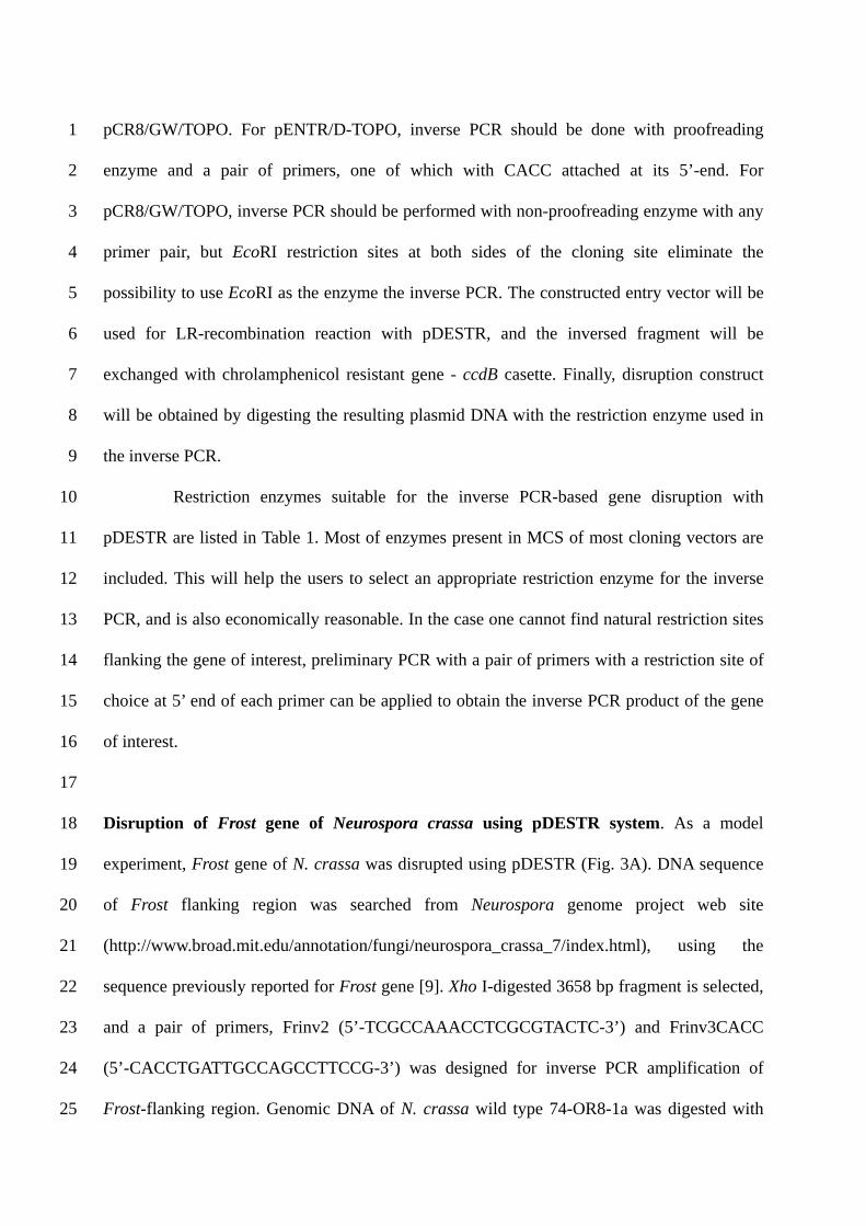
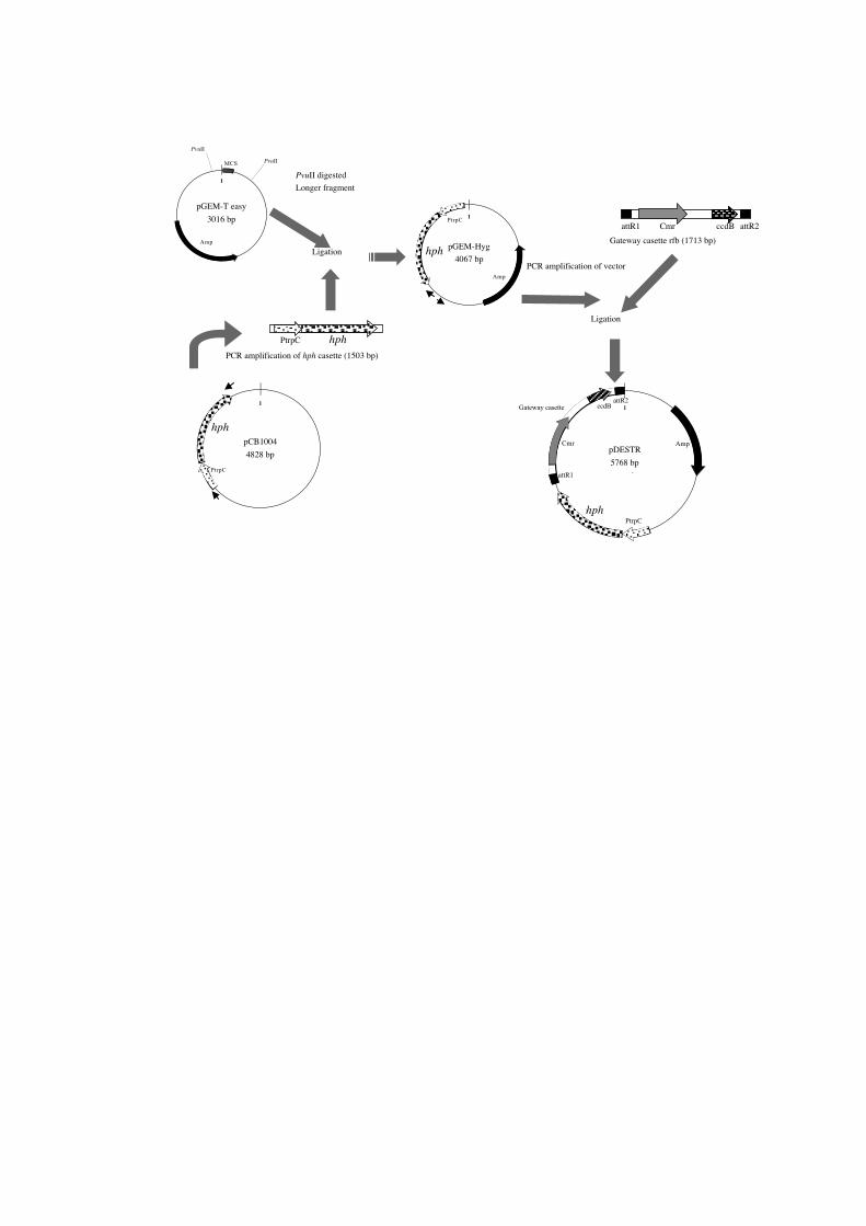
In order to achieve the necessary conditions above, a strategy of construction was
made (Fig. 2). For the basic plasmid structure, pGEM-T easy was used. This may be
substituted by other pUC-related vectors such as pUC, pBluescript and other pGEM vectors.
First, by digestion with Pvu II, MCS (multi-cloning sites) were removed. This effectively
reduced the number of restriction sites included in the plasmid. On the other hand, the hph
gene for hygromycin resistance was amplified from pCB1004, with primers HygF
(5’-CCGTGGAGGTAATAATTGA-3’) and HygR (5’-CGAAGAACGTTTTCCAATGA-3’).
The cassette was the modified version of that in pCSN43, with the reduced number of
restriction sites [1]. The amplified, blunted-kinated hph cassette was ligated into the blunt Pvu
II site in the pGEM-T easy/ Pvu II longer fragment to be named as pGEM-Hyg. A pair of
primers (DESTR1, 5’- GATCGGTGCGGGCCTCTTCG-3’ and DESTR2, 5’-
GTTGCGCAGCCTGAATGGCG-3’) was designed to amplify the almost full length of the
pGEM-Hyg, and the amplified DNA was blunted, kinated and ligated with the Gateway
cassette rfb in the Gateway vector conversion kit, to become pDESTR, the final product. The
vector was checked for the ability to transform N. crassa wild type strain and Magnaporthe
grisea Ina168 strain to hygromycin resistant transformants (data not shown).
For the cloning of the inverse PCR fragment, Gateway system is to be utilized, i.e.
inverse PCR fragment will be first ligated into entry vector, such as pENTR/D-TOPO or
pCR8/GW/TOPO. For pENTR/D-TOPO, inverse PCR should be done with proofreading
enzyme and a pair of primers, one of which with CACC attached at its 5’-end. For
pCR8/GW/TOPO, inverse PCR should be performed with non-proofreading enzyme with any
primer pair, but EcoRI restriction sites at both sides of the cloning site eliminate the
possibility to use EcoRI as the enzyme the inverse PCR. The constructed entry vector will be
used for LR-recombination reaction with pDESTR, and the inversed fragment will be
exchanged with chrolamphenicol resistant gene - ccdB casette. Finally, disruption construct
will be obtained by digesting the resulting plasmid DNA with the restriction enzyme used in
the inverse PCR.
1
2
3
4
5
6
7
8
9
10
11
12
13
14
15
16
17
18
19
20
21
22
23
24
25
Restriction enzymes suitable for the inverse PCR-based gene disruption with
pDESTR are listed in Table 1. Most of enzymes present in MCS of most cloning vectors are
included. This will help the users to select an appropriate restriction enzyme for the inverse
PCR, and is also economically reasonable. In the case one cannot find natural restriction sites
flanking the gene of interest, preliminary PCR with a pair of primers with a restriction site of
choice at 5’ end of each primer can be applied to obtain the inverse PCR product of the gene
of interest.
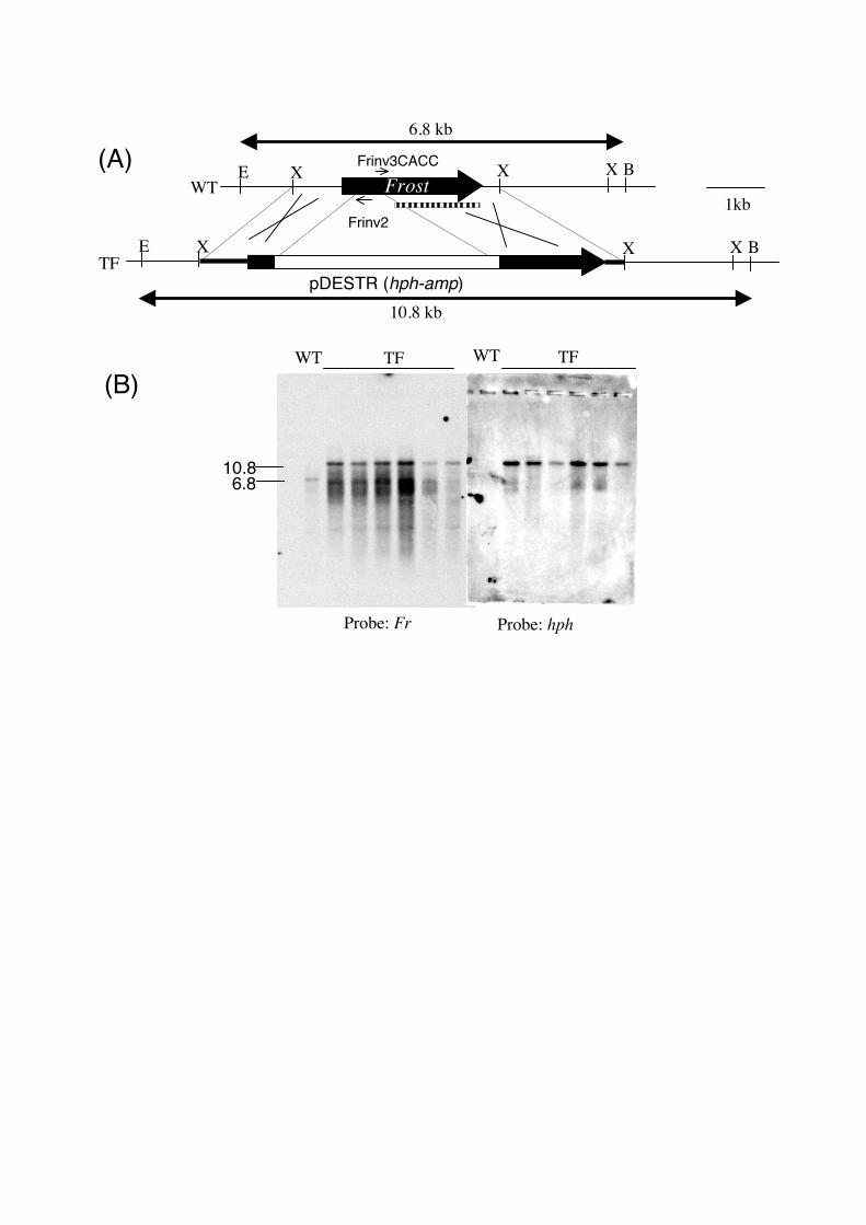
Disruption of Frost gene of Neurospora crassa using pDESTR system. As a model
experiment, Frost gene of N. crassa was disrupted using pDESTR (Fig. 3A). DNA sequence
of Frost flanking region was searched from Neurospora genome project web site
(http://www.broad.mit.edu/annotation/fungi/neurospora_crassa_7/index.html), using the
sequence previously reported for Frost gene [9]. Xho I-digested 3658 bp fragment is selected,
and a pair of primers, Frinv2 (5’-TCGCCAAACCTCGCGTACTC-3’) and Frinv3CACC
(5’-CACCTGATTGCCAGCCTTCCG-3’) was designed for inverse PCR amplification of
Frost-flanking region. Genomic DNA of N. crassa wild type 74-OR8-1a was digested with
Xho I at 0.1, 0.5, 1.0 ng DNA/μl and combined prior to inverse PCR. A clear amplification
was observed and amplicon was used for the cloning into pENTR/D-TOPO after the cleanup
with the Microspin S-400HR (Amersham Biosciences, Piscataway, NJ). The resulting plasmid,
pENTR-fr was used for the LR-recombination reaction, and a destination vector pDESTR-fr
was constructed. pDESTR-fr was digested with Xho I prior to the transformation of the N.
crassa wild type. Transformation of N. crassa was performed with electroporation. On the
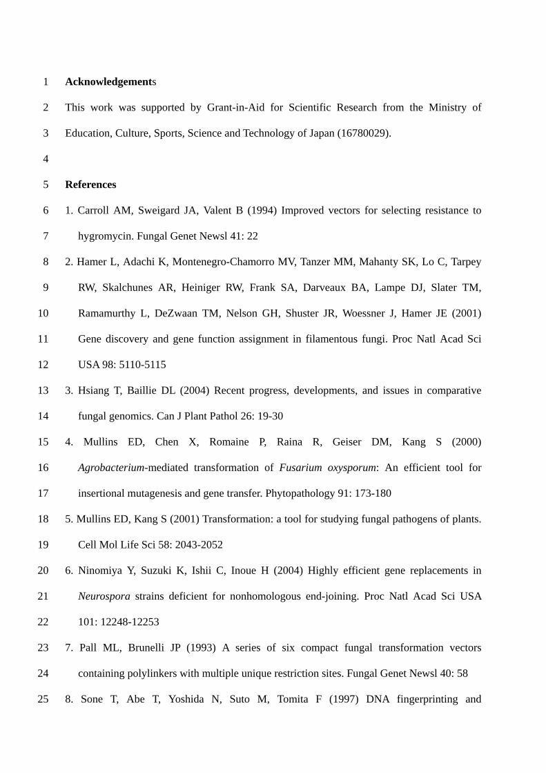
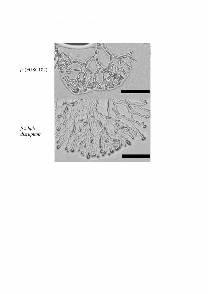
hygromycin containing selection medium, hygromycin resistant colonies appeared. Almost
half of the colonies showed abnormal, hyperbranching morphology similar to the frost mutant
(Fig.4). Six colonies with frost-like morphology were picked up and their genomic DNAs
were extracted. Southern hybridization was performed to confirm that the expected
recombination caused the frost-like morphology (Fig. 3B). In all 6 transformants, it was
revealed that the expected recombination happened at the flanking region of frost, and
contained fr::hph structure.
1
2
3
4
5
6
7
8
9
10
11
12
13
14
15
16
17
18
19
20
21
22
23
24
25
Conclusion. The gene disruption system using pDESTR, a novel Gateway destination vector
reported in this paper gives an efficient way of gene disruption in filamentous fungi. The
selection marker of hph cassette from pCB1004 was already known to work effectively in
other filamentous fungi [5, 11], indicating that pDESTR system will work also in many other
fungal strains. A similar Gateway destination vector for gene disruption harboring
phosphinothricin resistance gene (bar) cassette from pBARKS1 [7] is under construction.
This will enable us to disrupt multiple genes. These novel vectors may help the functional
characterization of hypothetical genes of genome-sequenced fungi, by combination with
non-homologous end joining (NHEJ) – deficient strains [6] and efficient transformation
methods [5].
Acknowledgements 1
2
3
4
5
6
7
8
9
10
11
12
13
14
15
16
17
18
19
20
21
22
23
24
25
This work was supported by Grant-in-Aid for Scientific Research from the Ministry of
Education, Culture, Sports, Science and Technology of Japan (16780029).
References
1. Carroll AM, Sweigard JA, Valent B (1994) Improved vectors for selecting resistance to
hygromycin. Fungal Genet Newsl 41: 22
2. Hamer L, Adachi K, Montenegro-Chamorro MV, Tanzer MM, Mahanty SK, Lo C, Tarpey
RW, Skalchunes AR, Heiniger RW, Frank SA, Darveaux BA, Lampe DJ, Slater TM,
Ramamurthy L, DeZwaan TM, Nelson GH, Shuster JR, Woessner J, Hamer JE (2001)
Gene discovery and gene function assignment in filamentous fungi. Proc Natl Acad Sci
USA 98: 5110-5115
3. Hsiang T, Baillie DL (2004) Recent progress, developments, and issues in comparative
fungal genomics. Can J Plant Pathol 26: 19-30
4. Mullins ED, Chen X, Romaine P, Raina R, Geiser DM, Kang S (2000)
Agrobacterium-mediated transformation of Fusarium oxysporum: An efficient tool for
insertional mutagenesis and gene transfer. Phytopathology 91: 173-180
5. Mullins ED, Kang S (2001) Transformation: a tool for studying fungal pathogens of plants.
Cell Mol Life Sci 58: 2043-2052
6. Ninomiya Y, Suzuki K, Ishii C, Inoue H (2004) Highly efficient gene replacements in
Neurospora strains deficient for nonhomologous end-joining. Proc Natl Acad Sci USA
101: 12248-12253
7. Pall ML, Brunelli JP (1993) A series of six compact fungal transformation vectors
containing polylinkers with multiple unique restriction sites. Fungal Genet Newsl 40: 58
8. Sone T, Abe T, Yoshida N, Suto M, Tomita F (1997) DNA fingerprinting and
electrophoretic karyotyping of Japanese isolates of rice blast fungus. Ann Phytopathol Soc
Jpn 63: 155-163
1
2
3
4
5
6
7
8
9
10
11
12
13
14
15
16
17
18
19
20
9. Sone T, Griffiths AJF (1999) The frost gene of Neurospora crassa is a homolog of yeast
cdc1 and affects hyphal branching via manganese homeostasis. Fungal Genet Biol 28:
227-237
10. Toews MW, Warmbold J, Konzack S, Rischitor P, Veith D, Vienken K, Vinuesa C, Wei H,
Fischer R (2004) Establishment of mRFP1 as a fluorescent marker in Aspergillus nidulans
and construction of expression vectors for high-throughput protein tagging using
recombination in vitro (GATEWAY). Curr Genet 45: 383-389
11. Wang HJ, Kim SH, Siu H Breuil C (1999) Transformation of sapstaining fungi with
hygromycin B resistance plasmids pAN791 and pCB1004. Mycol Res 103: 77-80
12. Wendland J (2003) PCR-based methods facilitate targeted gene manipulations and cloning
procedures. Curr Genet 44: 115-123
13. Yang L, Ukil L, Osmani A, Nahm F, Davies J, De Souza CPC, Dou X, Perez-Balaguer,
Osmani A (2004) Rapid production of gene replacement constructs and genetation of a
green fluorescent protein-tagged centromeric marker in Aspergillus nidulans. Eukaryot
Cell 3: 1359-1362
14. Yu JH, Hamari Z, Han KH, Seo JA, Domínguez YR, Scazzocchio C (2004) Double-joint
PCR: a PCR-based molecular tool for gene manipulation in filamentous fungi. Fungal
Genet Biol 41: 973-981
Figure Legends 1
2
3
4
5
6
7
8
9
10
11
12
13
14
15
16
17
18
19
20
21
22
23
24
25
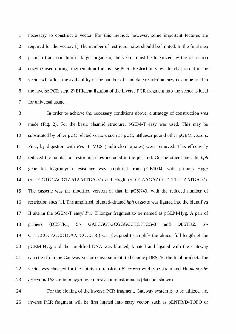
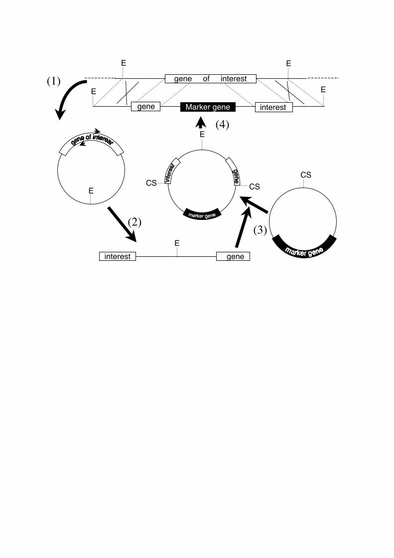
Fig.1 Schematic illustration of the inverse PCR-based gene disruption. (1) Selection of an
appropriate restriction enzyme (E), which cuts both upstream and downstream of the gene of
interest. Then digestion of genomic DNA or larger clone which includes the gene of the
interest such as cosmid clone, and self ligation by DNA ligase will be performed. A pair of
inverse primers will be designed at the central part of the restriction fragment. The position of
these primers will be the junction with the disruption DNA, thus primers should be designed
at the desired position for the disruption. (2) Inverse PCR will produce the DNA fragment
which connects the upstream and downstream DNA fragments in an opposite order, at the
central restriction site (E). (3) Ligation of the inverse PCR fragment into a cloning site (CS)
of the vector, which contains a selectable marker. (4) Digestion of the ligated plasmid with the
restriction enzyme used for the inverse PCR. This will produce the gene disruption construct.
Fig. 2 The construction of pDESTR. Small arrows indicate primers for PCR. See Results &
Discussion for details.
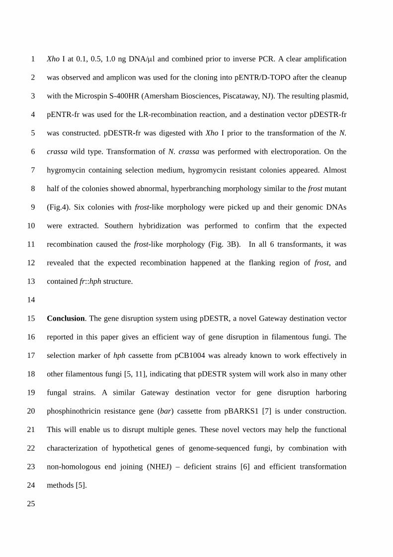
Fig. 3 Disruption of N. crassa Frost gene. (A) Maps of genomic DNA of wild type Frost
locus (WT) and the desired transformant with the disrupted frost locus (TF). Disruption
construct for the transformation was 7.7 kb Xho I fragment and is shown with thick line in TF
map. Frost gene is indicated with a black solid arrow. hph-Amp DNA from pDESTR was
indicated with white box. Position of primers for the inverse PCR was indicated with small
arrows. The probe for the Southern analysis was indicated with a dotted box. The length of
EcoR I-BamH I fragments detected in the Southern analysis were indicated. B, BamH I; E,
EcoR I; X, Xho I. (B) Southern analysis of the transformants. Genomic DNA (2μg) was
double-digested with EcoR I and BamH I and probed with Frost gene fragment (left) and hph

cassette (right). WT: 74-OR8-1a, TF: Six transformants with frost phenotype. Sizes are shown
in kb.
1
2
3
4
5
Fig 4 Morphology of fr::hph disruptant. Bar = 0.1mm.

Table. List of restriction enzymes suitable for the inverse-PCR based gene disruption using pDESTR* 1
**Aat I/Eco147 I/Pce I/SseB I/Stu I, Aau I/Bsp1407 I/BsrG I/SspB I, Acc65 I/Asp718 I,
Acv I/BbrP I/Eco72 I/PmaC I/Pml I, Afe I/Aor51H I/Eco47 III/Fun I,
Age I/AsiA I/BshT I/CspA I/PinA I, Ahl I/Bcu I/SpeI, Apa I, Asc I,
Asu II/Bpu14 I/BsiCI/Bsp119 I/BspT104 I/BstB I/Csp45 I/Lsp I/Nsp V/Sfu I, AsuNH I/Nhe I,
Avr II/Bln I/BspA2 I/XmaJ I, Bal I/Mls I/Msc I/Msp20 I, BamH I,
Ban III/Bsa29 I/Bsc I/BseC I/BsiX I/Bsp106 I/BspD I/BspX I/Bsu15 I/BsuTU I/Cla I/Zho I,
Bbe I, BbvC I, Bcl I/BsiQ I/Fba I/Kso22 I, Bgl II, BmgB I/Btr I, BsiW I/Pfl23 II/PspL I/Sun I,
BseP I/BssH II/Pau I, Bsp68 I/Nru I, Bsp120 I/PspOM I, BssH I/PaeR7 I/Sfr274 I/Sla I/Tli I/Xho I,
BssNA I/BstZ17 I/Bst1107 I, BstSN I/Eco105 I/SnaB I, CciN I/Not I, Cfr9 I/PspA I/Vma I/XmaC I,
Cfr42 I/Sac II, Eci36 II/EcoICR I, EcoR I/Fun II, EcoR V/Eco32 I, EcoT22 I/Mph1103 I/Nsi I/Zsp2 I,
Ege I/Ehe I/Sfo I, Fse I, Hind III, Kas I, Kpn I, Mfe I/Mun I, Mlu I, Mly113 I/Nar I, Mss I/Pme I, Pac I,
Pst I, Pvu II, Psp124B I/Sac I/Sst I, Sal I, Sbf I/Sda I/Sse8387 I, Sma I, Smi I/Swa I, Srf I, Xba I
* Enzymes often present in multi cloning sites of cloning vectors are listed with bold letters. Enzymes with
ambiguous recognition sequence (for example, Nci I (CCSGG)) were not included in this list because such
restriction fragments were not always self-ligated. **Isoschizomers are shown with “/”.
2
3
4
E
Einterest gene
CS
E
(1)
(2) (3)
(4)
CSCS
gene of interest
gene
E
interestMarker gene
EE
E
pGEM-Teasy
3016 bp
Plasmid name:pGEM-Teasy
Plasmid size:3016 bp
Constructed by:
Construction date:
Comment&Reference:
PvuII
PvuII
MCS
Amp
1
pCB1004
4828 bp
Plasmid name:pCB1004
Plasmid size:4828 bp
Constructed by:
Construction date:
Comment&Reference:
PtrpC
Hph
1
pDESTR
5768 bp
Plasmid name:pDESTR
Plasmid size:5768 bp
Constructed by:
Construction date:
Comment&Reference:
Amp
PtrpCHph
Gateway casette
attR1
Cmr
ccdBattR2
1
attR1 Cmr ccdB attR2
pGEM-Hyg
4067 bp
Plasmid name:pGEM-Hyg
Plasmid size:4067 bp
Constructed by:
Construction date:
Comment&Reference:
Amp
Hph
PtrpC1
PtrpC Hph
pDESTR5768 bp
PCR amplification of hph casette (1503 bp)
PvuII digestedLonger fragment
Ligation
Ligation
PCR amplification of vector
Gateway casette rfb (1713 bp)
hph
hph
hph
pGEM-Hyg4067 bp
hph
pCB10044828 bp
pGEM-T easy3016 bp
1kbFrost
E X X X B
X X
pDESTR (hph-amp)
Frinv2
Frinv3CACC
E X B
6.8 kb
10.8 kb
WT
TF
TF TFWT WT
6.810.8
(A)
(B)
Probe: Fr Probe: hph
fr (FGSC102)
fr:: hphdisruptant