Embed Size (px)
Citation preview

بسم االله الرحمن الرحيم
Contamination of Drinking Water by Coliform Bateria in Kassala City-Sudan
By:
Waleed Idriss Adam Salim
B.Sc., University of Al- fashir(2002)
Supervisor:
Dr. Abdel Hafeez Hassan Nimir
A thesis submitted to University of Khartoum in partial fulfillment of the requirement for the degree of M.Sc in Microbiology.
Department of Microbiology, Faculty of Veterinary Medicine
Aug-2009
brought to you by COREView metadata, citation and similar papers at core.ac.uk
provided by KhartoumSpace

2
DEDICATION
To my father, mother, brothers and sisters.
To all my friends who encouraged and helped me.
I dedicate this work.

3
ACKNOWLEDGEMENTS I would like to express my thanks to Allah, who gave me the health
and patience to achieve this work. Also, I am greatly indebted to my
supervisor Dr. Abdel Hafeez Hassan Nimir, for his constructive criticism,
guidance, encouragement and patience throughout this work. Thanks are
extended to workers at Water Corporation in Kassala State for their
generous unlimited help, and for allowing sample collection.
Acknowledgement is also extended to all Microbiology Department
members, Faculty of Veterinary Medicine, University of Khartoum for
thier valuable advice and assistance.

4
ABSTRACT
This study was conducted to investigate the coliform bacterial
contamination of drinking water of Kassala city during March to June
2008.
Seventy samples of water were taken from seven different areas
main station, El-shảbiya, El-halanga, El-ảmriya, El-gasor, El-kara, El-
brno (ten samples each), including chemically treated water from the
main station and untreated water from wells. Bacterial coliform
contamination was investigated by Multiple fermentation tube technique
to detect the existence of total and thermotolerant coliforms at 37◦C and
44◦C, respectively.
The total coliform bacteria were detected in 46 (65.7%) samples,
while thermotolerant coliform bacteria were detected in 31 (44.3%)
samples.
A total of 57 isolates were obtained from 46 samples, which were
identified according to their microscopic, cultural and biochemical
properties as: Escherichia coli (38.6%), Enterobacter aerogenes (17.5%),
Enterobacter cloacae (15.8%), Klebsiella pneumoniae subspecies
aerogenes (12.3%), Klebsiella pneumoniae subspecies ozaenae (8.8%)
and Citrobacter freundii (7%).
Citrobacter freundii and Klebsiella pneumoniae ssp. ozaenae were
found in untreated water only. While the other species were found in both
treated and untreated water with higher prevalence in untreated water.
The wells in the centeral city (El-halanga and El-ảmriya) contained more
coliform bacteria, especially Escherichia coli than wells which exist near
Gash river (El-gasor) in the side of the city.
5
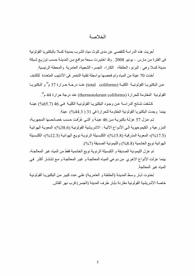
الخلاصة
القولونية ينة آسلا بالبكتيريا أجريت هذه الدراسة للتقصي عن مدى تلوث مياه الشرب بمد
ع من الم . 2008 يونيو –فى الفترة من مارس ع شبكة دوقد اختيرت سبعة مواق ة حسب توزي ين
.المحطة الرئيسية و ، العامريةة الشعبي، الجسر البرنو، الحلنقة، الكارا،:هي مدينة آسلا و
للكشفمتعددة ال الأنابيب التخمر فىتقنيةة عينة من المياه وتم فحصها بواسط70أخذت
ا ن البكتيري ة ع ة القولوني رارة) total coliforms(الكلي ة ح د درج ا و oم 37 عن البكتيري
.o م44ة حرارعند درجة ) thermotolerant coliforms( المقاومة للحرارة القولونية
و ن وج ة ع ائج الدراس شفت نت ادآ ة لونوق ال البكتيري ة الكلي ي ي ة%) 65.7( 46 ف عين
.عينة %)44.3 ( 31ة للحرارة في المقاومالقولونيةتيريا وجدت البك بينما
زل م ع ن 57ت ة م ة بكتيري ي ع46ُ عزل ة و الت ت حسب خصائرِّ عين ة، هصف ا المجهري
ي رالمز ة واعالأن عية و الكيموحيوية ال ة : الآتي شية القولوني ة المع ،%)38.6(الاشري ة الهوائي وي
ة ا المعوية ،%)17.5( ع ، %)15.8(لمذرقي ة نوي سيلة الرئوي ة الكلب سيلة %)(12.3 الهوائي ، الكلب
%).7( الصديقة الليمونيةو%) 8.8 ( الخشميةالهوائية نويع
،المعالجة المياه غير منفقطمية الخش الكبسيلة الرئوية نويع و ليمونية الصديقة لم عزل ا ت
اه الاخري الأنواع بينما عزلت وعي المي ر المعالجة و مع إ من ن شار المعالجة و غي ر نت في أآث
. غير المعالجةالمياه
وت ار إحت ة آب ة (وسط المدين ةوالحلنق دد ) العامري ي ع ن ريآبعل ة م ا القولوني البكتيري
.القاش قرب نهر)الجسر( طرف المدينةر بآبامقارنة الاشريشية القولونيةخاصة
6
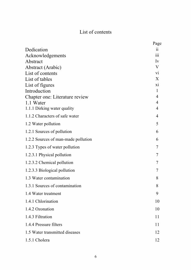
List of contents
Page
ii Dedication iii AcknowledgementsIv AbstractV Abstract (Arabic)vi List of contentsX List of tables xi List of figures1 Introduction4 Chapter one: Literature review4 1.1 Water4 1.1.1 Dirking water quality
4 1.1.2 Characters of safe water
5 1.2 Water pollution
6 1.2.1 Sources of pollution
6 1.2.2 Sources of man-made pollution
7 1.2.3 Types of water pollution
7 1.2.3.1 Physical pollution
7 1.2.3.2 Chemical pollution
7 1.2.3.3 Biological pollution
8 1.3 Water contamination
8 1.3.1 Sources of contamination
9 1.4 Water treatment
10 1.4.1 Chlorination
10 1.4.2 Ozonation
11 1.4.3 Filtration
11 1.4.4 Pressure filters
12 1.5 Water transmitted diseases
12 1.5.1 Cholera

7
13 1.5.2 Typhoid fever
13 1.5.3 Paratyphoid fever
13 1.5.4 Dysentery
13 1.5.5 E. Coli diarrhoeas
14 1.6 Water quality analysis
14 1.6.1 1Bacteria as indicators of water sanitary quality
14 1.6.1.1 The coliforms group
16 1.6.1.2 Non coliform bacteria
16 1.6.1.2.2 Clostridium perfringens
16 1.6.2 Importance of coliforms as faecal indicators
17 1.7 Methods for detection of faecal pollution in water
18 1.7.1 Standard plate count (SPC)
18 1.7.2 Membrane filters technique (M F)
19 1.7.3 Multiple fermentation tube technique (Most Probable Number,
MPN)
20 1.7.4 Colilert methods
21 1.7.5 Gas-liquid chromatography
21 1.7.7 Nucleic acid-based methods
21 1.7.7.1 Polymerase chain reaction (PCR)
22 1.7.7.2 Fluorescence in situ hybridization (FISH)
23 1.8 Determination of faecal pollution sources
24 1.6 Drinking water standards
25 Chapter two: Materials and Methods
25 2.1 Collection of samples
25 2.1.1 Collection of samples from taps
25 2.1.2 Collection of samples from tanks
26 2.2 Culture media
26 2.2.1 Liquid media

8
26 2.2.1.1 MacConkey's broth
26 2.2.1.2 Brilliant Green Lactose Bile broth (BGLB)
27 2.2.1.3 MR-VP medium (Glucose-phosphate medium)
27 2.2.1.4 Peptone water
28 2.2.1.5 Nitrate medium
28 2.2.2 Semi-solid media
28 2.2.2.1 Oxidation-fermentation (O/ F) medium
28 2. 2.2.2 Motility medium (Craigie tube medium)
29 2.2.3 Solid medium
29 2.2.1.1 Eosin Methylene Blue
29 2.2.3.2 MacConkey's agar
30 2.2.5.3 Nutrient agar
30 2.2.3.4 Simmons's citrate agar
31 2.2.3.4.5 Urea agar base
31 2.3 Reagents and buffers
31 2.3.1 Kovac's reagent
31 2.3.2 Normal saline
32 2.3.3 Hydrogen peroxide (H2O2)
32 2.3.4 Potassium hydroxide
32 2.3.5 Lead acetate
32 2.3.6 Methyl red solution
32 2.3.7 Alpha-naphthol solution
32 2.3.8 Tetramethyle-P-phenyline diaminehydrochloride
33 2.3.2 Phosphate-buffered dilution water
33 2.4 Sterilization
33 2.4.1 Flaming
33 2.4.2 Red heat
33 2.4.3 Hot air oven

9
33 2.4.4 Moist heat (Autoclaving)
34 2.4.5 Disinfectants
34 2.6 Cultivation of samples
34 2.6.1 Multiple fermentation tube technique
34 2.6.1.1 Materials
34 2.6.1.2 Method
35 2.6.1.3 Confirmatory tests
35 2.6.1.3.1 Brilliant green lactose bile broth (BGLB)
35 2.6.1.3.2 Indole test
36 2.6.1.4 Testing in Eosin Methylene Blue
36 2.6 Identication of isolates
36 2.6.1 Microscopic examination
37 2.6.1.1 Staining method
37 2.6.1.2 Cultural characteristics
37 2.6.2 Biochemical testing
37 2.6.2.1 Primary tests
37 2.6.2.1.1 Catalase test
38 2.6.2.1.2 Oxidase test
38 2.6.2.1.3 Oxidation-fermentation (O/F) test
38 2.6.2.1.4 Gas from glucose
38 2.6.2.1.5 Motility test
39 2.6.2.2 Secondary tests
39 2.6.2.2.1 Citrate test
39 2.6.2.2.2 Urease test
39 2.6.2.2.3 Indole test
39 2.6.2.2.4 Methyl red (MR) test
40 2.6.2.2.5 Voges-Proskaeur (V. P.) test
40 2.6.2.2.6 Hydrogen sulphide (H2S) production test

10
40 2.6.2.2.7 Sugar fermentation test
41 2.6.2.2.8 Nitrate reduction test
44 Chapter three: Results 44 3.1 Total and thermotolerant coliforms
44 3.2 Isolates
44 3. 2.1 Identification of isolates
45 3.2.2 Characteristics of bacterial species
45 3.2.2.1 E. coli isolates
45 3.2.2.1.1 Microscopic examination
45 3.2.2.1.2 Cultural characteristics of E. coli
45 3.2.2.1.3 Biochemical reactions
45 3.2.2.2 Enterobacter aerogenes
45 3.2.2.2.1 Microscopic examinations
45 3.2.2.2.2 Cultural characteristics
45 3.2.2.2.3 Biochemical reactions
46 3.2.2.3 Enterobacter cloacae
46 3.2.2.3.1 Microscopic examination
46 3.2.2.3.2 Cultural characteristics
46 3.2.2.3.3 Biochemical reactions
46 3.2.2.4 Klebsiella isolatess
46 3.2.2.4.1 Microscopic examination
46 3.2.2.4.2 Cultural characteristics
46 3.2.2.4.3 Biochemical reactions
46 3.2.2.5 Citrobacter freundii isolates
46 3.2.2.5.1 Microscopic examination
47 3.2.2.5.2 Cultural characteristics
47 3.2.2.5.3 Cultural characteristics
55 Chapter four: Discussion

11
57 CONCLUSION
57 RECOMMENDATIONS
58 References
12
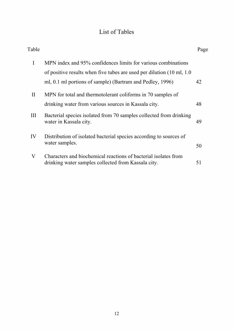
List of Tables
Table Page
I
MPN index and 95% confidences limits for various combinations
of positive results when five tubes are used per dilution (10 ml, 1.0
ml, 0.1 ml portions of sample) (Bartram and Pedley, 1996)
42
II MPN for total and thermotolerant coliforms in 70 samples of
drinking water from various sources in Kassala city.
48
III Bacterial species isolated from 70 samples collected from drinking water in Kassala city.
49
IV Distribution of isolated bacterial species according to sources of water samples.
50
V Characters and biochemical reactions of bacterial isolates from drinking water samples collected from Kassala city.
51

13
List of Figures
Page Figure
52
Bacterial species isolated from 70 samples collected from drinking water in Kassala city.
1
53
Multiple Tube Fermentation test in MacConkey's broth. Positive result indicated by production of gas and acid (right tube).
2
53
Multiple Tube Fermentation test in Brilliant green lactose bile broth. Positive result indicated by production of gas (right tube).
3
54 Growth of E. coli on Eosin methylene blue agar.
4
54 Growth of coliform bacteria on MacConkey's agar.
5

14
Introduction Water is the universal solvent as they call it. It covers 70% of the
earth's surface and is also present in varying amounts in the atmosphere.
It is an essential component of all cells and a requirement for life. It
represents about 45% to 95% of living cell (Lim, 1998).
The drinking water must be safe and it is cosidered polluted if it
contains toxic substances or pathogens (Black, 1999). Good quality water
is odourless, colorless, tasteless and free from faecal pollution and
chemicals in harmful amounts (Cheesbrough, 1994). Black (1999) has
estimated that up to 80% of all sicknesses and diseases in the world are
caused by polluted water. The World Health Organization (WHO) has
estimated that up to 80% of all sicknesses and diseases in the world was
caused by inadequate sanitation, polluted water or unavailability of water
(Cheesbrough, 1994).
Many water sources in developing countries are unhealthy because
they contain harmful physical, chemical and biological agents. It has been
reported that the death of most of children in Africa who die under the
age of 5 is caused by inadequate and unsafe water supplies (Loucks,
1994).
UNICEF reported that about 90% of major epidemics in the Sudan
are water-borne and water-related, causing the death of some 40% of
children under five years of age (El Tayeb, 2002). When water is
contaminated with faecal material, any pathogen that leaves the body
through the faeces, (many bacteria, viruses and some protozoa) can be
present. The most common diseases that are transmitted by water are
typhoid fever, salmonellosis, shigellosis, cholera, legionnaire's diseases
and gastroenteritis which is induced by Vibrio parahaemolyticus,
Escherichia coli, Yersinia enterocolitica and Campylobacter fetus.
15
Also, some viral and parasitic diseases may be transmitted like hepatitis,
poliomyelitis, giardiasis and amoebic dysentery (Black, 1999).
Coliform bacteria may not cause disease but can be indicators of
pathogenic organisms that cause intestinal infections, dysentery, hepatitis,
typhoid fever, cholera and other illnesses. These illnesses are not limited to
diseases-causing organisms in drinking water. However, other factors that
may not associate with drinking water may be the cause. Previous studies
in Sudan have dealt only with certain aspects of water pollution problems.
Detailed considerations have come to light as a result of the work done by
Dirar (1986) and Abdelmagid, Ibrahim and Dirar (1984) on the pollution
of water from wells in Khartoum area. Routine microbiological analysis of
water is necessary once water treatment system has been installed in order
that problem in the treatment process can be anticipated (WHO, 1996).
Different microbiological methods have been developed since the
early 1900s to assess water quality with regard to public health. Most
methods rely on enumerating coliforms and E. coli as indicators for faecal
pollution. There is a large number of techniques that are used in
bacteriological analysis of water such as standard plate count, membrane
filter technique and multiple fermentation tube technique. The latter two
methods are recommended by the WHO. However, culture-based methods,
which include the detection of only live bacteria, have sevral problems
such as lack of specificity for detection of free faecal coliform (E. coli) and
in addition time is required for detection and conformation (Roszake and
Colwell, 1987). Polymerase Chain Reaction (PCR) has been used for
detection of the target nucleotide sequence associated with coliform
bacteria or specifically with E. coli using specific primers (Schochetman
and Jones, 1988).

16
Objectives of the study:
1- To investigate drinking water for colifrom bacterial pollution in
Kassala city at different locations during four months
2- Investigation of ground water before its pumping through public
pipelines.

17
Chapter one
Literature review
1.1 Water:
Clean hygienic water denotes that it is free of pollutants. Water is
said to be polluted either due to the presence of harmful chemicals or
bacterial contamination (WHO, 1976).
Unhygienic water contributes to animal and human hazards like
microbiological ones (Bartram and Wheeler, 1993).
1.1.1 Dirking water quality:
Surveillance of drinking water quality is continuous and vigilant
public health assessment and overview of safety and acceptability of
drinking water by world health organization (WHO, 1976).
Water quality control devotes regular sampling and analyzing of water
samples as well as recording of result obtained. However, it also involves
assessing how good a method is and how well is operating in practice
(WHO, 1976; WHO, 1984).
1.1.2 Characters of safe water:
Water is usually demanded to meet different needs such as
drinking, agricultural and industrial activities. When it is polluted it
becomes not acceptable for these uses or for human consumption. So,
good quality water for human consumption must fulfill the following
requirements:
• It must be acceptable with good taste, not turbid, colorless and
with no smell.
• Microbiologically safe: It should not contain any pathogenic
bacteria, viruses, fungi, protozoa or helminthes eggs.

18
• Chemically hygienic: Water content of toxic materials, organic
or inorganic, must be zero or at a minimum; it should contain
appropriate content of iodine, fluoride and harmless ions.
• It must be safe radiologically (White and Godfree, 1985).
1.2 Water pollution:
The more acceptable definition of water pollution ''The presence
of any substance (organic, inorganic, biological, thermal or radiological)
in water at intensity levels which tend to impair, degrade, or adversely
affect its quality or use fullness for specific purposes''. (Food and
Agricultural Organization (FAO), 1979).
Water under natural conditions contains different microbes which
include protozoa, bacteria, fungi, and viruses. The number and kind of
microbes present depends on the source of water, the contaminations by
the excreta from humans, animals and addition of other contaminated
materials (Smith, 1981).
For purposes of simplicity, scientists classify water into major
types, ground water and surface water. Ground water originates from
deep wells and subterranean springs and because of the filtering action of
soil, deep sand and rocks, it is visually free of microorganisms. As water
flows up long channels, contaminants may enter it and alter its quality
(Alcamo, 1997). Microbial contamination may occur when a well is
situated within 200 feet of the source of contamination (Smith, 1981).
Surface water is found in lakes, streams and shallow well. Its
microbial population may reflect the air through which rain has passed, or
the sewage treatment facility located along rivers bank (Alcamo, 1997).
Generally, surface water contains more microbes than ground water
and rain water since the majority of soil microorganisms are found in the
upper crust (6 inches) of the earth. Surface water contains many

19
nonpathogenic microbes from soil, and in the vicinity of cities it is often
contaminated with sewage bacteria (Smith, 1981).
A major type of bacteria in polluted water is coliform bacteria, a
group of Gram-negative bacteria and non-spore-forming bacilli which
inhabit human and animal intestines. They usually ferment lactose to acid
and gas. The most important species of this group are E. coli, Klebsiella
spp. and Enterobacter spp.
Noncoliform bacteria are also common in polluted water and
include Streptococcus, Proteus and Pseudomonas species (Alcamo,
1997). Bifidobacteria are one of the most common bacterial types found
in the intestines of human and other animals and may be used as indicator
for human faecal pollution (Bonjoch, Balleste and Blanch, 2004).
1.2.1 Sources of pollution:
The nature, man, agriculture and industry all contribute to the
degradation of water quality.
1.2.2 Sources of man-made pollution:
There are wide number of activities that are associated with man,s
introduction of foreign chemical and biological material by direct or
indirect route in surface or subsurface water environment. According to
the FAO (1979) and Katz (1971) there are four main sources of water
pollution:
1. Agriculture (animal and crop waste and fertilizers)
2. Industry (serves as another originator of chemicals).
3. Waste generated domestically.
4. Radioactive materials.

20
All these constitute major sources of chemical or biological
material (can be induced by man as artificial water contamination) which
threaten to degrade surface and underground water supply.
It is known that toxic sources may enter surface water from air
drifting of chemicals and by direct application for the control of
undesired aquatic life. The expanding food industry necessitates the
application of chemicals for food preservation. As a result, highly toxic
substances find access to the aquatic environment (Dutt, 1982).
1.2.3 Types of water pollution:
Pollution of water includes
1.2.3.1 Physical pollution:
Physical pollution occurs when a particulate matter such as sand
or soil makes the water cloudy. Living material may also be involved
such as cyanobacteria, which give water consistency of pea-soap
(Alcamo, 1994).
1.2.3.2 Chemical pollution:
Chemical pollution results from the introduction of organic and
inorganic waste to the water especially when water supplies are drawn
from rivers that have received up stream effluent of industrial wastes.
Although water looks clear in some cases, chemical analysis may detect
many contaminating substances (Black, 1999).
1.2.3.3 Biological pollution:
This type of pollution develops from organisms that enter water
from human or animal wastes. The organism finds a good environment
for growth such as water useful ions and organic matter which can be
digested by heterophic organisms and produce carbon dioxide, so water is
considered biologically polluted (Alcamo, 1994).
21
1.3 Water contamination:
The term contamination is defined as presence, in water, of
bacteria from intestinal tract of warm-blood animals including man. The
presence of such bacteria means the water may carry human disease
agents. The fact that water looks clear and sparkling is no assurance of its
purity. Disease agents are invisible to the unaided eye (Forrest, 1956).
The movement of animal wastes into the surface water is often cited as a
major factor contributing to the pollution of available water in many rural
areas (Doran, and Linn, 1979; Fernandez-Alvarez, Carballo-Cuervo,
Carmendela Rosa-Jorge, and Rodrignez-Delecea, 1991).
Studies in the Sudan have clearly demonstrated the close
association of biological contamination of drinking water with high
prevalence of diarrhoeal diseases and certain enteric pathogens (El
Shazali and Erwa, 1971).
Although the terms contamination and pollution are often used
synonymously, health authorities make the definition of water pollution
as any undesirable quality of water other than contamination. Dirt, silt,
organic matter, minerals, objectionable colours, odours or tastes, acidity
and alkalinity are causes of pollution. Although pollution is not
necessarily a health hazard, it is often accompanied by contamination
which is a health hazard (Forrest, 1956). The most common pollutants
are bio-degradable organic matter, suspended solids, nutrients, heat and
bacteria from industrial, agricultural sewage waste effluents and land run
off.
1.3.1 Sources of contamination:
Water receives faecal pollution from a variety of sources, including
humans and animals. Knowledge of pollution sources could aid in
restoration of the water quality, reduce the amount of nutrients leaving

22
watersheds, and reduce the danger of infectious disease resulting from
exposure to contaminated water. The sources of bacterial contamination
include:
• Human and animal wastes which are primary sources of bacteria.
These sources include runoff from feedlots, dog run and other land
areas where animal wastes are deposited.
• Insects, rodents or animals enter wells.
• Discharge from septic tanks and sewage treatment centres. It was
found that the major feature of safe water supply is the separation
of sewage (human excreta) from drinking water (Duerden, Reid,
Jewsbury, and Turk, 1988).
• Natural soil/plant bacteria. Bacteria from these sources can enter
wells that open at land surface.
• Infiltration by flood waters or by surface runoff. Flood waters
commonly contain high levels of bacteria.
1.4 Water treatment:
For small communities, it is generally preferable to protect a
ground water source that requires little or no treatment than to treat
surface water that has been exposed to faecal contamination and is
usually of poor quality. In many circumstances, however, surface
water is the only practicable source of supply and requires affordable
treatment and disinfection. The range of treatments available for
small-community supplies is necessarily limited by technical and
financial considerations; the most appropriate and commonly used
treatments are summarized below. Installation of packaged treatment
plants is not a suitable means of dealing with the typical water-quality
problems that prevail in rural areas (WHO, 2004).

23
1.4.1 Chlorination: Chlorination can be achieved by using liquefied chlorine gas,
sodium hypochlorite solution or calcium hypochlorite granules and
on-site generators. Liquefied chlorine gas is supplied in pressurized
containers. The gas is withdrawn from the cylinder and dosed into
water by a chlorinator, which both controls and measures the gas flow
rate. Sodium hypochlorite solution is dosed using a positive-
displacement electric dosing pump or gravity feed system. Calcium
hypochlorite has to be dissolves in water and then mixed with the
main supply. Chlorine, whether in the form of chlorine gas from a
cylinder, sodium hypochlorite or calcium hypochlorite, dissolves in
water to form hypochlorous acid (HOCI) and hypochlorite ion (OCI)
(WHO, 2004).
Chlorination is employed primarily for microbial disinfection.
However, chlorine also acts as an oxidant and can remove or assist in
the removal of some chemicals. A disadvantage is its ability to react
with natural organic matter. However, by product formation may be
controlled by optimization of the treatment system (WHO, 2004).
1.4.2 Ozonation:
Ozone is a powerful oxidant and has many uses in water
treatment, including oxidation of organic chemicals. Ozone can be
used as a primary disinfectant. Ozone gas is formed by passing dry air
or oxygen during a high-voltage electric field. The resultant ozone-
enriched air is dosed directly into the water by means of porous
diffusers at the base of baffled contractor tanks. The contractor tanks,
typically about 5 m deep, provide 10 – 20 min of contact time.
Dissolution of at least 80% of the applied ozone should be possible,

24
with the remainder contained in the off-gas, which is passed during an
ozone destructor and vented to the atmosphere (WHO, 2004).
Ozone reacts with natural organics to increase their bio-
degradability, measured as assimilable organic carbon. To avoid
undesirable bacterial growth in distribution, ozonation is normally
used with subsequent treatment, such as filtration to remove
biodegradable organics, followed by a chlorine residual, since it does
not provide a disinfectant residual. Ozone is effective for the
degradation of a wide range of pesticides and other organic chemicals
(WHO, 2004).
1.4.3 Filtration:
Particulate matter can be removed from raw water by rapid
gravity horizontal, pressure or slow sand filters. Slow sand filtration is
essentially a biological process, whereas the others are physical
treatment processes (WHO, 2004).
Rapid gravity, horizontal and pressure filter can be used for
direct filtration of raw water, without pre-treatment. Rapid gravity and
pressure filter are commonly used to filter water that has been pre-
treated by coagulation, which passes directly onto the filter where the
precipitated flock is removed. The application of direct filtrations is
limited by the available storage within the filter to accommodate
solids (WHO, 2004).
1.4.4 Pressure filters:
Pressure filters are sometimes used where it is necessary to
eliminate the need for pumping into supply. The filter bed is enclosed in
cylindrical shell. Small pressure filters, capable of treating up to 15 m3/h,
can be manufactured in glass-reinforced plastics. Larger pressure filters,

25
up to 4 m in diameter, are manufactured in specially coated steel.
Operation and performance are generally as described for the rapid
gravity filter, and similar facilities are required for backwashing and
disposal of the dilute sludge (WHO, 2004).
1.5 Water transmitted diseases:
Polluted water, tainted food and malnutrition account for high
infant mortality when sanitation measures are lacking. Throughout
history, water-borne diseases have important restraint on population
growth (Deming, 1975).
Water borne infectious diseases are those in which the pathogen, or
causative organism, is present in water and ingested when the water is
consumed. Most of the pathogens involved are derived from human
stools, and the diseases transmitted by consumption of faecal
contaminated water are called faecal-oral diseases (Meybeck, Kuusisto,
Makela, and Malkki, 1996).
Typhoid fever, cholera and shigelosis are the most important
bacterial diseases that spread by contaminated water. However, water
borne epidemics of these diseases are rare due to continual familiar
bacteria such as species of Yersinia and Campylobacter, and toxin-
producing strains of E. coli. An emerging pathogen associated with
contaminated water is Vibrio vulnificus, a Gram-negative bacterium that
can cause serious illness in persons with pre-existing liver diseases or
immuncompromised persons (Alcamo, 1997).
1.5.1 Cholera:
The disease is caused by Vibrio cholerae and its variant the Eltor
vibrio. The infection is usually contracted by ingestion of water
contaminated by infected human faecal material (Twort, Law and

26
Crowly, 1985). It gives rise to sudden and profuse diarrhoea which leads
to severe dehydration and death within 1 or 2 days if fluids are not
replaced (Cairncross, Carruthers, Curtis, Feachem, Breadley, and
Baldwin, 1980).
1.5.2 Typhoid fever:
The disease is caused by Salmonella typhi. The infection is usually
contracted by ingestion of material contaminated by human faeces or
urine, including water and food (Twort et al., 1985).
1.5.3 Paratyphoid fever:
Paratyphoid fever is caused by Salmonella paratyphi A, B or
C. Infection may exceptionally be via contaminated water, but more
commonly due to ingestion of contaminated food (Twort et al., 1985).
Typhoid and paratyphoid fever are again specific severe fevers
sometimes with intestinal symptoms, which have a high mortality if
untreated (Cairncross et al., 1980).
1.5.4 Dysentery:
Dysentery is caused by Shigella spp. Infection is occasionally
contracted via water contaminated by human faeces, but more commonly
it is due to ingestion of food contaminated by flies or by unhygienic food
handlers who are carriers (Twort et al., 1985). The disease is
characterized by severe bloody diarrhoea accompanied by abdominal
pain. A severe illness is affected by access to domestic water and its
quality (Cairncross et al., 1980).
1.5.5 E. coli diarrhoeas:
Enterotoxingenic and enteroinvasive strains of E. coli
represent the major cause of infant diarrhoea. The produced diarrhoea

27
leads to dehydration and salt imbalance which is substantial enough to
threaten life (Alcamo, 1997). Other reports pointed out of E. coli causes
enteritis in children and adults (Ogden and Watt, 1991). Traveller's
diarrhoea represents one of the conditions that can be caused by E. coli. It
is transmitted in the same way as dysentery and water may sometime be
the vehicle (Twort et al., 1985).
1.6 Water quality analysis:
Although water can contain unwanted chemicals, the greatest risk
to human health is from faecal contamination of water supplies. Serious
ill health can be caused by water becoming contaminated from faeces
being passed or washed into rivers and pools. The most important aspect
of microbial analysis is therefore to determine whether faecal
contamination is present (Cheesbrough, 1994).
1.6.1 Bacteria as indicators of water sanitary quality:
Water which looks clear and pure may be sufficiently contaminated
with pathogenic organisms and may constitute a health hazard (Brock,
Madigan, Martinko, and Parke, 1994)). Detection of all possible
pathogens would be a costly and very time consuming process. Methods
have, therefore, been developed which detect organisms, which are
indicative of presence of faecal pollution, such as normal intestinal
bacteria. If evidence for faecal material is found in the water sample, it
can be assumed that other dangerous faecal pathogens may be present. If
no evidence is found it is likely, although not totally certain, that the
water is safe for human use.

28
1.6.1.1 The coliforms group:
The coliforms group of bacteria has remained the corner stone of
the national drinking water regulation all over the world and is used in the
water supply industry as criteria of operational parameters.
This group is defined as the aerobic and facultative anaerobic
Gram-negative, rod-shaped, bacteria that ferment lactose with gas
productions within 24-48 h at 35 or 37 oC (Madigan, Martinko and
Parker, 1997). They are also oxidase negative and non-spore-forming. By
definition, coliform bacteria display B-galactose activity.
Traditionally, coliform bacteria were regarded as belonging to the
genera Escherichia, Citrobacter, Enterobacter and Klebsiella. However,
as defined by modern taxonomical method, the group is heterogenous. It
includes lactose-fermenting bacteria, such as Enterobacter and
Citrobacter freundi that can be found both in faeces and environment
(nutrient, rich water, soil, decaying plant material). Due to these facts,
coliforms used as indicators of faecal pollution in water are classified into
total coliform and thermotolerant coliform.
The term'' total coliform'' refers to a large group of Gram-negative,
rod-shaped bacteria that share several characteristics. The group of
coliforms includes thermotolerant coliforms and of faecal origin, as well
as some bacteria that may be isolated from environmental sources. Thus
the presence of total coliforms may or may not indicate faecal
contamination. In extreme cases a high count for the total coliform group
may be associated with a low or even zero count for thermotolerant
coliforms. Such a result would not necessarily indicate the presence of
faecal contamination. It might be caused by entry of soil or organic
matter into the water or by conditions suitable for the growth of other
types of coliform.

29
The term'' Thermo tolerant coliform'' has been used in water
microbiology to denote coliform organisms which grow at 44 or 44.5 oC
and ferment lactose to produce acid and gas. In practice, some organisms
with these characteristics may not be of faecal origin and the term
''thermotolerant coliform'' is, therefore, more correct and is becoming
more commonly used. Nevertheless, the presence of thermotolerant
coliforms nearly always indicates faecal contamination. Usually, more
than 95% of thermotolerant coliforms isolated from water are the gut
organism E. coli, the presence of which is definitive proof of faecal
contamination. As result, it is often unnecessary to undertake further
testing to confirm the specific presence of E. coli.
The coliform group was proposed as an indictor; mainly because it
is a common inhabitant of man and animal intestines, and because of its
presence in the intestinal tract in large numbers (Buttiaux and Mossel,
1961). It is, therefore, the presence of these organisms in water that is
taken to indicate faecal contamination. Since the water borne diseases are
generally intestinal diseases, the existence of pollution is taken to indicate
the possibility that the etiologic agents of these diseases may be present
(Jay, 1986). The presence of any member of organisms from coliform
group in treated potable water is not acceptable regardless of their source,
and that their presence in potable water indicates important practices
(Kabler, Clark and Geldreich, 1960).
1.6.1.2 Non coliform bacteria:
1.6.1.2.1 Faecal Streptococci:
The presence of this group is an evidence of faecal contamination.
They tend to persist longer in the environment than thermotolerant or
total coliform and are highly resistant to drying (Bartram and Pedley,
1999).

30
1.6.1.2.2 Clostridium perfringens:
Clostridial spores are also able to survive in water longer than
coliform organisms and resist disinfection. Detection of these bacteria
may give strong evidence that the investigated water is contaminated with
faeces (Friedrich, Chapman and Beim, 1992).
1.6.2 Importance of coliforms as faecal indicators:
The coliforms are suitable as indicators because they are common
inhabitant of the intestinal tract of both human and animal, and are
generally present in the intestinal tract in large numbers, they grow easily
in simple media in the laboratory. When excreted into the water
environment, these organisms eventually die, but they do not die at any
faster rate than pathogenic bacteria such as Salmonella and Shigella
which are difficult to grow in the laboratory. Therefore, detection of
coliforms indicates the possible presence of these highly pathogenic
bacteria (Brock et al., 1994).
Escherichia coli is the most important indicator organism within
the group. The presence of E. coli is considered the principal test for
faecal coliform, which is not indigenous to water supplies and assessing
its survival in aquatic environments is important, especially with regard
to interpretation of water quality data (Fish and Pettibone, 1995).
1.7 Methods for detection of faecal pollution in water:
Many methods are available for the detection of bacterial
contamination of water, and various ways are selected according to the
capability of the testing laboratory. Since it is impossible to test for
pathogenic organisms, water quality bacteriologists have adapted to
practice of testing for certain indicator bacteria normally found in human

31
intestinal tract. If these bacteria are present, faecal contamination has
probably taken place (Alcamo, 1997).
These procedures are based on the fact that faecal E. coli is able to
survive in water and is usually much more numerous than other
pathogens in faeces. Therefore, it is more practical to test for E. coli as an
indicator of faecal contamination than to try to isolate the relatively few
pathogens which may be present in the water (Nester, Roberts, Pearsall
and McCarthy, 1978).
1.7.1 Standard plate count (SPC):
Standard plate count has not been suggested for water
analysis because water with a few pathogenic bacteria is obviously more
dangerous than water containing many saprophytic bacteria (Pelcazar,
Reid and Chan, 1977).
Samples of water are diluted in sterile buffer solution and
carefully measured amounts are pipetted into Petri dishes. Agar medium
is added and allowed to set, and then plates are incubated at 37oC
overnight. The total number of bacteria per ml of the original sample is
calculated by multiplying the average count of the colonies by the
reciprocal of dilution (Alcamo, 1994).
1.7.2 Membrane filters technique (M F):
The membrane filter (M F) is an approved method to detect
total coliform numbers in water samples. This technique is used in
association with several different media and different incubation
temperatures. Water in volumes that range from 0.0001 – 100 ml,
depending on the source, is passed through a sterile membrane filter. The
filter is then placed onto a sterile absorbent pad saturated with the
enrichment medium (lauryl tryptose broth).

32
If an agar based medium (e.g. LES Endo agar) is used, the
filter is separated from the pad and ''rolled'' on the agar surface. If a liquid
medium (e.g. M- Endo medium) is used, the filter is transferred to a new
absorbent pad saturated with the liquid medium. The membrane agar
plates are then inverted and incubated for 22 to 24 hours at 37oC for total
coliform or at 44 oC for thermotolerant coliforms. Pink to dark red
colonies with metallic sheen are enumerated by direct count. The
combined MF procedure, which tests for total coliform or thermotolerant
coliforms, may require 24 to 72 h to complete.
The main drawbacks of this method for detection of E. coli
are that the stressed organisms may be sensitive to high incubation
temperatures, and reagents such as those for indole test are bactericidal,
to an extent that further confirmatory examination cannot be performed
(Mates and Shaffer, 1989).
This technique gives results comparable to multiple tube
method and has advantage of providing rapid results. However, it is not
suitable for highly turbid waters, or when there are many other organisms
capable of growing on the medium as it may interfere with the coliforms
(Friedrich, et al., 1992).
1.7.3 Multiple fermentation tube technique (Most Probable Number,
MPN):
It is a serial tube test for the detection of faecal coliforms.
The method requires 72 h to complete and is based upon gas production
by coliforms from the utilization of lactose. It is a statistical approach to
enumerate bacteria using selective and differential broth media along
with a series of confirmatory tests. The most probable number technique
consists of presumptive, confirmed and completed tests. The presumptive

33
test demonstrates gas production through use of Lauryl tryptose broth or
MacConkey broth after 48 h of inoculation. The positive tubes of each
inoculation volume are applied to an MPN table to estimate the number
of organisms in 100 ml sample. Samples from positive tubes are then
transferred to tubes of brilliant green lactose bile broth to confirm the
presence of total coliforms through gas production (confirmation test).
This formulation is designed to inhibit the growth of non-coliforms.
Following 48 h incubation, samples demonstrating gas production are
then tested for indole (only thermotolerant coliform E. coli is positive for
the test). Eosin methylene blue (EMB) agar can be used in complete test
(standard method). The MPN is cheap and simple but is subject to
considerable error, e.g. contamination during cultivation (Friedrich et al.,
1992).
1.7.4 Colilert methods:
These are new rapid methods that are developed by IDEXX
laboratories, (Westborn, Miami, USA). Colilert methods detect a
particular enzyme; each enzyme is able to metabolize a specific substrate
(Eckner, 1998).
These methods have centred on enzymatic hydrolysis of the
substrate with galactosidase and ß-D- glucuronidase (Brenner, Rankin,
Roybal, Stelma, Scarpino, and Dufour, 1993). These assays were
predicted on the subposition that ß-D- galactosidase and ß-D-
glucuronidase enzymatic activities indicate the presence of coliforms and
E. coli.
Microorganisms other than the target cannot grow and
metabolise the substrate, hence do not affect the test. A major
characteristic of this technology is that more than one enzyme can be

34
assayed at the same time providing that each is attached to chromogen of
a different colour (Hanko, 2002).
False-negative results of colilert test may be due to a variety
of reasons including injury, substrate specificity and substrate sensitivity.
Any increase in sensitivity of the procedure would be likely to increase
the number of water treatment failures and would therefore lead to a need
to relax the standards which determine whether water is fit for human
consumption (Walter, Fricker and Fricker, 1994).
1.7.5 Gas-liquid chromatography:
It is used for the analysis of volatile and non-volatile fatty
acids that are produced by bacteria. There are also approaches, which use
gas-liquid chromatography profiles of cellular fatty acids to identify
various bacterial species (Wang, Cao and Cerniglia, 1996).
1.7.6 Monoclonal and polyclonal antibodies:
Antibodies possess highly specific binding and recognition
domains that target specific surface of pathogen (antigen). Immunological
methods using antibodies are widely used to detect pathogens in clinical,
agricultural and environmental samples.
Another option for detection of viable indicators in water is
the combination of immunofluorescence with respiratory activity
compound. This approach has been described for the detection of E. coli
0157:H7, S. Typhimurium and K. pneumoniae in water (Pyle, Broadway
and Mcfters, 1995).
1.7.7 Nucleic acid-based methods:
1.7.7.1 Polymerase chain reaction (PCR):
Polymerase Chain Reaction (PCR) is recently widely used in
research laboratories. It is rapid, convenient and cost-effective. The

35
technology relies on the ability of DNA-copying enzymes to remain
stable at high temperatures for amplification of target DNA, and has been
applied to detect a wide range of organisms. There is a considerable
interest in the application of PCR technology for the detection of bacteria
in water, in particular, E. coli and coliform group which are used to
indicate faecal contamination and lack of system integrity (Fricker and
Fricker, 1994).
Amplification of a segment of coding region of E. coli (laZ)
detected E.coli and other coliform bacteria (including Shigella spp.) but
not Salmonella spp. and noncoliform bacteria. Amplification of a region
of E. coli (lamB) detected E. coli, Salmonella and Shigella spp.
The PCR technique has been extensively used to detect stx
genes either in Stxonoly technique or in multiplex PCR techniques
incorporating primes eae or flic. Numerous primers and PCR protocols
have been designed to amplify these genes (Gannon, D,souza, Graham,
King, Rahn, and Read, 1997). PCR is generally considered to be the
most sensitive means of determining whether a faecal specimen or food
sample or environment sample contain E. coli 0157:H7 (Paton and Paton,
2002).
If PCR is to be used for monitoring water quality, it is
essential that the procedures are extensively tested to determine their
relative sensitivity and specificity (Fricker and Fricker, 1994).
1.7.7.2 Fluorescence in situ hybridization (FISH):
This detection method uses gene probes with fluorescent
marker, typically targeting the 16S ribosomal RNA (16S rRNA) (Amann,
Ludwing and Scleifer, 1995). Concentrated and fixed cells are
permeabilised and mixed with the probe. Incubation temperature and
addition of chemicals can influence the stringency of the match between
36
the gene pribe and target sequence. A number of FISH methods for
detection of coliforms and enterococci have been developed (Meier,
Koob, Ludwing, Amann, Frahm, Hoffmann, Obst. and Scleifer, 1997).
FISH detection based methods might be better and indicate the presence
of infective pathogen and viable bacterial indicators (Steinertz, Emody,
Amann, and Hacker, 1997).
1.8 Determination of faecal pollution sources:
For water received from a variety of sources, including
human and animal, different methods are available to differentiate
between these contamination sources.
Discriminate analysis of patterns of antibiotic resistance in
faecal Streptococci is used to differentiate between human and animal
sources of faecal pollution in natural water (Wiggins, 1996; Hagedorn,
Robinson, Filtz, Grubbs, Angier, and Reneau, 1999).
Escherichia coli was used to differentiate human from animal
sources by horizontal fluorophore-enhanced, repetitive exragenic
palindromic PCR (rep PCR) DNA fingerprinting technique (HFERP).
Genetic diversity in E. coli is very great and is most likely accounting for
the inability to correctly classify many environmental E. coli isolates
(Johnson, Brown, Carruthers, Ferguson, Dombek, and Sadowsky, 2004 ).
Bifidobacterium is used as a new, simple and specific
protocol to discriminate between human and animal faecal pollution by a
procedure based on the detection of certain Bifidobacterium species in
water samples. Two 16S rRNA gene-targeted probes are described. One
of these probes (BDE) targets a region of the 16S rRNA gene of
Bifidobacterium dentium. The other probe (BAN) is based on the
sequence of a region in 16S rRNA gene for several Bifidobacterium
species related with animal origins (Nebra, Bonoch and Blanch, 2003).

37
The presence of nine human-related Bifidobacterium species was
analyzed; only B. adolescentis and B. dentium were found exclusively in
human sewage (Bonjoch et al., 2004).
1.6 Drinking water standards: Drinking water must contain no impurity that would offend
sight, taste or smell, and substances with deleterious physiologic effects
must be eliminated or not introduced (Smith, 1981).
Drinking water in United States is specified under the Safe
Drinking Water Act, which provides a framework for the development by
the environmental Protection Agency (EPA) of drinking water standards.
Current standards prescribe that when the membrane filter technique is
used, 100ml volume samples must be filtered and the number of coliform
bacteria shall not exceed any of the following (Brok et al., 1994):
1. One per 100ml as the arithmetic mean of all samples examined per
month.
2. Four per 100ml in more than one sample when less than 20 samples
are examined per month.
3. Four per 100ml in more than 5% of samples when 20 sample or more
are examined per month.

38
Chapter two
Materials and Methods
2.1 Collection of samples:
Seventy samples of water were collected from different
sources in Kassala city during the period between March and June 2008.
Sources of samples were as follows:
1- 10 from main station.
2- 10 from El-shảbiya well.
3- 10 from El-halanga well.
4- 10 from El-ảmriya well.
5- 10 from El-gasor well.
6- 10 from tap water of main station origin (El-kara).
7- 10 from mixed tap water of two origins (well and main station from
El-brno).
2.1.1 Collection of samples from taps:
A volume of 250 ml water was collected in a sterile glass
bottle. Collection was done as follows (WHO, 1996):
1- The out side nozzle of the tap was cleaned carefully.
2- The tap was turned on full, and the water was allowed to run to waste
for one minutes.
3- The sample bottle was oppened and filled from the gentle flow water.
4- Contamination was avoided by not allowing any surface to touch the
screw thread of the bottle neck or the inside of the cap.
5- The bottle cap was then replaced.
2.1.2 Collection of samples from tanks:
1- The cap was removed and the mouth of the bottle was faced up.
2- The bottle was pushed forward horizontally until it was filled.

39
The samples from taps and tanks were labelled with a sample
number and transported in ice to the laboratory.
2.2 Culture media:
2.2.1 Liquid media:
2.2.1.1 MacConkey's broth [Oxoid]:
This medium was composed of:
Peptone 20 g
Lactose 10 g
Bile 5 g
Sodium chloride 5 g
Bromo-cresol blue 0.075 g
Forty grams of solids were dissolved in one litre of distilled water,
pH was adjusted to 7.4 and then the bromo-cresol purple was added.
Also, a medium of double strength was prepared (80g of solids). The
medium was distributed in tubes containing an inverted Durhamִיs tube, in
each tube 10 ml of medium were poured. Then the medium was sterilized
by autoclaving at 121oC for 15 minutes.
2.2.1.2 Brilliant Green Lactose Bile Broth (BGLB) [Oxoid]:
This medium was composed of:
Peptone 10 g
Lactose 10 g
Ox- bile 20 g
Brilliant green 0.0133 g

40
Brilliant green lactose bile broth was prepared by dissolving
40 g of dehydrated medium in one litre of distilled water by heating. The
pH was adjusted to 7.4. Then the medium was distributed in 10 ml
volumes into test tubes containing an inverted Durham's tube and then
sterilized by autoclaving at 121oC for 15 minutes. It was used as
confirmatory medium for multiple tube fermentation method.
2.2.1.3 MR-VP medium (Glucose-phosphate medium):
Ingredient (g/1)
Peptone 5 g
K2HPO4 5 g
Glucose 5 g
The solid substances were dissolved in one litre of distlled
water and the pH was adjusted to 7.2 and then distributed into 1.5 ml
portions in test tubes. The tubes were then sterilized by autoclaving at for
10 minutes.
2.2.1.4 Peptone water:
Composition:
Peptone 2 g
Sodium chloride 19 g
The medium was prepared by dissolving 21 g in one litre of
distilled water and then the medium was distributed in 10 ml volumes
into test tubes, and sterilized by autoclaving at 121oC for 15 minutes. The
medium was kept at 4◦C and used for indole test.

41
2.2.1.5 Nitrate medium: Potassium nitrate (0.2 g) and peptone (0.5 g) were dissolved
into 1 litre distilled water. The medium mixture was distributed in 5 ml
amounts in test tubes and then autoclaved for 15 minutes at 121oC.
2.2.2 Semi-solid media:
2.2.2.1 Oxidation-fermentation (O/ F) medium:
This medium contained peptone (2.0 g), sodium chloride (5.0
g), di-potassium hydrogen phosphate, anhydrous K2HPO4 (0.3 g), agar
(2.5 g), glucose (10.0 g), bromothymol blue solution (1% w/v) (3 ml) and
distilled water 1 litre.
The medium was prepared according to Barrow and Feltham
(1993) by dissolving the solids in one litre of distilled water and pH was
adjusted to 7.1, then the indicator was added and mixed. The medium was
then distributed into 10 ml volumes into test tubes and sterilized by
autoclaving at 115oC for 10 minutes.
2.2.2.2 Motility medium (Craigie tube medium):
This medium contained nutrient broth (Oxoid) (13 g) and
agar (Oxoid agar No. 1) (5 g).
The medium was prepared as described by Barrow and
Feltham (1993). Thirteen grams of dehyetrated nutrient broth were
dissolved in one litre of distilled water. The pH was adjusted to 7.4; this
medium was dispensed in volumes of 5 ml into 20 ml test tubes
containing the appropriate Craigie tubes. The medium was then sterilized
by autoclaving at 121oC for 15 minutes.

42
2.2.3 Solid medium:
2.2.1.1 Eosin Methylene Blue (EMB) agar [Oxoid]:
Composition:
Peptone 12.5 g
Lactose 10 g
Di- potassium hydrogen phosphate 2 g
Eosin yellow 0.4 g
Methylene blue 0.065 g
Agar 15 g
The medium was prepared by dissolving 40 grams of
dehydrated medium in one litre of distilled water, dissolved by heating
and then autoclaved at 121oC for 15 minutes. The medium was allowed
to cool to 55oC and poured gently in 15 ml amounts into sterile Petri
dishes. The medium was used as confirmatory medium to isolate
coliforms.
2.2.3.2 MacConkey's agar [Oxoid]:
Composition:
Peptone 20 g
Lactose 10 g
Bile salts 5 g
Sodium chloride 5 g
Neutral red 0.075 g
Agar No.3 15 g
The medium was prepared by dissolving 52 grams in one litre
of distilled water by heating. The pH adjusted to 7.4 and then autoclaved
at 121oC for 15 minutes. The medium was allowed to cool to 55 ◦C and

43
poured gently in 15 ml amounts into sterile Petri dishes. The medium was
used as selective medium to isolate coliforms.
2.2.3.3 Nutrient agar [Oxoid]:
Composition:
Peptone 5 g
Lab-lemco powder 1 g
Yeast extracts 2 g
Sodium chloride 5 g
Agar No.3 15 g
The medium was prepared by dissolving 28 grams in one litre
distilled water, and the pH was adjusted to 7.4 and then autoclaved at
121oC for 15 minutes. The medium was allowed to cool to 55oC and
poured gently in 15 ml amounts into sterile Petri dishes.
2.2.3.4 Simmons's citrate agar:
It is a dehydrated medium obtained from Difco. It was
composed of MgSO4 (0.2 g), NH4H2PO4 (2.0 g), NaNH4PO4 (0.8 g),
sodium citrate (tri-basic) (0.08 g), agar No. 3 (15 g) and bromothymol
blue (0.08 g).
Eighteen grams of the dehydrated medium were suspended in
one litre of distilled water, brought to boiling to dissolve completely. The
pH was adjusted to 7.0 and then the medium was sterilized by
autoclaving at 121oC for 15 minues. The medium was then dispensed into
Bijou bottles in portions of 5 ml each and left to solidify in a slope
position.

44
2.2.3.5 Urea agar base:
This medium contained peptone (1.0 g), dextrose (1.0 g),
sodium chloride (5.0 g), phenol red (0.012 g), di-sodium phosphate (1.2
g), potassium dihydrogen phosphate (0.8 g) and agar (15.0 g).
The medium was prepared according to manufacturerִיs
instructions by dissolning 2.4 g of the dehydrated powder in 95 ml of
distilled water, and then dissolved by boiling. It was then sterilized by
autoclaving at 121oC for 15 minutes. Then it was cooled to 50oC and 5
ml of sterilized 40% urea solution (Oxoid SR 20) were added under
aseptic condition. The complete medium was distributed into sterile Bijou
bottles in 5 ml amounts and allowed to solidify in a slope position.
2.3 Reagents and buffers:
2.3.1 Kovac's reagent:
This reagent contains:
Paradimethyl amino benzaldahyde 5 g
Amyl alcohol 75 ml
Concentrated hydrochloric acid 25 ml
The aldehyde was dissolved in alcohol by gentle warming in
water bath (50- 55oC). It was then cooled and acid was added with care.
This reagent was protected from light, stored at 4oC and used for indole
test.
2.3.2 Normal saline:
Normal, physiological, or isotonic saline was prepared as
described in Oxiod manual by dissolving 8.5 g of sodium chloride in one
liter of distilled water to obtain 0.85% concentration.

45
2.3.3 Hydrogen peroxide (H2O2):
It was perpared as 3% aqueous solution, protected from light
and stored in a cool place and used for catalase test.
2.3.4 Potassium hydroxide:
This was obtained from Hopkins and Williams, London; it
was prepared as 40% solution and it was used for Voges-Prokaeur (V P)
test.
2.3.5 Lead acetate paper:
Filter paper strips, 4-5 mm wide and 50-60 mm long were
impregnated in lead acetate saturated solution and then dried and it was
used for hydrogen sulfide test.
2.3.6 Methyl red solution:
This solution was prepared by dissolving 0.04 g of methyl red
in 40 ml ethanol and the volume was made up to 100 ml with distilled
water.
2.3.7 Alpha-naphthol solution:
This solution was obtained from Hopkin and Williams,
London; and it was prepared as 5% solution. It was used for V. P. test.
2.3.8 Tetramethyle-P-phenyline diaminehydrochloride:
This reagent was obtained from Hopkin and Williams,
London; and it was prepared as 1% aqueous solution and it was used to
impregnate strips of filter paper, which were then over-dried and used for
oxidase test.
46
2.3.2 Phosphate-buffered dilution water: A stock solution of buffered dilution water was prepared by
dissolving 34 g of potassium dihydrogen phosphate in 500 ml of distilled
water. The pH was adjusted to 7.2 and then distilled water was added to
bring the final volume to one litre. The buffered water was stored in
tightly stopper bottle in the refrigerator. Then bottles of dilution water
were prepared by adding 1.25 ml of stock solution to one litre of distilled
water, then mixed well and dispensed into dilution bottles in quantities of
9 ml. The bottles were loosely capped and sterilized by autoclaving at
121oC for 15 minutes and then after the bottles were removed from the
autoclave, the caps were tightened and the bottles were stored in a clean
place until needed.
2.4 Sterilization:
2.4.1 Flaming:
It was used to fix smears on glass slides and prevent
contamination during cultivation of different media.
2.4.2 Red heat:
It was used to sterilize loop wires and points of forceps by
holding them over Bunsen burner flame until became red-hot.
2.4.3 Hot air oven:
It was used to sterilize glass Petri dishes and pipettes by
holding them at 170oC for one hour.
2.4.4 Moist heat (Autoclaving):
It was used to sterilize media, bottles and cultures at 121oC
for 15 minutes.
47
2.4.5 Disinfectants: They were used to disinfect laboratory surfaces, pipette
discard containers.
2.5 Cultivation of samples:
2.5.1 Multiple fermentation tube technique:
2.5.1.1 Materials:
1. Incubators at 37oC and 44oC.
2. Test tubes, 20×150 mm were used for 10 ml of sample + 10 ml
of culture medium (double strength broth), and test tubes 10 ×
150 mm were used for 1ml of sample + 10 ml of culture
medium (single strength broth).
3. MacConkey's broth.
4. Phosphate- buffered dilution water.
2.5.1.2 Method:
The test was done according to the method described by Bartram
and Pedley (1996), three volumes of a sample were prepared. Two sets of
tubes were cultured, one incubated at 37oC and the other at 44oC.
1. Required number of MacConkey,s broth tubes was prepared
(enough for two sets).
2. Serial dilution of sample in phosphate buffered dilution water was
prepared (to prepare 0.1 ml of sample).
3. The appropriate volumes of sample and diluted sample were
pipetted into the tubes of medium. The sample and the medium
were shaken gently to mix them.
4. The tubes were labelled with the sample numbers and also the
dilution and volume were added.

48
5. The two batches of the tubes were incubated at 37◦C for 24-48
hours (for the detection of total coliforms) and the other was
incubated at 44oC for 24-48 h. (for detection of thermotolerants).
6. After 24 hours the tubes which showed growth (turbidity and gas
production or colour change) were regarded as positive, the
number of positive tubes at each dilution were recorded, then the
tubes (in incubator) were returned to the incubator and re-examined
after 48 hours of incubation.
7. The pattern of positive results was compared with Most Probable
Number (MPN) table (Table 1).
2.5.1.3 Confirmatory tests:
2.5.1.3.1 Brilliant green lactose bile broth (BGLB):
It was used to confirm the presence of coliforms in MPN
positive tubes. Any tube in presumptive test which gave a positive result
was used to inoculate one tube from brilliant green lactose broth (BGLB).
Sterile wire loop was used to transfer an inoculate from positive tubes
into the BGLB medium tubes. These tubes were labelled carefully with
the same code used in the presumptive test. The tubes were incubated at
37oC for total coliform bacteria and at 44oC for thermotolerant coliforms
for 24 h. After incubation, the positive results (turbidity and gas
production) were recorded.
2.5.1.3.2 Indole test:
It was used to confirm the presence of thermotolerant coliforms
(E. coli). Positive MPN tubes were used to inoculate peptone water which
was incubated at 37◦C for 24 hours. 0.2 ml of kovac's reagent was

49
dropped a long the side of the culture tubes to detect the production of
indole (formation of red ring on the surface).
2.5.1.4 Testing in Esoin Methylene Blue:
Any sample that gave positive result in presumptive test for
thermotolerant coliforms was taken to streak Eosin Methylene Blue agar
plates, which were incubated at 37oC for 24 hours (Fig. 4). The result
recorded by observing the colonies characteristics.
After confirmation, inocula from BGLB (positive results) were taken to
streak MacConkey's agar plates, which were incubated at 37oC for 24 h.
The purified isolates were further identified partially to the genus level by
the procedure described by Barrow and Feltham (1993) and then the
isolates were stored in slants at 4oC.
2.6 Identification of isolates:
The purified isolates were identified according to criteria
described by Barrow and Feltham (1993). This included staining reaction,
organism morphology, growth condition, colony characteristics on
different media, motility and biochemical characteristics.
2.6.1 Microscopic examination:
A smear was made from each type of colony on primary
culture and from purified colonies, fixed by heating and stained by
Gramִיs method. Then the stained smears were examined microscopically
under oil immersion lens. The smears were examined for cell morphology
and arrangement, presence of capsule and staining reaction.

50
2.6.1.1 Staining method: Gram's staining method:
1. Crystal violet was added to fixed smear for 30 sec.
2. Washed with distilled water.
3. Lugolִיs iodine was added for 30 sec.
4. Decolorized with acetone-alcohol for 2-3 seconds.
5. Washed with distilled water.
6. Counter stained with dilute carbol fuchsin for 30 sec.
7. Washed with distilled water.
8. Dried with filter paper and examined under microscope by oil
immersion objective lens.
Gram-positive bacteria appear purple, while Gram-negative bacteria
appeared red.
2.6.1.2 Cultural characteristics:
The colony characteristics of all isolates (shape, size,
consistency, opacity, pigments and type of growth on different media)
were observed, and used for identification.
2.6.2 Biochemical testing:
All the following biochemical tests were conducted and
performed according to Barrow and Feltham (1993).
2.6.2.1 Primary tests:
2.6.2.1.1 Catalase test:
A drop of 3% H2O2 was placed on a clean slide and a colony
of test organism cultured on nutrient agar was picked by glass rod and
added to the drop of H2O2. A positive result was indicated by immediate
production of air bubbles.

51
2.6.2.1.2 Oxidase test: A strip of filter paper, which was soaked in 1% solution of
tetra-methyl-P-phenylene diamine dihyrochoride (oxidase reagent) and
dried in hot air oven, was placed on a clean glass slide by forceps. A fresh
young test culture from nutrient agar was picked off with a sterile glass
rod and rubbed on the filter paper strip. If blue-purple color developed
within 5-10 seconds, the reaction considered positive.
2.6.2.1.3 Oxidation-fermentation (O/F) test:
The test organism was inoculated by stabbing with straight
wire into two test tubes of Hugh and Leifsonִיs medium. To one test tube,
a layer of melted sterile soft paraffin oil was added to seal the medium
from air, both test tubes were incubated at 37oC and were examined daily
for up to two weeks. Yellow color in the open tube only indicated
oxidation reaction, yellow color in both tubes indicateed fermentation
reaction and green or blue in the open tube and yellow color in sealed one
indicated production of alkali, while green in both tubes indicated that no
oxidation or fermentation of glucose.
2.6.2.1.4 Gas from glucose:
Tubes of glucose sugar medium were inoculated with the test
culture and incubated for up to 7 days at 37 oC, production of acid and
gas was indicated by the presence of empty space in the inverted
Durhamִיs tubes.
2.6.2.1.5 Motility test:
By a sterile straight wire, a small piece of colony was picked
and stabbed in the center of the semi-solid agar in the Craigie tube. This
52
preparation was incubated at 37oC overnight. The growth out side Craigie
tube and turbidity in the medium indicated the organism was motile.
2.6.2.2 Secondary tests:
2.6.2.2.1 Citrate test:
The test organism (grown in nutrient agar) was heavily
inoculated into a slope of Simmonִיs citrate agar, then incubated at 37oC
and examined after 24 h and daily for up to 7 days. Blue color and growth
of the organism indicated positive result, green and no growth indicated
negative result.
2.6.2.2.2 Urease test:
A slope of Christensenִיs urea agar medium was heavily
inoculated with test organism, incubated at 37oC and examined after 24 h
and daily for 5 days. If the color changed from yellow to red-pink, it was
considered as a positive result.
2.6.2.2.3 Indole test:
The test organism was cultured into peptone water which
contains tryptophan and incubated at 37oC for 48 h. One milliliter of
Kovac's reagent which contains 4-P-dimethylamine benzaldehyde was
run down along side of the test tube. Appearance of pink color in the
reagent layer whithin a minute indicated a positive reaction.
2.6.2.2.4 Methyl red (MR) test:
Tubes containing glucose-phosphate peptone water medium
were inoculated with 24 h peptone water cultures and then incubated at
37oC for 24 h. Two drops of methyl red reagent were added, shaken well

53
and examined. Appearance of bright red color indicated a positive
reaction whereas orange yellow color indicated a negative reaction.
2.6.2.2.5 Voges-Proskaeur (V. P.) test:
The test culture was inoculated into glucose phosphate
medium and incubated at 37oC for 48 h 0.6 ml of 5% α-naphthol
followed by 0.2 ml of 40% potassium hydroxide (KOH) aqueous solution
were added to 1 ml of culture medium. The mixture was well shaken and
examined after 15 minutes and after one hour. When bright pink color
developed, the reaction was regarded as positive, indicated production of
acetyl-methyl carbinol.
2.6.2.2.6 Hydrogen sulphide (H2S) production test:
This test was performed by two methods:
a. Production in Kligler's Ion Agar (KIA):
A tube of KIA was inoculated by stabbing the butt and
streaking the slope, then incubated at 37oC. The tubes were observed
daily for up to 7 days for blackening of the butt which indicated H2S
production.
b. H2S production was also examined by lead acetate paper method:
A tube of peptone water medium was inoculated by test
organism and a lead acetate paper was inserted between the cotton plug
and the tube, incubated at 37oC and examined daily for a week.
Blackening of the paper was considered positive result.
2.6.2.2.7 Sugar fermentation test:
Carbohydrate medium was inoculated with 24 h peptone
water culture, then incubated at 37oC and examined daily for up to 7

54
days. Production of acid was indicated by appearance of reddish or pink
color.
2.6.2.2.8 Nitrate reduction test:
The test culture was inoculated into nitrate broth and
incubated at 37oC for 24 h. Then 1 ml of solution A followed by 1 ml of
solution B of the nitrate test reagents were addded. After few minutes,
development of red color was considered indicative to reduction of nitrate
to nitrite. If a red color didn't develop, zinc was added to see whether
there was residual nitrate or not. Red color development indicated
reaction by zinc but not by organism, while unchanged color indicated
nitrate in original medium had been reduced completely and nitrite was
further broken down by the test organism.

55
Table1. MPN index and 95% confidences limits for various combinations
of positive results when five tubes are used per dilution (10 ml,
1.0 ml, 0.1 ml portions of each sample) (Bartram and Pedley,
1996).
95%
Confidence
Limits
95%
Confidence
Limits
Combination
of
Positives
MPN
Index
Per
100ml
Upper Lower
Combination
of
Positives
MPN
Index
Per
100ml
Upper Lower
0-0-0 <2 _ _ 4-2-0 22 9.0 56
0-0-1 2 1.0 10 4-2-I 26 12 65
0-1-0 2 1.0 10 4-3-0 27 12 67
0-2-0 4 1.0 13 4-3-1
4-4-0
33
34
15
16
77
80
1-0-0 2 1.0 11 5-0-0 23 9.0 86
1-0-1 4 1.0 15 5-0-1 30 10 110
1-1-0 4 1.0 15 5-0-2 40 20 140
1-1-1 6 2.0 18 5-1-0 30 10 120
1-2-0 6 2.0 18 5-1-1
5-1-2
50
60
20
30
150
180
2-0-0 4 1.0 17 5-2-0 50 20 170
2-0-1 7 2.0 20 5-2-1 70 30 210
2-1-0 7 2.0 21 5-2-2 90 40 250
2-1-1 9 3.0 24 5-3-0 80 30 250
2-2-0 9 3.0 25 5-3-1 110 40 300
2-3-0 12 5.0 29 5-3-2 140 60 360
3-0-0 8 3.0 24 5-3-3 170 80 410
3-0-1 11 4.0 29 5-4-0 130 50 390
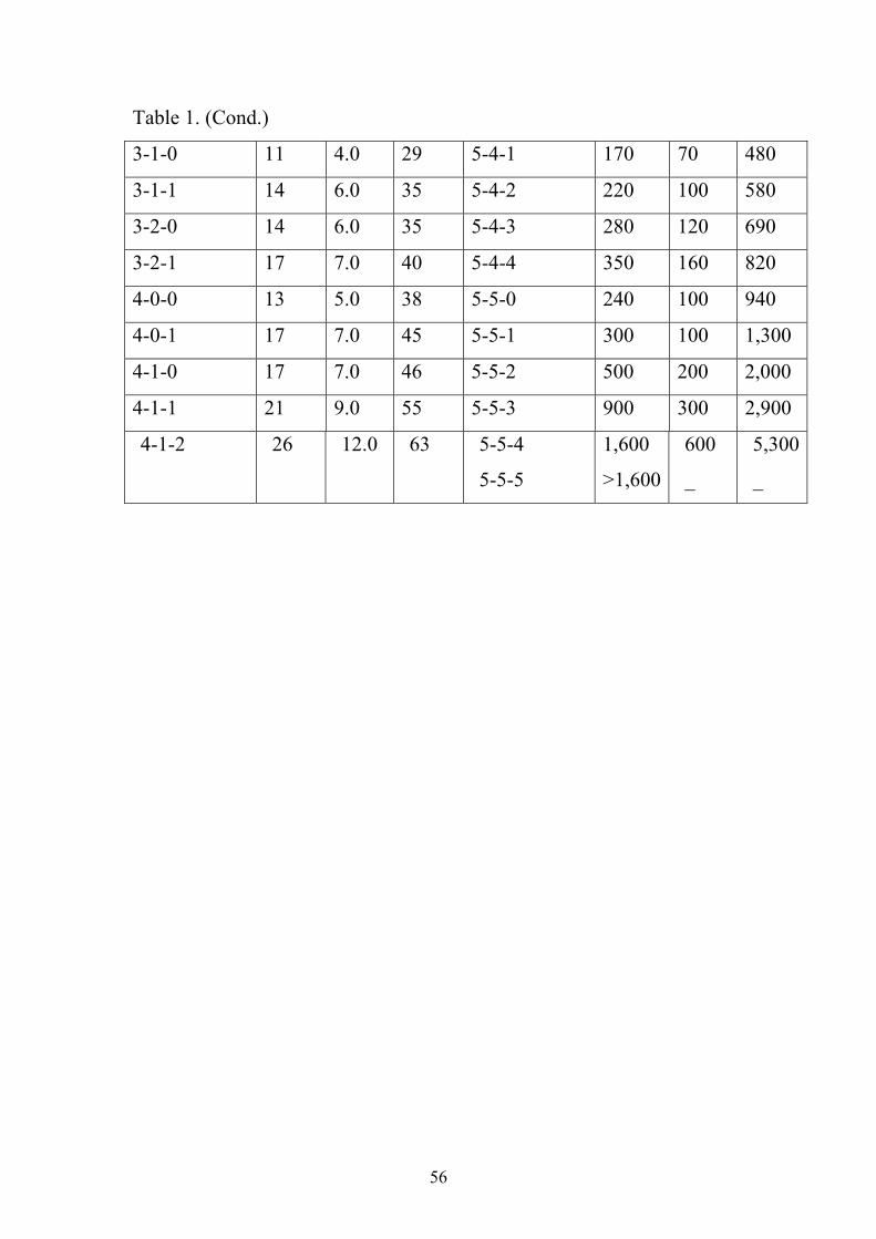
56
Table 1. (Cond.)
3-1-0 11 4.0 29 5-4-1 170 70 480
3-1-1 14 6.0 35 5-4-2 220 100 580
3-2-0 14 6.0 35 5-4-3 280 120 690
3-2-1 17 7.0 40 5-4-4 350 160 820
4-0-0 13 5.0 38 5-5-0 240 100 940
4-0-1 17 7.0 45 5-5-1 300 100 1,300
4-1-0 17 7.0 46 5-5-2 500 200 2,000
4-1-1 21 9.0 55 5-5-3 900 300 2,900
4-1-2 26 12.0 63 5-5-4
5-5-5
1,600
>1,600
600
_
5,300
_

57
Chapter three
Results
3.1 Total and thermotolerant coliform:
Out of 70 drinking water samples collected from various
sources and areas in Kassala city, total coliforms were detected in 46
samples (65.7%) (Table II). Total coliforms were detected in 50-80% in
the samples of the different sources, while thermotolerant coliforms were
found to be less prevalent, 30-60%.
3.2 Isolates:
A total of 57 isolates were obtained from 46 samples out of
70 (Table III). Highest number of isolates was from El-ảmriya well and
the lowest was from El-gasor well (Table IV).
3.2.1 Identification of isolates:
According to Gram's method of staining, all of the isolates
were Gram-negative. Microscopic examination of Gram's stained smears,
study of cultural characteristics on the media and conditions specified and
the biochemical properties (Table V) of each isolate were the identifiction
tools. Accordingly, the 57 isolates were identified as follows: 22 (38.6%)
isolates were identified as E. coli, followed by 10 (17.5%) as
Enterobacter aerogenes, 9 (15.8%) as Enterobacter cloacae, 7 (12.3%)
as Kleblsiella pneumoniae ssp. aerogenes, 5 (8.8%) as Klebsiella
pneumoniae ssp. ozaenae, 4 (7%) as Citrobacter freundii.
Table IV shows the distribution of isolated bacterial species
according to source of water samples.

58
3.2.2.1 E. coli isolates:
3.2.2.1.1 Microscopic examination:
Under the microscope, E. coli isolates appeared as Gram-
negative, short to medium slender rods, arranged singly and rarely in
pairs. Encapsulated isolates were not detected.
3.2.2.1.2 Cultural characteristics of E. coli:
On MacConkey's agar medium, large, pink, circular, convex
and smooth colonies were observed indicating lactose fermenting
colonies, which were pink and small, were observed.
3.2.2.1.3 Biochemical reactions:
All isolates were found to be lactose-fermenters, indole-
positive, Vages-Proskauer-negative, methyl red-positive, oxidase-
nagative, citrate-negative, urease-negative and H2S-negative. Acid and
gas were produced from glucose.
3.2.2.2 Enterobacter aerogenes:
3.2.2.2.1 Microscopic examinations:
Non spore-forming, Gram-negative short rods were seen.
3.2.2.2.2 Cultural characteristics:
On MacConkey's agar medium, lactose fermenting colonies
with irregular round edges and mucoid in nature were seen.
3.2.2.2.3 Biochemical reactions:
This isolate was citrate-positive but urease and indole-
negative.

59
3.2.2.3 Enterobacter cloacae:
3.2.2.3.1 Microscopic examination:
Non spore-forming, Gram-negative short rods were seen.
3.2.2.3.2 Cultural characteristics:
On MacConkey's agar medium, lactose fermenting colonies
with irregular edges, mucoid in nature were seen.
3.2.2.3.3 Biochemical reactions:
This species was citrate and urease-positive but indole-
negative.
3.2.2.4 Klebsiella isolates:
3.2.2.4.1 Microscopic examination:
Encapsulated, Gram-negative straight short rods that occurred
singly, in pairs or in short chains were seen in Gram's stained smears.
3.2.2.4.2 Cultural characteristics:
On MacConkey's agar medium, colonies of Klebsiella
appeared large, mucoid, glistening and pink in color indicating lactose
fermentation and capsule formation.
3.2.2.4.3 Biochemical reactions:
The Klebsiella isolates were Voges-Proskauer-negative and
methyl-red-positive, indole-nagative and utilized some sugars. Klebsiella
pneumoniae ssp aerogenes urease-positive but K. ozaenae was urease-
negative.
60
3.2.2.5 Citrobacter freundii isolates:
3.2.2.5.1 Microscopic examination:
Non spore-forming, non-capsulated, Gram-negative rods
were observed.
3.2.2.5.2 Cultural characteristics:
On MacConkey's agar medium they gave pink-red colonies.
3.2.2.5.3 Biochemical characteristics:
All isolates were indole-positive, methyl red-positive,
produce gas from fermentation of glucose, H2S was produced.

61
Table II. MPN for total and thermotolerant coliforms in 70 samples
of drinking water from various sources in Kassala city.
MPN/100ml
Source of
Samples*
Positive total
Coliforms (37C)
Positive thermotolerant
coliforms (44C)
Main station 6 3
El-shảbiya 8 5
El-halanga 7 6
El-ảmriya 7 5
El-gasor 5 3
El-kara 7 4
Mixed sources (main
station + El-brno well)
6 5
Total 46 31
* Number of samples from each source was 10.
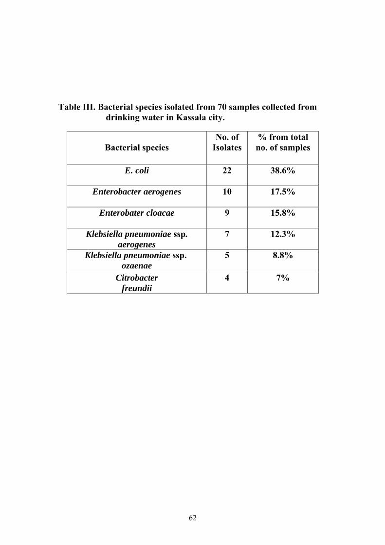
62
Table III. Bacterial species isolated from 70 samples collected from drinking water in Kassala city.
% from total no. of samples
No. of Isolates
Bacterial species
38.6% 22 E. coli
17.5% 10
Enterobacter aerogenes
15.8% 9 Enterobater cloacae
12.3% 7 Klebsiella pneumoniae ssp. aerogenes
8.8% 5
Klebsiella pneumoniae ssp. ozaenae
7% 4 Citrobacter freundii

63
Table IV. Distribution of isolated bacterial species according to sources of water samples.
Total
Citrobacter
freundii
K.
ozaenae
Klebsiella aerogenes
E.
cloacae
Enterobacter
aerogenes
E.
coli
Bacteria
Sources
4 0 0 1 1 1 1 Main station
11 1 1 1 1 2 5 El-shảbiya 12 1 0 2 2 2 5 El-halanga 14 1 3 2 2 2 4 El-ảmriya 3 0 0 0 1 0 2 El-gasor 4 0 0 0 1 1 2 El-kara 9
1
1
1
1
2
3
Mixed sources (main
station + El-brno well)
57 4 5 7 9 10 22 Total

64
Table V. Characters and biochemical reactions of bacterial isolates from drinking water samples collected from Kassala city.
Citrobacter freundii
K. ozaenae
Klebsiella aerogenes
E. cloacae
Enterobacter aerogenes
E.coli
Bacterial species
Character
- ve - ve - ve - ve - ve - ve Gram stain Rod Rod Rod Rod Rod Rod Shape + ve - ve - ve + ve + ve + ve Motolity + ve + ve + ve + ve + ve + ve Catalase - ve - ve - ve - ve - ve - ve Oxidase F F F F F F O/F
A&G A&G A&G A&G A&G A&G Glucose fermentation
+ ve + ve + ve + ve - ve - ve Urease + ve + ve + ve + ve - ve - ve Citrate - ve - ve - ve + ve - ve - ve V.P - ve - ve - ve - ve - ve -ve Nitrate + ve + ve - ve - ve + ve + ve M.R - ve - ve - ve - ve - ve + ve Indole
production + ve - ve - ve - ve - ve - ve H2S (paper
method) Acid from:
+ ve + ve + ve + ve + ve + ve Arabinose
+ ve + ve + ve + ve + ve + ve Mannitol + ve + ve + ve + ve + ve + ve Maltose + ve + ve + ve + ve + ve + ve Lactose - ve + ve + ve + ve + ve + ve Salicin + ve + ve + ve + ve + ve + ve Sorbitol - ve - ve + ve + ve + ve - ve Sucrose + ve + ve + ve + ve + ve + ve Xylose - ve + ve + ve - ve + ve - ve Inositol + ve + ve + ve + ve + ve + ve Rafinose
- ve: Negative + ve: Positive F: Fermentative A&G: Acid and Gas

65
0
5
10
15
20
25
30
35
Percentage
Bacterial species
E. coli
Enterobacteraerogenes
Enterobactercloacae
KlebsiellapneumoniaeaerogenesKlebsiellapneumoniaessp.ozaenaeCitrobacterfreundii
fig. 1: Bacterial species isolated from 70 samples collected from drinking
water in Kassala city.

66
fig. 2: Multiple Tube Fermentation test in MacConkey's broth. Positive result indicated by production of gas and acid (right tube).
Fig. 3 : Multiple Tube Fermentation test in Brilliant green lactose bile broth. Positive result indicated by production of gas (right tube).

67
Fig. 4: Growth of E. coli on Eosin methylene blue agar. .
Fig. 5: Growth of E. coli on MacConkey's agar.
68
Chapter four
Discussion
Water can be considered as food stuff which is consumed in the
greatest quantity around the world. Therefore, it comes as no surprise that
the health risks associated with consumption of contaminated water are of
great importance.
This study aimed to investigate drinking water for bacterial
pollution in Kassala city at different locations and investigation of ground
water before its pumping through public pipeline. Results
showed that 65.7% of samples were contaminated with total coliform
while 44.3% were contaminated with thermotolerant coliform (E. coli).
This result is similar to the studies of El-amin (2005) and Sulieman
(2005) who reported that the total coliform bacteria and thermotolerant
coliform bacteria constituted 66%, 46% and 60%, 30%, respectively.
The highest number of total coliforms was detected in
samples from EL shảbiya. This is may be due to the irregular cleaning of
tanks or contaminated with human wastes.
Comparing samples from different water sources (tap water
from main station, wells), the lowest numbers of coliforms was detected
in tap water samples of main station. This could be explained by the fact
that deep-well water usually contains few organisms because microbes
are filtered out as the water trickles through the earth layers (Smith,
1981). In addition to treatment of water with suitable chemicals (e.g.
chlorine).
The presence of coliforms in water samples indicates either
that water was contaminated with human and animal wastes or the water
might not have been properly treated with suitable chemicals (e.g.

69
chlorine) using the suitable concentration. The consumption of such poor
quality water may expose the public to many dangerous diseases. Similar
explanations were reported by Champan (1992).
The highest isolation frequency of E. coli was detected in samples
from EL halanga and El-shảbiya . This is may be due to contamination
with human wastes as the deep-well usually contains few organisms
(Smith, 1981).
Isolation of thermotolerant E. coli in this study represents another
public health hazard. It may cause outbreaks of diarrhea, especially the
serotype O157: H7 (WHO, 1998).
The highest number of total coliforms were detected in samples
from tap water (EL kara, EL brno). This finding may be indicate that
water was contaminated with human wastes or contaminated through
damage in pipelines.
In this work, six bacterial species were isolated from the different
sources and types of water. Citrobacter freundii and Klebsiella
pneumoniae ssp. ozaenae were found in untreated water only. This may
be because these two species are more susceptible to the chemical
treatment (Chlorine). E. coli was the most prevalent species (38.6%) are
found in both types of water, while Citrobcter freundii was the least
prevalent species (7%).
The wells in the centre of the city (El-ảmriya and El-halanga) were
found to be the most contamination water sources. This could be
attributed to conamination with human wastes.
CONCLUSION:
Total and thermotolerant coliforms were detected in 65.7%
and 44.3% of water samples investigated in this study, respectively.
Coliforms were detected in tap water from different wells in Kassala city,
detection rate was higher in untreated water. Potential pathogens were

70
among the isolates and this contamination may represent a real public
health hazard.
RECOMMENDATIONS:
1. Routine and periodic investigation of drinking water from all
sources by health authorities for microbial contamination is
important.
2. Following up of water pipelines to ckeck for and repair their
damage to avoid bacterial contamination of water.
3. Ground water have to be treated before it is pumbing through the
net work to avoid bacterial contamination.
4. Further studies are needed to be carried out for investigation of E.
coli, especially serotype O157:H7, because it has been associated
clinically with severe bloody diarrhoea, which leads to hemolytic
uraemic syndrome (HUS).

71
References
Abdelmagid. H.M.; Ibrahim, I.S. and Dirar, H.A. (1984). Chemical and
microbiological examination of well and Nile water. Environ.
Internat. 10: 259-263.
Alcamo, E.I. (1994). Fundamentals of Microbiology. 4th ed.
Benjamic/cummings, publshing company Inc, Red wood city.
Alcamo, E.I. (1997). Fundamentals of Microbiology. 5th ed.
Benjamic/cummings, an imprint of Addison Wesley Longman, Inc.
Amann, R.I.; Ludwig, W.; and Scleifer, K.H. (1995). Phylogenetic
identification and in situ detection of individual microbial cell with
cultivation. Microbiol. Review. 59: 143-146.
Barrow, G.L.; and Felthman, R.K.A. (1993). Cowan and Steels Manual
for the Identification of Medical Bacteria. 3rd ed. Cambridge
Univerisity Press, U. K.
Bartram, J.; and Pedley, S. (1999). Microbiological Analysis In:
UNEP/WHO monitoring Water quality monitoring- A practical
guide to the design and implementation of fresh water quality
studies and programmes.
Bartram, J.; and Wheeler, D. (1993). Microbiological aspects of drinking
water quality monitoring. WHO, Cyprus.
Black, G.J. (1999). Microbiology Principles and Exploration. 2nd ed.
Prentice Hall, Englewood cliffs, New Jersey.
Bonjoch, X.; Balleste, B.; and Blanch, A. R. (2004). Multiplex PCR
with 16S rRNA gene-targeted primers of Bifidobacterium spp. to
identify sources of fecal pollution. Applied and Environ.
Microbiolo. 70: 3171-3175.
Brenner, K. P.; Rankin, C. C.; Roybal, Y. R.; Stelma, G. N.; Scarpino,
P.V.; and Dufour, A. P.(1993). New medium for the simultaneous

72
detection of total coliforms and Escherichia coli in water. Applied
and Environ. Microbiol. 59: 3534-3544.
Brock, T.D.; Madigan, M.T.; Martinko, J. M.; and Parke, J. (1994).
Biology of Microorganisms. 7th ed. Prentice-Hall, Inc. A paramount
communications company- Englewood cliffs, New Jersy.
Buttiaux, R.; and Mossel, D. (1961). The significance of various
organisms of fecal origin in foods and drinking water. J. Applied
Bactriol. 24: 353-364.
Cairncross, S.; Carruthers, I.; Curtis, D.; Feachem, R.; Breadley, D.;
and Baldwin, G. (1980). Evaluation for village water supply
planning. Published in association with International Reference
Centre for Community Water Supply, by John Wiley and SONS.
Chichester. New York. Brisbane. Toronto.
Chapman. D. (1992). Water quality assessment. A guide to the use of
Biota, sediments and water in environmental monitoring. Published
on behalf of UNESCO, United Nation.
Cheesbrough, M. (1994). Medical Laboratory Manual for Tropical
Countries. Part 2. 1st eds. Cambridge University Press.
Deming, H.G. (1975). Water the Fountain of Opportunity. Oxford
University Press, USA.
Dirar, H.A. (1986). Coliform bacterial counts in the Nile water at
Khartoum. Environ. Internat. 12: 571-576.
Doran, J.; and Linn, D. (1979). Bacteriological quality of run-off water
from pastureland. Applied and Environ. Microbiolo. 37: 985-991.
Duerden, B.I.; Reid, T.M.S.; Jewsbury, J.M.; and Turk, D.C. (1988). A
New Short Text Book of Microbical and Parasitic Infection. 2nd ed.
Edward Arnold, London.
Dutt, G. (1982). Mapping of the Environment. Internat. Environ. Studies.
19: 117.

73
Eckner, K.F. (1998). Comparisons of membrane filtration and multiple-
tube fermentation by the Colilert and Enterolert methods for
detection of waterborne coliform bacteria, Escherichia coli, and
Enterococci used in drinking and bathing water quality monitoring
in southen Sweden. Applied Environ. Microbiol. 64: 3079-3083.
El-amin, A.A. (2005), Investigation on Echerichia coli O157: H7 in
drinking water in Omdurman-Sudan. M. Sc. Thesis, University of
Khartoum.
El Shazali, H.; and Erwa, H. (1971). An over looked source of
gastroenteritis in Sudanese Children. Sudan Med. J. 9: 46.
El Tayeb, G. (2002). Human development in Sudan from agenda 21
perspective. In: Abel ati, H.A Sustainable Development in Sudan:
10 years after Rio summit a civil society perspective.
Environmentalists Society, EDGE for Consultancy and Research;
Heinrich Boll Foundation Regional Office, Horn of Africa.
Fernandez-Alvarez, R.M.; Carballo-Cuervo, S.; Carmendela Rosa-Jorge,
M.; and Rodrignez-Delecea, J. (1991). The influence of
agricultural runoff on bacterial populations in a river. J. Applied
Bacteriol. 70: 437-445.
Fish, J.T.; and Pettibone, G.W. (1995). Influence of water sediment on
the survival of Echerichia coli and Salmonella spp. as measured
by three methods of enumeration. Letter in Applied Microbiol.
20: 277-281.
Food and Agricultural Organization (FAO) (1979). Ground Water
Pollution, No. 31, FAO. Rome.
Forrest, B. (1956). Rural Water Supply and Sanitation. John Wiley and
Sons, Inc. New York.

74
Fricker, E,J.; and Fricker, C.R. (1994). Application of polymerase chain
reaction to the identification of Escherichia coli and coliform in
water. Letters in Applied Microbiol. 19: 44-46.
Friedrich, G.; Chapman, D.; and Beim, A. (1992). The use of biological
material in water quality assessments-a guide to use of biota,
sediments and water environmental monitoring. 2nd ed.
UNESO/WHO/UNEP.
Gannon, V.P.; D,souza, S.; Graham, T.; King, R.K., Rahn, k.; and
Read, S. (1997). Use of the flagella H7 antigen as a target in
multiplex PCR assay and improved specificity in identification of
enterohemorrhagic Escherichia coli strains. J. Clin. Microbiol.
33(3): 656-662.
Hagedorn, C.; Robinson, S,L.; Filtz, J.R.; Grubbs, S.M.; Angier,
T.A.; and Reneau, R.B. (1999). Determining Sources of Faecal
Pollution in a Rural Virginia Watershed with Antibiotic Resistance
Patterns in Faecal Streptococci. Applied Environ. Microbiol. 65:
5522-5531.
Hanko, E. (2002). Colilert: What is it all about? Water Industry
Operators Association (WIOA) conference papers. Australia.
Jay, J.M. (1986). Modern food Microbiology. 3rd ed. Van Nostrand
Reinhold Company. New York.
Johnson, L.K.; Brown, M.B.; Carruthers, E.A.; Ferguson, J.A.;
Dombek, P.E.; and Sadowsky, M.J. (2004). Sample size, library
composition and genotypic diversity among natural populations of
Escherichia coli from different animals. Influence accuracy of
determining sources of faecal pollution. Applied Environ.
Microbiol. 70: 4478-4485.

75
Kabler, P.; Clark, H.; and Geldreich, E. (1960). Coliforms in sanitary
bacteriology, J. Amer. Wt. Wks. Assoc. 52: 105.
Katz, M. (1971). The effect of pollution upon aquatic life. In: Ciaccio, L.
Water and Water Pollution Handbook, vol. 1, Marcel Dekker, Inc.
USA. 297-326.
Lim, D. (1998). Microbiology. 2nd ed. The McGraw-Hill companies, Inc.
Loucks, P. (1994). Water quality-drinking water and sanitation. Water
Resources Management: Focusing on Sustainability, UNESCO,
Paris, 13-14.
Madigan, M.; Martinko, J.; and Parker, J. (1997). Brock Biology of
Microorganisms. 8th ed. Hall Int., USA. 902-984
Mates, A.; and Shaffer, M. (1989). Membrane filtration differentiation of
Escherichia coli from coliforms in the examination of water. J.
Applied Bacteriol. 67: 343-346.
Meier, H.; Koob, C.; Ludwing, W.; Amann, R.; Frahm, E.; Hoffmann, S.;
Obst. U.; and Scleifer, K. H. (1997). Detection of enterococci with
rRNA targeted DNA probes and thier use for hygienic drinking
water control. Water Science Technology. 35: 437-444.
Meybeck, M.; Kuusisto, E.; Makela, A.; and Malkki, E. (1996). Water
Quality. In: UNEP/WHO Water Quality Monitoring-a Practical
Guide to the Design and Implementation of Freshwater Quality
Studies and Monitoring Programmes.
Nebra, Y.; Bonjoch, X.; and Blanch, A.R. (2003). Use of Bifidobacterium
dentium as an Indicator of the Origin of Faecal Water Pollution.
Applied Environ. Microbiol. 69: 2651- 2656.
Nester, E.W.; Roberts, C.E.; Pearsall, N.N.; and McCarthy, B.J. (1978).
Microbiology. 2nd ed. Holt, Rinehart and Winston.

76
Ogden, I.D.; and Watt, A.J. (1991). An evalution of fluorogenic and
chromogenic assays for the direct enumeration of Escherichia coli.
Applied Microbiol. 13: 212-215.
Paton, A.W.; and Paton, J.C. (2002). Direct detection and
charachterization of shiga toxingenic Escherichia coli by multiplex
PCR for stx 1, stx 2.eae, ehxh and saa. J. Clin. Microbiol. 40 (1) :
271-174.
Pelcazar, M.J.; Reid, R.D.; Chan, E.C.S. (1977). Microbiology. 4th ed.
McGraw-Hill, Inc.
Pyle, B.H.; Broadaway, S.C.; and Mcfters, G.A. (1995). A rapid, direct
method for enumerating respiring enterohemorrhagic Escherichia
coli 0157:H7 in water. Applied Environ Microbiol. 61: 2614-1619.
Rosazak, D.B.; and Colwell, R.R. (1987). Survival strategies of bacteria
in natural environment. Microbiology Review. 51: 365-379.
Schochetman, G.; Jones, W.K. (1988). Polymerase chain reaction. J.
Infectious diseases. 158: 1154-1157.
Smith, A.L. (1981). Principles of Microbiology. 9th ed. C.V. Mosby
company. St. Louis. Toronto. London.
Steinertz, M.; Emody, L.; Amann, R.; and Hacker, J. (1997).
Resuscitation of viable but non-culturable Leza\gionella
pneumonphila philadelphia JR32 by acanthamoeba castellanii.
Applied Environ Microbiol. 63: 2047-2053.
Sulieman, A.H. (2005). Bacteriological studies on drinking water in
Khartoum State- Sudan. M. Sc. Thesis, Univeristy of Khartoum.
Twort, A.C.; Law, F.M.; and Crowly, F.W. (1985). Water supply. 3rd ed.
Edward Arnold, London, USA, Australia.
Walter, k.S.; Fricker, E.J.; and Fricker, C.R. (1994). Observations on
the use of a medium detecting Bita-glucuronidase activity and

77
lactose fermentation for the simultaneous detection of Escherichia
coli and coliforms. Letters in Applied Microbiol. 19: 47-49.
Wang, R.F.; Cao, W.; and Cerniglia, C.E. (1996). PCR detection and
quantification of predominant anaerobic bacteria in human and
animal faecal samples. Applied Environ Microbiol. 62: 1242-1247.
White, W.R.; and Godfree, A.F. (1985). Pollution of fresh water and
estuaries. J. Applied Bacteriol. 8: 77-81.
Wiggins, B.A. (1996). Discriminate analysis of antibiotic resistance
patterns in faecal Sterptococci, a method to differentiate human
and animal sources of faecal pollution in natural waters. Applied
and Environ. Microbiol. 62:3979-4002.
World Health Organization. (1976). Surveillance of drinking water
quality. WHO press, Geneva.
World Health Organization. (1984). Guideline for drinking water quality.
WHO press, Geneva.
World Health Organization. (1996). Giudelines for drinking water
quality. WHO press, Geneva.
World Health Organization. (1998). Zoonotic (non-O157 shiga toxin
producing Esherichia coli (STEC). Report of a WHO, scientific
working group meeting. http//www.who.int/emc.
World Health Organization. (2004). Guidelines for drinking water
quality. 3rd edition, vol. 1, p. 532.