Embed Size (px)
Citation preview
1
2
3
4
56789
10
11121314151617181920212223242526
47
48
49
50
51
52
53
54
55
56
Progress in Neuro-Psychopharmacology & Biological Psychiatry xxx (2010) xxx–xxx
PNP-07719; No of Pages 6
Contents lists available at ScienceDirect
Progress in Neuro-Psychopharmacology & BiologicalPsychiatry
j ourna l homepage: www.e lsev ie r.com/ locate /pnp
The increase in theta/beta ratio on resting-state EEG in boys withattention-deficit/hyperactivity disorder is mediated by slow alpha peak frequency
Marieke M. Lansbergen a,⁎, Martijn Arns b,c, Martine van Dongen-Boomsma a,d,Desirée Spronk a,b, Jan K. Buitelaar d,e
a Department of Psychiatry, Radboud University Nijmegen Medical Centre, Donders Institute for Brain, Cognition and Behaviour, Nijmegen, The Netherlandsb Research Institute Brainclinics, Nijmegen, The Netherlandsc Department of Experimental Psychology, Utrecht University, Utrecht, The Netherlandsd Karakter Child and Adolescent Psychiatry University Centre, Nijmegen, The Netherlandse Department of Cognitive Neuroscience, Radboud University Nijmegen Medical Centre, Donders Institute for Brain, Cognition and Behaviour, Nijmegen, The Netherlands
Abbreviations: ADHD, attention-deficit/hyperactivityInternational Database; CPRS, Conners' Parent Rating Smance test; EEG, electroencephalography; IAF, indivSPHERE-12, Somatic and Psychological Health Report qu⁎ Corresponding author.
E-mail addresses: [email protected] ([email protected] (M. Arns), M.vanDongen-Boo(M. van Dongen-Boomsma), [email protected] ([email protected] (J.K. Buitelaar).
0278-5846/$ – see front matter © 2010 Published by Edoi:10.1016/j.pnpbp.2010.08.004
Please cite this article as: Lansbergen MMhyperactivity disorder is mediated..., Prog N
a b s t r a c t
a r t i c l e i n f o27
28
29
30
31
32
33
34
35
36
37
38
Article history:Received 1 June 2010Received in revised form 21 July 2010Accepted 5 August 2010Available online xxxx
Keywords:Alpha peak frequencyAttention-deficit/hyperactivity disorder(ADHD)Electroencephalography (EEG)IntegNeuroTheta/beta ratio
39
40
41
42
43
Attention-deficit/hyperactivity disorder (ADHD) was found to be characterized by a deviant pattern ofelectrocortical activity during resting state, particularly increased theta and decreased beta activity. The firstobjective of the present study is to confirm whether individuals with slow alpha peak frequency contributeto the finding of increased theta activity in ADHD. The second objective is to explore the relation betweenresting-state brain oscillations and specific cognitive functions. From 49 boys with ADHD and 49 healthycontrol boys, resting-state EEG during eyes open and eyes closed was recorded, and a variety of cognitivetasks were administered. Theta and beta power and theta/beta ratio were calculated using both fixedfrequency bands and individualized frequency bands. As expected, theta/beta ratio, calculated using fixedfrequency bands, was significantly higher in ADHD children than control children. However, this group effectwas not significant when theta/beta ratio was assessed using individualized frequency bands. No consistentrelation was found between resting-state brain oscillations and cognition. The present results suggest thatprevious findings of increased theta/beta ratio in ADHD may reflect individuals with slow alpha peakfrequencies in addition to individuals with true increased theta activity. Therefore, the often reported theta/beta ratio in ADHD can be considered a non-specific measure combining several distinct neurophysiologicalsubgroups such as frontal theta and slowed alpha peak frequencies. Future research should elucidate thefunctional role of resting-state brain oscillations by investigating neurophysiological subgroups, which mayhave a more clear relation to cognitive functions than single frequency bands.
44
disorder; BRID, Brain Resourcecale; CPT, continuous perfor-idual alpha peak frequency;estionnaire.
. Lansbergen),[email protected]. Spronk),
lsevier Inc.
, et al, The increase in theta/beta ratio oneuro-Psychopharmacol Biol Psychiatry (201
© 2010 Published by Elsevier Inc.
4546
57
58
59
60
61
62
63
64
65
1. Introduction
Attention-deficit/hyperactivity disorder (ADHD) is the mostcommon psychiatric disorder in childhood, affecting 5–10% of allchildrenworldwide (Faraone et al. 2003). In 40–60% of all cases ADHDpersists in adolescence and adulthood (Faraone et al. 2006).Electrophysiological studies have revealed consistent evidence forabnormal brain oscillations during resting state in individuals withADHD (Barry et al. 2003). The EEG of the majority of children with
66
67
68
69
70
71
72
73
74
ADHD is characterized by a deviant pattern of baseline corticalactivity, specifically increased slow-wave activity, primarily in thetheta band, and decreased fast-wave activity, primarily in the betaband, often coupled (i.e., increased theta/beta ratio; Barry et al. 2003).A meta-analysis of EEG and ADHD including 9 studies (1498participants) reported significant effect sizes for theta and betapower, and theta/beta ratio (effect size=1.31, −0.51, 3.08, respec-tively; Snyder and Hall 2006). However, recently, it has beensuggested that at least two different EEG subtypes in ADHD, asubgroup with true frontal slow EEG (i.e., enhanced theta activity)and a subgroup with slow alpha peak frequency, might lead to thefinding of increased ‘theta’ power (Arns et al., 2008), and thusincreased theta/beta ratio, in ADHD. Moreover, these two EEGsubtypes differed in their response to stimulant medication (Arnset al. 2008). So, the robust finding of increased theta and theta/betaratio in ADHD may largely depend on a subgroup of children withADHDwho have a slow alpha peak frequency. In earlier studies, basedon visual inspection of EEG data, it has already been reported that
resting-state EEG in boys with attention-deficit/0), doi:10.1016/j.pnpbp.2010.08.004
75
76
77
78
79
80
81
82
83
84
85
86
87
88
89
90
91
92
93
94
95
96
97
98
99
100
101
102
103
104
105
106
107
108
109
110
111
112
113
114
115
116
117
118
119
120
121
122
123
124
125
126
127
128
129
130
131
132
133
134
135
136
137
138
139
140
141
142 Q1143
144
145
146
147
148
149
150
151
152
153
154
155
156
157
158
159
160
161
162
163
164
165
166
167
168
169
170
171
172
173
174
175
176
177
178
179
180
181
182
183
184
185
186
187
188
189
190
191
192
193
194
195
196
197
198
199
200
201
2 M.M. Lansbergen et al. / Progress in Neuro-Psychopharmacology & Biological Psychiatry xxx (2010) xxx–xxx
slow alpha peak frequency was correlated to hyperactive behavior,whereas the frontal EEG abnormalities such as frontal slow EEGshowed no relation to the ADHD symptomatology (Stevens et al.1968). When EEG power is calculated from individual adjustedfrequency bands (based on the individual alpha peak frequency)rather than from fixed frequency bands (Klimesch 1999), the findingof increased theta power in one group relative to another group is notcontaminated by participants with slow alpha peaks. Especially inchildren it is known that the alpha peak frequency matures starting at4–6 Hz at age 2–12 months to 10 Hz at 10 years of age (Niedermeyerand Da Silva 2004), and hence the use of individual adjustedfrequency bands is especially important. So far, all studies comparingresting-state EEG between children with and without ADHD usedfixed frequency bands to estimate EEG power. The first objective ofthe present study is to address the question whether the robustfindings of increased theta activity in ADHD are still present whencontrolling for children who exhibit slower alpha peak frequencies.
Few studies have addressed the functional role of resting-statebrain oscillations. Do specific resting-state brain oscillations relate tospecific cognitive functions?
Decades of research have established well-replicated findingsof several cognitive deficits in attention-deficit/hyperactivity disorder(ADHD) such as attention deficits, working memory problemsand deficient inhibitory control (Nigg 2005). However, the questionremains whether cognitive impairments may relate to abnormal brainoscillations.
It has been argued that alpha activity reflects arousal (i.e., “thecurrent energetic level of the organism”) and theta and beta activityreflect task- or situation-specific activation changes resulting fromstimulus processing (Barry et al. 2007). Based on findings from animalandhuman research, it has been suggested that task-induced increasesin theta power and the phase relationship between theta and gammaoscillations are important for memory processes, particularly episodiclong-term memory and working memory (Knyazev 2007; Sausenget al. 2010). Recently, it has also been postulated that theta oscillationsreflect a “more general brain integrative mechanism” rather than anintegrative mechanism specific for memory processes (Sauseng et al.2010). Event-related increases in alpha power have been associatedwith top-down inhibitory control processes of visual information(Jensen et al. 2002).
Klimesch (1999) argued that low levels of theta activity and highlevels of alpha activity during resting state predict increased thetapower and decreased alpha power during task performance, thatsubsequently lead to improved cognitive performance. However, fewstudies investigated directly the relation between brain oscillations ina resting human (not during task performance) and subsequentcognitive performance. Consistent with the hypothesis of Klimesch(1999), increased theta power and increased alpha power at rest havebeen related to impaired cognitive performance in children withADHD as well as in control children (Hermens et al., 2005; Loo andSmalley 2008; Sumich et al. 2009). However, discrepant findings havealso been reported (Swartwood et al. 1998; Wienbruch et al. 2005).So, the functional role of EEG oscillations during resting state is stillunclear. One possible explanation for the inconsistent findings is thewide variety of behavioral paradigms that have been used, which alltap different cognitive functions. The second goal is to explore therelation between resting-state brain oscillations (calculated usingindividualized frequency bands) and task performance on a variety ofcognitive tasks. By setting this goal, the present study may revealbaseline EEG markers of specific neurocognitive dysfunctions inADHD, and give more insight into the functional role of resting-stateEEG.
Based on previous independent findings of increased slow-waveand decreased fast-wave activity as well as impaired cognitiveperformance in ADHD, we expect that increased theta power,decreased beta power, and increased theta/beta ratios will be
Please cite this article as: Lansbergen MM, et al, The increase in thehyperactivity disorder is mediated..., Prog Neuro-Psychopharmacol Biol
associated with decreased cognitive performance in ADHD patients.Based on the assumption of Klimesch et al. (1999) that increasedalpha activity at rest predicts decreased event-related alpha activitywhich reflects increased cognitive performance, we hypothesized thatalpha power at rest correlates positively with cognitive performance.
2. Method
2.1. Participants
Forty-nine boys diagnosed with ADHD (M=12.2 years; SD=3.0;range 6–18) were matched on age, gender, and education with 49healthy control boys (M=12.5 years; SD=2.8; range 7–18). Perfor-mance on the Spot the Real Word Test, which is a good indicator ofpremorbid IQ (Paul et al. 2005), did not differ significantly between thegroups (36.8 and 37.4 for the ADHD and control group, respectively;F(1,91)b1). The data from the participants in the present study wereacquired as part of the Brain Resource International Database (BRID;http://www.brainresource.com) and have already partially beenpublished (Arns et al. 2008). Data acquisition for the BRID is performedin a standardized manner with identical hardware, software, para-digms, and experimental procedures (Gordon et al. 2005).
All children were recruited from the Sydney metropolitan region.Two pediatricians evaluated the children with ADHD using a semi-structured interview based on DSM-IV criteria for ADHD (Williamset al., 2010) and Conners' Parent Rating Scale (CPRS; Conners et al.1998) (T-scores 1 SD above the norm for either inattentive orhyperactive/impulsive subscores). Twenty-one participants metcriteria for ADHD combined subtype, 22 met the criteria for ADHDpredominantly inattentive type, and 2 boys met the criteria for ADHDpredominantly hyperactive/impulsive subtype. The classification ofADHD subtype was missing for 3 participants. The average number ofinattentive and hyperactive/impulsivity DSM-IV symptoms for theADHD group was 8.0 (SD=1.3) and 5.2 (SD=2.9), respectively. Theaverage scores on the cognitive problems/inattentive, hyperactive,and impulsive subscales of the CPRS were 70.3 (SD=7.4), 73.5(SD=14.9), and 73.0 (SD=9.8), respectively.
Exclusion criteria for ADHD and healthy control children includeda personal history of physical brain injury, neurological disorder,genetic disorder, or other serious medical condition and a personalhistory of substance abuse or dependency. Additionally, ADHDchildren were excluded if they had an Axis I psychiatric disorder(other than ADHD), assessed by two pediatricians in a semi-structured interview. Children in the control group were excluded ifthe Somatic and Psychological Health Report questionnaire (SPHERE-12; Hickie et al. 2001) revealed an Axis I disorder. All children weremedication free for at least 48 h before testing. Moreover, for at least2 h prior to testing participantswere required to refrain from caffeine-intake and smoking.
All subjects and their caretakers provided written informed consentto participate in the study. In the informed consent, permission isasked to add the participant's de-linked data to the brain database,and to use their de-linked data for the specified and other scientificinvestigations. The study was approved by the Western Sydney AreaHealth Service Human Research Ethics Committee.
2.2. Intelligence
2.2.1. Spot the Real Word TestThis test is a computerized adaptation of the Spot the Word Test
(Baddeley et al. 1993). The estimated IQ derived from this testcorrelates highly with full scale IQ, as assessed by the WAIS-III(r=0.76; Paul et al. 2005). On each of the 60 trials, a real word ispresented simultaneously with a nonsense word. Participants wererequired to select the real word. The estimated IQ is derived fromthe total correct score.
ta/beta ratio on resting-state EEG in boys with attention-deficit/Psychiatry (2010), doi:10.1016/j.pnpbp.2010.08.004
202
203
204
205
206
207
208
209
210
211
212
213
214
215
216
217
218
219
220
221
222
223
224
225
226
227
228
229
230
231
232
233
234
235
236
237
238
239
240
241
242
243
244
245
246
247
248
249
250
251
252
253
254
255
256
257
258
259
260
261
262
263
264
265
266
267
268
269
270
271
272
273
274
275
276
277
278
279
280
281
282
283
284
285
286
287
288
289
290
291
292
293
294
295
296
297
298
299
300
301
302
303
304
305
306
307
308
309
310
311
312
313
314
315
316
317
318
319
320
321
322
323
324
3M.M. Lansbergen et al. / Progress in Neuro-Psychopharmacology & Biological Psychiatry xxx (2010) xxx–xxx
2.3. Neuropsychological tests
The cognitive tests were part of the IntegNeuro battery, a fullycomputerized and standardized neuropsychological battery that ispresented on a touch-screen computer. The IntegNeuro batteryconsists of 12 tests that tap five domains of cognitive functions:sensori-motor functions, attention and working memory, executivefunction, learning and memory, and language skills (for more details,see Clark et al. 2006; Paul et al. 2005;Williams et al. 2005). Test–retestreliability and convergent and divergent validity of the IntegNeurobattery have been reported (Paul et al. 2005; Williams et al. 2005).Moreover, it has recently been demonstrated that the IntegNeurobattery could distinguish patients with ADHD from control partici-pants with relatively high sensitivity and specificity (Williams et al.,2010).
Standardized visual and auditory task instructions were presented.Each test was preceded by a practice trial. The test trials werepresented only if participants passed the practice trial accurately. Thepresent paper presents and discusses EEG data and the results of6 cognitive tests covering the sensori-motor, attention/memory,and language domains. Since details of the neuropsychological tasksof the IntegNeuro battery have been extensively described previously(Clark et al. 2006; Paul et al. 2005), here only the most importantdetails are reported.
Sensori-motor functions were assessed by the Choice ReactionTime test. Dependent variable is the mean reaction time across trials(i.e., mean choice reaction time [Choice RT]).
The digit span task (forward and backward version) and span ofvisual memory task (forward and backward version) were used to tapverbal and visuo-spatial short-term memory (forward version) andverbal and visuo-spatial working memory (backward version),respectively. Dependent variables are the maximum number of digitsand maximum number of squares correctly recalled.
The continuous performance test (CPT) and a Go/No-Go paradigmwere used to tap attention, as reflected in mean reaction time tocorrect target/go-stimuli.
Finally, language skills (verbal fluency) were assessed by the wordgeneration task in which the total number of correct words generatedacross three letters was the variable of interest.
2.4. Electrophysiological recordings
EEG and EOG activity were recorded using a Quickcap (NuAmps)with 26 electrodes according to the 10–20 electrode internationalsystem. Data were referenced to averaged mastoids with the groundelectrode placed at Fpz. Horizontal electrooculogram (HEOG) wasrecorded from electrodes placed lateral to the outer canthi of botheyes and vertical electrooculogram (VEOG) from electrodes attachedabove and below the left eye. Electrode impedancewas kept below 5 kΩ. The sampling rate was 500 Hz and the data were filtered onlinewith a 100 Hz low-pass filter (40 dB attenuation).
2.5. Procedure
Participants were placed in a sound-attenuated testing room andasked to sit quietly for 4 min, 2 min with their eyes open and 2 minwith their eyes closed during which baseline EEG was recorded.Afterwards, participants completed the IntegNeuro battery in front ofthe touch-screen computer in approximately 50 min. Details of theprocedure have been published (Gordon et al. 2005; Paul et al. 2005).
2.6. EEG analyses
Since EEG data from one boy with ADHD were not recorded atall electrode sites of interest (i.e., Fz, F3, F4, Cz, C3, C4, Pz, P3, and P4),he was excluded from the EEG analyses. EEG data during the eyes
Please cite this article as: Lansbergen MM, et al, The increase in thehyperactivity disorder is mediated..., Prog Neuro-Psychopharmacol Biol
open and eyes closed resting condition were processed and analyzedseparately using BrainVision Analyzer software (www.brainproducts.com). EEG data were filtered using a band-pass filter of 0.5–100 Hzand a notch filter of 50 Hz. The sampling rate was changed to 256 Hz.The continuous EEG data were segmented into 2-s epochs, separatelyfor the eyes open and eyes closed condition. Epochs were rejectedfrom further analyses if data exceeded 100 μV and ocular artifactcorrection was conducted according to the Gratton et al. (1983)algorithm. The average number of EEG epochs used for the FFTanalyses was 57.3 (SD=9.6) for the eyes open and 57.6 (SD=9.0) forthe eyes closed condition in the ADHD group and 57.8 (SD=5.9) forthe eyes open and 56.3 (SD=6.8) for the eyes closed condition inthe control group. EEG data were Fourier transformed (Hanningwindow length of 20%) and subsequently ln-transformed (Gasseret al., 1982).
First, mean theta and beta power were calculated using fixedfrequency bands (4–7.5 Hz for theta and 12.5–25 Hz for beta).Additionally, since alpha peak frequency and consequently alsoEEG frequency bands vary as a function of age (Klimesch 1999;Niedermeyer and Da Silva 2004) and may differ between individuals(Arns et al. 2008), separately for each participant, the average powervalues of the different frequency bands were also calculated usingindividual alpha peak frequency as anchor point (Doppelmayr et al.1998). First, the frequency was assessed at which alpha power wasmaximum within 5–15 Hz at the parietal and occipital electrodes(Pz, P3, P4, Oz, O1, and O2) in the eyes closed condition. Second, EEGdata in the eyes open resting condition were subtracted from the EEGdata in the eyes closed resting condition to find the frequency (within5–15 Hz) at which alpha power was most attenuated by opening ofthe eyes (Posthuma et al. 2001). When peak frequencies derived fromthe two methods and different electrode sites did not differ morethan 0.5 Hz, themean peak frequency at which alpha power wasmostattenuated by opening of the eyes across the parietal and occipitalelectrodes was used as individual alpha peak frequency (IAF). Forpeak frequencies occurring at the boundaries of the search windowand for peak frequencies which differed between electrode sites andthe two methods, visual inspection of the EEG data was conducted todetermine the true individual alpha peak frequency.
Three boys with ADHD did not show clear alpha peak frequenciesand were excluded from the analysis of the individual frequencybands. For 2 boys with ADHD and 1 control boy, who did not showalpha attenuation after opening the eyes, but did show a clear alphapeak, individual alpha peak frequencies were assessed using the EEGdata in the eyes closed resting condition. Further, for 2 ADHD boys and2 control boys, who showed only alpha attenuation at occipitalelectrode sites and not at parietal sites, mean individual alpha peakfrequencies were calculated from the difference power spectrum(eyes closed minus eyes open) across occipital electrode sites. Thebandwidth for the theta, alpha, and beta bands were defined using IAFas anchor point: 0.4*IAF–0.6*IAF, 0.6*IAF–0.8*IAF, 0.8*IAF–IAF, IAF–1.2*IAF, and 1.2*IAF–25 Hz for theta, lower 1-alpha, lower-2-alpha,upper alpha, and beta, respectively (Doppelmayr et al. 1998).
For both fixed frequency and individualized frequency bands,average power spectra were calculated at frontal (Fz, F3, and F4),central (Cz, C3, and C4) and parietal (Pz, P3, and P4) sites. Separatelyfor each electrode, theta/beta power ratio was calculated by dividingthe power of the slower frequency band by the power of the fasterfrequency band.
2.7. Statistical analyses
Since the behavioral data were not normally distributed, Mann–Whitney U tests were conducted for each dependent variable withgroup as between-subjects factor (ADHD vs. matched controls) toexamine differences between boys with and without ADHD oncognitive performance in several domains.
ta/beta ratio on resting-state EEG in boys with attention-deficit/Psychiatry (2010), doi:10.1016/j.pnpbp.2010.08.004
325
326
327
328
329
330
331
332
333
334
335
336
337
338
339
340
341
342
343
344
345
346
347
348
349
350
351
352
353
354
355
356
357
358
359
360
361
362
363
364
365
366
367
368
369
370
371
372
373
374
375
376
377
378
379
380
381
382
383
384
385
386
387
388
389
390
391
392
393
t1:1
t1:2t1:3
t1:4
t1:5
t1:6
A. Eyes open
B. Eyes closed
ADHD
Controls
ADHD
Controls
3
The
ta/b
eta
ratio
4
5
6
3
The
ta/b
eta
ratio
4
5
6
F3 Fz F4 Cz C4C3 PzP3 P4
F3 Fz F4 Cz C4C3 PzP3 P4
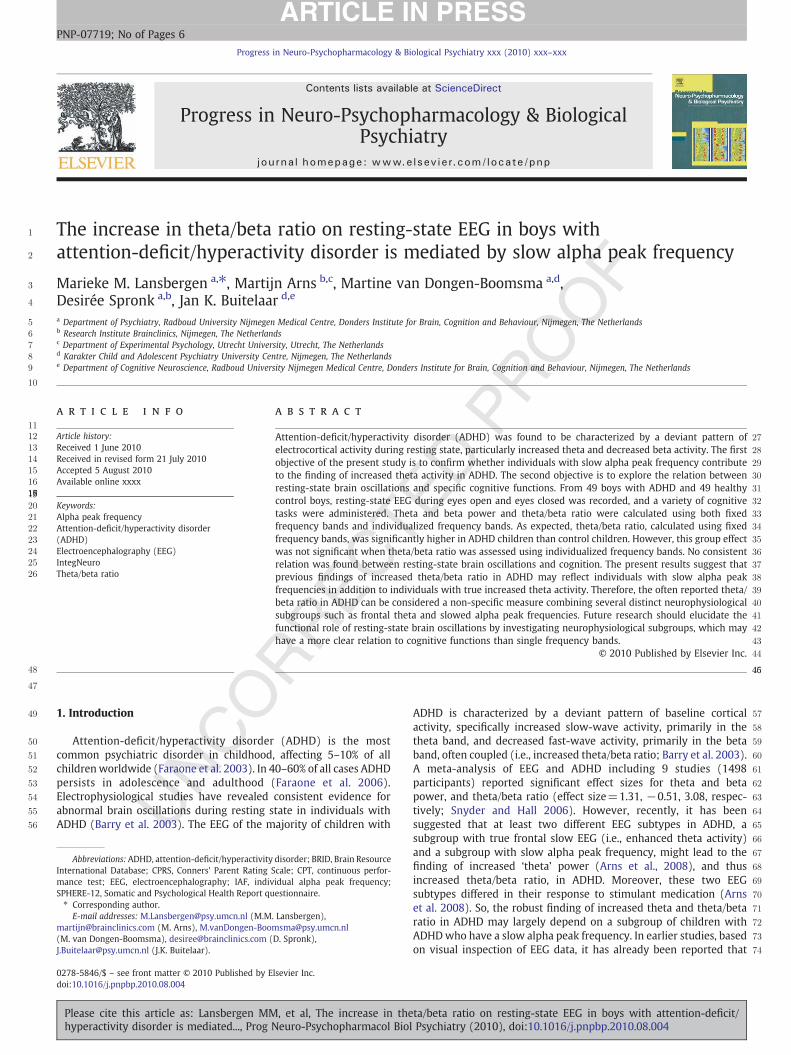
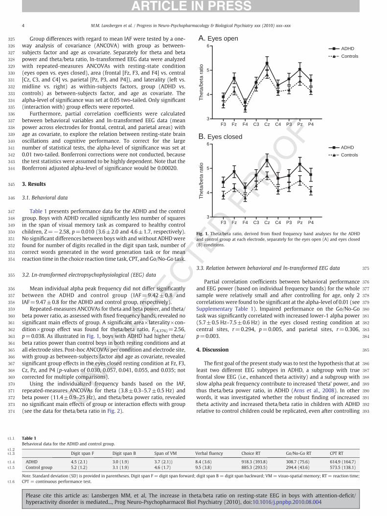
Fig. 1. Theta/beta ratio, derived from fixed frequency band analyses for the ADHDand control group at each electrode, separately for the eyes open (A) and eyes closed(B) conditions.
4 M.M. Lansbergen et al. / Progress in Neuro-Psychopharmacology & Biological Psychiatry xxx (2010) xxx–xxx
Group differences with regard to mean IAF were tested by a one-way analysis of covariance (ANCOVA) with group as between-subjects factor and age as covariate. Separately for theta and betapower and theta/beta ratio, ln-transformed EEG data were analyzedwith repeated-measures ANCOVAs with resting-state condition(eyes open vs. eyes closed), area (frontal [Fz, F3, and F4] vs. central[Cz, C3, and C4] vs. parietal [Pz, P3, and P4]), and laterality (left vs.midline vs. right) as within-subjects factors, group (ADHD vs.controls) as between-subjects factor, and age as covariate. Thealpha-level of significance was set at 0.05 two-tailed. Only significant(interaction with) group effects were reported.
Furthermore, partial correlation coefficients were calculatedbetween behavioral variables and ln-transformed EEG data (meanpower across electrodes for frontal, central, and parietal areas) withage as covariate, to explore the relation between resting-state brainoscillations and cognitive performance. To correct for the largenumber of statistical tests, the alpha-level of significance was set at0.01 two-tailed. Bonferroni corrections were not conducted, becausethe test statistics were assumed to be highly dependent. Note that theBonferroni adjusted alpha-level of significance would be 0.00020.
3. Results
3.1. Behavioral data
Table 1 presents performance data for the ADHD and the controlgroup. Boys with ADHD recalled significantly less number of squaresin the span of visual memory task as compared to healthy controlchildren, Z=−2.58, p=0.010 (3.6±2.0 and 4.6±1.7, respectively).No significant differences between boys with andwithout ADHDwerefound for number of digits recalled in the digit span task, number ofcorrect words generated in the word generation task or for meanreaction time in the choice reaction time task, CPT, and Go/No-Go task.
3.2. Ln-transformed electropsychophysiological (EEG) data
Mean individual alpha peak frequency did not differ significantlybetween the ADHD and control group (IAF=9.42±0.8 andIAF=9.47±0.8 for the ADHD and control group, respectively).
Repeated-measures ANCOVAs for theta and beta power, and theta/beta power ratio, as assessed with fixed frequency bands, revealed nosignificant main effects of group. A significant area×laterality×con-dition×group effect was found for theta/beta ratio, F(4,376)=2.56,p=0.038. As illustrated in Fig. 1, boys with ADHD had higher theta/beta ratios power than control boys in both resting conditions and atall electrode sites. Post-hoc ANCOVAs per condition and electrode site,with group as between-subjects factor and age as covariate, revealedsignificant group effects in the eyes closed resting condition at Fz, F3,Cz, Pz, and P4 (p-values of 0.030, 0.057, 0.041, 0.055, and 0.035; notcorrected for multiple comparisons).
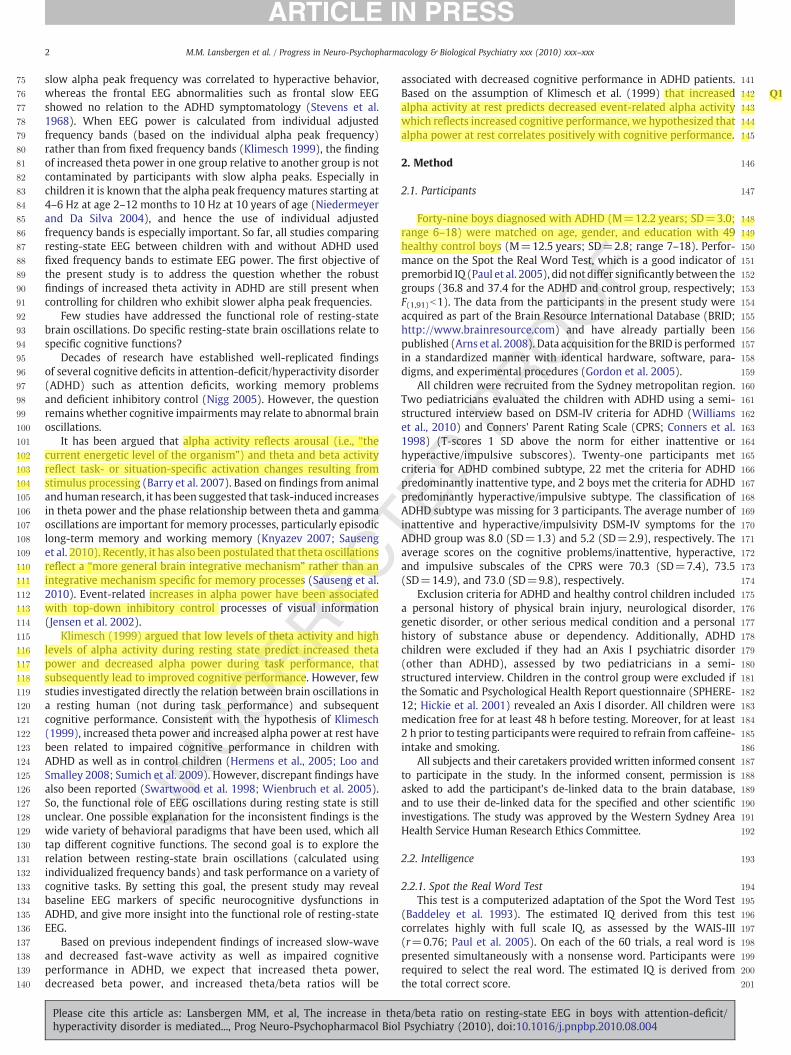
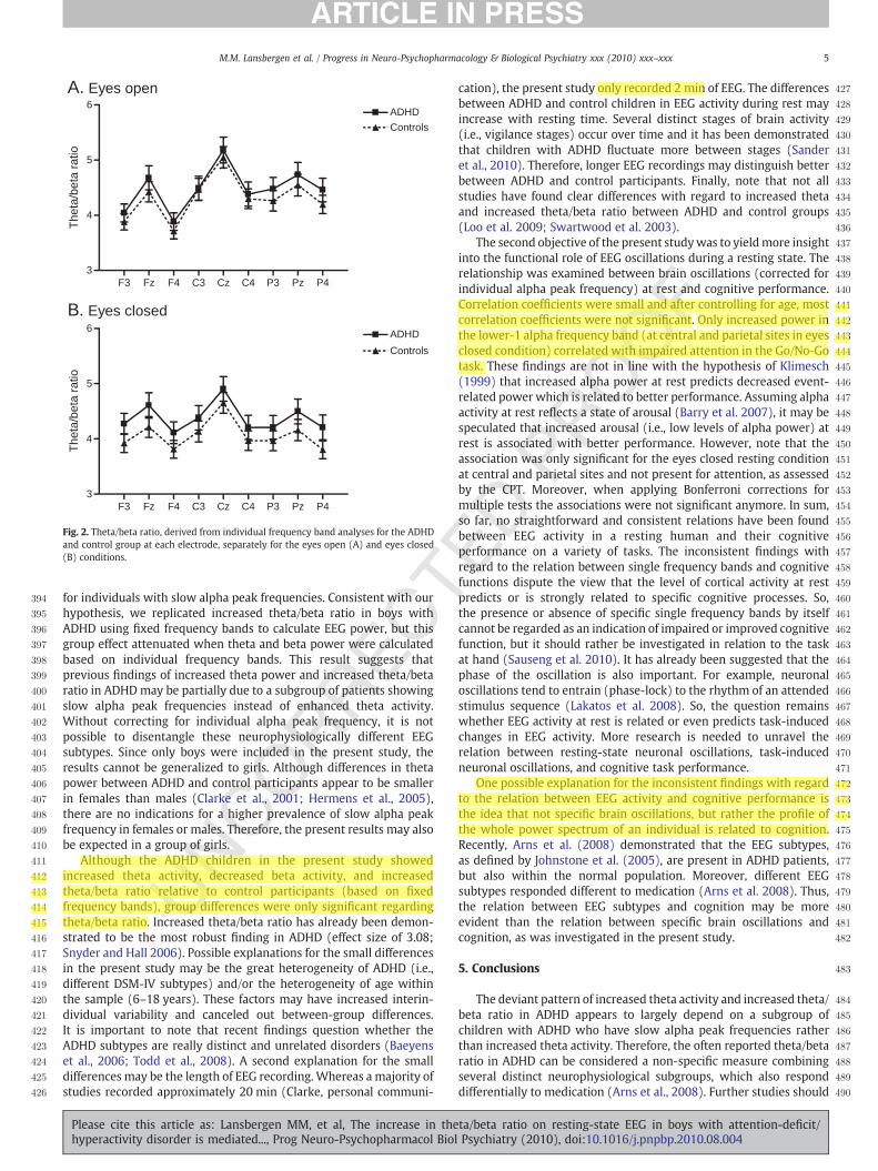
Using the individualized frequency bands based on the IAF,repeated-measures ANCOVAs for theta (3.8±0.3–5.7±0.5 Hz) andbeta power (11.4±0.9–25 Hz), and theta/beta power ratio, revealedno significant main effects of group or interaction effects with group(see the data for theta/beta ratio in Fig. 2).
Table 1Behavioral data for the ADHD and control group.
Digit span F Digit span B Span of VM
ADHD 4.5 (2.1) 3.0 (1.9) 3.7 (2.1))Control group 5.2 (1.2) 3.1 (1.9) 4.6 (1.7)
Note. Standard deviation (SD) is provided in parentheses. Digit span F = digit span forward;CPT = continuous performance test.
Please cite this article as: Lansbergen MM, et al, The increase in thehyperactivity disorder is mediated..., Prog Neuro-Psychopharmacol Biol
3.3. Relation between behavioral and ln-transformed EEG data
Partial correlation coefficients between behavioral performanceand EEG power (based on individual frequency bands) for the wholesample were relatively small and after controlling for age, only 2correlations were found to be significant at the alpha-level of 0.01 (seeSupplementary Table 1). Impaired performance on the Go/No-Gotask was significantly correlated with increased lower-1 alpha power(5.7±0.5 Hz–7.5±0.6 Hz) in the eyes closed resting condition atcentral sites, r=0.294, p=0.005, and parietal sites, r=0.306,p=0.003.
4. Discussion
The first goal of the present study was to test the hypothesis that atleast two different EEG subtypes in ADHD, a subgroup with truefrontal slow EEG (i.e., enhanced theta activity) and a subgroup withslow alpha peak frequency contribute to increased ‘theta’ power, andthus theta/beta power ratio, in ADHD (Arns et al., 2008). In otherwords, it was investigated whether the robust finding of increasedtheta activity and increased theta/beta ratio in children with ADHDrelative to control children could be replicated, even after controlling
Verbal fluency Choice RT Go/No-Go RT CPT RT
8.4 (3.6) 918.3 (393.8) 308.7 (75.6) 614.9 (164.7)9.5 (3.8) 885.3 (293.5) 294.4 (43.6) 573.5 (138.1)
digit span B = digit span backward; VM= visuo-spatial memory; RT = reaction time;
ta/beta ratio on resting-state EEG in boys with attention-deficit/Psychiatry (2010), doi:10.1016/j.pnpbp.2010.08.004
394
395
396
397
398
399
400
401
402
403
404
405
406
407
408
409
410
411
412
413
414
415
416
417
418
419
420
421
422
423
424
425
426
427
428
429
430
431
432
433
434
435
436
437
438
439
440
441
442
443
444
445
446
447
448
449
450
451
452
453
454
455
456
457
458
459
460
461
462
463
464
465
466
467
468
469
470
471
472
473
474
475
476
477
478
479
480
481
482
483
484
485
486
487
488
489
490
F33
The
ta/b
eta
ratio
4
5
6
3
The
ta/b
eta
ratio
4
5
6
A. Eyes open
B. Eyes closed
Fz F4 Cz C4C3 PzP3 P4
F3 Fz F4 Cz C4C3 PzP3 P4
ADHD
Controls
ADHD
Controls
Fig. 2. Theta/beta ratio, derived from individual frequency band analyses for the ADHDand control group at each electrode, separately for the eyes open (A) and eyes closed(B) conditions.
5M.M. Lansbergen et al. / Progress in Neuro-Psychopharmacology & Biological Psychiatry xxx (2010) xxx–xxx
for individuals with slow alpha peak frequencies. Consistent with ourhypothesis, we replicated increased theta/beta ratio in boys withADHD using fixed frequency bands to calculate EEG power, but thisgroup effect attenuated when theta and beta power were calculatedbased on individual frequency bands. This result suggests thatprevious findings of increased theta power and increased theta/betaratio in ADHDmay be partially due to a subgroup of patients showingslow alpha peak frequencies instead of enhanced theta activity.Without correcting for individual alpha peak frequency, it is notpossible to disentangle these neurophysiologically different EEGsubtypes. Since only boys were included in the present study, theresults cannot be generalized to girls. Although differences in thetapower between ADHD and control participants appear to be smallerin females than males (Clarke et al., 2001; Hermens et al., 2005),there are no indications for a higher prevalence of slow alpha peakfrequency in females or males. Therefore, the present results may alsobe expected in a group of girls.
Although the ADHD children in the present study showedincreased theta activity, decreased beta activity, and increasedtheta/beta ratio relative to control participants (based on fixedfrequency bands), group differences were only significant regardingtheta/beta ratio. Increased theta/beta ratio has already been demon-strated to be the most robust finding in ADHD (effect size of 3.08;Snyder and Hall 2006). Possible explanations for the small differencesin the present study may be the great heterogeneity of ADHD (i.e.,different DSM-IV subtypes) and/or the heterogeneity of age withinthe sample (6–18 years). These factors may have increased interin-dividual variability and canceled out between-group differences.It is important to note that recent findings question whether theADHD subtypes are really distinct and unrelated disorders (Baeyenset al., 2006; Todd et al., 2008). A second explanation for the smalldifferences may be the length of EEG recording. Whereas a majority ofstudies recorded approximately 20 min (Clarke, personal communi-
Please cite this article as: Lansbergen MM, et al, The increase in thehyperactivity disorder is mediated..., Prog Neuro-Psychopharmacol Biol
cation), the present study only recorded 2 min of EEG. The differencesbetween ADHD and control children in EEG activity during rest mayincrease with resting time. Several distinct stages of brain activity(i.e., vigilance stages) occur over time and it has been demonstratedthat children with ADHD fluctuate more between stages (Sanderet al., 2010). Therefore, longer EEG recordings may distinguish betterbetween ADHD and control participants. Finally, note that not allstudies have found clear differences with regard to increased thetaand increased theta/beta ratio between ADHD and control groups(Loo et al. 2009; Swartwood et al. 2003).
The second objective of the present studywas to yieldmore insightinto the functional role of EEG oscillations during a resting state. Therelationship was examined between brain oscillations (corrected forindividual alpha peak frequency) at rest and cognitive performance.Correlation coefficients were small and after controlling for age, mostcorrelation coefficients were not significant. Only increased power inthe lower-1 alpha frequency band (at central and parietal sites in eyesclosed condition) correlated with impaired attention in the Go/No-Gotask. These findings are not in line with the hypothesis of Klimesch(1999) that increased alpha power at rest predicts decreased event-related power which is related to better performance. Assuming alphaactivity at rest reflects a state of arousal (Barry et al. 2007), it may bespeculated that increased arousal (i.e., low levels of alpha power) atrest is associated with better performance. However, note that theassociation was only significant for the eyes closed resting conditionat central and parietal sites and not present for attention, as assessedby the CPT. Moreover, when applying Bonferroni corrections formultiple tests the associations were not significant anymore. In sum,so far, no straightforward and consistent relations have been foundbetween EEG activity in a resting human and their cognitiveperformance on a variety of tasks. The inconsistent findings withregard to the relation between single frequency bands and cognitivefunctions dispute the view that the level of cortical activity at restpredicts or is strongly related to specific cognitive processes. So,the presence or absence of specific single frequency bands by itselfcannot be regarded as an indication of impaired or improved cognitivefunction, but it should rather be investigated in relation to the taskat hand (Sauseng et al. 2010). It has already been suggested that thephase of the oscillation is also important. For example, neuronaloscillations tend to entrain (phase-lock) to the rhythm of an attendedstimulus sequence (Lakatos et al. 2008). So, the question remainswhether EEG activity at rest is related or even predicts task-inducedchanges in EEG activity. More research is needed to unravel therelation between resting-state neuronal oscillations, task-inducedneuronal oscillations, and cognitive task performance.
One possible explanation for the inconsistent findings with regardto the relation between EEG activity and cognitive performance isthe idea that not specific brain oscillations, but rather the profile ofthe whole power spectrum of an individual is related to cognition.Recently, Arns et al. (2008) demonstrated that the EEG subtypes,as defined by Johnstone et al. (2005), are present in ADHD patients,but also within the normal population. Moreover, different EEGsubtypes responded different to medication (Arns et al. 2008). Thus,the relation between EEG subtypes and cognition may be moreevident than the relation between specific brain oscillations andcognition, as was investigated in the present study.
5. Conclusions
The deviant pattern of increased theta activity and increased theta/beta ratio in ADHD appears to largely depend on a subgroup ofchildren with ADHD who have slow alpha peak frequencies ratherthan increased theta activity. Therefore, the often reported theta/betaratio in ADHD can be considered a non-specific measure combiningseveral distinct neurophysiological subgroups, which also responddifferentially to medication (Arns et al., 2008). Further studies should
ta/beta ratio on resting-state EEG in boys with attention-deficit/Psychiatry (2010), doi:10.1016/j.pnpbp.2010.08.004
491
492
493
494
495
496
497
498
499
500
501
502
503
504
505
506
507
508
509
510
511
512
513
514
515516517518519520521522523524525526527528529530531532533534535536537538539540541542543544545546547548
549550551552553554555556557558559560561562563564565566567568569570571572573574575576577578579580581582583584585586587 Q2588589590591592593594595596597598599600601602603604605606607608609610611612613614615616
617
6 M.M. Lansbergen et al. / Progress in Neuro-Psychopharmacology & Biological Psychiatry xxx (2010) xxx–xxx
replicate this finding in larger samples and investigate whetherthese two subgroups also differ clinically and neuropsychologically.Furthermore, the present results suggest that EEG activity in a restinghuman does not have a clear association with cognitive processes.Future research might address the relation between resting-stateand task-induced neuronal oscillations, and subsequent behavioralperformance, also in larger samples. Furthermore, looking at differentEEG subtypes rather than specific frequency bands may elucidate aclearer relationship between brain oscillations and cognitive deficits.
Acknowledgments
The authors gratefully acknowledge the support of the BrainGainSmart Mix Programme of the Netherlands Ministry of EconomicAffairs and the Netherlands Ministry of Education, Culture andScience. Further, we acknowledge the support of the Brain ResourceInternational Database (under the auspices of Brain Resource—www.brainresource.com) for use of normative and clinical data. We alsothank the individuals who gave their time to participate in thedatabase. Access to the database for scientific purposes is adminis-tered independently via the scientific network (BRAINnet; www.brainnet.net), which is coordinated independently of the commercialoperations of Brain Resource.
Supplementarymaterials related to this article can be found onlineat doi:10.1016/j.pnpbp.2010.08.004.
References
Arns M, Gunkelman J, Breteler M, Spronk D. EEG phenotypes predict treatmentoutcome to stimulants in children with ADHD. J Integr Neurosci 2008;7:421–38.
Baddeley A, Emslie H, Nimmo-Smith I. The Spot-the-Word test: a robust estimate ofverbal intelligence based on lexical decision. Br J Clin Psychol 1993;32(Pt 1):55–65.
Baeyens D, Roeyers H, Walle JV. Subtypes of attention-deficit/hyperactivity disorder(ADHD): distinct or related disorders across measurement levels? Child PsychiatryHum Dev 2006;36:403–17.
Barry RJ, Clarke AR, Johnstone SJ. A review of electrophysiology in attention-deficit/hyperactivity disorder: I. Qualitative and quantitative electroencephalography. ClinNeurophysiol 2003;114:171–83.
Barry RJ, Clarke AR, Johnstone SJ, Magee CA, Rushby JA. EEG differences between eyes-closed and eyes-open resting conditions. Clin Neurophysiol 2007;118:2765–73.
Clark CR, Paul RH, Williams LM, Arns M, Fallahpour K, Handmer C, et al. Standardizedassessment of cognitive functioning during development and aging using anautomated touchscreen battery. Arch Clin Neuropsychol 2006;21:449–67.
Clarke AR, Barry RJ, McCarthy R, Selikowitz M. Age and sex effects in the EEG:differences in two subtypes of attention-deficit/hyperactivity disorder. ClinNeurophysiol 2001;112:815–26.
Conners CK, Sitarenios G, Parker JD, Epstein JN. The revised Conners' Parent Rating Scale(CPRS-R): factor structure, reliability, and criterion validity. J Abnorm Child Psychol1998;26:257–68.
Doppelmayr M, Klimesch W, Pachinger T, Ripper B. Individual differences in braindynamics: important implications for the calculation of event-related band power.Biol Cybern 1998;79:49–57.
Faraone SV, Sergeant J, Gillberg C, Biederman J. Theworldwide prevalence of ADHD: is itan American condition? World Psychiatry 2003;2:104–13.
Faraone SV, Biederman J, Mick E. The age-dependent decline of attention deficithyperactivity disorder: a meta-analysis of follow-up studies. Psychol Med 2006;36:159–65.
Gasser T, Bächer P, Möcks J. Transformations towards the normal distribution of broadband spectral parameters of the EEG. Electroencephalogr Clin Neurophysiol 1982;53:119–24.
Gordon E, Cooper N, Rennie C, Hermens D, Williams LM. Integrative neuroscience: therole of a standardized database. Clin EEG Neurosci 2005;36:64–75.
Please cite this article as: Lansbergen MM, et al, The increase in thehyperactivity disorder is mediated..., Prog Neuro-Psychopharmacol Biol
Gratton G, Coles MG, Donchin E. A new method for off-line removal of ocular artifact.Electroencephalogr Clin Neurophysiol 1983;55:468–84.
Hermens DF, KohnMR, Clarke SD, Gordon E, Williams LM. Sex differences in adolescentADHD: findings from concurrent EEG and EDA. Clin Neurophysiol 2005a;116:1455–63.
Hermens DF, Soei EX, Clarke SD, Kohn MR, Gordon E, Williams LM. Resting EEG thetaactivity predicts cognitive performance in attention-deficit hyperactivity disorder.Pediatr Neurol 2005b;32:248–56.
Hickie IB, Davenport TA, Naismith SL, Scott EM. SPHERE: a national depression project.SPHERE National Secretariat. Med J Aust 2001;175(Suppl):S4–5.
Jensen O, Gelfand J, Kounios J, Lisman JE. Oscillations in the alpha band (9–12 Hz)increase with memory load during retention in a short-term memory task. CerebCortex 2002;12:877–82.
Johnstone J, Gunkelman J, Lunt J. Clinical database development: characterization ofEEG phenotypes. Clin EEG Neurosci 2005;36:99-107.
Klimesch W. EEG alpha and theta oscillations reflect cognitive and memoryperformance: a review and analysis. Brain Res Brain Res Rev 1999;29:169–95.
Knyazev GG. Motivation, emotion, and their inhibitory control mirrored in brainoscillations. Neurosci Biobehav Rev 2007;31:377–95.
Lakatos P, Karmos G, Mehta AD, Ulbert I, Schroeder CE. Entrainment of neuronaloscillations as a mechanism of attentional selection. Science 2008;320:110–3.
Loo S, Smalley S. Preliminary report of familial clustering of EEGmeasures in ADHD. AmJ Med Genet B (Neuropsychiatr Genet) 2008;147:107–9.
Loo SK, Hale TS, Macion J, Hanada G, McGough JJ, McCracken JT, et al. Cortical activitypatterns in ADHD during arousal, activation and sustained attention. Neuropsy-chologia 2009;47:2114–9.
Niedermeyer E, Da Silva FHL. Electroencephalography: basic principles, clinicalapplications, and related fields; 2004.
Nigg JT. Neuropsychologic theory and findings in attention-deficit/hyperactivitydisorder: the state of the field and salient challenges for the coming decade. BiolPsychiatry 2005;57:1424–35.
Paul RH, Lawrence J, Williams LM, Richard CC, Cooper N, Gordon E. Preliminary validityof “integneuro”: a new computerized battery of neurocognitive tests. Int J Neurosci2005;115:1549–67.
Posthuma D, Neale MC, Boomsma DI, de Geus EJ. Are smarter brains running faster?Heritability of alpha peak frequency, IQ, and their interrelation. Behav Genet2001;31:567–79.
Sander C, Arns M, Olbrich S, Hegerl U. EEG-vigilance and response to stimulants inpaediatric patients with attention deficit/hyperactivity disorder. Clin Neurophysiol2010.
Sauseng P, Griesmayr B, Freunberger R, Klimesch W. Control mechanisms in workingmemory: a possible function of EEG theta oscillations. Neurosci Biobehav Rev2010;34:1015–22.
Snyder SM, Hall JR. A meta-analysis of quantitative EEG power associated withattention-deficit hyperactivity disorder. J Clin Neurophysiol 2006;23:440–55.
Stevens JR, Sachdev K, Milstein V. Behavior disorders of childhood and theelectroencephalogram. Arch Neurol 1968;18:160–77.
Sumich A, Matsudaira T, Gow RV, Ibrahimovic A, Ghebremeskel K, Crawford M, et al.Resting state electroencephalographic correlates with red cell long-chain fattyacids, memory performance and age in adolescent boys with attention deficithyperactivity disorder. Neuropharmacology 2009;57:708–14.
SwartwoodMO, Swartwood JN, Lubar JF, Timmermann DL, Zimmerman AW,MuenchenRA. Methylphenidate effects on EEG, behavior, and performance in boys withADHD. Pediatr Neurol 1998;18:244–50.
Swartwood JN, Swartwood MO, Lubar JF, Timmermann DL. EEG differences in ADHD-combined type during baseline and cognitive tasks. Pediatr Neurol 2003;28:199–204.
Todd RD, Huang H, Todorov AA, Neuman RJ, Reiersen AM, Henderson CA, et al.Predictors of stability of attention-deficit/hyperactivity disorder subtypes fromchildhood to young adulthood. J Am Acad Child Adolesc Psychiatry 2008;47:76–85.
Wienbruch C, Paul I, Bauer S, Kivelitz H. The influence of methylphenidate on the powerspectrum of ADHD children — an MEG study. BMC Psychiatry 2005;5:29.
Williams LM, Simms E, Clark CR, Paul RH, Rowe D, Gordon E. The test–retest reliability ofa standardized neurocognitive and neurophysiological test battery: “neuromarker”.Int J Neurosci 2005;115:1605–30.
Williams LM, Hermens DF, Thein T, Clark CR, Cooper NJ, Clarke SD, et al. Using brain-based cognitive measures to support clinical decisions in ADHD. Pediatr Neurol2010;42(2):118–26.
ta/beta ratio on resting-state EEG in boys with attention-deficit/Psychiatry (2010), doi:10.1016/j.pnpbp.2010.08.004


![Progress in Materials Sciencebib-pubdb1.desy.de/record/320708/files/NiTi_Welding... · UNCORRECTED PROOF Progress in Materials Science xxx (2017) xxx-xxx 3 Fig. 2. Stress-strain-temperatureplotexhibitingsuperelasticityandshapememoryeffect[8]](https://img.pdfslide.net/doc/110x75/60185b1b81f2fc0baa30d4d9/progress-in-materials-sciencebib-uncorrected-proof-progress-in-materials-science.jpg)