Embed Size (px)
Citation preview
The American Journal of Pathology, Vol. 183, No. 5, November 2013
ajp.amjpathol.org
See related Commentary on page 1378.
TUMORIGENESIS AND NEOPLASTIC PROGRESSION
Continuous Exposure to Chrysotile Asbestos Can CauseTransformation of Human Mesothelial Cells via HMGB1 andTNF-a SignalingFang Qi,*y Gordon Okimoto,* Sandro Jube,* Andrea Napolitano,*y Harvey I. Pass,z Rozalia Laczko,* Richard M. DeMay,x
Ghazal Khan,x Maarit Tiirikainen,* Caterina Rinaudo,{ Alessandro Croce,{ Haining Yang,*k Giovanni Gaudino,* andMichele Carbone*k
From the University of Hawai’i Cancer Center,* and the Department of Molecular Biosciences and Bioengineering,y University of Hawai’i, Honolulu,Hawaii; the Department of Pathology,k John A. Burns School of Medicine, University of Hawai’i, Honolulu, Hawaii; the Division of Thoracic Surgery,z
Department of Cardiothoracic Surgery, Langone Medical Center, New York University, New York, New York; the Section of Cytopathology,x Department ofPathology, University of Chicago Medical Center, University of Chicago, Chicago, Illinois; and the Department of Science and Technological Innovation,{
University of Piemonte Orientale “Amedeo Avogadro,” Alessandria, Italy
Accepted for publication
C
P
h
July 17, 2013.
Address correspondence toMichele Carbone, M.D., Ph.D.,University of Hawai’i CancerCenter, University of Hawai’i,701 Ilalo St., Honolulu,HI 96813. E-mail: [email protected].
opyright ª 2013 American Society for Inve
ublished by Elsevier Inc. All rights reserved
ttp://dx.doi.org/10.1016/j.ajpath.2013.07.029
Malignant mesothelioma is strongly associated with asbestos exposure. Among asbestos fibers,crocidolite is considered the most and chrysotile the least oncogenic. Chrysotile accounts for more than90% of the asbestos used worldwide, but its capacity to induce malignant mesothelioma is still debated.We found that chrysotile and crocidolite exposures have similar effects on human mesothelial cells.Morphological and molecular alterations suggestive of epithelialemesenchymal transition, such asE-cadherin down-regulation and b-catenin phosphorylation followed by nuclear translocation, wereinduced by both chrysotile and crocidolite. Gene expression profiling revealed high-mobility group box-1protein (HMGB1) as a key regulator of the transcriptional alterations induced by both types of asbestos.Crocidolite and chrysotile induced differential expression of 438 out of 28,869 genes interrogated byoligonucleotide microarrays. Out of these 438 genes, 57 were associated with inflammatory and immuneresponse and cancer, and 14 were HMGB1 targeted genes. Crocidolite-induced gene alterations weresustained, whereas chrysotile-induced gene alterations returned to background levels within 5 weeks.Similarly, HMGB1 release in vivo progressively increased for 10 or more weeks after crocidolite exposure,but returned to background levels within 8 weeks after chrysotile exposure. Continuous administrationof chrysotile was required for sustained high serum levels of HMGB1. These data support the hypothesisthat differences in biopersistence influence the biological activities of these two asbestos fibers.(Am J Pathol 2013, 183: 1654e1666; http://dx.doi.org/10.1016/j.ajpath.2013.07.029)
Supported by NIH grants NCI R01 CA106567 (M.C.), NCI R01CA160715-0A (H.Y.), P01 CA114047 (M.C.), and the P30 CA071789(UHCC Genomics Shared Resource); the Mesothelioma Applied ResearchFoundation (H.Y.), the United-4 A Cure (H.Y.), the Hawai’i CommunityFoundation (H.Y. and G.G.), the V foundation (H.Y.), and the University ofHawai’i Foundation (M.C.).Current address of R.L., Cardiovascular Research Center, University of
Hawai’i, Honolulu, HI.
Malignant mesothelioma (MM) is an aggressive cancer of thepleura and peritoneum, and less frequently of other meso-thelial linings; it is strongly associated with asbestos expo-sure and affects approximately 3200 individuals annually inthe United States.1 The median survival of MM patients isapproximately 1 year from diagnosis, despite surgical re-section, chemotherapy, and radiotherapy.2,3
Asbestos is a nonspecific term commonly used to describeany of six types of naturally occurring fibrous silicate mineralsthat were widely used commercially during the 20th century.4
Asbestos fibers are divided into two major groups, serpentine
stigative Pathology.
.
and amphibole, and are further distinguished based on theirchemical composition and crystalline structure.5 Serpentineasbestos is chrysotile (white asbestos); amphibole asbestos
Transforming Potential of Chrysotile
includes crocidolite (blue asbestos), amosite (brown asbestos),anthophyllite, actinolite, and tremolite. It has been esti-mated that chrysotile accounts for approximately 95% ofall asbestos used in the United States6 and 90% of asbestosused worldwide.7,8 In the human body, amphibole fibers tendto persist at sites of deposition, with fiber concentrationincreasing with prolonged exposure, whereas chrysotilefibers are usually rapidly cleared from the lungs.6 It is wellaccepted that amphibole asbestos cause MM.9 Althoughchrysotile can induce MM in animal experiments,10e16 itscarcinogenic role in humans is still debated, because epide-miological studies have not proven a definitive causal asso-ciation between chrysotile and MM.6,17,18 It has beenproposed that the mechanisms of asbestos carcinogenesismay vary among different species19; however, few studieshave investigated the molecular pathways induced bychrysotile that may eventually lead to MM.5,20
Exposure to crocidolite induces necrosis of primary humanmesothelial (HM) cells, which is accompanied by passiverelease of the damage-associated molecular pattern high-mobility group box-1 protein (HMGB1).21 In the extra-cellular space, HMGB1 leads to chronic inflammationthrough the recruitment and accumulation of macro-phages, which in turn actively secrete HMGB1 alongwith several other cytokines, including tumor necrosisfactor (TNF-a), which plays a critical role in crocidolite-mediated carcinogenesis.22
Epithelialemesenchymal transition (EMT) is a physio-pathological process by which epithelial cells acquire mes-enchymal shape and properties associated with cell migrationand cancer progression.23 EMT contributes to the histo-morphological features ofMM(ie, epithelioid versus biphasicand sarcomatoid subtypes).23e25 TNF-a has been shown toinduce EMT in epithelial cells26,27 and in mesothelial cells,28
and HMGB1 has been also associated with EMT in alveolarepithelial cells.29,30 EMT is characterized by increased ex-pression of mesenchymal markers such as the cytoskeletalproteins, vimentin, and a-smooth muscle actin31 and bydecreased expression of the epithelial cell adhesion moleculeE-cadherin, either at the transcriptional level26,32,33 or throughubiquitin-mediated degradation.34,35 E-cadherin forms ad-herent junctions that maintain cell adhesion in a multiproteincomplex that includes b-catenin.36 During EMT, phosphor-ylation of b-catenin on tyrosine 142 (Y142) by receptortyrosine kinases (eg, c-Met) eventually results in disassem-bling of the adhesion junction complex, degradation ofE-cadherin, and release of b-catenin.37 Depending on theupstream messages, b-catenin can be either degraded ortranslocated to the nucleus, where it is transcriptionallyactive.34,38 The majority of established MM cell lines exhibitnuclear accumulation ofb-catenin,39e41 suggesting a possiblecontributory role of b-catenin in MM development.
In the present study, we compared the biological,morphological, and transcriptional effects of crocidoliteand chrysotile on primary HM cells in tissue culture and inmice.
The American Journal of Pathology - ajp.amjpathol.org
Materials and Methods
Cell Lines and Culturing Conditions
HM cells are routinely cultured in our laboratory (Universityof Hawai'i Cancer Center), isolated from pleural fluids frompatients with congestive heart failure or other benignconditions.42 HM cells are established in cell culture inDulbecco’s modified Eagle’s medium containing 20% fetalbovine serum (FBS) and are characterized morphologicallyand by positive immunostaining for cytokeratin, HBME-1,and calretinin and negative staining for LeuM1, Ber-Ep4,B72.3, and carcinoembryonic antigen.42 THP-1 humanmonocytes (ATCC TIB202; ATCC, Manassas, VA) arecultured in RPMI 1640 medium supplemented with 10%FBS and 0.05 mmol/L 2-mercaptoethanol. Experiments wereperformed using Dulbecco’s modified Eagle’s mediumcontaining 10% FBS, unless otherwise specified.
Fiber Preparation
Chrysotile and crocidolite fibers were obtained from theInternational Union against Cancer (Union InternationaleContre le Cancer; UICC Lyon, France). Fibers were pro-cessed as described previously.22,43 In brief, fibers werebaked at 150�C for 18 hours, suspended in PBS at 4 mg/mL,triturated 10 times through a 22-gauge needle, and auto-claved. Reference chrysotile B, Canadian, consisted ofa mixture of fibers from the following companies or mines:Bell, Carey, Cassiar, Flintkote, Johns-Manville, Lake,Normandie, and National. Reference chrysotile A, histori-cally called Rhodesian, consisted of asbestos fibers mined inZvishavane (also spelled Shabani or Shavani), MatabelelandSouth Province, Zimbabwe. Both chrysotile A and B arepredominantly made up of hydrous silicates of magnesia.Reference crocidolite consisted of fibers from the Koegasmine, a large asbestos mine in the Northern Cape province ofSouth Africa. This mineral is predominantly made up ofhydroxyl silicates of sodium, magnesium, and iron. The cell-culture experiments were performed with 5 mg/cm2 for eachof the asbestos fiber types, unless specified otherwise. Cellswere cultured in the presence of fibers for different durations,depending on the type of assay performed.
Asbestos Fiber Electron Microscopy
A drop of fiber suspension (40 mg/mL) was deposited ontocarbon conductive tape and placed on pin stubs for analysisunder a scanning electron microscope. Finally, the liquidphase was evaporated by placing the sample in a dryingglass cloche for 24 hours. The chrysotile dimensional studywas performed under an Quanta 200 environmental scan-ning electron microscope (FEI, Hillsboro, OR), equippedwith an energy-dispersive spectroscopy (EDS) microprobesystem (EDAX, Mahwah, NJ) in low-vacuum operationalmode, allowing sample characterization without coating by
1655
Qi et al
carbon or gold. The pressure used was 90 Pa, with accel-erating voltage of 20 kV and a working distance of 10 mm.The fibers were identified at �2000 magnification andcounted. Length and diameter of each fiber were determinedat �500, �2000, or �4000, depending on the dimensions.In each sample, 200 fibers were measured, for a total of 600crystals for each mineral phase. Samples were analyzed intriplicate. Average length and diameter of chrysotile B fiberswere 19.9 � 19.6 mm and 0.47 � 0.27 mm, respectively(Supplemental Figure S1). Average length and diameter ofreference crocidolite fibers were 13.60 � 20.24 mm and0.60 � 0.45 mm, respectively (Supplemental Figure S2).
TNF-a Signaling Studies
To stimulate TNF-a signaling, HM cells were incubated with10 ng/mL recombinant TNF-a (Sigma-Aldrich, St. Louis,MO). To inhibit endogenous TNF-a activity, a neutralizingantibody (antieTNF-a; R&D Systems, Minneapolis, MN)was used at a concentration of 1 mg/mL.
Immunofluorescence Staining
Asbestos StainingAsbestos fibers were stained as described.44 In brief,asbestos fibers were preincubated with normal rabbit serum(Santa Cruz Biotechnology, Santa Cruz, CA) and added toHM cells for 24 hours. The HM cells were then fixed in 4%paraformaldehyde,washed in PBS, permeabilized using 0.2%Triton X-100, and saturated with bovine serum albumin.Incubation was performed with Alexa Fluor 594econjugatedanti-rabbit secondary antibody and Alexa Fluor 488econjugated phalloidin (Life Technologies, Carlsbad, CA).Slides were washed in PBS and mounted with aqueousmounting medium premixed with the nuclear marker DAPI(Vector Laboratories, Burlingame, CA) for fluorescencemicroscopy.
b-Catenin StainingCells were cultured and fixed as described above. Bovineserum albumin (1% solution) was used to block nonspecificimmunoreactivity. Incubation with antieb-catenin or controlrabbit IgG (Santa Cruz Biotechnology) was followed bylabeling with Alexa Fluor 594econjugated secondary anti-body and Alexa Fluor 488econjugated phalloidin (LifeTechnologies). Slides were mounted as described above forfluorescence microscopy.
Cell Proliferation and Viability Assay
To evaluate and compare effects on cell proliferation andviability, HM cells were seeded in 96-well plates with sixreplicates per treatment condition. Experiments were per-formed in the presence or absence of crocidolite or chrysotileasbestos fibers. Cells were preincubated with 10 ng/mLTNF-a or PBS for 24 hours and thenwere exposed to asbestos
1656
for an additional 24 or 48 hours before viability was assessedusing anMTS assay according to the manufacturer’s protocol(Promega, Madison, WI). MTS assays were quantified byusing a colorimetric reaction mix and microplate reader at490 nm (Bio-Rad Laboratories, Hercules, CA). To avoid theintrinsic variability of this method, we performed the MTSassay for all time points in one measurement on the same day.
Cell Cytotoxicity Assay
Cytotoxicity was assessed by measuring the amount oflactate dehydrogenase (LDH) released from the cytosol ofdamaged cells. A LDH cytotoxicity detection kit (RocheDiagnostics, Indianapolis, IN) was used according to themanufacturer’s protocol. In brief, HM cells were seeded ina 96-well tissue culture plate at a density of 5 � 103 cells perwell. The next day, the medium was changed, and 100 mL ofasbestos fiber suspensions at different concentrations wasadded. After 24 hours, 100 mL of supernatant per well washarvested and transferred into a new 96-well, flat-bottomplate. LDH substrate was added to each well and incubatedat room temperature protected from light. The absorbance ofthe samples was measured at 490 nm with an enzyme-linkedimmunosorbent assay (ELISA) reader. Cytotoxicity wascalculated as % cytotoxicity Z [(experimental value � lowcontrol) �100]/(high control � low control), where the lowcontrol is assay medium plus cells and the high control isassay medium with 2% Triton X-100 plus cells.
Western Blotting
For total cell protein extraction, cells were washed with coldPBS, scraped in radioimmunoprecipitation assay lysis buffer(100 mmol/L NaCl, 10 mmol/L Tris, 0.1% SDS, 1% TritonX-100, and 5 mmol/L EDTA at pH 7.2) containing aprotease inhibitor cocktail (Roche Diagnostics), and incu-bated for 30 minutes on ice.A nuclear extraction kit (Active Motif, Carlsbad, CA)
was used according to the manufacturer’s protocol to per-form cell fractionation. In brief, cultured cells were rinsed incold PBS, scraped, and centrifuged in PBS supplementedwith phosphatase inhibitors. The cell pellets were lysed inhypotonic buffer supplemented with detergents for 15minutes on ice to access cytoplasmic proteins. The nuclearpellets were lysed separately in nuclear lysis buffer for 30minutes on ice to access the nuclear proteins.Equal amounts (20e50 mg) of cell lysate per lane were
applied on 4% to 12% gradient gels (NuPAGE; Life Tech-nologies). Proteins separated on bis-tris gels were transferredto polyvinylidene difluoride membranes (Immobilon-P;EMDMillipore, Billerica, MA) and blocked with 5% bovineserum albumin before incubation with specific primary andsecondary antibodies. The antibodies used were used histone1, E-cadherin, b-catenin, vimentin (Santa Cruz Biotech-nology); GAPDH (Chemicon; EMD Millipore); Y142-phosphorylated b-catenin (ECM Biosciences, Versailles,
ajp.amjpathol.org - The American Journal of Pathology
Transforming Potential of Chrysotile
KY); and HMGB1 and a-smooth muscle actin (Abcam,Cambridge, MA). The relative density of Western blot bandswas evaluated by the ImageJ software version 1.45s (NIH,Bethesda, MD). Briefly, the density of target bands werenormalized versus the corresponding loading control banddensity and compared to vehicle exposed controls andexpressed as relative density.
Quantitative RT-PCR
HM cells were exposed to asbestos fibers at 5 mg/cm2 for24 hours. Total RNAs were isolated using an RNeasy kit(Qiagen, Valencia, CA) according to the manufacturer’sprotocol and were treated with RNase-free DNase (Qiagen).For each sample, 1 mg RNA was used. GAPDH was used asthe internal control. Quantitative RT-PCR (RT-qPCR) forTNF-a and GAPDH mRNA was performed in triplicate in25-mL total reaction volumes using a SYBR Green mastermix (Bio-Rad Laboratories). A LightCycler 480 RT-qPCRsystem (Roche Diagnostics) was used according to standardprocedures. The primers were as described previously.22,45
Cell Transformation Assay
HM cells were cultured and seeded in six-well plates ata density of 3 � 105 cells per well. The next day, the mediumwas switched to Dulbecco’s modified Eagle’s medium with10% FBS. In the TNF-aedependent transformation assays,HM cells were pretreated with 10 ng/mL TNF-a or with PBS(vehicle) control for 24 hours before exposure to 5 mg/cm2
asbestos fibers or PBS control for another 48 hours. Culturemedium containing 10 ng/mL TNF-a was replaced twicea week.
For coculture cell transformation assays, the HMemacrophage coculture assay was performed as describedpreviously.46 THP-1 peripheral blood monocytes were dif-ferentiated into macrophages as described previously.46 Inbrief, tissue-culture inserts containing differentiated macro-phages were placed above HM cell cultures. Membranes ofthe inserts allowed the cytokines and growth factors producedby macrophages to reach the lower chamber, where HM cellswere cultured. HM cells were cocultured with macrophagesand then were exposed to crocidolite fibers, chrysotile fibers,or vehicle alone for 48 hours. Fibers not engulfed by cells andfloating in the medium were washed off. Freshly differenti-ated macrophages and culture medium were replaced twicea week. After 6 to 8 weeks, foci were identified by crystalviolet staining and were counted under a light microscope.
Microarray Profiling
Whole-genome expression arrays were used to characterizethe transcriptional response of HM cells exposed to crocidoliteor chrysotile fibers. HM cells were cultured in six-well plateswith 2� 105 cells/well. Experimental conditionswereHMcellswith PBS (vehicle control), HMemacrophage cocultures46
The American Journal of Pathology - ajp.amjpathol.org
prepared as described above, and HM cells cultured in thepresence of TNF-a exposed to crocidolite, chrysotile, or PBSfor 48 hours and 5 weeks. The experiments were performed induplicate to verify reproducibility of the findings. RNA wasextracted using a miRNeasy micro kit (Qiagen), and RNAquality was determined using an Agilent Bioanalyzer systemwith RNA 6000 Nano or Pico chips (Agilent Technologies,Santa Clara, CA). The average RNA integrity number (RIN)was 9.2. GeneChip human gene 1.0 ST arrays (Affymetrix,Santa Clara, CA) were used to interrogate expression of 28,869genes, with two replicate samples for each condition, byapplying the Affymetrix GeneChip whole-transcript sensetarget labeling protocol with 100 ng of total RNA.
Microarray Data Analysis
The raw .CEL file data for each array were quantified,quantile-normalized, and log2etransformed, to facilitatecomparisons between different samples. The array data weremodeled using two-factor analysis of variance with experi-mental condition (seven levels) and time (two levels) as thetwo factors. The experimental design also included twobiological replicates per level for each factor, as required forsuch an analysis. The two-factor analysis of variance modelwas applied to all 28 arrays. A set of 57 genes were even-tually identified that were both highly variable between twoor more of the seven conditions [false discovery rate (FDR)<0.05] and highly enriched for biological processes associatedwith inflammatory and immune response and cancer asdetermined by Ingenuity Pathway Analysis software version6 (IPA; Ingenuity Systems, Redwood City, CA). The falsediscovery rate based on the BenjaminieHochberg methodwas used to control for multiple comparisons. Subsequentpairwise analyses that compared fiber-treated HM cells withvehicle and HMemacrophage cocultures using a noise-adaptive fold change to identify differentially expressedgenes (DEGs) for each comparison.47 IPA was then used topredict upstream regulators and downstream effects based onthe observed patterns of pairwise differential expression.
Microarray Data Validation
For the technical and biological validation and replication ofgene expression levels generated in the microarray expres-sion profiling assay, RT-qPCR was used to measure geneexpression levels for six genes. These genes were chosenbecause they were either differentially expressed between thedifferent treatment conditions (ATL3, CCL20, IL24, andPTGS2 from 438 DEGs) or represented a stable gene acrossour various experimental conditions (TERF2IP). In addition,a reference gene (GAPDH ) was used to normalize mRNAexpression levels. In brief, validation RT-qPCR was per-formed on the same samples that were used in the microarrayassays and on the samples from repeated experiments.The cDNA was prepared from 1 mg of untreated total RNAusing an Applied Biosystems high-capacity cDNA reverse
1657
Qi et al
transcription kit (Life Technologies). qPCR was performedfor the seven genes using TaqMan gene expression assaysand TaqMan universal PCR master mix with the recom-mended thermal profiles on a 7900HT fast real-time PCRsystem (Life Technologies). Relative normalized expressionlevels between samples were calculated using the 2(�DDCT)
method.
Prediction of Upstream Transcriptional Factors
IPA was used to predict the likely upstream transcriptionfactors (TFs) for a given set of DEGs (for here, 438 DEGs) interms of activation z-score and number of downstream targetsthat are also DEGs. For a given TF, the direction of change ofeach target in our set of DEGs was compared with the pre-dicted effect based on the current literature to arrive at a z-score that implies activation or inactivation of the TF ofinterest. If the change in expression across all target genes ispositively correlated with prediction, then the TF is viewed asrelatively more activated in the treated samples, comparedwith the controls. If the target genes are negatively correlatedwith prediction, then the TF is viewed as less activated,compared with the controls. The intersection P valuemeasures the probability that an observed number of down-stream targets in the data are due to chance. A given TF issignificant if it has a z-score of>2 in absolute value and has anintersection P value of <0.01.
Animal Experiments and Cytokine Detection in Serum
Human HMGB1 ELISA was used to measure the levels ofHMGB1 in asbestos-exposed mice sera. BALB/c (BALB/cAnNCrl) female mice aged 6 to 8 weeks (Charles RiverLaboratories International, Wilmington, MA) were housedand handled under aseptic conditions, in accordance withUniversity of Hawai’i Institutional Animal Care and UseCommittee (IACUC) guidelines. Mice were weighed andrandomly assigned to three different groups: i) negativecontrol (vehicle PBS); ii) high-dose, short-term injectionwith crocidolite or chrysotile; and iii) low-dose, long-terminjection with crocidolite or chrysotile (n Z 10 animals pergroup). Mice in the high-dose, short-term groups receivedtwo intraperitoneal injections, 1 week apart, of 2.5 mg of therespective asbestos fiber; mice in the low-dose, long-termgroups received 10 intraperitoneal injections, 1 week apart,of 0.5 mg of the respective asbestos fiber. Thus, animals inall of the asbestos-exposed groups received a total of 5 mgof fiber. Each fiber injection had a volume of 500 mL. Thecontrol group received intraperitoneal injection of 500 mLPBS vehicle on the same schedule as the correspondinghigh-dose or low-dose treatment protocol. In all groups,blood was drawn every 2 weeks. The sera were collectedand used for the detection of HMGB1 levels with an ELISAkit (IBL International, Toronto, ON, Canada). Aftercompletion of the experiment, mice were euthanized andnecropsied according to IACUC regulations.
1658
An ELISA kit (R&D Systems) was used to measureTNF-a levels in the conditioned medium of HM cells ex-posed to either crocidolite or chrysotile asbestos fibers. Forthe detection of extracellular TNF-a released by HM cells,2 � 105 cells were cultured in Dulbecco’s modified Eagle’smedium with 1% FBS for 24 hours. The culture mediumwas then collected and concentrated by ultrafiltration usingAmicon Ultra centrifugal filters (EMD Millipore), and50-mL aliquots were assayed in duplicate by ELISA. Allculture media were collected under identical conditions.
Statistical Analysis
All experiments were performed at least three times. One-way analysis of variance was used for MTS and LDH dataanalysis. UnpairedU-test was used for microarray validation.For other data analysis, unpaired Student’s t-test was per-formed using GraphPad Prism software version 5 (GraphPadSoftware, La Jolla, CA). P< 0.05 was considered significant.
Results
Chrysotile Induces Cell Death and MorphologicalChanges in Primary HM Cells
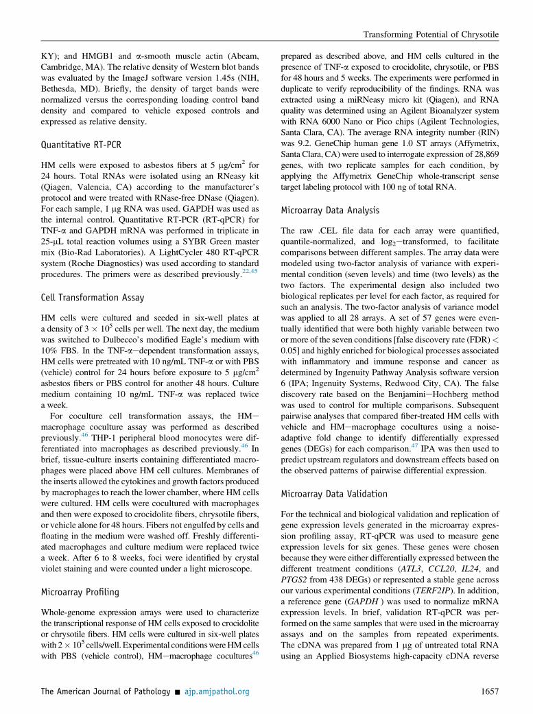
To compare the biological effects of crocidolite and chrys-otile on HM cells, the cells were first exposed to 5 mg/cm2 ofeach of the asbestos fibers for 48 hours and analyzed for theviability and morphology. Fiber analyses showed that themajority of fibers were short and measured within the rangesfound in human MM48 (Supplemental Figures S1 and S2).Fewer chrysotile-exposed HM cells than crocidolite-exposedHM cells were adherent on tissue culture dishes (46 � 9%versus 73 � 12%; P < 0.0001). Moreover, in chrysotile-exposed HM cells, a greater number of attached (ie,surviving) cells acquired a spindle-shaped morphology,compared with crocidolite-exposed cells (48� 6% versus 26� 8%; PZ 0.0173), a possible indication of EMT (discussedbelow) (Figure 1A and Supplemental Figure S3). The greaternumbers of dead and spindle-shaped surviving cellssuggest that chrysotile induces higher cellular stress thancrocidolite, eventually resulting in cell death. Viability andcytotoxicity assays revealed that both fibers are cytotoxic ina density-dependent manner, with chrysotile being signif-icantly more cytotoxic than crocidolite (Figure 1, B and C).
TNF-a Significantly Reduces Chrysotile Cytotoxicity
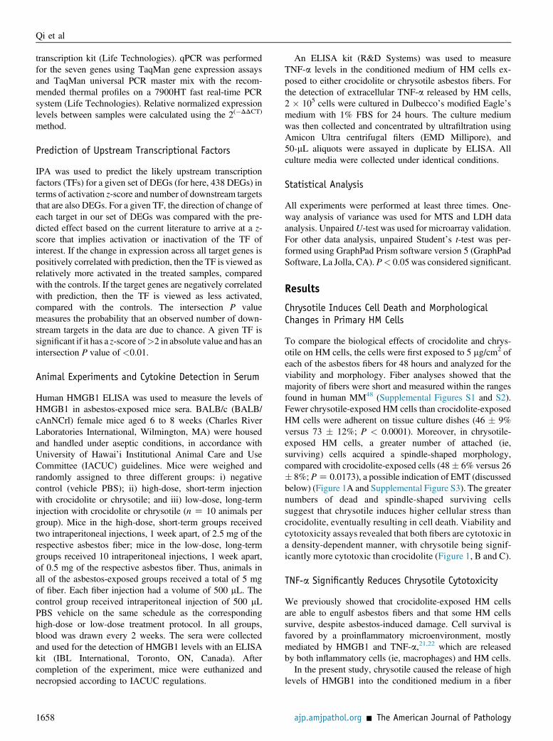
We previously showed that crocidolite-exposed HM cellsare able to engulf asbestos fibers and that some HM cellssurvive, despite asbestos-induced damage. Cell survival isfavored by a proinflammatory microenvironment, mostlymediated by HMGB1 and TNF-a,21,22 which are releasedby both inflammatory cells (ie, macrophages) and HM cells.In the present study, chrysotile caused the release of high
levels of HMGB1 into the conditioned medium in a fiber
ajp.amjpathol.org - The American Journal of Pathology
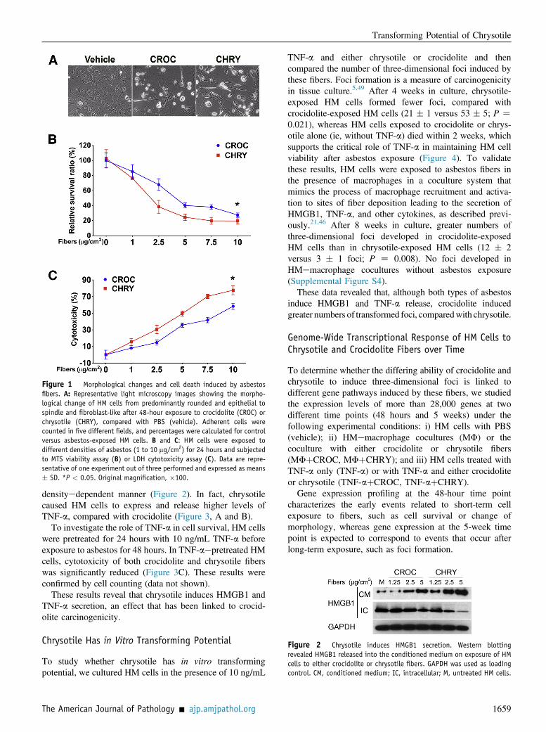
Figure 2 Chrysotile induces HMGB1 secretion. Western blottingrevealed HMGB1 released into the conditioned medium on exposure of HMcells to either crocidolite or chrysotile fibers. GAPDH was used as loadingcontrol. CM, conditioned medium; IC, intracellular; M, untreated HM cells.
Figure 1 Morphological changes and cell death induced by asbestosfibers. A: Representative light microscopy images showing the morpho-logical change of HM cells from predominantly rounded and epithelial tospindle and fibroblast-like after 48-hour exposure to crocidolite (CROC) orchrysotile (CHRY), compared with PBS (vehicle). Adherent cells werecounted in five different fields, and percentages were calculated for controlversus asbestos-exposed HM cells. B and C: HM cells were exposed todifferent densities of asbestos (1 to 10 mg/cm2) for 24 hours and subjectedto MTS viability assay (B) or LDH cytotoxicity assay (C). Data are repre-sentative of one experiment out of three performed and expressed as means� SD. *P < 0.05. Original magnification, �100.
Transforming Potential of Chrysotile
densityedependent manner (Figure 2). In fact, chrysotilecaused HM cells to express and release higher levels ofTNF-a, compared with crocidolite (Figure 3, A and B).
To investigate the role of TNF-a in cell survival, HM cellswere pretreated for 24 hours with 10 ng/mL TNF-a beforeexposure to asbestos for 48 hours. In TNF-aepretreated HMcells, cytotoxicity of both crocidolite and chrysotile fiberswas significantly reduced (Figure 3C). These results wereconfirmed by cell counting (data not shown).
These results reveal that chrysotile induces HMGB1 andTNF-a secretion, an effect that has been linked to crocid-olite carcinogenicity.
Chrysotile Has in Vitro Transforming Potential
To study whether chrysotile has in vitro transformingpotential, we cultured HM cells in the presence of 10 ng/mL
The American Journal of Pathology - ajp.amjpathol.org
TNF-a and either chrysotile or crocidolite and thencompared the number of three-dimensional foci induced bythese fibers. Foci formation is a measure of carcinogenicityin tissue culture.5,49 After 4 weeks in culture, chrysotile-exposed HM cells formed fewer foci, compared withcrocidolite-exposed HM cells (21 � 1 versus 53 � 5; P Z0.021), whereas HM cells exposed to crocidolite or chrys-otile alone (ie, without TNF-a) died within 2 weeks, whichsupports the critical role of TNF-a in maintaining HM cellviability after asbestos exposure (Figure 4). To validatethese results, HM cells were exposed to asbestos fibers inthe presence of macrophages in a coculture system thatmimics the process of macrophage recruitment and activa-tion to sites of fiber deposition leading to the secretion ofHMGB1, TNF-a, and other cytokines, as described previ-ously.21,46 After 8 weeks in culture, greater numbers ofthree-dimensional foci developed in crocidolite-exposedHM cells than in chrysotile-exposed HM cells (12 � 2versus 3 � 1 foci; P Z 0.008). No foci developed inHMemacrophage cocultures without asbestos exposure(Supplemental Figure S4).
These data revealed that, although both types of asbestosinduce HMGB1 and TNF-a release, crocidolite inducedgreater numbers of transformed foci, comparedwith chrysotile.
Genome-Wide Transcriptional Response of HM Cells toChrysotile and Crocidolite Fibers over Time
To determine whether the differing ability of crocidolite andchrysotile to induce three-dimensional foci is linked todifferent gene pathways induced by these fibers, we studiedthe expression levels of more than 28,000 genes at twodifferent time points (48 hours and 5 weeks) under thefollowing experimental conditions: i) HM cells with PBS(vehicle); ii) HMemacrophage cocultures (MF) or thecoculture with either crocidolite or chrysotile fibers(MFþCROC, MFþCHRY); and iii) HM cells treated withTNF-a only (TNF-a) or with TNF-a and either crocidoliteor chrysotile (TNF-aþCROC, TNF-aþCHRY).
Gene expression profiling at the 48-hour time pointcharacterizes the early events related to short-term cellexposure to fibers, such as cell survival or change ofmorphology, whereas gene expression at the 5-week timepoint is expected to correspond to events that occur afterlong-term exposure, such as foci formation.
1659
Figure 3 TNF-a is induced in HM cells after asbestos exposure and signifi-cantly reduces asbestos cytotoxicity. A: HM cells were exposed to 5 mg/cm2 ofcrocidolite or chrysotilefibers or toPBS (vehicle control) for 24hours andqPCR forTNF-amRNAwas performed, using GAPDH as reference.B: TNF-a protein levels inconditioned medium from vehicle control HM cells and HM cells exposed tocrocidolite or chrysotile at 5 mg/cm2 for 24 hours. C: HM cells were preincubatedfor 24 hours either with 10 ng/mL TNF-a (solid lines) or with PBS (dashed lines),and then5mg/cm2crocidoliteor chrysotilefiberswereaddedandcell viabilitywasmeasured by MTS assay after an additional 24 and 48 hours. As control, HM cellswere pretreated with PBS for 24 hours. Data are representative of one experimentout of three performed and expressed as means� SD. *P< 0.05.
Figure 4 Transforming potential of crocidolite and chrysotile fibers inpresence of TNF-a. HM cells were pretreated with 10 ng/mL TNF-a andexposed to crocidolite, chrysotile, or PBS (vehicle). A: HM cells werepretreated with PBS and exposed to PBS (top left image) or were pre-treated with TNF-a and exposed to PBS (top right image), to crocidolite(bottom left image), or to chrysotile (bottom right image). After 4weeks, three-dimensional foci were observed in both crocidolite- andchrysotile-exposed HM cells, but not in PBS-exposed cells. B: Foci wereidentified by crystal violet staining and were counted under lightmicroscopy. Data are expressed as means � SD. Experiments were per-formed three times, and data are representative of one experiment out ofthree performed. *P < 0.05. Original magnification: �100.
Qi et al
Total RNA was extracted from cells for each conditionof interest at each time point, and global gene expressionwas assayed using Affymetrix GeneChip human gene 1.0ST arrays. After data normalization and log2 transformation,
1660
two-way analysis of variance, hierarchical cluster analysis,438 DEGs were identified, and among these 438 DEGs, IPAwere used to identify a subset of 57 genes that were highlyvariable over the seven experimental conditions of interest andthat followed a coordinate expression pattern of geneexpression. These 57 genes were statistically enriched forbiological processes associated with carcinogenesis, inflam-matory response, and immune response (Figure 5A andTable 1). Heat mapping of the 57 genes (Figure 5B) showedpersistent gene activation over time (ie, from the 48-hour tothe 5-week time point) in TNF-aetreated HM cells, inde-pendent of asbestos fiber exposure (TNF-a, TNF-aþCROC,TNF-aþCHRY), as well as in crocidolite-exposed HMemacrophage coculture (MFþCROC). In chrysotile-exposedHMemacrophage coculture (MFþCHRY), however, acti-vation of those genes was observed at 48 hours but haddeclined to baseline levels at 5 weeks (Figure 5, A and B). Tovalidate the microarray experiment, we performed RT-qPCRfor 4 of the 438 differentially expressed genes (DEGs) and 1
ajp.amjpathol.org - The American Journal of Pathology
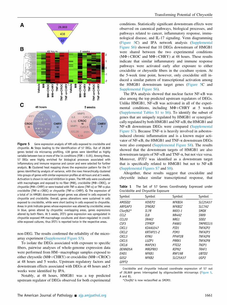
Figure 5 Gene expression analysis of HM cells exposed to crocidolite andchrysotile. A: Steps leading to the identification of 57 DEGs. Out of 28,869genes tested via microarray profiling, 438 genes were identified as highlyvariable between two or more of the 14 conditions (FDR< 0.05). Among these,57 DEGs were highly enriched for biological processes associated withinflammatory and immune response and cancer and were selected for furtheranalysis. B: Clustered heat mapping shows the expression pattern for the 57genes identified by analysis of variance, with the rows hierarchically clusteredinto groups of genes with similar expression profiles at 48 hours and at 5 weeks.Activation is shown in red and inhibition in green. The HM cells were coculturedwith macrophages and exposed to no fiber (MF), crocidolite (MFþCROC), orchrysotile (MFþCHRY) or were treated with TNF-a alone (TNF-a) or TNF-a pluscrocidolite (TNF-aþCROC) or chrysotile (TNF-aþCHRY). C: The expression ofa total of 14 HMGB1 downstream target genes was altered in cells exposed tochrysotile and crocidolite. Overall, genes alterations were sustained in cellsexposed to crocidolite, while were short lasting in cells exposed to chrysotile.Areas in pink indicate genes whose expression was altered by crocidolite; areasin blue, genes altered by chrysotile; overlapping areas, genes expressionsaltered by both fibers. At 5 weeks, IFIT1 gene expression was upregulated inchrysotile exposed HM-macrophage cocultures and down-regulated in crocid-olite exposed cultures, thus IFIT1 is reported twice in the respective areas.
Table 1 The Set of 57 Genes Coordinately Expressed underCrocidolite and Chrysotile Exposure
Symbol Symbol Symbol Symbol
AMIGO2 HIVEP2 NFKBIA SLC25A37ARFGAP3 IFNGR2 NFKBIZ SLC7A2C5orf62* IL7R NKX3-1 SMOXCCL2 IL8 NR4A2 SNX9CCL20 IRAK2 NRG1 SOD2CSF3 ITPRIP PANX1 TMPRSS15CXCL1 KIAA0247 PID1 TNFAIP2CXCL2 KRTAP21-2 PIM3 TNFAIP3CXCL3 KYNU PPAP2B TNFAIP6CXCL5 LUZP1 PRRX1 TNFAIP8CXCL6 MAP2K1 PTGS2 TNIP1DENND5A MRGPRX3 RIPK2 TRAF1EHD1 NFKB1 RNF19B UBTD2G0S2 NFKB2 SLC25A37 UGP2GFPT2
Crocidolite and chrysotile induced coordinate expression of 57 outof 28,869 genes interrogated by oligonucleotide microarrays (Figure 5,A and B).
*C5orf62 is now reclassified as SMIM3.
Transforming Potential of Chrysotile
non-DEG. The results confirmed the reliability of the micro-array experiment (Supplemental Figure S5).
To isolate the DEGs associated with exposure to specificfibers, pairwise analyses of whole-genome expression datawere performed from HMemacrophage samples exposed toeither chrysotile (MFþCHRY) or crocidolite (MFþCROC)at 48 hours and 5 weeks. Upstream regulatory factors anddownstream effects associated with DEGs at 48 hours and 5weeks were identified by IPA.
Notably, at 48 hours, HMGB1 was a top predictedupstream regulator of DEGs observed for both experimental
The American Journal of Pathology - ajp.amjpathol.org
conditions. Statistically significant downstream effects wereobserved on canonical pathways, biological processes, andpathways related to cancer, inflammatory response, immu-nological disease, and IL-17 signaling. Venn diagramming(Figure 5C) and IPA network analysis (SupplementalFigure S6) showed that 10 DEGs downstream of HMGB1were shared between the two experimental conditions(MFþCROC and MFþCHRY) at 48 hours. These resultsindicate that similar inflammatory and immune responsepathways were activated early after exposure to eithercrocidolite or chrysotile fibers in the coculture system. Atthe 5-week time point, however, only crocidolite still in-duced a similar pattern of transcriptional activation amongthe HMGB1 downstream target genes (Figure 5C andSupplemental Figure S6).
The IPA analysis showed that nuclear factor NF-kB wasalso among the top predicted upstream regulators of DEGs.Unlike HMGB1, NF-kB was activated in all of the experi-mental conditions, including MFþCHRY at 5 weeks(Supplemental Tables S1 to S6). To identify the subset ofgenes that are uniquely regulated by HMGB1 or synergisti-cally regulated by bothHMGB1 andNF-kB, theHMGB1 andNF-kB downstream DEGs were compared (SupplementalFigure S7). Because TNF-a is heavily involved in asbestos-induced chronic inflammation and is a known major acti-vator of NF-kB, the HMGB1 and TNF-a downstream DEGswere also compared (Supplemental Figure S8). The resultsshowed that the downstream targets of HMGB1 are alsodownstream targets of NF-kB and TNF-a, but not vice versa.Moreover, IFIT1 was identified as a downstream targetthat is specifically related to HMGB1 but not to NF-kB(Supplemental Figures S7 and S8).
Altogether, these results suggest that crocidolite andchrysotile induce similar transcriptional response, that
1661
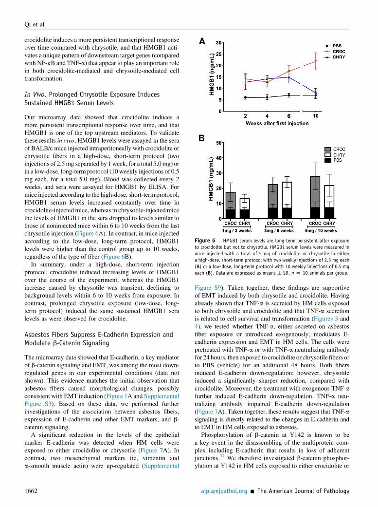
Figure 6 HMGB1 serum levels are long-term persistent after exposureto crocidolite but not to chrysotile. HMGB1 serum levels were measured inmice injected with a total of 5 mg of crocidolite or chrysotile in eithera high-dose, short-term protocol with two weekly injections of 2.5 mg each(A) or a low-dose, long-term protocol with 10 weekly injections of 0.5 mgeach (B). Data are expressed as means � SD. n Z 10 animals per group.
Qi et al
crocidolite induces a more persistent transcriptional responseover time compared with chrysotile, and that HMGB1 acti-vates a unique pattern of downstream target genes (comparedwith NF-kB and TNF-a) that appear to play an important rolein both crocidolite-mediated and chrysotile-mediated celltransformation.
In Vivo, Prolonged Chrysotile Exposure InducesSustained HMGB1 Serum Levels
Our microarray data showed that crocidolite induces amore persistent transcriptional response over time, and thatHMGB1 is one of the top upstream mediators. To validatethese results in vivo, HMGB1 levels were assayed in the seraof BALB/c mice injected intraperitoneally with crocidolite orchrysotile fibers in a high-dose, short-term protocol (twoinjections of 2.5mg separated by 1week, for a total 5.0mg) orin a low-dose, long-term protocol (10weekly injections of 0.5mg each, for a total 5.0 mg). Blood was collected every 2weeks, and sera were assayed for HMGB1 by ELISA. Formice injected according to the high-dose, short-term protocol,HMGB1 serum levels increased constantly over time incrocidolite-injectedmice, whereas in chrysotile-injectedmicethe levels of HMGB1 in the sera dropped to levels similar tothose of noninjected mice within 6 to 10 weeks from the lastchrysotile injection (Figure 6A). In contrast, in mice injectedaccording to the low-dose, long-term protocol, HMGB1levels were higher than the control group up to 10 weeks,regardless of the type of fiber (Figure 6B).
In summary, under a high-dose, short-term injectionprotocol, crocidolite induced increasing levels of HMGB1over the course of the experiment, whereas the HMGB1increase caused by chrysotile was transient, declining tobackground levels within 6 to 10 weeks from exposure. Incontrast, prolonged chrysotile exposure (low-dose, long-term protocol) induced the same sustained HMGB1 seralevels as were observed for crocidolite.
Asbestos Fibers Suppress E-Cadherin Expression andModulate b-Catenin Signaling
The microarray data showed that E-cadherin, a key mediatorof b-catenin signaling and EMT, was among the most down-regulated genes in our experimental conditions (data notshown). This evidence matches the initial observation thatasbestos fibers caused morphological changes, possiblyconsistent with EMT induction (Figure 1A and SupplementalFigure S3). Based on these data, we performed furtherinvestigations of the association between asbestos fibers,expression of E-cadherin and other EMT markers, and b-catenin signaling.
A significant reduction in the levels of the epithelialmarker E-cadherin was detected when HM cells wereexposed to either crocidolite or chrysotile (Figure 7A). Incontrast, two mesenchymal markers (ie, vimentin anda-smooth muscle actin) were up-regulated (Supplemental
1662
Figure S9). Taken together, these findings are supportiveof EMT induced by both chrysotile and crocidolite. Havingalready shown that TNF-a is secreted by HM cells exposedto both chrysotile and crocidolite and that TNF-a secretionis related to cell survival and transformation (Figures 3 and4), we tested whether TNF-a, either secreted on asbestosfiber exposure or introduced exogenously, modulates E-cadherin expression and EMT in HM cells. The cells werepretreated with TNF-a or with TNF-a neutralizing antibodyfor 24 hours, then exposed to crocidolite or chrysotile fibers orto PBS (vehicle) for an additional 48 hours. Both fibersinduced E-cadherin down-regulation; however, chrysotileinduced a significantly sharper reduction, compared withcrocidolite. Moreover, the treatment with exogenous TNF-afurther induced E-cadherin down-regulation. TNF-a neu-tralizing antibody impaired E-cadherin down-regulation(Figure 7A). Taken together, these results suggest that TNF-asignaling is directly related to the changes in E-cadherin andto EMT in HM cells exposed to asbestos.Phosphorylation of b-catenin at Y142 is known to be
a key event in the disassembling of the multiprotein com-plex including E-cadherin that results in loss of adherentjunctions.37 We therefore investigated b-catenin phosphor-ylation at Y142 in HM cells exposed to either crocidolite or
ajp.amjpathol.org - The American Journal of Pathology
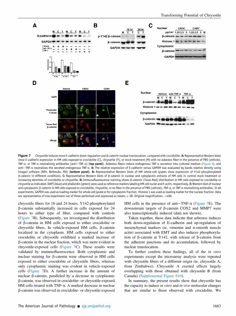
Figure 7 Chrysotile induces more E-cadherin down-regulation and b-catenin nuclear translocation, compared with crocidolite. A: Representative Western blotsshow E-cadherin expression in HM cells exposed to crocidolite (C), chrysotile (Y), or mock treatment (M) with no asbestos fiber in the presence of PBS (vehicle),TNF-a, or TNF-a neutralizing antibodies (antieTNF-a) (top panel). Asbestos fibers induce endogenous TNF-a secretion into cultured medium (Figure 3), andantieTNF-a neutralizes the secreted endogenous TNF-a. A: The relative expression of E-cadherin versus GAPDH was evaluated by bands relative density usingImageJ software (NIH, Bethesda, MD) (bottom panel). B: Representative Western blots of HM whole-cell lysates show expression of Y142-phosphorylatedb-catenin in different conditions. C: Representative Western blots of b-catenin in nuclear and cytoplasmic extracts of HM cells in control mock treatment orincreasing densities of crocidolite or chrysotile. D: Immunofluorescence staining shows b-catenin (Texas Red) distribution in HM cells exposed to crocidolite orchrysotile as indicated. DAPI (blue) and phalloidin (green)were used as referencemarkers labelingHM cell nuclei and F-actin, respectively. E:Western blot of nuclearand cytoplasmic b-catenin in HM cells exposed to crocidolite, chrysotile, or no fiber in the presence of PBS (vehicle), TNF-a, or TNF-a neutralizing antibodies. In allexperiments, GAPDHwas used as loading marker for whole cell lysate or for cytoplasmic fraction. Histone 1 was used as loadingmarker for the nuclear fraction. Dataare representative of one experiment out of three performed and expressed as means � SD. Original magnification, �400.
Transforming Potential of Chrysotile
chrysotile fibers for 16 and 24 hours. Y142-phosphorylatedb-catenin substantially increased in cells exposed for 24hours to either type of fiber, compared with controls(Figure 7B). Subsequently, we investigated the distributionof b-catenin in HM cells exposed to either crocidolite orchrysotile fibers. In vehicle-exposed HM cells, b-cateninlocalized in the cytoplasm. HM cells exposed to eithercrocidolite or chrysotile exhibited a marked increase ofb-catenin in the nuclear fraction, which was more evident inchrysotile-exposed cells (Figure 7C). These results werevalidated by immunofluorescence. Both cytoplasmic andnuclear staining for b-catenin were observed in HM cellsexposed to either crocidolite or chrysotile fibers, whereasonly cytoplasmic staining was evident in vehicle-exposedcells (Figure 7D). A further increase in the amount ofnuclear b-catenin, paralleled by a decrease in cytoplasmicb-catenin, was observed in crocidolite- or chrysotile-exposedHM cells treated with TNF-a. A marked decrease in nuclearb-catenin was observed in crocidolite- or chrysotile-exposed
The American Journal of Pathology - ajp.amjpathol.org
HM cells in the presence of antieTNF-a (Figure 7E). Thedownstream targets of b-catenin COX2 and MMP7 werealso transcriptionally induced (data not shown).
Taken together, these data indicate that asbestos inducesboth down-regulation of E-cadherin and up-regulation ofmesenchymal markers (ie, vimentin and a-smooth muscleactin) associated with EMT and also induces phosphoryla-tion of b-catenin at Y142, with release of b-catenin fromthe adherent junctions and its accumulation, followed bynuclear translocation.
To further confirm these findings, all of the in vitroexperiments except the microarray analysis were repeatedwith chrysotile fibers of a different origin (ie, chrysotile A,from Zimbabwe). Chrysotile A exerted effects largelyoverlapping with those obtained with chrysotile B (fromCanada) (Supplemental Figure S10).
In summary, the present results show that chrysotile hasthe capacity to induce in vitro and in vivo molecular changesthat are similar to those observed with crocidolite. We
1663
Qi et al
characterized HMGB1 and TNF-a as key regulators of the-se processes, and our data suggest that b-catenin and E-cad-herin signaling are early cellular events that follow asbestosexposure and contribute to oncogenic transformation.However, crocidolite-induced effects last over the course ofseveral weeks or longer, whereas chrysotile signaling is ofonly short duration, unless exposure is continued over time.
Discussion
Chrysotile accounts for 90% or more of commercially usedasbestos fibers worldwide. Although its use is banned in theEuropean Union and in many other countries, chrysotile isstill widely mined and is still exported to developingcountries,8 under the assumption that its carcinogenicity isnot conclusively proven. Some argue that because the greatmajority of asbestos exposure comes from chrysotile, evenif chrysotile were to be significantly less carcinogenic thancrocidolite, it would still account for a large percentage ofMM cases.50 Others argue that epidemiological and bio-logical data do not support a causative role for chrysotile inMM.6 The issue is complicated by many billions of dollarsin litigation and chrysotile exports that might be influencedby research linking or not linking chrysotile to MM.5,7,51
Our present results show that, compared with crocidolite,chrysotile causes more mesothelial cell death and increasedrelease of HMGB1 and TNF-a (at picogram levels). TNF-areduced asbestos cytotoxicity. On long-term HM cell culturewith exogenous TNF-a or macrophages, crocidolite ex-hibited greater transforming potential than chrysotile, asmeasured by the number of HM three-dimensional foci thatwere induced by these fibers. A possible explanation is that,because chrysotile is more cytotoxic than crocidolite,chrysotile exposure results in fewer surviving HM cells,which in turn may account for the fewer foci observed inchrysotile-exposed cells.
When HM cells were exposed to asbestos fibers in theHMemacrophage coculture system (in which the macro-phages are also exposed to asbestos and thus releaseHMGB1), or when HM cells were cultured in the presenceof exogenous TNF-a, we observed the activation of up-stream regulatory elements associated with MM, includingHMGB1. A number of genes differentially expressed inHMemacrophage coculture exposed to asbestos were pre-dicted to be downstream targets of HMGB1. There was nobias toward HMGB1-inducible targets, because each pair-wise analysis was based on an unbiased, genome-wide scanof more than 28,000 genes over seven experimental condi-tions, with two time points per condition and two replicatesper time point. IPA of the top 200 DEGs identified in eachanalysis predicted HMGB1 as a significant regulator of theobserved DEGs (Supplemental Tables S1 to S6) for bothfibers at 48 hours, and only crocidolite at 5 weeks.
Similarly, 57 DEGs were identified based on an unbiased,genome-wide scan of gene expression. The row-clustered
1664
heat map of these 57 genes with samples grouped by timerevealed that, although both fibers showed similar activationpatterns at 48 hours, only crocidolite showed persistentactivation at the 5-week time point. The IPA knowledgebase to predict which transcriptional factors are involved inregulation of the DEGs showed that HMGB1 was amongthe seven top predicted regulators in all but two of theconditions, the exceptions being MF only and MFþchry-sotile at 5 weeks (Supplemental Tables S1 to S6).As expected, NF-kB was also among the top predicted
regulators of DEGs, but with a different temporal pattern ofactivation than was observed for HMGB1. Long-termcoculture of HM cells and MF with crocidolite resulted inpersistent transcriptional activity of both NF-kB andHMGB1 and a large number of transforming foci. In thecase of chrysotile, NF-kB transcriptional activity wasmaintained, but fewer foci were observed, correlating withthe reduced activation of HMGB1. This finding suggeststhat NF-kB signaling alone is not sufficient to fully accountfor asbestos carcinogenesis, and that sustained HMGB1signaling seems to be required. HMGB1 selection as a targetin our investigation was therefore unbiased (Figure 5A). Thecritical role of HMGB1 is also reinforced by the evidencethat HMGB1 specifically regulates only a subset of NF-kBeregulated genes, and that not all of the HMGB1 targets areshared with other transcriptional regulators.The microarray data also showed that persistent transcrip-
tional activation of HMGB1 and downstream genes 5 weeksfrom exposure was present in HM cells exposed to high doseof exogenous TNF-a, regardless of fiber presence. Theobservation that TNF-a is able to induce persistent tran-scriptional activation validates the crucial role of TNF-a inpromoting mesothelial cell survival and transformation.22
A possible explanation for the absence of persistentHMGB1 signaling in HM cells cocultured with macro-phages and exposed to chrysotile is that chrysotile formssilky fibril bundles (in contrast to the needle-shaped fibersof crocidolite), which are more easily washed out duringcell culture procedures, resulting in a loss of physicalpersistence and biological signaling over time. Also,crocidolite fibers persist at sites of deposition in vivo, withthe fiber concentration increasing with prolonged expo-sure,6 whereas chrysotile fibers are rapidly cleared from thelung.6 We therefore, further tested in vivo the hypothesisthat the biopersistence of chrysotile fibers is required forsustained HMGB1 signaling, comparing HMGB1 serumlevels in short-term and long-term injection protocols.Increasing the biopersistence of chrysotile fibers throughrepeated injections resulted in HMGB1 secretion to thesame extent as that of crocidolite.Finally, we observed that cells surviving exposure to either
type of asbestos fibers developed a spindle-like morphologysuggestive of EMT. Chrysotile appeared to have a strongereffect than crocidolite in decreasing E-cadherin expression.We concluded that the observed morphological changes wereassociated with E-cadherin down-regulation, a result that was
ajp.amjpathol.org - The American Journal of Pathology
Transforming Potential of Chrysotile
further promoted by exogenous TNF-a. One of the hallmarksof EMT is E-cadherin down-regulation also at the geneexpression level,33 a marker we found in the microarray geneexpression profiling, supporting the occurrence of EMT inHM cells exposed to asbestos. Chrysotile- and crocidolite-induced E-cadherin down-regulation (possibly both at tran-scriptional and post-translational levels) was also associatedwith b-catenin phosphorylation, nuclear translocation, andtranscriptional activity.
Potential limitations to the present study involve bothin vitro and in vivo aspects. First, the concentration of as-bestos used in our experiments, although suitable for in vitrostudies to allow measurement of a biological response withina limited time span, likely exceeds concentrations achieved inthe pleura of individuals exposed to asbestos. A secondlimitation is the absence of a long-term experiment assessingMM incidence in vivo with the two different injectionprotocols. (Our research group is currently conducting suchan experiment, with results expected within approximately2 years.) The chrysotile samples used in this study closelymatch the average dimensions of chrysotile fibers found inoccupation-exposed MM patients to asbestos, strengtheningthe validity of our results (Supplemental Figure S1 andSupplemental Figure S2). Moreover, samples froma different chrysotile source (chrysotile A; Zvishavane,Matabeleland South, Zimbabwe) produced similar results(see Supplemental Figure S10 for a representative experi-ment), supporting the reliability of our findings.
In summary, our results show that, in HM cells, chrysotilehas the capacity to induce molecular changes similar to themolecular changes induced by crocidolite, but that thesechanges are transitory. HMGB1 and TNF-a proved to bekey mediators of these processes, for both chrysotile andcrocidolite. Moreover, E-cadherin down-regulation and b-catenin signaling pathways were induced by both chrysotileand crocidolite, and were enhanced by TNF-a.
Although our results do not address the overall issue ofchrysotile carcinogenesis in humans, they highlight for thefirst time similarities and differences between crocidoliteand chrysotile in inducing biological changes that may leadto MM. Sporadic exposure to crocidolite was sufficient toinduce some of the molecular changes associated with HMtransformation, such as sustained gene alterations andHMGB1 secretion. Sporadic exposure to chrysotile did notinduce these effects. Instead, repeated exposure to chrysotileand crocidolite led to similar molecular changes and similaramount of HMGB1 secretion in vitro and in vivo.Our data suggest that fiber biopersistence is one of the
main differences between the carcinogenicities of crocido-lite and chrysotile. The different fiber physical characteris-tics are associated with different biopersistence,52 but itcannot be excluded that different morphometric parametersmay result in different biological activities per se.
The present results underscore the importance of asbestosfiber biopersistence in inducing sustained HMGB1 levels andHM malignant transformation, and support the notion that
The American Journal of Pathology - ajp.amjpathol.org
only continuous exposure to chrysotile is able to maintain theprocesses that may lead to MM over a prolonged time span.
Acknowledgments
We thank Dr. Zeyana Rivera for help in conducting animalexperiments, Brian Kendrick for help in characterizingprimary human mesothelial cells, and Dr. Yurii Shvetsov,Dr. Thomas Wenska, and Michael Loomis for fruitfuldiscussions and advice on microarray data management andbioinformatics analysis.
Supplemental Data
Supplemental material for this article can be found athttp://dx.doi.org/10.1016/j.ajpath.2013.07.029.
References
1. Henley SJ, Larson TC, Wu M, Antao VC, Lewis M, Pinheiro GA,Eheman C: Mesothelioma incidence in 50 states and the District ofColumbia, United States, 2003-2008. Int J Occup Environ Health2013, 19:1e10
2. Flores RM, Pass HI, Seshan VE, Dycoco J, Zakowski M, Carbone M,Bains MS, Rusch VW: Extrapleural pneumonectomy versus pleur-ectomy/decortication in the surgical management of malignant pleuralmesothelioma: results in 663 patients. J Thorac Cardiovasc Surg2008, 135. 620e626, 626.e1e3
3. Pass HI, Vogelzang NJ, Hahn SM, Carbone M: Benign and malignantmesothelioma. DeVita, Hellman, and Rosenberg’s Cancer: Principlesand Practice of Oncology. ed 9. Edited by De Vita VT Jr,Lawrence TS, Rosenberg SA, DePinho RA, Weinberg RA. Phila-delphia, Lippincott Williams & Wilkins, 2011, pp 2052e2080
4. Baumann F, Ambrosi JP, Carbone M: Asbestos is not just asbestos:an unrecognised health hazard. Lancet Oncol 2013, 14:576e578
5. Carbone M, Ly BH, Dodson RF, Pagano I, Morris PT, Dogan UA,Gazdar AF, Pass HI, Yang H: Malignant mesothelioma: facts, myths,and hypotheses. J Cell Physiol 2012, 227:44e58
6. Britton M: The epidemiology of mesothelioma. Semin Oncol 2002,29:18e25
7. Tweedale G: Asbestos and its lethal legacy. Nat Rev Cancer 2002,2(4):311e315
8. Burki T: Health experts concerned over India’s asbestos industry.Lancet 2010, 375:626e627
9. McDonald JC: Epidemiology of malignant mesotheliomaean outline.Ann Occup Hyg 2010, 54:851e857
10. Smith WE, Miller L, Elsasser RE, Hubert DD: Tests for carcinoge-nicity of asbestos. Ann N Y Acad Sci 1965, 132:456e488
11. Wagner JC, Berry G, Skidmore JW, Timbrell V: The effects of theinhalation of asbestos in rats. Br J Cancer 1974, 29:252e269
12. Davis JM: Histogenesis and fine structure of peritoneal tumorsproduced in animals by injections of asbestos. J Natl Cancer Inst1974, 52:1823e1837
13. Glickman LT, Domanski LM, Maguire TG, Dubielzig RR, Churg A:Mesothelioma in pet dogs associated with exposure of their owners toasbestos. Environ Res 1983, 32:305e313
14. Friemann J, Brinkmann O, Pott F, Müller KM: Peritoneale Differ-enzierungsstörungen als Reaktion auf Asbest- und Asbestersatzstoffe.Tierexperimentelle Untersuchungen [Disturbances in peritonealdifferentiation as a reaction to asbestos and asbestos substitutes.Experimental animal studies]. German. Verh Dtsch Ges Pathol 1988,72:312e316
1665
Qi et al
15. Minardi F, Maltoni C: Results of recent experimental research on thecarcinogenicity of natural and modified asbestos. Ann N Y Acad Sci1988, 534:754e761
16. Hasanoglu HC, Bayram E, Hasanoglu A, Demirag F: Orally ingestedchrysotile asbestos affects rat lungs and pleura. Arch Environ OccupHealth 2008, 63:71e75
17. Pott F: Asbestos use and carcinogenicity in Germany and a compar-ison with animal studies. Ann Occup Hyg 1994, 38:589e600
18. Yano E, Wang ZM, Wang XR, Wang MZ, Lan YJ: Cancer mortalityamong workers exposed to amphibole-free chrysotile asbestos. Am JEpidemiol 2001, 154:538e543
19. Mossman BT, Churg A: Mechanisms in the pathogenesis of asbes-tosis and silicosis. Am J Respir Crit Care Med 1998, 157:1666e1680
20. Nagai H, Ishihara T, Lee WH, Ohara H, Okazaki Y, Okawa K,Toyokuni S: Asbestos surface provides a niche for oxidative modi-fication. Cancer Sci 2011, 102:2118e2125
21. Yang H, Rivera Z, Jube S, Nasu M, Bertino P, Goparaju C,Franzoso G, Lotze MT, Krausz T, Pass HI, Bianchi ME, Carbone M:Programmed necrosis induced by asbestos in human mesothelial cellscauses high-mobility group box 1 protein release and resultantinflammation. Proc Natl Acad Sci USA 2010, 107:12611e12616
22. Yang H, Bocchetta M, Kroczynska B, Elmishad AG, Chen Y, Liu Z,Bubici C, Mossman BT, Pass HI, Testa JR, Franzoso G, Carbone M:TNF-alpha inhibits asbestos-induced cytotoxicity via a NF-kappaB-dependent pathway, a possible mechanism for asbestos-inducedoncogenesis. Proc Natl Acad Sci USA 2006, 103:10397e10402
23. Fassina A, Cappellesso R, Guzzardo V, Dalla Via L, Piccolo S,Ventura L, Fassan M: Epithelial-mesenchymal transition in malignantmesothelioma. Mod Pathol 2012, 25:86e99
24. Merikallio H, Pääkkö P, Salmenkivi K, Kinnula V, Harju T, Soini Y:Expression of snail, twist, and Zeb1 in malignant mesothelioma.APMIS 2013, 121:1e10
25. Casarsa C, Bassani N, Ambrogi F, Zabucchi G, Boracchi P,Biganzoli E, Coradini D: Epithelial-to-mesenchymal transition, cellpolarity and stemness-associated features in malignant pleuralmesothelioma. Cancer Lett 2011, 302:136e143
26. Baran B, Bechyne I, Siedlar M, Szpak K, Mytar B, Sroka J, Laczna E,Madeja Z, Zembala M, Czyz J: Blood monocytes stimulate migrationof human pancreatic carcinoma cells in vitro: the role of tumournecrosis factor-alpha. Eur J Cell Biol 2009, 88:743e752
27. Wu Y, Deng J, Rychahou PG, Qiu S, Evers BM, Zhou BP: Stabili-zation of snail by NF-kappaB is required for inflammation-inducedcell migration and invasion. Cancer Cell 2009, 15:416e428
28. Demir AY, Groothuis PG, Dunselman GA, Schurgers L, Evers JL, deGoeij AF: Molecular characterization of soluble factors from humanmenstrual effluent that induce epithelial to mesenchymal transitions inmesothelial cells. Cell Tissue Res 2005, 322:299e311
29. Lynch J, Nolan S, Slattery C, Feighery R, Ryan MP, McMorrow T:High-mobility group box protein 1: a novel mediator ofinflammatory-induced renal epithelial-mesenchymal transition. Am JNephrol 2010, 32:590e602
30. He M, Kubo H, Ishizawa K, Hegab AE, Yamamoto Y, Yamamoto H,Yamaya M: The role of the receptor for advanced glycation end-products in lung fibrosis. Am J Physiol Lung Cell Mol Physiol2007, 293:L1427eL1436
31. Kalluri R, Weinberg RA: The basics of epithelial-mesenchymaltransition [Erratum appeared in J Clin Invest 2010, 120:1786]. JClin Invest 2009, 119:1420e1428
32. Bellovin DI, Bates RC, Muzikansky A, Rimm DL, Mercurio AM:Altered localization of p120 catenin during epithelial to mesenchymaltransition of colon carcinoma is prognostic for aggressive disease.Cancer Res 2005, 65:10938e10945
33. Cano A, Pérez-Moreno MA, Rodrigo I, Locascio A, Blanco MJ, delBarrio MG, Portillo F, Nieto MA: The transcription factor snailcontrols epithelial-mesenchymal transitions by repressing E-cadherinexpression. Nat Cell Biol 2000, 2:76e83
1666
34. Cavallaro U, Christofori G: Cell adhesion and signalling by cadherinsand Ig-CAMs in cancer. Nat Rev Cancer 2004, 4:118e132
35. Pece S, Gutkind JS: E-cadherin and Hakai: signalling, remodeling ordestruction? Nat Cell Biol 2002, 4:E72eE74
36. Reynolds AB, Daniel JM, Mo YY, Wu J, Zhang Z: The novel cateninp120cas binds classical cadherins and induces an unusual morpho-logical phenotype in NIH3T3 fibroblasts. Exp Cell Res 1996, 225:328e337
37. Lilien J, Balsamo J: The regulation of cadherin-mediated adhesion bytyrosine phosphorylation/dephosphorylation of beta-catenin. CurrOpin Cell Biol 2005, 17:459e465
38. Clevers H, Nusse R: Wnt/beta-catenin signaling and disease. Cell2012, 149:1192e1205
39. Uematsu K, Kanazawa S, You L, He B, Xu Z, Li K, Peterlin BM,McCormick F, Jablons DM: Wnt pathway activation in mesothe-lioma: evidence of Dishevelled overexpression and transcriptionalactivity of beta-catenin. Cancer Res 2003, 63:4547e4551
40. Abutaily AS, Collins JE, Roche WR: Cadherins, catenins and APC inpleural malignant mesothelioma. J Pathol 2003, 201:355e362
41. Dai Y, Bedrossian CW, Michael CW: The expression pattern of beta-catenin in mesothelial proliferative lesions and its diagnostic utilities.Diagn Cytopathol 2005, 33:320e324
42. Bocchetta M, Di Resta I, Powers A, Fresco R, Tosolini A, Testa JR,Pass HI, Rizzo P, Carbone M: Human mesothelial cells are unusu-ally susceptible to simian virus 40-mediated transformation andasbestos cocarcinogenicity. Proc Natl Acad Sci USA 2000, 97:10214e10219
43. Xu A, Zhou H, Yu DZ, Hei TK: Mechanisms of the genotoxicity ofcrocidolite asbestos in mammalian cells: implication from mutationpatterns induced by reactive oxygen species. Environ Health Perspect2002, 110:1003e1008
44. MacCorkle RA, Slattery SD, Nash DR, Brinkley BR: Intracellularprotein binding to asbestos induces aneuploidy in human lungfibroblasts. Cell Motil Cytoskeleton 2006, 63:646e657
45. Zhang L, Qi F, Gaudino G, Strianese O, Yang H, Morris P, Pass HI,Nerurkar VR, Bocchetta M, Carbone M: Tissue tropism of SV40transformation of human cells: role of the viral regulatory region andof cellular oncogenes. Genes Cancer 2010, 1:1008e1020
46. Carbone M, Baris YI, Bertino P, Brass B, Comertpay S, Dogan AU,Gaudino G, Jube S, Kanodia S, Partridge CR, Pass HI, Rivera ZS,Steele I, Tuncer M, Way S, Yang H, Miller A: Erionite exposure inNorth Dakota and Turkish villages with mesothelioma. Proc NatlAcad Sci USA 2011, 108:13618e13623
47. Szauter KM, Jansen MK, Okimoto G, Loomis M, Kimura JH,Heller M, Ku T, Tiirikainen M, Boyd CD, Csiszar K, Girton RA:Persistent inflammatory pathways associated with early onsetmyocardial infarction in a medicated multiethnic Hawaiian cohort.Biochem Insights 2011, 2011(4):13e27
48. Suzuki Y, Yuen SR, Ashley R: Short, thin asbestos fibers contributeto the development of human malignant mesothelioma: pathologicalevidence [Erratum appeared in Int J Hyg Environ Health 2005, 208:439e444]. Int J Hyg Environ Health 2005, 208:201e210
49. Jube S, Rivera Z, Bianchi ME, Powers A, Wang E, Pagano IS,Pass HI, Gaudino G, Carbone M, Yang H: Cancer cell secretion of theDAMP protein HMGB1 supports progression in malignant meso-thelioma. Cancer Res 2012, 72:3290e3301
50. Kanarek MS: Mesothelioma from chrysotile asbestos: update[Erratum appeared in Ann Epidemiol 2012, 22:377]. Ann Epidemiol2011, 21:688e697
51. Lagnese JA: Economic aspects of mesothelioma. Malignant Meso-thelioma: Advances in Pathogenesis, Diagnosis, and TranslationalTherapies. Edited by Pass HI, Vogelzang NJ, Carbone M. New York,Springer, 2005, pp 821e832
52. Bernstein DM, Rogers R, Smith P: The biopersistence of Canadianchrysotile asbestos following inhalation [Erratum appeared in InhalToxicol 2004, 16:67]. Inhal Toxicol 2003, 15:1247e1274
ajp.amjpathol.org - The American Journal of Pathology