Embed Size (px)
Citation preview
[CANCER RESEARCH 44, 5702-5706, December 1984]
Contributions of Glycolysis and Oxidative Phosphorylation to Adenosine5'-Triphosphate Production in AS-30D Hepatoma Cells1
Richard A. Nakashima,2 Marco G. Paggi,3 and Peter L. Pedersen4
Department of Biological Chemistry, The Johns Hopkins University School of Medicine, Baltimore, Maryland 21205
ABSTRACT
The AS-30D rat hepatoma cell line is characteristic of thatclass of rapidly growing tumors which exhibit high rates ofaerobic glucose utilization and lactic acid production (Busta-
mante, E., Morris, H. P., and Pedersen, P. L., J. Biol. Chem.,256: 8699-8704, 1981). In this study, we have examined thecoupling properties of the mitochondria in intact AS-30D hepatoma cells and the relative contributions of cytoplasmic (glyco-
lytic) and mitochondrial compartments to total cellular ATP production in the presence of glucose and glutamine. All respirationin AS-30D cells was inhibited by inhibitors of mitochondrial
electron transport, ruling out significant rates of respiration fromother cellular components. Moreover, cellular respiration wasfound to be coupled to phosphorylation of ADP, as demonstratedby its inhibition by oligomycin and aurovertin, inhibitors of themitochondrial ATP synthetase (FoFi-ATPase). When intact cells
were supplied with glucose as the only added energy source, itwas estimated that about 60% of the total cell ATP was derivedfrom glycolysis and 40% from oxidative phosphorylation. Addition of physiological concentrations of glutamine in the presenceof glucose had little effect on the relative contributions of glycolysis and oxidative phosphorylation to total cellular ATP production. In the absence of added glucose, glutamine alone couldmaintain the same ATP production rates by supporting mitochondrial oxidative phosphorylation. It is concluded that, in theAS-30D hepatoma cell line, glucose is the preferred energysource, with the larger portion of ATP production being suppliedby glycolytic reactions. Although oxidative substrates such asglutamine can replace glucose in maintaining total cell ATPproduction, they do not appear to be the major fuel sourceswhen hepatoma AS-30D cells are exposed to concentrations of
substrates which occur in vivo.
INTRODUCTION
It has been known for over 50 years that tumor cells possessan abnormal energy metabolism in comparison with most normalmammalian cells, which derive most of their energy supplies frommitochondrial oxidative phosphorylation (1, 8, 27). Cancer cellsexhibit an increased utilization of anaerobic (glycolytic) metabolism for cell energy production (1-5,22), and a direct correlation
has been observed between cancer cell growth rate and glucoseutilization rate (2, 3). The most rapidly growing tumor cell linesobtain up to 50% of their total ATP production from glycolytic
1Supported by Grant CA 32742 from the National Cancer Institute, NIH.2 Supported by NIH National Research Service Award 1 F32 CA 07506-01 from
the National Cancer Institute.3 Permanent address: Istituto Regina Elena per lo Studio e la Cura dei Tumori,
V. le Regina Elena 291,00161 Rome, Italy. Fellow of the Consiglio Nazionale delleRicerche, sponsored by the Associazione Italiana per la Ricerca sul Cancro.
' To whom requests for reprints should be addressed.
Received April 24,1984; accepted August 20.1984.
metabolism (1,11,21), with a corresponding decrease in oxidative phosphorylation and in cell mitochondrial content (21, 22).The increase in glucose consumption rate is not strictly a consequence of the increased rate of cell division (3,21,22). Rather,it reflects a basic difference in the metabolic properties of transformed cells (3, 21, 22).
A recent novel approach to the chernotherapeutic treatmentof cancer has focused upon the aberrant energy metabolism oftransformed cells (9,10,13,26). Preliminary results suggest thatit may be possible to selectively inhibit tumor cell energy production, growth rate, and survival by targeting tumor-specific en
zyme systems (9,10,13,26). In order to maximize the effectiveness of such an approach, it is important to determine whatenergy substrates tumor cells utilize and what the relative contributions of aerobic and glycolytic metabolism are to tumor cellenergy production under in vivo conditions. In the present study,we have addressed the latter question in the rapidly growing,highly glycolytic AS-30D rat hepatoma cell line. The results
presented indicate that, at the concentrations of substratesfound in vivo (24), glucose is a preferred substrate for this cellline, with over 50% of total cell ATP production coming fromglycolytic metabolism, even in the presence of high levels ofglutamine.
MATERIALS AND METHODS
Chemicals. Aurovertin was purchased from the Pitman-Moore Com
pany (Indianapolis, IN), oligomycin and rotenone were from Sigma Chemical Company, and FCCP6 was the generous gift of P. G. Heytter of E. I.
Dupont de Nemours and Co. (Inc.) of Wilmington, DE. All other chemicalswere of reagent grade or better and were purchased from commonchemical sources.
Tumor Cells. The AS-30D hepatoma cell line was obtained from Dr.
A. L. Lehninger of the Johns Hopkins University School of Medicine,Baltimore, MD. The line was maintained by injection of 2 ml of asciticfluid i.p. into 125- to 150-g female Sprague-Dawley rats purchased from
Holtzman Laboratories (Madison, Wl). The animals were sacrificed bycervical dislocation at 10 to 14 days after inoculation, and the cells werecollected and suspended in Chance-Hess medium [6.2 mw KCI, 154 HIM
NaCI, 11 mW sodium phosphate, pH 7.4] (6) at room temperature. Thecells were centrifuged 4 to 7 times at 300 x g for 10 min in a swinging-
bucket rotor at room temperature in the same medium in order toeliminate RBC. The cells were counted in a Neubauer counting chamberand then resuspended in Chance-Hess medium to a final concentrationof 2 x 10°cells/ml. Cell viability was greater than 97%, as indicated by
trypan blue dye exclusion.Mitochondrial Isolation. Mitochondria were isolated from purified AS-
30D cells according to the method of Parry and Pedersen (20).Assay of Glycolysis. Aerobic glycolysis in hepatoma cells was carried
out in a shaking water bath in 25-ml Erlenmeyer flasks at 30°. Thereaction mixture contained AS-30D cells (4 x 10⁷ cells) in Chance-Hess
5The abbreviation used is: FCCP, carbonyl cyanide p-triftuoromethoxyphenyl-
hydrazone.
CANCER RESEARCH VOL. 44 DECEMBER 1984
5702
on August 3, 2020. © 1984 American Association for Cancer Research. cancerres.aacrjournals.org Downloaded from
ENERGY SOURCES FOR CANCER CELLS
medium (pH 7.4) and other components, as indicated in the charts, in afinal volume of 3.0 ml. After a 10-min prior incubation period at 30°,the
reaction was started by the addition of substrates. Incubation wasallowed to proceed for the times indicated in the charts. At the end ofthe incubation, the reaction was stopped by immediate chilling of thesampleson ice and sedimentation of the cells in an Eppendorf centrifugefor 1 min. The supematants were frozen with dry ice-isopropyl alcoholand stored prior to analysis for lactic acid concentration. Lactic acidconcentration was determined enzymaticallyaccording to the method ofHohorst(12).
Measurement of Respiration Rate. Oxygen consumption rates weremeasuredpolarographicallyusing a Clark-type electrode (Yellow SpringsInstruments, OH) inserted into a water-jacketed sealed glass chamber.The oxygen electrode apparatus was connected to a chart recorderwhich was calibrated between 0 and 100% saturation with atmosphericoxygen at 30°.Rates of oxygen consumption in change in percentage
of saturation/min were read directly from the chart recording. Thesewere converted to ng atoms of oxygen/min using a conversion factor of435 ng-atoms of oxygen/ml (9) according to the following formula.
AS-30D Cells
A % of saturation/min x435 ng-atoms of oxygen/ml
100% saturationx 1.3ml
= ng-atoms of oxygen/min
Cells (2.2 mg of protein) or mitochondria (1.3 mg of protein) wereincubated at 30°with magnetic stirring in a total volume of 1.3 ml of
medium, with additions as noted in the legends to the charts.Protein Determination. Proteinconcentrationwas determinedaccord
ing to the method of Upsky and Pedersen(17), using the modified biuretprocedure of Schmukler and Yiengst (25).
RESULTS
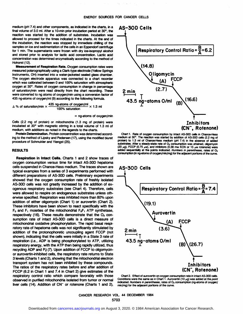
Respiration in Intact Cells. Charts 1 and 2 show traces ofoxygen consumption versus time for intact AS-30D hepatomacells suspended in Chance-Hess medium. The traces shown are
typical examples from a series of 3 experiments performed withdifferent preparations of AS-30D cells. Preliminary experiments
showed that the oxygen consumption rate of freshly isolatedAS-30D cells was not greatly increased by the addition of ex
ogenous respiratory substrates (see Chart 4). Therefore, cellswere allowed to respire on endogenous substrates unless otherwise specified. Respiration was inhibited more than 80% uponaddition of either oligomycin (Chart 1) or aurovertin (Chart 2).These inhibitors have been shown to react specifically with theFOand F! moieties of the mitochondria! FoF-\ ATP synthetase,
respectively (16). These results demonstrate that the O2 consumption rate of intact AS-30D cells is a direct measure of
mitochondria! oxidative phosphorylation. The rapid basal respiratory rate of hepatoma cells was not significantly stimulated byaddition of the protonophoretic uncoupling agent FCCP (notshown), indicating that the cells were initially in a State 3 rate ofrespiration (I.e., ADP is being phosphorylated to ATP, utilizingrespiratory energy, with the ATP then being rapidly utilized, thusrecycling ADP and P¡)(7). Upon addition of FCCP to oligomycin-or aurovertin-inhibited cells, the respiratory rate returns to State
3 levels (Charts 1 and 2), showing that the mitochondria! electrontransport system has not been inhibited by these compounds.The ratios of the respiratory rates before and after addition ofFCCP (6.2 in Chart 1 and 7.4 in Chart 2) give estimates of therespiratory control ratio which compare favorably with thoseobserved in purified mitochondria isolated from tumor or normalliver cells (14). Addition of CNT or rotenone (Charts 1 and 2),
BRespiratory Control Ratio --r-6.2
2 min
J14.8)
Oligomycin
J(2.7)
T43.5 ng-atoms O/ml /g\ (16.6)
Inhibitors(CN~, Rotenone)
Chart 1. Rate of oxygen consumption by intact AS-30D cells in Chance-Hessmedium at 30°.The reaction was started by addition of AS-30D cells (2.2 mg ofprotein) to 1.3 ml of Chance-Hess medium (pH 7.4) in the absence of addedsubstrates. After a steady-state rate of 02 consumption was attained, oligomyan(20 /¿g),FCCP (0.75 MM),and inhibitors (0.38 mm KCN or 15 MMrotenone) wereadded sequentially at the points indicated. Numbers in parentheses, rates of 0?consumption (inng-atomsof oxygen/min/mg) for the adjacentportions of the curve.
AS-30D Cells
I
Respiratory Control Ratio=T=7.4B
FCCP
2 min
43.5nig-atomsO/m. (
Inhibitors(CN~, Rotenone)
Chart 2. Effect of aurovertin on oxygen consumption rate in intact AS-30Dcells.Conditions were the same as in Chart 1. Aurovertin (10 /ig) was added at the pointindicated.Numbers in parentheses, rates of O2consumption (ng-atoms of oxygen/min/mg) for the adjacent portions of the curve.
CANCER RESEARCH VOL. 44 DECEMBER 1984
5703
on August 3, 2020. © 1984 American Association for Cancer Research. cancerres.aacrjournals.org Downloaded from
ENERGY SOURCES FOR CANCER CELLS
which are specific inhibitors of mitochondria! electron transport,produces a complete block of O2 consumption which is notaffected by FCCP. These results demonstrate that the respirationof purified AS-30D cells is due entirely to the activity of the
mitochondria! electron transport system and is predominantlycoupled to the production of intracellular ATP by oxidative phosphorylation.
Mitochondria! Respiration. The ability of various substratesto support tumor oxidative metabolism was examined usingpurified AS-30D mitochondria. In the absence of added substrate, AS-30D mitochondria showed a low rate of oxygen
consumption, which was not stimulated by addition of ADP(Table 1, Experiments 2 to 5). The subsequent addition of anoxidative substrate such as succinate resulted in a large (28-
fold) stimulation of oxygen consumption rate (Table 1, Experiment 2), consistent with the ability of succinate to supportmitochondria! oxidative phosphorylation. A similar stimulation ofmitochondria! respiration in the presence of ADP was observedwith glutamine and, to a lesser extent, with pyruvate (Table 1,Experiments 3 and 5), but not with 0-hydroxybutyrate (Table 1,Experiment 4). Variation of /3-hydroxybutyrate concentration between 0.8 and 20.0 mM had no effect on ADP-dependent respi
ration, while increasing pyruvate concentration above 0.08 ITIMcaused a slight inhibition of respiration rate (not shown).
When the order of addition was reversed, the low basal rateof mitochondria! respiration was stimulated 3-fold by succinate
addition (Table 1, Experiment 1). Subsequent addition of ADPresulted in a large stimulation of respiration which was inhibited75% by oligomycin (Table 1, Experiment 1). We conclude fromthese results that, in the AS-30D hepatoma line, succinate,
glutamine, and pyruvate can support mitochondria! ATP production by oxidative phosphorylation, while /3-hydroxybutyrate can
not.Table 1
Substrate-dependent oxygen consumption by isolated AS-30D mitochondria
Purified AS-30D mitochondria (1.0 mg/ml) were incubated in 1.3 ml of mediumcontaining 180 mM sucrose, 40 mM KCI, 0.5 mM ethyleneglycol bis(0-aminoethyl-ether)-Af,/v,/V',N'-tetraacetic acid, 5 mM MgCI»,10 mM K2HPO«,and 2 mM 4-(2-hydroxyethyl)-1-piperazineethanesulfonic acid (pH 7.1) at 30°. Respiration was
initiated by the sequential addition of substrates in the order noted below. Therates of oxygen consumption were calculated as described in 'Materials andMethods.* The experiments were performed at least twice, with similar results.
ExperimentExperiment
1Mitochondria+5
mMsuccinate+0.4mMADP+10M9
ofoligomycinExperiment2Mitochondria+1.6mMADP+5
mMsuccinateExperimentsMitochondria+1.6mM
ADP+5mMglutamine+7.7
MMrotenoneExperiment4Mitochondria+1.6mMADP+5
mMfi-hydroxybutyrate+5mMglutamineExperiment
5Mitochondria+1.6mMADP+0.08
mMpyruvate+7.7MMrotenoneRespiration
rate (ng-atomsof oxygen/min/mg)5.216.097.224.74.63.085.23.83.058.31.24.02.92.962.43.72.39.10.0
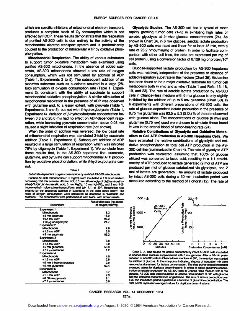
Glycolytic Studies. The AS-30D cell line is typical of mostrapidly growing tumor cells (1-5) in exhibiting high rates of
aerobic glycolysis at in vivo glucose concentrations (24). Asshown in Chart 34, in 6 HIM glucose, aerobic lactate productionby AS-30D cells was rapid and linear for at least 45 min, with a
rate of 26.2 nmol/min/mg of protein. In order to facilitate comparison with other cell lines, the data are expressed per mg ofcell protein, using a conversion factor of 0.129 mg of protein/106
cells.Glucose-supported lactate production by AS-30D hepatoma
cells was relatively independent of the presence or absence ofadded respiratory substrate in the medium (Chart 3B). Glutaminehas been found to be a major oxidative substrate for tumor cellmetabolism both in vivo and in vitro (Table 1 and Refs. 15,18,19, and 23). The rate of aerobic lactate production by AS-30Dcells in Chance-Hess medium with 6 HIMglucose is only slightly
inhibited by the addition of up to 5 mw glutamine (Chart 33). In6 experiments with different preparations of AS-30D cells, therate of glucose-dependent lactate production in the presence of
0.75 mM glutamine was 93.5 ±5.3 (S.D.) % of the rate observedwith glucose alone. The concentrations of glucose (6 mM) andglutamine (0.75 mM) used were chosen to simulate those foundin vivo in the arterial blood of tumor-bearing rats (24).
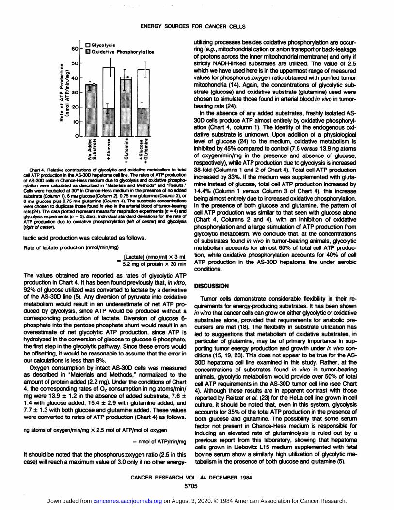
Relative Contributions of Glycolytic and Oxidative Metabolism to Cell ATP Production ¡nAS-30D Hepatoma Cells. We
have estimated the relative contributions of glycolytic and oxidative phosphorylation to total cell ATP production in the AS-30D cell line (summarized in Chart 4). The rate of glycolytic ATPproduction was calculated, assuming that 100% of glucoseutilized was converted to lactic acid, resulting in a 1:1 stoichi-ometry of ATP produced to lactate generated (2 mol of ATP areproduced per mol of glucose catabolized via glycolysis, and 2mol of lactate are generated). The amount of lactate producedby intact AS-30D cells during a 30-min incubation period was
measured according to the method of Hohorst (12). The rate of
I500 30 L B
L_
IO 20 30 40 50 60Minutes
O I 2 3 4 5Glutamine Concentration (mM)
Chart 3. A, time course for lactate production by intact AS-30D cells incubatedin Chance-Hess medium supplemented with 6 HIM glucose. After a 10-min prein-cubation of AS-30D cells in Chance-Hess medium at 30°,the reaction was started
by addition of glucose. At the time points indicated, aliquots of incubation mix wereremoved and analyzed for lactate concentration. The data points plotted representaveraged values for duplicate determinations. 8, effect of added glutamine concentration on lactate production by AS-30D cells in Chance-Hess medium with 6 mMglucose. AS-30D cells were incubated in Chance-Hess medium at 30°with glucose
and the indicated concentrations of glutamine. The rate of lactate production overa 30-min incubation period is plotted as a function of glutamine concentration. Thedata points represent averaged values for duplicate determinations.
CANCER RESEARCH VOL. 44 DECEMBER 1984
5704
on August 3, 2020. © 1984 American Association for Cancer Research. cancerres.aacrjournals.org Downloaded from
ENERGY SOURCES FOR CANCER CELLS
60c
500TP
ProductTP/min/mgWA 00o
o20«Cci-
IOOD
Glycolysis0 OxidativePhosphorylati--i*Tièr
J.ii
.?
fì —
03 OZOT +
^ S3o +o•f +
Chart 4. Relative contributions of glycolytic and oxidative metabolism to totalcell ATP production in the AS-30D hepatoma cell line. The rates of ATP productionof AS-30D cells in Chance-Mess medium due to glycolysis and oxidative phospho-rylatton were calculated as described in 'Materials and Methods" and 'Results.'Cells were incubated at 30°in Chance-Hess medium in the presence of no added
substrate (Column 1), 6 mM glucose (Column 2), 0.75 RIM glutamina (Column 3), or6 mM glucose plus 0.75 mM glutamina (Column 4). The substrate concentrationswere chosen to duplicate those found in vivo in the arterial blood of tumor-bearingrats (24). The data plotted represent means for respiration experiments (n = 4) andglycolysis experiments (n = 5). Bars, individual standard deviations for the rate ofATP production due to oxidative prtosphorylation (left of center) ana glycolysis(right of center).
lactic acid production was calculated as follows.
Rate of láclateproduction (nmol/min/mg)
[Lactate] (nmol/ml) x 3 ml5.2 mg of protein x 30 min
The values obtained are reported as rates of glycolytic ATPproduction in Chart 4. It has been found previously that, in vitro,92% of glucose utilized was converted to lactate by a derivativeof the AS-30D line (5). Any diversion of pyruvate into oxidative
metabolism would result in an underestimate of net ATP produced by glycolysis, since ATP would be produced without acorresponding production of lactate. Diversion of glucose 6-
phosphate into the pentose phosphate shunt would result in anoverestimate of net glycolytic ATP production, since ATP ishydrolyzed in the conversion of glucose to glucose 6-phosphate,
the first step in the glycolytic pathway. Since these errors wouldbe offsetting, it would be reasonable to assume that the error inour calculations is less than 8%.
Oxygen consumption by intact AS-30D cells was measuredas described in "Materials and Methods," normalized to the
amount of protein added (2.2 mg). Under the conditions of Chart4, the corresponding rates of 02 consumption in ng atoms/min/mg were 13.9 ±1.2 in the absence of added substrate, 7.6 ±1.4 with glucose added, 15.4 ±2.9 with glutamine added, and7.7 ±1.3 with both glucose and glutamine added. These valueswere converted to rates of ATP production (Chart 4) as follows.
ng atoms of oxygen/min/mg x 2.5 mol of ATP/mol of oxygen
= nmol of ATP/min/mg
It should be noted that the phosphorus:oxygen ratio (2.5 in thiscase) will reach a maximum value of 3.0 only if no other energy-
utilizing processes besides oxidative phosphorylation are occurring (e.g., mitochondria! cation or anióntransport or back-leakage
of protons across the inner mitochondria! membrane) and only ifstrictly NADH-linked substrates are utilized. The value of 2.5
which we have used here is in the uppermost range of measuredvalues for phosphorus:oxygen ratio obtained with purified tumormitochondria (14). Again, the concentrations of glycolytic substrate (glucose) and oxidative substrate (glutamine) used werechosen to simulate those found in arterial blood in vivo in tumor-
bearing rats (24).In the absence of any added substrates, freshly isolated AS-
30D cells produce ATP almost entirely by oxidative phosphorylation (Chart 4, column 1). The identity of the endogenous oxidative substrate is unknown. Upon addition of a physiologicallevel of glucose (24) to the medium, oxidative metabolism isinhibited by 45% compared to control (7.6 versus 13.9 ng atomsof oxygen/min/mg in the presence and absence of glucose,respectively), while ATP production due to glycolysis is increased38-fold (Columns 1 and 2 of Chart 4). Total cell ATP production
increased by 33%. If the medium was supplemented with glutamine instead of glucose, total cell ATP production increased by14.4% (Column 1 versus Column 3 of Chart 4), this increasebeing almost entirely due to increased oxidative phosphorylation.In the presence of both glucose and glutamine, the pattern ofcell ATP production was similar to that seen with glucose alone(Chart 4, Columns 2 and 4), with an inhibition of oxidativephosphorylation and a large stimulation of ATP production fromglycolytic metabolism. We conclude that, at the concentrationsof substrates found in vivo in tumor-bearing animals, glycolytic
metabolism accounts for almost 60% of total cell ATP production, while oxidative phosphorylation accounts for 40% of cellATP production in the AS-30D hepatoma line under aerobic
conditions.
DISCUSSION
Tumor cells demonstrate considerable flexibility in their requirements for energy-producing substrates. It has been shown
in vitro that cancer cells can grow on either glycolytic or oxidativesubstrates alone, provided that requirements for anabolic pre-
cursers are met (18). The flexibility in substrate utilization hasled to suggestions that metabolism of oxidative substrates, inparticular of glutamine, may be of primary importance in supporting tumor energy production and growth under in vivo conditions (15,19, 23). This does not appear to be true for the AS-
30D hepatoma cell line examined in this study. Rather, at theconcentrations of substrates found in vivo in tumor-bearing
animals, glycolytic metabolism would provide over 50% of totalcell ATP requirements in the AS-30D tumor cell line (see Chart
4). Although these results are in apparent contrast with thosereported by Reitzer ef al. (23) for the HeLa cell line grown in cellculture, it should be noted that, even in this system, glycolysisaccounts for 35% of the total ATP production in the presence ofboth glucose and glutamine. The possibility that some serumfactor not present in Chance-Hess medium is responsible for
inducing an elevated rate of glutaminolysis is ruled out by aprevious report from this laboratory, showing that hepatomacells grown in LJebovitz L15 medium supplemented with fetalbovine serum show a similarly high utilization of glycolytic metabolism in the presence of both glucose and glutamine (5).
CANCER RESEARCH VOL. 44 DECEMBER 1984
5705
on August 3, 2020. © 1984 American Association for Cancer Research. cancerres.aacrjournals.org Downloaded from
ENERGY SOURCES FOR CANCER CELLS
The AS-30D Å“il line used in this study can be considered tobe representative of the category of rapidly growing tumorswhich exhibit high rates of aerobic glycolysis and lactate production at in vivo blood glucose levels (1-5, 21, 22, 27, 28). We do
not propose that the results obtained with this cell line areapplicable to the metabolism of all forms of cancer cells. Asnoted in "Introduction," cancer cell lines exhibit a broad spectrum
of aerobic glycolytic activity, with slowly growing, minimal-devia
tion tumors resembling normal cells in exhibiting low rates ofglycolysis and high utilization of oxidative metabolism for cellenergy production (1, 2, 4, 5, 21, 22, 28).
The finding that glucose at in vivo concentrations is a preferredsubstrate for the AS-30D cell line does not preclude an importantrole for oxidative metabolism in these hepatoma cells. This reportpresents the first measurement of respiratory efficiency (respiratory control ratio) in intact tumor cells (Charts 1 and 2), utilizingthe inhibitors oligomycin and aurovertin, which are specific foroxidative phosphorylation (16). The high values obtained forrespiratory control ratios indicate that AS-30D hepatoma mitochondria are well-coupled in situ, allowing efficient energy pro
duction by oxidative metabolism as well as by glycolysis. In fact,although glycolysis is of primary importance in providing ATP forthe AS-30D hepatoma cell line, about 40% of the total cell ATPproduction is still derived from oxidative phosphorylation. Theseresults suggest that any attempt to inhibit tumor cell growth andsurvival by interfering with tumor energy production must takeinto account the ability of transformed cell lines to utilize simultaneously both oxidative and glycolytic substrates to support cellgrowth. Significantly, this point has been emphasized in severaldetailed reviews dealing with the energy metabolism of cancercells (1,21, 28).
REFERENCES
1. Aisenberg, A. C. The Glycolysis and Respiration of Tumors. New York:Academic Press, Inc., 1961.
2. Burk. 0., Woods, M.. and Hunter, J. On the significance of glucolysis forcancer growth, with special referenceto Morris rat hepatomas.J. Nati. CancerInst., 38: 839-863,1967.
3. Bustamante. E., Morris, H. P., and Pedersen,P. L. Hexokinase: the direct linkbetween mitochondria!and glycolytic reactions in rapidly growing cancer cells.Adv. Exp. Mod. Btol., 92: 363-380,1977.
4. Bustamante, E., Morris, H. P., and Pedersen, P. L. Energy metabolism oftumor cells. Requirement for a form of hexokinase with a propensity formitochondrial binding. J. Biol. Chem.,256: 8699-8704,1981.
5. Bustamante, E., and Pedersen, P. L. High aerobic glycolysis of rat hepatomacells in culture: role of mitochondrial hexokinase. Proc. Nati. Acad. Sci. USA,74: 3735-3739,1977.
6. Chance, B.. and Hess, B. Metabolic control mechanisms. I. Electron transfer
in the mammaliancell. J. Bol. Chem., 234: 2404-2412,1959.7. Chance, B., and Williams,G. R. Respiratory enzymes in oxidative phosphoryl
ation. I. Kinetics of oxygen utilization. J. Biol. Chem., 277: 383-393,1955.8. Con, C. F.,and Con, G. T. The carbohydratemetabolismof tumors. II.Changes
in the sugar, lactic acid, and COj-cornbining powers of blood passing througha tumor. J. Btol.Chem., 65: 397-405,1925.
9. Floridi, A., Paggi, M. G., D'Atri, S., DeMartìno,C., Marcante, M. L, Silvestrini,
B., and Caputo, A. Effect of Lomdammeon the energy metabolism of Ehrlichascites tumor cells. Cancer Res., 47: 4661-4666, 1981.
10. Floridi, A., Paggi. M. G., Marcante, M. L., Silvestrini, B., Caputo, A., andDeMartino,C. Lonidamine,a selective inhibitor of aerobic glycolysisof murinetumor cells. J. Nati. Cancer Inst., 66:497-499,1981.
11. Gumaa, K. A., and McLean, P. A possible interrelationshipbetween bindingofhexokinase and the site of ATP formation in Krebs ascites cells. Biochem.Btophys. Res. Commun., 36:771-779,1969.
12. Hohorst. H. J. Determination with lactic-dehydrogenase and DPN. In: H. V.Bergmeyer(ed.),Methods of EnzymaticAnalysis,p. 266. New York: AcademicPress, Inc., 1965.
13. Johnson,J. H., Zimniak,A., and Racker,E. Inhibitionof hexokinaseand proteinkinase activities of tumor cells by a chtoromethyl ketone derivative of lacticacid. Biochemistry, 27: 2984-2989,1982.
14. Kaschnitz, R. M., Hatefi, Y., and Morris. H. P. Oxidative phosphorylationproperties of mitochondria isolated from transplanted hepatoma. Biochim.Btophys.Acta, 449: 224-235,1976.
15. Kovacevto, Z., and Monis, H. P. The role of glutamine in the oxidativemetabolism of malignantcells. Cancer Res., 32: 326-333,1972.
16. LJnnett,P. E., and Beechey, R. B. Inhibitors of the ATP synthetase system.Methods Enzymol.,55: 472-518,1979.
17. Lipsky, N. G., and Pedersen, P. L. Perturbation by clofibrate of mitochondriallevels in animalcells. Implicationsfor a model of mitochondrialgenesis.J. Btol.Chem.,257:1473-1481,1982.
18. McKeehan,W. L. Glycolysis, glutaminolysisand cell proliferation. Cell Btol. Int.Rep., 6: 635-650,1982.
19. Moreadith, R. W., and Lehninger, A. L. The pathways of glutamate andglutamineoxidation by tumor cell mitochondria.Roleof mitochondrialNAD(P)'-dependent maltoenzyme. J. Biol. Chem., 259: 6215-6221,1984.
20. Parry, D. M., and Pedersen, P. L. Intracellular localization and properties ofpaniculate hexokinase in the Novikoff ascites tumor. Evidence for an outermitochondrialmembranelocation. J. Btol.Chem., 256:10904-10912,1983.
21. Pedersen, P. L. Tumor mitochondria and the btoenergetics of cancer cells.Prog. Exp. Tumor Res., 22: 190-274,1978.
22. Pedersen,P. L., and Bustamante,E. Hexokinaseand the abnormalmetabolismof hepatoma cells. In: W. E. Jacobus and J. S. Ingwall (eds.), Heart CreatineKinase: the Integration of Isozymes for Energy Distribution, pp. 147-154.Baltimore: Williams & Wilkins, 1980.
23. Reitzer, L. J., Wice, B. M., and Kenneil.D. Evidencethat glutamine, not sugar,is the major energy source for cultured HeLacells. J. Btol.Chem., 254:2669-2676,1979.
24. Sauer, L. A., and Dauchy, R. T. Ketone body, glucose, lactic acid, and aminoacid utilization by tumors in vivo in fasted rats. Cancer Res., 43: 3497-3503,1983.
25. Schmukler.M., and Yiengst, M. J. A biuret method for determinationof proteinin hyamine hydroxide and NCS solutions used for liquid scintillation countingin toluene. Anal. Biochem.,25:406-411,1968.
26. Tuszynski, G. P., and Cossu, G. Differential cytotoxic effect of gossypol onhuman melanoma,colon carcinoma,and other tissue culture cell lines. CancerRes.. 44: 768-771,1984.
27. Warburg, 0., Posener, K., and Negelein,E. Ãœberden Stoffwechsel der Carci-nomzelle.Biochem. Z., 752: 309-344,1924.
28. Wemhouse.S. Glycolysis, respiration, and enzyme deletions in slow-growinghepatic tumors in biological and biochemical evaluation of malignancy inexperimental hepatomas. Gann Monogr., 7: 99-115,1966.
CANCER RESEARCH VOL. 44 DECEMBER 1984
5706
on August 3, 2020. © 1984 American Association for Cancer Research. cancerres.aacrjournals.org Downloaded from
1984;44:5702-5706. Cancer Res Richard A. Nakashima, Marco G. Paggi and Peter L. Pedersen Cells
-Triphosphate Production in AS-30D Hepatoma′Adenosine 5Contributions of Glycolysis and Oxidative Phosphorylation to
Updated version
http://cancerres.aacrjournals.org/content/44/12_Part_1/5702
Access the most recent version of this article at:
E-mail alerts related to this article or journal.Sign up to receive free email-alerts
Subscriptions
Reprints and
To order reprints of this article or to subscribe to the journal, contact the AACR Publications
Permissions
Rightslink site. Click on "Request Permissions" which will take you to the Copyright Clearance Center's (CCC)
.http://cancerres.aacrjournals.org/content/44/12_Part_1/5702To request permission to re-use all or part of this article, use this link
on August 3, 2020. © 1984 American Association for Cancer Research. cancerres.aacrjournals.org Downloaded from