Embed Size (px)
Citation preview
ARCHIVES OF BIOCHEMISTRY AND BIOPHYSICS
Vol. 337, No. 2, January 15, pp. 169–175, 1997Article No. BB969784
Control of Hepatic Fatty Acid Oxidation by 5*-AMP-Activated Protein Kinase Involves a Malonyl-CoA-Dependent and a Malonyl-CoA-Independent Mechanism
Guillermo Velasco,* Math J. H. Geelen,†,1 and Manuel Guzman**Department of Biochemistry and Molecular Biology I, Faculty of Biology, Complutense University, 28040-Madrid, Spain;and †Laboratory of Veterinary Biochemistry, Graduate School of Animal Health, Utrecht University, P.O. Box 80.176,3508 TD Utrecht, The Netherlands
Received July 30, 1996, and in revised form October 25, 1996
The 5*-AMP-activated protein kinase (AMPK)2 playsa major role in the regulation of lipid metabolism inIncubation of rat hepatocytes with 5-aminoimidaz-mammals. Thus, AMPK phosphorylates and inacti-ole-4-carboxamide ribonucleoside (AICAR), an activa-vates key regulatory enzymes of lipid metabolism suchtor of the 5*-AMP-activated protein kinase (AMPK),as acetyl-CoA carboxylase (fatty acid synthesis), 3-hy-produced a twofold stimulation of palmitate oxidationdroxy-3-methylglutaryl-CoA reductase (sterol/isopren-and of the activity of carnitine palmitoyltransferase
I (CPT-I), together with a profound decrease of the oid synthesis), and hormone-sensitive lipase (triacyl-activity of acetyl-CoA carboxylase and of the intracel- glycerol/cholesteryl ester breakdown) [reviewed in Ref.lular level of malonyl-CoA. AICAR-induced CPT-I stim- (1)]. Although several protein kinases can phosphory-ulation progressively blunted with time after cell per- late purified acetyl-CoA carboxylase and 3-hydroxy-3-meabilization, pointing to reversal of conformational methylglutaryl-CoA reductase in vitro, it is currentlyconstraints of the enzyme in control cells due to the accepted that in intact hepatocytes and in the liver inpermeabilization-triggered dilution of intracellular vivo this phosphorylation is mainly performed bymalonyl-CoA. The stimulation stabilized at a steady AMPK [cf. Refs. (1–3)].20–25%. This 20–25% increase in CPT-I activity sur- Several studies have been performed on the potentialvived upon complete removal of malonyl-CoA from the involvement of AMPK in the control of fatty acid oxida-permeabilized cells, indicating that it was not depen- tion in the ischemic heart (4, 5) and the working muscledent on the malonyl-CoA concentration of the cell. This (6). However, the possible role of this kinase in themalonyl-CoA-independent activation of CPT-I was not control of hepatic fatty acid oxidation has not beenevident when mitochondria were isolated for assay of studied to date. Unlike the heart and the skeletal mus-enzyme activity or when cells were disrupted by vigor- cle, the liver is capable of expressing either high ratesous sonication. In addition, the microtubule stabilizer
of lipogenesis or high rates of fatty acid oxidation de-taxol prevented the malonyl-CoA-independent stimu-pending on the hormonal and nutritional status of thelation of CPT-I induced by AICAR. Hence, stimulationanimal, and hence regulation of fatty acid oxidationof hepatic fatty acid oxidation by AMPK seems to relyseems to be more complex in liver [reviewed in Ref. (7)]on the activation of CPT-I by two different mecha-than in heart [reviewed in Ref. (8)] and skeletal musclenisms: deinhibition of CPT-I induced by depletion of[reviewed in Ref. (9)]. Carnitine palmitoyltransferaseintracellular malonyl-CoA levels and malonyl-CoA-in-I (CPT-I) is the key regulatory enzyme in the transportdependent stimulation of CPT-I, which might involveof long-chain fatty acids into the mitochondrial matrixmodulation of interactions between CPT-I and cy-(7, 10, 11). This enzyme is subject to allosteric inhibi-toskeletal components. q 1997 Academic Presstion by malonyl-CoA, the product of the reaction cata-
2 Abbreviations used: AMPK, 5*-AMP-activated protein kinase;CoA, coenzyme A; CPT-I, carnitine palmitoyltransferase I; AICAR,
1 To whom correspondence should be addressed. Fax: **31-30- 5-aminoimidazole-4-carboxamide ribonucleoside; ZMP, 5-aminoimid-azole-4-carboxamide ribonucleoside monophosphate.2535492. E-mail: [email protected].
1690003-9861/97 $25.00Copyright q 1997 by Academic PressAll rights of reproduction in any form reserved.
AID ABB 9784 / 6b28$$$261 12-12-96 17:00:01 arcas

170 VELASCO ET AL.
Hepatocyte isolation and incubation. Male Wistar rats (250–300lyzed by acetyl-CoA carboxylase (7, 10, 11). During theg) which had free access to food and water were used throughout inpast few years, a novel mechanism of short-term con-this study. Hepatocytes were isolated by the collagenase perfusiontrol of hepatic CPT-I has been put forward [reviewed method described in Ref. (19). Because lipogenesis is markedly de-
in Ref. (7)]. Several studies using permeabilized hepa- pressed just after hepatocyte isolation, cells were incubated for 15min at 377C in a gyratory metabolic shaker and subsequently filteredtocytes have shown that various agents exert short-through nylon mesh prior to use (20). Cell viability, as determined byterm effects on CPT-I activity in parallel with changestrypan blue exclusion, always exceeded 95% in the final hepatocytein the rate of long-chain fatty acid oxidation measuredsuspension.
in the same cell preparations. Thus, cAMP analogs Hepatocytes were incubated in Krebs–Henseleit bicarbonate(e.g., dibutyryl-cAMP) (12), effectors that increase in- buffer supplemented with 10 mM glucose and 1% (w/v) defatted and
dialyzed bovine serum albumin. Incubations (4–6 mg of cellular pro-tracellular cAMP levels (e.g., glucagon and forskolin)tein/ml) were performed in a total volume of 2 ml at 377C, with(12), and protein phosphatase inhibitors (e.g., okadaicconstant shaking (85 oscillations/min) and under an atmosphere ofacid) (13) are able to stimulate hepatic CPT-I, whereas O2/CO2 (19:1). Stock solutions of AICAR, okadaic acid, and taxol were
Ca2/-mobilizing agents (e.g., vasopressin, a1-adrener- prepared in Me2SO. Therefore, control incubations had the corre-gic agonists, and extracellular ATP) inhibit hepatic sponding Me2SO content. No significant influence of Me2SO on any
of the experimentally determined parameters was observed at theCPT-I (14). Since these short-term changes in CPT-Ifinal concentration used (0.1%, v/v).activity are very stable and survive complete removal
Rate of fatty acid oxidation. For determination of the rate of fattyof malonyl-CoA from the medium, they are assumed toacid oxidation, hepatocytes were incubated for 15 min with the cellu-be mediated by a malonyl-CoA-independent mecha-lar effectors indicated in every case. Reactions were subsequently
nism which might involve phosphorylation of putative started by the addition to cell incubations of [1-14C]fatty acid (eithermediator protein(s) (7, 16). palmitate or octanoate, 0.05 Ci/mol, 0.5 mM final concentration)
bound to albumin. After 10 min, reactions were stopped with 0.5 mlIdentification of physiological substrates of AMPKof 2 M perchloric acid and oxidation products were extracted andhas been hampered by the lack of specific methods forquantified exactly as described before (13). Total oxidation productsactivating the kinase in intact cells. AMPK was rou- were calculated as the sum of acid-soluble products and CO2. Acid-
tinely activated in intact hepatocytes by incubation soluble products (mostly ketone bodies) routinely accounted for 90–with fructose or by heat shock or arsenite [cf. Ref. (1)]. 95% of total oxidation products (13).These treatments all deplete intracellular ATP and, CPT-I assay. The activity of CPT-I was determined as the tetra-
decylglycidate-sensitive incorporation of radiolabeled L-carnitinetherefore, have many nonspecific side effects. However,into palmitoylcarnitine by four different methods (A, B, C, and D).a more specific method for activating AMPK in intactIn brief, hepatocytes were preincubated for 20 min in the absence orcells has been recently reported. Incubation of intactin the presence of 10 mM tetradecylglycidate, a specific irreversible
hepatocytes with 5-aminoimidazole-4-carboxamide ri- inhibitor of CPT-I (13, 21). Incubations were continued for an addi-bonucleoside (AICAR) causes uptake of this compound tional 15-min period in the presence of the cellular effectors indicated
in every case. Subsequently, aliquots were removed from the incuba-and subsequent accumulation within the cell of itstions to monitor CPT activity with the four different types of assay.monophosphorylated form, 5-aminoimidazole-4-car-
In methods A and B, CPT activity was measured in digitonin-boxamide ribonucleoside monophosphate (ZMP) (16). permeabilized hepatocytes. Both methods were performed using theThe latter has been shown to mimic the effect of 5*- same detergent/cell protein ratio (about 40 mg digitonin/mg cell pro-AMP on allosteric activation of rat liver AMPK without tein). Enzyme activity was routinely determined by method A (‘‘one-
step assay’’). In this method, 100 ml of hepatocyte suspension is di-changing the levels of ATP, ADP, and AMP within therectly added to 100 ml of prewarmed digitonin-containing assay me-hepatocyte (16, 17). Thus, exposure to AICAR has beendium exactly as described in Ref. (13). Hence, monitoring of enzymeshown to inactivate acetyl-CoA carboxylase and 3-hy- activity is performed immediately upon permeabilization of the cells
droxy-3-methylglutaryl-CoA reductase in isolated he- (13, 22). In method B (‘‘two-step assay’’), hepatocytes are permeabil-ized and thoroughly washed prior to determination of enzyme activ-patocytes (3, 17) as well as hormone-sensitive lipase inity. Thus, 1.0 ml of hepatocyte suspension was permeabilized withisolated adipocytes (18). In the present report we show0.20 mg of digitonin dissolved in 1.0 ml of 5 mM Tris–HCl (pH 7.4),that activation of AMPK produces a stimulation of he-50 mM KF, 100 mM KCl, 2.5 mM EDTA, and 2.5 mM EGTA (F0patic long-chain fatty acid oxidation, which relies in medium). The resulting mix was gently shaken for 5 s and rapidly
turn on activation of CPT-I by malonyl-CoA-dependent diluted by transfer to tubes containing 40 ml of ice-cold F0 medium.Cell ghosts were sedimented by centrifugation at 350g for 15 s, andand independent mechanisms.pellets were taken up in 1.0 ml of prewarmed F0 medium. The re-sulting suspensions of cell ghosts were incubated at 377C for 5–15min and then CPT activity was monitored (15).MATERIALS AND METHODS
In method C, CPT activity was measured in sonicated hepatocytes.Cells were sedimented by low-speed centrifugation (50g, 2 min). TheMaterials. [1-14C]Palmitic acid, [1-14C]octanoic acid, L-[Me-14C]-
carnitine, [1-14C]acetyl-CoA, and 3H2O were supplied by Amersham cell pellet was resuspended in F0 medium and sonicated for twoperiods of 10 s while being cooled in ice/water. Samples of sonicatedInternational (Amersham, Bucks, UK). Digitonin, collagenase (type
I), AICAR, ZMP, 5*-AMP, and taxol were purchased from Sigma preparations were used for determination of CPT activity (15).In method D, CPT activity was measured in mitochondria isolatedChemical Co. (St. Louis, MO). Okadaic acid was supplied by Calbio-
chem (San Diego, CA). Tetradecylglycidic acid was a kind gift from from hepatocyte suspensions exactly as described before (15). Prepa-rations of mitochondria were practically devoid of peroxisomes, asDr. J. M. Lowenstein (Brandeis University, Waltham, MA).
AID ABB 9784 / 6b28$$$261 12-12-96 17:00:01 arcas

171CONTROL OF FATTY ACID OXIDATION BY AMP-ACTIVATED PROTEIN KINASE
judged from measurements of recovery of catalase activity (alwaysless than 10% of total cellular activity).
Other analytical methods. Intracellular levels of malonyl-CoAwere determined in neutralized perchloric acid cell extracts by aradioenzymatic method as described in Ref. (19). Rates of de novofatty acid and cholesterol synthesis were monitored as the incorpora-tion of 3H2O into total fatty acids and digitonin-precipitable sterols,respectively (23). Acetyl-CoA carboxylase activity was measured indigitonin-permeabilized hepatocytes as the incorporation of radiola-beled acetyl-CoA into fatty acids in a reaction coupled to the fattyacid synthase reaction (23). Fatty acid synthase activity was deter-mined in digitonin-permeabilized hepatocytes as the malonyl-CoA-dependent incorporation of radiolabeled acetyl-CoA into fatty acids(23). Protein was determined by the method of Lowry et al. (24), withbovine serum albumin as a standard.
Statistical analysis. Results shown represent the means { SD ofthe number of hepatocyte preparations indicated in every case. Cell FIG. 1. Dose-dependent effect of AICAR on fatty acid oxidation andincubations and/or enzyme assays were always carried out in tripli- on CPT-I activity. Hepatocytes were preincubated for 15 min in thecate. Statistical analysis was performed by Student’s t test. presence of different concentrations of AICAR. Subsequently, ali-
quots were removed to determine the rate of [14C]palmitate (s) and[14C]octanoate (l) oxidation, as well as CPT-I activity by method A
RESULTS AND DISCUSSION (h) (see Materials and Methods). Reaction time of the CPT-I assaywas 20 s. The 100% values of oxidation for [14C]palmitate and [14C]-The effect of AICAR on fatty acid oxidation was stud-octanoate were 58.7{ 6.4 and 122.0{ 16.5 nmol fatty acid convertedied in incubations of rat hepatocytes. Pilot experiments into total oxidation products/h/mg cellular protein, respectively. The
were performed to test the validity of our experimental 100% value of CPT-I activity was 1.37 { 0.36 nmol/min/mg cellularprotein. Results correspond to four different hepatocyte prepara-system. As previously described (3, 16), addition of 0.5tions.mM AICAR to the hepatocyte incubation medium
strongly depressed acetyl-CoA carboxylase activity (94{ 5% inhibition, nÅ 4) and de novo fatty acid synthesis(92 { 16% inhibition, n Å 4). De novo cholesterol syn- exposure of hepatocytes to AICAR. When CPT-I activ-thesis was similarly depressed (90 { 8% inhibition, n ity was determined at a very short assay time (20 sÅ 3). In addition, 0.5 mM AICAR had no effect on the in Fig. 1), a good correlation was observed betweenactivity of fatty acid synthase (results not shown), a maximal stimulation of enzyme activity (210 { 16%)lipogenic enzyme which is not believed to be a substrate and palmitate oxidation (217 { 22%). Furthermore, 0.5for AMPK. mM AICAR was found to depress intracellular malonyl-
CoA concentration from 87 { 12 pmol/mg cell proteinAICAR stimulates in parallel CPT-I and long-chainfatty acid oxidation. Figure 1 shows the effect of (incubations with no additions) to levels hardly detect-
able by the radioenzymatic assay (3 { 3 pmol/mg cellAICAR on hepatic fatty acid oxidation. AICAR pro-duced a dose-dependent increase in the rate of [14C]- protein, n Å 4, P õ 0.01 vs incubations with no addi-
tions). It is thus conceivable that the AICAR-inducedpalmitate oxidation. Similar results were obtainedwhen [14C]oleate was used as substrate (results not stimulation of long-chain fatty acid oxidation may be
mediated by the AMPK-dependent phosphorylationshown). In contrast, [14C]octanoate oxidation was notaffected by AICAR. It is well established that long- (inactivation) of acetyl-CoA carboxylase, thereby de-
creasing intracellular malonyl-CoA levels.chain fatty acids like palmitate and oleate are trans-ported into mitochondria by a carnitine-dependent pro- Hepatocytes take up AICAR, which is subsequently
phosphorylated to produce ZMP (16). In addition,cess, whereas medium-chain fatty acids like octanoateenter mitochondria independently of carnitine (7, 10, AICAR is not removed from the medium before enzyme
activity is determined by method A. Therefore, to rule11). Therefore, this observation suggests that the tar-get for AICAR action might be CPT-I, the enzyme that out the possibility that either AICAR or ZMP may have
a direct effect on CPT-I activity, enzyme activity wascatalyzes the pace-setting step of long-chain fatty acidtranslocation into the mitochondrial matrix (7, 10, 11). determined in isolated liver mitochondria incubated in
the presence of 0.5 mM AICAR or 10 mM ZMP [cf. Ref.The other components of the fatty acid-translocatingsystem, namely acyl-CoA synthetase, CPT-II, and car- (16)]. Assays on mitochondria incubated with 1 mM 5*-
AMP were also carried out as a control. CPT-I activitynitine:acylcarnitine translocase, are generally not con-sidered to play a significant regulatory role in the was also determined in the permeabilized-cell assay
(method A) after hepatocyte preincubation with no ad-transport of long-chain fatty acids into the mitochon-drial matrix (7, 10, 11). ditions but adding 1 mM AICAR, 20 mM ZMP, or 2
mM 5*-AMP to the digitonin-containing assay mixture.As shown in Fig. 1, CPT-I activity was enhanced by
AID ABB 9784 / 6b28$$$261 12-12-96 17:00:01 arcas
172 VELASCO ET AL.
However, no effect of AICAR, ZMP, or 5*-AMP on CPT-I activity in either of the two systems could be detected(results not shown).
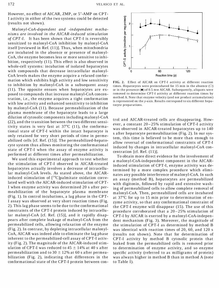
Malonyl-CoA-dependent and -independent mecha-nisms are involved in the AICAR-induced stimulationof CPT-I. It has been shown that CPT-I is reversiblysensitized to malonyl-CoA inhibition by malonyl-CoAitself [reviewed in Ref. (11)]. Thus, when mitochondriaare incubated in the absence or presence of malonyl-CoA, the enzyme becomes less or more sensitive to inhi-bition, respectively (11). This effect is also observed inwhole-cell systems: incubation of isolated hepatocyteswith compounds that decrease intracellular malonyl-CoA levels makes the enzyme acquire a relaxed confor-mation which exhibits high activity and low sensitivity FIG. 2. Effect of AICAR on CPT-I activity at different reactionto inhibition by malonyl-CoA in a subsequent assay times. Hepatocytes were preincubated for 15 min in the absence (s)
or in the presence (l) of 0.5 mM AICAR. Subsequently, aliquots were(11). The opposite ensues when hepatocytes are ex-removed to determine CPT-I activity at different reaction times byposed to compounds that increase malonyl-CoA concen-method A. Note that enzyme velocity (and not product accumulation)tration, i.e., the enzyme acquires a tight conformationis represented on the y-axis. Results correspond to six different hepa-
with low activity and enhanced sensitivity to inhibition tocyte preparations.by malonyl-CoA (11). Because permeabilization of theplasma membrane of the hepatocyte leads to a largedilution of cytosolic components including malonyl-CoA trol and AICAR-treated cells are disappearing. How-(22), and the transition between the two different sensi- ever, a constant 20–25% stimulation of CPT-I activitytivity states is very fast at 377C (11), the conforma- was observed in AICAR-treated hepatocytes up to 140tional state of CPT-I within the intact hepatocyte is s after hepatocyte permeabilization (Fig. 2). In our sys-only retained for very short periods of time in perme- tem, this time is believed to be more than enough toabilized cells [cf. Ref. (15)]. The permeabilized-hepato- allow reversal of conformational constraints of CPT-Icyte system thus allows monitoring the conformational induced by changes in intracellular malonyl-CoA con-state of CPT-I when the assay of enzyme activity is centration [cf. Ref. (11)].performed at different times after permeabilization. To obtain more direct evidence for the involvement of
We used this experimental approach to test whether a malonyl-CoA-independent component in the AICAR-the stimulation of CPT-I observed in AICAR-treated induced stimulation of CPT-I, enzyme activity was de-hepatocytes actually involves a decrease of intracellu- termined by a more complex procedure which elimi-lar malonyl-CoA levels. As stated above, the AICAR- nates any possible interference of malonyl-CoA. In suchinduced stimulation of [14C]palmitate oxidation corre- an assay (method B), hepatocytes are permeabilizedlated well with the AICAR-induced stimulation of CPT- with digitonin, followed by rapid and extensive wash-I when enzyme activity was determined 20 s after per- ing of permeabilized cells to allow complete removal ofmeabilization of the hepatocyte plasma membrane malonyl-CoA. Then, permeabilized cells are incubated(Fig. 1). In control incubations, a lag phase in the CPT- at 377C for up to 15 min prior to determination of en-I assay was observed at very short reaction times (Fig. zyme activity, so that any conformational constraint of2). This lag phase seems to be due to the conformational the CPT-I enzyme will disappear (15). The use of thisconstraints of the CPT-I protein induced by intracellu- procedure corroborated that a 20–25% stimulation oflar malonyl-CoA [cf. Ref. (15)], and it rapidly disap- CPT-I by AICAR is exerted by a malonyl-CoA-indepen-pears after complete leakage of malonyl-CoA from the dent mechanism (Fig. 3). Moreover, the magnitude ofpermeabilized cells, allowing relaxation of the enzyme the stimulation of CPT-I as determined by method B(Fig. 2). In contrast, by depleting intracellular malonyl- was identical with reaction times of 20, 60, and 120 sCoA, AICAR was indeed able to eliminate the lag phase (results not shown). Note that for determination ofinherent to the permeabilized-cell assay of CPT-I activ- CPT-I activity by method B cytosolic protein thatity (Fig. 2). The magnitude of the AICAR-induced stim- leaked from the permeabilized cells is removed priorulation of CPT-I was reduced to 45 { 14% at 40 s after to determination of enzyme activity, and so enzymepermeabilization and to 30 { 12% at 60 s after permea- specific activity (referred to as milligrams of protein)bilization (Fig. 2), indicating that differences in the was always higher in method B than in method A (note
to Table I).conformational state of the CPT-I protein between con-
AID ABB 9784 / 6b28$$$261 12-12-96 17:00:01 arcas

173CONTROL OF FATTY ACID OXIDATION BY AMP-ACTIVATED PROTEIN KINASE
AMPK) and protein phosphatases did not lead to anychange in the kinetic and regulatory properties of CPT-I (15). It has also been shown that the malonyl-CoA-independent increase of CPT-I activity observed in oka-daic acid-treated hepatocytes is not due to the directphosphorylation of the enzyme, but may involve modu-lation of interactions between CPT-I and nondiffusibleextramitochondrial components (15). Three observa-tions indicate that this may also be the case of themalonyl-CoA-independent component of the AICAR-in-duced stimulation of CPT-I:
(i) Stimulation of CPT-I was not apparent when en-zyme activity was measured in intact mitochondria iso-lated from AICAR-treated hepatocytes (method D), de-FIG. 3. Differential effect of AICAR on CPT-I activity as deter-
mined by methods A, B, C, and D. Hepatocytes were preincubated spite the presence of 50 mM fluoride in all the isolationfor 15 min in the absence or in the presence of 0.5 mM AICAR. buffers (Fig. 3). Likewise, when hepatocytes were vigor-Subsequently, aliquots were removed to determine CPT-I activity by ously disrupted by sonication after AICAR treatmentthe various methods. In the case of method B, the ghosts of perme-
(method C), the stimulatory effect of AICAR on CPT-Iabilized cells were incubated at 377C for 5 or 15 min prior to determi-was not preserved (Fig. 3). Thus, malonyl-CoA-inde-nation of enzyme activity. Reaction time was 20 s for method A and
60 s for methods B, C, and D. Results correspond to eight different pendent stimulation of CPT-I by AICAR is only evidenthepatocyte preparations. Significantly different versus incubations when mitochondria are still associated with other cellu-with no additions: *P õ 0.05; **P õ 0.01. lar fractions, i.e., in the ghosts of the permeabilized
cells, but not after separation of mitochondria fromother cell components. Therefore, it appears that themalonyl-CoA-independent stimulation of CPT-I byMalonyl-CoA-independent stimulation of CPT-I byAICAR requires components of the extramitochondrialAICAR may involve interaction of CPT-I with cytoskele-compartment of the cell.tal components. The observation that the phospha-
(ii) The possibility that cytoskeletal componentstase inhibitor okadaic acid stimulates hepatic CPT-I bymay be involved in the malonyl-CoA-independent stim-a stable mechanism led to the suggestion that inhibi-ulation of CPT-I by AICAR was tested by using taxol.tion of the phosphatases might have resulted in in-This complex diterpenoid binds to tubulin and stabi-creased phosphorylation of CPT-I, with a consequentlizes microtubules, preventing the disassembly of mi-increase of enzyme activity (7). However, incubationcrotubules in a very efficient fashion (25). Interestingly,of purified mitochondrial outer membranes or isolated
mitochondria with different protein kinases (including malonyl-CoA-independent activation of CPT-I induced
TABLE I
Combined Effects of AICAR, Okadaic Acid, and Taxol on the Activities of CPT-I and Acetyl-CoA Carboxylase
CPT-I activity (nmol/min/mg protein)Acetyl-CoA carboxylase activity
Additions Method A Method B (nmol/min/mg protein)
None (n Å 10) 1.44 { 0.36 2.37 { 0.37 0.26 { 0.14AICAR (n Å 8) 3.07 { 0.86* 2.94 { 0.31** 0.01 { 0.01*Okadaic acid (n Å 4) 2.38 { 0.46* 3.93 { 0.52* 0.03 { 0.01*Taxol (n Å 4) 1.43 { 0.23 2.35 { 0.26 0.26 { 0.02AICAR / okadaic acid (n Å 6) 4.51 { 1.15* 3.93 { 0.88* 0.01 { 0.01*AICAR / taxol (n Å 6) 2.82 { 0.40* 2.30 { 0.52 001 { 0.01*Okadaic acid / taxol (n Å 4) 1.50 { 0.26 2.44 { 0.33 0.06 { 0.03*
Note. Hepatocytes were preincubated for 20 min with or without 10 mM taxol. Incubations were continued for an additional 15 min withor without 0.5 mM AICAR and/or 0.5 mM okadaic acid. Subsequently, aliquots were removed to determine the activities of acetyl-CoAcarboxylase and CPT-I. The activity of CPT-I was assayed by methods A and B. In method A, assay time was 20 s. In method B, the ghostsof the permeabilized cells were incubated at 377C for 15 min prior to determination of enzyme activity in a 60-s assay. Results correspondto the number of hepatocyte preparations shown in parentheses.
Significantly different versus incubations with no additions: *P õ 0.01; **P õ 0.05.
AID ABB 9784 / 6b28$$$261 12-12-96 17:00:01 arcas

174 VELASCO ET AL.
by AICAR was completely abolished by pretreatment other still unidentified cytoskeletal or cytoplasmic ele-ments (27, 28). In liver cells, these interactions may inof hepatocytes with taxol (Table I, method B). Likewise,
taxol antagonized the okadaic acid-induced stimulation turn determine the permeability of the mitochondrialouter membrane to ADP (29). Mitochondria have alsoof CPT-I (Table I). It is also noteworthy that the inhibi-
tion of acetyl-CoA carboxylase by okadaic acid or been shown to contain specific binding sites for phalloi-din-stabilized F-actin (30) as well as for fodrin, an ac-AICAR was not prevented by taxol (Table I), indicating
that the antagonistic effect of taxol on okadaic acid- tin-binding protein (27). In hepatocytes and in severalcell lines, hyperphosphorylation of microtubules, actinand AICAR-induced CPT-I stimulation is independent
of changes in intracellular malonyl-CoA concentration. microfilaments, and intermediate filaments leads todisruption of the cytoskeleton (31–33). In fact, we haveHence, it seems that blockade of microtubule dynamics
prevents malonyl-CoA-independent stimulation of CPT-I recently obtained data indicating that cytoskeletalcomponents might be involved in the okadaic acid-in-by AICAR.
(iii) Stimulation of CPT-I by AICAR and okadaic duced stimulation of CPT-I (34). Whether AMPK mayphosphorylate cytoskeletal components related to theacid was nearly additive when enzyme activity was
determined by method A, but not when enzyme activity control of CPT-I activity is an intriguing possibilitywhich is currently under study in our laboratory.was determined by method B (Table I). This indicates
that the effect of AICAR observed in method A is mostly The results shown in this report must be interpretedwith caution, since AICAR may have effects apart fromdue to a malonyl-CoA-dependent mechanism, whereas
AICAR and okadaic acid seem to share a common malo- activation of AMPK. Thus, for example, ZMP mimicsthe inhibition of fructose 1,6-bis-phosphatase by AMP,nyl-CoA-independent pathway to stimulate CPT-I as
evidenced by method B. and thus treatment of hepatocytes with AICAR inhibitsgluconeogenesis (17). In other cell types such as Chi-nese hamster fibroblasts and ovary cells, exposure to
CONCLUDING COMMENTS AICAR has been shown to alter the levels of differentpurine and pyrimidine nucleotides (35–37). However,In the present report we show that exposure of intactas discussed by Corton et al. (16), none of these ancil-hepatocytes to AICAR produces a strong (twofold) stim-lary changes in nucleotides are evident in rat hepato-ulation of long-chain fatty acid oxidation. This effectcytes. In the latter, AICAR seems to be a rather specificseems to rely on the activation of CPT-I by two differentactivator of AMPK or at least the most specific com-mechanisms. On the one hand, AMPK-induced acetyl-pound for activation of AMPK available to date (16).CoA carboxylase inactivation would lead to the deple-
The AICAR-induced malonyl-CoA-dependent dein-tion of intracellular malonyl-CoA, with concomitanthibition of CPT-I is quantitatively more important thandeinhibition of CPT-I. This mechanism, which has beenthe malonyl-CoA-independent deinhibition of enzymeproposed to operate in the ischemic heart (4, 5) and theactivity. Nonetheless, the latter data are actuallyworking muscle (6), seems to make a major contribu-highly reproducible and statistically significant (Tabletion (ca. 80%) to the AICAR-induced stimulation of he-I). It is remarkable that the two mechanisms operatepatic long-chain fatty acid oxidation. On the othersimultaneously in the short-term control of hepatichand, AICAR induces a 20–25% stimulation of CPT-ICPT-I activity, especially since AICAR is not expectedby a stable, malonyl-CoA-independent mechanism. Weto produce any stable (covalent) modification of CPT-Ihave previously reported that incubation of purifiedor the putative regulatory proteins of the cytoskeleton.mitochondrial outer membranes or isolated mitochon-It is also worth noting that when CPT-I activity is mea-dria with AMPK under phosphorylation conditionssured by method B to determine the malonyl-CoA-inde-does not lead either to the phosphorylation of CPT-I orpendent component of the AICAR effect, cells are exten-to any change in the kinetic and regulatory propertiessively washed and further incubated for up to 15 minof CPT-I (15). Instead, data in this paper suggest thatat 377C. Hence, if the malonyl-CoA-independent dein-malonyl-CoA-independent modulation of CPT-I activ-hibition of CPT-I activity relies on protein–protein in-ity might rest on the modulation of interactions be-teractions, these may partially disappear after the po-tween CPT-I and cytoskeletal components. It has beentential modifications of the enzyme microenvironment.established that the dynamics of mitochondria in livingTherefore, it is conceivable that malonyl-CoA-indepen-cells (shape changes, dislocation, and fusion and fis-dent control of CPT-I activity may be quantitativelysion) may result from specific interactions of mitochon-more important within the cell than is evident underdria with components of the cytoskeleton (26). In addi-the conditions of our assay.tion, it has been shown that porin and hexokinase are
localized in domains of the mitochondrial outer mem- ACKNOWLEDGMENTSbrane which interact specifically with microtubule- and This study was financially supported by a grant from the Spanish
CICYT (SAF93/0281). We also acknowledge the support in part bymicrofilament-associated proteins, as well as with
AID ABB 9784 / 6b28$$$261 12-12-96 17:00:01 arcas

175CONTROL OF FATTY ACID OXIDATION BY AMP-ACTIVATED PROTEIN KINASE
the Netherlands Foundation for Chemical Research (SON) with fi- 19. Beynen, A. C., Vaartjes, W. J., and Geelen, M. J. H. (1979) Diabe-tes 28, 828–835.nancial aid from the Netherlands Organization for Scientific Re-
search (NWO). 20. Holland, R., Witters, L. A., and Hardie, D. G. (1984) Eur. J. Bio-chem. 140, 325–333.
21. Declercq, P. E., Falck, J. R., Kuwajima, M., Tyminski, H., Foster,REFERENCESD. W., and McGarry, J. D. (1987) J. Biol. Chem. 262, 9812–9821.
1. Hardie, D. G. (1992) Biochim. Biophys. Acta 1123, 231–238. 22. Guzman, M., and Geelen, M. J. H. (1988) Biochem. Biophys. Res.2. Woods, A., Munday, M. R., Scott, J., Yang, X., Carlson, M., and Commun. 151, 781–787.
Carling, D. (1994) J. Biol. Chem. 269, 19509–19515. 23. Bijleveld, C., and Geelen, M. J. H. (1987) Biochim. Biophys. Acta3. Henin, N., Vincent, M. F., Gruber, H. E., and van den Berghe, 918, 274–283.
G. (1995) FASEB J. 9, 541–546. 24. Lowry, O. H., Rosebrough, N. J., Farr, A. L., and Randall, R. J.4. Kudo, N., Barr, A. J., Barr, R. L., Desai, S., and Lopaschuk, G. D. (1951) J. Biol. Chem. 193, 265–275.
(1995) J. Biol. Chem. 270, 17513–17520. 25. Nogales, E., Grayer Wolf, S., Khan, I. A., Luduena, R. F., andDowning, K. H. (1995) Nature 375, 424–427.5. Kudo, N., Gillespie, J. G., Kung, L., Witters, L. A., Schulz, R.,
Clanachan, A. S., and Lopaschuk, G. D. (1996) Biochim. Biophys. 26. Bereiter-Hahn, J., and Voth, M. (1994) Microsc. Res. Technol.Acta 1301, 67–75. 27, 198–219.
6. Winder, W. W., and Hardie, D. G. (1996) Am. J. Physiol. 270, 27. Leterrier, J. F., Rusakov, D. A., Nelson, B. D., and Linden, M.E299–E304. (1994) Microsc. Res. Technol. 27, 233–261.
7. Guzman, M., and Geelen, M. J. H. (1993) Biochim. Biophys. Acta 28. Linden, M., Nelson, B. D., Loncar, D., and Leterrier, J. F. (1989)1167, 227–241. Biochem. J. 261, 167–173 (Erratum, Biochem. J. 262, 1002,
1989).8. Lopaschuk, G. D., Belke, D. D., Gamble, J., Itol, T., and Schone-29. Fontaine, E. M., Keriel, C., Lantuejoul, S., Rigoulet, M., Leverve,kess, B. O. (1994) Biochim. Biophys. Acta 1213, 263–276.
X. M., and Saks, V. A. (1995) Biochem. Biophys. Res. Commun.9. Bulow, J. (1993) Med. Sport Sci. 38, 158–185.213, 138–146.10. McGarry, J. D., Woeltje, K. F., Kuwajima, M., and Foster, D. W.
30. Lazzarino, D. A., Boldogh, I., Smith, M. G., Rosand, J., and Pon,(1989) Diabetes Metab. Rev. 5, 271–284.L. A. (1994) Mol. Biol. Cell 5, 807–818.
11. Zammit, V. A. (1994) Diabetes Rev. 2, 132–155.31. Yano, Y., Sakon, M., Kambayashi, J., Kawasaki, T., Senda, T.,
12. Guzman, M., and Castro, J. (1991) FEBS Lett. 291, 105–108. Tanaka, K., Yamada, F., and Shibata, N. (1995) Biochem. J. 307,13. Guzman, M., and Geelen, M. J. H. (1992) Biochem. J. 287, 487– 439–449.
492. 32. Seglen, P. O., and Bohley, P. (1992) Experientia 48, 158–172.14. Guzman, M., Velasco, G., and Castro, J. (1996) Am. J. Physiol. 33. Ohta, T., Nishiwaki, R., Yatsunami, J., Komori, A., Suganuma,
270, G701–G707. M., and Fujiki, H. (1992) Carcinogenesis 13, 2443–2447.15. Guzman, M., Kolodziej, M. P., Caldwell, A., Costorphine, C. G., 34. Velasco, G., Sanchez, C., Geelen, M. J. H., and Guzman, M.
and Zammit, V. A. (1994) Biochem. J. 300, 693–699. (1996) Biochem. Biophys. Res. Commun. 224, 754–759.16. Corton, J. M., Gillespies, J. G., Hawley, S. A., and Hardie, D. G. 35. Thomas, C. B., Meade, J. C., and Holmes, E. W. (1981) J. Cell
(1995) Eur. J. Biochem. 229, 558–565. Physiol. 107, 335–344.17. Vincent, M. F., Marangos, P. J., Gruber, H. E., and Van den 36. Sabina, R. L., Holmes, E. W., and Becker, M. A. (1984) Science
Berghe, G. (1991) Diabetes 40, 1259–1266. 223, 1193–1195.37. Sabina, R. L., Patterson, D., and Holmes, E. W. (1985) J. Biol.18. Sullivan, J. E., Brocklehurst, K. J., Marley, A. E., Carey, F., Car-
ling, D., and Beri, R. K. (1994) FEBS Lett. 353, 33–36. Chem. 260, 6107–6114.
AID ABB 9784 / 6b28$$$261 12-12-96 17:00:01 arcas