Embed Size (px)
Citation preview

Review
Control of Proliferation and Cancer Growth bythe Hippo Signaling PathwayUrsula Ehmer1,2,3 and Julien Sage1,2
Abstract
The control of cell division is essential for normal devel-opment and the maintenance of cellular homeostasis. Abnor-mal cell proliferation is associated with multiple pathologicalstates, including cancer. Although the Hippo/YAP signalingpathway was initially thought to control organ size andgrowth, increasing evidence indicates that this pathway alsoplays a major role in the control of proliferation indepen-dent of organ size control. In particular, accumulatingevidence indicates that the Hippo/YAP signaling pathway
functionally interacts with multiple other cellular pathwaysand serves as a central node in the regulation of cell division,especially in cancer cells. Here, recent observations arehighlighted that connect Hippo/YAP signaling to transcrip-tion, the basic cell-cycle machinery, and the control of celldivision. Furthermore, the oncogenic and tumor-suppressiveattributes of YAP/TAZ are reviewed, which emphasizes therelevance of the Hippo pathway in cancer. Mol Cancer Res; 14(2);127–40. �2015 AACR.
IntroductionA tight control of cell division is essential for the mainte-
nance of homeostasis in adult organs and tissues. Severalcellular mechanisms exist to regulate and fine-tune cell prolif-eration during physiologic growth and regeneration. Failures inone or more of these control mechanisms can result inunchecked cell divisions and, eventually, cancer development(1). A number of cellular pathways have been implicated in theregulation of cell division. In particular, the importance of theso-called "Hippo" signaling pathway in the control of prolif-eration was discovered a little more than 10 years ago inDrosophila (2, 3). Since then, it has become evident that theHippo signaling pathway is involved in a plethora of cellularfunctions, ranging from organ size control to cellular differen-tiation and metabolism (4–7). However, the control of cellproliferation—mainly by negatively regulating the pathway'sdownstream effectors YAP and TAZ—remains one of its centralfunctions (8). In this review, we provide a detailed overview ofthe role of the Hippo pathway in the control of proliferationand summarize recently published data that shed light on themolecular basis for Hippo-mediated cell-cycle control. Finally,we discuss the implication of these findings for our under-standing of the role of Hippo signaling in cancer and for thedevelopment of novel tumor therapies.
The Canonical Hippo/YAP SignalingPathway: A Brief Overview
The first big steps in our understanding of Hippo pathwayfunction were made in flies. Although many pathway functionsare conserved between flies and mammals (9), for historic rea-sons, mammalian and Drosophila orthologues differ in theirrespective names. With few exceptions, we use here the mamma-lian nomenclature and focus onmammalian systems. This shouldby no means make the major advances in the Drosophila field lessimportant.
The backbone of the Hippo signaling pathway consists of akinase cascades that works to control the activity of thedownstream effectors YAP and TAZ. The upstream kinases ofthe Hippo signaling pathway, MST1 and MST2, work togetherwith the adaptor protein SAV1/WW45 to phosphorylate andactivate LATS1 and LATS2. The activated LATS kinases togetherwith MOB1 then phosphorylate the YAP and TAZ effectorproteins (Fig. 1). This phosphorylation event results in thenuclear exclusion of YAP/TAZ mediated by 14-3-3 proteinsand, ultimately, their cytoplasmic degradation (5, 10). How-ever, not all cytoplasmic YAP/TAZ is degraded and the phos-phorylated, cytoplasmic proteins can associate with proteincomplexes of other pathways such as WNT or TGFb signalingto modify signaling through these pathways (11, 12). Never-theless, the main functions of YAP and TAZ can be attributed totheir unphosphorylated, nuclear state, where the proteinsfunction as transcriptional cofactors that can interact withseveral transcription factors. Their major transcriptional bind-ing partners are members of the TEAD/TEF transcription factorfamily (TEAD1-4) that are believed to mediate the majorityof pro-proliferative and oncogenic functions of YAP andTAZ (13).
Apart from so-called "canonical" Hippo signaling, severalcellular components independent of the classic pathway cancontrol YAP and TAZ. Importantly, the differential regulation islikely tissue specific (14). Particularly, the requirement of all
1Department of Pediatrics, Stanford University School of Medicine,Stanford, California. 2Department of Genetics, Stanford UniversitySchool of Medicine, Stanford, California. 3Department of Medicine II,Klinikum rechts der Isar, Technische Universit€at M€unchen, 81675Munich, Germany.
Corresponding Author: Ursula Ehmer, Klinikum rechts der Isar, TechnischeUniversit€at M€unchen, II. Medizinische Klinik und Poliklinik, Ismaninger Strasse22, 81675 Munich, Germany. Phone: 49-89-4140-4982; Fax: 49-89-4140-4910;E-mail: [email protected]
doi: 10.1158/1541-7786.MCR-15-0305
�2015 American Association for Cancer Research.
MolecularCancerResearch
www.aacrjournals.org 127
on July 13, 2018. © 2016 American Association for Cancer Research. mcr.aacrjournals.org Downloaded from
Published OnlineFirst October 2, 2015; DOI: 10.1158/1541-7786.MCR-15-0305

kinases of the coreHippo kinase cascade in the control of YAP andTAZ activity does not seem to be conserved in all mammalian celltypes. Although MST1/2 control the phosphorylation of YAP inthe liver, the function of the upstream Hippo kinases is dispens-able for YAP activation in mouse embryonic fibroblasts, kerati-nocytes, and possibly also melanocytes (15–17). Furthermore,the functional requirement of either YAP or TAZ varies betweendifferent cell types. Although many studies specifically highlightthe role of YAP in tumorigenesis—or consider YAP and TAZ asfunctionally redundant—at least in some cancers such as breastcancer, melanoma, or glioblastoma, TAZmay play a more impor-tant role (18–20).
Cellular Functions of Hippo SignalingIn many tissues, YAP and TAZ regulate key cellular functions
that include—but are not limited to—proliferation control, inhi-bition of apoptosis, and promotion ofmetastasis (2, 3, 18, 21). Bynegatively regulating oncogenic YAP and TAZ activities, thekinases of the Hippo pathway work as important tumor-suppres-sivemolecules. Inmany cancers, Hippo signaling is dysfunctionaland activation of YAP and/or TAZ is observed, with high levels ofnuclear proteins; in addition, direct genetic mutations in Hippopathway genes are not very frequent, with the exception of NF2(reviewed in ref. 22). This uniquemutation pattern forNF2 could
© 2015 American Association for Cancer Research
Molecular Cancer Research Review
TEAD
SMAD2/3
RUNX2
PAX3
β-catenin
TBX5
EGFR
NF2(Merlin)
PDK1
β-TrCP
β-catenin
Wnt
MST1/2
LATS1/2
SAV1
MOB
GPCR
RhoA
?
F-actin
AMOT
Degradation
Degradation
Proliferation
Proliferation, growth
ABL1
p73PP1A Apoptosis
PP2A
PP
P
P
SMAD2/3
YAP
PYAP
YAP
YAP
YAP
14-3-3
AKTCTGFBIRC5AREGMCM6YAP
TAZ
TAZP
TAZ
TAZ
TAZ
YAP
TAZ
YAP
TAZ
YAP
TAZAXIN
GSK3APC
Notch
Shh
PI3K
Figure 1.An overview of the regulation of transcription by YAP/TAZ transcriptional cofactors and the regulation of YAP/TAZ activity in mammalian cells. YAP/TAZ aredownstream mediators of numerous signaling pathways in cells, including from the cell surface. Their transcriptional targets have been involved in theregulation of proliferation and growth, but also cell death and differentiation.
Ehmer and Sage
Mol Cancer Res; 14(2) February 2016 Molecular Cancer Research128
on July 13, 2018. © 2016 American Association for Cancer Research. mcr.aacrjournals.org Downloaded from
Published OnlineFirst October 2, 2015; DOI: 10.1158/1541-7786.MCR-15-0305

either suggest that loss of NF2 function results in additionaloncogenic effects beyond inactivating Hippo signaling; alterna-tively, there may be an optimal level of altered Hippo pathwayactivity to promote cancer development and losing NF2 mayachieve this oncogenic level in cancer cells. Activation of Hip-po/YAP signaling can also be achieved through alterations inmany of the pathways that functionally interact with Hipposignaling (see below), which again may lead to just enoughperturbation in the pathway to be oncogenic. Nevertheless, moreandmore genetic and genomic alterations are being discovered inHippo pathway members, including amplification of the YAPgene (23, 24) or gene fusion events involving multiple pathwaymembers (25); it is also possible that a number of epigeneticevents lead to silencing of tumor-suppressive elements in thepathway (26).
Aside from its role in cancer, the physiologic role of Hipposignaling is no less important: by being involved in the mainte-nance of tissue specific stem cells, tissue regeneration, woundhealing as well as in the control of differentiation (6, 27–33), thispathway is a major player in the control of embryonic develop-ment and tissue homeostasis. YAP-deficientmouse embryos die atE8.5 with multiple developmental defects (34). Knockout ofMst1/2 or Sav1 results in liver overgrowth and the developmentof hepatocellular carcinoma (35, 36), a phenotype that is alsoobserved in transgenicmice overexpressing YAP (4, 37).However,organ size control only presents one outcome of deregulatedHippo signaling. The direct effects of unleashed YAP/TAZ activa-tion are tissue and often cell-type specific, but the control of cellproliferation remains a common theme. Interestingly, progenitorcell compartments seem to be especially sensitive to the loss ofHippo control mechanisms, including in the liver (36, 37), theintestine (37, 38), the nervous system (39), the skin, and the lung(40). Several studies further indicate that YAP and TAZ areinvolved in the maintenance of a stem cell phenotype and caninhibit cellular differentiation (6, 27, 32, 41, 42). Key functionsfor the Hippo signaling in the biology of stem/progenitor cellsinclude the regulation of cell cycle and interactions with othersignaling pathways (e.g., Hedgehog,Wnt, orNotch; see below). Inaddition, this control of stem cells byHippo/YAP signaling can beachieved in a cell-intrinsic manner but also by controlling thestem cell niche, as has been described in flies (43) and inmammals (44).
Keeping the balance between promotion of physiologic pro-liferation and prevention of unrestricted cell divisions that lead tocancer development is a key function of Hippo signaling andmakes it a promising therapeutic target in many human pathol-ogies. For instance, promotion of downstreamYAPor TAZ activitycould be useful to promote tissue regeneration; in contrast,strategies to activate of Hippo signaling or inactivate YAP/TAZmay help treat certain cancers (Fig. 2).
Regulation of Hippo SignalingThe core Hippo kinase cascade functions as a central hub that
relays input from the "outside world" of the cell and translates itinto specific cellular responses by controlling the activity ofdownstream YAP/TAZ and by interacting with other pathwayssuch as Wnt/b-catenin, Notch, AKT, or Hedgehog signaling (45–50). Although the regulation by extracellular receptors is sharedby many signaling pathways, Hippo signaling seems to be espe-cially sensitive to input from mechanical cues. How signals from
beyond the cell's borders feed into the Hippo signaling cascadehas been extensively studied over the recent years and is reviewedin detail elsewhere (14). Below, we provide an overview of themost important regulators.
The long quest to find receptors that directly activate or inac-tivate Hippo signaling only recently led to the identification of G-protein–coupled receptors (GPCR) as regulators of this pathway(51, 52). GPCRs are integral membrane receptors that are acti-vated by external ligands whose binding leads to activation of thealpha-subunit of one or more of the four subclasses of G proteinsGai/0, Ga12/13, Gas, and Gaq/11. In the regulation of Hippo sig-naling, activation of GPCRs linked to Gas results in the phos-phorylation of LATS and therefore inactivation of YAP. On theother hand, activation of Ga12/13-coupled GPCRs is correlatedwith inhibition of LATS and activation of YAP (51). Additionally,a role for mutated Gaq/11 in uveal melanoma carcinogenesismediated by YAP has been reported (53). How GPCR signalingtransduces into activation or inactivation of YAP is not wellunderstood, but at least for some GPCRs these effects seem tobe mediated by Rho GTPases and/or alterations in F-actin poly-merization (51, 52, 54). Targeting this large class of cell-surfacereceptorsmaybe apromising approach tomodifyHippopathwayactivity in cancer or regeneration.
Other membrane receptors that have been shown to regulateYAP expression and proliferation are the receptor tyrosine kinasesEGFR and ERBB4. Treatment with EGF leads to EGFR-mediatedactivation of PI3K and PDK1, which connects to the Hippopathway through the adaptor protein SAV1/WW45. PDK1 acti-vation triggers the dissociation of theHippo core kinases from thescaffolding protein SAV1/WW45 and results in inactivation ofLATS, dephosphorylation and nuclear translocation of YAP, andexpression of YAP target genes (55, 56). Interestingly, one of theYAP targets identified is the EGFR ligand amphiregulin (AREG),providing a positive feedback loop between YAP and EGFRsignaling that can work to amplify proliferative signals from bothsignaling pathways and also trigger cell proliferation in a non–cell-autonomous manner (57). Another member of the EGRreceptor family, ERBB4, has been shown to trigger YAP activationand expression of YAP target genes throughdirect interaction of itssoluble intracellular domain with YAP and possibly also TEAD1in the nucleus (58). Most likely, future research will identifyadditional receptors that feed signals into the Hippo signalingcascade. Manipulating these receptors to specifically alter Hipposignaling will then present the next big challenge.
In addition to receptor-mediated regulation, the Hippo signal-ing pathway receives multiple "mechanical" inputs from the cellsurface as well as from within the cell (reviewed in refs. 59, 60).The mechanical cues involved in the regulation of Hippo signal-ing include proteins that bind to intercellular junctions such astight and adherence junctions as well as components of thecytoskeleton, including F-actin, microtubules, actomyosin, andpossibly centrosomes (61–66). Specifically, the interactions withcentrosomes may also be relevant to the regulation of cell-cycleprogression by Hippo signaling. It is highly likely that thesebiomechanical regulators are also involved in the promotion ofoncogenic YAP and TAZ activity in cancer. It is tempting tospeculate that changes of the extracellular matrix found in cancer(67, 68) might influence cell proliferation through mechanicalregulation of Hippo pathway effectors (59, 69). Additionally,components of the cytoskeleton are often deregulated in cancercells, which could possibly contribute to the activation of YAP/
Hippo/YAP in Cell Cycle and Cancer
www.aacrjournals.org Mol Cancer Res; 14(2) February 2016 129
on July 13, 2018. © 2016 American Association for Cancer Research. mcr.aacrjournals.org Downloaded from
Published OnlineFirst October 2, 2015; DOI: 10.1158/1541-7786.MCR-15-0305

TAZ in carcinogenesis. In cancer-associated fibroblasts, matrixstiffening enhances YAP activation—which in turn promotesexpression of cytoskeletal regulators involved in remodeling ofthe extracellular matrix (69). Additionally, the connection ofintercellular junction-associated proteins to the Hippo pathwayseems to be essential for the maintenance of apical–basal cellpolarity (14). Several well-established upstream regulators ofHippo signaling, such as Merlin, Kibra, Expanded, the Angiomo-tin proteins, E-cadherin, and a-catenin, are known to associatewith tight and adherence junctions or other cell polarity com-plexes (16, 70–73). Alterations in this regulatory network may be
linked to the disruption of cell polarity that is linked to cancerprogression and invasiveness (74).
One of the best-studied upstream regulators of the Hipposignaling pathway is the apical membrane protein NF2/Mer-lin—a well-described tumor suppressor inactivated in many can-cers (75). In mammals as well in flies, NF2/Merlin can interactwith theHippo signaling pathway onmultiple levels (reviewed inref. 75). Importantly, Merlin directly binds and recruits the LATSkinases to the plasma membrane to promote their phosphoryla-tion and activation by MST1/2 and SAV1 (76). An additionalmechanism to directly regulate LATS activity is mediated by
Molecular Cancer Research Review
© 2015 American Association for Cancer Research
OncogenicYAP/TAZ
Tumor suppressiveYAP/TAZ
Brain:YAP functions as an oncogene inmedulloblastoma, glioblastoma,and meningioma
Breast:YAP has been shown topromote anoikis and toinhibit metastasisin breast cancer
Blood:YAP promotesABL1-inducedapoptosis in hematologicmalignancies
Intestine:Inhibition of colorectalcancer through inhibitionof Wnt signaling bycytoplasmic YAP
Breast:TAZ promotes drug resistancein breast cancer therapy.Both YAP and TAZ are linkedto increased proliferationand metastasis in breast cancer
Lung:YAP expression correlateswith survival in non-small celllung cancer and synergizeswith Kras to promote EMT
Liver:Inactivation of Hipposignaling and activationof YAP both induce liverovergrowth and cancerdevelopment
Bone:Up-regulation of YAPdownstream of deregulatedHedgehog signalingcontributes to carcinogenesisin osteosarcoma
Skin:YAP and TAZ enhanceproliferation and metastasisin melanoma
Figure 2.Oncogenic and tumor-suppressivefunctions of YAP and TAZ inhuman cancer (86, 151, 162). Seetext for details.
Ehmer and Sage
Mol Cancer Res; 14(2) February 2016 Molecular Cancer Research130
on July 13, 2018. © 2016 American Association for Cancer Research. mcr.aacrjournals.org Downloaded from
Published OnlineFirst October 2, 2015; DOI: 10.1158/1541-7786.MCR-15-0305

phosphorylatedMerlin that inhibits the tumorigenic effects of theE3 ubiquitin ligase CRL4-DCAF1 in the nucleus (77). Bound byMerlin, CRL2-DCAF1 is not able to ubiquitinate and inactivateLATS1/2 to promote YAP activity (78). Furthermore, Merlin canbind to other cell membrane and junction proteins, such asAngiomotin and a-catenin, and probably influences their inter-action with the Hippo signaling kinases (reviewed in ref. 75).Interestingly, tumor development upon NF2 inactivation in theliver and other tissues is suppressed by heterozygous deletion ofYAP, supporting the idea that NF2/Merlin inhibits proliferationby repression of YAP activity (79, 80).
The Angiomotin family of proteins AMOT (Angiomotin),AMOTL1 and AMOTL2 interact with tight junction proteins aswell as the actin cytoskeleton and play an important role in themaintenance of cell polarity. With exception of the AMOT-p80variant, all members of Angiomotin protein family can directlybind to YAP and TAZ and inhibit their activity, either by recruit-ment to the extranuclear environment or by inducing YAP/TAZphosphorylation (72, 81, 82). Interestingly, several recent reportsshow that AMOT proteins themselves are targets of the LATS1/2kinases (83, 84). Phosphorylation by LATS1/2 negatively regu-lates actin binding of Angiomotins and increases their ability toinhibit YAP (82, 84), providing an additional mechanism forLATS kinases to negatively regulate downstream YAP. Anotherconnection between Hippo signaling and intercellular junctionsis the adherence junction protein a-catenin, which is able todirectly bind and inhibit phosphorylated YAP by facilitating itssequestration in complex with 14-3-3 proteins (16). This mech-anism restricts proliferation in several different cell types andseems important to mediate the tumor suppressor activity ofa-catenin (12, 16, 85).
Other cell junction proteins or proteins involved in the main-tenance of apical–basal polarity such as SCRIB, PTPN14, LIN7C,PATJ, andMPDZ or E-cadherin, Crumbs, ZO1, ZO2, NPHP4, andLKB1, respectively, interact with the Hippo signaling cascade ondifferent levels (reviewed in refs. 14 and 86).
Sensing inputs from neighboring cells, the extracellular matrix,or the cytoskeleton is key to trigger several cellular responses, butmost importantly one decision—to grow and divide or to remainquiescent. Clearly, integration of these signals plays a key role inthe activity of the Hippo signaling pathway. Failures in thetranslationof these cues from theoutsideworld canbe deleteriousto the maintenance of tissue integration and furthermore under-line the importance of tight control mechanisms in this process.
The Many Nodes That Connect to HippoSignaling
In addition to the multitude of upstream inputs to Hipposignaling there are several pathways that directly interact withthe more downstream components of the signaling cascade tomodify the output of Hippo signaling.
Themost well-investigated partner of the Hippo pathway is theWnt signaling pathway. Binding ofWNT ligands to their receptorsin the cell membrane results in phosphorylation of the b-catenindegradation complex. The phosphorylation of this complex uponWnt activation, however, leads to stabilization ofb-catenin and itstranslocation into the nucleus, where it associates with TCFtranscription factors to promote the expression ofWNT/b-catenintarget genes (87). A large body of data now indicates that cyto-plasmic YAP and TAZ can inhibit Wnt pathway activation—
mainly through interaction with the b-catenin destructioncomplex.
A first study that gave evidence for a connection between Wntand Hippo signaling showed that phosphorylated, cytoplasmicTAZ binds to DVL, a member of the b-catenin degradationcomplex. TAZ binding inhibits the phosphorylation of DVL byCK1d/e upon Wnt activation, thereby stabilizes the b-catenindestruction complex and prevents the translocation of b-catenininto the nucleus (46). Interestingly, the b-catenin degradationcomplex also seems to be involved in the degradation of TAZ inthe cytoplasm, as it mediates the interaction of phosphorylatedTAZ with its ubiquitin ligase b-TrCP (88). Compelling evidencealso indicates that both YAP and TAZ are integral components ofthe b-catenin destruction complex and are required for the over-growth phenotype in APC-deficient intestines (11). In addition,phosphorylated YAP/TAZ can directly bind to b-catenin to sup-press its nuclear translocation in colorectal cancer cells (89). Athird mechanism by which cytoplasmic YAP can inhibit Wntsignaling output is by restricting DVL activity independent of theb-catenin degradation complex, likely through inhibition of thetranscriptional coactivator function of DVL (44). This lattermechanism seems to be highly important to counterbalanceactivated Wnt signaling in regenerating intestinal stem cells, butalso has implications in carcinogenesis as reexpression of previ-ously silenced YAP can inhibit cell proliferation in xenograftmodels of an aggressive subtype of human colorectal carcinomas(44). These findings indicate that YAP might have importanttumor-suppressive functions under certain conditions that arerelated to activated Wnt/b-catenin signaling. However, the role ofYAP in cancers with activated b-catenin is not fully understoodand likely highly context dependent: in a screen in b-catenin–activated cancer cell lines, YAP was essential for tumorigenicity(90). Mechanistically, YAP binds to the transcription factor TBX5in a complex with b-catenin to induce the transcription of pro-oncogenic and antiapoptotic target genes independent of TCFtranscription factors. The oncogenic activity of YAP may requirenuclear translocation and likely relies on tyrosine phosphoryla-tion by the YES protein kinase independent of canonical Hipposignaling (90). A similar interaction was observed in mice withcardiac deletion of Sav, where increased nuclear YAP was associ-ated with cardiac overgrowth that was dependent on Wnt signal-ing (45). In chromatin immunoprecipitation assays, both YAPand b-catenin were shown to bind to common targets, such asSnai2 and Sox2, to enhance their transcription and contribute toincreased proliferation in cardiomyocytes (45). The contradictingfindings on the interplay between YAP/TAZ and Wnt signalingshow that multiple factors might influence the outcome observedin Wnt-activated cells. To understand the mechanisms that divertYAP/TAZ activity toward tumor suppression or oncogenesis pre-sents a major challenge and will be of high importance fortherapeutic targeting of Hippo signaling.
Long before LATS kinaseswere identified as themain regulatorsof YAP/TAZ activation, AKT was shown to phosphorylate YAP atSer127, resulting in nuclear exclusion and degradation by 14-3-3proteins similar to the effect observed after LATS-mediated phos-phorylation (91). A recent publication indicates that YAP is able topromote PI3K–AKT signaling in cardiomyocytes by direct tran-scriptional activation of the catalytic PI3K-subunit p110b (92).This activation of AKT signaling is associated with increasedsurvival and proliferation in cardiomyocytes and can at least inpart explain the promotion of cardiac regeneration by YAP (93)
Hippo/YAP in Cell Cycle and Cancer
www.aacrjournals.org Mol Cancer Res; 14(2) February 2016 131
on July 13, 2018. © 2016 American Association for Cancer Research. mcr.aacrjournals.org Downloaded from
Published OnlineFirst October 2, 2015; DOI: 10.1158/1541-7786.MCR-15-0305

that is counteracted byoverexpressionofMst1, resulting indilatedcardiomyopathy (94). Recently, a cross-talk between mTOR andYAP has been described in TSC1/2-mutant cells. In this system,hyperactive mTOR results in activation of YAP—most likely in anautophagy-dependent manner—to promote proliferation andtumor formation (95).
There is strong evidence that the Notch pathway is activated byYAP through direct transcriptional targeting of JAG1 in the liver(27, 50). Additionally, high activity of YAP inMst1/Mst2-deficientintestinal epithelial cells correlates with activation of Notchsignaling (96). Although these finding indicate that Notch sig-naling is downstream of Hippo/YAP, the two pathways can alsoact in concert by regulating expression of CDX2 in the trophecto-derm (97). Notch signaling can also inhibit theDrosophila homo-log of the YAP/TAZ-TEAD complex—Yki/Sc—under specific con-ditions (98). Therefore, the interaction betweenHippo andNotchsignaling is likely context dependent and further research isneeded to investigate the interaction between these twopathways.
A connection between Hedgehog signaling and YAP has beenidentified in medulloblastoma and neural precursors, whereSonic hedgehog (SHH) promotes nuclear accumulation of YAPand results in increased proliferation (49). On the other hand,activation of SHH inversely correlates with the expression ofnuclear YAP in pancreatic adenocarcinoma (99). Gli2, a majortranscriptional mediator of Hedgehog signaling, has been iden-tified as a transcriptional target of YAP/TEAD in neural precursors(49), while YAP can bind to and inhibit GLI transcription factorsin fibroblasts, resulting in decreased expression of Hedgehogpathway targets (99). Importantly, Hippo upstream kinases aswell as YAP/TAZ are involved ciliogenesis of the primary cilium(100, 101), an organelle of the plasma membrane that is animportant determinant of Hedgehog signaling (and other signal-ing pathways). These findings indicate that the interactionbetween Hippo and Hedgehog signaling is highly complex andtissue- as well as context dependent.
In summary, the downstream effectors of Hippo signalinginteract with many key pathways that play important roles incell proliferation and also oncogenesis. In many cases, theseinteractions might contribute to the amplification of the pro-proliferative output of YAP/TAZ signaling. As this pathway acti-vation often seems to be dependent on the transcriptional activityof YAP/TAZ, it might be targetable by specific inhibition of YAP/TAZ-TEAD-mediated transcription. However, some interactionsseem to function outside canonical Hippo signaling. Their role incancer as well as possible means to target these interactions toinhibit proliferation or promote regeneration remain to beinvestigated.
Fine-tuning YAP/TAZ ActivityThemost important regulators of YAP/TAZ are LATS kinases. By
phosphorylation at one or more serine residues, LATS1/2 nega-tively regulate the transcriptional activity of YAP and TAZ. Inaddition to LATS1/2, other proteins such as AKT and JNK canmediate YAP serine phosphorylation (91, 102). PhosphorylatedYAP/TAZ proteins are retained in the cytoplasm, where they aretargeted for degradation by 14-3-3 protein. Interestingly, YAPmethylation by SET7 also results in cytoplasmic retention anddecreased YAP transcriptional activity, amechanism that seems tobe of relevance in the maintenance of tissue homeostasis in theintestine (103). Another level of YAP regulation is mediated by
tyrosine phosphorylation by YES, SRC, or c-ABL kinases (90, 104–106). However, the biologic effects of tyrosine phosphorylationseem to be context dependent and can divert YAP transcriptionalactivity toward expression of pro- or antiapoptotic genes, respec-tively (90, 104).
Because phosphorylation presents such an important deter-mining factor of YAP/TAZ activity, it is highly likely that one ormore proteins exist that control their dephosphorylation. Recent-ly, PP1A and PP2A have been identified as phosphatases of YAPand TAZ that promote nuclear translocation and expression oftarget genes (16, 107, 108). Further research is needed to deter-mine the in vivo relevance of PP1A, PP2A, and other Hippopathway phosphatases, including those interacting with kinasesin the pathway (109–111). The search for phosphatases in theHippo pathway is an area of active investigation that will givenovel insights into the mechanisms regulating Hippo activity.Ultimately, inhibition of oncogenic phosphatases in the Hippopathway could also provide new therapeutic options.
Transcriptional Control of ProliferationThe range of biologic functions of the YAP and TAZ effector
proteins we know about is ever expanding. YAP and TAZ can bindto several different transcription factors, each likely initiatingtranscriptional programs that contribute to the functional diver-sity of active YAP/TAZ (14). In general, transcription factor bind-ing requires nuclear translocation of dephosphorylated YAP andTAZ.Upon phosphorylation by LATS aswell as other kinases, YAPand TAZ are retained in the cytoplasm and become transcription-ally inactive, resulting in decreased expression of their targetgenes.
The main binding partners of transcriptionally active YAP andTAZ in the nucleus are TEA domain (TEAD) transcriptions factors(112). They mediate the majority of proliferative and oncogenicfunctions of YAP and TAZ (13, 113, 114). The TEAD family oftranscription factors consists of four highly homologous proteinsin humans and mice. TEAD1 is expressed in most adult tissues,while the expression of the other members of the protein familyvaries considerably between different tissues and developmentalstages (115). All TEADs share almost identical DNA-bindingdomains and seem to bind to the same DNA binding motif withcomparable affinity (115). However, binding to different TEADproteins may explain at least some of the tissue-specific outcomesthat are observed upon YAP/TAZ activation.
Overexpression studies lead to the identification of an array ofYAP and TAZ transcriptional targets, including CTGF, CYR61,BIRC5, AREG, and others. These genes are considered as canonicaltarget genes and are used as a readout of YAP/TAZ activity (116,117). Additionally, YAP/TAZ or TEAD target gene signatures havebeen identified in vitro and in vivo in different cells and organs (4,5, 118). However, these gene signatures rely on overexpressionstudies and only identify upregulated genes in general that mightnot represent direct transcriptional targets. Additionally, a sub-stantial number of identified target genes seem tobe expressed in atissue-specific fashion. Additional ChIP-seq analyses in differenttissue contexts are required to identify transcriptional programsdirectly regulated by YAP/TAZ-TEAD complexes.
Among the known transcriptional targets of YAP/TAZ andTEAD, several pro-proliferative target genes have been identified.However, the profound proliferation response upon activation ofdownstream Hippo effectors suggests that a larger transcriptional
Ehmer and Sage
Mol Cancer Res; 14(2) February 2016 Molecular Cancer Research132
on July 13, 2018. © 2016 American Association for Cancer Research. mcr.aacrjournals.org Downloaded from
Published OnlineFirst October 2, 2015; DOI: 10.1158/1541-7786.MCR-15-0305
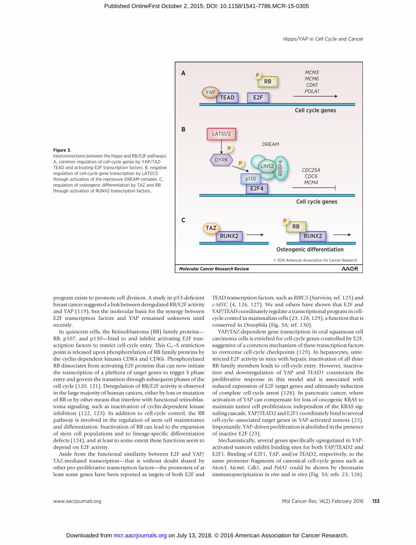
program exists to promote cell division. A study in p53-deficientbreast cancer suggested a linkbetweenderegulatedRB/E2F activityand YAP (119), but the molecular basis for the synergy betweenE2F transcription factors and YAP remained unknown untilrecently.
In quiescent cells, the Retinoblastoma (RB) family proteins—RB, p107, and p130—bind to and inhibit activating E2F tran-scription factors to restrict cell-cycle entry. This G1–S restrictionpoint is released upon phosphorylation of RB family proteins bythe cyclin-dependent kinases CDK4 and CDK6. PhosphorylatedRB dissociates from activating E2F proteins that can now initiatethe transcription of a plethora of target genes to trigger S phaseentry and govern the transition through subsequent phases of thecell cycle (120, 121). Deregulation of RB/E2F activity is observedin the large majority of human cancers, either by loss or mutationof RB or by other means that interfere with functional retinoblas-toma signaling, such as inactivation of cyclin-dependent kinaseinhibitors (122, 123). In addition to cell-cycle control, the RBpathway is involved in the regulation of stem cell maintenanceand differentiation. Inactivation of RB can lead to the expansionof stem cell populations and to lineage-specific differentiationdefects (124), and at least to some extent these functions seem todepend on E2F activity.
Aside from the functional similarity between E2F and YAP/TAZ-mediated transcription—that is without doubt shared byother pro-proliferative transcription factors—the promoters of atleast some genes have been reported as targets of both E2F and
TEAD transcription factors, such as BIRC5 (Survivin; ref. 125) andc-MYC (4, 126, 127). We and others have shown that E2F andYAP/TEADcoordinately regulate a transcriptional program in cell-cycle control inmammalian cells (23, 128, 129), a function that isconserved in Drosophila (Fig. 3A; ref. 130).
YAP/TAZ-dependent gene transcription in oral squamous cellcarcinoma cells is enriched for cell-cycle genes controlled by E2F,suggestive of a commonmechanism of these transcription factorsto overcome cell-cycle checkpoints (129). In hepatocytes, unre-stricted E2F activity in mice with hepatic inactivation of all threeRB family members leads to cell-cycle entry. However, inactiva-tion and downregulation of YAP and TEAD1 counteracts theproliferative response in this model and is associated withreduced expression of E2F target genes and ultimately inductionof complete cell-cycle arrest (128). In pancreatic cancer, whereactivation of YAP can compensate for loss of oncogenic KRAS tomaintain tumor cell proliferation independent of the KRAS sig-naling cascade, YAP/TEAD2andE2F1 coordinately bind to severalcell-cycle–associated target genes in YAP-activated tumors (23).Importantly, YAP-drivenproliferation is abolished in the presenceof inactive E2F (23).
Mechanistically, several genes specifically upregulated in YAP-activated tumors exhibit binding sites for both YAP/TEAD2 andE2F1. Binding of E2F1, YAP, and/or TEAD2, respectively, to thesame promoter fragments of canonical cell-cycle genes such asMcm3, Mcm6, Cdk1, and PolA1 could be shown by chromatinimmunoprecipitation in vivo and in vitro (Fig. 3A; refs. 23, 128).
Molecular Cancer Research Review
© 2015 American Association for Cancer Research
E2F4
TEADYAP
DREAM
RB
RUNX2RUNX2TAZ
p130
RB
BP4LIN52
DYRK
LATS1/2
RB
E2F
A
B
C
Cell cycle genes
Cell cycle genes
P
P
P
P
MCM3MCM6CDK1POLA1
CDC25ACDC6MCM4
Figure 3.Interconnections between the Hippo and RB/E2F pathways.A, common regulation of cell-cycle genes by YAP/TAZ-TEAD and activating E2F transcription factors. B, negativeregulation of cell-cycle gene transcription by LATS1/2through activation of the repressive DREAM complex. C,regulation of osteogenic differentiation by TAZ and RBthrough activation of RUNX2 transcription factors.
Hippo/YAP in Cell Cycle and Cancer
www.aacrjournals.org Mol Cancer Res; 14(2) February 2016 133
on July 13, 2018. © 2016 American Association for Cancer Research. mcr.aacrjournals.org Downloaded from
Published OnlineFirst October 2, 2015; DOI: 10.1158/1541-7786.MCR-15-0305

Additionally, bioinformatics analysis revealed that transcription-al start sites of YAP targets in the liver (4, 5) overlapwith E2FChIP-seq peaks in more than 60% of genes (128). Promoters of genesspecifically upregulated in YAP-activated pancreatic cancer areenriched for E2Fmotif containing gene signatures (23). However,it remains to be investigated if E2F and YAP/TEADdirectly bind toeachother in the coordinate regulationof cell-cycle genes or if theyare at least part of a common regulatory complex. An alternativeexplanation for the synergy of YAP/TAZ and E2F is that YAP/TAZ-TEAD in conjuncture with AP-1 bind tomore distant enhancers ofE2F-regulated cell-cycle genes to promote proliferation (131).Independent of the direct molecular mechanism, the commonactivation of pro-proliferative target genes by E2F and YAP/TEADtranscription factors is likely to play a central role in the oncogenicfunction of the YAP transcriptional cofactor.
A recent study in endothelial cells showed that YAP might playin role in the regulation of S-phase entry (132). They authorsidentified several differential regulated genes in YAP knockdowncells that are part of the replication machinery, such as CDC6,CDT1, MCM4, and MCM10 (132). The possibility of direct YAPbinding to the promoter of these genes as well as the transcriptionfactors involved remains to be investigated. Interestingly, a num-ber differentially regulated S-phase genes also present E2F targetgenes in linewith E2F requirement for S-phase progression (133).Beyond S-phase entry and progression, there is increasing evi-dence that the Hippo pathway is involved in the regulation of theG2–M phase (134). However, these functions seem to be inde-pendent of the Hippo downstream effectors YAP and TAZ andmainly mediated by MST1/2 and NDR kinases (reviewed inref. 134). Additionally, LATS2 seems to be required for mitoticprogression and cytokinesis (135). Some of the cell-cycle func-tions of the Hippo pathway may also be linked to interactionswith transcriptional complexes such as the DREAM complex(dimerization partner, RB-like, E2F and multi-vulval class B;ref. 136) and related complexes involved at various steps ofcell-cycle progression. In particular, LATS2 has been shown tophosphorylate DYRK to promote the assembly of the DREAMrepressor complex at promoters of E2F-regulated cell-cycle genes(136), providing an additional level of cell-cycle control byupstream Hippo kinases (Fig. 3B).
In summary, an important role of YAP/TAZ-induced prolifer-ation might be transcriptional activation of cell-cycle genes in aTEAD- andmaybe also E2F-dependent fashion. However, furtherresearch is needed to understand the interplay between the cell-cycle regulation machinery and Hippo pathway components.
In addition to direct control of the cell cycle, YAP and TAZ canprobably influence cell proliferation less directly by transcription-al regulation of several pathways that promote growth, cell divi-sions, and other pro-oncogenic functions. In the liver, a complexof YAP and TEAD4 directly targets JAG1 and NOTCH2 genes fortranscriptional activation (27, 50). Recent data indicate that YAP-mediated activation of Notch signaling can differentiate maturehepatocytes into cells with characteristics of liver progenitor cellsand propagate their expansion (27). In hepatocellular carcinoma,activation of YAP directly targets Notch signaling and is correlatedwith shorter survival times in human patients (50). In addition toNotch signaling, YAP as well as TAZ can activate the EGFRsignaling axis by targeting the promoter of the amphiregulin(AREG) gene (57, 137). In breast cancer cells, this activationcontributes to cell proliferation and migration independent ofEGF (137). Thus, many transcriptional targets of YAP and TAZ
work together to promote proliferation by directly activatinggenes important in cell division and simultaneously targetingother pro-proliferative signaling pathways, such as Notch andEGFR, to sustain a robust proliferative response.
Transcriptional Regulation by YAP/TAZbeyond TEAD
In addition to TEADs, several other transcriptional bindingpartners of YAP and TAZ have been identified. Interestingly,binding to transcription factors other than TEADs seem to medi-ate functions different from proliferation in many cases.
Both activated YAP and TAZ have been reported to bind toSMAD proteins to promote signaling from TGFb and BMP path-ways, adding another level to promote proliferation and onco-genic signaling (12, 138).On the other hand, phosphorylated andtranscriptionally inactive YAP/TAZ can bind and retain SMAD2/3proteins in the cytoplasm, inhibit their transcriptional activity,and induce their sequestration (12). Recently, YAP has beenshown to interact with the AP-1 transcription factor family mem-ber FOS to promote epithelial–mesenchymal transition (EMT)and metastasis (139). In models of KRAS-driven lung and colo-rectal cancer, activation of YAPmediates resistance to inactivationof oncogenic KRAS, highlighting its role in tumor plasticity.Mechanistically, both KRAS and YAP converge on the transcrip-tion factor FOS to activate transcriptional targets that are involvedin EMT, such as Vimentin, Slug, and Snail (139).
Although nuclear YAP/TAZ play important roles in oncogenicproliferation and EMT, they can also trigger expression of proa-poptotic genes to counteract tumorigenesis. In DNA damage, YAPcan interact with and stabilize p73 to enhance the apoptosisresponse (140). The interaction with p73 is greatly amplified byYAP tyrosine phosphorylation that is induced by c-ABL uponDNA damage (105). This mechanism presents an importantsystem to divert YAP transcriptional control away from prolifer-ation in the context of DNA damage. Interestingly, YAP is down-regulated in several hematologic malignancies with activatedDNA damage signaling and high nuclear c-ABL kinase activity(141). Restoration of YAP activity in these tumor cells by inhibi-tionof theupstreamHippoMST1kinase is sufficient todrive theminto apoptosis (141). Therefore, YAP phosphorylation by theHippo core kinase cascade may not only exert tumor-suppressivefunctions, butmaybeoncogenic under certain conditions (Fig. 2).In addition to upstream Hippo kinases, AKT can phosphorylateYAP on Ser127 to inhibit the proapoptotic synergy between YAPand p73 (91, 142).
Apart from the control of oncogenic signaling, YAP and TAZplay a role in differentiation in several organs. Although bothtranscriptional cofactors generally seem to drive cells toward amore stem cell–like phenotype (6, 18, 143), they can also inducelineage-specific differentiation. TAZ interacts with RUNX2 tocoactivate RUNX2-dependent gene transcription and direct mes-enchymal stem cells toward osteogenic differentiation (144). Atthe same time, TAZ inhibits PPARg-dependent gene expressionand adipogenic differentiation (144). Interestingly, similar effectshave been reported for phosphorylated RB that can bind toRUNX2 to promote osteogenic differentiation and can inhibitPPARg-driven adipogenic differentiation in binding to E2Fs(145), presenting another example for functional convergenceof Hippo and RB/E2F signaling (Fig. 3C). Besides RUNX2, severalother developmental transcription factors are bound by the
Ehmer and Sage
Mol Cancer Res; 14(2) February 2016 Molecular Cancer Research134
on July 13, 2018. © 2016 American Association for Cancer Research. mcr.aacrjournals.org Downloaded from
Published OnlineFirst October 2, 2015; DOI: 10.1158/1541-7786.MCR-15-0305

Hippo signaling downstream effectors. YAP and TAZ both coac-tivate PAX3, a transcription factor important in the developmentof the neuronal crest (146), as well as TBX5, which initiatestranscriptional programs in heart development (147). Addition-ally, a transcriptional complex of b-catenin–YAP–TBX5 isrequired for the maintenance of b-catenin–driven cancers andcan bind to pro-proliferative target genes independent of TEADtranscription factors (90). In the intestine, YAP/TAZ cooperatewith KLF4 in promoting differentiation into goblet cells (38). Inaddition to direct binding of transcription factors involved indevelopment and differentiation, a complex of YAP and TEAD2has been reported to influence the binding of HNF4a and FOXA2to enhancers of specific target genes in embryonic liver (148).Although this YAP/TEAD2-induced target gene switch likelyinvolves direct binding to DNA, it remains unclear to date if theYAP/TEAD complex might be part of a larger transcriptionalcomplex with FOXA2 or HNF4a.
It is highly likely that the number of developmental transcrip-tion factors identified as binding partners of YAP and TAZ will beexpanding in the future. Even from what we know to date it isobvious that the Hippo downstream effectors have a key role indevelopment to promote differentiation into distinct cellularlineages dependent on the transcriptional binding partner.
Targeting Hippo SignalingAn increasing amount of data highlights the important role of
Hippo signaling in the control of proliferation, including unre-stricted proliferation found in cancer. Inhibiting oncogenicYAP and TAZ to control proliferation is therefore a promisingapproach in cancers that show increased activity of these tran-scriptional cofactors. Additionally, YAP has been shown to be acritical mediator of Ras-driven cancer and tomediate resistance tosuppression of RAS andRAF signaling and could therefore be usedin combination therapy with inhibitors of these oncogenic path-ways to prevent or break drug resistance (23, 139, 149, 150).
Several different mechanisms are known to activate YAP andTAZ in human cancers to promote proliferation, including geneticamplification of YAP or genetic inactivation of upstream Hipporegulators such as LATS1, SAV1, NF2, or GPCRs (reviewed inref. 151). Additionally, input signals from other pathways (e.g.,Wnt) are likely involved in deregulation of Hippo signaling.However, all these different effects on Hippo signaling convergeonto one specific outcome: activation of YAP and/or TAZ topromote tumorigenesis. It therefore seems logical to target thesedownstream effectors directly to inhibit tumor development.Although YAP/TAZ bind to different transcription factors, theirinteraction with TEADs is likely the most important one incarcinogenesis as it promotes proliferation as well as metastasis(13, 114, 118, 152). Efforts to find specific inhibitors of YAP/TAZ-TEAD function have led to the identification of verteporfin. Thecompound was identified in a drug-screening approach andscored together with other porphyrins in inhibiting the interac-tion between YAP and TEAD (114).Most importantly, verteporfinalso inhibits proliferation and liver overgrowth in vivo in mousemodels with either YAP overexpression or inactivation ofupstream Nf2/Merlin, resulting in disinhibition of endogenousYAP (114). An antitumorigenic effect of verteporfin was shown invitro, where the drug was able to block tumor growth in uvealmelanoma cells exhibiting activated YAP (53). Therefore, verte-porfin could present a promising agent in the treatment of cancers
dependent on high YAP activity. However, as a drug used as aphotosensitizer in photodynamic therapy for macular degenera-tion the phototoxic properties of verteporfin are likely to limit itsapplication in a therapeutic setting. The development of alterna-tive compounds that specifically inhibit the interaction betweenYAP/TAZ and TEAD is therefore of high interest in cancer therapy.In this line, several alternative approacheswere described recently.Engineered YAP-like peptides are able to disrupt the YAP–TEADinteraction in vitro (153). However, their efficiency to target thetranscriptional complex in vivo remains unclear to date. Anotheroption to target YAP–TEAD-induced transcription might be bymimicking the interaction between vestigial-like proteins (VGLL)andTEADs. As alternative bindingpartners for TEAD transcriptionfactors that compete with YAP for the same binding sites VGLLproteins counteract the activity of the Hippo downstream effec-tors at least in some tissues. VGLL4 has recently been identified asa tumor suppressor in lung cancer, where the transcriptionalcofactor negatively regulates YAP–TEAD activity (154). A peptidemimicking VGLL4 function was reported to inhibit growths ofgastric cancer cells in vitro and in vivo and to restrain tumordevelopment in aHelicobacter pylorimousemodel of gastric cancer(155). Even though the effects to VGLL4 might be tissue specificandneed tobe evaluated inother cancers, this novel peptide couldpresent a promising option to inhibit YAP–TEAD-driven tran-scription in cancer.
Direct inhibition of YAP by genetic targeting was effective in anin vivo HCC model with deregulated Hippo signaling: in Mst1/2-conditional knockoutmice, small interfering RNA-lipid nanopar-ticles targeting YAPwere able to significantly reduce tumorburdenin comparison to controls (156). Although this method did notspecifically inhibit the YAP/TEAD interaction, transcriptionalprograms that are likely dependent on TEAD were significantlyaltered in thismodel, including E2F target genes (23, 128), as wellas targets of HNF4a and FOXA2 (153).
In addition to direct inhibition of YAP and TAZ transcriptionalactivity, modifying upstream Hippo signaling could presentanother approach to restrict, or activate, YAP/TAZ function. Theidentification of specific receptors that regulate Hippo signalingsuch as GPCR and EGFR (51, 56, 58) offers novel opportunities tofunctionally target this pathway. However, these receptors aremostly not pathway specific, and the complexity of signalingnetworks that are influenced for example even by a singlememberof the GPCR receptor family might limit this therapeuticapproach. Additionally, dependent on the cellular context, theoutcomes of YAP/TAZ inhibition might vary. This is of highimportance in cancers where the transcriptional cofactors alsomight have a tumor-suppressive role (Fig. 2), such as in somehematologic malignancies where YAP promotes apoptosis (141).Additionally, the role of YAP in breast cancer remains controver-sial and the protein might act as a tumor suppressor possibly inthe presence of additional genetic changes (157, 158). In theintestine, YAP can function to restrict the expansion of intestinalstem cells and to inhibit the growth of colorectal carcinomaxenografts (44). However, from what we know to date, thesetumor suppressor functions may not depend on the interactionbetween YAP/TAZ and TEAD. Instead, they do at least in partdepend on interactionwith other transcription factors such as p73in the apoptotic response (141) or KLF4 and RUNX2 in theinduction of differentiation (38, 144). Additionally, the tumor-suppressive activity can be independent of any transcriptionalactivity at all and stem from functions specific to cytoplasmic YAP
Hippo/YAP in Cell Cycle and Cancer
www.aacrjournals.org Mol Cancer Res; 14(2) February 2016 135
on July 13, 2018. © 2016 American Association for Cancer Research. mcr.aacrjournals.org Downloaded from
Published OnlineFirst October 2, 2015; DOI: 10.1158/1541-7786.MCR-15-0305

(44). Given themultitude of interactions of Hippo signaling withdifferent pathways and the diverse functions of YAP/TAZ, it seemsto be the safest and most predictable approach to target theinteraction with TEAD transcription factors to inhibit prolifera-tion and other oncogenic effects in cancer. However, furtherstudies have to show the efficiency and feasibility of this approachin a clinical setting.
It is also worth considering the possibility to inhibit Hippopathway function to enhance the activity of YAP and TAZ. Appli-cations for this approach include c-ABL–driven malignancies, topromote the induction of an apoptotic response dependent on aYAP/TAZ interaction with p73 (141) as well as targeting Hipposignaling functions beyond cancer. This is of special interest inorgans where high YAP/TAZ activity is associated with tissuegrowth and regeneration, such as the heart (45, 159) or the liver(160, 161). Specific receptors such as GPCRs are the most prom-ising targets to dampen the activity of upstreamHippo kinases: atleast in vitro, GPCR ligands such as LPA promote the activity ofYAP and TAZ (51). However, a large number of different GPCRsexist and the effects on the Hippo pathway differ in quality,dependent on the class of GPCR, as well as in quantity (51).Therefore, further research is needed to identify specific andligands to effectively inhibit Hippo signaling upstream of YAP/TAZ, especially in vivo. Even if we succeed to successfully induceYAP/TAZ activation, the pro-oncogenic side effects associatedwith any drug that promotes proliferation should be consideredcarefully—even when the treatment is only given for a shortperiod of time.
ConclusionThere has been an explosion of reports on the Hippo/YAP
pathway in the past few years. These studies have brought thissignaling pathway to the forefront of cancer research. Similar toother key cancer pathways, the Hippo/YAP pathway is complexand coordinates multiple cellular functions, including prolifera-tion, cell death, anddifferentiation. This plethoraof functions and
recent evidence strongly indicate that theHippo/YAP pathway is acentral player in tumor plasticity and how cycling cells interactwith their environment. Superficially, the Hippo/YAP pathwayseems to serve similar functions to other central pathways such asthe Rb/E2F pathway. However, accumulating evidence points tounique functions for this pathway, even though the basis of thisspecificity is still not fully understood. A key aspect of the Hippo/YAP pathway may be its ability to sense and mediate uniqueextracellular signals, includingmechanical forces. TheHippo/YAPpathway canbe both tumor suppressive andoncogenic, and it willbe key in the future to better understand the tissue specificity ofaction of this pathway, as well as cell-intrinsic and non–cell-autonomous effects on cell proliferation. This will be crucialbefore anticancer therapies targeting this pathway can be imple-mented. However, early success repurposing a drug such asverteporfin to regulate the Hippo pathway suggests that, in thefuture, this pathway is "druggable" and may thus expand thera-peutic options in cancer patients.
Disclosure of Potential Conflicts of InterestNo potential conflicts of interest were disclosed.
AcknowledgmentsThe authors thank M. Butte and P. Mazur for their input on the manuscript
and apologize to the authors for their work not being cited owing to spacelimitations.
Grant SupportThis work was supported by the NIH (grant CA114102 to J. Sage), the Lucile
Packard Foundation for Children's Health (Ernest and Amelia Gallo EndowedPostdoctoral Fellowship CTSA grant number UL1 RR025744 to U. Ehmer), andthe Dr. Mildred Scheel fellowship from Deutsche Krebshilfe (to U. Ehmer). Dr.Sage is the Harriet and Mary Zelencik Scientist in Children's Cancer and BloodDiseases.
Received July 10, 2015; revised September 8, 2015; accepted September 25,2015; published OnlineFirst October 2, 2015.
References1. Hanahan D, Weinberg RA. Hallmarks of cancer: the next generation. Cell
2011;144:646–74.2. Wu S, Huang J, Dong J, Pan D. hippo encodes a Ste-20 family protein
kinase that restricts cell proliferation and promotes apoptosis in conjunc-tion with salvador and warts. Cell 2003;114:445–56.
3. Harvey KF, Pfleger CM, Hariharan IK. The Drosophila Mst ortholog,hippo, restricts growth and cell proliferation and promotes apoptosis.Cell 2003;114:457–67.
4. Dong J, Feldmann G, Huang J, Wu S, Zhang N, Comerford SA, et al.Elucidation of a universal size-control mechanism in Drosophila andmammals. Cell 2007;130:1120–33.
5. Zhao B, Wei X, Li W, Udan RS, Yang Q, Kim J, et al. Inactivation of YAPoncoprotein by the Hippo pathway is involved in cell contact inhibitionand tissue growth control. Genes Dev 2007;21:2747–61.
6. Lian I, Kim J, Okazawa H, Zhao J, Zhao B, Yu J, et al. The role of YAPtranscription coactivator in regulating stem cell self-renewal and differ-entiation. Genes Dev 2010;24:1106–18.
7. Sorrentino G, Ruggeri N, Specchia V, Cordenonsi M, Mano M, Dupont S,et al. Metabolic control of YAP and TAZ by the mevalonate pathway. NatCell Biol 2014;16:357–66.
8. Huang J,WuS, Barrera J,MatthewsK, PanD. TheHippo signaling pathwaycoordinately regulates cell proliferation and apoptosis by inactivatingYorkie, the Drosophila homolog of YAP. Cell 2005;122:421–34.
9. Badouel C, Garg A, McNeill H. Herding Hippos: regulating growth in fliesand man. Curr Opin Cell Biol 2009;21:837–43.
10. Kanai F, Marignani PA, Sarbassova D, Yagi R, Hall RA, Donowitz M, et al.TAZ: a novel transcriptional co-activator regulated by interactions with14-3-3 and PDZ domain proteins. EMBO J 2000;19:6778–91.
11. Azzolin L, Panciera T, Soligo S, Enzo E, Bicciato S, Dupont S, et al. YAP/TAZ incorporation in the beta-catenin destruction complex orchestratesthe Wnt response. Cell 2014;158:157–70.
12. Varelas X, Samavarchi-Tehrani P, Narimatsu M, Weiss A, Cockburn K,Larsen BG, et al. The Crumbs complex couples cell density sensing toHippo-dependent control of the TGF-beta-SMAD pathway. Dev Cell2010;19:831–44.
13. Zhao B, Ye X, Yu J, Li L, Li W, Li S, et al. TEAD mediates YAP-dependentgene induction and growth control. Genes Dev 2008;22:1962–71.
14. Yu FX, Guan KL. The Hippo pathway: regulators and regulations. GenesDev 2013;27:355–71.
15. Zhou D, Conrad C, Xia F, Park JS, Payer B, Yin Y, et al. Mst1 and Mst2maintain hepatocyte quiescence and suppress hepatocellular carcinomadevelopment through inactivation of the Yap1 oncogene. Cancer Cell2009;16:425–38.
16. Schlegelmilch K, Mohseni M, Kirak O, Pruszak J, Rodriguez JR, Zhou D,et al. Yap1 acts downstream of alpha-catenin to control epidermalproliferation. Cell 2011;144:782–95.
17. Feng X, Degese MS, Iglesias-Bartolome R, Vaque JP, Molinolo AA, Rodri-gues M, et al. Hippo-independent activation of YAP by the GNAQ uvealmelanoma oncogene through a trio-regulated rho GTPase signalingcircuitry. Cancer Cell 2014;25:831–45.
Ehmer and Sage
Mol Cancer Res; 14(2) February 2016 Molecular Cancer Research136
on July 13, 2018. © 2016 American Association for Cancer Research. mcr.aacrjournals.org Downloaded from
Published OnlineFirst October 2, 2015; DOI: 10.1158/1541-7786.MCR-15-0305

18. Cordenonsi M, Zanconato F, Azzolin L, Forcato M, Rosato A, Frasson C,et al. The Hippo transducer TAZ confers cancer stem cell–related traits onbreast cancer cells. Cell 2011;147:759–72.
19. Nallet-Staub F, Marsaud V, Li L, Gilbert C, Dodier S, Bataille V, et al. Pro-invasive activity of theHippopathway effectors YAP andTAZ in cutaneousmelanoma. J Invest Dermatol 2014;134:123–32.
20. Bhat KP, Salazar KL, Balasubramaniyan V, Wani K, Heathcock L,Hollingsworth F, et al. The transcriptional coactivator TAZ regulatesmesenchymal differentiation in malignant glioma. Genes Dev 2011;25:2594–609.
21. Wang Y, Dong Q, Zhang Q, Li Z, Wang E, Qiu X. Overexpression of yes-associated protein contributes to progression and poor prognosis of non-small-cell lung cancer. Cancer Sci 2010;101:1279–85.
22. Harvey KF, ZhangX, ThomasDM. TheHippopathway andhuman cancer.Nat Rev Cancer 2013;13:246–57.
23. Kapoor A, YaoW, YingH, Hua S, Liewen A,WangQ, et al. Yap1 activationenables bypass of oncogenic Kras addiction in pancreatic cancer. Cell2014;158:185–97.
24. Cancer Genome Atlas N. Comprehensive genomic characterization ofhead and neck squamous cell carcinomas. Nature 2015;517:576–82.
25. Dhanasekaran SM, Balbin OA, Chen G, Nadal E, Kalyana-Sundaram S,Pan J, et al. Transcriptome meta-analysis of lung cancer reveals recurrentaberrations in NRG1 and Hippo pathway genes. Nat Commun 2014;5:5893.
26. Seidel C, Schagdarsurengin U, Blumke K, Wurl P, Pfeifer GP, HauptmannS, et al. Frequent hypermethylation of MST1 and MST2 in soft tissuesarcoma. Mol Carcinog 2007;46:865–71.
27. Yimlamai D, Christodoulou C, Galli GG, Yanger K, Pepe-Mooney B,Gurung B, et al. Hippo pathway activity influences liver cell fate. Cell2014;157:1324–38.
28. Karpowicz P, Perez J, Perrimon N. The Hippo tumor suppressorpathway regulates intestinal stem cell regeneration. Development2010;137:4135–45.
29. Cai J, Zhang N, Zheng Y, de Wilde RF, Maitra A, Pan D. The Hipposignaling pathway restricts the oncogenic potential of an intestinal regen-eration program. Genes Dev 2010;24:2383–8.
30. JudsonRN, TremblayAM,KnoppP,White RB,Urcia R,DeBari C, et al. TheHippo pathwaymember Yap plays a key role in influencing fate decisionsin muscle satellite cells. J Cell Sci 2012;125:6009–19.
31. Gao T, Zhou D, Yang C, Singh T, Penzo-Mendez A, Maddipati R, et al.Hippo signaling regulates differentiation and maintenance in the exo-crine pancreas. Gastroenterology 2013;144:1543–53, 53.e1.
32. Mahoney JE, Mori M, Szymaniak AD, Varelas X, Cardoso WV. The hippopathway effector Yap controls patterning and differentiation of airwayepithelial progenitors. Dev Cell 2014;30:137–50.
33. Lee MJ, Ran Byun M, Furutani-Seiki M, Hong JH, Jung HS. YAP and TAZregulate skin wound healing. J Invest Dermatol 2014;134:518–25.
34. Morin-Kensicki EM, Boone BN, Howell M, Stonebraker JR, Teed J, Alb JG,et al. Defects in yolk sac vasculogenesis, chorioallantoic fusion, andembryonic axis elongation in mice with targeted disruption of Yap65.Mol Cell Biol 2006;26:77–87.
35. Lu L, Li Y, Kim SM, Bossuyt W, Liu P, Qiu Q, et al. Hippo signaling is apotent in vivo growth and tumor suppressor pathway in the mammalianliver. Proc Natl Acad Sci U S A 2010;107:1437–42.
36. Lee KP, Lee JH, Kim TS, Kim TH, Park HD, Byun JS, et al. The Hippo–Salvador pathway restrains hepatic oval cell proliferation, liversize, and liver tumorigenesis. Proc Natl Acad Sci U S A 2010;107:8248–53.
37. Camargo FD, Gokhale S, Johnnidis JB, Fu D, Bell GW, Jaenisch R, et al.YAP1 increases organ size and expands undifferentiated progenitor cells.Curr Biol 2007;17:2054–60.
38. Imajo M, Ebisuya M, Nishida E. Dual role of YAP and TAZ in renewal ofthe intestinal epithelium. Nat Cell Biol 2015;17:7–19.
39. Cao X, Pfaff SL, Gage FH. YAP regulates neural progenitor cell number viathe TEA domain transcription factor. Genes Dev 2008;22:3320–34.
40. Lee JH, Kim TS, Yang TH, Koo BK, Oh SP, Lee KP, et al. A crucial role ofWW45 in developing epithelial tissues in the mouse. EMBO J 2008;27:1231–42.
41. Seo E, Basu-Roy U, Gunaratne PH, Coarfa C, Lim DS, Basilico C, et al.SOX2 regulates YAP1 to maintain stemness and determine cell fate in theosteo-adipo lineage. Cell Rep 2013;3:2075–87.
42. Zhao R, Fallon TR, Saladi SV, Pardo-Saganta A, Villoria J, Mou H, et al.Yap tunes airway epithelial size and architecture by regulating theidentity, maintenance, and self-renewal of stem cells. Dev Cell 2014;30:151–65.
43. Sarikaya DP, Extavour CG. The Hippo pathway regulates homeostaticgrowth of stem cell niche precursors in the Drosophila ovary. PLoS Genet2015;11:e1004962.
44. Barry ER, Morikawa T, Butler BL, Shrestha K, de la Rosa R, Yan KS, et al.Restriction of intestinal stem cell expansion and the regenerative responseby YAP. Nature 2013;493:106–10.
45. Heallen T, Zhang M, Wang J, Bonilla-Claudio M, Klysik E, Johnson RL,et al. Hippo pathway inhibits Wnt signaling to restrain cardiomyocyteproliferation and heart size. Science 2011;332:458–61.
46. Varelas X, Miller BW, Sopko R, Song S, Gregorieff A, Fellouse FA, et al. TheHippo pathway regulates Wnt/beta-catenin signaling. Dev Cell 2010;18:579–91.
47. Hergovich A, Hemmings BA. TAZ-mediated crosstalk between Wnt andHippo signaling. Dev Cell 2010;18:508–9.
48. Strassburger K, Tiebe M, Pinna F, Breuhahn K, Teleman AA. Insulin/IGFsignaling drives cell proliferation in part via Yorkie/YAP. Dev Biol2012;367:187–96.
49. Fernandez LA, Northcott PA, Dalton J, Fraga C, Ellison D, Angers S, et al.YAP1 is amplified and up-regulated in hedgehog-associated medullo-blastomas and mediates Sonic hedgehog-driven neural precursor prolif-eration. Genes Dev 2009;23:2729–41.
50. Tschaharganeh DF, Chen X, Latzko P, Malz M, Gaida MM, Felix K, et al.Yes-associated protein up-regulates Jagged-1 and activates the Notchpathway in human hepatocellular carcinoma. Gastroenterology 2013;144:1530–42 e12.
51. Yu FX, Zhao B, PanupinthuN, Jewell JL, Lian I, Wang LH, et al. Regulationof the Hippo-YAP pathway by G-protein-coupled receptor signaling. Cell2012;150:780–91.
52. Miller E, Yang J, DeRan M, Wu C, Su AI, Bonamy GM, et al. Identificationof serum-derived sphingosine-1-phosphate as a small molecule regulatorof YAP. Chem Biol 2012;19:955–62.
53. Yu FX, Luo J, Mo JS, Liu G, Kim YC, Meng Z, et al. Mutant Gq/11 promoteuveal melanoma tumorigenesis by activating YAP. Cancer Cell 2014;25:822–30.
54. Mo JS, Yu FX, Gong R, Brown JH, Guan KL. Regulation of the Hippo-YAPpathway by protease-activated receptors (PARs). Genes Dev 2012;26:2138–43.
55. Fan R, Kim NG, Gumbiner BM. Regulation of Hippo pathway bymitogenic growth factors via phosphoinositide 3-kinase and phos-phoinositide-dependent kinase-1. Proc Natl Acad Sci U S A 2013;110:2569–74.
56. Reddy BV, Irvine KD. Regulation of Hippo signaling by EGFR-MAPKsignaling through Ajuba family proteins. Dev Cell 2013;24:459–71.
57. Zhang J, Ji JY, Yu M, Overholtzer M, Smolen GA, Wang R, et al. YAP-dependent induction of amphiregulin identifies a non-cell-autono-mous component of the Hippo pathway. Nat Cell Biol 2009;11:1444–50.
58. Haskins JW, Nguyen DX, Stern DF. Neuregulin 1-activated ERBB4 inter-acts with YAP to induce Hippo pathway target genes and promote cellmigration. Sci Signal 2014;7:ra116.
59. Guo X, Zhao B. Integration of mechanical and chemical signals by YAPand TAZ transcription coactivators. Cell Biosci 2013;3:33.
60. Low BC, Pan CQ, Shivashankar GV, Bershadsky A, Sudol M, Sheetz M.YAP/TAZ as mechanosensors and mechanotransducers in regulatingorgan size and tumor growth. FEBS Lett 2014;588:2663–70.
61. Dupont S, Morsut L, Aragona M, Enzo E, Giulitti S, Cordenonsi M,et al. Role of YAP/TAZ in mechanotransduction. Nature 2011;474:179–83.
62. Aragona M, Panciera T, Manfrin A, Giulitti S, Michielin F, Elvassore N,et al. A mechanical checkpoint controls multicellular growth throughYAP/TAZ regulation by actin-processing factors. Cell 2013;154:1047–59.
63. Mana-Capelli S, Paramasivam M, Dutta S, McCollum D. Angiomotinslink F-actin architecture to Hippo pathway signaling. Mol Biol Cell2014;25:1676–85.
64. Mohseni M, Sun J, Lau A, Curtis S, Goldsmith J, Fox VL, et al. A geneticscreen identifies an LKB1-MARK signalling axis controlling the Hippo-YAP pathway. Nat Cell Biol 2014;16:108–17.
Hippo/YAP in Cell Cycle and Cancer
www.aacrjournals.org Mol Cancer Res; 14(2) February 2016 137
on July 13, 2018. © 2016 American Association for Cancer Research. mcr.aacrjournals.org Downloaded from
Published OnlineFirst October 2, 2015; DOI: 10.1158/1541-7786.MCR-15-0305

65. GanemNJ, Cornils H, Chiu SY, O'Rourke KP, Arnaud J, Yimlamai D, et al.Cytokinesis failure triggers hippo tumor suppressor pathway activation.Cell 2014;158:833–48.
66. Deng H, WangW, Yu J, Zheng Y, Qing Y, Pan D. Spectrin regulates Hipposignaling by modulating cortical actomyosin activity. Elife 2015;4:e06567.
67. Bonnans C, Chou J, Werb Z. Remodelling the extracellular matrixin development and disease. Nat Rev Mol Cell Biol 2014;15:786–801.
68. Levental KR, Yu H, Kass L, Lakins JN, Egeblad M, Erler JT, et al. Matrixcrosslinking forces tumor progression by enhancing integrin signaling.Cell 2009;139:891–906.
69. Calvo F, EgeN,Grande-Garcia A,Hooper S, Jenkins RP, Chaudhry SI, et al.Mechanotransduction and YAP-dependent matrix remodelling isrequired for the generation and maintenance of cancer-associated fibro-blasts. Nat Cell Biol 2013;15:637–46.
70. Hamaratoglu F,WilleckeM, Kango-SinghM,Nolo R, Hyun E, Tao C, et al.The tumour-suppressor genes NF2/Merlin and Expanded act throughHippo signalling to regulate cell proliferation and apoptosis. NatCell Biol2006;8:27–36.
71. Yu J, Zheng Y, Dong J, Klusza S, Deng WM, Pan D. Kibra functions as atumor suppressor protein that regulates Hippo signaling in conjunctionwith Merlin and Expanded. Dev Cell 2010;18:288–99.
72. Zhao B, Li L, Lu Q, Wang LH, Liu CY, Lei Q, et al. Angiomotin is a novelHippo pathway component that inhibits YAP oncoprotein. Genes Dev2011;25:51–63.
73. Kim NG, Koh E, Chen X, Gumbiner BM. E-cadherin mediates contactinhibition of proliferation through Hippo signaling-pathway compo-nents. Proc Natl Acad Sci U S A 2011;108:11930–5.
74. Martin-Belmonte F, Perez-Moreno M. Epithelial cell polarity, stem cellsand cancer. Nat Rev Cancer 2012;12:23–38.
75. Cooper J, Giancotti FG. Molecular insights into NF2/Merlin tumorsuppressor function. FEBS Lett 2014;588:2743–52.
76. Yin F, Yu J, Zheng Y, Chen Q, Zhang N, Pan D. Spatial organization ofHippo signaling at the plasma membrane mediated by the tumor sup-pressor Merlin/NF2. Cell 2013;154:1342–55.
77. Li W, You L, Cooper J, Schiavon G, Pepe-Caprio A, Zhou L, et al. Merlin/NF2 suppresses tumorigenesis by inhibiting the E3 ubiquitin ligase CRL4(DCAF1) in the nucleus. Cell 2010;140:477–90.
78. Li W, Cooper J, Zhou L, Yang C, Erdjument-Bromage H, Zagzag D, et al.Merlin/NF2 loss-driven tumorigenesis linked toCRL4(DCAF1)-mediatedinhibition of the hippo pathway kinases Lats1 and 2 in the nucleus.Cancer Cell 2014;26:48–60.
79. Benhamouche S, Curto M, Saotome I, Gladden AB, Liu CH, GiovanniniM, et al. Nf2/Merlin controls progenitor homeostasis and tumorigenesisin the liver. Genes Dev 2010;24:1718–30.
80. Zhang N, Bai H, David KK, Dong J, Zheng Y, Cai J, et al. The Merlin/NF2tumor suppressor functions through the YAP oncoprotein to regulatetissue homeostasis in mammals. Dev Cell 2010;19:27–38.
81. Moleirinho S, Guerrant W, Kissil JL. The Angiomotins–from discovery tofunction. FEBS Lett 2014;588:2693–703.
82. Chan SW, Lim CJ, Guo F, Tan I, Leung T, Hong W. Actin-binding and cellproliferation activities of angiomotin family members are regulated byHippo pathway-mediated phosphorylation. J Biol Chem 2013;288:37296–307.
83. Dai X, She P, Chi F, Feng Y, Liu H, Jin D, et al. Phosphorylation ofangiomotin by Lats1/2 kinases inhibits F-actin binding, cell migration,and angiogenesis. J Biol Chem 2013;288:34041–51.
84. Adler JJ, JohnsonDE,Heller BL, Bringman LR, RanahanWP, ConwellMD,et al. Serum deprivation inhibits the transcriptional co-activator YAP andcell growth via phosphorylation of the 130-kDa isoform of Angiomotinby the LATS1/2 protein kinases. Proc Natl Acad Sci U S A 2013;110:17368–73.
85. Li J, Gao E, Vite A, Yi R, Gomez L, Goossens S, et al. Alpha-catenins controlcardiomyocyte proliferation by regulating yap activity. Circ Res 2015;116:70–9.
86. Johnson R, Halder G. The two faces of Hippo: targeting the Hippopathway for regenerative medicine and cancer treatment. Nat Rev DrugDiscov 2014;13:63–79.
87. Clevers H, Nusse R. Wnt/beta-catenin signaling and disease. Cell 2012;149:1192–205.
88. Azzolin L, Zanconato F, Bresolin S, Forcato M, Basso G, Bicciato S, et al.Role of TAZ as mediator of Wnt signaling. Cell 2012;151:1443–56.
89. Imajo M, Miyatake K, Iimura A, Miyamoto A, Nishida E. A molecularmechanism that links Hippo signalling to the inhibition of Wnt/beta-catenin signalling. EMBO J 2012;31:1109–22.
90. Rosenbluh J, Nijhawan D, Cox AG, Li X, Neal JT, Schafer EJ, et al. beta-Catenin-driven cancers require a YAP1 transcriptional complex for sur-vival and tumorigenesis. Cell 2012;151:1457–73.
91. Basu S, Totty NF, Irwin MS, Sudol M, Downward J. Akt phosphor-ylates the Yes-associated protein, YAP, to induce interaction with 14-3-3 and attenuation of p73-mediated apoptosis. Mol Cell 2003;11:11–23.
92. Lin Z, Zhou P, von Gise A, Gu F, Ma Q, Chen J, et al. Pi3kcb links Hippo-YAP and PI3K-AKT signaling pathways to promote cardiomyocyte pro-liferation and survival. Circ Res 2015;116:35–45.
93. XinM, Kim Y, Sutherland LB, MurakamiM, Qi X, McAnally J, et al. Hippopathway effector Yap promotes cardiac regeneration. Proc Natl Acad Sci US A 2013;110:13839–44.
94. Yamamoto S, Yang G, Zablocki D, Liu J, Hong C, Kim SJ, et al. Activationof Mst1 causes dilated cardiomyopathy by stimulating apoptosis withoutcompensatory ventricular myocyte hypertrophy. J Clin Invest 2003;111:1463–74.
95. Liang N, Zhang C, Dill P, Panasyuk G, PionD, Koka V, et al. Regulation ofYAP by mTOR and autophagy reveals a therapeutic target of tuberoussclerosis complex. J Exp Med 2014;211:2249–63.
96. Zhou D, Zhang Y, Wu H, Barry E, Yin Y, Lawrence E, et al. Mst1 and Mst2protein kinases restrain intestinal stem cell proliferation and colonictumorigenesis by inhibition of Yes-associated protein (Yap) overabun-dance. Proc Natl Acad Sci U S A 2011;108:E1312–20.
97. Rayon T, Menchero S, Nieto A, Xenopoulos P, Crespo M, Cockburn K,et al. Notch and hippo converge on Cdx2 to specify the trophectodermlineage in the mouse blastocyst. Dev Cell 2014;30:410–22.
98. Djiane A, Zaessinger S, Babaoglan AB, Bray SJ. Notch inhibits Yorkieactivity in Drosophila wing discs. PLoS One 2014;9:e106211.
99. Tariki M, Dhanyamraju PK, Fendrich V, Borggrefe T, Feldmann G, LauthM. The Yes-associated protein controls the cell density regulation ofHedgehog signaling. Oncogenesis 2014;3:e112.
100. Kim M, Kim M, Lee MS, Kim CH, Lim DS. The MST1/2-SAV1 complex ofthe Hippo pathway promotes ciliogenesis. Nat Commun 2014;5:5370.
101. Kim J, Jo H, Hong H, Kim MH, Kim JM, Lee JK, et al. Actin remodellingfactors control ciliogenesis by regulating YAP/TAZ activity and vesicletrafficking. Nat Commun 2015;6:6781.
102. Tomlinson V, Gudmundsdottir K, Luong P, Leung KY, Knebel A, Basu S.JNK phosphorylates Yes-associated protein (YAP) to regulate apoptosis.Cell Death Dis 2010;1:e29.
103. Oudhoff MJ, Freeman SA, Couzens AL, Antignano F, Kuznetsova E, MinPH, et al. Control of the hippo pathway by Set7-dependent methylationof Yap. Dev Cell 2013;26:188–94.
104. Zaidi SK, Sullivan AJ, Medina R, Ito Y, van Wijnen AJ, Stein JL, et al.Tyrosine phosphorylation controls Runx2-mediated subnuclear targetingof YAP to repress transcription. EMBO J 2004;23:790–9.
105. Levy D, Adamovich Y, ReuvenN, Shaul Y. Yap1 phosphorylation by c-Ablis a critical step in selective activation of proapoptotic genes in response toDNA damage. Mol Cell 2008;29:350–61.
106. Keshet R, Adler J, Ricardo Lax I, Shanzer M, Porat Z, Reuven N, et al. c-Ablantagonizes the YAP oncogenic function. Cell Death Differ 2015;22:935–45.
107. Liu CY, Lv X, Li T, Xu Y, Zhou X, Zhao S, et al. PP1 cooperates withASPP2 to dephosphorylate and activate TAZ. J Biol Chem2011;286:5558–66.
108. Wang P, Bai Y, Song B, Wang Y, Liu D, Lai Y, et al. PP1A-mediateddephosphorylation positively regulates YAP2 activity. PLoS One 2011;6:e24288.
109. Huang JM, Nagatomo I, Suzuki E, Mizuno T, Kumagai T, Berezov A, et al.YAPmodifies cancer cell sensitivity to EGFR and survivin inhibitors and isnegatively regulated by the non-receptor type protein tyrosine phospha-tase 14. Oncogene 2013;32:2220–9.
110. Couzens AL, Knight JD, Kean MJ, Teo G, Weiss A, Dunham WH, et al.Protein interaction network of the mammalian Hippo pathway revealsmechanisms of kinase-phosphatase interactions. Sci Signal 2013;6:rs15.
Mol Cancer Res; 14(2) February 2016 Molecular Cancer Research138
Ehmer and Sage
on July 13, 2018. © 2016 American Association for Cancer Research. mcr.aacrjournals.org Downloaded from
Published OnlineFirst October 2, 2015; DOI: 10.1158/1541-7786.MCR-15-0305
111. Jung S, Kang JG, Lee JH, SongKJ, Ko JH, KimYS. PHLPP1 regulates contactinhibition by dephosphorylating Mst1 at the inhibitory site. BiochemBiophys Res Commun 2014;443:1263–9.
112. Li Z, Zhao B, Wang P, Chen F, Dong Z, Yang H, et al. Structural insightsinto the YAP and TEAD complex. Genes Dev 2010;24:235–40.
113. Chan SW, Lim CJ, Loo LS, Chong YF, Huang C, Hong W. TEADs mediatenuclear retention of TAZ to promote oncogenic transformation. J BiolChem 2009;284:14347–58.
114. Liu-Chittenden Y, Huang B, Shim JS, Chen Q, Lee SJ, Anders RA, et al.Genetic and pharmacological disruption of the TEAD-YAP complexsuppresses the oncogenic activity of YAP. Genes Dev 2012;26:1300–5.
115. Vassilev A, Kaneko KJ, Shu H, Zhao Y, DePamphilis ML. TEAD/TEFtranscription factors utilize the activation domain of YAP65, a Src/Yes-associated protein localized in the cytoplasm. Genes Dev 2001;15:1229–41.
116. Urtasun R, LatasaMU, DemartisMI, Balzani S, Goni S, Garcia-IrigoyenO,et al. Connective tissue growth factor autocriny in human hepatocellularcarcinoma: oncogenic role and regulation by epidermal growth factorreceptor/yes-associated protein-mediated activation. Hepatology2011;54:2149–58.
117. Piccolo S, Dupont S, Cordenonsi M. The biology of YAP/TAZ: hipposignaling and beyond. Physiol Rev 2014;94:1287–312.
118. OtaM, SasakiH.Mammalian Teadproteins regulate cell proliferation andcontact inhibition as transcriptional mediators of Hippo signaling.Development 2008;135:4059–69.
119. Cheng L, Zhou Z, Flesken-Nikitin A, Toshkov IA, Wang W, Camps J, et al.Rb inactivation accelerates neoplastic growth and substitutes for recurrentamplification of cIAP1, cIAP2 and Yap1 in sporadicmammary carcinomaassociated with p53 deficiency. Oncogene 2010;29:5700–11.
120. Dick FA, Rubin SM. Molecular mechanisms underlying RB proteinfunction. Nat Rev Mol Cell Biol 2013;14:297–306.
121. Weinberg RA. The retinoblastoma protein and cell cycle control. Cell1995;81:323–30.
122. Burkhart DL, Sage J. Cellular mechanisms of tumour suppression by theretinoblastoma gene. Nat Rev Cancer 2008;8:671–82.
123. Viatour P, Sage J. Newly identified aspects of tumor suppression by RB.Dis Models Mech 2011;4:581–5.
124. Sage J. The retinoblastoma tumor suppressor and stem cell biology.GenesDev 2012;26:1409–20.
125. Jiang Y, Saavedra HI, Holloway MP, Leone G, Altura RA. Aberrantregulation of survivin by the RB/E2F family of proteins. J Biol Chem2004;279:40511–20.
126. Thalmeier K, Synovzik H, Mertz R, Winnacker EL, Lipp M. Nuclear factorE2F mediates basic transcription and trans-activation by E1a of thehuman MYC promoter. Genes Dev 1989;3:527–36.
127. Neto-Silva RM, de Beco S, Johnston LA. Evidence for a growth-stabilizingregulatory feedback mechanism between Myc and Yorkie, the Drosophilahomolog of Yap. Dev Cell 2010;19:507–20.
128. Ehmer U, Zmoos AF, Auerbach RK, Vaka D, Butte AJ, KayMA, et al. Organsize control is dominant over Rb family inactivation to restrict prolifer-ation in vivo. Cell Rep 2014;8:371–81.
129. Hiemer SE, Zhang L, Kartha VK, Packer TS, AlmershedM, Noonan V, et al.A YAP/TAZ-regulated molecular signature is associated with oral squa-mous cell carcinoma. Mol Cancer Res 2015;13:957–68.
130. Nicolay BN, Bayarmagnai B, Islam AB, Lopez-Bigas N, Frolov MV.Cooperation between dE2F1 and Yki/Sd defines a distinct transcrip-tional program necessary to bypass cell cycle exit. Genes Dev 2011;25:323–35.
131. Zanconato F, Forcato M, Battilana G, Azzolin L, Quaranta E, BodegaB, et al. Genome-wide association between YAP/TAZ/TEAD and AP-1at enhancers drives oncogenic growth. Nat Cell Biol 2015;17:1218–27.
132. Shen Z, Stanger BZ. YAP regulates S-phase entry in endothelial cells. PLoSOne 2015;10:e0117522.
133. MudryjM,Devoto SH,Hiebert SW,Hunter T, Pines J, Nevins JR. Cell cycleregulation of the E2F transcription factor involves an interaction withcyclin A. Cell 1991;65:1243–53.
134. Hergovich A, Hemmings BA. Hippo signalling in the G2/M cell cyclephase: lessons learned from the yeast MEN and SIN pathways. Semin CellDev Biol 2012;23:794–802.
135. YabutaN,OkadaN, Ito A, Hosomi T, Nishihara S, Sasayama Y, et al. Lats2is an essential mitotic regulator required for the coordination of celldivision. J Biol Chem 2007;282:19259–71.
136. Tschop K, Conery AR, Litovchick L, Decaprio JA, Settleman J, Harlow E,et al. A kinase shRNA screen links LATS2 and the pRB tumor suppressor.Genes Dev 2011;25:814–30.
137. Yang N, Morrison CD, Liu P, Miecznikowski J, Bshara W, Han S, et al.TAZ induces growth factor-independent proliferation throughactivation of EGFR ligand amphiregulin. Cell Cycle 2012;11:2922–30.
138. Alarcon C, Zaromytidou AI, Xi Q, Gao S, Yu J, Fujisawa S, et al. NuclearCDKs drive Smad transcriptional activation and turnover in BMP andTGF-beta pathways. Cell 2009;139:757–69.
139. Shao DD, XueW, Krall EB, Bhutkar A, Piccioni F, Wang X, et al. KRAS andYAP1 converge to regulate EMT and tumor survival. Cell 2014;158:171–84.
140. Strano S, Monti O, Pediconi N, Baccarini A, Fontemaggi G, Lapi E, et al.The transcriptional coactivator Yes-associated protein drives p73 gene-target specificity in response to DNA Damage. Mol Cell 2005;18:447–59.
141. Cottini F, Hideshima T, Xu C, Sattler M, Dori M, Agnelli L, et al. Rescue ofHippo coactivator YAP1 triggers DNA damage-induced apoptosis inhematological cancers. Nat Med 2014;20:599–606.
142. Lapi E,Di Agostino S,Donzelli S, GalH,Domany E, RechaviG, et al. PML,YAP, and p73 are components of a proapoptotic autoregulatory feedbackloop. Mol Cell 2008;32:803–14.
143. Mo JS, Park HW, Guan KL. The Hippo signaling pathway in stem cellbiology and cancer. EMBO Rep 2014;15:642–56.
144. Hong JH, Hwang ES, McManus MT, Amsterdam A, Tian Y, Kalmukova R,et al. TAZ, a transcriptional modulator of mesenchymal stem cell differ-entiation. Science 2005;309:1074–8.
145. Calo E, Quintero-Estades JA, Danielian PS, Nedelcu S, Berman SD, LeesJA. Rb regulates fate choice and lineage commitment in vivo. Nature2010;466:1110–4.
146. Manderfield LJ, Engleka KA, Aghajanian H, Gupta M, Yang S, Li L, et al.Pax3 and hippo signaling coordinate melanocyte gene expression inneural crest. Cell Rep 2014;9:1885–95.
147. Murakami M, Nakagawa M, Olson EN, Nakagawa O. A WW domainprotein TAZ is a critical coactivator for TBX5, a transcription factorimplicated in Holt-Oram syndrome. Proc Natl Acad Sci U S A 2005;102:18034–9.
148. Alder O, Cullum R, Lee S, Kan AC, Wei W, Yi Y, et al. Hippo signalinginfluences HNF4A and FOXA2 enhancer switching during hepatocytedifferentiation. Cell Rep 2014;9:261–71.
149. Zhang W, Nandakumar N, Shi Y, Manzano M, Smith A, Graham G, et al.Downstream of mutant KRAS, the transcription regulator YAP is essentialfor neoplastic progression to pancreatic ductal adenocarcinoma. SciSignal 2014;7:ra42.
150. Lin L, Sabnis AJ, Chan E, Olivas V, Cade L, Pazarentzos E, et al. The Hippoeffector YAP promotes resistance to RAF- and MEK-targeted cancer ther-apies. Nat Genet 2015;47:250–6.
151. Plouffe SW, Hong AW, Guan KL. Disease implications of the Hippo/YAPpathway. Trends Mol Med 2015;21:212–22.
152. Lamar JM, Stern P, Liu H, Schindler JW, Jiang ZG, Hynes RO.The Hippo pathway target, YAP, promotes metastasis through itsTEAD-interaction domain. Proc Natl Acad Sci U S A 2012;109:E2441–50.
153. ZhouZ,Hu T, Xu Z, Lin Z, Zhang Z, Feng T, et al. TargetingHippo pathwayby specific interruption of YAP-TEAD interaction using cyclic YAP-likepeptides. FASEB J 2015;29:724–32.
154. Zhang W, Gao Y, Li P, Shi Z, Guo T, Li F, et al. VGLL4 functions as a newtumor suppressor in lung cancer by negatively regulating the YAP–TEADtranscriptional complex. Cell Res 2014;24:331–43.
155. Jiao S, Wang H, Shi Z, Dong A, Zhang W, Song X, et al. A peptidemimicking VGLL4 function acts as a YAP antagonist therapy againstgastric cancer. Cancer Cell 2014;25:166–80.
156. Fitamant J, Kottakis F, Benhamouche S, Tian HS, Chuvin N, Para-choniak CA, et al. YAP inhibition restores hepatocyte differentiation inadvanced HCC, leading to tumor regression. Cell Rep 2015 Mar 10.pii: S2211-1247(15)00173-4. doi: 10.1016/j.celrep.2015.02.027.[Epub ahead of print].
www.aacrjournals.org Mol Cancer Res; 14(2) February 2016 139
Hippo/YAP in Cell Cycle and Cancer
on July 13, 2018. © 2016 American Association for Cancer Research. mcr.aacrjournals.org Downloaded from
Published OnlineFirst October 2, 2015; DOI: 10.1158/1541-7786.MCR-15-0305
157. YuanM, Tomlinson V, Lara R, Holliday D, Chelala C, Harada T, et al. Yes-associated protein (YAP) functions as a tumor suppressor in breast. CellDeath Differ 2008;15:1752–9.
158. Chen Q, Zhang N, Gray RS, Li H, Ewald AJ, Zahnow CA, et al. A temporalrequirement for Hippo signaling in mammary gland differentiation,growth, and tumorigenesis. Genes Dev 2014;28:432–7.
159. von Gise A, Lin Z, Schlegelmilch K, Honor LB, Pan GM, Buck JN, et al.YAP1, the nuclear target of Hippo signaling, stimulates heart growththrough cardiomyocyte proliferation but not hypertrophy. Proc NatlAcad Sci U S A 2012;109:2394–9.
160. Grijalva JL, Huizenga M, Mueller K, Rodriguez S, Brazzo J, Camargo F,et al. Dynamic alterations inHippo signaling pathway and YAP activationduring liver regeneration. Am J Physiol Gastrointest Liver Physiol2014;307:G196–204.
161. Wang C, Zhang L, He Q, Feng X, Zhu J, Xu Z, et al. Differe-nces in Yes-associated protein and mRNA levels in regenera-ting liver and hepatocellular carcinoma. Mol Med Rep 2012;5:410–4.
162. Moroishi T, Hansen CG, Guan KL. The emerging roles of YAP and TAZ incancer. Nat Rev Cancer 2015;15:73–9.
Mol Cancer Res; 14(2) February 2016 Molecular Cancer Research140
Ehmer and Sage
on July 13, 2018. © 2016 American Association for Cancer Research. mcr.aacrjournals.org Downloaded from
Published OnlineFirst October 2, 2015; DOI: 10.1158/1541-7786.MCR-15-0305

2016;14:127-140. Published OnlineFirst October 2, 2015.Mol Cancer Res Ursula Ehmer and Julien Sage PathwayControl of Proliferation and Cancer Growth by the Hippo Signaling
Updated version
10.1158/1541-7786.MCR-15-0305doi:
Access the most recent version of this article at:
Cited articles
http://mcr.aacrjournals.org/content/14/2/127.full#ref-list-1
This article cites 161 articles, 55 of which you can access for free at:
Citing articles
http://mcr.aacrjournals.org/content/14/2/127.full#related-urls
This article has been cited by 5 HighWire-hosted articles. Access the articles at:
E-mail alerts related to this article or journal.Sign up to receive free email-alerts
Subscriptions
Reprints and
To order reprints of this article or to subscribe to the journal, contact the AACR Publications Department at
Permissions
Rightslink site. Click on "Request Permissions" which will take you to the Copyright Clearance Center's (CCC)
.http://mcr.aacrjournals.org/content/14/2/127To request permission to re-use all or part of this article, use this link
on July 13, 2018. © 2016 American Association for Cancer Research. mcr.aacrjournals.org Downloaded from
Published OnlineFirst October 2, 2015; DOI: 10.1158/1541-7786.MCR-15-0305