Embed Size (px)
Citation preview

Conversion of Xanthine Dehydrogenase to Oxidase in Ischemic Rat TissuesTodd D. Engerson, T. Greg McKelvey, Darryl B. Rhyne, Elizabeth B. Boggio, Stephanie J. Snyder, and Harold P. JonesDepartments of Biomedical Sciences and Biochemistry, University of South Alabama, Mobile, Alabama 36688
Abstract
In response to global ischemia, tissue xanthine dehydrogenasewas converted to xanthine oxidase in all tissues with half-timesof conversion at 370C of - 3.6, 6, 7, and 14 h for the liver,kidney, heart, and lung, respectively. The time course of enzymeconversion at 40C was greatly extended with half-conversiontimes of 6, 5, 5, and 6 d for the respective tissues. Increases inxanthine oxidase activity were accompanied by the appearanceof a distinct new protein species with greater electrophoreticmobility. The oxidase from ischemic rat liver was purified 781-fold and found to migrate with a higher mobility on native gelsthan the purified native dehydrogenase. Sodium dodecyl sulfateprofiles revealed the presence of a single major band of 137 kDfor the native dehydrogenase, whereas the oxidase had been par-tially cleaved generating polypeptides of 127, 91, and 57 kD.Polypeptide patterns for the oxidase resemble those seen fol-lowing limited in vitro proteolysis of the native dehydrogenasesupporting a proteolytic mechanism for the conversion of xan-thine dehydrogenase to oxidase in ischemic rat liver.
Introduction
Superoxide has been implicated as playing a major role in post-ischemic or reperfusion injury in a wide variety of tissues in-cluding intestine, heart, kidney, liver, and brain. Superoxide dis-mutase, an enzymatic scavenger of superoxide, provides exten-sive protection against reperfusion injury in both in vivo and invitro heart, kidney, intestine, and liver models (1-7).
One major source of superoxide in these reperfused tissuesis the enzyme xanthine oxidase. Allopurinol and pterinaldehyde,two structurally dissimilar inhibitors of xanthine oxidase, areboth highly protective against certain types of reperfusion injury.Allopurinol blocks reperfusion injury in the heart, kidney, in-testine, and liver (3, 4, 7, 8), improves posttransplant renal func-tion and graft survival (9, 10), and increases survivability inresponse to circulatory shock (1 1), whereas pterinaldehyde po-tently decreases postischemic injury in the intestine (12). In theheart, allopurinol also provides protection against reperfusion-induced arrhythmias (13).
The mechanism of xanthine oxidase-mediated reperfusioninjury was first proposed by Granger et al. (5). According to theirhypothesis, two important events were required during the isch-emic period to poise the tissue for injury once oxygen returned.One of these events, the breakdown of ATP to AMPto hypo-
Address correspondence to Dr. Harold P. Jones, Chairman, Departmentof Biomedical Sciences, University of South Alabama, Mobile, AL 36688.
Receivedfor publication 25 September 1986 and in revisedform 22January 1987.
xanthine, would provide substrate for xanthine oxidase. Thesecond event, the intracellular conversion of xanthine dehy-drogenase to xanthine oxidase, would cause the enzyme to useoxygen, rather than NAD, as an oxidant. As a result of the con-version, the active enzyme would produce superoxide, ratherthan NADH, as hypoxanthine was oxidized. Thus when oxygenwas reintroduced into the tissue the action of the oxidase wouldprovide a burst of superoxide and subsequently derived activeoxgen species that would inflict damage on the organ.
In support of this model, Roy and McCord (14) demonstratedthat a dramatic conversion of xanthine dehydrogenase to xan-thine oxidase activity occurred in rat intestinal segments thathad been subjected to global ischemia with almost total con-version of dehydrogenase to oxidase occurring within 1 min.However similar studies with rat liver, lung, and kidney showedlittle conversion (only 10%) during a 1-h period of global isch-emia.
In their original paper, Granger et al. (5) proposed that thisconversion might be due to proteolytic processing of the nativedehydrogenase. This was suggested by the irreversibility of theconversion with dithiothreitol and by previous reports by otherinvestigators showing that conversion of xanthine dehydrogenaseto oxidase can be accomplished in vitro by limited proteolysis(15-17). However, dehydrogenase-to-oxidase (D-to-O)' conver-sion can be promoted in vitro by a variety of other treatmentsincluding heating at 370, anaerobiosis, perturbation with organicsolvents, and sulfhydryl oxidation (17).
If the model as proposed by Granger et al. (5) is to be gen-eralized to explain reperfusion injury in tissues other than theintestine, it is important to demonstrate that D-to-O conversionoccurs in these tissues in a time frame consistent with the isch-emia-reperfusion injury process. Also, because the model callsfor enzyme modification via proteolysis, it is important to doc-ument that physical changes in the xanthine dehydrogenase/xanthine oxidase enzyme do accompany changes in dehydrog-enase and oxidase activities within the tissue and that thosechanges are due to proteolysis.
In this paper, we report the kinetic parameters of the con-version of xanthine dehydrogenase to xanthine oxidase in isch-emic rat liver, lung, heart, and kidney, the temperature depen-dence of this conversion, and the appearance of an ischemia-induced xanthine oxidase that is structurally distinct from thexanthine dehydrogenase found in normal tissue. In addition, wereport the purification of this converted oxidase form from isch-emic rat livers and present evidence that the conversion of xan-thine dehydrogenase to oxidase in ischemic tissues is mediatedby proteolysis.
Methods
Materials. DEAESephacel and G-25 Sephadex were purchased fromPharmacia Inc., Piscataway, NJ, and HA-Ultrogel from LKB Instruments,
1. Abbreviation used in this paper: D-to-O, dehydrogenase-to-oxidase.
1564 Engerson, McKelvey, Rhyne, Boggio, Snyder, and Jones
J. Clin. Invest.© The American Society for Clinical Investigation, Inc.0021-9738/87/06/1564/07 $1.00Volume 79, June 1987, 1564-1570
Inc., Gaithersburg, MD. Acrylamide and ammonium persulfate wereobtained from Boerhinger Mannheim Biochemicals, Indianapolis, IN,and bisacrylamide, sodium dodecyl sulfate, and molecular weight stan-dards from Bio-Rad Laboratories, Richmond, CA. a-chymotrypsin wasobtained from Worthington Diagnostic Systems, Inc., BiochemicalProducts Division, Freehold, NJ, and bovine pancreatic trypsin fromSigma Chemical Co., St. Louis, MO. Protein was assayed with the Bio-Rad Laboratories protein assay kit using bovine serum albumin purchasedfrom Sigma Chemical Co. as a standard. Leupeptin was obtained fromBoerhinger Mannheim Biochemicals. All other protease inhibitors, an-tibiotics, and chemicals were purchased from Sigma Chemical Co.
Tissue preparation and homogenation for measurement of conversion.Tissues were surgically excised from male Sprague-Dawley rats, 250-275 g, that had been euthanized by decapitation. Tissues were rinsed ina phosphate-buffered saline (PBS) solution (0.15 Mpotassium phosphate,0.9% NaCl, pH 7.4) to remove residual blood elements and then incubatedfor various time periods in the same buffer. Incubations were conductedat either 37, 25, or 4VC. After the prescribed period of incubation, in-dividual organs were frozen in liquid nitrogen and then homogenizedwith a Polytron Tissue Homogenizer, Brinkmann Instruments Co.,Westbury, NY, (30 s at high speed) in an ice-cold buffered solution con-taining 0.05 Mpotassium phosphate, 0.1 MEDTA, 0.5 mMdithiothreitol(to prevent reversible D-to-O conversion), 0.5 mg/liter leupeptin, and0.2 mMphenylmethylsulfonylfluoride, pH 7.8. The homogenate wascentrifuged at 27,000 g at 4VC for 30 min and the pellet discarded. Thesupernatant fraction was chromatographed on Sephadex G-25 in thesame buffer to remove endogenous substrates. The resultant eluate wasutilized for measurement of xanthine dehydrogenase and oxidase activity,protein measurements, and electrophoresis samples.
Enzyme and protein assays. Xanthine dehydrogenase and oxidaseactivities were assayed by measurement of uric acid formation at 295nm in the presence and absence of NADat 370C as described by Waudand Rajagopalan (16). 1 U of enzyme activity is defined as the amountof enzyme required to convert I umol of xanthine to uric acid per minuteat 37°C, and specific activity as micromoles of xanthine converted touric acid per minute per milligram protein. Protein was assayed accordingto Bradford (18) using bovine serum albumin (BSA) as a standard.
Data display. Conversion data were plotted as scattergrams fitted tofourth order curves generated by the curve-fitting function of IBM-PCStatistician (Danbury, CT). Each curve fits the prescribed data with asignificance value of P < 0.005.
Purification ofxanthine dehydrogenase and oxidase. Livers were ex-cised from male Sprague-Dawley rats, 200-250 g, and washed in cold(40C) buffer A (10 mMTris, I mMEDTA, I mMdithiothreitol, 0.5mg/liter leupeptin, 0.7 mg/liter pepstatin, and 0.2 mMphenylmethyl-sulfonylfluoride, pH 7.8). The washed livers were then homogenized in4 vol (wt/vol) of cold (4°C) buffer A with 0.25 Msucrose added. Liverswere homogenized with a Polytron disruptor and then further homog-enized with a motor-driven Teflon pestle in a glass-grinding vessel (threestrokes, high speed). The homogenate was centrifuged at 27,000 g for30 min at 4°C and the resultant supernatant fraction was used as thestarting material.
The supernatant fraction was applied to 300 ml of DEAE-Sephacelpreequilibrated with buffer A in a Buchner funnel (diameter, 9 cm).After application, the gel was washed with three 200-ml aliquots of bufferA and the enzyme was then eluted by washing the column with successivel00-ml aliquots of buffer A with 0.1 MNaCl. Each 100-ml fraction wascollected and assayed for xanthine dehydrogenase plus xanthine oxidaseactivity and active fractions (fractions 2-6) pooled.
The pooled fractions were then fractionated with cold acetone(-20°C). Cold acetone was added with vigorous stirring to the enzymefraction until an acetone/homogenate ratio of 43:57 was achieved. Thepreparation was immediately centrifuged at 5,000 g for 10 min at 4°C.The resultant supernate was discarded and the pellet resuspended in 50ml of buffer A. Material that would not redissolve was then removed bycentrifugation at 27,000 g for 10 min at 4°C.
The redissolved acetone precipitate was applied to a 45 X 2.5-cmG-25 Sephadex column preequilibrated with buffer A. The void volume,
light brown in coloration, was applied to a 35 X 2.5-cm DEAE-Sephacelcolumn preequilibrated with buffer A enzyme activity and eluted witha 500 ml, 0.05-0.25 MNaCl gradient in buffer A. 5-ml fractions werecollected and assayed for activity.
Pooled fractions from DEAE-Sephacel were desalted by chromatog-raphy on G-25-equilibrated with 50 mMpotassium phosphate, 1 mMdithiothreitol, 1 mMEDTA, and 0.2 mMphenylmethylsulfQnylfluoride,pH 7.0. The enzyme preparation was then applied to an hydroxylapatitecolumn (HA-Ultrogel) (48 X 1.5 cm). The column was washed with thesame buffer until no detectable protein eluted from the column asjudgedby absorbance measurements at 280 mM. Enzyme activity was theneluted from HA-Ultragel with a 500-ml, 0.05-0.40 Mpotassium phos-phate gradient, pH 7.0, containing 1 mMEDTA, 1 mMdithiothreitol,and 0.2 mMphenylmethylsulfonylfluoride. 5-ml fractions were collectedand assayed for xanthine dehydrogenase plus xanthine oxidase activityand protein estimated by spectral absorbance at 280 nM.
Samples of each pooled fraction were assayed for xanthine dehy-drogenase activity, xanthine oxidase activity, and protein, and sampleswere frozen for future electrophoretic examination.
Purification procedures for the xanthine oxidase from ischemic liverswere identical with two exceptions. First, the excised livers were subjectedto warm ischemia (37°C) in PBS, pH 7.4, (0.15 Mpotassium phosphate,0.9% NaCl) for 6 h before homogenization, and secondly, 0.175 MNaClwas used to elute the enzyme from the initial DEAEbatch step.
Gel electrophoresis. Protein samples were electrophoresed on 6.0%polyacrylamide slab gels according to the method of Gabriel (19) andthen stained for either protein with Coomassie Brilliant Blue or for xan-thine dehydrogenase plus xanthine oxidase activity as described by Waudand Rajagopalan (16). Sodium dodecyl sulfate (SDS) gel electrophoresiswas performed in the presence of 5% fl-rnercaptoethanol on 7.5% poly-acrylamide gels according to Laemmli (20). Molecular weight standardsincluded myosin (200,000), B-galactosidase (I 16,000), phosphorylase b(88,000), BSA (66,000), and ovalbumin (44,000).
Preparation of chymotrypsin and trypsin: converted oxidase for elec-trophoresis. Conversion of rat liver xanthine dehydrogenase to oxidaseby limited chymotryptic and tryptic digestion was accomplished as de-scribed by Waud and Rajagopalan (16). Xanthine dehydrogenase wasincubated with each protease at 37°C for 1 h (1 part protease: 100 partsxanthine dehydrogenase [wt/wt]) in a buffer containing 0.05 Mpotassiumphosphate, pH 7.8, and 10 mMdithiothreitol. Digested samples wereassayed and found to be converted to the oxidase form. Digested sampleswere immediately prepared for electrophoresis by denaturation in SDS.
Results
Dehydrogenase to oxidase conversion at 37°C. Incubation ofischemic livers, kidneys, hearts, and lungs at 37°C resulted in afour- to six-fold increase in xanthine oxidase activity in thosetissues (Fig. 1). In each case, at time 0, xanthine oxidase rep-resented only 10-20% of the total xanthine dehydrogenase plusxanthine oxidase activity. However xanthine oxidase activityrose to 80-100% of the total activity given a sufficient period ofischemia at 37°C. The increase in oxidase activity was judgedto have resulted from conversion of xanthine dehydrogenase tooxidase because xanthine dehydrogenase activity fell and thetotal activity of xanthine oxidase plus xanthine dehydrogenaseremained relatively constant throughout the ischemic period.Specific activities for liver, kidney, heart, and lung were 5.8, 1.9,1.1, and 1.8 mU/mgprotein, respectively. Similar studies con-ducted in the presence of antibiotics (10,000 U penicillin G, 10mg streptomycin sulfate, and 25 gg amphotericin B per ml di-luted 1:100 in PBS) showed no appreciable difference in eitherthe rate or extent of conversion.
Of the four tissues studied, the conversion process at 37°Cproceeded most rapidly in the liver with - 50% of the enzymeexisting in the oxidase form after 3.6 h of warm (370C) ischemia.
Xanthine Oxidase and Ischemia 1565

a)70 OT w
O 6001060 -vur
0 4 8 12 16 20Duration of Ischemia at 37 'C (Hours)
Figure 1. Tissue comparisons of rates of conversion of xanthine dehy-drogenase to oxidase. Rat tissues subjected to various lengths of isch-emia at 370 were assayed for xanthine oxidase activity and xanthinedehydrogenase plus xanthine oxidase activity. Data is plotted as thatpercentage of xanthine dehydrogenase plus xanthine oxidase in eachtissue present in the oxidase form as a function of length of ischemiaat 370.
Conversion in the kidney and heart proceeded at a somewhatslower rate with half conversion occurring after 6 h in the kidneyand after 7 h in the heart. Xanthine dehydrogenase was mostslowly converted in the lung where - 14 h of ischemia were re-quired for 50% conversion of the enzyme.
Conversion at 4 and 250C. A similar conversion process oc-curred in each of the tissues when they were kept ischemic at4VC. However, as one might expect, the length of ischemia re-quired for conversion was greatly extended. Liver, kidney, heart,and lung were 50% in the oxidase form following ischemic pe-riods of 6, 5, 5, and 6 d at 4VC (Fig. 2, A and B). At 250C, theconversion process in liver was 50% complete after 20 h (datanot shown). In each case the total concentration of xanthinedehydrogenase plus xanthine oxidase did not appreciably changeover the time course.
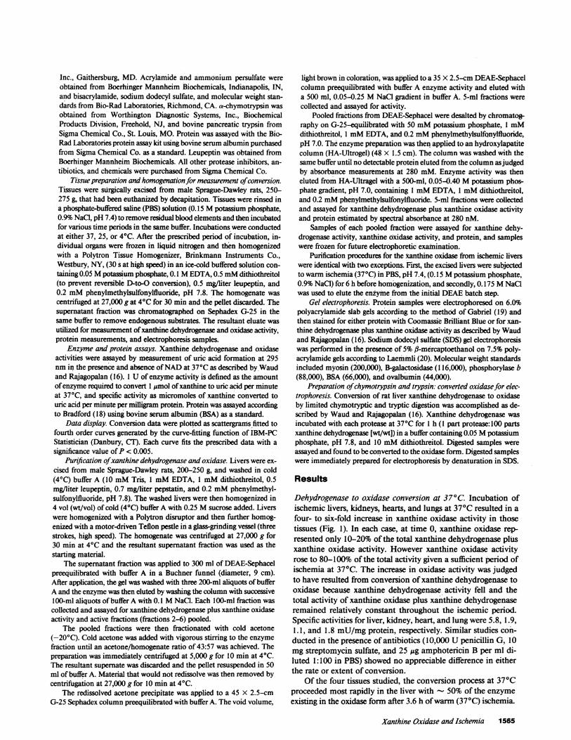
Electrophoretic examination ofxanthine dehydrogenase andoxidase in control and ischemic tissues. Samples from controllivers, kidneys, hearts, and lungs and from livers, kidneys, hearts,and lungs subjected to warm ischemia (8 h for liver, 24 h for allother tissues) were electrophoresed on 6% discontinuous poly-acrylamide gels and stained for xanthine dehydrogenase and ox-idase activity. As shown in Fig. 3, xanthine-oxidizing activitymigrated with a higher mobility in each of the ischemic tissuesthan in their matched controls, with the change in mobility leastpronounced in the ischemic heart.
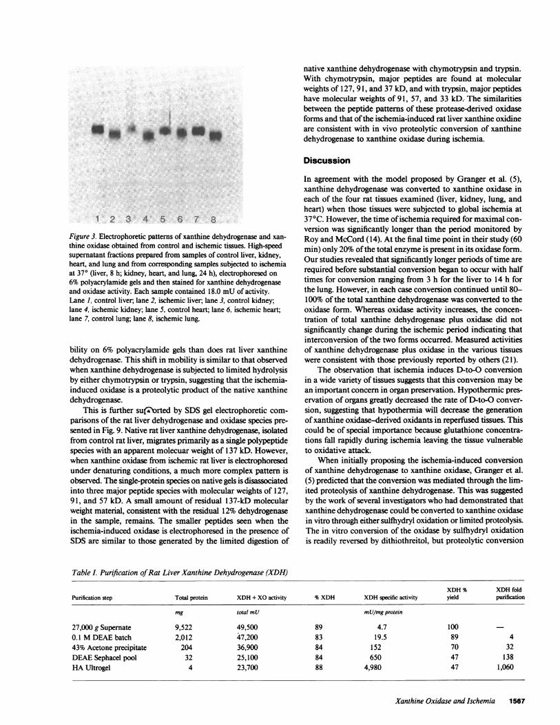
Purification of rat liver xanthine dehydrogenase. Results ofthe purification of xanthine dehydrogenase from control rat liversare shown in Table I. The enzyme was purified 1,060-fold relative
100- AX . . 100- B90 - A
X9
,80- x x 80-
g70-__ 7060
-XI
,
x 60 -2
50 5'40 X~ )' 40--
X 0 X So
Duration of Ischemia at 41C (Days) Duration of Ischemia
to the original 27,000 g supernatant fraction and has a specificactivity for dehydrogenase activity of 4,980 mUper mgprotein.47% of the initial activity was recovered using this purificationprotocol with - 4 mgof pure enzyme recovered from the 100g of fresh liver.
The inclusion of protease inhibitor and the sulfhydryl reagentdithiothreitol throughout the procedure successfully preventedthe conversion of xanthine dehydrogenase to xanthine oxidaseduring purification. The percentage of activity present in thedehydrogenase form remained relatively constant at - 88%throughout the entirety of the process.
The xanthine dehydrogenase preparation was judged to bepure based upon its elution from hydroxylapatite as a singlepeak of protein coincident with activity (Fig. 4) and upon elec-trophoresis of samples collected at various stages of protein pu-rification that showed the progressive purification of a singleprotein species (Fig. 5 A) comigrating with xanthine dehydrog-ehase activity (Fig. 5 B). Fig. 5 Balso indicates that no structuralchanges that can be detected by electrophoresis occur within thedehydrogenase during the purification procedure.
Purification of rat liver xanthine oxidasefrom ischemic livers.Results of the purification of xanthine oxidase from ischemicrat liver are shown in Table II. The ischemia-induced oxidasewas purified 78 1-fold with a specific activity for oxidase of 3,300mU/mgprotein. The yield of xanthine oxidase from the startingmaterials was 19 percent or 3 mgof protein from 140 g of isch-emic rat liver. As with the purification of the dehydrogenase,under the condition utilized for purification, there was little in-terconversion between oxidase and dehydrogenase forms, withthe final product being 88% oxidase.
The oxidase from ischemic livers was judged pure, basedupon its elution as a single, coincident peak of activity and pro-tein from hydroxylapatite (Fig. 6) and upon its appearance as asingle major band of protein on native polyacrylamide gels (Fig.7 A) comigrating with xanthine oxidase activity (Fig. 7 B). Asjudged by activity staining, no change in electrophoretic mobilityoccurred during the course of enzyme purification. This suggeststhat the final product is indeed the "native" form of the ischemia-induced xanthine oxidase. A small minor band of both proteinand coincident activity (< 5%) appeared at a slightly lower mo-bility than the major activity.
Electrophoretic comparisons ofxanthine dehydrogenase andischemia-induced xanthine oxidase. Fig. 8 shows an electropho-retic comparison on native gels of xanthine dehydrogenase fromcontrol rat livers and xanthine oxidase from ischemic rat liver.Also shown are rat liver xanthine oxidase forms generated bythe limited proteolysis of rat liver xanthine dehydrogenase bychymotrypsin and trypsin. Under nondenaturing conditions,xanthine oxidase from ischemic liver migrates with greater mo-
° . ~~~Figure 2. Conversion of xanthine dehydrogenase toxanthine oxidase at 4°. Rat tissues subjected tovarious lengths of ischemia at 40 were assayed forxanthine oxidase activity and xanthine dehydroge-nase plus xanthine oxidase activity. Data is plotted
- Heart as the percentage of xanthine dehydrogenase plusLung (*)
xanthine oxidase present in the oxidase form as a7 8 9 10 11 12 function of duration of ischemia at 4C. (A) kid-
at 4°C (Days) ney and liver; (B) heart and lung.
1566 Engerson, McKelvey, Rhyne, Boggio, Snyder, and Jones
native xanthine dehydrogenase with chymotrypsin and trypsin.With chymotrypsin, major peptides are found at molecularweights of 127, 91, and 37 kD, and with trypsin, major peptideshave molecular weights of 91, 57, and 33 kD. The similaritiesbetween the peptide patterns of these protease-derived oxidaseforms and that of the ischemia-induced rat liver xanthine oxidineare consistent with in vivo proteolytic conversion of xanthinedehydrogenase to xanthine oxidase during ischemia.
Discussion
1 2 3 4 5 6 7 8
Figure 3. Electrophoretic patterns of xanthine dehydrogenase and xan-thine oxidase obtained from control and ischemic tissues. High-speedsupernatant fractions prepared from samples of control liver, kidney,heart, and lung and from corresponding samples subjected to ischemiaat 370 (liver, 8 h; kidney, heart, and lung, 24 h), electrophoresed on6% polyacrylamide gels and then stained for xanthine dehydrogenaseand oxidase activity. Each sample contained 18.0 mUof activity.Lane 1, control liver, lane 2, ischemic liver; lane 3, control kidney;lane 4, ischemic kidney; lane 5, control heart; lane 6, ischemic heart;lane 7, control lung; lane 8, ischemic lung.
bility on 6% polyacrylamide gels than does rat liver xanthinedehydrogenase. This shift in mobility is similar to that observedwhen xanthine dehydrogenase is subjected to limited hydrolysisby either chymotrypsin or trypsin, suggesting that the ischemia-induced oxidase is a proteolytic product of the native xanthinedehydrogenase.
This is further su(-Zorted by SDS gel electrophoretic com-parisons of the rat liver dehydrogenase and oxidase species pre-sented in Fig. 9. Native rat liver xanthine dehydrogenase, isolatedfrom control rat liver, migrates primarily as a single polypeptidespecies with an apparent molecuar weight of 137 kD. However,when xanthine oxidase from ischemic rat liver is electrophoresedunder denaturing conditions, a much more complex pattern isobserved. The single-protein species on native gels is disassociatedinto three major peptide species with molecular weights of 127,91, and 57 kD. A small amount of residual 1 37-kD molecularweight material, consistent with the residual 12% dehydrogenasein the sample, remains. The smaller peptides seen when theischemia-induced oxidase is electrophoresed in the presence ofSDSare similar to those generated by the limited digestion of
In agreement with the model proposed by Granger et al. (5),xanthine dehydrogenase was converted to xanthine oxidase ineach of the four rat tissues examined (liver, kidney, lung, andheart) when those tissues were subjected to global ischemia at370C. However, the time of ischemia required for maximal con-version was significantly longer than the period monitored byRoy and McCord (14). At the final time point in their study (60min) only 20%of the total enzyme is present in its oxidase form.Our studies revealed that significantly longer periods of time arerequired before substantial conversion began to occur with halftimes for conversion ranging from 3 h for the liver to 14 h forthe lung. However, in each case conversion continued until 80-100% of the total xanthine dehydrogenase was converted to theoxidase form. Whereas oxidase activity increases, the concen-tration of total xanthine dehydrogenase plus oxidase did notsignificantly change during the ischemic period indicating thatinterconversion of the two forms occurred. Measured activitiesof xanthine dehydrogenase plus oxidase in the various tissueswere consistent with those previously reported by others (21).
The observation that ischemia induces D-to-O conversionin a wide variety of tissues suggests that this conversion may bean important concern in organ preservation. Hypothermic pres-ervation of organs greatly decreased the rate of D-to-O conver-sion, suggesting that hypothermia will decrease the generationof xanthine oxidase-derived oxidants in reperfused tissues. Thiscould be of special importance because glutathione concentra-tions fall rapidly during ischemia leaving the tissue vulnerableto oxidative attack.
When initially proposing the ischemia-induced conversionof xanthine dehydrogenase to xanthine oxidase, Granger et al.(5) predicted that the conversion was mediated through the lim-ited proteolysis of xanthine dehydrogenase. This was suggestedby the work of several investigators who had demonstrated thatxanthine dehydrogenase could be converted to xanthine oxidasein vitro through either sulfhydryl oxidation or limited proteolysis.The in vitro conversion of the oxidase by sulfhydryl oxidationis readily reversed by dithiothreitol, but proteolytic conversion
Table I. Purification of Rat Liver Xanthine Dehydrogenase (XDH)
XDH% XDHfoldPurification step Total protein XDH+ XOactivity %XDH XDHspecific activity yield purification
mg total mU mU/mgprotein
27,000 g Supernate 9,522 49,500 89 4.7 1000.1 MDEAEbatch 2,012 47,200 83 19.5 89 443% Acetone precipitate 204 36,900 84 152 70 32DEAESephacel pool 32 25,100 84 650 47 138HAUltrogel 4 23,700 88 4,980 47 1,060
Xanthine Oxidase and Ischemia 1567

60 80 100 120FRACTION NUMBER
Figure 4. Elution profile of xanthine dehydrogenase from HA-Ultrogel.
is irreversible (16, 22). Because the newly formed oxidase activityin ischemic intestine can not be reconverted to the dehydrogenaseform with dithiothreitol (5), it was proposed that the activationevent is proteolytic in nature.
The discovery of a structurally distinct species of the enzymethat arises concurrently with conversion of the enzyme from itsdehydrogenase to oxidase form is consistent with that proposal.This distinct form, isolated from the various ischemic tissues,migrates with a higher electrophoretic mobility, as does xanthineoxidase generated from in vitro proteolysis of xanthine dehy-drogenase ( 15, 20).
In an attempt to study the nature of the alteration resultingin this distinct form, the dehydrogenase form of the enzymefrom control rat liver and the oxidase form from ischemic rat
A
* -. aW t *9of
*. ..
B
Figure 6. Elution profile of ischemia-induced xanthine oxidase fromHA-Ultrogel.
liver were purified. The utilized protocols produced highly pu-rified enzyme species with specific activities and fold purificationsgreater than those previously reported (16, 23). More importantlythough, through the use of protease inhibitors and dithiothreitol,it was possible to prevent any interconversion or alterations ofthe enzyme forms during the course of purification. The enzymefrom control liver was maintained as 88% dehydrogenasethroughout the procedure and represents the highest percentageof dehydrogenase activity in any preparation to date. The purifiedenzymes were stable for > 8 wk when stored at -70°C in thepresence of dithiothreitol. Xanthine oxidase from the ischemictissue remained in the oxidase form as well during the purifi-
_-B
1A ,6* in*~~~0-1
1 2 3 4 5 1 2 3 4 5
Figure 5. Electrophoretic examination of the purification protocol forxanthine dehydrogenase. Samples at various stages of purification ofxanthine dehydrogenase were electrophoresed on 6% native polyacryl-amide gels and then stained for either protein with Coomassie Bril-liant Blue or for activity according to Waudand Rajagopalan (5). (A)is the protein stain, with lane I containing 150yg of the 27,000 g su-pernate; lane 2, 100 ,ug of the pooled DEAE-batched step; lane 3, 501g of the resuspended acetone pellet; lane 4, 20 Mg of the DEAE-col-umn pool; and lane 5, 5 ug of the HA-Ultrogel pool. (B) is activity-stained fractions. Each fraction contains 1.25 mUof total activity.Lane 1, 27,000 g supernate; lane 2, DEAE-batch step; lane 3, acetoneprecipitate; lane 4, DEAE-column pool; and lane 5, HA-Ultrogel pool.
1 2 3 4 5 1 2 3 4 5
Figure 7. Electrophoretic examination of the protocol used for purifi-cation of xanthine oxidase from ischemic rat livers. Samples at variousstages of purification of ischemia-induced xanthine oxidase were elec-trophoresed on 6%native polyacrylamide gels and then stained for ei-ther protein with Coomassie Brilliant Blue or for activity according toWaudand Rajagopalan (5). (A) is the protein stain, with lane I con-taining 150 Mug of the 27,000 g supernate; lane 2, 100 ug of the pooledDEAE-batched step; lane 3, 50 Mug of the resuspended acetone pellet;lane 4, 20 Mgof the DEAE-column pool; and lane 5, 5 Mug of the HA-Ultrogel pool. (B) is activity-stained fractions. Each fraction contains1.25 mUof total activity. Lane 1, 27,000 g supernate; lane 2, DEAE-batch step; lane 3, acetone precipitate; lane 4, DEAE-column pool;and lane 5, HA-Ultrogel pool.
1568 Engerson, McKelvey, Rhyne, Boggio, Snyder, and Jones

Table I. Purification of Rat Liver Xanthine Oxidase (XO)
XO% XOfoldPurification step Total protein XDH+ XOactivity %XO XOspecific activity yield purification
mg total mU mU/mgprotein
27,000 g Supernate 12,130 52,000 100 4.3 1000.175 MDEAEbatch 1,357 51,800 100 38.1 99 943% Acetone precipitate 87 28,600 84 277 46 64DEAESephacel pool 20 15,400 75 579 22 135HAUltrogel 3 12,000 84 3,360 19 781
cation process. Gel patterns of samples electrophoresed andstained for activity showed that no physical changes resulting inchanges in electrophoretic mobility occurred during the coursesof purification. This integrity of the nature of the starting materialin the final preparation was critical if comparisons were to bemade between the two forms because in vitro interconversionof the dehydrogenase to the oxidase could have occurred if properprecautions were not taken (15-17, 23). Webelieve that thesedata demonstrate that the utilized purification protocols main-tained the general integrity of both the dehydrogenase and theischemia-induced oxidase.
A comparison of rat liver xanthine dehydrogenase and itsischemic-induced oxidase form indicated that the oxidase is aproteolytic product of the dehydrogenase. Whereas both formsof the enzyme (dehydrogenase and oxidase) migrated as singlespecies on nondenaturing polyacrylamide gels, denaturation re-vealed that the oxidase form had been cleaved. Xanthine de-hydrogenase has one major subunit of molecular weight 137kD, whereas the ischemia-induced oxidase slowed multiplepolypeptides of 127, 91, and 57 kD along with some residual137 kD polypeptide. Cleavage of the enzyme during ischemiaproduced an SDSgel profile similar to that observed when xan-thine dehydrogenase was converted to the oxidase form by lim-ited digestion with trypsin or chymotrypsin and resulted in ashift in mobility for the enzyme on native gels similar to thatobserved when the dehydrogenase was converted to the oxidaseby limited proteolysis.
The molecular weight for the rat liver dehydrogenase andits proteolytic products are comparable to those previously re-ported by Waud and Rajagopalan (16). The native dehydroge-
Figure 8. Electrophoreticcomparisons of native ratliver xanthine dehydroge-nase and ischemia-inducedxanthine oxidase. 10-,gsamples of the native xan-
4 thine dehydrogenase (lanesI and 6), ischemia-inducedxanthine oxidase (lanes 2and 5), trypsin-treated xan-thine dehydrogenase (lane3), and chymotrypsin-treated xanthine dehydroge-nase (lane 4) were electro-phoresed as 6%native poly-acrylamide gels and stained
1 2 3 4 5 6for protein with Coomassie
123456 Brilliant Blue.
nase in those studies was reported to have a subunit molecularweight on SDS-urea gels of 150 kD compared with our mea-surement of 137 kD. Differences in molecular weight estimatesbetween the studies may reflect differences in the electrophoreticsystems (the presence or absence of urea) or differences in stan-dards because in the study of Waudand Rajagopalan (16) bovinemilk xanthine oxidase was used as the upper limit standard andwas ascribed a molecular weight value of 150 kD. Nevertheless,the molecular weights of the degradation products were similar.In each case a major protein species is present at a molecularweight 10-15 kD smaller than the native dehydrogenase. Similarbands are also present at molecular weights of - 90 and 60 kD.
There are at least two potential types of proteases that mightmediate the conversion process in ischemic tissues. Lysosomalproteases, physically separated from the dehydrogenase in nor-mal tissues, could be released from lysosomes damaged duringischemia. The released proteases could then mediate the con-
rn *wo
1 2 3 4 5 6- 7 8 9Figure 9. SDSgel profiles of native rat liver xanthine dehydrogenaseand ischemia-induced xanthine oxidase. Samples of native rat liverxanthine dehydrogenase (5 ,ug), ischemia-induced xanthine oxidase (15ug), trypsin-treated xanthine dehydrogenase (20 Mg), and chymotryp-sin-treated xanthine dehydrogenase were electrophoresed on 7.5%polyacrylamide gels in the presence of SDSand B-mercaptoethanoland then stained with Coomassie Brilliant Blue. Standards in lanes Iand 9 include myosin (200,000), B-galactosidase (116,000), phospho-rylase b (88,000), BSA (66,000), and ovalbumin (44,000). Lanes 2, 3,and 8 contain the native dehydrogenase; lanes 4 and 7, the ischemia-induced oxidase; lane 5, trypsin-treated dehydrogenase; and lane 6,chymotrypsin-treated dehydrogenase.
Xanthine Oxidase and Ischemia 1569

version process. Alternatively, conversion could be mediated bya cytosolic protease whose activity is stimulated during ischemia.One potential candidate would be the cytosolic calcium-depen-dent proteases.
Cytosolic proteases activated by micromolar concentrationsof calcium are present in the liver (24), and it is well documentedthat intracellular calcium concentrations rise during ischemiato levels that would trigger the action of these proteases. Schafferet al. have shown that the dehydrogenase can be converted tothe oxidase by increases in intracellular calcium even in theabsence of ischemia (25), and recently, we have found in pre-liminary studies that calcium-dependent proteases cause theconversion of rat liver xanthine dehydrogenase to xanthine ox-idase in vitro (data not shown). Nevertheless, convincing evi-dence for the involvement of the calcium-dependent proteasesin the conversion process must await careful analysis of thecleavage products of the ischemia-induced oxidase and com-parison of those products to those obtained by the in vitro con-version of the enzyme by the calcium-dependent proteases.
In conclusion, proteolytic conversion of xanthine dehy-drogenase to xanthine oxidase occurs during ischemia in theliver. This conversion may be an important factor in reperfusioninjury in both the liver and other organs. Therefore, understand-ing the conversion process and its regulation may prove impor-tant in designing new agents and devising new strategies for organpreservation and for limiting reperfusion injury in a variety ofother pathological situations leading to reperfusion injury.
Acknowledgments
The authors wish to thank Dr. Matthew Grisham and Dr. D. N. Grangerfor their help in preparation of the manuscript.
References
1. Chambers, D. E., D. A. Parks, G. Patterson, S. Yoshida, K. Burton,L. F. Parmley, J. M. McCord, and J. M. Downey. 1983. Role of oxygenderived radicals in myocardial ischemia. Fed. Proc. 42:1093.
2. Jolly, S. R., W. J. Kane, M. B. Bailie, G. D. Abrams, and B. R.Lucchesi. 1984. Canine myocardial reperfusion injury: its reduction bythe combined administration of superoxide plus superoxide dismutase.Circ. Res. 54:277-286.
3. Hearse, D. J., A. S. Manning, J. M. Downey, and D. M. Yellon.1986. Xanthine oxidase: a critical mediator of myocardial injury duringischemia and reperfusion? Acta Physiol. Scand. 126:65-78.
4. Hansson, R. O., 0. Jonsson, J. Lundstam, S. Petterson, and T.Schersten. 1983. Effects of free radical scavengers on renal circulationafter ischemia in the rabbit. Clin. Sci. 65:605-6 10.
5. Granger, D. N., G. Rutili, and J. M. McCord. 1981. Role of su-peroxide radicals in intestinal ischemia. Gastroenterology. 81:22-29.
6. Parks, D. A., G. B. Bulkley, D. N. Granger, S. R. Hamilton, andJ. M. McCord. 1982. Ischemic injury in the cat small intestine: role ofsuperoxide radicals. Gastroenterology. 82:9-15.
7. Adkison, D., M. E. Hollwarth, J. N. Benoit, D. A. Parks, J. M.
McCord, and D. N. Granger. 1986. Role of free radicals in ischemia-reperfusion injury to the liver. Acta Physiol. Scand. 126:101-107.
8. Parks, D. A., and D. N. Granger. 1983. Ischemia-induced vascularchanges: role of xanthine oxidase and hydroxyl radicals. Am. J. Physiol.245:G285-289.
9. Owens, M. L., H. Lazarus, M. W. Wolcott, J. G. Maxwell, and B.Taylor. 1974. Allopurinol and hypoxanthine pretreatment of caninekidney donors. Transplantation 17:424-427 (1974).
10. Koyama, I., G. B. Bulkley, G. M. Williams, and M. J. Im. 1985.The role of oxygen free radicals in mediating the reperfusion injury ofcold-preserved ischemic kidneys. Transplantation (Baltimore). 40:590-595.
11. Crowell, J. W., C. E. Jones, and E. E. Smith. 1969. Effect ofallopurinol of hemorrhagic shock. Am. J. Physiol. 216:744-748.
12. Granger, D. N., M. E. Hollwarth, and D. A. Parks. 1986. Ischemia-reperfusion injury: role of oxygen-derived free radicals. Acta Physiol.Scand. 126:47-63.
13. Manning, A. S., D. J. Coltart, and D. J. Hearse. 1984. Ischemiaand reperfusion-induced arrhythmias in the rat. Effects of xanthine ox-idase inhibition with allopurinol. Circ. Res. 55:545-548.
14. Roy, R. S., and J. M. McCord. 1983. Superoxide and ischemia:conversion of xanthine dehydrogenase to xanthine oxidase. In Proceed-ings of the Third International Conference on Superoxide and SuperoxideDismutases. R. Greenwald and G. Cohen, editors. Elsevier/North HollandBiomedical Press, NewYork. 145-153.
15. Batelli, M. G., E. Lorenzoni, and F. Stirpe. 1973. Milk oxidasetype D (Dehydrogenase) and type 0 (Oxidase). Purification, intercon-version, and some properties. Biochem. J. 131:191-198.
16. Waud, W. R., and K. V. Rajagopalan. 1976. Purification andproperties of the NAD+-dependent (Type D) and O2-dependent (Type0) form of rat liver xanthine dehydrogenase. Arch Biochem. Biophys.172:354-364.
17. Della Corte, E., and F. Stirpe. 1972. The regulation of rat liverxanthine oxidase. Involvement of thiol groups in the conversion of theenzyme activity from dehydrogenase (type D) into oxidase (type 0) andpurification of the enzyme. Biochem. J. 126:739-745.
18. Bradford, M. 1970. A rapid and sensitive method for the quan-titation of microgram quantities of protein utilizing the principle of pro-tein-dye binding. Anal. Biochem. 72:240-254.
19. Gabriel, 0. 1971. Analytical disc gel electrophoresis. MethodsEnzymol. 22:565-577.
20. Laemmli, U. K. 1970. Cleavage of structural proteins during theassembly of the head of bacteriophage T4. Nature (Lond.). 227:680-685.
21. Krenitsky, T. A., J. V. Tuttle, E. L. Catlau, and P. Wang. 1974.A comparison of the distribution and electron acceptor specificities ofxanthine oxidase and aldehyde oxidase. Comp. Biochem. Physiol. 49B:687-703.
22. Batelli, M. G., E. Lorenzoni, and F. Stirpe. 1973. Milk oxidasetype D (Dehydrogenase) and type 0 (Oxidase). Purification, intercon-version, and some properties. Biochem. J. 131:191-198.
23. Della Corte, E., and F. Stirpe. 1972. The regulation of rat liverxanthine oxidase. Biochem. J. 126:739-745.
24. DeMartino, G. N. 1981. Calcium-dependent proteolytic activityin rat liver: identification of two proteases with different calcium re-quirements. Arch Biochem. Biophys. 211:253-257.
25. Schaffer, S. W., R. S. Roy, and J. M. McCord. 1983. Possiblerole for calmodulin in calcium paradox-induced heart failure. Eur. HeartJ. 4:81-87.
1570 Engerson, McKelvey, Rhyne, Boggio, Snyder, and Jones