Embed Size (px)
Citation preview
Copyright 2009, John Wiley & Sons, Inc.
Chapter 22: The Lymphatic System and Immunity

Copyright 2009, John Wiley & Sons, Inc.
Immunity or Resistance Ability to ward off damage or disease through our
defenses 2 types of immunity Innate or nonspecific immunity – present at birth
(Table 22.1, p891) No specific recognition of invaders, no memory
component 1st and 2nd line of defenses
Adaptive or specific immunity Specific recognition of invaders with a memory
component

Copyright 2009, John Wiley & Sons, Inc.
Lymphatic system structure and function Consists of lymph (liquid), lymphatic
vessels, structures and organs containing lymphatic tissue, red bone marrow
Functions of the lymphatic system1. Drain excess interstitial fluid
2. Transport dietary lipid
3. Carry our immune responses
Copyright 2009, John Wiley & Sons, Inc.
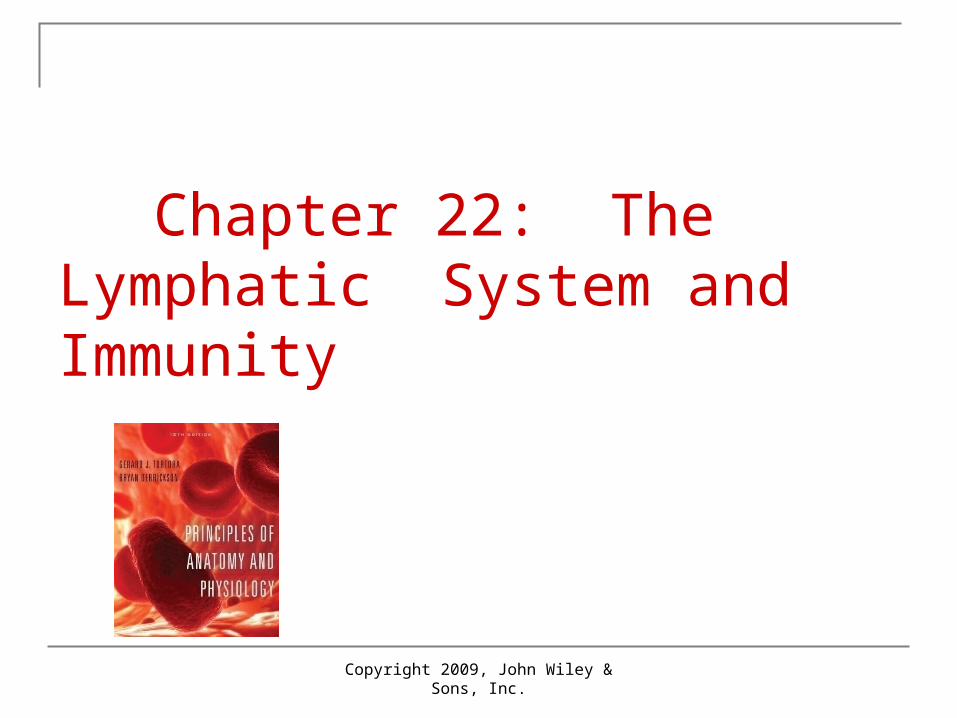
Components of the Lymphatic System
See page 833 for potential problem

Copyright 2009, John Wiley & Sons, Inc.
Lymphatic vessels and lymph circulation
Vessels begin as lymphatic capillaries Closed at one end
Unite to form large lymphatic vessels Resemble veins in structure but thinner
walls and more valves Passes through lymph nodes
Encapsulated organs with masses and B and T cells
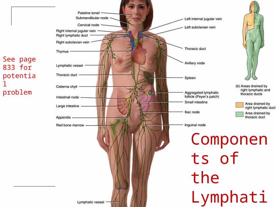
Definitions: T Cells = lymphocytes that mediate celluar immunity;
including helper and cytotoxoic (aka T lymphocytes). Origin – Thymus.
B Cells = (aka B lymphocytes) oversee humoral (fluids) immunity; their descendants differentiate into antibody-producing plasma cells. Origin – Red Bone marrow.
Lymphocyte – agranular white blood cell via bone marrow maturing in lymphoid organs of the body
Pluripotent stem cells - can develop into most of the specialized cells and tissues of the body, are self-renewing (see page Blood Chapter)
Antibody (protein), Antigen (foreign substance)
Copyright 2009, John Wiley & Sons, Inc.
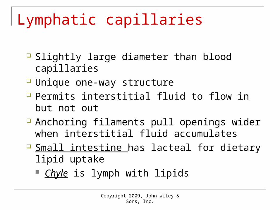
Lymphatic capillaries
Slightly large diameter than blood capillaries Unique one-way structure Permits interstitial fluid to flow in but not out Anchoring filaments pull openings wider when
interstitial fluid accumulates Small intestine has lacteal for dietary lipid
uptake Chyle is lymph with lipids
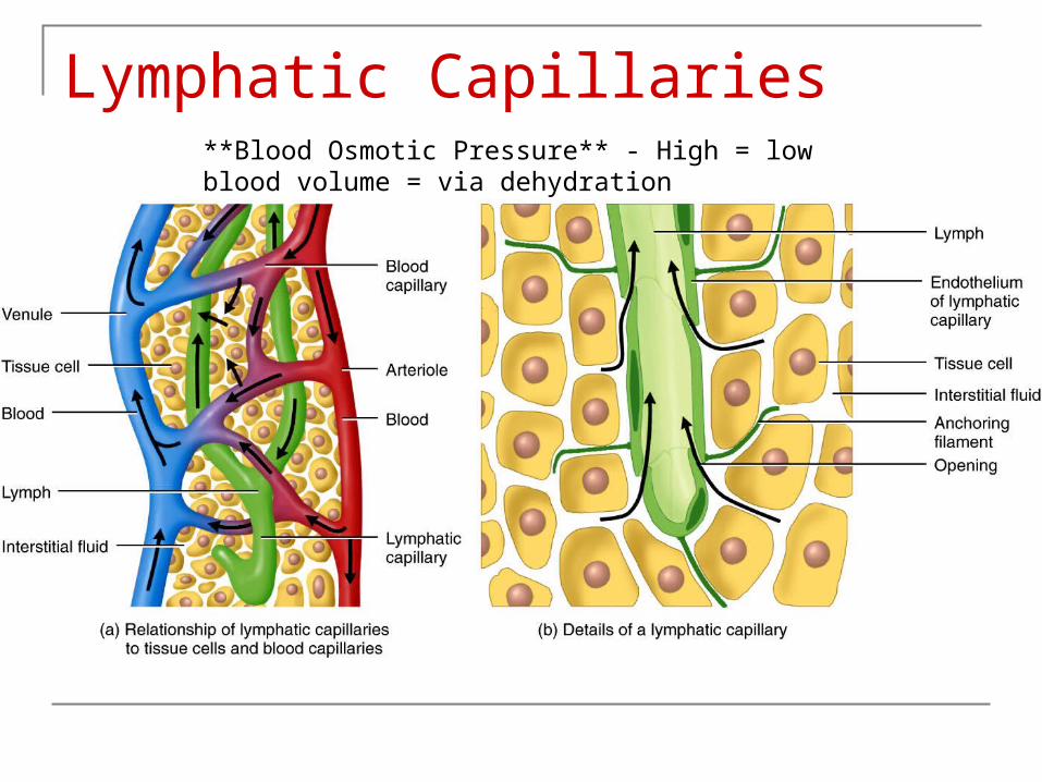
Lymphatic Capillaries**Blood Osmotic Pressure** - High = low blood volume = via dehydration

Copyright 2009, John Wiley & Sons, Inc.
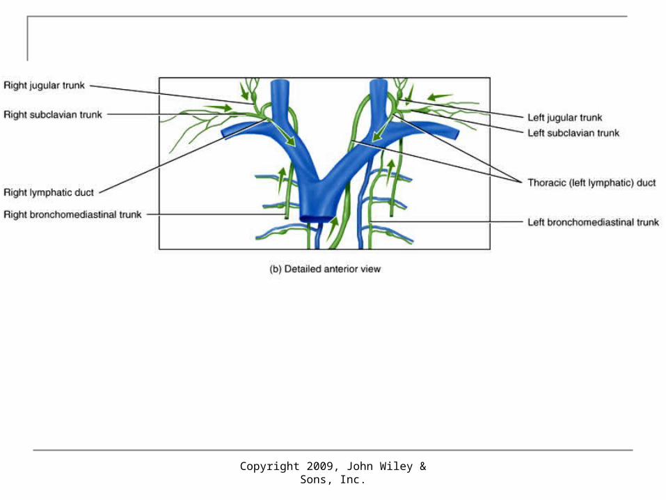
Lymph trunks and ducts
Vessels unite to form lymph trunks Principal trunks are the lumbar, intestinal,
bronchomediastinal, subclavian and jugular Passes from lymph trunks into 2 main
channels (thoracic and right lymphatic ducts) before draining into venous blood

Routes for drainage of lymph
Copyright 2009, John Wiley & Sons, Inc.

Copyright 2009, John Wiley & Sons, Inc.
Formation and flow of lymph More fluid filters out of blood capillaries than
returns to them by reabsorption Excess filtered fluid – about 3L/day – drains into
lymphatic vessels and become lymph Important function of lymphatic vessels to return
lost plasma proteins to blood stream Contain valves Same 2 “pumps” aiding venous return also used
Skeletal muscle pump – milking action Respiratory pump – pressure changes during breathing
Copyright 2009, John Wiley & Sons, Inc.
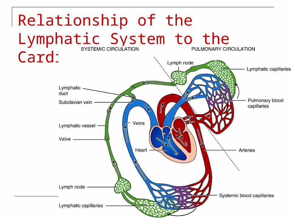
Relationship of the Lymphatic System to the Cardiovascular System
Copyright 2009, John Wiley & Sons, Inc.
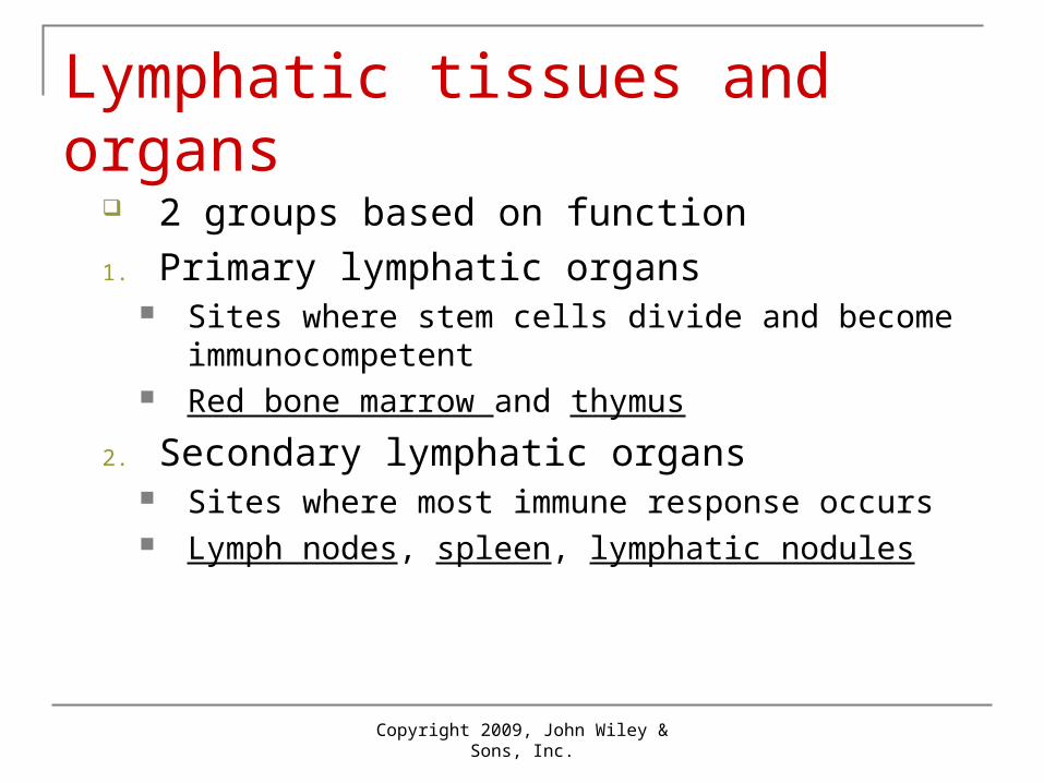
Lymphatic tissues and organs
2 groups based on function
1. Primary lymphatic organs Sites where stem cells divide and become
immunocompetent Red bone marrow and thymus
2. Secondary lymphatic organs Sites where most immune response occurs Lymph nodes, spleen, lymphatic nodules

Copyright 2009, John Wiley & Sons, Inc.
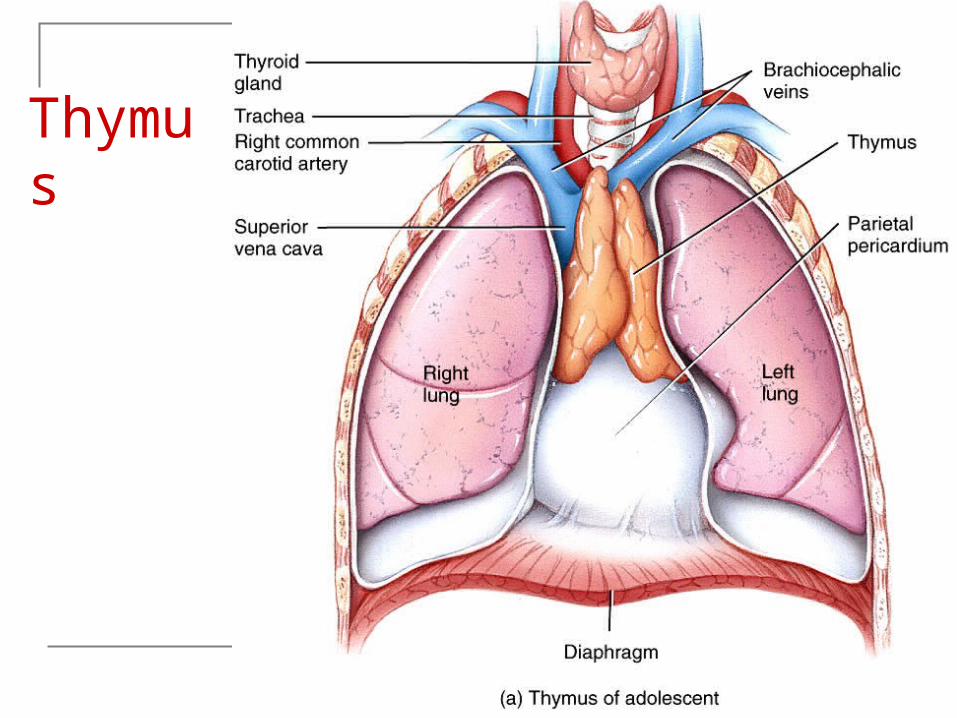
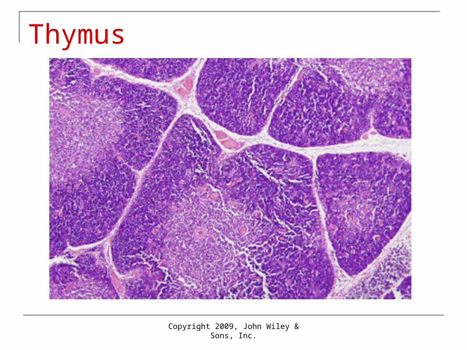
Thymus and Medulla
Thymus Outer cortex composed of large number of T cells
Immature T cells migrate here from red bone marrow where they proliferate and begin to mature
Dendritic cells derived from monocytes (largest WBC) assist in T cell maturation
Specialized epithelial cells help educate T cells through positive selection – only about 2% survive
Macrophages (phagocyte derived from monocyte, free or fixed) clear out dead and dying cells
Medulla More mature T cells migrate here from cortex More epithelial cells, dendritic cells and macrophages
Thymus shrinks with age from 70g in infants to 3g in old age
Copyright 2009, John Wiley & Sons, Inc.
Thymus

Copyright 2009, John Wiley & Sons, Inc.
Copyright 2009, John Wiley & Sons, Inc.
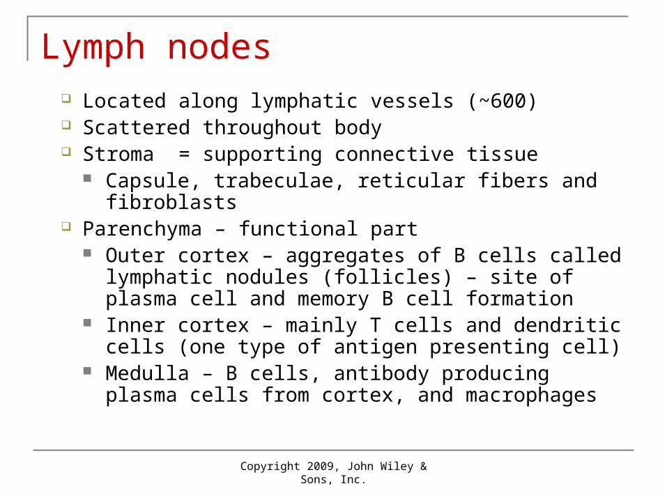
Lymph nodes Located along lymphatic vessels (~600) Scattered throughout body Stroma = supporting connective tissue
Capsule, trabeculae, reticular fibers and fibroblasts Parenchyma – functional part
Outer cortex – aggregates of B cells called lymphatic nodules (follicles) – site of plasma cell and memory B cell formation
Inner cortex – mainly T cells and dendritic cells (one type of antigen presenting cell)
Medulla – B cells, antibody producing plasma cells from cortex, and macrophages

Copyright 2009, John Wiley & Sons, Inc.
Lymph Lymph flows through a node in 1 direction only
Enters through afferent lymphatic vessels Directs lymph inward Lymph enters sinuses (irregular channels) Into medulla Medullary sinuses drain into efferent lymphatic vessels Conveys lymph, antibodies and activated T cells out of the
node Lymph nodes function as a filter
Foreign substances trapped Destroyed by macrophages or immune response of
lymphocytes

Copyright 2009, John Wiley & Sons, Inc.
Structure of a Lymph Node

Copyright 2009, John Wiley & Sons, Inc.
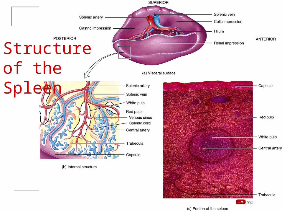
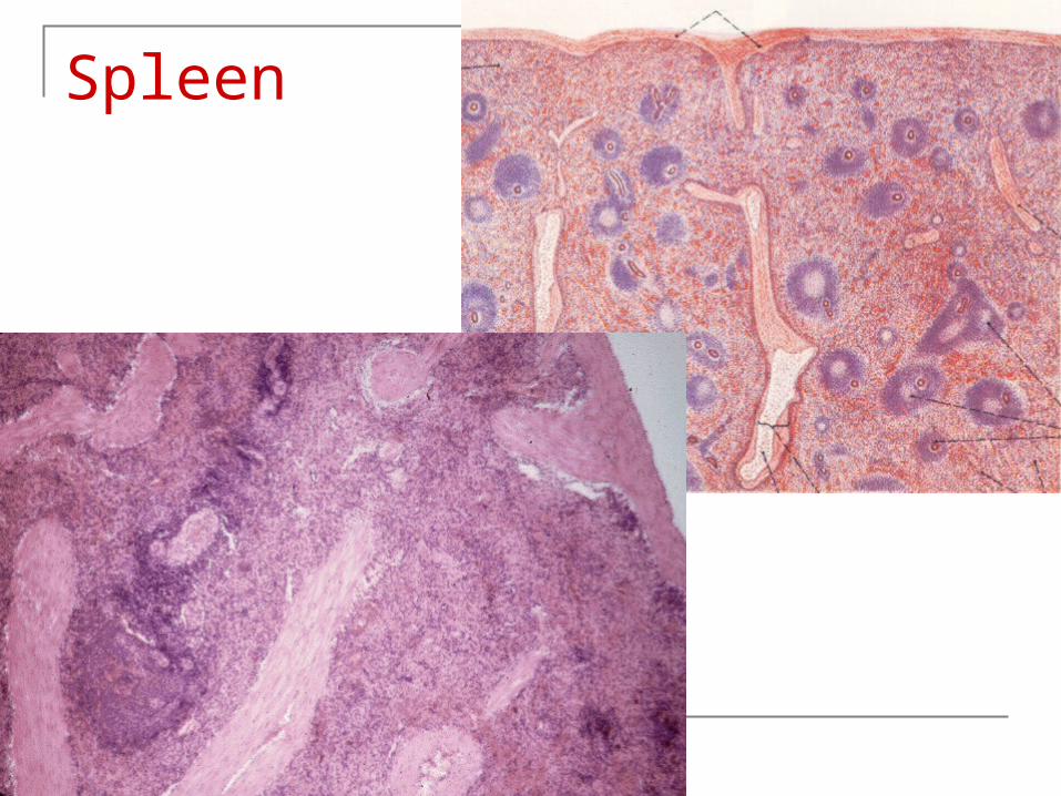
Spleen
Largest single mass of lymphatic tissue in the body
Stroma – capsule, trabeculae, reticular fibers, and fibroblasts
Parenchyma White pulp – lymphatic tissue (lymphocytes
and macrophages) B cells and T cells carry out immune
function Red pulp…
Copyright 2009, John Wiley & Sons, Inc.
Red Pulp
Red pulp – blood-filled venous sinuses and splenic (Bilroth’s) cords – red blood cells, macrophages, lymphocytes, plasma cells, and granulocytes. Functions: Macrophages breakdown ruptured, worn out or
defective blood cells Storage of up to 1/3 of body’s platelet supply Production of blood cells during fetal life
Copyright 2009, John Wiley & Sons, Inc.
Structure of the Spleen
Copyright 2009, John Wiley & Sons, Inc.
Lymphatic nodules
Not surrounded by a capsule Scattered throughout lamina propria
(connective tissue) of mucous membranes lining GI, urinary, reproductive tract
Mucosa-associated lymphatic tissue (MALT) of respiratory tract
Most small and solitary Some larger – tonsils, Peyer’s patches,
appendix

Copyright 2009, John Wiley & Sons, Inc.
Innate immunity
First line of defenses: Skin and mucous membranes Provide both physical and chemical barriers Physical barriers
Epidermis – closely packed, keratinized cells Periodic shedding
Mucous membranes Mucus traps microbes and foreign substances
Nose hairs trap and filter Cilia of upper respiratory tract propel trapped particles
up and out
Copyright 2009, John Wiley & Sons, Inc.
Innate Immunity Fluids
Lacrimal apparatus of eye Washing action of tears Lysozyme breaks down bacterial cell walls – also present in
saliva, perspiration, nasal secretions, and tissue fluids Saliva washes mouth Urine cleanses urinary system Vaginal secretions defecation vomiting
Chemicals Sebaceous (oil) glands secrete sebum – protective film, acid Perspiration, gastric juice, vaginal secretions – all acidic
Copyright 2009, John Wiley & Sons, Inc.
Second line of defenses: Internal defenses Antimicrobial substances
1. Interferons Produced by lymphocytes, macrophages, and fibroblasts
infected by viruses Prevents replication in neighboring uninfected cells
2. Complement Proteins in blood plasma and plasma membranes “complement” or enhance certain immune reactions Causes cytolysis of microbes, promotes phagocytosis,
contributes to inflammation

Copyright 2009, John Wiley & Sons, Inc.
Internal Defenses
3. Iron-binding proteins Inhibit growth of bacteria by reducing available
iron
4. Antimicrobial proteins (AMPs) Short peptides that have a broad spectrum of
antimicrobial activity Can attract dendritic cells and mast cells that
participate in immune responses
Copyright 2009, John Wiley & Sons, Inc.
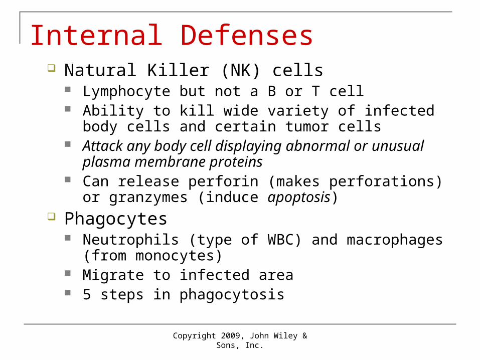
Internal Defenses Natural Killer (NK) cells
Lymphocyte but not a B or T cell Ability to kill wide variety of infected body cells and
certain tumor cells Attack any body cell displaying abnormal or unusual
plasma membrane proteins Can release perforin (makes perforations) or granzymes
(induce apoptosis) Phagocytes
Neutrophils (type of WBC) and macrophages (from monocytes)
Migrate to infected area 5 steps in phagocytosis

Copyright 2009, John Wiley & Sons, Inc.
Phagocytosis of a microbe
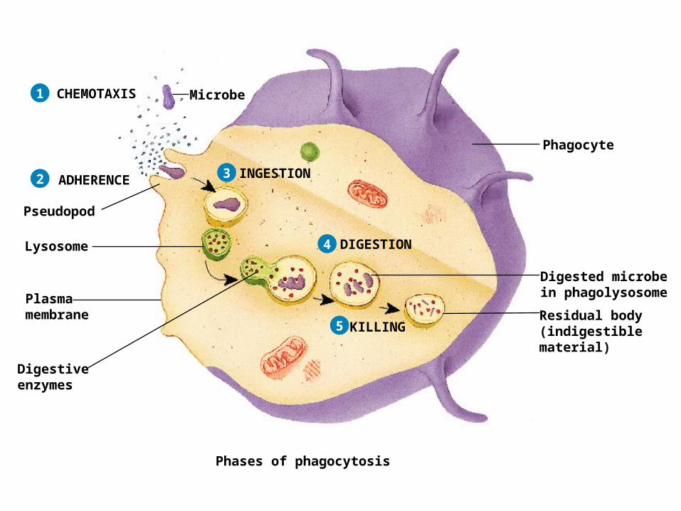
1 MicrobeCHEMOTAXIS
Phagocyte
Phases of phagocytosis
1
Phases of phagocytosis
MicrobeCHEMOTAXIS
Pseudopod
Phagocyte
ADHERENCE2
1 MicrobeCHEMOTAXIS
Lysosome
Pseudopod
Phagocyte
ADHERENCE INGESTION2 3
Phases of phagocytosis
1 MicrobeCHEMOTAXIS
Lysosome
Digestiveenzymes
Pseudopod
Phagocyte
ADHERENCE INGESTION
Plasmamembrane
DIGESTION
2 3
4
Phases of phagocytosis
1 MicrobeCHEMOTAXIS
Lysosome
Digestiveenzymes
Pseudopod
Phagocyte
ADHERENCE INGESTION
Plasmamembrane
DIGESTION
KILLINGResidual body(indigestiblematerial)
Digested microbein phagolysosome
2 3
4
5
Phases of phagocytosis
Copyright 2009, John Wiley & Sons, Inc.
Inflammation
Nonspecific, defensive response of body to tissue damage
4 signs and symptoms – redness, pain, heat and swelling
Attempt to dispose of microbes, prevent spread, and prepare site for tissue repair
3 basic stages Vasodilation and increased blood vessel permeability Emigration Tissue repair

Copyright 2009, John Wiley & Sons, Inc.
Vasodilation and increased permeability of blood vessels
Increased diameter of arterioles allows more blood flow through area bringing supplies and removing debris
Increased permeability means substances normally retained in the blood are permitted to pass out – antibodies and clotting factors
Histamine – released due to injury via mast cells (cell in areolar connective tissue), platelets, and Basophil (type of WBC)
Kinins – polypeptides, that VD and promote chemotaxis prostaglandins (PGs) – chemotaxis and enhance response of 1st two. leukotrienes (LTs) – chemotaxis, phagocyte attachment, via basophils. complement - similar to above traits and can also kill some bacteria.

Inflammation
Copyright 2009, John Wiley & Sons, Inc.
Emigration of phagocytes
Depends on chemotaxis Neutrophils predominate in early stages but
die off quickly Monocytes transform into macrophages
More potent than neutrophils Pus – pocket of dead phagocytes and
damaged tissue
Copyright 2009, John Wiley & Sons, Inc.
Adaptive immunity
Ability of the body to defend itself against specific invading agents
Antigens (Ags) – substances recognized as foreign and provoking an immune response
Distinguished from innate immunity by Specificity Memory
Copyright 2009, John Wiley & Sons, Inc.
Maturation of T cells and B cells
Both develop from pluripotent stem cells originating in red bone marrow B cells complete their development in red bone marrow T cells develop from pre-T cells that migrate from red
bone marrow to the thymus 2 types - Helper T cells (CD4 T cells) and cytotoxic T cells
(CD8 T cells)
Immunocompetence – ability to carry out adaptive immune response Have antigen receptors to identify specific antigen
Mechanisms of Antigen Entrance Enters Skin - Lymph vessels – L. Nodes
Lymph nodules Enter mucus membrane – M.A.L.T. Enters blood – Spleen
Copyright 2009, John Wiley & Sons, Inc.
Copyright 2009, John Wiley & Sons, Inc.
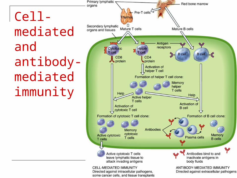
Cell-mediated and antibody-mediated immunity

Copyright 2009, John Wiley & Sons, Inc.
Copyright 2009, John Wiley & Sons, Inc.

Copyright 2009, John Wiley & Sons, Inc.

Copyright 2009, John Wiley & Sons, Inc.
2 types of adaptive immunity
Cell-mediated Cytotoxic T cells directly attack invading antigens Particularly effective against intracellular pathogens, some
cancer cells and foreign tissue transplants Antibody-mediated (B cells stay in secondary LT)
B cells transform into plasma cells making antibodies (Abs) or immunoglobulins
Works against extracellular pathogens in fluids outside cells Helper T cells aid in both types
2 types of immunity work together

Copyright 2009, John Wiley & Sons, Inc.
Clonal selection
Process by which a lymphocyte proliferates and differentiates in response to a specific antigen Clone – population of identical cells all recognizing the same
antigen as original cell Lymphocyte undergoes clonal selection to produce
Effector cell – active helper T cell, active cytotoxic T cell, plasma cell; all die after immune response
Memory cell – do not participate in initial immune response, respond to 2nd invasion by proliferating and differentiating into more effector and memory cells, long life spans (decades)

Copyright 2009, John Wiley & Sons, Inc.
Antigens
Antigens have 2 characteristics Immunogenicity – ability to
provoke immune response Reactivity – ability of
antigen to react specifically with antibodies it provoked
Entire microbes may act as antigen
Typically, just certain small parts of large antigen molecule triggers response (epitope or antigenic determinant)

Copyright 2009, John Wiley & Sons, Inc.

Copyright 2009, John Wiley & Sons, Inc.
Diversity of antigen receptors
Human immune system able to recognize and bind to at least a billion different epitopes
Result of genetic recombination – shuffling and rearranging of a few hundred versions of several small gene segments
Major Histocompatibility Complex Antigens MHC or human leukocyte antigens (HLA) Normal function to help T cells recognize foreign or self Class I MHC (MHC-I) – built into all body cells except
RBCs Class II MHC (MHC-II) – only on antigen presenting cells
(APC)

Copyright 2009, John Wiley & Sons, Inc.
Pathways of antigen processing
B cells can recognize and bind to antigens T cells must be presented with processed
antigens Antigenic proteins are broken down into peptide
fragments and associated with MHC molecules Antigen presentation – antigen-MHC complex
inserted into plasma membrane Pathway depends on whether antigen is outside or
inside body cells
Copyright 2009, John Wiley & Sons, Inc.
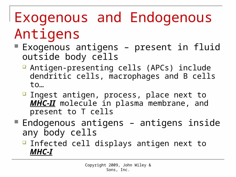
Exogenous and Endogenous Antigens Exogenous antigens – present in fluid outside
body cells Antigen-presenting cells (APCs) include dendritic
cells, macrophages and B cells to… Ingest antigen, process, place next to MHC-II
molecule in plasma membrane, and present to T cells
Endogenous antigens – antigens inside any body cells Infected cell displays antigen next to MHC-I

Copyright 2009, John Wiley & Sons, Inc.
Exogenous Antigens

Phagocytosis orendocytosis ofantigen
APCs present exogenous antigens in association with MHC-II molecules
Antigen-presentingcell (APC)
MHC-IIself-antigen
Antigenpeptidefragments
Key:
Exogenousantigen
1 Phagocytosis orendocytosis ofantigen
Digestion ofantigen intopeptide fragments
Phagosomeor endosome
APCs present exogenous antigens in association with MHC-II molecules
Antigen-presentingcell (APC)
MHC-IIself-antigen
Antigenpeptidefragments
Key:
1
2
Exogenousantigen
Phagocytosis orendocytosis ofantigen
Digestion ofantigen intopeptide fragments
Phagosomeor endosome
APCs present exogenous antigens in association with MHC-II molecules
Antigen-presentingcell (APC)
Synthesis of MHC-II molecules
MHC-IIself-antigen
Antigenpeptidefragments
Key:
Endoplasmicreticulum
1
3
2
Exogenousantigen
Phagocytosis orendocytosis ofantigen
Digestion ofantigen intopeptide fragments
Phagosomeor endosome
APCs present exogenous antigens in association with MHC-II molecules
Antigen-presentingcell (APC)
Packaging of MHC-IImolecules into a vesicle
Synthesis of MHC-II molecules
MHC-IIself-antigen
Antigenpeptidefragments
Key:
Endoplasmicreticulum
1
4
3
2
Exogenousantigen
Phagocytosis orendocytosis ofantigen
Digestion ofantigen intopeptide fragments
Phagosomeor endosome
APCs present exogenous antigens in association with MHC-II molecules
Antigen-presentingcell (APC)
Vesicles containing antigenpeptide fragments andMHC-II molecules fuse
Packaging of MHC-IImolecules into a vesicle
Synthesis of MHC-II molecules
MHC-IIself-antigen
Antigenpeptidefragments
Key:
Endoplasmicreticulum
1
5
4
3
2
Exogenousantigen
Phagocytosis orendocytosis ofantigen
Digestion ofantigen intopeptide fragments
Antigen peptidefragments bind toMHC-II molecules
Phagosomeor endosome
APCs present exogenous antigens in association with MHC-II molecules
Antigen-presentingcell (APC)
Vesicles containing antigenpeptide fragments andMHC-II molecules fuse
Packaging of MHC-IImolecules into a vesicle
Synthesis of MHC-II molecules
MHC-IIself-antigen
Antigenpeptidefragments
Key:
Endoplasmicreticulum
1
5
6
4
3
2
Exogenousantigen
Phagocytosis orendocytosis ofantigen
Digestion ofantigen intopeptide fragments
Antigen peptidefragments bind toMHC-II molecules
Phagosomeor endosome
APCs present exogenous antigens in association with MHC-II molecules
Antigen-presentingcell (APC)
Vesicles containing antigenpeptide fragments andMHC-II molecules fuse
Packaging of MHC-IImolecules into a vesicle
Synthesis of MHC-II molecules
MHC-IIself-antigen
Antigenpeptidefragments
Key:
Endoplasmicreticulum
Vesicle undergoesexocytosis andantigen–MHC-IIcomplexes are insertedinto plasma membrane
1
5
6
7
4
3
2
Exogenousantigen
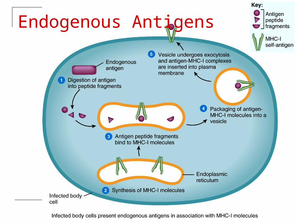
Endogenous Antigens
Copyright 2009, John Wiley & Sons, Inc.
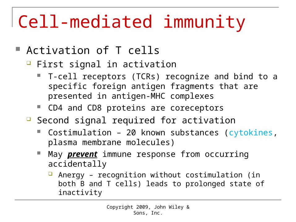
Cell-mediated immunity Activation of T cells
First signal in activation T-cell receptors (TCRs) recognize and bind to a specific
foreign antigen fragments that are presented in antigen-MHC complexes
CD4 and CD8 proteins are coreceptors Second signal required for activation
Costimulation – 20 known substances (cytokines, plasma membrane molecules)
May prevent immune response from occurring accidentally Anergy – recognition without costimulation (in both B and T cells)
leads to prolonged state of inactivity
Copyright 2009, John Wiley & Sons, Inc.
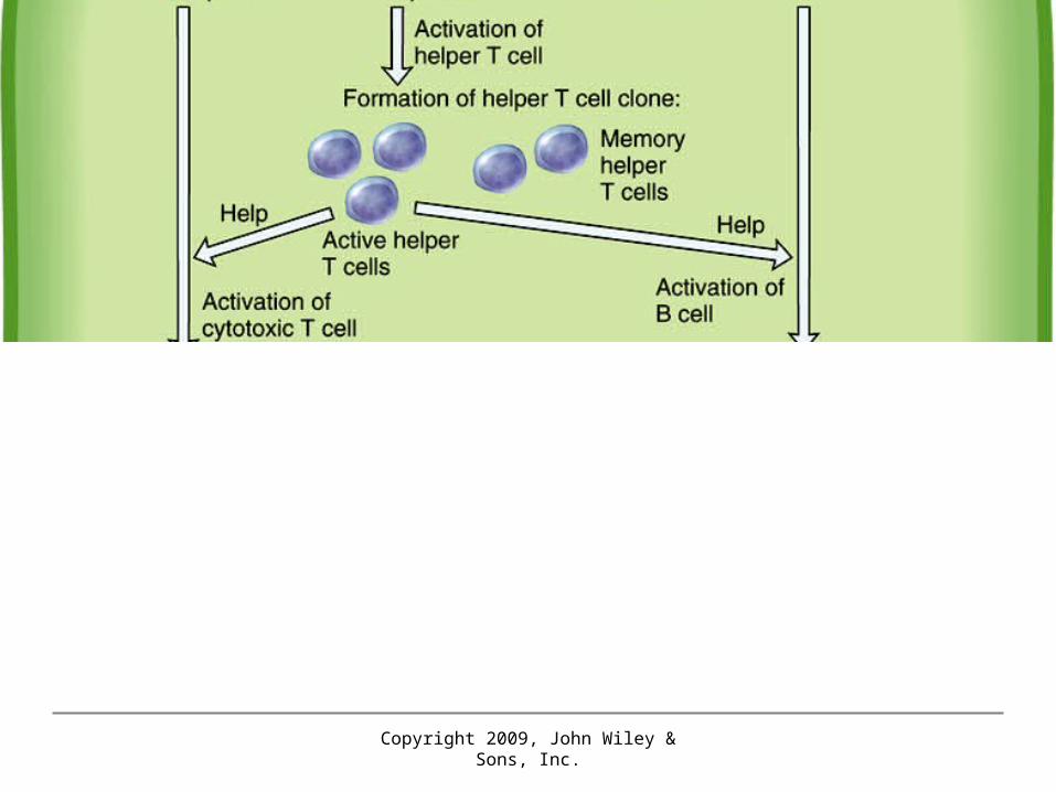
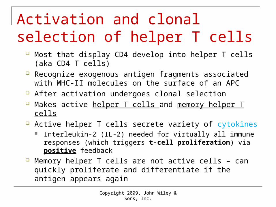
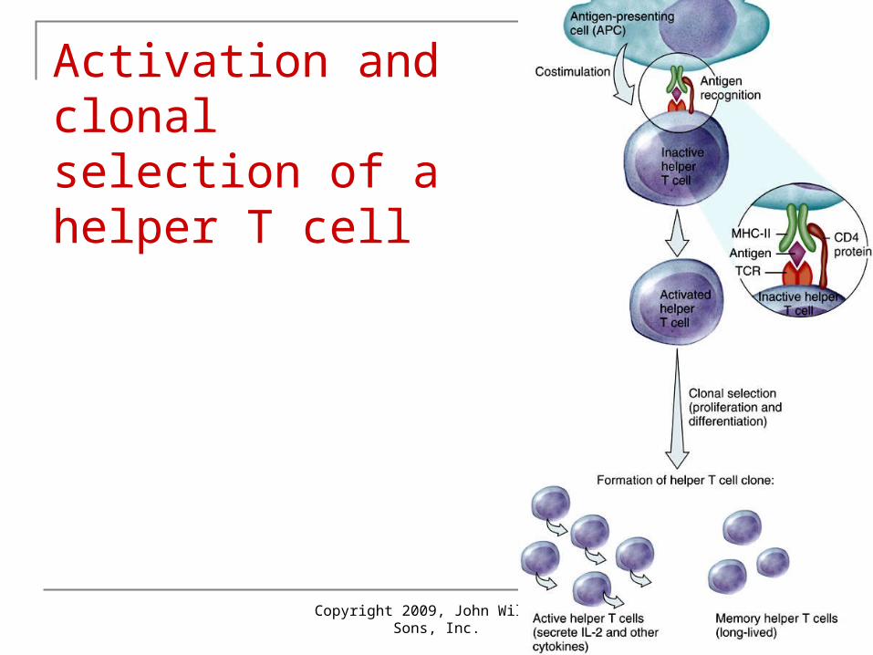
Activation and clonal selection of helper T cells
Most that display CD4 develop into helper T cells (aka CD4 T cells)
Recognize exogenous antigen fragments associated with MHC-II molecules on the surface of an APC
After activation undergoes clonal selection Makes active helper T cells and memory helper T cells Active helper T cells secrete variety of cytokines
Interleukin-2 (IL-2) needed for virtually all immune responses (which triggers t-cell proliferation) via positive feedback
Memory helper T cells are not active cells – can quickly proliferate and differentiate if the antigen appears again
Copyright 2009, John Wiley & Sons, Inc.
Activation and clonal selection of a helper T cell

Copyright 2009, John Wiley & Sons, Inc.
Copyright 2009, John Wiley & Sons, Inc.
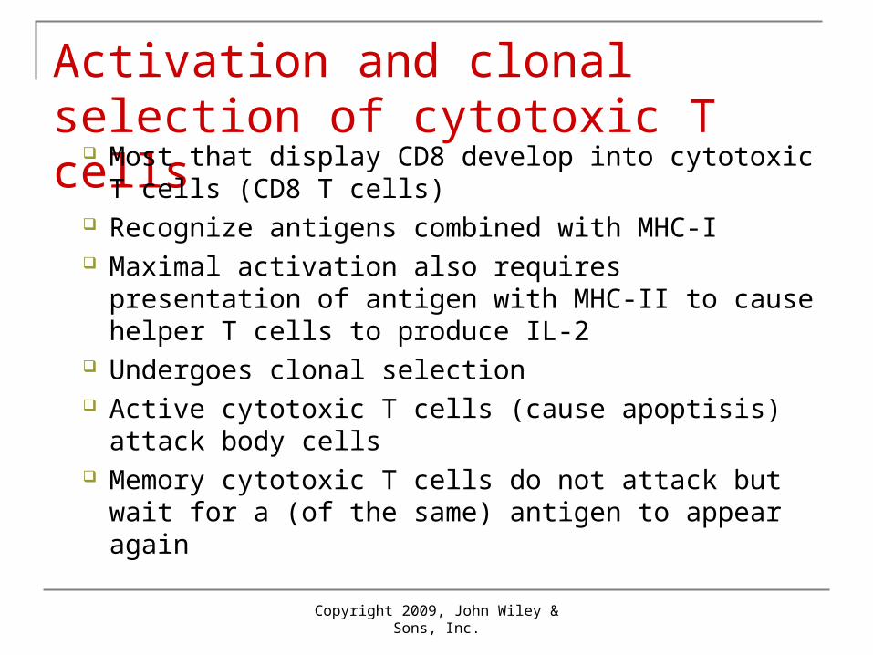
Activation and clonal selection of cytotoxic T cells
Most that display CD8 develop into cytotoxic T cells (CD8 T cells)
Recognize antigens combined with MHC-I Maximal activation also requires presentation of
antigen with MHC-II to cause helper T cells to produce IL-2
Undergoes clonal selection Active cytotoxic T cells (cause apoptisis) attack
body cells Memory cytotoxic T cells do not attack but wait for a
(of the same) antigen to appear again
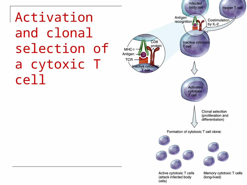
Activation and clonal selection of a cytoxic T cell

Copyright 2009, John Wiley & Sons, Inc.
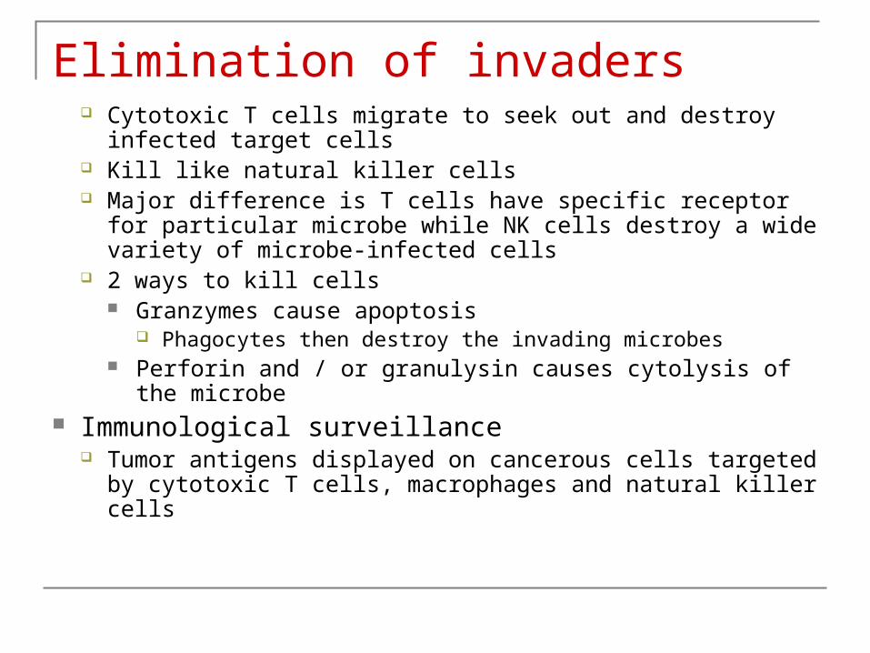
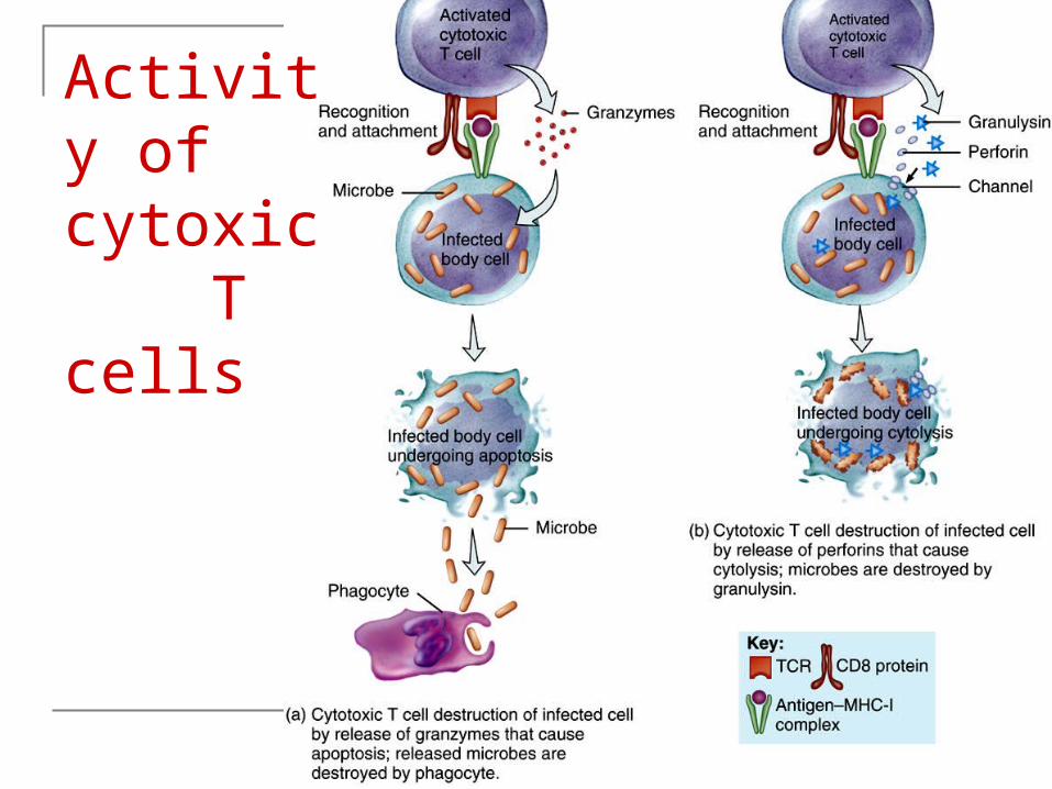
Elimination of invaders Cytotoxic T cells migrate to seek out and destroy infected
target cells Kill like natural killer cells Major difference is T cells have specific receptor for particular
microbe while NK cells destroy a wide variety of microbe-infected cells
2 ways to kill cells Granzymes cause apoptosis
Phagocytes then destroy the invading microbes Perforin and / or granulysin causes cytolysis of the microbe
Immunological surveillance Tumor antigens displayed on cancerous cells targeted by
cytotoxic T cells, macrophages and natural killer cells
Copyright 2009, John Wiley & Sons, Inc.
Activity of cytoxic T cells
Copyright 2009, John Wiley & Sons, Inc.
Antibody-mediated immunity
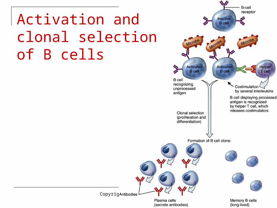
Activation and clonal selection of B cells During activation, antigen binds to B cell receptor (BCRs) B Cell can respond to unprocessed antigen (no APC) Response much more intense when B cell processes
antigen from APC Antigen taken into B cell, combined with MHC-II, moved to
plasma membrane, helper T cell binds and delivers costimulation (interleukin-2 and other cytokines)
B cell undergoes clonal selection Plasma cells secrete antibodies Memory B cells do not secrete antibodies but wait for
reappearance of antigen
Copyright 2009, John Wiley & Sons, Inc.
Activation and clonal selection of B cells
Copyright 2009, John Wiley & Sons, Inc.
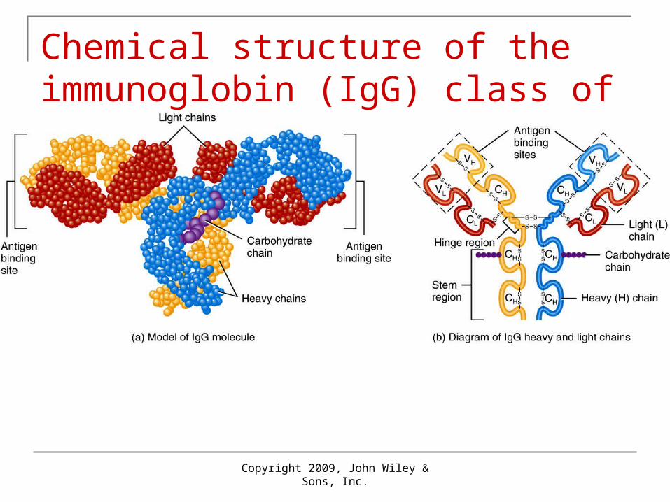
Chemical structure of the immunoglobin (IgG) class of antibody

Antibodies There are 5 classes of antibodies:
• IgG – a monomer with two antigen-binding sites Comprises 80% of total antibody Only class able to cross the placenta Provides long-term immunity against bacteria and
viruses, by enhancing phagocytosis, neutralizing toxins, and triggers compliment systems
• IgA – a dimer with four antigen-binding sites prevalent in body secretions like sweat, tears, saliva, breast
milk and gastrointestinal fluids Provides localized protection of mucous membranes
against B and V See Table 22.3
Copyright 2009, John Wiley & Sons, Inc.
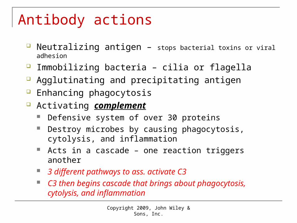
Antibody actions
Neutralizing antigen – stops bacterial toxins or viral adhesion
Immobilizing bacteria – cilia or flagella Agglutinating and precipitating antigen Enhancing phagocytosis Activating complement
Defensive system of over 30 proteins Destroy microbes by causing phagocytosis, cytolysis, and
inflammation Acts in a cascade – one reaction triggers another 3 different pathways to ass. activate C3 C3 then begins cascade that brings about phagocytosis,
cytolysis, and inflammation

Copyright 2009, John Wiley & Sons, Inc.
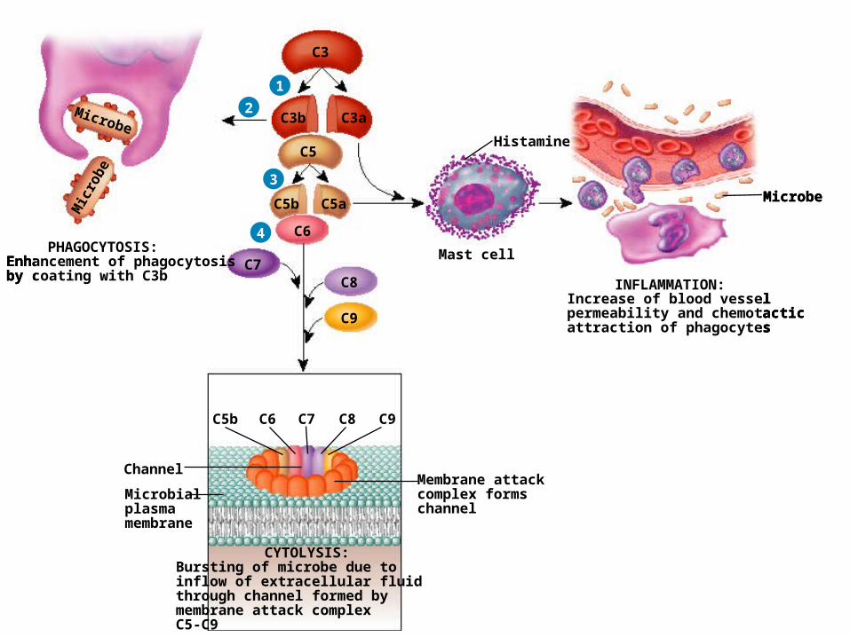
Complement activation and results of activation
1
C3
C3b C3a
C5
1
Microbe
Mast cell
C3
Microbe
Mic
robe
C3b C3a
C5Histamine
INFLAMMATION:Increase of blood vesselpermeability and chemotacticattraction of phagocytes
PHAGOCYTOSIS:Enhancement of phagocytosisby coating with C3b
2
1
Microbe
Mast cell
C3
Microbe
Mic
robe
C3b C3a
C5
C5b C5a
Histamine
INFLAMMATION:Increase of blood vesselpermeability and chemotacticattraction of phagocytes
PHAGOCYTOSIS:Enhancement of phagocytosisby coating with C3b
2
3
1
Microbe
Mast cell
C3
Microbe
Mic
robe
C3b C3a
C5
C5b
C6
C7C8
C9
C5b C6 C7 C8 C9
C5a
Histamine
INFLAMMATION:Increase of blood vesselpermeability and chemotacticattraction of phagocytes
PHAGOCYTOSIS:Enhancement of phagocytosisby coating with C3b
CYTOLYSIS:Bursting of microbe due toinflow of extracellular fluid through channel formed by membrane attack complex C5-C9
Membrane attackcomplex formschannel
Microbialplasmamembrane
Channel
2
3
4
Copyright 2009, John Wiley & Sons, Inc.
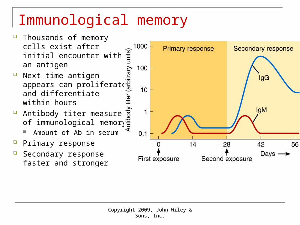
Immunological memory Thousands of memory cells
exist after initial encounter with an antigen
Next time antigen appears can proliferate and differentiate within hours
Antibody titer measure of immunological memory Amount of Ab in serum
Primary response Secondary response faster
and stronger
Copyright 2009, John Wiley & Sons, Inc.
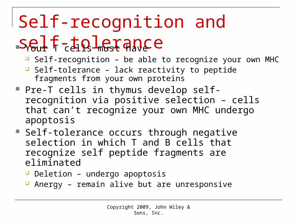
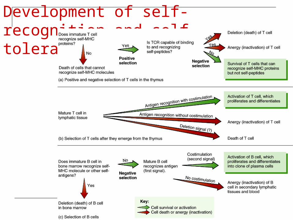
Self-recognition and self-tolerance Your T cells must have
Self-recognition – be able to recognize your own MHC Self-tolerance – lack reactivity to peptide fragments from
your own proteins Pre-T cells in thymus develop self-recognition via
positive selection – cells that can’t recognize your own MHC undergo apoptosis
Self-tolerance occurs through negative selection in which T and B cells that recognize self peptide fragments are eliminated Deletion – undergo apoptosis Anergy – remain alive but are unresponsive
Copyright 2009, John Wiley & Sons, Inc.
Development of self-recognition and self-tolerance

Nova Science Now
http://www.pbs.org/wgbh/nova/body/replacing-body-parts.html
Thymus
Copyright 2009, John Wiley & Sons, Inc.

Lymph node
Copyright 2009, John Wiley & Sons, Inc.
Spleen
Copyright 2009, John Wiley & Sons, Inc.


![22 [chapter 22 the lymphatic system and immunity]](https://img.pdfslide.net/doc/110x75/5a6495f87f8b9a27568b6f3b/22-chapter-22-the-lymphatic-system-and-immunity.jpg)