Embed Size (px)
Citation preview
J. Physiol. (1968), 199, pp. 511-532 511With 7 text-ftgure8Printed in Great Britain
CORD CELLSRESPONDING TO FINE MYELINATED AFFERENTS FROM
VISCERA, MUSCLE AND SKIN
By B. POMERANZ, P. D. WALL AND W. V. WEBERFrom the M.R.C. Cerebral Functions Research Group, Department ofAnatomy, University College London, W.C. 1, England and Department ofBiology and Research Laboratory of Electronics, M.I.T., Cambridge, Mass.
U.S.A.
(Received 21 February 1968)
SUMMARY
1. Micro-electrode recordings were made in the thoracic cord of acutespinal cats. Cells, which were located in the histologically defined lamina 5,responded both to the fine myelinated afferents from the splanchnicnerve and to afferents from the skin. Splanchnic afferents inhibit the effectof converging cutaneous inputs for periods up to 150 msec. Skin stimulimay also inhibit the effect of afferent nerve impulses from viscera. Somecells respond monosynaptically to the splanchnic afferents, othersindirectly.
2. Fine myelinated afferents from gastrocnemius (group 3) stimulatelamina 5 cells which also have cutaneous receptive fields. Cutaneous andgroup 3 muscle afferents interact by mutual inhibition in their effect onthe cells.
3. Fine myelinated afferents from skin excite lamina 5 cells. Thecutaneous responses of lamina 5 cells contrast with those of lamina 4 cellsin the following respects: (a) the receptive fields are larger, (b) they respondwith an increased latency to A,8 afferents, (c) there is a low pressurethreshold at the edge, (d) they respond to a wide range of pressure stimulifrom light brush to heavy pinch applied to the centre of the receptivefields and (e) they respond to AA afferents.
4. Lamina 5 cells receive fine myelinated afferents either from visceraor from muscle or from skin. Lamina 4 receives large myelinated afferentsfrom skin and lamina 6 receives large myelinated afferents from muscle.The results suggest the hypothesis that some fine myelinated afferentsform a class of afferents which signal the state of tissue, and end onlamina 5 cells.
Phy. i9933
512 B. POMERANZ, P. D. WALL AND W. V. WEBER
INTRODUCTION
Rexed (1952) subdivided the gray matter of the spinal cord of the caton the basis of the size, shape and distribution of the cell bodies. He con-cluded that the large cell region of the dorsal horn was subdivided intothree laminae which he numbered laminae 4, 5 and 6. Wall (1967) investi-gated these laminae by recording from single cells with micro-electrodesand found that cells in Rexed's laminae 4 and 5 responded to cutaneousstimulation while cells in lamina 6 responded to limb movement. In thepresent paper, we have continued this physiological analysis of the sub-divisions of the dorsal horn cells by searching for those cells which respondto the small myelinated afferent fibres from viscera, muscle and skin. Thesplanchnic, gastrocnemius and sural nerves were chosen as convenientsources of these afferent fibres since the splanchnic nerve originates inviscera, the gastrocnemius nerve in muscle, and the sural nerve in skin.
Small myelinated afferent fibres with diameters of 1-5 ,u and conductionvelocities of 4-35 m/s are found in the splanchnic, gastrocnemius and suralnerves (Patton, 1965). In the splanchnic nerve, some of these fibres arestimulated by movements of the small intestine (Bessou & Perl, 1966)while others respond only to intense stimuli (Gernandt & Zotterman, 1946).In muscle nerves, the small myelinated afferents are the group 3 afferentsand most are stimulated by pressure or noxious stimulation (Paintal,1960; Bessou & Laporte, 1961). In skin nerves, the small myelinatedfibres form the delta group which contains both fibres which respond tolight pressure (Hunt & McIntyre, 1960) and others which only transmitimpulses when the skin is damaged (Burgess & Perl, 1967).The region of termination of fine visceral afferents in the spinal cord has
been investigated by Weber (1966) and by Selzer & Spencer (1967) usingboth the techniques of single unit recording and of electrical field plotting.They conclude that cells receiving visceral afferents lie in the ventral partof the dorsal horn. Fine muscle afferent fibres and the interneurones onwhich they end have been included in the flexor reflex afferent pathwaysby Lundberg (1964). Wickelgren (1967) reports that some lamina 5 cellsrespond to group 3 muscle afferents. Lundberg (1964) has also placed thecutaneous delta fibres and the interneurones which receive them in theflexor reflex afferent pathway. Fetz (1968) noted that the dorsal horn cellswhich responded to a wide range of cutaneous pressure stimuli lay ventralto a layer of cells which responded only to light and medium pressure. It istherefore apparent that evidence has been accumulating which points tothe ventral part of the dorsal horn as containing cells which respond tothe fine myelinated afferents from viscera, muscle and skin.The central effects of visceral afferents may interact with those of soma-
CORD CELL RESPONSES TO DELTA AFFERENTS 513
tic origin. Downman (1955) showed that stimulation of the smallermyelinated fibres in the splanchnic nerve or stimulation of an intercostalnerve evokes a discharge in other intercostal nerves. He further showedthat these two types of afferent volley interact with each other so that inthe decerebrate cat one volley facilitates the reflex effects of the other,while in the spinal animal there was an inhibitory interaction. Franz,Evans & Perl (1966) recorded from sympathetic preganglionic rami inspinal cats and showed that certain fibres responded both to somatic andto splanchnic nerve stimulation. Fine myelinated afferents in both thesomatic and the visceral nerves had to be stimulated in order to evoke thereflex effects. The reflex discharge evoked by one nerve was markedlydepressed for several hundred milliseconds by a conditioning volley toanother nerve. Weber (1966) discovered certain single cells in the spinalcord which responded to both splanchnic and to somatic nerve stimulation.Similarly, Selzer & Spencer (1967) recorded from single cells in the upperlumbar cord which responded to pelvic visceral afferents and to somaticstimulation.
In the experiments described in this paper, we set out to locate thosecells which respond to the fine myelinated afferents and to describe theinteractions on those cells of inputs of different origins.
METHODS
All experiments were carried out on cats prepared under ether anaesthesia with occlusionof the basilar and carotid arteries and section of the spinal cord at C 1. The animals were thengiven artificial respiration, paralysed with gallamine triethiodide and the administration ofether was stopped. Extracellular single unit recordings were made with fluid filled, 2-5 MQ),glass micro-electrodes by the methods previously described (Wall, 1967). The stimulus siteson peripheral nerves were prepared by dissecting the nerve free from surrounding tissueover a distance of 15-20 mm, without section of the nerve. A sheet of paraffin wax (Parafilm'M', American Can Co.) was then slipped under the freed section of nerve. Fine insulatedwire with a diameter of 200 ,t was brought from the stimulus isolation unit to the stimulussite. On the end of each wire, 3-4 mm of 50 ,u platinum wire was attached. The platinumwire was slipped under the nerve and gently wrapped around it so that no tension wasexerted on the nerve. Two such wires separated by 5 mm were placed around the nerve andthen embedding wax, at a temp. of 40-45' C, was dropped on until all exposed nerve andwire was covered. The wax did not affect impulse conduction and prevented drying orflooding of the exposed region. The lightness and flexibility of the electrodes and their fixedapposition to the nerve allowed free movement of the region without change of the positionof the electrodes with respect to the nerve. In many experiments, additional pairs of elec-trodes were placed on more proximal parts of the nerve so that they could be used either torecord the volley generated by the first pair or to generate afferent volleys with a shorterperipheral conduction distance. Four types of natural stimulation of skin were used; brush,touch, mild pressuire produced by picking up a fold of skin, heavy pressure by pinching askin fold. For electrical stimulation of skin, fine hypodermic needles or Michel wound clipswere used as stimulating electrodes.Method of recording. A method of recording and display (Wall, 1960) allows the response
33-2

514 B. POMERANZ, P. D. WALL AND W. V. WEBERto 150 or more pairs of stimuli to be presented in one figure so that one can see any variabilityin the response and the effects of interaction as the time interval between the stimuli ischanged. The system can be explained by reference to Fig. 4A. This is the record of anexperiment in which there was one recording micro-electrode near a dorsal horn cell, andtwo sites of electrical stimulation, the splanchnic nerve and the skin. The figure is read fromabove downwards. The stimulus artifact from cutaneous stimulation gives a spot whosesize and position on the screen is adjusted to give a continuous vertical line as the stimulusis repeated once every second. This line is at the zero point on the abscissa which representsthe separation in time of the two stimuli; it is scaled + 100 msec on either side of the zero.The artifact from splanchnic nerve stimulation similarly produces a continuous line whichapproaches the vertical line as the interval between the pair of stimuli is decreased on repeti-tion. The recording of the cell firing produces a dot on the screen. At the start of the experi-ment (top of Fig. 4A), the splanchnic stimulus leads the cutaneous stimulus by 100 msecand the cell fires about 15 msec after the splanchnic stimulus. There is no firing of the cellin response to the cutaneous stimulus. The same result is found as the time interval betweenthe stimuli is reduced by equal increments until they are coincident (first crossing point ofthe continuous lines). In the following section the splanchnic stimulus progressively lagsbehind the cutaneous stimulus, and the unit now responds to both stimuli. In the lower halfof the figure, the temporal relationship between the stimuli is progressively reversed untilthe initial condition is reached after 150 pairs of stimuli.
RESULTS
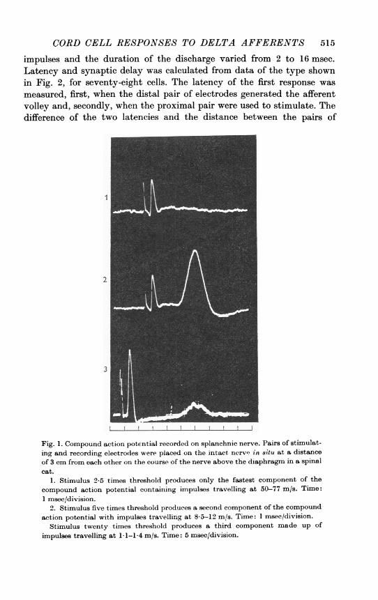
Thoracic cord1. Effects of splanchnic nerve stimulationA. Afferent volley. Two pairs of electrodes, separated byabout 3 cm, were
placed on the splanchnic nerve above the diaphragm. When stimulatingpulses were applied to one pair of electrodes, a compound action potentialwas recorded on the other pair (Fig. 1). When the stimulus strength wastwenty times threshold, the compound action potential had three com-ponents produced by impulses travelling at 50-77 m/s, 8-5-12 m/s and1-1-1-4 m/s.B. Cord cells responding to splanchnic stimulation. The dorsal horn of
thoracic segments 7, 8 and 9 was searched with micro-electrodes. If thestimulus intensity to the splanchnic was low so that only the fastestcomponent of the compound action potential was observed on the nerve,a small brief field potential was recorded in the ipsilateral dorsal quadrantof the cord at the expected time of the arrival of the high velocity afferentvolley. This showed that the impulses had arrived in the spinal cord butno single cells were ever discovered which responded to these impulsestransmitted by the larger myelinated fibres in the splanchnic nerve. If thestimulus intensity on the splanchnic nerve was raised so that an afferentvolley was generated which travelled at 8-5-12 m/s many cells weredetected in deeper parts of the dorsal horn which responded with a burstof repetitive firing after the arrival in the cord of this slower afferent volley.The number of impulses in the repetitive discharge varied from 2 to 12
CORD CELL RESPONSES TO DELTA AFFERENTS 515
impulses and the duration of the discharge varied from 2 to 16 msec.Latency and synaptic delay was calculated from data of the type shownin Fig. 2, for seventy-eight cells. The latency of the first response wasmeasured, first, when the distal pair of electrodes generated the afferentvolley and, secondly, when the proximal pair were used to stimulate. Thedifference of the two latencies and the distance between the pairs of
Fig. 1. Compound action potential recorded on splanchnic nerve. Pairs of stimulat-ing and recording electrodes were placed on the intact nerv' in situ at a distanceof 3 cm from each other on the course of the nerve above the diaphragm in a spinalcat.
1. Stimulus 2-5 times threshold produces only the fastest component of thecompound action potential containing impulses travelling at 50-77 m/s. Time:1 msee/division.
2. Stimulus five times threshold produces a second component of the compoundaction potential with impulses travelling at 85-12 im/s. Time: 1 msec/division.
Stimulus twenty times threshold produces a third component made up of
impulses travelling at 1-1-1-4 m/s. Time: 5 msec/division.
516 B. POMERANZ, P. D. WALL AND W. V. WEBER
stimulating electrodes allowed the calculation of the conduction velocityof those fibres responsible for firing the cell. The result showed that cells
were fired by afferents travelling at a conduction velocity of 10-30 m/s.The sympathetic chain and rami had been cut so that splanchnic afferentimpulses could only enter the cord over the dorsal root of segments T8and T9. This limitation of entry point allowed the conduction distancebetween the stimulus point and the observed cell to be measured fairly
2
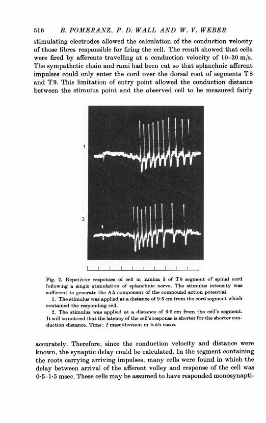
Fig. 2. Repetitive responses of cell in 'amina 5 of T8 segment of spinal cordfollowing a single stimulation of splanchnic nerve. The stimulus intensity wassufficient to generate the AA component of the compound action potential.
1. The stimulus was applied at a distance of 9-5 cm from the cord segment whichcontained the responding cell.
2. The stimulus was applied at a distance of 6-5 cm from the cell's segment.It will be noticed that the latency of the cell's response is shorter for the shorter con-duction distance. Time: 2 msec/division in both cases.
accurately. Therefore, since the conduction velocity and distance wereknown, the synaptic delay could be calculated. In the segment containingthe roots carrying arriving impulses, many cells were found in which thedelay between arrival of the afferent volley and response of the cell was0-5-1-5 msec. These cells may be assumed to have responded monosynapti-

CORD CELL RESPONSES TO DELTA AFFERENTS 517
cally to the afferent fibres. Many other cells were detected in the sameregion which responded with a latency of 2-15 msec after the arrival ofthe entering volley and these cels must be presumed to be in indirectcontact with splanchnic afferents. No lateral horn cells were detectedwhich responded antidromically to the peripheral stimulus.
C. Location of cell3 responding to &planchnic afferenrt. Mapping experi-ments were carried out by the method previously used (Wall, 1967). Thesplanchnic nerve was stimulated once a second at an intensity well abovethe threshold for fine myelinated afferents. First of all, a micro-electrodewas made to penetrate the cord along a track close to the mid line andrecordings were made at 50 ,u intervals during the penetration. When theelectrode had penetrated to a depth of 2 mm, it was cut off and left inposition for later location by the clearing technique (Wall, 1967). Next, asecond micro-electrode was placed in the cord on a track parallel to thefirst and slightly lateral to it. This process was continued until the entiredorsal horn had been searched for cells responding to the splanchnicafferent volley. Maps were completed in six cats and three of the maps areshown in Fig. 3. The outlines of the dorsal and ventral horns were tracedfrom the actual specimen of the cleared spinal cord containing the micro-electrodes. A point was marked on each electrode track at which theelectrode had first recorded cells in the dorsal part of the dorsal horn whichresponded to cutaneous stimulation but not to splanchnic nerve stimula-tion. These cells will be discussed in the next section. A line was drawnacross the dorsal horn connecting these points on each electrode track.This line is the more dorsal of the two lines drawn in the dorsal hornsshown in Fig. 3. As each electrode penetrated more deeply into the dorsalhorn, a point was reached at which cells were encountered which respondedto the splanchnic volley. The depth at which these cells were first en-countered was marked on each electrode track and a second more ventralline was drawn across the dorsal horn. The zone between the two lineswhich had been located by micro-electrode recording corresponds roughlyto Rexed's histological lamina 4. The remainder of dorsal horn ventral tothe second line which contained cells responding to the splanchnicafferents is roughly the same as Rexed's histological lamina 5. Thoracicdorsal horn does not contain a histologically recognizable lamina 6.
2. Cells responding to cutaneous stimuliWhile searching for cells responding to splanchnic stimulation, all cells
were examined for cutaneous responses. The dorsal horn was mapped forcells responding to cutaneous stimulation by the same method and at thesame time as it was mapped for splanchnic responses. The zone betweenthe two lines drawn in the dorsal horn in Fig. 3 was found to contain cells

518 B. POMERANZ, P. D. WALL AND W. V. WEBER
with the characteristics of lamina 4 cells previously described in thelumbar enlargement (Wall, 1967). The receptive fields were relativelysmall, 1-2 cm, and the cells within the lamina were arranged in a cleartopographic map with medial cells having ventrally placed receptive fields
Fig. 3. Cord maps showing the location of regions in which calls were encounteredwhich responded to cutaneous and sp.anchnic stimulation. The outline maps aretraced from three separate experiments on thoracic segments 7. 8 and 9. Eachdorsal horn had been explored by at least eight parallel electrode tracks which hadbeen located. The more dorsal line in each dorsal horn is drawn between points onthe electrode tracks at which cells were first encountered which responded to lightbrushing of the skin. In the region between the two lines in the dorsal horn,recordings were made from cells which generated action potentials of more than50 ,uV, responded to brushing of the skin and never responded to splanchnic nervestimulation.When the electrodes penetrated to the more ventral of the two lines, cells were
encountered which responded to splanchnic nerve stimulation. These cells alsohad cutaneous receptive fields considerably larger than those of the more dorsalcells.
and lateral cells receiving from dorsal areas within the dermatome of thesegment. Electrical stimulation of either the receptive field or the inter-costal nerve produced firing of these cells within 0-51-5 msec of theentering volley so that they can be considered to be monosynapticallyconnected to A,f afferent fibres. The cells responded to brush, touch andlight pressure within their receptive field. Some cells adapted rapidly ifthe stimulus was maintained. If the pressure on the skin fold was increasedto a heavy pinch, no cells in this lamina increased their firing rate.When the electrodes penetrated to the region whose upper boundary is

CORD CELL RESPONSES TO DELTA AFFERENTS 519
marked by the more ventral line in Fig. 3, there were five changes in theproperty of the cells which differentiated these deeper cells from the moresuperficial cells. The size of the receptive field expanded to involve aboutone third of the dermatome. This was the same sudden expansion whichhad been observed at the lumbar lamina 4-5 junction (Wall, 1967). Thereceptive fields changed from having a relatively uniform threshold overthe entire field as seen in lamina 4 to having a marked gradient of pressurethreshold. Most responded to brush in the centre but heavy pinching wasrequired at the edge of the receptive field. Some cells did not respond tobrushing and required a light pressure stimulus even in the middle of theirreceptive fields. In the middie of the receptive field, the cells exhibited a'wide dynamic range' (Mendell, 1966). That is to say that, unlike thelamina 4 cells, if the pressure was increased from light touch to heavypinch, the firing frequency of the cell increased with each increment ofpressure stimulus. As in the lumbar region, the cells in lamina 5 respondedto electrical stimulation of A,8 afferents but with a longer latency than thecells in lamina 4. Twenty-six pairs of cells were examined; one cell of eachpair was in lamina 4 and the other cell was immediately ventral to the firstin lamina 5. These pairs of cells were examined for their latency of responseto an afferent A,8 volley generated by electrical stimulation applied directlyto the skin of the cells' receptive fields. Two pairs showed no latency shiftbut the remainder showed a shift of 0-5-20 msec, average 09 msec. Thislatency shift had been previously interpreted as suggesting that thelamina 5 cells were stimulated by the lamina 4 cells which received theA,@ input. In some preparations, the lamina 5 cells showed a markedhabituation to repeated light brushing within their receptive field, aphenomenon previously described in lumbar lamina 5 cells in the decere-brate cat (Wall 1967) and the spinal rat (Wall, Freeman & Major, 1967).The final difference between lamina 4 and lamina 5 cells was that many ofthe cells with lamina 5 cutaneous characteristics responded to splanchnicnerve stimulation.
3. Interaction of splanchnic and cutaneous volleysSince the cells which responded to splanchnic afferents also responded
to cutaneous stimulation, it was important to study the interactions ofthese convergent inputs. This was done by stimulating both the splanchnicnerve and the cutaneous receptive field of the single cells under examina-tion and by varying the time interval between the two stimuli. The resultsfrom two cells are shown in Fig. 4 where cell A showed the most commonlyobserved interaction and cellB a less frequent type. For cell A, the stimulusstrengths were adjusted so that the cell responded with one or two

520 B. POMERANZ, P. D. WALL AND W. V. WEBER
impulses when either the splanchnic or the cutaneous stimuli were appliedby themselves. If the splanchnic stimulus preceded the cutaneous stimulus,the cutaneous response was inhibited. For this cell, the duration of theinhibition was found to be 150 msec. In the illustration, the responses ofthe cell are shown when the splanchnic stimulus was gradually shifted
Fig. 4. Interaction of splanchnic and cutaneous afferent volleys on two lamina5 cells, A and B, in segment T 8. Paired stimuli were delivered once a second to AAsplanchnic fibres and to the cutaneous receptive field of the cell. One hundred andfifty pairs of stimuli and responses are shown. The time interval between condition-ing and test stimuli was progressively varied. The response to cutaneous stixnula-tion is shown at a fixed position while the splanchnic stimulus was moved. At thetop, splanchnic stimulation was given 100 msec before cutaneous stimulationand then moved step by step until, in the middle of the picture, cutaneous stimula-tion is given 100 msec before splanchnic stimulation (see Methods). The splanchnicvolley inhibited the cutaneous response while the cutaneous volley had no effect onthe splanchnic response in cell A and produced a weak inhibition in cell B. Timemarks: 100 msec.
from 100 msec before the cutaneous stimulus to 100 msec after it. Wherethe splanchnic stimulation occurred earlier than the cutaneous stimulation,the cutaneous response was completely inhibited. When the two stimuliwere synchronous, there was a partial summation of the two responses.Where the cutaneous stimulus preceded the splanchnic stimulus, therewas no observable effect of the cutaneous stimulus on the responses of the
CORD CELL RESPONSES TO DELTA AFFERENTS 521
cell to the splanchnic volley. In oell B; the-two inputs given by themselveseach produced two impulses from the cell. If the splanchnic stimuluspreceded the cutaneous by 15-100 msec, the illustration shows that theresponse of the cell to the cutaneous volley was partially inhibited so thatthe cell fired only once instead of twice. If the splanchnic stimulus precededthe cutaneous by 0-15 msec, the cutaneous response was completelyinhibited. When the cutaneous stimulus preceded the splanchnic, it willbe seen that in this cell, unlike cell A, the cutaneous stimulus inhibited thesplanchnic response for a period of about 25 msec. In these experimentsthe cutaneous stimulus was firing off a far smaller number of peripheralnerve fibres than the splanchnic nerve stimulus.We wondered if the reason why the splanchnic volley inhibited the
cutaneous response while the cutaneous afferents usually failed to inhibitthe splanchnic response was because the size of the cutaneous volley wasso much smaller than the splanchnic volley. To test this suggestion, westimulated the entire intercostal nerve at the costovertebral junctioninstead of stimulating the skin through hypodermic needles placed in thereceptive field of the cell under observation. Under these conditions, it wasfound that the volley from the intercostal nerve always inhibited responsesproduced by splanchnic stimulation. The duration of the inhibition pro-duced by intercostal nerve stimulation was about equal to that whichfollowed splanchnic stimuli. The duration varied from cell to cell between100 and 200 msec.
Lumbar cord1. Effects of gastrocnemius nerve stimulationA. Afferent volley. The nerve to the gastrocnemius was dissected free in
the popliteal fossa and placed on pairs of stimulating and recordingelectrodes. When a stimulus at twice the threshold intensity was appliedto the nerve, a compound action potential was recorded from the nerve.This wave was produced by impulses in the large motor fibres and thegroup 1 and 2 sensory fibres. When the stimulus intensity was raised toten times threshold, a later component of the compound action potentialwas recorded. This component was produced by impulses in the group 3afferents.
B. Cord cells responding to the afferent volley. Gastrocnemius afferentsenter the cord in the rostral S 1 and caudal L 7 dorsal root filaments. Theexact location of the entry points was determined in each cat by localstimulation on the surface of dorsal rootlets and by recording the size ofthe antidromic volley on the nerve to gastrocnemius. A search was thencarried out in segments L 7 and S 1 for cells which responded to afferentimpulses from gastrocnemius. The method of mapping was the same asthat used to search for cells in the thoracic cord which responded to

522 B. POMERANZ, P. D. WALL AND W. V. WEBER
splanchnic afferents. Cells were located in the dorsal horn which respondedto group 3 afferent nerve impulses but did not respond to group 1 or 2afferents. These cells would only respond to gastrocnemius nerve stimula-tion if the intensity of the stimulus was 2-5-10-0 times the threshold forthe largest axons. The cells fired repetitively for as long as 10 msec follow-ing the arrival of a maximal group 3 volley. In the lateral part of dorsalhorn, close to the root entry zone of the gastrocnemius afferent fibres,many cells responded within less than 1 msec of the entry of the group 3afferent impulse volley. These cells can therefore be assumed to be mono-synaptically connected to the group 3 afferents. In other parts of segmentsL 7 and S 1, cells could be found which also responded when, and only when,a group 3 volley was generated, but these cells responded with a longerlatency and were presumed to be fired indirectly by a polysynapticpathway.
C. Location of cells responding to group 3 muscle afferents. The L 7-S 1junction was mapped with regularly spaced micro-electrode tracks. Themethod used and the results obtained with cutaneous stimulation weresimilar to those previously reported (Wall, 1967). In the dorsal horn therewas a dorsal lamina containing cells with small cutaneous receptive fields,a middle lamina with cells with greatly expanded cutaneous receptivefields and a ventral lamina containing cells which responded to passivemovement of the leg. These three laminae which were delineated byrecording from single cells corresponded roughly to Rexed's histologicallaminae 4, 5 and 6. The cells in lamina 4 never responded under anycircumstances to stimulation of the gastrocnemius nerve. As soon aslamina 5 was entered and cells were encountered with enlarged cutaneousreceptive fields, it was found that these cells were the ones which respondedto group 3 afferent volleys but showed no response whatsoever to stimula-tion of group 1 or 2 afferent volleys. On deeper penetration, provided thatthe electrode was in the lateral half of the dorsal horn and close to the rootentry zone of gastrocnemius afferents, cells were encountered with lamina 6characteristics, that is to say, they responded to passive movement of thelegs, particularly ankle flexion. Lamina 6 cells in the lateral half of thelamina responded to group 1 afferent volleys with a central latency ofless than 1 msec after the arrival in the cord of the group 1 afferent volley.It is apparent therefore that in segments L 7 and S 1, lamina 5 contains thecells which respond monosvnaptically to group 3 gastrocnemius afferents.Lamina 5 also contains cells which respond polysynaptically to theseafferents.
CORD CELL RESPONSES TO DELTA AFFERENTS 523
2. Cells responding both to cutaneous stimuli and to group 3 muscle afferentsAll those cells in lamina 5 which responded monosynaptically to gastro-
cnemius group 3 afferents had cutaneous receptive fields on the calf, Achillestendon or heel. In other words they had a cutaneous receptive field whichoverlay some part of the muscle or its insertion. In contrast to the cells indirect contact with the group 3 afferents, the cells which responded witha latency of more than 1 msec after the arrival of the afferent volley hadreceptive fields on the sole of the foot and on the toes. Details of thesereceptive fields will be given below.
3. Interaction of group 3 muscle afferent volleys and cutaneous volleysThe cutaneous receptive fields of the lamina 5 cells receiving gastro-
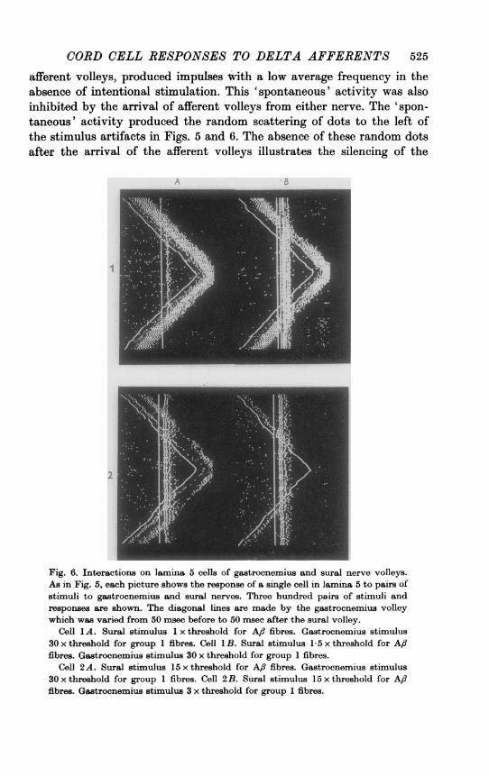
cnemius group 3 afferents mainly lie in skin supplied by sural nerve. Wetherefore chose to study the interaction of volleys from the sural andgastrocnemius nerves rather than to stimulate the skin directly. Theadvantage of sural stimulation was, first, that we could generate a largerand more synchronous volley than could have been produced from skinstimulation and, secondly, that we could ensure by insulation of thestimulating electrodes that no stimulus spread to underlying muscleoccurred. The results are shown for four different cells in Figs. 5 and 6.In Fig. 5, cell 1A, the gastrocnemius nerve was fired at thirty times thestimulus intensity for group 1 afferents and the cell responded with amultiple burst. The sural nerve was stimulated at 1-5 times the thresholdfor the largest Af, fibres and the cell responded twice. When the twovolleys interacted, there was a slight inhibition of the sural response bythe group 3 afferents and a weak inhibition of the group 3 response byconditioning from the sural. If the stimulus intensity on the sural wasraised to fifteen times threshold (cell 1B) the inhibition of the gastroc-nemius response by the preceding sural volley became very much strongerand the gastrocnemius inhibition of the sural effect disappeared. Theother three cells, viz. Fig. 5, cell 2 and Fig. 6, cells 1 and 2, show variationsof this balance between the mutual inhibitory effects of one volley oxi theother. Figure 6 cell LA shows an inhibition of the sural response bygroup 3 afferents while there is no inhibition of the response to the group 3afferent volley produced by threshold sural stimulation. This result issimilar to that shown in Fig. 4A where splanchnic stimulation inhibitedthe response to cutaneous stimulation but cutaneous stimulation failedto inhibit the splanchnic response. However, if the stimulus strength tothe sural nerve was raised to 1 5 times threshold, as in Fig. 6, 1 B, the suralstimulus was then followed by an inhibition of the response to the group 3afferents from gastrocnemius. Many of the cells, which responded to both

524 B. POMERANZ, P. D. WALL AND W. V. WEBER
Fig. 5. Interactions on lamina 5 cells of gastrocnemius and sural nerve volleys.The method of recording is the same as that shown in Fig. 4. The vertical 1ines arethe records of the stimulus artifacts and responses of a single cell to sural nervestimulation. The timing of the gastrocnemius nerve stimulation was varied withrespect to the sural nerve stimulus. Stimuli were given at 3/sec in pairs. At the topof each picture the stimulus to the gastrocnemius preceded that to the sural by50 msec. The time interval between the gastrocnemius and sural stimuli was shiftedregularly until half way down each picture the sural stimulus preceded that to thegastrocnemius bv 50 msec. The lower half of each picture shows the effect ofreversing the direction of shift of time interval between the two stimuli. Eachpicture shows the response of the cell to 300 pairs of stimuli.
Cell 1A. Sural stimulus 1-5 x threshold for A,8 fibres. Gastrocnemius stimulus30 x threshold for group 1 fibres. Cell 1B. Sural stimulus 15 x threshold for Aflfibres. Gastrocnemius stimulus 30 x threshold for group 1 fibres.
Cell 2 A. Sural stimulus 1-5 x threshold for A,8 fibres. Gastrocnemius stimulus 50 xthreshold for group 1 fibres. Cell 2B. Sural stimulus 15 x threshold for A,8 fibres.Gastrocnemius stimulus 50 x threshold for group 1 fibres.
CORD CELL RESPONSES TO DELTA AFFERENTS 525
afferent volleys, produced impulses wvith a low average frequency in theabsence of intentional stimulation. This 'spontaneous' activity was alsoinhibited by the arrival of afferent volleys from either nerve. The 'spon-taneous' activity produced the random scattering of dots to the left ofthe stimulus artifacts in Figs. 5 and 6. The absence of these random dotsafter the arrival of the afferent volleys illustrates the silencing of the
Fig. 6. Interactions on lamina 5 cells of gastrocnemius and sural nerve volleys.As in Fig. 5, each picture shows the response of a single cell in lamina 5 to pairs ofstimuli to gastrocnemius and sural nerves. Three hundred pairs of stimuli andresponses are shown. The diagonal lines are made by the gastrocnemius volleywhich was varied from 50 msec before to 50 msec after the sural volley.
Cell 1A. Sural stimulus 1 x threshold for A/? fibres. Gastrocnemius stimulus30 x threshold for group 1 fibres. Cell 1B. Sural stimuilus 1-5 x threshold for AA8fibres. Gastrocnemius stimulus 30 x threshold for group 1 fibres.
Cell 2A. Sural stimulus 15 x threshold for A/8 fibres. Gastrocnemius stimulus30 x threshold for group 1 fibres. Cell 2B. Sural stimulus 15 x threshold for A,/fibres. Gastrocnemius stimulus 3 x threshold for group 1 fibres.

526 B. POMERANZ, P. D. WALL AND W. V. WEBER
'spontaneous' activity of the cells. In summary, the results show that ahigh intensity stimulus to either nerve inhibited the responses producedby a low intensity stimulus to the other nerve for periods of 45-200 msec.
Lumbar cord BEffects of sural nerve stimulation
A. Afferent volley. The sural nerve was dissected free either in thepopliteal fossa or on the surface of gastrocnemius, and stimulating andrecording electrodes were installed on the nerve. The compound actionpotential produced by gradually increasing strengths of stimuli wasrecorded. Here, as with the splanchnic and gastrocnemius nerves, a slowlyconducting group of nerve impulses, the A delta group, could be recorded.
B. Cord cells responding to the afferent volley. The dorsal horn was searchedwith micro-electrodes in the L 7-S 1 segments and stimuli of variousamplitudes were given to the sural nerve. The search and mapping pro-cedure was the same as that used for the splanchnic and gastrocnemiusnerves. Two clearly separated types of cells were found. One respondedwith a brief repetitive discharge when an A,/ volley was generated in thesural nerve but, when the stimulus was increased above this level, therewas no further prolongation of the burst length. The second type, illus-trated in Fig. 7, also responded to an A,f volley. However, as the stimulusintensity was increased, so that the afferent volley contained impulses insmaller and smaller fibres, the second type of cell, unlike the first, re-sponded with a more and more prolonged burst of repetitive firing. Theextent of this prolongation varied from cell to cell. In some cells the repeti-tive discharge did not extend beyond 50 msec and in others it lastedlonger than 1 sec. As the stimulus strength increased, the repetitive dis-charge did not increase in a smooth fashion. The prolongation took placein a series of stages marked by the appearance of relatively high frequencybursts. In the cell shown in Fig. 7A there were seven clear stages in theprolongation of the repetitive charge. The last of these stages was triggeredby the inclusion in the afferent volley of impulses in non-myelinatedC fibres when the stimulus intensity exceeded 10 V for 0-1 msec. In thesecond cell, Fig. 7B, only two bursts were set off by the myelinated fibresand a third was evoked by non-myelinated afferents. Most of the cells ofthe second tvpe produced 3-7 bursts of discharge during their repetitivedischarge following the arrival of a volley set off by a supramaximalstimulus to the sural nerve. AMendell (1966) has reported this 'banding' ofthe repetitive discharge. The extension of the duration of repetitive firingwas caused by recruitment of more and more fibres in the sural volleyand was not due to current spread to other nerves. To test for the possi-bility of stimulus spread, the nearby gastrocnemius muscle was inspected
CORD CELL RESPONSES TO DELTA AFFERENTS 527
under a dissecting microscope for signs of twitches associated with thestimulus and, since none were seen it is reasonable to assume that thestimulating current was confined to the sural nerve.
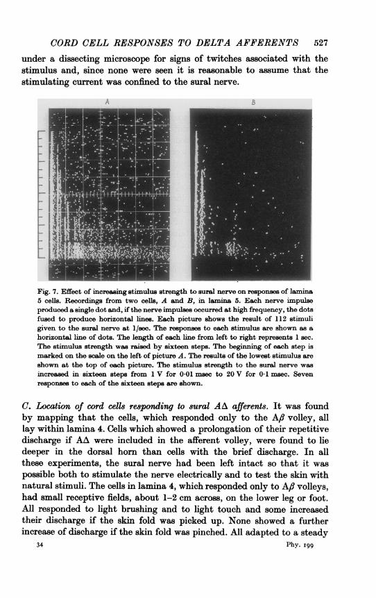
Fig. 7. Effect of increasing stimulus strength to sural nerve on responses of lamina5 cells. Recordings from two cells, A and B, in lamina 5. Each nerve impulseproduced a single dot and, if the nerve impulses occurred at high frequency, the dotsfused to produce horizontal lines. Each picture shows the result of 112 stimuligiven to the sural nerve at 1/sec. The responses to each stimulus are shown as ahorizontal line of dots. The length of each line from left to right represents 1 sec.The stimulus strength was raised by sixteen steps. The beginning of each step ismarked on the scale on the left of picture A. The results of the lowest stimulus areshown at the top of each picture. The stimulus strength to the sural nerve wasincreased in sixteen steps from 1 V for 0.01 msec to 20 V for 0.1 msec. Sevenresponses to each of the sixteen steps are shown.
C. Location of cord cells responding to sural AA afferents. It was foundby mapping that the cells, which responded only to the A,f volley, alllay within lamina 4. Cells which showed a prolongation of their repetitivedischarge if AA were included in the afferent volley, were found to liedeeper in the dorsal horn than cells with the brief discharge. In allthese experiments, the sural nerve had been left intact so that it waspossible both to stimulate the nerve electrically and to test the skin withnatural stimuli. The cells in lamina 4, which responded only to A,8 volleys,had small receptive fields, about 1-2 cm across, on the lower leg or foot.All responded to light brushing and to light touch and some increasedtheir discharge if the skin fold was picked up. None showed a furtherincrease of discharge if the skin fold was pinched. All adapted to a steady
34 Phy. I99
528 B. POMERANZ, P. D. WALL AND W. V. WEBER
stimulus, some within a few impulses while others maintained their dis-charge for seconds. The threshold within the whole area of the receptivefield seemed uniform when tested by manual stimulation. No attempt wasmade to subdivide these cells into subclasses based on location, receptivefield size, adaptation, pressure range or length of repetitive discharge.
If the deeper cells responding to AA fibres were tested with naturalstimulation of the skin they contrasted with the lamina 4 celLs in severalways. The receptive fields expanded proximo-distally within the S 1-L 7dermatomes up to a length of 4-5 cm. There was a marked gradient ofpressure sensitivity within the receptive field so that light brushing in thecentre stimulated the cell but pressure was required at the edge. Finally,and perhaps most significantly, if a skin fold was picked up in the receptivefield and the pressure gradually increased to a hard pinch, the firingfrequency of the cell increased with each increment of pressure in contrastto lamina 4 cells. The frequency of discharge gradually decreased if aheavy pressure stimulus was maintained but some cells would maintain anincreased firing frequency for at least 1 hr.
DISCUSSION
The results show that there are celLs in the region of lamina 5 which areexcited by fine myelinated afferents from either viscera, muscle or skin.All of these cells also had a cutaneous receptive field and there wereinhibitory interactions between the fine myelinated input and the afferentsfrom the cutaneous receptive field. We failed to detect cells which re-sponded only to visceral afferents or to group 3 muscle afferents. Suchspecific cells may exist in the population of cells from which recordingscannot be made with presently available micro-electrodes.The acute spinal cat was chosen as a preparation on which to begin the
investigation of these cells. There are reasons to believe that their responsesmay be different in the decerebrate or freely moving animal. Downman(1955) showed that viscero-somatic reflexes and the interaction of inputswere strikingly different in the spinal and decerebrate cat. Lundberg (1964)has reviewed the mechanisms responsible for the classical observation thatsection of spinal cord in the decerebrate animal facilitates flexor reflexesand inhibits stretch reflexes. Wall (1967) showed that spinal cord blockin the decerebrate animal greatly facilitated the cutaneous response oflamina 5 cells and found that impulses descending from the head inhibitedthe cutaneous input and facilitated the proprioceptive input to lamina 6cells. For these reasons, we now plan to extend the observations on thesecells so that the effect of convergent inputs may be observed in decerebrateand other preparations. In describing the responses of these cells, it is
CORD CELL RESPONSES TO DELTA AFFERENTS 529
important to consider not only the activity of other parts of the nervoussystem but also the state of the cardiovascular system. We have notreported here results obtained from animals in shock with poor circulationobserved in cord surface vessels because we noted that cells in lamiina 4and lamina 5 failed to respond to natural cutaneous stimulation in theseanimals although brief repetitive discharges could still be recorded fromlamina 5 cells following splanchnic stimulation.We failed to detect cells responding to the large diameter group of
myelinated afferents in the splanchnic nerve. This fits with other types ofevidence suggesting that these afferents may not play a role in generatinglocal segmental reflexes. Widen (1955) showed that they did not producea slow wave in the cord. Downman (1955) stimulated the largest group ofsplanchnic afferents without producing muscular reflexes and Franz et al.(1966) were unable to record a sympathetic discharge produced by theirstimulation.We have shown here that cells in the region of Rexed's lamina 5 contrast
in their response properties with the celLs in lamina 4 in the following ways:(1) larger cutaneous receptive fields, (2) response to smaller diametermyelinated afferents, (3) longer latency of response to cutaneous Aftafferents, (4) wider dynamic range of response to cutaneous pressurestimuli, (5) slower adaptation of responses to continuous pressure stimuli.In a previous paper (Wall, 1967), the distinction between the cells in thetwo laminae had depended on the interpretation of quantitative differencesof response and it remained a possibility that there was a continuousdorso-ventral gradient of gradually shifting properties as suggested byA. Taub (1966, personal communication). The results presented here nowprovide a clear qualitative difference between the two populations of cellssince the more dorsal group do not respond to the fine myelinated afferentsof visceral and muscle origin while the more ventral group does respond.Similarly, a clear ventral border to the region of lamina 5 cells can bedrawn because lamina 5 cells respond only to group 3 muscle afferentswhile lamina 6 cells respond to groups 1 and 2. We did not search in lateraland ventral horns for cells responding to visceral afferents.
In previous papers by Wall (1960, 1967) there was no report of theimportant difference between lamina 4 and lamina 5 cells with respect tothe range of pressure stimuli to which they responded. There were tworeasons for this. Much of the earlier work depended on recording fromaxons in the dorso-lateral column which at that time were thought to havea uniform origin so that variations of the range of pressure sensitivitywere thought of as variations within a single group of cells. In the morerecent mapping experiments (Wall, 1967) intense pressure stimuli to theskin were avoided so that the skin would not be damaged for subsequent
34-2
530 B. POMERANZ, P. D. WALL AND W. V. WEBER
stimuli and so that the stimulus would not spread mechanically to distantstructures. Mendell (1966) defined a type of fibre in the dorsolateral tract(DLT) which had a wide dynamic range of response to pressure stimuliand also responded to unmyellnated fibres. Lundberg & Oscarsson (1961)reported that only those units in the DLT with wide dynamic range couldbe fired by group 3 muscle afferents while the units with a narrow rangecould only be fired by skin A,t fibres (i.e. lamina 4). Fetz (1968) found thatsome 20% of DLT fibres originated in lamina 5 and the rest in lamina 4.He suggests that the deeper cells tended to have a wider dynamic rangealthough he did not use heavy pinch stimuli. He also showed that pyrami-dal tract stimulation had a greater effect on cells with wide dynamicrange. Wickelgren (1967) showed that a group of dorsal interneuroneswhich habituated to repeated cutaneous stimuli tended to have the com-mon properties of wide dynamic range, long latency of response, prolongedrepetitive discharge, high spontaneous activity and perhaps larger recep-tive field size. The results of these workers tend to support the contentionof this paper that there are in the dorsal part of the dorsal horn two typesof cell responding to cutaneous stimulation.The observation of the region of termination of splanchnic afferents
confirms the slow wave analysis of Weber (1966) that the main activitywas set off in the neck region of the dorsal horn. Similar conclusions werealso reached for a visceral input entering the cord over the L 4 dorsal rootby Selzer & Spencer (1967) and by Selzer (1967). These authors also noteda powerful inhibition of the visceral input by the somatic and a mutualdepolarization of the terminals of the two types of afferent fibres suggest-ing that at least part of the inhibition was presynaptic. We would nowattribute the preponderant inhibition of the visceral input by the somatic,reported by Selzer & Spencer (1967), as being due to the larger size of thesomatic input volley. The results reported here tend to support thesuggestion made by Wall (1967) that lamina 4 units project on to lamina 5cells and that this pathway explains why lamina 5 cells respond to A,8cutaneous afferents with a delay. More experiments are required to makeclear the synaptic relations between Af, and delta cutaneous afferents andthe cells of lamina 4 and lamina 5. We have presented evidence that somecells respond directly to small myelinated afferents while others respondwith such a long latency that they cannot be directly in contact withafferents. Lamina 5 is histologically characterized by its reticulatedappearance caused by the many longitudinally running bundles of axons(Rexed, 1952). These axons may interconnect the cells of the region andmay be responsible for the spread of firing within the lamina at consider-able distances from the cells which have received the input volley fromfine myelinated afferents.

CORD CELL RESPONSES TO DELTA AFFERENTS 531If the cells described in lamina 5 are the only ones to receive visceral
afferents, then it must be that their activity triggers visceral pain reac-tions. It is therefore of considerable interest to note that these cells alsohave a cutaneous receptive field. Of the many theories of referred pain,this finding lends support to the theory of Ruch (1965) that interneuroneswould be found in the cord on which visceral and cutaneous impulsesconverge. If these are the cells which trigger pain reactions, then it is clearthat the signal for pain must be some level of prolonged high frequencyfiring since very light pressure stimuli also evokes some discharge. It isalso clear that the interactions between arriving afferent volleys and theeffects of descending impulses which control the cells' excitability (Wall,1967; Fetz, 1968) must be studied in detail.Lamina 5 runs as a continuous structure through all segments of the
spinal cord. We have shown that its cells in different regions receive smallmyelinated fibres from a visceral, a muscle and a skin nerve. One mighttherefore speculate that lamina 5 receives fine myelinated afferents fromall types of tissue. Muscle is a specialized tissue with specialized end organssuch as spindles and Golgi tendon organs but it may also contain generaltissue receptors which may be attached to group 3 afferents. Similarly,skin has specialized endings such as those around hair follicles and touchcorpuscles but in addition it may have general tissue receptors attached tosome of the AA fibres. A final step in this train of speculation would be tosuggest that lamina 4 receives the specialized cutaneous afferents, lamina 5receives general tissue afferents and lamina 6 receives the specializedmuscle afferents.
The authors would like to thank Miss Diane Major for help with the experiments andDrs Downman, Fetz, Mendell, Taub and Wickelgren for helpful discussions. The work wassupported by the Medical Research Council of Great Britain and the Foundation Fund forResearch in Psychiatry. One of us (W.V.W.) began work on this topic as part of his Ph.D.project directed by Professor C. B. B. Downman at the Royal Free Hospital Medical School.
REFERENCES
BESSOU, P. & LAPORTE, Y. (1961). Etude des recepteurs musculaires innerves par les fibresafferents du Groupe III (fibres myelinis6es). Archs ital. Biol. 99, 293-321.
BESSOU, P. & PERL, E. R. (1966). A movement receptor of the small intestine. J. Physiol.182, 404-426.
BURGESS, P. R. & PERL, E. R. (1967). Cutaneous nociceptors. Fedn Proc. 26, 492.DOWNMAN, C. B. B. (1955). Skeletal muscle reflexes of splanchnic and intercostal nerve origin.
J. Neurophysiol. 18, 218-235.FETZ, E. (1968). Pyramidal tract effects on interneurons in the cat lumbar dorsal horn.
J. Neurophysiol. 31, 69-80.FRAXZ, D. N., EVANS, M. H. & PERL, E. R. (1966). Characteristics of viscerosympathetic
reflexes in the spinal cat. Am. J. Physiol. 211, 1292-1298.GERN'A-NDT, B. & ZOTTERMAN, Y. (1946). Intestinal pain: an electrophysiological investiga-
tion on mesenteric nerves. Acta physiol. scand. 12, 56-72.
532 B. POMERANZ, P. D. WALL AND W. V. WEBBERHuNT, C. C. & MCINTYRE, A. K. (1960). An analysis of fibre diameter and receptor charac-
teristics of myelinated cutaneous afferents in cat. J. Physiol. 153, 99-112.LUNDBERG, A. (1964). Physiology of spinal neurons. Prog. Brain Res. 12, 197-221.LUNDBERG, A. & OSCARSSON, 0. (1961). Three ascending spinal pathways. Acta physiol.
8cand. 51, 1-16.MENDELL, L. M. (1966). Physiological properties of unmyelinated fibre projection to the
spinal cord. Expl Neurol. 16, 316-332.PAINTAT, A. S. (1960). Functional analysis of Group 3 afferent fibres of mammalian muscles.
J. Physiol. 152, 250-270.PATTON, H. D. (1965). Special properties of nerve trunks and tracts. Physiology and Bio-
physics, ed. RuCH, T. C. & PATTON, H. D. 19th ed.. pp. 73-91. Philadelphia: Saunders.REXED, B. (1952). The cytoarchitectonic organization of spinal cord in cat. J. comp. Neurol.
96, 415-456.RUCE, T. C. (1965). Pathophysiology of pain. Physiology and Biophysics, ed. RUCH, T. C.& PATTON, H. D. 19th edn., pp. 345-353. Philadelphia: Saunders.
SELZER, M. E. (1967). Interactions between visceral and cutaneous afferent nerve fibres inthe spinal cord of cat. Ph.D. thesis, New York University Medical School.
SELZER, M. E. & SPENCER, W. A. (1967). Convergence and reciprocal inhibition of visceraland cutaneous afferents in the spinal cord. Fedn Proc. 26, 433.
WALL, P. D. (1960). Cord cells responding to touch, damage and temperature of the skin.J. Neurophysiol. 23, 197-210.
WALL, P. D. (1967). The laminar organisation of dorsal horn and effects of descendingimpulses. J. Physiol. 188, 403-423.
WALL, P. D., FREEMAN, J. H. & MAJOR, D. (1967). Dorsal horn cells in spinal and freelymoving rats. Expl Neurol. 19, 519-529.
WEBER, W. V. (1966). Comparison of activity in thoracic spinal cord of visceral and somaticorigins. Ph.D. Thesis. University of London.
WICKELGREN, B. G. (1967). Habituation of spinal interneurons. J. Neurophysiol. 30, 1404-1423 and 1424-1438.
WIDEN, L. (1955). Cerebellar representation of high threshold afferents from splanchnicnerve. Acta physiol. sc8nd. suppl.. 117, 1-69.