Embed Size (px)
Citation preview

Title Osmotic stress tolerance mechanisms in Lactococcus lactis
Author(s) O'Callaghan, John
Publication date 1998
Original citation O'Callaghan, J. 1998. Osmotic stress tolerance mechanisms inLactococcus lactis. PhD Thesis, University College Cork.
Type of publication Doctoral thesis
Link to publisher'sversion
http://library.ucc.ie/record=b1290615Access to the full text of the published version may require asubscription.
Rights © 1998, John O'Callaghanhttp://creativecommons.org/licenses/by-nc-nd/3.0/
Embargo information No embargo required
Item downloadedfrom
http://hdl.handle.net/10468/1620
Downloaded on 2017-02-12T13:42:35Z

DP
UNIVERSITY COLLEGE
-ecrk1M'" I. " N l>
COLAISTE NA hOllSCOILE CORCAIGH
DeAL
Osmotic Stress Tolerance Mechanismsin Lactococcus lactis
A Dissertation Presented For The Degree of
Doctor of Philosophy
by
John Q'Callaghan B.Se
Department of Microbiology
University College Cork
Supervisor: Professor Seamus Condon.
November 1998

This Thesis is dedicated to my parents.

Contents
Abstract.
Chapter I. Literature Review. 1
Chapter II. Materials and Methods. 60
Chapter III. Factors influencing the survival of Lactococcus lactis 68
cells exposed to high NaCI concentrations.
Chapter IV. The role of potassium in the adaptation of Lactococcus 90
lactis subsp. cremoris to osmotic stress
Chapter V. Measurement of the potassium concentration required 113
for growth and characterisation of potassium transport in
Lactococcus lactis subsp. cremoris NCDO 712.
Chapter VI. Utilisation of glycine betaine and other compatible 139
solutes by Lactococcus lactis: correlation between low aw
sensitivity and absence of glycine betaine transport activity
Chapter VII. Concluding remarks. 180
Literature cited.
Acknowlegements.
187
222

Abstract.
The response of Lactococcus lactis subsp. cremoris NCDO 712 to low water
activity (aw) was investigated, both in relation to growth following moderate reductions
in the aw and in terms of survival following substantial reduction of the aw with NaCI.
Lc.lactis NCDO 712 was capable of growth in the presence of ~ 4% w/v NaCI
and concentrations in excess of 4% w/v were lethal to the cells. The presence of
magnesium ions significantly increased the resistance of NCDO 712 to challenge with
NaCI and also to challenge with high temperature or low pH. Survival of Lco lactis
NCDO 712 exposed to high NaCI concentrations was growth phase dependent and
cells were most sensitive in the early exponential phase of growth. Pre-exposure to 3%
w/v NaCI induced limited protection against subsequent challenge with higher NaCI
concentrations. The induction was inhibited by chloramphenicol and even when
induced, the response did not protect against NaCI concentrations> 10% w/v.
When growing at low aWt potassium was accumulated by Lco lactis NCDO 712
growing at low a w, if the a w was reduced by glucose or fructose, but not by NaCI.
Reducing the potassium concentration of chemically defined medium from 20 to 0.5
mM) produced a substantial reduction in the growth rate, if the aw was reduced with
NaCI, but not with glucose or fructose. The reduction of the growth rate correlated
strongly with a reduction in the cytoplasmic potassium concentration and in cell
volume. Addition of the compatible solute glycine betaine, partially reversed the
inhibition of growth rate and partially restored the cell volume.
The potassium transport system was characterised in cells grown in medium at
both high and low awo It appeared that a single system was present, which was induced
approximately two-fold by growth at low aw. Potassium transport was assayed in vitro
using cells depleted of potassium; the assay was competitively inhibited by Na+ and by
the other monovalent cations NH4+, Li+, and Cs+.
There was a strong correlation between the ability of strains of Leo lactis subspo
lactis and subsp. cremoris to grow at low a w and their ability to accumulate the
compatible solute glycine betaine. The Leo lactis subsp. cremoris strains incapable of
1

growth at NaCI concentrations> 2% w/v did not accumulate glycine betaine when
growing at low aw, whereas strains capable of growth at NaCI concentrations up to 4%
w/v did. A mutant, extremely sensitive to low aw was isolated from the parent strain
Le. lactis subsp. cremoris MG 1363, a plasmid free derivative of NCDO 712. The
parent strain tolerated up to 4% w/v NaCI and actively accumulated glycine betaine
when challenged at low aw. The mutant had lost the ability to accumulate glycine
betaine and was incapable of growth at NaCI concentrations >2% w/v or the equivalent
concentration of glucose. As no other compatible solute seemed capable of substitution
for glycine betaine, the data suggest that the traditional; phenotypic speciation of strains
on the basis of tolerance to 4% w/v NaCI can be explained as possession or lack of a
glycine betaine transport system.
n

Chapter I
Literature Review
1

TABLE OF CONTENTS.
1. INTRODUCTION. 4
2. OSMOTIC STRESS AND ITS EFFECTS ON THE CELL. S
3. RESPONSE OF BACTERIA TO OSMOTIC STRESS 7
4. COMPATIBLE SOLUTES. 8
4.1 GLYCINE BETAINE. 9
4.1.1 Synthesis of glycine betaine. 9
4.1.2 Transport of glycine betaine. 13
4.2 PROLINE. 21
4.2.1 Transport of proline in E. coli and S. typhimurium 22
4.2.2 Transport of proline in gram positive bacteria. 23
4.3 POTASSIUM 2S
4.3.1 Potassium transport by E. coli in response to osmotic stress. 2S
4.3.2 Potassium as a compatible solute in Gram-positive bacteria. 29
4.3.3 Potassium efflux systems. 32
4.3.4 Glutamate accumulation. 33
s. OTHER COMPATIBLE SOLUTES 34
5.1 CARNITINE. 34
5.2 ECTOINE. 36
5.3 TREHALOSE. 37
6. INTEGRATION OF OSMOTIC STRESS RESPONSES. 38
7. PROTEIN SYNTHESIS DURING OSMOTIC STRESS. 40
8. SURVIVAL OF LETHAL ENVIRONMENTAL STRESS. 42
8.1 INDUCED STRESS TOLERANCE IN BACTERIA. 42
8.2 GROWTH PHASE DEPENDENT RESPONSES. 44
2

8.2.1 Growth phase dependent sttess tolerance in Lactococcus lactis. 45
8.2.2 Growth phase dependent gene expressien in Escherichia coli: the effect of rpoS. 46
8.3 COMPOUNDS WHICH PROTECT AGAINST LETHAL STRESS. 48
8.4 THE EFFECT OF COMPATIBLE SOLUTES ON SURVIVAL DURING
LETHAL OSMOTIC STRESS. 49
9. THE GENUS LACTOCOCCUS. 50
9.1 LACTOCOCCUS LACTlS SUBSP LACTlS AND L. LACTlS SUBSP.
CREMORlS. 50
9.2 L. LACTlS AND ENVIRONMENTAL STRESS. 52
9.3 OSMOTIC TOLERANCE IN LACTOCOCCUS. 53
9.4 INSERTIONAL MUTAGENESIS IN LACTOCOCCUS. 54
10. CONCLUDING REMARKS 55
3

1. INTRODUCTION.
Bacteria exist in a wide range of natural environments, many of which involve
either transient or continuous exposure to environmental stress(es). Osmotic stress,
which can result from drying or increased extracellular solute concentrations is
commonly encountered during growth in natural environments. The means by which
the lactic acid bacteria (LAB) respond to osmotic stress is of interest as industrial
processes involving these organisms often involve exposure to osmotic stress, e.g
following salting during cheese production or during the production of lyophilised
starter cultures. The osmotic tolerance response of the genus Lactococcus is of
particular interest as the two most commonly used subspecies Le. lactis subsp. lactis
and Le. lactis subsp. cremoris have traditionally been separated on the basis of their
ability to grow in the presence of sodium chloride. Strains traditionally regarded as
lactis strains are eXPected to grow in broth containing up to 4% w/v NaCI whereas the
upPer limit for growth of the cremoris subspecies is traditionally regarded as
approximately 2% w/v. As the ability to grow in the presence of sodium chloride will
influence the activity of a Lactococcus culture following salting of cheese, a
characterisation of the osmotic tolerance response of this genus and elucidation of the
mechanism(s) involved is of great interest to us.
This review is concerned with the general area of osmotic stress in bacteria and
the various mechanisms by which bacteria adapt to osmotic stress are discussed. The
osmoregulatory mechanisms of bacteria are outlined both at the genetic and
physiological levels. In addition to describing the mechanisms which facilitate growth at
high osmolarity, the mechanisms which PrOtect the cell during lethal intensities of
osmotic stress are examined. Where possible, the osmotolerance of Gram-positive
bacteria, including Lactococcus lactis is described; however, study of the physiology
and genetics of Gram-positive organisms is not as advanced as that in Escherichia coli
and Salmonella and accordingly much of the literature reviewed concerns these two
organisms. The susceptibility of bacteria to lethal levels of environmental stress is often
growth phase dependent and in consequence, the phenomenon of growth phase
4

dependent gene expression and its influence on bacterial resistance to environmental
stress is also reviewed.
2. OSMOTIC STRESS AND ITS EFFECTS ON THE CELL.
The bacterial cytoplasmic membrane is freely permeable to water while being
impermeable to many solutes. The solute concentration of the bacterial cytoplasm is
normally higher than that of the surrounding medium and therefore, water will tend to
flow into the cell. The classic demonstration of this situation is with a model system
consisting of two compartments open to the atmosphere and separated by a semi
permeable membrane. If the solute concentration of one of the compartments is
increased, the influx of water required to adjust the solute concentration results in a
difference in water level between the two compartments. This difference in level
(hydrostatic pressure) is referred to as osmotic pressure. In the case of a bacterial cell,
the volume cannot expand indefinitely therefore the pressure within the cell increases.
This is known as turgor pressure and is Proportional to the gradient of solute across the
cell membrane.
The solute concentration of a solution can also be expressed as the activity (a) of
the solvent. This may be expressed by the formula (1- moles solute/moles solvent). In most
biological systems the solvent in question is water and the osmolarity is referred to as
the water activity (aw).
The osmolarity of a solution can be expressed as the osmotic potential (x), which is a
function of the water activity.
x =(RTN) In aw.,
Where V =partial molar volume of the solvent
R =the universal gas constant
T =the absolute temPerature (K)
5

Turgor pressure is the difference between the osmotic potential of the cytoplasm and
that of the the environment
Turgor pressure =(RTV)(ln aw (medium) - In aw(cytoplasm»
From this, an approximate expression for turgor pressure may be derived.
Turgor pressure - RT (Ccells - Cmedium )
Where Ccells and Cmedium are the solute concentrations of the cell cytoplasm and the
medium respectively (Csonka, 1989)
It follows that any increase in the extracellular solute concentration (decreased
aw) will lead to a decrease in turgor pressure. This may be achieved by addition of extra
solutes such as salt or sugars, or by removal of water (drying). Turgor pressure has
been suggested as the driving force required for expansion of the cell during growth
(Koch et al., 1981); if this is the case, then maintenance of turgor pressure is intrinsic to
cell growth.
Hyperosmotic shock, Le. a reduction in the aw of the environment such that the
transmembrane solute gradient decreases, causes a decrease in turgor pressure. This
results in shrinkage of the cytoplasmic volume (plasmolysis). As there is a loss of water
from the cytosol during plasmolysis its aw decreases. Once the cytoplasmic aw reaches
a value outside its normal range it is likely that cellular activity will be impaired by a
reduction in several enzyme activities. Loss of membrane tension may also affect the
conformation of membrane bound proteins leading to a total or partial loss of activity.
Carbohydrate uptake and glycolysis in Escherichia coli (Roth et al., 1985a,b) and
Clostridium pasteurianum (Walter et al., 1987) were inhibited at low aWe DNA
replication and cell division in E. coli were also inhibited by osmotic stress. (Meury,
1988). Evidence that biosynthetic pathways are inhibited by loss of turgor pressure was
provided by a report that intracellular ATP levels in E. coli are increased following
osmotic stress (Ohwada and Sagisak 1988). This result suggests that ATP producing
pathways are unaffected while pathways that consume ATP are inhibited and is in
6

conflict with the reports of inhibited glycolytic activity in E. coli and C. pasteurianum
by Roth et al. (1985 a,b) and Walter et al. (1987) respectively.
If the aw of the environment is reduced to a level outside the range that permits
growth, cell death will probably occur. However, survival of cells under conditions of
severe hyperosmotic stress has not been as extensively studied as the growth of cells
under moderate osmotic stress.
3. THE RESPONSE OF BACTERIA TO OSMOTIC STRESS.
In order to cope with moderate fluctuations in the aw of their environment,
bacteria must possess a mechanism to maintain turgor pressure at, or close to, the
optimal level. The simplest way of achieving this is to adjust the solute concentration of
the cytoplasm such that the value Ccells - Cmedium remains at its optimalleveI. The cell
therefore accumulates certain solutes in the cytoplasm in proportion to the increase in
extracellular solute concentration. These solutes can be accumulated to relatively high
levels without adverse effect on cellular metabolism and consequently, are known as
compatible solutes (Brown and Simpson, 1972). Several compounds have been
identified as compatible solutes. not alone for bacteria but also for a wide range of
eukaryotic cells. The most important compatible solutes will be discussed below.
In contrast, when cells are subjected to more severe osmotic stress (Le. that
which may result in cell death) it is likely that maintenance of turgor may be less
important. Under these conditions preservation of the integrity of cell constituents may
be more important, as this will allow growth of the cell if conditions become more
favourable. Protection against other lethal agents such as low pH or high temperature is
achieved by synthesis of chaperone proteins whose function may be to stabilise cellular
macromolecules (Minton et al., 1982; Georgopoulos, 1992). There is a degree of cross
protection between the different stress responses, i.e. synthesis of some proteins is
induced by more than one environmental stress. In addition to these general stress
proteins there are some proteins which are induced by one specific stress e.g. acid
shock proteins, or heat shock proteins.
7

4. COMPATIBLE SOLUTES.
Compatible solutes differ in their effects on the growth of cells under osmotic
stress. Some compatible solutes are accumulated without greatly affecting the specific
growth rate of the cell. Others, if present in the medium, will substantially stimulate the
growth of osmotically stressed cells and are regarded as osmoprotectants. There are
several reasons why compatible solutes differ in their ability to stimulate growth. Some
compatible solutes may inhibit cellular processes if accumulated to high levels e.g. the
addition of glycine betaine or proline had little effect on the activity of plant and
vertebrate enzymes, even at relatively high concentrations (1 M), whereas ions such as
sodium and potassium and some other amino acids did inhibit enzyme activity (Yancey
et al., 1982). Glycine betaine contains a trimethyl amino group (N,N,N-trimethyl
glycine) and other osmoprotectants including carnitine also possess methylated amino
groups. The extent of methylation appears to influence the ability of these compounds
to act as osmoprotectants. Glycine betaine and dimethyl glycine reversed NaCI
inhibition of barley malate dehydrogenase, with glycine betaine being the more effective
of the two; in contrast, sarcosine (N-methylglycine) and glycine had no protective effect
(Yancey et al., 1982). In addition to the methylation of the amino group, the
hydrocarbon chain length of the molecule is also a factor in determining its
osmoprotective abilities. Fully N-methylated derivatives of propionate, butYrate and
caproate differ in their osmoprotective abilities; the protective effect decreases with
increasing chain length. Propionyl betaine is more stimulatory than butyrobetaine,
which is in turn more stimulatory than caproic betaine (Peddie et al., 1994).
4.1 GLYCINE BETAINE.
This compound (N,N,N-trimethyl glycine) is probably the most widely used
osmoprotectant, not only in bacteria, but also in plants. Glycine betaine accumulation in
bacteria can occur either by synthesis or by accumulation from the environment;
however, not all bacteria can synthesise glycine betaine. The earliest reports of
8

osmoprotection of bacteria by glycine betaine were for Pediococcus soyae (Sakaguchi,
1960) and for an unnamed halotolerant bacterium Ba-l (Rafael-Eshkol and Avi-Dor,
1968). Betaine accumulation in response to osmotic stress was fIrst demonstrated in the
halophilic species Ectothiorhodospira halochloris by Galinski and Truper (1983).
These observations were extended to the enteric bacteria by Le Rudelier and Bouillard
(1983) who observed stimulation of E. coli, Salmonella typhimurium and Klebsiella
pneumoniae by glycine betaine. At the present time, there are numerous reports in the
literature of the use of glycine betaine as an osmoprotectant by both Gram-positive and
Gram-negative bacteria. One of the advantages of glycine betaine is that it is
metabolically inert in most bacteria and therefore once accumulated, it is not degraded.
4.1.1 Synthesis of glycine betaine.
Few bacteria are capable of de novo synthesis of glycine betaine, this is
confined to autotrophic bacteria such as cyanobacteria (Galinski and Truper, 1982;
Reed and Stewart, 1985; Imhoff, 1986). Many bacteria incapable of de novo glycine
betaine synthesis can convert choline (which contains a trimethylated amino group) to
glycine betaine. It has also been reported that some pseudomonads can synthesise
glycine betaine from carnitine (Kleber, 1997). In E. coli, glycine betaine is synthesised
from choline in two steps. First choline is oxidised to glycine betaine aldehyde by the
enzyme choline dehydrogenase; a second oxidation catalysed by either choline
dehydrogenase, which is bifunctional, or glycine betaine aldehyde dehydrogenase,
converts glycine betaine aldehyde to glycine betaine (Landfald and Strom, 1986).
Salmonella typhimurium cannot oxidise choline to glycine betaine, as it lacks the genes
encoding the pathway. (Styrvold et al., 1986; Andresen et al., 1988).
Choline is transported into the cytoplasm of E. coli by two transport systems, a
high affinity system (Km, 8.0 J.1M) and a low affinity system (Km, 1.5 mM) (Styrvold
et al., 1986). The high affinity system was named BetT and the gene encoding this
transporter was located close to an operon containing the genes encoding choline
dehydrogenase and glycine betaine aldehyde dehydrogenase (Andresen et al., 1988,
9
Lamark et al., 1991). Mutants of E. coli in which the high affinity glycine betaine
transport system ProU was inactivated did not display low affinity choline uptake
(Lamark et al., 1992). It is therefore likely that the low affinity transport of choline
occurs via the ProU system.
The choline - glycine betaine pathway in E. coli is regulated by the osmotic
strength of the medium, and maximal activity of choline dehydrogenase and glycine
betaine aldehyde dehydrogenase was only observed in extracts from cells grown in
medium of high osmolarity (Landfald and Str~m, 1986). Increased activity of both
enzymes was observed in cells grown in medium of high osmolarity, even if choline
was absent. However, inclusion of choline in the growth medium greatly increased the
specific activity of both enzymes (Landfald and Strom, 1986). No stimulation of
growth, or increased cytoplasmic levels of glycine betaine were observed in E. coli cells
grown anaerobically in the presence of choline, indicating that the oxidation of choline
only occurred under aerobic conditions. Conversion of glycine betaine aldehyde to
glycine betaine was not oxygen dePendent. Addition of glycine betaine aldehyde to
anaerobic cultures both stimulated growth, and increased the cellular glycine betaine
content. Growth of cultures in medium of low osmolarity followed by transfer to
medium of high osmolarity in the presence of chloramphenicol did not result in the
synthesis of glycine betaine, indicating that the increased enzyme levels resulted from
de novo protein synthesis. Analysis of the choline dehydrogenase activity and glycine
betaine aldehyde activities in subcellular fractions revealed that the former was
membrane associated whereas the latter was a soluble enzyme indicating a cytoplasmic
location (Landfald and Strom, 1986).
More recent work has focussed on the genetics of the choline-glycine betaine
pathway. In E. coli the synthesis of glycine betaine from choline is under the control of
the bet genes of which there are four, bet T which encodes the choline transport protein,
bet B which encodes the betaine aldehyde dehydrogenase gene, bet A which encodes
choline dehydrogenase, and bet I, a regulatory gene. The bet I, A, and B genes are
located in an operon, while bet T is located upstream of bet IBA. The operons are
10

transcribed divergently under the control of separate, but partially overlapping
promoters (Lamark et al., 1991). Expression of bet A, bet B and bet T was increased
seven to ten fold in response to increases in the osmotic strength of the medium.
Addition of choline during osmotic stress further induced the expression of the berT and
betA genes, while addition of glycine betaine prevented the induction by choline.
Expression of betA, betB and betT was reduced under anaerobic conditions (Eshoo et
al., 1988). Choline stimulated binding of the Bet! regulatory protein to the promoter
region (R~kenes et al., 1996). Expression from both promoters was reduced under
anaerobic conditions, by the AreA regulatory protein (Lamark et al., 1996). AreA is the
regulator protein of a two component regulatory system which controls the activity of
E. coli genes repressed under anaerobic conditions. Osmotic induction of the bet genes
occurred independently of betl (Lamark et al., 1996).
The Gram positive organism Bacillus subtilis is also capable of synthesising
glycine betaine from choline (or glycine betaine aldehyde). In this organism, choline is
transported by two transport systems, OpuB and OpuC. Both of these transport
systems belong to the ATP binding cassette (ABC) superfamily of prokaryotic and
eukaryotic transporters. OpuC also transports the compatible solutes glycine betaine
and ectoine (Jebbar et al., 1997). B. subtilis cells transported choline in a highly
substrate SPeCific manner and competition experiments indicated that a fully methylated
quaternary ammonium group was intrinsic to substrate recognition (Boch et al., 1994).
A substantial amount of choline transport was observed even in cells grown at low
osmolarity; however, a three- to four-fold increase in the rate of choline transport was
observed in cells grown at high osmolarity. The Km value for choline transport
remained approximately the same irrespective of the osmolarity of the growth medium
(Boch et al., 1994).
In B. subtilis the genes involved in the choline-glycine betaine pathway were
cloned by functional complementation of E. coli mutants incapable of glycine betaine
synthesis (Boch et al., 1996). Two genes gbsA and gbsB were identified. Sequence
analysis indicated that gbsA belonged to a superfamily of aldehyde dehydrogenases
11

which includes glycine betaine aldehyde dehydrogenases from plants and animals, and
the E. coli betB gene (39% homology) gbsB showed a significant similarity to the type
In alcohol dehydrogenases. Over-expression of gbsA and gbsB revealed a 62 kDa and
a 43 kDa protein respectively. Deletion of the gbsAB genes abolished the
osmoprotective effect of choline indicating that the gbsAB genes encoded the only
glycine betaine synthesis pathway in B. subtilis. There were significant differences
between the E. coli bet genes and the gbsAB genes. B subtilis did not appear to have
any equivalent to the gene for the membrane bound betA protein from E. coli and in
addition, although gbsAB appeared to be arranged in an operon, analysis of both
upstream and downstream sequences did not reveal any homologues to the E. coli
choline transporter berT or the regulatory gene betl. However, transcription of the gbsA
and gbsB genes was increased in the presence of choline which suggests the presence
of a regulatory protein in B. subtilis , similar to the BetI protein of E. coli (Boch et al.,
1996). It is clear that although B. subtilis can convert choline to the osmoprotectant
glycine betaine, both the enzymes involved and the organisation of the genes encoding
the pathway differ substantially from those that have been observed in E. coli.
The gbsA gene has been cloned and over-expressed in E. coli which allowed
extensive in vitro studies of factors affecting GbsA activity. The effect of high
concentrations of other osmoprotectants on the activity of GbsA was determined; 88%
of the enzyme activity was retained in the presence of 2.4M potassium and 66% of its
activity in 1M glycine betaine. Proline, which was accumulated to high levels by
osmotically stressed B. subtilis cells (Whatmore et al., 1990) was stimulatory to the
enzyme, which increased its activity to 167% in 0.5 M proline (Boch et al., 1997). The
enzyme is therefore exceptionally well suited to its role under the conditions likely to be
found in the cytoplasm of osmotically stressed B. subtilis.
4.1.2 Transport of glycine betaine.
4.1.2.1 Transport in Escherichia coli and Salmonella.
12

In general, if exogenous glycine betaine is present, expression of the E. coli bet
genes is down-regulated. Under these conditions, glycine betaine transport becomes the
major contributor to the cytoplasmic glycine betaine pool. Bacteria which cannot
synthesise glycine betaine rely entirely on transport of glycine betaine to increase their
intracellular glycine betaine content during osmotic stress.
In E. coli and S. typhimurium transport of glycine betaine (and other
compatible solutes) occurs via the ProU and ProP transport systems (Csonka and
Hanson, 1991). ProU is a member of the ABC transporter family and belongs to a
subgroup referred to as binding-protein dependent ABC transporters. Members of this
family of transport systems share common structural features. In Gram negative
bacteria a typical binding protein dependent ABC transporter contains a soluble
periplasmic binding protein (PBP) which binds the substrate to be transported with
high affinity, a permease composed of two membrane bound proteins which are not
substrate specific and one or two hydrophilic polypeptides which have ATPase activity.
The membrane bound permease proteins may be homo- or heterodimeric and can
recognise more than one PBP, each with a different substrate specificity. In Gram
positive bacteria the periplasmic binding protein is replaced by an extracellular binding
protein anchored to the cytoplasmic membrane.
ProU transports betaine with high affinity (Km, 1.3 flM) and transcription of
proU is stimulated in proportion to the osmolarity of the medium, up to approx 100
fold (Cairney et al., 1985b). Transport did not occur in low osmolarity medium even in
cells pregrown in medium of high osmolarity (Cairney et al., 1985b). ProP is a single
membrane protein driven by cation symport. Transcription of proP was also induced
(three to five fold) by increasing the osmolarity of the growth medium, and the rate of
transport was higher in high osmolarity medium than in low osmolarity medium
(Dunlap and Csonka, 1985; Caimey et al., 1985a; Grothe et al., 1986). ProP has a
lower affinity for glycine betaine than does ProU (Km, 44 flM) but the Vmax value is
greater (37 nmoVmg prt/min) as opposed to 12 nmoVmg prt/min for ProU. (Caimeyet
al., 1985a, 1985b). The higher Vmax by ProP suggests that it is probably the major
13
contributor to the cytoplasmic pool of glycine betaine, when the external glycine betaine
concentration is high (May et ai., 1986). Under limiting glycine betaine concentrations,
cells with only ProU grew better than ProP+ cells. At high glycine betaine
concentrations, ProU+ cells grew as well as ProP+ cells (May et aI., 1986). Both the
ProP and ProU systems were originally identified as proline transponers (Anderson et
al., 1980; Menzel and Roth, 1980). The affinity of these transpon systems for glycine
betaine was much higher than that for proline and in the presence of osmotic stress the
affinity of ProP for proline decreased and that for glycine betaine increased (Cairney et
ai., 1985a ,1985b). However, there has been a repon from another laboratory in which
it was claimed that the ProP system in E. coli had approximately equal affinities for
glycine betaine and proline (Milner et al., 1987).
Regulation of glycine betaine transpon in E. coli is achieved both at the genetic
and physiological levels. Both the ProP and ProU systems required increased
osmolarity for maximal activity even with cells in which maximal expression of proP
and proU was achieved, suggesting that the activity of the transpon system was
osmotically regulated (Caimey et ai. 1985a; 1985b). The regulation of proU has been
studied intensively by a number of laboratories. The components of the proU transport
system are encoded by an operon containing three cistrons, proV, proW and proX
(Gowrishankar, 1985). In E. coli, two promoters have been identified upstream of the
operon, together with a negative regulatory sequence within the proV gene (Dattananda
et ai., 1991). The major promoter is located 60 nucleotides upstream of the operon and
the second promoter, which is dependent on the alternative sigma factor as, is 250
nucleotides upstream (Dattananda et ai., 1991; Manna and Gowrishankar, 1994). In S
typhimurium a negative regulator within proU was also identified but no as dependent
promoter was found (Overdier et ai., 1989; Overdier and Csonka, 1992). Deletion of
the negative regulator increased expression of the proU operon in both E. coli and S.
typhimurium by up to 4O-fold in low osmolarity medium (Dattananda et ai., 1991;
Overdier and Csonka, 1992). Panial osmotic control of the proU operon was retained
even in the absence of the negative regulatory region (Dattananda et aI., 1991; Overdier
14

and Csonka, 1992; Owen-Hughes et al., 1992). If the negative regulatory region was
replaced with the luxAB region which is also AT rich, a significant degree of osmotic
control was restored (Owen-Hughes et al., 1992). Expression of proU at low
osmolarity was repressed less effectively in mutants defective in the DNA binding
protein H-NS (Dattananda et al., 1991; Overdier and Csonka, 1992; Owen-Hughes et
al., 1992); this protein binds preferentially to curved DNA and the negative regulatory
region is AT rich which favours DNA curving. There is evidence that another DNA
binding protein HU activates proU expression in E. coli by interaction with the major
promoter (Manna and Gowrishankar, 1994). H-NS/HU double mutants exhibited a
level of expression equivalent to the effects of each as individual mutations (Manna and
Gowrishankar, 1994).
Intracellular potassium concentration was suggested as a possible signal for
proU expression by Sutherland et ale (1986) and later by Ramirez et ale (1989), who
reported that expression of proU in vitro was increased in proportion to the potassium
glutamate concentration in the assay buffer. Other workers have disputed this proposal
and eliminated glutamate (but not potassium) as the inducing signal (Csonka et al.,
1994). Interaction between potassium and H-NS has been reported; high potassium
concentrations prevented inhibition of proU expression by H-NS in vitro; this occurred
irrespective of whether potassium was supplied as potassium glutamate or potassium
chloride (Ueguchi and Mizuno, 1993).
DNA supercoiling has also been suggested as a regulator of proU expression in
S. typhimurium (Higgins et al., 1988; Ni Bhrlan et al., 1989); mutations that alter DNA
supercoiling have been shown to alter the expression of proU and the extent of DNA
supercoiling increased when S. typhimurium was grown in high osmolarity medium
(Higgins et al., 1988). DNA gyrase deficient mutants displayed reduced supercoiling
and decreased proU expression. This effect was also observed in wild type cells treated
with the DNA gyrase inhibitors novobiocin and nalidixic acid (Higgins et al., 1988). In
contrast to S. typhimurium, DNA gyrase mutants of E. coli did not display enhanced
15

proU expression and neither did wild type E. coli treated with DNA gyrase inhibitors
(Ramirez and Villarejo, 1989).
In summary, it is not yet completely clear how expression of proU is regulated.
There is a certain amount of evidence to support all the models outlined above, however
no model has as yet gained universal acceptance. There seems to be strong evidence
from several laboratories in support of the Proposal that potassium accumulation
stimulates proU expression. This hypothesis is attractive in that potassium
accumulation is the primary response to osmotic upshock in E. coli and S.
typhimurium, and regulation of proU by the cytoplasmic potassium concentration
would facilitate integration of the different osmotic tolerance responses.
Activity of the proP system is also dependent on the osmolarity of the growth
medium (Cairney et al., 1985a). Expression of this system was stimulated only 2-3
fold by osmotic upshock (Cairney et al., 1985a). Transport of both glycine betaine andt
proline by the system was dependent on external potassium; little uptake of either
osmoprotectant was observed if potassium was absent (Koo et al., 1991). In addition
to osmotic stress, proP was inducible by amino acid stalVation (Anderson et al., 1980),
indicating a probable role for ProP in supplYing proline for nutritional purposes.
4.1.2.2 Transport in Gram-positive bacteria.
Investigation of glycine betaine transport in Gram-positives has been initiated relatively
recently and while much work has been done on the physiology of the transport
systems, genetic analysis is at a very early stage.
Staphylococcus'aureus is the most osmotolerant of the non halophilic bacteria
and will grow at aw values as low as 0.86. Staph. aureus is a leading cause of food
borne illness, particularly in high salt foods where competition from other bacteria is
reduced. The osmotolerance mechanisms of Staph. aureus have been the subject of
much investigation for these reasons.
Glycine betaine acts as an osmoprotectant in Staph. aureus and is accumulated
to high levels following osmotic upshock (Miller et al., 1991; Graham and Wilkinson
16

1992). Uptake of glycine betaine in Staph. aureus occurs via two transport systems,
one of low affinity and the other of high affinity (Pourkomailan and Booth, 1992;
Townsend and Wilkinson, 1994). Transport of glycine betaine is controlled by
feedback regulation, the rate of uptake being inversely dePendent on the cytoplasmic
concentration of glycine betaine (Pourkomailan and Booth, 1994; Stimeling et al.,
1994). Cells preloaded with glycine betaine did not transport glycine betaine via the
high affinity system, and activity of the low affinity system was greatly reduced
(Pourkomailan and Booth, 1994). Both transport systems displayed only a low level of
transport activity in the absence of sodium ions (Pourkomailan and Booth, 1992); the
activity of the high affinity system was unaffected by the osmolarity of the assay or by
the presence of proline in the assay. The low affinity system displayed increased
activity with increasing osmolarity of the assay buffer, and transport was inhibited by
proline at a concentration 10-fold higher than that of glycine betaine. This suggested
that the low affinity system could also transport proline; further evidence to support this
suggestion came from the observation that mutants defective in the low affinity glycine
betaine transport system were also incapable of low affinity proline uptake
(Pourkomailan and Booth, 1992; 1994).
As is the case with Staph. aureus, Listeria monocytogenes is a cause of
foodborne illness, which has led to a significant amount of research into the
mechanisms by which this organism adapts to environmental stresses, including
osmotolerance. Glycine betaine stimulates growth of L. monocytogenes at high
osmolarity (Patchett et al., 1992; Ko et al., 1994; Beumer et al., 1994). Transport of
glycine betaine was stimulated under conditions of high osmolarity, by cells grown on
either low, or high osmolarity medium. The highest rate of transport was by cells
grown and assayed at high osmolarity (Patchett et al., 1994). Increased initial rates of
glycine betaine transport, when assayed at high osmolarity, were also observed by
Gerhardt et ale (1996) in membrane vesicles. Transport of glycine betaine by membrane
vesicles was dePendent on the trans-membrane electrical potential (~'I') and occurred
with co-transport of sodium ions (Gerhardt et al., 1996). There has been no evidence to
17

suggest the presence of more than one transport system in L. monocytogenes; the
kinetics of uptake did not vary over a range of glycine betaine concentrations from 2 to
800 J.1M (Patchett et al ., 1994). Transport apPeared to be regulated by intracellular
glycine betaine concentration as transport by cells preloaded with glycine betaine
occurred at a rate inversely proportional to the cytoplasmic concentration of glycine
betaine (Verheul et al., 1997). Verheul et aI. (1997) also reported that glycine betaine
uptake by cells grown in the absence of glycine betaine was unaffected by the
osmolarity of the assay buffer or the osmolarity of the medium in which the cells had
been grown. This observation was in conflict with that of Patchett et al., (1994); the
differences were ascribed to the fact that Patchett et al. (1994) had grown the cells in
complex medium (which contained glycine betaine) rather than the glycine betaine-free
defined medium of Verheul et al. (1997). Cells preloaded with glycine betaine prior to
adding radiolabelled glycine betaine, accumulated glycine betaine at rates dePendent on
the osmolarity of the assay buffer (Verheul et al., 1997) and this was in agreement with
the results of Patchett et al. (1994) assuming that pregrowth in complex medium had
resulted in some accumulation of glycine betaine. However, the results of Gerhardt et
al. (1996) who also reported stimulation of uptake by the osmolarity of the medium in
which the cells were grown prior to preparation of membrane vesicles and by the
osmolarity of the assay buffer, still suggest that both the level of the Permease and its
activity are controlled by osmolarity. As do the results of Ko et al. (1994) who reported
that cells grown in medium of elevated osmotic strength accumulated betaine at higher
rates than those grown in low osmolarity medium. Glycine betaine is also reported to
enhance cryotolerance in L. monocytogenes (Ko et al., 1994)..
Growth of Lactobacillus acidophilus (Hutkins et al., 1987) and Lb. plantarum
(Glaasker et al., 1996a) was stimulated by glycine betaine in medium of high
osmolarity. The rate of glycine betaine transport in Ih. plantarum was increased three
fold by pregrowth of the cells in high osmolarity medium and was increased even
further if the osmolarity of the assay buffer was raised, even in the presence of
chloramphenicol (Glaasker et al.,1996a). Activation of glycine betaine transpon was
18

independent of the extent of the osmotic upshock, but the amount of glycine betaine
accumulated was proportional to the extent of upshock. The time interval between
upshock and glycine betaine addition did not influence the rate of accumulation by Lb.
plantarum but there was a slight decrease in the final level accumulated, suggesting that
osmotic equilibrium was partially restored before addition of glycine betaine, possibly
by potassium uptake (Glaasker et al., 19900). A rapid efflux of glycine betaine resulted
when lb. plantarum cells were subjected to hypo-osmotic stress. The efflux ocurred at
a more rapid rate than was observed for uptake and the extent of glycine betaine loss
was dependent on the extent of downshock (Glaasker et al., 1996a). Further
examination of glycine betaine fluxes in lb. plantarum revealed that steady state glycine
betaine concentrations were maintained by the combined action of influx and efflux
systems. Osmotic upshock resulted in stimulation of glycine betaine uptake and
inhibition of efflux. Osmotic down shock resulted in a brief inhibition of influx together
with a rapid efflux. The efflux was not due to cell lysis and the influx rate began to
return to the steady state level approximately two minutes following downshock
probably due to restoration of osmotic equilibrium (Glaasker et al., 1996b). Efflux of
glycine betaine appeared to occur in two distinct phases, an initial extremely rapid phase
and a second slower phase. The rapid phase occurred too rapidly to permit analysis, but
the secondary phase appeared to be dependent on the internal glycine betaine
concentration at the end of the rapid phase, i.e. higher cytoplasmic glycine betaine
concentrations resulted in a faster rate of efflux. The fact that activation of uptake
following osmotic stress and the efflux following downshock occurred at rates
independent of extracellular osmolarity suggests that these mechanisms are regulated by
an on/off type mechanism, possibly related to turgor pressure/membrane tension
(Glaasker et al., 1996b).
Bacillus subtilis is the only Gram-positive bacterium in which genetic studies of
glycine betaine synthesis and transport are well advanced. These studies were facilitated
by the construction of an E. coli strain with mutations in all the genes essential for
glycine betaine transport and synthesis, which facilitated the cloning of the
19

corresponding B. subtilis genes by functional complementation. B. subtilis has three
known glycine betaine uptake systems (Kappes et al., 1996). One of the systems,
OpuC, is an ABC transporter similar to the ProU system of E. coli and S. ryphimuriwn
(Lin and Hansen, 1995). Another of the systems, OpuA, is also an ABC transporter,
with similarities to the ProU system (Kempf and Bremer, 1996). The third system,
OpuD, does not share any characteristics with previously identified glycine betaine
transport systems but does share similarities with the BetT (choline) and the CaiT
(carnitine) transport systems in E. coli. The substrates for these three systems are all
trimethylammonium compounds; however, OpuD does not display any choline or
carnitine transport activity (Kappes et al., 1996). Mutant strains were constructed
which possessed each of the three systems alone, which allowed the kinetic parameters
of each to be determined, along with the contribution of each to osmoregulation.
OpuC+ or OpuD+ strains were stimulated by glycine betaine in high osmolarity
medium but not to the same extent as the OpuA+ strain. This was reflected in the Vmax
values for OpuA, OpuC and OpuD of 282, 65 and 61 nmol/min/mg protein,
respectively. The Km values for all three strains were in the range 2.4 to 13 flM,
demonstrating that all three strains had a high affinity for glycine betaine.
Lactococcus lactis, the subject of this thesis, was stimulated by glycine betaine
in medium of elevated osmolarity when the osmolarity was increased by addition of
NaCI or KCl. Interestingly however, no stimulation was observed when the osmolarity
was increased by addition of sorbitol or sucrose (Molenaar et al., 1993). Glycine
betaine appears to be the preferred osmoprotectant in Le. lactis, as proline was only
accumulated in medium from which glycine betaine was absent. Transport of glycine
betaine in Le. lactis occurred via a high affinity transport system with a Km value of
1.5 flM; neither the rate of transport nor the Km was affected by pregrowth in complex
medium of increased osmolarity. This high affinity system was energy dependent but
independent of the proton-motive force. Growth in defined medium with high
osmolarity however, resulted in the induction of very rapid glycine betaine transport
that was independent of an energy source. It is interesting that this high velocity
20
transport of glycine betaine only occurred when proline transport was also stimulated
(i.e in high osmolarity chemically defined medium), and that proline transport was
inhibited by glycine betaine (Molenaar et al., 1993). These data suggested to the
authors that in addition to the high affinity non-inducible uptake system observed in
cells grown in complex medium, there is a second, inducible system, which also
transports proline. A proline auxotrophic mutant of Le. lactis was isolated, which was
unable to grow in the absence of exogenous proline, this mutant was capable of
growth in the presence of 0.5 mM KCI in the absence of glycine betaine but was
inhibited if glycine betaine was added. Growth of the mutant was not inhibited in low
osmolarity medium in the presence of glycine betaine, which suggested that inhibition
of proline uptake by glycine betaine may depend on the osmolarity of the growth
medium (Molenaar et al., 1993). The data of Molenaar et aI. (1993) suggest that the the
uptake system(s) for glycine betaine may be similar to those reported for Staph. aureus
by Pourkomailan and Booth (1992; 1994) and Stimeling et al. (1994).
4.2 PROLINE.
Proline was first reported as an osmoprotectant in S. oranienburg by Christian
(1955). The effect of increased osmolarity on the amino acid content of a number of
Gram-positive and -negative bacteria was investigated by Measures (1975) who
reported that proline and glutamate were the amino acids whose intracellular levels were
most influenced by the osmolarity of the growth medium. Csonka (1981) observed a
doubling of the specific growth rate ofS. typhimurium when proline was added to high
osmolarity medium. S.typhimurium mutants which overproduced proline were more
osmotolerant than the wild type on proline free medium (Csonka, 1981). In general,
accumulation of proline as a compatible solute by Gram-negative bacteria occurs as a
result of transport, while some Gram-positive bacteria are capable of osmotically
induced synthesis of proline (Csonka, 1989). In B. subtilis, proline synthesis was
responsible for its accumulation following osmotic stress (Whatmore et ai., 1990).
Unlike glycine betaine, proline can be used by some bacteria as a carbon or nitrogen
21

source, in addition to its incorporation into proteins. This creates a situation whereby
accumulated free proline levels decrease due to catabolism and macromolecule
synthesis.
4.2.1 Proline transport in E. coli and S. typhimurium.
In E. coli and S. typhimurium proline uptake in response to osmotic stress
occurs via the transport systems ProU and ProP. Another proline transport system
PutP transports proline for use as a carbon and/or nitrogen source and has no role in
osmoprotection as PutP- mutants of both E. coli and S. typhimurium were stimulated
by proline in high osmolarity medium (Csonka, 1982; Gowrishankar, 1985). ProP was
identified first by Menzel and Roth (1980) in S. typhimurium and ProU by Csonka
(1982), also in S. typhimurium. The regulation of these systems has already been
outlined above in relation to the role of these systems in glycine betaine uptake.
Measurement of growth in high osmolarity medium by mutants deficient in either ProU
or ProP revealed that ProP was the major contributor to osmoprotection by proline
(Csonka et al., 1982). The affinity of these systems for glycine betaine was reponed to
be greater than for proline (Cairney et al.,1985a); however, other workers claimed that
ProP has approximately similar affinities for both substrates (Milner et al., 1987). In
any event, it is not disputed that ProU preferentially transports glycine betaine, as the
Km value for glycine betaine is 44 JlM (Cairney et a11985a) and that for proline is 340
JlM (Anderson et al., 1980); therefore, the net effect is a higher rate of glycine betaine
uptake when both osmoprotectants are present.
4.2.2 Proline transport in Gram positive bacteria.
Among the Gram-positive bacteria, osmoprotection by proline has been best
studied in Staph. aureus.; the first reports were by Measures (1975) and Koujima et ale
(1978). The total amino acid pool increased 6-fold when the NaCI concentration of the
medium was increased from 0.1 to 1.8M, practically all of the increase being due to
proline accumulation. Uptake of proline from the medium increased immediately
22

following upshock (Koujima et al., 1978). These results were confirmed by Anderson
and Witter (1982), who also reported that proline accumulation was entirely due to
accumulation of exogenous proline rather than by synthesis. These workers also
reported an increase in the intracellular glutamic acid pool by synthesis in high
osmolarity medium. Measures et ale (1975), Koujima et ale (1978) and Anderson and
Witter (1982) relied on amino acid analysis to detennine the cytoplasmic pools of amino
acids. Miller et al. (1991) used natural abundance 13C nuclear magnetic resonance
spectroscopy to analyse the cytoplasmic content of Staph. aureus under conditions of
osmotic stress. This analysis identified glycine betaine as another osmoprotectant. The
effect of adding proline and glycine betaine to defined medium with high osmolarity
was then examined, and proline was found to substantially stimulate growth, but not to
the same extent as glycine betaine (Miller et al., 1991). Staph. aureus was found to
possess two proline transport systems, one being a high affinity system with a Km
value of 7tlM and the other of a low affinity with a Km value of 420 tlM (Bae and
Miller, 1992). The Vmax for the high affinity system was 10 nmoUmin/mg Prt. whereas
the low affinity system had a much higher Vmax of 110 nmoUmin/mg Prt. These results
were similar to those of Townsend and Wilkinson (1992) who also reported the
presence of high- and low-affinity proline uptake systems in Staph. aureus. Both
laboratories reported that sodium ions were essential for the activity of both systems
and that activity of the low affinity system was stimulated by high osmolarity.
Inhibition of proline uptake by glycine betaine was not observed for either system (Bae
and Miller, 1992; Townsend and Wilkinson 1992). Conversely, the results presented
by Pourkomailan and Booth (1992; 1994) in relation to glycine betaine transport, seem
to suggest that the low affinity uptake of proline and glycine betaine by Staph. aureus is
due to a single system. The gene for the high affinity proline uptake system from Staph.
aureus has been cloned and the DNA sequence shared 49% homology with the PutP
proline transport gene from E. coli. This observation, together with the lack of any
significant osmotic stimulation of the Staph. aureus high affinity system, suggests its
role may be to transport proline for use as a carbon or nitrogen source i.e. a similar
23

function to that of PutP (Wengender and Miller, 1995). In summary, proline transport
in Staph. aureus is due to two systems, only one of which apPears to be responsible for
osmotic-stress related proline transport. It is probable that this system also transports
glycine betaine.
Proline accumulation in response to osmotic stress has been reported for Lc.
lactis (Molenaar et al., 1993). Stimulation of growth was not investigated by these
workers as the strain used in their study was partially auxotrophic for proline and it
would not have been possible to separate stimulation due to a nutritional requirement
from that due to osmoprotection. Proline accumulation by Le. lactis in high osmolarity
medium only occurred in the absence of glycine betaine. The proline transport system
was inducible by pregrowth in defmed medium at high osmolarity and in addition, high
osmolarity during the assay was also necessary for maximal uptake. Proline uptake was
not induced by growth in high-osmolarity complex medium. As outlined above,
induction of the proline transport system coincided with induction of extremely rapid
glycine betaine uptake, and proline transport was strongly inhibited by glycine betaine
which suggested that the proline uptake system also transported betaine. The proline
transport system had an extremely low affinity for proline (Km .. 5 mM), it had
previously been thought that proline entered Le. lactis by passive diffusion and not by
active transport (Smid and Konings, 1990). This low affinity for proline and repression
of the proline transport system may reflect the use of Le.lactis as a dairy starter culture.
The milk protein casein is particularly rich in proline and regular growth in milk would
have led to 1.£. lactis becoming more reliant on proline-eontaining peptides, than on free
proline.
1lJ. plantarum also accumulates proline in response to osmotic stress (Glaasker
et al., 1996a); accumulation only occurred if glycine betaine was absent. Induction of
proline uptake was observed following growth in high osmolarity medium and
activation of proline transport by the osmolarity of the assay buffer was also observed.
This occurred in a very similar manner to that observed for the induction and activation
of glycine betaine transport as outlined above for Le. lactis. A rapid efflux of proline
24

occurred following osmotic downshock; again this was similar to that observed for
glycine betaine, in that the extent of the efflux was dependent on the magnitude of the
downshock (Glaasker et al., 1996a).
4.3 POTASSIUM.
Potassium is accumulated by a wide range of bacteria in response to osmotic
stress. Potassium accumulation usually occurs as a primary response to increases in
osmolarity by which cells maintain turgor pressure until other osmoprotectants have
been accumulated. Potassium accumulation in response to osmotic upshock has been
reported for both Gram-negative and Gram-positive bacteria. Potassium is essential for
bacterial growth and is the most prevalent intracellular cation, which suggests that
accumulation of potassium is possibly less deleterious to the cell than other cations. As
with most other aspects of bacterial physiology, the accumulation of potassium as a
compatible solute has been best studied in E. coli.
4.3.1 Potassium accumulation by E. coli in response to osmotic stress.
In growing E. coli cells, an increase in the extracellular osmolarity from 0.1 to
1.2 osM resulted in an increase in the cytoplasmic potassium concentration from 0.15
to 0.55 M. This increase was only observed when the osmolarity was increased with
membrane-impermeant solutes such as NaCI or sucrose, rather than glycerol which
being membrane permeant is not osmotically active (Epstein and Schultz, 1966).
Sudden osmotic upshock resulted in an immediate influx of potassium and osmotic
downshock caused an efflux of potassium (Epstein and Schultz, 1966). The immediate
influx of potassium indicates that activation of existing systems rather than protein
synthesis is involved. The accumulation of potassium imposes a requirement for a
negatively charged counter-ion. In a number of bacteria, including E. coli, glutamate is
thought to fulfil this role (Measures, 1975). Accumulation of glutamate ocurred in
conjunction with potassium accumulation in E. coli (McLaggan et al., 1994), the
glutamate concentration reaching approximately 70% of the potassium concentration.
25

Decreased glutamate catabolism was responsible for the accumulation of glutamate as
accumulation appeared to be independent of the pathway by which glutamate synthesis
occurred (McLaggan et al., 1994) The cytoplasmic glutamate pool began to increase
within one minute of osmotic upshock. E. coli possesses four constitutive low affinity
transport systems for potassium, TrkG, TrkH, Kup and TrkF and an inducible high
affinity system Kdp.
The high affinity Kdp system has a Km value for potassium of 2J.1M, a maximal
rate of 150 J.1rnol/g cells/min (Rhoads et al., 1976; Epstein et al., 1978) and is extremely
specific for potassium in that it does not transport the potassium analogue, rubidium.
The driving force for potassium transport by the Kdp system comes from ATP
hydrolysis (Epstein et aI., 1978). The Kdp system is a member of a family called the P
type ATPases, which are characterised by the formation of a phosphorylated
intermediate during catalysis and sensitivity to vanadate (Siebers and Altendorf, 1992).
The Kdp transport complex consists of three cytoplasmic membrane proteins KdpA,
KdpB and KdpC (Laimins et al., 1978). The genes for the structural proteins kdpA, -B
and -C are arranged as an operon (Laimins et al., 1978), which also contains kdpD and
-E, genes which positively regulate kdpABC and are located at the promoter-distal end
of the operon (Rhoads et aI., 1978; Polarek et aI.,1992). The kdpDE genes are
expressed as an operon from a promoter located within kdpC; kdpDE is expressed at
high levels along with kdpABC. The kdpE and -D gene products are members of the
two component sensor-effector class of regulators (Walderhaug et aI.,1992). The kdpD
gene encodes a membrane bound sensor while kdpE is a soluble transcriptional
effector.
Expression of the Kdp system is inducible by conditions of potassium
limitation, and also by hyperosmotic stress (Laimans et al., 1981). Potassium
concentrations had to be reduced to a level which reduced the growth rate before
induction occurred. Increasing the potassium concentration during osmotic stress
repressed kdp expression (Sutherland et al., 1986; Gowrishankar, 1985) and non-ionic
solutes did not induce Kdp expression as effectively as did ionic solutes. It was
26

proposed by Laimins et al., (1981) that regulation of kdp expression was by the turgor
pressure of the cell. The model assumed that low internal K+ concentrations resulting
from low transport rates by the low affinity uptake systems led to decreased turgor
pressure. Changes in membrane tension in response to a reduction in turgor were
thought to affect conformation of the membrane bound sensor. The external or internal
potassium concentrations did not directly affect transcription from the kdp promoter and
so it appeared that turgor was the most likely signal for de-repression of kdp. Hyper
osmotic stress would also reduce turgor pressure, which would therefore be a common
consequence of both inducing conditions. More recently however, evidence has been
presented which implies that turgor pressure does not regulate kdp expression (Asha
and Gowrishankar, 1993). These workers presented data which indicated that
expression of kdp remained dependent on the extracellular potassium concentration in
cells grown in the presence of glycine betaine. The added glycine betaine restored the
specific growth rate to its normal level; therefore, it was reasonable to assume that the
turgor pressure had been restored as well. In S. ryphimurium, mutants in sapG (the
equivalent of the E. coli Trk system) in which the Kdp system was functional, became
progressively more inhibited as the extracellular potassium concentration increased. It
appears from this result that high extracellular potassium levels decreased kdp
expression, and in the absence of the low affinity system (SapO), potassium starvation
occurred and the growth rate decreased (Frymier et al., 1997). These results suggest
that the regulation of the kdp genes is more complex than previously thought and
support a model proposed by Sugiura etal. (1994), in which potassium acts as a
negative regulator of kdp and osmotic stress acts as an inducer. The positive effect of
osmotic stress in this model may still be due to reduced turgor pressure, but the
negative regulation by potassium is clearly not due to restoration of turgor (Asha and
Gowrishankar, 1993; Frymier et al., 1997).
The low-affinity Trk systems are constitutive in E. coli; however, the activities
of the systems are regulated by turgor pressure and the Vmax values in K+ replete cells
are much lower than in K+ depleted cells. The rate of potassium influx is independent
27

of the extent of osmotic upshock, but is dependent on the potassium concentration of
the cytoplasm (Meury et al., 1985). It was once thought that the major low affinity
transport system was TrkA; however, more recent work has revealed that the potassium
uptake attributed to TrkA was in fact due to the action of two transport systems TrkG
and TrkH (Dosch et al., 1991). The TrkG and TrkH proteins are thought to be
membrane proteins with up to twelve membrane-spanning domains. The DNA
sequences were 49% identical at the nucleotide level and the gene products were 40%
identical in their amino acid sequences and a further 18% are conservative substitutions.
The trkA gene product was involved in both systems and was thought to mediate
activation by ATP or act as a protein kinase (Bossemeyer et al., 1989a; Bakker, 1993).
The product of another gene, trkE, was essential for TrkH activity but TrkG was
capable of a reduced level of activity in its absence (Dosch et aI., 1991). The KIn values
for potassium ofTrkG and TrkH were 0.3-1 mM and 2.2-3.0 mM respectively and the
Vmax values were >200 nmoVmg prt fmin (frk G) and >300 nmoVmg prt fmin (frkH)
(Bossemeyer et al., 1989a; Dosch et al., 1991).
The Kup system (formerly TrkD) was differentiated from the other low affinity
systems on the basis of its additional ability to transport cesium (Bossemeyer et al.,
1989b). This system has a Km value of 0.3-0.4 mM and a Vmax of approx 30
nmoVmg prt fmin (Bossemeyer et al., 1989a; Dosch et aI., 1991) which is only approx.
10% of the uptake rate of the TrkG and TrkH systems. Kup does not participate in
osmoadaptation of E. coli as Kdp, Trk mutants accumulated little potassium following
osmotic upshock (Dinnbier et al., 1988; Schleyer et al., 1993) and were inhibited
completely by 0.3M sodium chloride (Bakker, 1993). The inhibition of Kup by sodium
chloride was not due to reduced turgor, as cells which had restored turgor pressure did
not display any Kup activity (Bakker, 1993).
The final potassium transport system in E. coli TrkF, has a very low affinity for
potassium. Mutant strains in which TrkF is the only potassium transport system require
potassium concentrations >15 mM for growth (Epstein and Kim, 1971; Rhoads et al.,
1976). Little is known about this transport system as no gene has been linked to its
28

activity. The KIn value is between 20 and 30 mM and the Vmax extremely low «15
nmoVmin/mg prt).
Potassium transport in E. coli is therefore chiefly attributable to the activity of
the TrkG/H systems along with Kdp. This combination of transport systems allows
optimal growth at a range of potassium concentrations. Osmoadaptation is facilitated by
the rapid activation of the Trk systems along with activation of Kdp at lower potassium
concentrations.
4.3.2 Potassium as a compatible solute in Gram-positive bacteria.
Potassium accumulation by Gram-positive bacteria following osmotic upshock
has been observed but little is known about the transport systems involved. Staph.
aureus is extremely osmotolerant; however, no significant changes in its cytoplasmic
potassium content under osmotic stress have been observed (Graham and Wilkinson,
1992; Kunin and Rudy, 1993). This may be due to the fact that even in the absence of
osmotic stress,the potassium content of Staph. aureus was quite high (-1.0 M), which
may account for the extremely high turgor pressure maintained by this species.
Potassium accumulation in response to osmotic stress has been reported for B.
subtilis (Whatmore et al., 1990) and B. acidocaldarius (Michels and Bakker, 1987). B.
subtilis accumulated potassium to approximately double the concentration (350mM
increasing to 750 mM) observed in non-stressed cells (Whatmore et al., 1990). The
concentration of glutamate increased by approximately 60% following osmotic
upshock; however, the major organic compound accumulated was proline. The
accumulation of potassium was also observed when glycine betaine was present in the
growth medium, although the initial and final levels (135 mM and 350 mM
respectively) were lower than those of cells grown in the absence of glycine betaine
(Whatmore et al., 1990). Recovery of turgor pressure following osmotic upshock was
dependent on potassium accumulation which in tum was essential for the secondary
stage of osmoadaptation, as proline synthesis did not occur following osmotic stress
unless potassium was present (Whatmore et al., 1990). Potassium transport in the
29

genus Bacillus has been investigated, not in B. subtilis, but in the acidophilic, moderate
thermophile B. acidocaldarius (Michels and Bakker, 1987; Bakker et al., 1987). Two
potassium transport systems were identified, one being of low affinity and the other of
high affinity for potassium. The high affinity system was very similar to the E. coli
Kdp system, even to the extent of immunological crossreactivity with the KdpB subunit
from E. coli but no immunoprecipitation of KdpA and -C homologues from B.
acidocaldarius protein was observed. A more detailed examination has subsequently
revealed that B. acidocaldarius does possess subunits almost identical in size to the E.
coli KdpA and KdpC subunits (Hafer et al., 1989). The low affinity system had a Km
of 1.0 mM and a Vmax of 50-80 J,1mol/min/g prt for potassium, and transported
rubidium with the same Vmax but with a Km of 6 mM. The kinetics and substrate
specificity of this system are similar to those of the E. coli TrkG and -H systems
(Michels and Bakker, 1987).
The intracellular solute content of L. monocytogenes grown under osmotic
stress was determined by Patchett et al. (1992). The cytoplasmic potassium content
increased from 0.163 M in medium without added sodium chloride to 0.319 M in
medium containing 7.5% NaCl. The cytoplasmic potassium concentration of
Enterococcus faecalis doubled when grown in medium containing 0.9 M NaCI (Kunin
and Rudy, 1993). The potassium content of lh. plantarwn increased almost two-fold
over that observed in the absence of osmotic stress when grown in medium containing
0.8 M potassium chloride, but not when grown in medium containing sodium chloride
(Glaasker et al., 1993). This somewhat unusual observation was further investigated by
measuring potassium transport immediately following osmotic upshock; a rapid
increase in the potassium content of the cytoplasm was observed when upshocked with
potassium chloride but not if upshocked with sodium chloride. Glaasker et al., (1993)
concluded that the increase in potassium content observed following upshock with
potassium chloride was not due to accumulation as a compatible solute, but was
possibly due to non-specific binding of potassium to the cell surface. Another possible
explanation, not considered by Glaasker et al. (1993), is the phenomenon of 'ion
30

antagonism' reported almost 50 years ago by MacLeod and Snell (1948). These
workers observed that the potassium requirement of the lactic acid bacteria Lb.
arabinosus, Lb. casei, Ent.faecaiis and Leuconostoc mesenteroides was dePendent on
the concentration of other monovalent cations such as sodium or ammonium.
Ammonium ions were more toxic than sodium ions, and the toxic effects of either ion
were relieved by increasing the potassium concentration of the medium, indicating some
form of comPetition for uptake of potassium ions by the bacterial cells. Inhibition of
potassium transport seems to be the most likely reason for 'ion-antagonism' and the
observation that potassium transport by Ent. hirae was competitively inhibited by
sodium ions (Harold and Baarda, 1967) seems to support this assumption. The failure
of Glaasker et ale (1993) to observe potassium transport/accumulation in the presence of
NaCI may be due to 'ion-antagonism'. If lb. plantarum cells were upshocked with
0.85 M potassium chloride 10 min before addition of glycine betaine, less glycine
betaine was accumulated compared to cells which had glycine betaine present at the time
of upshock (Glaasker et al., 1993). This suggested that partial restoration of turgor had
occurred before the glycine betaine was added. The assays were performed in
potassium phosphate buffer, in the absence of any other osmoprotectant; therefore,
potassium was the sole means of turgor restoration until glycine betaine was added. It is
therefore possible, that the accumulation of potassium by the lactic acid bacteria in
response to osmotic stress may be dependent on the solute added to impose the osmotic
stress. The report by Kunin and Rudy (1991) of potassium accumulation by Ent.
faecalis in medium containing added NaCI may reflect the known ability of enterococci
to resist higher NaCI concentrations than lactocoeci.
4.3.3 Potassium efflux systems.
Potassium accumulation is the primary response of many bacteria to osmotic
stress; subsequent stages of the response in such bacteria involve the accumulation of
other compatible solutes and the requirement for potassium accumulation is therefore
31

reduced. Similarly osmotic downshock (hypo-osmotic stress) makes it necessary for
the cell to release some or all of the compatible solutes accumulated in response to
osmotic upshock. The most rapid method is probably the use of stretch activated
channels. As the name implies, an increase in turgor pressure causes these channels to
open due to stretching of the cell membrane and results in an efflux of small
cytoplasmic molecules. Osmotic downshock of E. coli cells which had previously
accumulated potassium, resulted in an extremely rapid loss of potassium ions (Epstein
and Schultz, 1965; Meury et al., 1985; Bakker, 1993; Schleyer et al., 1993). Loss of
potassium occurred to the extent that the potassium content of downshocked cells
decreased below that observed in steady state conditions at the lower osmolarity
(Bakker et al., 1993; Schleyer et al., 1993). The extent of potassium loss was
dePendent on the magnitude of the downshock. Cells upshocked by addition of 0.2 M
NaCI to the basal medium followed by a downshock to 0.1 M sodium chloride lost only
25% of their potassium, compared to cells downshocked from 0.6 M sodium chloride
to 0.3 M which lost >80% of their potassium (Schleyer et al., 1993). The rate of efflux
was also reported to be dependent on the osmolarity prior to downshock, but
indePendent of the extent of downshock (Meury et al., 1985). Almost immediately
following downshock, there was a certain amount of potassium re-accumulation,
presumably to stabilise the cellular potassium content at the required level for the new
osmolarity (Schleyer et al., 1993). The efflux of solutes was quite selective in that the
compatible solutes potassium, glutamate and trehalose were lost by the cells in large
quantities; yet only 5% of cellular ATP was lost (Schleyer et al., 1993). In addition to
stretch activated channels, E. coli has at least two K+/H+ antiporters Kern and KefC
which are not thought to have a role in turgor regulation (Booth et al., 1993); efflux of
potassium following osmotic downshock occurred even in mutants with inactive Kern
and -C systems. There is evidence that a third system, referred to as KefA, is the
predominant efflux mechanism which contributes to turgor but it is difficult to quantify
the contribution of a particular efflux system to turgor regulation as the number of
systems is not known for certain (Booth et al., 1993).
32

4.3.4 Glutamate.
The accumulation of glutamate following osmotic stress has been reported for a
wide range of both Gram-negative and -positive bacteria (Measures, 1975). In general
the greatest increases in the cytoplasmic glutamate concentration were observed in
Gram-negative bacteria. Gram-positive bacteria maintain a higher amino acid pool under
non-stressed conditions and while some accumulation of glutamate occurs following
osmotic stress the increases are generally lower than in Gram-negatives (Measures,
1975). Glutamate is negatively charged at neutral pH (i.e the situation that prevails in
the cell cytoplasm) and therefore, to maintain eleetroneutrality a counterion must also be
accumulated. Accumulation of glutamate by E. coli is usually associated with potassium
uptake, and usually occurs as a result of synthesis rather than transport (McLaggan et
al., 1994). Following osmotic upshock, the immediate increase in the cytoplasmic
potassium concentration is accompanied by alkalinisation of the cytoplasm, presumably
as a result of proton extrusion. Accumulation of glutamate by E. coli co-incides with a
reduction in the cytoplasmic pH, indicating that glutamate accumulation allows re
accumulation of protons and restoration of the pH and electroneutrality to their normal
levels (Dinnbier et al., 1988). The effect of limiting potassium accumulation on
glutamate accumulation and vice versa was examined by McLaggan et ale (1994).
Glutamate accumulation did not occur in a K+ free medium, but accumulated when
potassium was added 10 minutes after upshock. Prevention of glutamate accumulation
by using ammonium-free medium did not prevent potassium uptake; however, the level
of potassium uptake was slightly lower. These results appear to indicate that glutamate
accumulation is a secondary event following potassium accumulation. S. typhimurium
mutants which lacked the glutamine synthase-GOGAT system had much lower
cytoplasmic glutamate pools than did the wild type,when grown in medium with sub
optimal levels of ammonia. These mutants were osmotically sensitive and addition of
glycine betaine or proline did not reverse the growth inhibition (Csonka et al., 1995).
Yan et ale (1996) showed that growth of glutamine synthase-GOGAT mutant strains
33

was inhibited even at low osmolarity when ammonium concentrations were low, and
further inhibition was observed when 0.5 M sodium chloride was added to the growth
medium. Glutamate accumulation following osmotic upshock was much lower than in
the wild-type; potassium accumulation occurred in the mutant strains; however, the
concentration soon declined to the level prior to upshock. In contrast, the wild tyPe
strain maintained a higher cytoplasmic potassium concentration at high osmolarity.
These results suggest that the role of glutamate in osmoprotection is to act as a
counterion to maintain electroneutrality following potassium uptake.
5. OTHER COMPATIBLE SOLUTES.
In addition to the aforementioned compatible solutes, there are others which because of
their relatively recent discovery or because they are only utilised by a narrow range of
bacterial SPecies, have been less well studied. A brief summary of the most significant
of these is given below.
5.1 CARNITINE.
The importance of carnitine as a compatible solute has only come to be
recognised relatively recently. Carnitine, in common with glycine betaine has a
trimethyl-amino group, a feature seemingly important in compatible solutes. The first
reports of osmoprotection by carnitine was by Kets et ale (1994), following NMR
sPeCtroscopy of cell extracts from Lb. plantarum grown in medium containing added
NaCI and by Beumer et ale (1994) who reported stimulation of L. monocytogenes by
carnitine in medium containing NaCI. Addition of D or L carnitine to chemically
defined medium containing 0.5 M NaCI produced an approximate doubling of both the
SPeCific growth rate and the final cell numbers of Lb. plantarum (Kets et al., 1994).
Carnitine was also found to protect Lb. plantarum and other lactic acid bacteria during
drying (Kets et al., 1996). The carnitine analogues acetyl carnitine and propionyl
34

camitine also stimulated growth of Lb. plantarum, not however, to the same extent as
did camitine (Kets and de Bont, 1997). .
The level of stimulation of growth ofL. monocytogenes by carnitine in medium
containing high concentrations of NaCI was less than that observed when glycine
betaine was added (Beumer et al.,1994). Tombras-Smith (1996) observed that carnitine
protected L. monocytogenes cells against low temperature, high osmolarity or a
combination of both; however, the extent of stimulation was less than that observed for
glycine betaine.
Transport of carnitine by L. monocytogenes was via a high affinity system (Km
of 10JlM) and appeared to be ATP dependent (Verheul et al., 1995). Addition of
glycine betaine and proline at a loo-fold excess did not inhibit carnitine transport,
demonstrating that carnitine uptake was independent of the glycine betaine transport
system. The kinetics of carnitine transport were not altered by pregrowth in medium of
increased osmolarity (Verheul et al., 1995). Accumulation of betaine by L.
monocytogenes occurred in preference to camitine, where both were available at equal
concentrations. As with glycine betaine uptake, transport of carnitine was regulated by
feedback inhibition and efflux of carnitine occurred rapidly when cells were subjected to
an osmotic downshock (Verheul et al., 1997). Bacillus subtilis was also stimulated by
carnitine in high osmolarity medium; the structurally related compounds crotonobetaine
and y-butyrobetaine had a similar effect to that of camitine. Transport of carnitine,
crotonobetaine and y-butyrobetaine was via the OpuC transport system (Kappes and
Bremer, 1998), which also transports glycine betaine, ectoine and choline (Jebbar et
al., 1997). Addition of carnitine to medium containing 2.5% sodium chloride stimulated
the growth of the Gram-negative Yersinia enterocolitica; but not as well as did glycine
betaine (2-fold stimulation with carnitine compared to a 4-fold stimulation with glycine
betaine).
35

5.2 ECTOINE.
Ectoine belongs to a relatively novel family of osmoprotectants, the 1,4,5,6
tetrahydro-2-methyl-4-pyrimidine carboxylic acids. Osmoprotection by ectoine and
hydroxy-ectoine was ftrSt reported for halophilic bacteria by Galinski et al. (1985) and
subsequently by Severin et al. (1992). In high osmolarity medium, addition of
exogenous ectoine stimulated growth of E. coli, the cytoplasmic ectoine concentration
increasing in proportion to the osmolarity of the medium (Jebbar et al., 1992). Ectoine
was as effective as glycine betaine as an osmoprotectant (Jebbar et al., 1992). Both the
ProU and ProP systems were involved in ectoine transport, but with lower affinities
than for glycine betaine and ProU was the more significant of the two. Analysis of the
cytoplasmic content of Streptomyces strains grown under osmotic stress revealed the
presence of tetrahydropyrimidine derivatives similar in structure to ectoine. These
compounds stimulated the growth of E. coli in medium of high osmolarity and also at
high temPeratures (Malin and Lapidot, 1996). The genes for ectoine synthesis from the
Gram-positive halophile Marinococcus halophilus were cloned by functional expression
in E. coli.. The cytoplasmic ectoine content of E. coli cells containing the cloned genes
increased in proportion to the extracellular osmolarity, demonstrating that osmotic
control of these genes occurred in E. coli. (Louis and Galinski, 1997). The halophile
Brevibacterium linens accumulated ectoine when grown in high osmolarity medium.
The cytoplasmic concentrations of other compatible solutes such as glycine betaine did
not increase to the same extent as did ectoine indicating that ectoine may be the major
osmoprotectant in Brevibacterium linens (Bernard et al., 1993; Nagata et al., 1996).
The plant pathogen Erwinia chrysanthemi accumulated ectoine by two transport
systems one of which was similar in many resPects to the ProP systems of E. coli/So
typhimurium (Gousbet et al., 1996). Ectoine also functions as an osmoprotectant in B.
subtilis and is accumulated by the OpuC uptake system (Jebbar et al., 1997), which
also transports glycine betaine, carnitine and choline (KapPeS and Bremer, 1998)and
apPears to have a role as a multipurpose osmoprotectant uptake system. Ectoine was
not as effective as glycine betaine in stimulating the growth of B. subtilis at high
36

osmolarity. Another soil bacterium, Rhizobium meliloti, possesses a high affinity
binding protein dependent transport system for ectoine (Talibart et al., 1994). The
evolution of such a system indicates that ectoine is probably available in soil at levels
which allow its regular use as a compatible solute.
5.3 TREHALOSE.
The cytoplasmic concentration of trehalose in E. coli increased in proportion to
the osmolarity of the growth medium, when the growth medium did not contain glycine
betaine. Trehalose accumulation was the result of synthesis rather than accumulation as
exogenous trehalose was not supplied (Larsen et al., 1987). Mutants unable to
synthesise trehalose were osmotically sensitive in glucose mineral salts medium
(Gicever et al., 1988). Two genes, otsA and otsB, are involved in trehalose synthesis in
E. coli. Expression of these genes was dependent on the osmolarity of the growth
medium; increasing the sodium chloride concentration from 0 to 0.3 M produced a 6
fold increase in expression (Gicever et al., 1988). The otsA gene encodes trehalose-6
phosphate synthase which catalyses the condensation of glucose-6-phosphate and UDP
glucose; otsB encodes trehalose-6-phosphate phosphatase which generates free
trehalose (Gicever et al., 1988). Trehalose accumulation did not occur when an otsAB
constitutive strain was grown at low osmolarity, demonstrating that trehalose synthesis
was controlled both at the genetic and protein levels (Gicever et al., 1988). Expression
of otsAB was RpoS dePendent; the level of expression was increased 4 to 5-fold from
mid exponential phase to stationary phase. Growth phase dePendent expression was
not observed in rpoS mutants (Hengge-Aronis et al., 1991). In addition to abolishing
growth phase dePendent expression, the inactivation of the rpoS gene also prevented
osmotic induction of otsA and otsB (Hengge-Aronis et al., 1991).
6. INTEGRATION OF OSMOTIC STRESS RESPONSES
Maintenance of the cytoplasmic solute concentration at the optimum level so as
to maintain turgor pressure requires the integration of the activities of multiple systems
37
for compatible solute accumulation/efflux. In E. coli the primary signal of osmotic
stress is thought to ~ loss of turgor pressure. The activity of the constitutively
expressed KdpG and-H systems is <5% of the maximum rate in the absence of osmotic
stress and potassium transport occurs almost instantaneously following osmotic
upshock (Meury et al., 1985). The transport of potassium is mirrored by accumulation
of glutamate so as to maintain electroneutrality (Dinnbier et aI., 1988; McLaggan et al.,
1994; Yan et al., 1995). Because of the charges on potassium and glutamate,
accumulation of these solutes results in an increase in the ionic strength of the
cytoplasm. The second stage of adaptation to increased osmotic stress entails
replacement of potassium/glutamate with neutral compatible solutes. In the absence of
exogenous compatible solutes, synthesis of trehalose occurs, which allows the cell to
reduce the cytoplasmic potassium/glutamate concentration (Dinnbier et al., 1988). The
accumulation of trehalose was inhibited by addition of chloramphenicol at the time of
upshock, indicating that trehalose synthesis was inducible (Girever et al., 1988). The
activity of trehalose-6-phosphate synthase was stimulated by potassium and other
monovalent cations (Girever et al., 1988); therefore, the bacterial cytoplasm
immediately following osmotic upshock provides a stimulatory milieu for the enzyme.
The high cytoplasmic potassium concentration is thought to enhance the expression of
some genes involved in osmotolerance but this means of stimulation is unlikely to be
involved in trehalose SYnthesis, as trehalose is accumulated normally by kdp/trk double
mutants that accumulate potassium at very low rates (Dinnbier et al., 1988). Trehalose
accumulation is usually only observed in minimal medium, from which other
osmoprotectants are absent (Larsen et al., 1987). In complex medium the predominant
solutes accumulated are glycine betaine and proline, with glycine betaine being
preferred. In E. coli and S. typhimurium the ProU and ProP systems are the means of
glycine betaine uptake (Cairney et al., 1985a; 1985b). The ProU system is detected
only in cells grown at high osmolarity i.e cells that have a high cytoplasmic potassium
concentration. Sutherland et al. (1986) proposed that ProU is induced by the potassium
content of the cytoplasm. This was also suggested by Ramirez et al., (1989) who
38

observed that transcription of proU in vitro was stimulated by potassium glutamate. An
alternative theory is that DNA supercoiling varies with osmolarity, and that expression
of genes involved in osmoregulation is stimulated by changes in the level of
supercoiling (Higgins et al., 1988; Nf Bhrlan et al., 1989). Another process that must
be controlled in bacteria in which it is present, is the synthesis of glycine betaine.
Expression of the bet genes was reduced by the presence of glycine betaine or proline in
the growth medium (Eshoo, 1988). Whether this was due to repression by glycine
betaine, or increased turgor as a consequence of glycine betaine uptake is not clear.
Choline acted as an inducer of glycine betaine synthesis, and this effect was repressed
by glycine betaine (Eshoo, 1988).
The control of the response to increased osmolarity, is relatively complex. In
addition to the possible role of potassium ions on transcription, there is also the effect
of the DNA binding proteins such as HU or H-NS on proU transcription (Dattananda et
al., 1991; Overdier and Csonka 1992; Owen-Hughes et al., 1992; Manna and
Gowrishankar 1994) particularly as the potassium concentration influences interaction
between the proU negative regulator and H-NS in vitro (Ueguchi and Mizuno, 1993).
In addition to control at the genetic level, the uptake systems for most osmoprotectants
are subject to control at the enzyme level as well. The ProU and ProP systems of E. coli
and S. typhimurium require high osmolarity for maximal activity (Cairney et al., 1985a;
1985b). Membrane tension which is directly affected by turgor pressure is a likely
effector in regulation at this level. In contrast, efflux of solutes can occur very rapidly
by stretch activated channels. This is important, as without a rapid solute efflux system,
Gram-negative cells in particular would be liable to burst following osmotic
downshock. In the Gram-positive bacteria, there has been little or no study at the
genetic level but control at the enzyme level has received some attention. In L.
monocytogenes, accumulation of glycine betaine and camitine appears to be regulated
by feedback inhibition (Verheul et al., 1997). A similar observation has been reported
for Staph. aureus by Pourkomailan and Booth (1994) and Stimeling et al. (1994).
Glycine betaine accumulation by lb. plantarum was finely controlled by a combination
39

of influx and efflux systems (Glaasker et al., 1996b). Another important aspect in the
regulation of compatible solute uptake is the fact that while a fall in turgor pressure is
the obvious trigger for the induction of these systems, this signal will be diminished
following restoration of turgor by the primary response. Cells growing at high
osmolarity must continue to express the genes encoding compatible solute uptake
systems even after turgor has been restored (Csonka and Hanson, 1991).
7 PROTEIN SYNTHESIS DURING OSMOTIC STRESS.
It has been reported that synthesis of some proteins in bacteria is regulated by
osmotic stress. It is obvious that the induction of any genes required for compatible
solute synthesis or uptake will be accompanied by synthesis of the corresponding
protein. There is also evidence of some overlap between the proteins synthesised during
different environmental stresses.
Proteins induced by osmotic stress in E. coli were analysed using two
dimensional polyacrylamide gel electrophoresis. Three proteins were identified which
were absent from cells grown in low osmolarity medium, but which were induced
following osmotic upshock and remained present during growth at high osmolarity
(Clark and Parker, 1984). Another, more comprehensive study of osmotically induced
proteins in E. coli revealed that at least 41 proteins were induced by osmotic stress. In
cultures grown in the absence of NaCI these proteins comprised 0.26% of the total cell
protein whereas in cultures grown in 0.6 M NaCI they accounted for 23% of total cell
protein (Botsford, 1990). A detailed analysis of osmotically induced proteins has been
Performed in B. subtilis. Twenty three proteins were induced by high osmolarity, three
of which were exclusively induced by osmotic stress; the remaining 20 proteins were
also inducible by heat-shock or other environmental stresses (Hecker et al., 1988).
From this study it is clear that osmotic stress is a good inducer of general stress proteins
as well as those specifically associated with osmotic stress. A similar study of proteins
induced in Ent.faecaiis by osmotic stress identified 20 proteins induced more than 10
fold following a 2 hour incubation in 6.5% w/v NaCl (Flahaut et al., 1996). Osmotic
40

upshock with 52% sucrose produced an identical pattern of protein synthesis,
demonstrating that the induction was due to increased osmolarity rather than a specific
effect of NaCl. Osmotic upshock (2.5% w/v sodium chloride) of Le. lactis subsp.
cremoris MG1363 resulted in the induction of at least 12 proteins, among which were
the well known heat shock proteins DnaK, GroEL and GroES; no proteins specific to
salt stress were detected and all the heat-shock proteins were induced to lower levels by
NaCI than by heat (Kilstrup et al., 1997).
The disadvantage of 2-D gel electrophoresis is that apart from demonstrating the
presence of proteins and the rate of induction, little other information on the
physiological function is provided. Analysis of mutants with impaired growth in high
osmolarity medium is probably a better approach. It is obvious that mutants in
compatible solute uptake systems are likely to display osmo-sensitive phenotypes;
however, other mutations may be in the regulatory mechanisms of the osmotolerance
response and the activity of numerous systems may be affected. A membrane protein
which appears to be essential for the growth of Lc. lactis subsp. cremoris MG1363 in
medium of elevated osmolarity has been identified. The deduced amino acid sequence
of this protein was homologous to the essential E. coli protein Ftsh (Nilsson et al.,
1994). 1£. lactis ftsH mutants grew in medium without added sodium chloride but were
incapable of growth on 4% w/v sodium chloride. In addition to being osmotically
sensitive,ftsH mutants were incapable of growth at 38°C or 16°C. A gene homologous
to the E. coli Ftsh has also been identified in B. subtilis. Inactivation of this gene by
antisense RNA resulted in impaired growth at high osmolarity; however, stimulation by
the osmoprotectants glycine betaine and proline was still observed, (Deuerling et al.,
1995). The B. subtilis ftsH gene was transiently induced by osmotic or heat stress,
implying that it is a general stress gene rather than being induced specifically by one
stress. It was also classified as being a class three heat shock gene, ie one that is not
regulated by the upstream heat shock regulatory element CIRCE (controlling inverted
repeat of chaperone expression) or by the alternative sigma factor O'B. The regulator of
class In heat shock genes is not known (Deuerling et aI., 1995). The phenotypic effects
41

of ftsH inactivation in B. subtilis are pleomorphic; ftsH mutants were incapable of
growth in minimal medium, were twice as long as wild type cells, did not sporulate,
had altered levels of Penicillin-binding proteins and were unable to recover from
osmotic upshock (Lysenko et al., 1997). The expression of ftsH was not affected by
SpoA and was induced by osmotic stress. Cultures subjected to osmotic upshock (7%
w/v NaCl) entered a transient lag phase, after which growth resumed. Expression of
ftsH increased immediately following upshock and reached a peak value at
approximately the same time that growth resumed, after which expression decreased to
the normal level (Lysenko et al., 1997). This suggests that ftsH was required for
recovery from osmotic upshock, but not for growth at the higher osmolarity. The FtsH
protein belongs to the AAA-protein family of ATPases. Members of this family are
found both in prokaryotes and eukaryotes; they are involved in a range of activities,
ranging from protein assembly to cell cycle control. The E. coli FtsH protein was the
first AAA-protein identified in bacteria (Tomoyasu et al., 1993).
8. SURVIVAL OF LETHAL OSMOTIC STRESS.
8.1 INDUCED STRESS TOLERANCE IN BACTERIA.
The function of many proteins synthesised in response to adverse environmental
conditions extends to protecting the cell against lethal intensities of environmental
stress(es) rather than merely facilitating growth at more moderate levels of a particular
stress. In recent years, the phenomenon of induced stress resistance has been
investigated in many bacteria. The best examples of induced stress tolerance are the heat
shock (HSR) and the acid tolerance (ATR) responses. The former response has been
observed in all organisms examined to date. Pre incubation of bacteria at a temPerature
in excess of the optimal growth temperature results in greater resistance to a
subsequent, lethal, temperature challenge (Mackey and Derrick, 1986). The fIrst report
of an ATR was for E. coli by Goodson and Rowbury (1989); since then the presence of
an ATR has been reported in a number of bacteria, including 1..£. lactis (0' Sullivan and
42

Condon, 1997), L. monocytogenes (0' Driscoll et al., 1996) and S. typhimurium
(Foster and Hall, 1990). Inhibition of de novo protein synthesis during the adaptation
step prevents induction of the tolerance response.
Pre-exposure of E. coli to 0.35 M NaCI produced increased resistance to
subsequent challenge with 2..5 M NaCI (Jenkins et al., 1990). A similar observation
was made for B. subtilis (Volker et al., 1992) and a slight increase in the osmotolerance
of Ent.faecalis was observed following pre-incubation at 6.5% w/v NaCl, although the
equivalent concentration of sucrose resulted in increased sensitivity (Flahaut et al.,
1996). Comparison of the data obtained on stress-induced protein synthesis outlined
above, shows that there is a certain level of overlap between the proteins induced by
different stresses. This suggests that pre-exposure to a sublethal intensity of a specific
environmental stress might also protect against other stresses. This has proved to be the
case and tolerance to multiple environmental stresses was induced in a number of
bacteria. Tolerance to osmotic stress has been induced in E. coli by nutrient starvation
(Jenkins et al., 1990) and exposure to sublethal pH (Leyer and Johnson, 1993). In
addition, E. coli mutants defective in the DnaK protein which is induced by heat shock,
were unable to accumulate potassium in response to osmotic upshock (Meury and
Kohiyama, 1991). Overlap between stress responses has been quite well studied in the
lactic acid bacteria. Carbohydrate starved Le. Iactis cultures showed increased resistance
to challenge with 3.5 M NaCI, and also to challenge with heat, ethanol, low pH and
oxidative stresses (Hartke et al., 1994). Induction of the acid tolerance response in Le.
Iactis also induced resistance to 20% w/v NaCI, along with tolerance to heat (42°C),
ethanol and hydrogen peroxide (0' Sullivan and Condon, 1997). Osmotic stress was
also a potent inducer of tolerance to other stresses; pre-exposure to 6.5% w/v NaCI
enhanced the resistance of Ent. faecalis to heat (62°C), ethanol, bile salts, hydrogen
peroxide and sodium dodecyl-sulphate. Interestingly though, only a relatively minor
increase in tolerance to 28.5% NaCI (20% survival following 24h exposure for the
control and 45% sUlVival for the adapted cells) was observed. (Aahaut et al., 1996). In
Le. lactis, pre-exposure to 2.5% w/v NaCI induced the heat shock proteins DnaK,
43

GroEL and GroES (Kilstrup et al., 1997), In Listeria monocytogenes thennotolerance
was enhanced by pre-exposure to osmotic stress (J~rgenson et al., 1995). Induction of
DnaK by osmotic shock in Lc. lactis was notable because of the role this protein is
thought to have in osmotically induced potassium transport in E. coli (Meury and
Kohiyama, 1991). There appears to be a difference in the inducing efficiency of
different environmental stresses; for example, acid adaptation conferred resistance to a
number of other stresses on Le. lactis, but pre-exposure to these stresses did not
necessarily result in acid tolerance (0' Sullivan and Condon, 1997). It is also
interesting that pre-exposure to osmotic stress only induced a relatively minor degree of
osmotolerance in Ent. faecalis, but induced much more significant protection against
other stresses (Flahaut et al., 1996). Up until recently, little was known about the
mechanisms by which stress responses were induced in bacteria; however, the pH of
the cytoplasm (pHi) is thought to be important in the induction of the ATR in Le. lactis
(0' Sullivan and Condon, 1997). In the light of this report it may well be worth
examining the induction of stress responses for a correlation between the effect of the
inducing stress on pHi and the level of induction observed.
8.2 GROWTH PHASE DEPENDENT RESPONSES OF BACTERIA.
In recent years it has become apparent that many bacterial genes are regulated in
a growth phase dependent manner. Survival of bacteria subjected to lethal stress
challenges appears to be one example of this. The tolerance of bacteria to environmental
stresses is usually dependent on the phase of growth from which the cells were taken.
The tolerance of Le, lactis to acid challenge undergoes quite considerable variation
during growth from inoculation to stationary phase, and this will be discussed in more
detail later. Growth phase dependent tolerance to environmental stress has been
reported for other bacteria also. The acid tolerance of L. monocytogenes varied during
growth, with mid-exponential phase cells being most sensitive (0' Driscoll et al., 1996;
Davis et al., 1996). The thermotolerance of L. monocytogenes was reported to follow a
similar pattern during growth (Lou and Yousef, 1996). Growth phase dePendent acid
44

tolerance has also reponed for E. coli 0157:H7 (Benjamin and Datta, 1995; Arnold and
Kaspar, 1995). Stationary phase or starved E. coli cells were more resistant to osmotic,
heat or oxidative stress than were exponentially growing cells (Jenkins et al., 1988;
1990).
8.2.1 Growth phase dependent stress tolerance in Lc. lactis.
Growth phase dependent variation in the tolerance of Lc. lactis to various
environmental stresses was reported by Hartke et al. (1994). This response has been
studied in more detail for acid tolerance in Lc. lactis by 0' Sullivan (1996), the
sensitivity of Lc. lactis to acetic acid varied quite dramatically during growth from
inoculation to stationary phase. Immediately following inoculation the cells were quite
resistant to acid challenge, this resistance was gradually lost during growth until a
period of maximal sensitivity was reached in early to mid exponential phase of growth.
Following this period of sensitivity, the resistance of the cells gradually increased until
stationary phase was reached, at this point almost all the cells survived challenge at pH
4.0 for two hours. These cultures were grown at constant pH 7.0 so as to eliminate
induction by the decrease in pH of the growth medium that would result from lactic acid
production during growth. A decrease in cytoplasmic pH has been reported to induce
acid tolerance in Le. lactis (0' Sullivan and Condon, 1997) but measurement of the
cytoplasmic pH of the cultures grown at constant pH 7.0 revealed no variation in the
cytoplasmic pH that would have induced an acid tolerance response (0' Sullivan, 1996;
Alemayehu, 1997). Inoculation of cells from the sensitive point in the growth curve into
medium harvested from late exponential phase cultures led to rapid acquisition of acid
tolerance. Inoculation of sensitive cells into medium supplemented with yeast extract or
tryptone delayed acquisition of acid tolerance (Alemayehu, 1997). These results suggest
that growth phase dependent acid tolerance observed in Le. lactis resulted from some
fonn of starvation stress response.
45

8.2.2 Growth phase regulation of gene expression in E. coli: the effect
of rpoS.
Expression of many genes in E. coli is under the control of the stationary-phase
sigma factor as encoded by the rpoS gene (Lange and Hengge-Aronis, 1992). The
cellular content of as varies in a growth-phase dependent manner; levels are low during
exponential phase, but increase substantially on entry to the stationary phase
(Yamashino et al., 1995). as is essential for the stationary phase stress tolerance of E.
coli, Yersinia enterocolitica, and Shigella flexneri (Cheville et a/., 1996; Badger and
Miller, 1995; Small et a/., 1994). The growth phase control of as levels was dependent
on the DNA-binding nucleoprotein H-NS. Mutants which do not produce H-NS have
constitutively high levels of as. The effect of H-NS on as levels was post-
transcriptional, with both the rate of translation and protein stability increasing in hns
mutants (Lange and Hengge-Aronis, 1994; Yamashino et al., 1995)
There has also been a report of positive regulation of rpoS; guanosine 3',5'
bispyrophosphate (ppGpp) levels increase in E. coli following starvation for amino
acids, nitrogen source or an energy source, the so-called stringent response. Because of
the accumulation of ppGpp under conditions known to induce as production, the effect
of ppGpp on expression of rpoS was examined (Gentry et a/., 1993). Mutants deficient
in ppGpp produced little as in response to starvation for glucose, phosphate or amino
acids. Mutants in which ppGpp was over-produced had higher levels of as than the
wild-type culture and gratuitous induction of ppGpp led to greatly increased as levels
(Gentry et al., 1993). Gentry et al. (1993) measured as levels using western blots but
transcription of rpoS was not measured. It was therefore not clear where ppGpp acted
to increase as levels, ie. did regulation occur at the transcriptional, translational or post-
translational levels. More recent work has indicated that the initiation of rpoS
transcription was not affected in ppGpp deficient mutants, but that uncoupling of
transcription and translation occurred, leading to a decreased level of transcript (Lange
et al., 1995).
46

transcription and translation occurred, leading to a decreased level of transcript (Lange
et ai., 1995).
Other factors thought to playa role in regulation of as levels are UDP-glucose,
which negatively regulated as levels. Mutants deficient in UDP-glucose had increased
as levels and increased rpoS transcription (Bohringer et ai.,1995). Cyclic-AMP also
appeared to function as a negative regulator of rpoS; mutants with deletions in the cya
and crp loci were unable to produce cyclic-AMP and displayed increased transcription
of rpoS (Lange and Hengge-Aronis, 1994). It has been proposed that negative
regulation by cAMP acts to prevent induction of rpoS during glucose starvation and
allows the cell to switch to another energy source following exhaustion of available
glucose (Lange and Hengge-Aronis, 1994).
The increase in as levels observed in late exponential phase/early stationary
phase appeared to be due in part to the cell density of the culture. The induction of
translational rpoS::lacZ fusions was observed when the growth rate of the culture had
not yet decreased. Induction of these translational fusions was not observed when
grown in medium containing a lower glucose concentration, in which the cells entered
stationary phase at a lower cell density (Lange and Hengge-Aronis, 1994). Cell density
mediated induction of bacterial genes is normally due to the production of signal
molecules by the cells. The basic mechanism entails low level production of the signal
molecule by the cells during growth. When cell density increases, so does the
concentration of the inducer in the medium. Increasing inducer concentration results in
autoinduction of its synthesis, leading to a rapid rise in the level of the signal molecule,
which above a threshold value activates a particular cellular target.
The expression of rpoS was increased by a dialysable heat-stable factor present
in medium harvested from stationary phase cultures (Mulvey et ai., 1990). Mutants in
the threonine biosynthesis pathway of E. coli which could not produce homoserine and
homoserine phosphate produced little or no as unless homoserine lactone was added
(Huisman and Kolter, 1994) Homoserine lactones are well known as autoinducers of
gene expression. For example, expression of virulence genes and secondary metabolite
47
production by Pseudomonas aeruginosa are also regulated by homoserine lactones
(Winson et al., 1995) These reports appear to suggest that there may be a cell density
dependent signal for as induction.
8.3 COMPOUNDS WHICH PROTECT AGAINST LETHAL STRESS.
The effect of compatible solutes on the growth of bacteria in an environment with
elevated osmolarity has already been discussed. In this section the effect of the
extracellular milieu on the survival of bacteria subjected to lethal levels of osmotic stress
is discussed. This aspect of the survival of bacteria has not been well studied and there is
little published information available. The availability of magnesium seems to be an
important factor in the survival of Le. lactis; during starvation in phosphate buffer
survival was increased if magnesium was present in the buffer. The presence of arginine
further increased survival during starvation (Thomas and Batt, 1968). The
thermotolerance of S. typhimurium was also greatly increased by the presence of
magnesium during the challenge. The stability of ribosmes was increased in the cells to
which magnesium had been added. A considerable reduction in 16S rRNA was observed
in cells heat-shocked in the· absence of magnesium which demonstrated a link between
magnesium induced ribosomal stability and increased survival of a lethal challenge
(Tolker-Nielsen and Molin, 1996).
Magnesium is essential for the activity of the heat-shock protein DnaK; this
protein belongs to the hsp70 family of heat-shock proteins which prevent aggregation of
heat-damaged proteins. DnaK has ATPase activity which is dependent on the free
magnesium content of the assay buffer (Skowyra and Wickner, 1995). The importance
of this protein in the heat-shock response suggests another target on which magnesium
could act to protect cells during stress challenge. The thermal stability of the E. coli
ribonuclease HI was also markedly increased by the presence of magnesium and a
similar degree of stability was provided by calcium and manganese (Kanaya et al.,
1996). Manganese could only partially replace magnesium as a stimulator of RNAase
activity (Berkower et aI., 1973). RNase proteins may also stabilise other proteins;
denaturation of bovine serum albumin during heat treatment was reduced in the presence
of heat-stable RNase (Minton et al., 1982).
48

8.4 THE EFFECT OF COMPATIBLE SOLUTES ON SURVIVAL AT
HIGH OSMOLARITY.
Survival during osmotic stress/drying was reported to be increased when Lb.
plantarum cells were dried in the presence of the osmoproteetant glycine betaine (Kets et
al., 1994; 1996). A related effect was observed for the survival of E. coli during
incubation in seawater. Pre-incubation in high osmolarity medium, which caused the
cells to accumulate potassium and glutamate, decreased the extent of the loss of viability
observed when the cells were incubated in seawater. The presence of glycine betaine in
the pre-incubation medium provided protection even if the pre-incubation was at low
osmolarity (Munro et aI., 1989). Mutations in sPecific compatible solute transporters
(proU, proP, trk and kdp) reduced or eliminated the protection resulting from
preincubation in high osmolarity medium (Munro et al., 1989) Osmotic downshock
prior to inoculation into seawater (which caused loss of pre-accumulated compatible
solutes), abolished the protective effect (Gauthier et al., 1991). These reports suggest
that it was the accumulation of the compatible solutes that was osmoprotective rather
than synthesis of other protective mechanisms. The presence of glycine betaine during
the challenge did not protect the cells, providing further evidence that it was
accumulation of the compatible solutes during the preincubation that provided protection
(Munro et al., 1989).
9. THE GENUS LACTOCOCCUS.
The lactococci are Gram-positive catalase negative coccoid bacteria which
produce L (+) lactic acid during the fennentation of carbohydrates. The frrst description
of a pure bacterial culture was of an organism named Bacterium lactis by Lister in 1873
which was later to be named Streptococcus lactis. An investigation of spontaneous
fermentation of sour cream, sour milk and cheese by workers in Denmark (Storch) and
Germany (Weigmann) led to the isolation of the lactic streptococci responsible for these
fermentations. In 1909 the genus was renamed as Streptococcus lactis and by 1919 the
49

mesophilic lactic streptococci had been subdivided into two species namely S. Iactis and
S. cremoris (Orla-Jensen). Serological classification by Lancefield in 1933 assigned the
lactic streptococci to group N which was serologically distinct from groups A, Band C
which contained the pathogenic streptococci (for review see Teuber, 1995).
In 1981 the cremoris and lactis species were reclassified as subspecies of S.
lactis (Garvie, 1981). The classification of the genus Streptococcus was altered greatly
in the mid 1980's when a new genus, Lactococcus was created which contained what
had previously been referred to as the mesophilic dairy streptococci. The faecal
streptococci were placed in the new genus Enterococcus leaving only the pathogenic
streptococci in the genus Streptococcus (Schleifer et al., 1985; Schleifer and Kilpper
Balz, 1987).
9.1 LACTOCOCCUS LACTIS SUBSP. LACTIS AND L. LACTIS
SUBSP. CREMORIS.
The traditional means of differentiating Le.lactis subsp. lactis from Le. lactis
subsp. cremoris is by means of biochemical tests, many of which are based on the
ability to grow during various environmental stresses. One differentiating characteristic
was that the lactis subspecies strains were capable of growth at 4% w/v sodium chloride
while the cremoris subspecies had an upper limit of 2% wlv. Growth at 40 °C was
another characteristic used to distinguish between the lactis and cremoris subsPecies as
most lactis strains grew at this temperature while cremoris strains did not. In recent
years many of the strains classified as belonging to either the lactis or cremoris
subspecies based on phenotypic characteristics have been reclassified on the basis of
nucleic acid hybridisation and 16s rRNA sequences. Comparison of 16s rRNA
sequences indicated that one lactis strain examined was more homologous to the
cremoris strains examined than to other lactis strains (Salama et al., 1991). DNA
hybridisation experiments using cloned chromosomal genes as probes, led to the
conclusion that certain members of the lactis subspecies were misclassified and
belonged in the cremoris subspecies and vice versa (Godon et al., 1992). As a result,
50

the subspecies cremoris now contains some strains which are phenotypically similar to
the traditionallactis subspecies. Another report on the chromosomal organisation ofLc.
lactis subsp. lactis and Le. lactis subsp. cremoris revealed the presence of a large
chromosomal inversion in the cremoris subspecies, compared to the lactis subspecies,
(Le Bourgeois et al., 1995). It has been reported that the chromosome of Le. lactis
subsp. cremoris has a mosaic structure and contains regions in which the G+C content
and codon usage are atypical of the genus Lactococcus (Delorme et al., 1994). It is not
clear how the evolutionary divergence between the cremoris and lactis subspecies came
about; however it is almost impossible to isolate cremoris strains from natural sources
other than dairy environments while the lactis subspecies has been isolated readily from
plant material (Sandine et al., 1972). This suggests that the cremoris subspecies may
have arisen as a result of the use of naturally occurring lactococci in dairy
fermentations. Another reason why cremoris strains are difficult to isolate may be that
they are present in low numbers and the absence of a selective medium for cremoris
strains renders it impossible to distinguish them from the more abundant lactis strains.
As mentioned earlier, cremoris strains may be distinguished from lactis strains by the
use of 16s rRNA probes (Salama et al., 1991). This approach has been used to identify
new cremoris strains by colony blotting of Lactococcus isolates. It was notable that all
the new cremoris strains isolated by this technique originated from raw milk samples
from Morocco and China, while raw milk originating in the U.S.A did not yield any
cremoris isolates (Salama et al., 1993).
9.2 LACTOCOCCUS LACTIS AND ENVIRONMENTAL STRESS.
The response of L. lactis to environmental stress has been quite well studied in
recent years. An inducible heatshock response was triggered by incubation at 42°C
which conferred protection against a subsequent challenge at 52 °c. The synthesis of
approximately 13 proteins was enhanced following incubation at 42°C; among these
proteins were homologs of the GroEL and DnaK, chaperone proteins of E. coli.
(Whitaker and Batt, 1991). The GroEL chaperon requires the co-chaperonin GroES for
51
folding of newly synthesised polypeptides (Zeilstra-Ryalls and Georgeopoulos, 1991)
and the genes for GroES and GroEL are arranged in an operon. Unlike the organisation
in Gram-negative bacteria the heat-shock genes in Gram-positive bacteria are not under
the control of an alternative sigma-factor, but appear to be negatively regulated at an
inverted repeat sequence (IR) upstream of the genes (Hecker et al., 1996). In B. subtilis
this IR has been named CIRCE (controlling inverted repeat of chaperone expression).
The groESL operon of Le. lactis has been cloned and was found to be preceded by a
CIRCE element and the expression of groESL was derepressed by heat shock (Kim and
Batt, 1993). Synthesis of the GroES and GroEL proteins was induced 12 and 5 fold
respectively by heat shock at 42 or 50 °C. In addition to heat shock, GroES and GroEL
were induced to a lesser extent by a pH shift from 7.0 to 5.5 and by UV irradiation
(Hartke et al., 1997). GroES, GroEL and DnaK were induced by incubation of Lc.
lactis subsp. cremoris MG1363 in 2.5% w/v NaCI. It was also noted that all NaCI
induced proteins were inducible by heat shock indicating that there is a substantial
overlap between heat shock and salt stress responses in Le. lactis (Kilstrup et al.,
1997).
Acid stress resulting from lactic acid production by the cells themselves is
probably the most common stress encountered by the lactococci during milk
fermentation. Le. lactis possesses an inducible acid tolerance response (ATR) that was
induced by exposure to sublethal pH values and was dependent on de novo protein
synthesis (O'Sullivan, 1996; Rallu et al., 1996). Another report claimed that the
induced ATR in Le. lactis did not require protein synthesis as the induction by acid was
not prevented by chloramphenicol (Hartke et al., 1996) This may be discounted as the
chloramphenicol addition was at the time of the pH downshift and it has been
demonstrated that for total inhibition of the ATR, chloramphenicol must be added 30
minutes prior to the pH shift (O'Sullivan and Condon, 1997). Induction of the ATR in
Le. lactis also conferred tolerance to other environmental stresses, heat, NaCl, H202
and ethanol (O'Sullivan and Condon, 1997). Cross protection induced by carbohydrate
starvation (Hartke et al., 1994) and UV irradiation (Hartke et aI., 1995) has also been
52
reported in Le. lactis. Little is known about the mechanism by which stress tolerance
responses are induced in Le. lactis, or how the cells sense environmental stress. The
cytoplasmic pH apPeared to be an important factor in the induction of the ATR in Le.
lactis with concomitant induction of tolerance to other environmental stresses
(O'Sullivan and Condon, 1997), indicating that at least in the case of acid stress,
internal pH is an important signal in induction of the response. It is not known if cross
protection results from one signal (e.g internal pH) inducing multiple responses, or
multiple signals, each sPeCific to a particular stress and capable of inducing more than
one stress response.
9.3 OSMOTIC TOLERANCE IN LACTOCOCCUS LACTIS.
Little work has been done on the osmotolerance of L lactis. As discussed
already, proline and glycine betaine are accumulated in response to osmotic stress
(Molenaar et al., 1993) but apart from this, nothing is known of the role of other
potential compatible solutes such as potassium. Another feature of interest in the case of
Le. lactis is the fact that some members of the SPecies are unable to grow at NaCI
concentrations in excess of 2% wlv. These strains comprise the 'traditional' cremoris
subSPeCies some of which are now reclassified as Le. lactis subsp lactis (Godon et al.,
1992). Other strains are capable of growth on 4% w/v NaCI and these include some Le.
lactis subsp. cremoris strains reclassified from the 'traditional' phenotypically classified
lactis subSPecies. Each subSPecies of Lc. lactis therefore, contains strains that differ
quite considerably in their tolerance to NaCl. It is not unreasonable to assume that the
differences in salt tolerance are related to the osmotolerance mechanisms of these
strains. Investigation of the differences in salt tolerance between different strains would
be of value given the economic importance of these strains in dairy manufacture.
During the manufacture of cheese, salting of the curd is a part of the
manufacturing process. The method of salt addition differs, dePending on the cheese
type and is an important determinant of the rate of lactose fermentation (Cogan, 1995).
The salt in moisture (s/m) ratio is the important parameter and obviously depends on
53

both the amount of salt added and the moisture content of the curci. The lactose content
of cheddar cheese during ripening is strongly affected by the s1m ratio. A s1m content of
4.1% wlv allows rapid metabolism of lactose and practically all available lactose is
exhausted within the first 10 days of ripening. Increased s1m content results in greatly
reduced lactose metabolism and lactose levels remain high for over 20 days (furner and
Thomas, 1980). The slm ratio is important in determining the quality of cheddar
cheese, as a low « 4.5%) slm value results in high starter numbers following salting,
which can favour the development of bitter flavour. In general, a slm value of 4.5
5.5% will ensure good cheddar quality cheese, provided that other parameters affecting
quality are also in the acceptable range.
9.4 INSERTIONAL MUTAGENESIS IN LACTOCOCCUS LACTIS.
The isolation of mutants is important both in the laboratory study of bacterial
metabolism and in the manipulation of strains for industrial purposes. Transposon
mutagenesis is a particularly useful means of generating mutants and transposable
genetic elements are present in the lactococci. The development of a reliable
transposition system for generating chromosomal mutations in 1.£. lactis has been
reported relatively recently. Tn919 which originated in Streptococcus sanguis was
capable of random insertion in the chromosome of Lc. lactis subsp. lactis biovar
diacetylactis (Hill et al., 1987); insertion of Tn919 was not random in all strains
however, which limited its use to those strains in which insertion was random. A high
frequency conjugation system was also necessary for efficient transposition. Tn919
was however used to isolate mutants incapable of citrate and maltose metabolism (Hill
et al., 1991).
The insertion sequence IS946 was used to construct a suicide integration vector
(pTRKI45) capable of insertion into the Lc.lactis chromosome; insertion appeared to
be random and the system was used to isolate mutants deficient in maltose utilisation
(Romero and Klaenhammer, 1992; Dinsmore et al., 1993). A similar system was
developed using the insertion sequence ISS1 and the temperature sensitive replicon
54

pG+host containing an antibiotic resistance gene. The advantage of this system is that
the vector replicates at 30 but not at 37 °C, and by manipulating the incubation
temperature of the culture it is possible to eliminate unintegrated plasmid from the host
cell. This allows selection of cells containing insertions by means of the antibiotic
resistance marker on the integrated vector. Excision of the vector is possible which
leaves a copy of ISS1 at the integration site, thus generating a food grade insertional
mutant (Maguin et al., 1996). This system has been used to generate mutants defective
in DNA repair (Maguin et al., 1996) and also to isolate acid resistant mutants (Rallu et
al., 1996). The frequency of transposition observed was high and transposition was
observed in Ent. jaeealis and Strep. thermophilus as well as in Le. Iaetis. This
transposition system therefore appears to hold great promise as a means of generating
chromosomal mutants of Le.Iaetis.
to.CONCLUDING REMARKS.
A comprehensive understanding of the means by which bacteria adapt to changing
environmental conditions is of great benefit in the use of microorganisms in industry.
The growth and/or metabolism of industrially important microorganisms may then be
manipulated by adjustment of the conditions under which they are grown. Le. Iactis is
extremely important in the dairy industry, and in recent years the responses of Le. laetis
to various stresses has been investigated in some detail. To our knowlege however,
there has only been one publication to date in the area of osmotolerance in Laetoeoeeus
(Molenaar et at, 1993). There are several aspects of the response of Le. Iaetis to
osmotic stress which warrant investigation. The difference in NaCI tolerance between
Lc.laetis subsp lactis and Le. Iaetis subsp. eremoris suggests that there may well be a
difference in the osmotic tolerance of these subspecies. The role of potassium in the
adaptation of Le. laetis to osmotic stress also needs to be clarified. The results of
MacLeod and Snell (1948) and others, suggest that NaCl may reduce the ability of Le.
laetis and other lactic acid bacteria to accumulate potassium. In addition, the factors
55

which facilitate survival during exposure to aw values less than those that pennit growth
are of interest.
56

Chapter II
Materials and Methods
57

2.1 Bacterial strains, plasmids and media.
The bacterial strains and plasmids used in this study are listed in Table 2. 1.
Lactococcus lactis strains were routinely grown in the M-I7 medium of Terzaghi and
Sandine (1975) modified by using glucose (0.5% w/v) as the energy source (GM-I7),
Two chemically defined media were used. The first was that of Thomas et al. (1979)
modified by replacement of Na2HP04 and KH2P04 with disodium 8
glycerophosphate (7.2 g/l) and NH4H2P04 (1.0 g/l) (MTEL medium); potassium was
added to this medium as KCl to the required concentration. In some experiments MfEL
medium was buffered with O.OIM 3-[N-Morpholino]propane-sulfonic acid (MOPS).
The second chemically defmed medium (COM) was that of Molenaar et al. (1993).
Escherichia coli strains were grown in Luria-Bertani (LB) broth (Sambrook et aI.,
1989). The antibiotics erythromycin and chloramphenicol were added where necessary
at the concentrations SPeCified in the text. Culture stocks were maintained in Protect™
vials (Technical service consultants Ltd, Lancashire, UK) at -20°C. Some lactococcal
cultures were stocked in 100/0 reconstituted skim milk at -20°C. The water activity of
medium containing added solutes was determined using a water activity meter
(Labcell).
2.2 Growth conditions.
Unless otherwise SPeCified, all Lactococcal cultures were grown without
aeration at 30°C in plastic universal containers containing 20ml of growth medium. E.
coli cultures were grown at 37°C with vigorous shaking to ensure aeration. Growth
was measured as 00580 in a Beckman OU 640 UV / Vis spectrophotometer. The
specific growth rates (J!, h-1) of cultures were calculated from semi-logarithmic plots
of 00 580 against time using the formula: J! =In X2 -In Xl / t2 - tl where X2 and Xl
were 00 580 values of the cultures at times t2 and tl respectively. Cultures at constant
pH were grown in a 1.5 1fermenter (Braun Biolabs) under a nitrogen head. The pH
was continuously adjusted with IN NaOH.
58

V.\0
Table 2.1 Bacterial strains and plasmids
Strain or plasmid
Strain
L. lactis subsp. lactis
CI0 (NCOO 509)
BAI
BA2
L. lactis subsp. cremoris
NCOO712
MGl363
HP (NCDO 607)
US3 (NCOO 1197)
BK5
OSM2X
Plasmids
pGhost9:ISS1
pCPL 1
pCPL3
pCI372
• UCC culture collection
Characteristics
Plasmid free derivative of NCOO 712
NaCI sensitive mutant of MG1363
2.5kb Sau3A fragment from wlerill monocylogenescloned in BamH1 digested pUC 18
2.5 kb Xmal - Xbal fragment from pCPL 1clonedin pCI 372.
Escherichill coli - Le. lactis shuttle vector Em r
Source and/or reference
UCC·
uccuee
UCC (Davies et al., 1981»
UCC (Gassoo, 1983»
ueeUCC
UCC
This study
Maguin et al. (1996)
R. Sleator U.C.C
This study

2.3 Challenge with lethal NaCI concentrations.
Aliquots of the culture to be challenged were diluted 10-fold into medium or
buffer as appropriate containing XlO.9% w/v NaCI, where X was the desired sodium
chloride concentration. This resulted in the correct sodium chloride concentration
following the addition of the cells. Viable cell numbers were determined using the spot
plate technique described by O'Sullivan and Condon (1997). Survival was expressed
as a Percentage of the initial cell numbers. Death rates were calculated from the equation
Death rate =InX2-lnXl/u-tl
where Xl and X2 are the cell numbers at times tl and t2 reSPectively.
2.4 Determination of intracellular potassium concentration.
3.0 ml of exponentially growing culture was filtered through a 0.45 J.1m
membrane filter (Millipore) and washed through with 5.0 ml 0.01 M MOPS-Tris pH
7.0 buffer containing NaCI or fructose at identical concentrations to those of the growth
medium. The filters were placed in 10 ml of 0.5 M Perchloric acid to precipitate cell
protein. The K+ content of the resulting extracts was determined using a PFP 2 flame
photometer (Jenway).
2.5 Transport assays.
Overnight cultures were depleted of potassium by loading with either Na+ or
choline (in O.IM sodium phosphate buffer pH 7.0 or 0.05 M choline citrate adjusted
with choline to pH 7.0 respectively) in the presence of 2,4-dinitrophenol (5mM) and
25 mM glucose as described by Bakker and Harold (1980). The Na+ loaded cells were
reSUSPended in 0.01 M MOPS-Tris buffer (MTB) at pH 7.0; glucose was added to a
concentration of 25 mM and after 10 minutes incubation at 30°C KCI was added to start
the assay. Samples were taken at intervals for determination of K+ content as outlined
above.
60
2.6 Glycine betaine transport assays.
Exponential phase cells were harvested, washed in assay buffer (0.05M
K2HP04,-0.05M NaH2P04,-2mM MgS04 at pH 6.0), resuspended to a high cell
density in the same buffer and stored on ice. When assays were being started 0.15 ml
of cell suspension was added to 1.35 ml of assay buffer at JOOC and incubated for 5
minutes. Glucose was added to a concentration of 25 mM (30JlI of a 1.25 M stock) and
incubation continued for a further 5 minutes after which 14C labelled glycine betaine
was added to start the assay, to a fmal concentration of 10~ (except for experiments
designed to measure Km). Samples were removed at the required intervals, filtered
through a pre-wetted 0.45 ~ polyethersulfone filter (Gelman Sciences, Ann Arbor
Michigan, USA) and washed with 3 ml of assay buffer. The filters were placed in 3 ml
scintillation fluid (Ultima Gold, Packard), and the radioactivity counted in a scintillation
counter (LS 6500, Beckman).
2.7 Glycine betaine accumulation measurements.
4 ml of chemically defined medium (Molenaar et al., 1993) containing 1 mM unlabelled
glycine betaine was inoculated from an overnight culture. A 2 ml aliquot of culture was
removed and added to a tube containing 20JlI of a l00mM solution of 14C labelled
glycine betaine (22.7 JlCi/mM). The cultures were grown to mid exponential phase at
which time Iml aliquots were filtered through 0.45JlM filters, washed twice with 3 ml
volumes of isotonic medium and the radioactivity per filter determined by scintillation
counting. The protein content was determined from the remaining 2ml aliquot of the
culture that did not contain radiolabelled betaine.
2.8 Measurement of cell protein and dry weight.
Exponentially growing cells were harvested, washed three times with distilled water
and resuspended in distilled water. A series of dilutions was then prepared, each at a
different optical density. The cellular protein content of the suspensions was determined
using a conunercial protein assay (Biorad, Laboratories GmbH, Munchen, Germany)
61

using bovine serum albumin as a standard. A standard curve relating protein
concentration (mg / mI) to 0.0.58Onm was prepared. The protein content of a culture at
an 0.0.580nm of 1.0 was 0.16 mg / mI. The dry weight was estimated by drying 10 ml
aliquots of the cell suspensions on aluminium foil dishes at 105°C and measuring the
mass of the dishes before and after drying. A standard curve relating dry weight to
00580nm was prepared from which the dry weight of a culture with an 00580 of 1.0
was determined to be 0.33 mg/mI.
2.9 Measurement of cytoplasmic volume.
Cytoplasmic volume was detennined by measuring the difference in
accumulation of the cytoplasmic impermeable marker 14C sorbitol and the permeable
marker 3H20, using the method of Patchett et al. (1992).
2.10 Measurement of cytoplasmic pH and 4'1'.
The cytoplasmic ~pH and ~'II were determined simultaneously by measuring
accumulation of 14C benzoic acid and 3H tetra-phenyl-phosphonium (TPP+). Cultures
were grown in MIEL medium until the pH approached 5.8 at which point the culture
was concentrated to an 00 580 of approximately 2.0 and the pH was maintained at 5.8
by automatic titration with NaOH. An aliquot of culture was removed for determination
of the cytoplasmic volume, as described previously. 14C benzoate and 3H TPP+ were
added (approximately 0.2J.LCi/mI of each) and the cells separated from the medium by
centrifugation through I-bromododecane. The cell pellets were resuspended in 200J.LI
MIEL medium; lOOJ.L1 aliquots were added to 3mI scintillation cocktail (Ultima Gold,
Packard) and the 14C and 3H radioactivity measured in a scintillation counter using full
spectrum counting (Beckman LS 65(0). lOOJ.L1 aliquots of the supernatant were
similarly counted. The resulting data were corrected for spillover from the 14C channel,
for non specific binding of TPP+ by measuring the TPP+ accumulated in toluenised
cells, and for 14C and 3H counts due to medium present in the interstitial space. The
~pH was calculated as described by O'Sullivan and Condon (1997) and the ~'II
62

determined using the fonnula L\'I' = Z. [TPP+]in/[TPP+]out. [TPP+]in and [TPP+]out
are the internal and extracellular TPP+ concentrations respectively while Z = 2.3RTIF
where F= 96.519 J mV-I eq-l, R = 8.314 J mol- l K-I and T = culture temperature (Z =
60 at 30°C/303K).
2.11 Insertional mutagenesis of L. lactis and isolation of osmotic stress
sensitive mutants.
Insertional mutagenesis of L. lactis subsp. cremoris MG 1363 was carried out
using the insertion sequence ISS1 contained in the temperature sensitive vector
pVE6156 (pGhost 9) as described by Maguin et al. (1996). The procedure of Maguin
et al was modified by using 35°C as the non permissive temperature for plasmid
replication instead of 37 °c as the plating efficiency of MG 1363 was only = 10% at 37
°c while it was =100% at 35°C. Following an overnight incubation at 30 °c to allow
transposition of the vector, the culture was inoculated into fresh medium and grown at
35°C which inhibited plasmid replication and caused elimination of non integrated
plasmid from the cells. Cells containing pGhost 9:ISS1 insertions were erythromycin
resistant and following plating on GM-17 agar with 2 J.lg erythromycinlml 1000
resistant colonies were replica plated on GM-17 agar and GM-17 agar containing 3.5%
NaCl. Colonies unable to produce growth on 3.5% NaCI were screened by two further
replica platings in the presence and absence of 3.50/0 NaCI and colonies retaining the
NaCI sensitive phenotype were taken to be osmotic stress sensitive mutants.
2.12 Isolation of DNA.
Chromosomal DNA from lactococcal cells was prepared using the method of
Leenhouts et al. (1989) except that mutanolysin was omitted during the lysis step.
Plasmid DNA from Lactococcal cells was prepared by the method of Anderson and
McKay (1983). Plasmid DNA from E. coli was prepared either using the alkaline lysis
mini-prep procedure described in Sambrook et al. (1989) or using a plasmid DNA
isolation kit (Qiagen spin-prep, Qiagen GmBH).
63

2.13 DNA manipulation.
Restriction enzyme digests and ligation reactions were performed as described in
Sambrook et al (1989), all molecular biology enzymes were obtained from Promega
(Madison WI, U.S.A). DNA extraction from agarose gels was performed using the
GeneClean™ kit (Bio 101 CA. U.S.A). DNA fragments were labelled with 32p using
the Prime-A-Gene kit (Promega) and the 32P-ATP was obtained from Amersham Ltd
(United Kingdom). Inverse PCR reactions were performed using a long template PCR
kit (Boehringer Mannheim GmbH, Germany).
2.14 Statistical treatment of results.
All data presented are the means ± standard deviations of triplicate experiments.
Where error bars are not shown, the extent of the errors is smaller than the data point.
In some cases the data presented was representative of triplicate experiments rather than
being the mean and this is indicated in the legends where appropriate.
64

Chapter III
Factors influencing the survival of Lactococcus lactis cells
exposed to high NaCI concentrations.
65

Abstract.
The survival of Le.lactis subsp. cremoris NCDO 712 during challenge with 20% w/v
NaCI was strongly influenced by the growth phase of the culture, with mid-exponential
cells being extremely sensitive to the challenge. NaCI concentrations as low as 5% w/v
were lethal to exponential phase cells. It was possible to induce tolerance to the NaCI
challenge by pre-exposure to 3% w/v NaCI for one hour. Induction of the tolerance
response was protein synthesis dePendent. The protection afforded by the induced
response dePended on the intensity of the challenge conditions; at NaCI concentrations
>10% no protection was observed. The sensitivity of the cells to NaCl, as well as other
stresses such as low pH and high temperature was dependent on the presence of
magnesium ions. Unlike NCDO 712 most strains of Le. lactis subsp. cremoris are
incapable of growth at 4% w/v NaCI; these strains were more sensitive to challenge at
20% w/v NaCI than was NCDO 712.
66

3.1 Introduction.
While the responses involved in maintaining the ability of bacteria to grow when
subjected to relatively mild osmotic stress, have been extensively studied, much less is
known about the response(s) necessary to ensure survival during exposure to more
extreme osmotic stress. The response of bacteria to other lethal environmental stresses,
in particular heat shock and acid stress have been the subject of intensive research. In
many bacteria the induction of stress-tolerance responses, whereby pre-exposure to
non-lethal intensities of environmental stress induces tolerance to a subsequent lethal
challenge, has been reported. In addition, the induced tolerance is sometimes not
specific to the stress causing induction; induction of tolerance to acid, for example, may
also result in enhanced thermotolerance. The response of Le Iactis to environmental
stress has been quite well studied recently. An inducible acid tolerance response (ATR)
which also conferred protection against other stresses, including 20% w/v NaCI was
reported by 0' Sullivan and Condon (1997). A starvation-induced stress response
which increased survival during nutrient starvation, with concomitant resistance to a
number of other stresses, including NaCI has also been described for Le. Iactis (Hartke
et al., 1994). In many bacteria, induction of stress tolerance is associated with the
synthesis of stress proteins, the best known being DnaK, GroES and GroEL.
Synthesis of GroES and GroEL was induced by exposure of Le. Iactis to high
temperature (Kim and Batt, 1993) and by acid stress (Hartke et al., 1997). Exposure of
1.£ Iactis to 2.5% w/v; NaCI resulted in the induction of GroES, GroEL and DnaK
(Kilstrup et al., 1997). Twelve proteins in all were induced by exposing Le. Iactis to
2.5% NaCI and it was notable that all of these proteins were also inducible by heat
(Kilstrup et al., 1997).It appears from the results outlined above, that as with most
other bacteria, there is a considerable overlap between the responses induced by
different stresses in 1.£. Iactis. What is also clear is that induction of protection against
lethal concentrations of NaCI is possible in 1.£ Iactis. However, to our knowlege there
are no reports of protection against higher concentrations of NaCI in Le. Iactis, by
67

lower concentrations of NaCI although such induced tolerance to NaCI has been
reported for Escherichia coli (Jenkins et al., 1990).
A major factor to be considered in determining the resistance of bacteria to a
particular environmental stress, is that the lethality of the stress is often dependent on
the growth phase of the culture. This has been reported for acid tolerance in Le. lactis
by O'Sullivan (1996) and also for acid tolerance in Listeria monocytogenes (O'Driscoll
et al.,1996) and E. coli (Benjamin and Datta, 1995; Amold and Kaspar, 1995). These
reports indicate that to make valid comparisons between different experiments it is
necessary to ensure that the culture is always at a similarly sensitive point when the
sample is taken for challenge.
The composition of the medium in which cells are challenged may have a
bearing on their sensitivity to the challenge. It has been reported that the presence of
compatible solutes such as glycine betaine and camitine protected several lactic acid
bacteria against loss of viability during drying (Kets et al., 1996). In addition, there is a
certain amount of evidence in the literature to suggest that magnesium may be an
important protective compound during exposure to lethal stress. The heat resistance of
Salmonella ryphimurium (Tolker-Nielsen et al., 1996) and the resistance of Le. lactis to
starvation (Thomas and Batt, 1968) were both increased in the presence of magnesium
ions.
In this chapter, we report the results of an investigation into factors which may
affect the survival of Le. lactis during challenge with high concentrations of NaCI. As a
preliminary experiment, the sensitivity of Le. lactis NCDO 712 to different NaCI
concentrations was assessed and whether or not NaCI tolerance was growth phase
dependent was determined. It was then attempted to induce tolerance to NaCI by pre
exposure to sublethal concentrations of NaCl. The influence of the challenge medium
on survival during osmotic stress was examined, with an emphasis on the role of
magnesium. In the final section of the chapter, The abilities of strains fronl the
subspecies lactis and cremoris to survive challenge with high concentrations of NaCI
were compared.
68

3.2 RESULTS.
3.2.1 Examination of Le. lactis subsp. eremoris for growth phase
dependent sensitivity to osmotic stress.
To eliminate the possibility that a cross-protective response could be induced by a
decrease in the pH of the growth medium (due to acid production during growth), Lc
lactis NCDO 712 was grown at constant pH 7.0 in complex broth (GM-17) in a 1.5L
fermenter. Samples were removed at intervals and survival was determined following 2
hours exposure to 20% w/v NaCI in GM-17 broth. The overnight culture was quite
resistant to NaCI, but this resistance was lost rapidly following inoculation into the
fresh medium. The culture reached a point of maximal sensitivity in early exponential
phase (OD 580, 0.2-0.3), after which it became progressively more resistant until
stationary phase was reached (Fig. 3.1). In all further experiments, cultures were
grown to an OD 580 of 0.2-0.3 before being challenged.
3.2.2 The effect of NaCI concentration on growth and survival.
In the initial experiment (3.2.1), 20% w/v NaCI was selected for the challenge
in order that cell death would occur rapidly and hence shorten the duration of the
eXPeriment. Once the point in the growth cycle at which maximal sensitivity occurred
was identified, we then sought to determine the threshold NaCI concentration at which
cell death occurred. NaCI concentrations between 5 to 20% w/v, added to GM-17 were
lethal to NCDO 712 cells. The rate of killing was proportional to the NaCI concentration
at concentrations S; 10% w/v (Fig. 3.2). Addition of NaCI to the growth medium at
concentrations between 1 and 4% w/v caused a reduction in the rate of growth. It is
obvious from Fig. 3.2 that NaCI concentrations even slightly in excess of those that
Permit growth (4% w/v) were lethal to NCDO 712. Increasing the NaCI concentration
above 10% w/v did not greatly increase the rate of killing.
69

10 100
7654321o-+--.....-----I_-...--__- .....-----I_-+_ 0.10.01
1 10 "iI~ >
0.~
0 C'I)
~
0.1 1
Time (hours)
Fig. 3.1 The influence of growth phase on the survival of Le. lactis subsp. cremorisNCOO 712 cells exposed to 20% w/v NaCl. The culture was grown in OM-17 medium atconstant pH 7.0 t anaerobically. Samples were assayed for optical density ([J) andsurvival following 2 hours exposure to 20% NaCI at pH 7.0 in OM-I7 (e ).
70
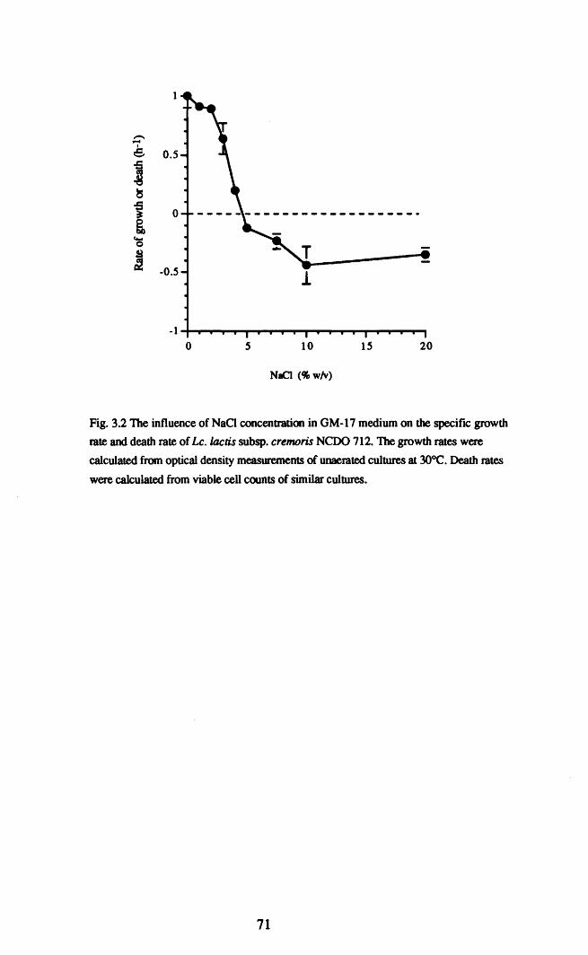
2015105o-1 ..........-I.-..........................,...~..............,.-......~.....,
--Ie- 0.5
~~
t 0
'-0
! -0.5
NaCl (% wlv)
Fig. 3.2 The influence of NaO concentration in GM-17 medium on the specifIC growth
rate and death rate of Le. laetis subsp. eremoris NeOO 712. The growth rates were
calculated from optical density measurements of unaerated cultures at 3OOC. Death rates
were calculated from viable cell counts of similar cultures.
71

3.2.3 Examination of NCDO 712 for an inducible osmotic stress
tolerance response.
Exponentially growing cells in OM-I7 medium were removed at an OD 580 of
0.2-0.4 and exposed to 3% w/v NaCI in fresh OM-17 for 1 hour, prior to being
challenged at various NaCI concentrations. The survival of these cells was compared to
that of cells which had not been pre-exposed to 3% w/v NaCl. A substantial level of
tolerance was induced by exposure to 3% w/v NaCI for one hour (Fig. 3.3). The
induced tolerance apPeared to be only effective at challenge conditions of up to 10%
w/v NaCI (Fig. 3.3). When challenged at 20% w/v NaCI the prior exposure to 3% w/v
NaCI sensitised the cells to the lethal salt challenge. The induced response appeared to
be protein synthesis dependent, as induction was prevented by addition of
chloramphenicol to the growth medium during the adaptation step (Fig. 3.4).
3.2.4 The effect of magnesium on survival during challenge with NaCI
and other environmental stresses.
For these experiments we switched to HEPES buffer or a chemically defined
medium in order to control the Mg2+ concentration during the NaCI challenge. The
effect of adding an energy source (glucose) and MgS04 to HEPES buffer containing
20% w/v NaCI on survival was examined, Addition of glucose did not significantly
enhance the tolerance of NCDO 712 to high NaCI concentrations, whereas addition of
MgS04 led to a significant increase in the tolerance of the cells. The protective effect of
magnesium was neither enhanced nor diminished by the presence of glucose in the
buffer (Fig. 3.5). The protective effect of magnesium was concentration dependent and
the maximum degree of protection was provided by 10 mM M g2+ (Fig. 3.6). The
protective effect appeared to be specific to magnesium; calcium had no effect on
survival while manganese rendered the cells slightly more sensitive than the control
(Fig. 3.7). Magnesium chloride was as effective as magnesium sulphate in protecting
the cells (Fig. 3.7). To be effective in protecting the cells, it appeared that magnesium
had to be present throughout the challenge. Addition of magnesium prior to challenge
72

(A)100
";J>.~
V,)
~
....
100 2 4 6
Time (hours)
(8)100
'iJ>.~
V,) 10~ ....
o 2 4 6
Time (hours)
Fig. 3.3 Induction of tolerance to high concentrations of NaCI by pre-exposure to 3% w/v
NaCI. Le. lactis subsp. cremoris NCOO 712 was grown in OM-17 medium and either
challenged with 7.5% (A). 10% (B) or 20% w/v NaCl (C) directly Cl ). or following
exposure to 3% w/v NaCl in OM-17 for 1 hour (_).
73

100~~__~ ..... ..
10
0.1 ~------r----.....,r-------,o 2
Time (hours)
4 6
Fig. 3.4 The effect of l~g chloramphenicoVml on the induction of tolerance to lethal
NaCl concentrations. Le. lactis subsp. cremoris NCDO 712 was grown in GM-17 medium
followed by challenge with 10% w/v NaCl in GM-17. The cells were challenged directly
( []), or following exposure to 3% w/v NaCl for 1 hour(_) or following exposure to
100 J,1g/ml chloramphenicol for 30 minutes prior to being exposed to 3% w/v NaCI in GM
17 for 1 hour (~).
74

100
10
1
0.1-t----r------.-----r---......
Time (hours)
Fig. 3.5 The influence of magnesium with or without glucose on the survival of Le.
lactis subsp. cremoris NeOO 712 in HEPES buffer pH 7.0 containing 20% w/v NaQ.
Exponential phase cells were challenged with 20% w/v NaCl without additions {[] ),with
25 mM glucose (0 ), with 10 mM MgS04 (.) or with 10 mM MgS04 plus 25 mM
glucose (. ).
75

8642o
10
0.1 .....---......--------.......----
Time (hours)
Fig. 3.6 The influence of the magnesium concentration on the survival of Le. laetis
subsp. eremoris NCOO 712 in HEPES buffer containing 20% w/v NaCI. The
exponential phase cells grown in GM-17 broth were challenged with 20% NaCI in the
absence of magnesium 0 ) and in the presence of 0.1 mM «». 1 mM (0 ). 10 mM
(Iii). or 100 mM (6) magnesium sulphate.
76

100~~_
10
0.1-+----_----_---_--
o 2 4
Time (hours)
6
Fig. 3.7 Survival of exponential phase Le. lactis subsp. cremoris NCD0712 cells grown
in GM-17 medium when exposed to 20% NaCI in HEPES buffer containing 20% w/v
NaCI alone (0 ) or in HEPES buffer with 10 mM MgCI (0 ), 10 mM MgS04 (0),
10 mM CaCl2 (6 ) or 10 mM MnCl2 (II).
77

and its removal at 30 or 60 minutes did not protect the cells beyond the time of removal
(Fig. 3.8). Similarly, if magnesium was not present at the initiation of the challenge but
was added during the challenge, resistance to the high salt was gained from the time of
magnesium addition (Fig. 3.9a). The degree of protection decreased as the interval
between exposure to the challenge and addition of magnesium, increased (3.9b) The
protective effect of magnesium was not a phenomenon unique to the HEPES buffer
system. When the cells were challenged in the chemically defined medium MTEL
omission of magnesium also resulted in increased sensitivity to the NaCl challenge;
however, the difference between survival of cells challenged in the presence of
magnesium and those challenged in its absence was less marked than in the HEPES
buffer (Fig. 3.10). Protection by magnesium was not confined to NaCl stress; its
presence also resulted in increased tolerance to acid stress (200 mM acetate, pH 3.7) or
heat stress (42°C) (Fig. 3.11a and b).
3.2.5 A comparison of the sensitivities to NaCI of Lc lactis subsp.
cremoris strains capable of growth at 4% w/v NaCI with those of Lc.
lactis subsp.cremoris inhibited by 4% w/v NaCI.
This eXPeriment investigated the possibility that there may be a link between the
ability to grow at relatively high concentrations of NaCI and survival of a lethal NaCI
challenge. NCDO 712 is capable of growth at 4% w/v NaCl whereas strains HP, US3
and BK5 are inhibited by NaCl concentrations >2% w/v. When challenged with 20%
w/v NaCI in OM-I7 broth, NCDO 712 was noticeably more resistant, particularly
during the frrst 6 hours of challenge than the other cremoris strains (Fig. 3.12). An
interesting feature of this result was that the loss of viability in NCDO 712 was
biphasic, in that initially the cell numbers decreased slowly followed by a more rapid
decline after 6 hours of challenge. This was noticeable in this eXPeriment in which the
the challenge with 20% NaCI was continued for 9 hours rather than the shorter times in
earlier eXPeriments.
78

3060
100
10
764320.11--......-....,,---......--......- .....---.----.
o
Time (hours)
Fig. 3.8 The effect of magnesium removal during challenge with 20% w/v NaCl on the
survival of Lc. lactis subsp. cremoris NCOO 712. The cells were challenged in HEPES
buffer with 20% NaCI in the presence (. ) or absence (IJ ) of 10 mM magnesium
sulphate. At 30 (0) and 60 (6) minutes after initiation of the challenge aliquots of the
cells challenged in HEPES buffer containing magnesium were removed. centrifuged and
washed twice with magnesium free HEPES buffer and resuspended in HEPES with 20%
w/v NaCl and without magnesium.
79

76432o0.1 .....- .....--..-~.....- .....- .....- ....- .....
1
100
Time (hours)
Fig. 3.9a The effect of delaying the addition of magnesium on the survival of exponential
phase Le. lactis subsp. cremoris NeDO 712 cells in HEPES buffer containing 20% w/v
NaCl. 10 mM magnesium sulphate was added at the start (0 ) at 1~ ), 2 (6 ), 3 (III ),
or 4 (.) hours after addition of the cells to the buffer. Magnesium was completely
omitted from the control sample (. ).
432o-l-t----.,....---.......-----,r----.....
-0.2
-0.4--e! -0.6
~
-0.8
Delay in addition of Mg2+ (hours)
Fig. 3.9b The influence of delaying the addition of Mg2+ to the HEPES buffer with
20% NaCl challenge system on the subsequent death rate of NeDO 712 cells. The death
rates were calculated from the data shown in Fig. 3.9a The horizonta1line indicates the
death rate of cells challenged in the absence of magnesium.
80

o 2
Time (hours)
4 6
Fig. 3.10 Survival of exponential phase Le. lactis subsp. cremoris NCOO 712 cells
grown in MTEL medium when challenged in MTEL medium containing 20% w/v NaCI
in the presence (. ) and absence C ) of 10 mM MgS04.
81

10
0.1
0.01 -+----.......---......---.......--~o 1 2
Time (hours)
3 4
Fig. 3.11a Survival of exponential phase Le. laetis subsp. eremoris NCDO 712 cellsgrown in MTEL medium during challenge in MTEL medium containing 200 mM aceticacid (pH 3.7) in the presence. ) and absence 0 ) of 10 mM MgS04.
100 'UIii::-----...----.....
o 30
Time (min)
60 90
Fig. 3.11b Survival of exponential phase Le. lactis subsp. eremoris NCOO 712 cells grown
in MTEL medium when challenged in MTEL medium at 420C in the presence (_ ) and
absence ([]) of 10 mM magnesium sulphate. The data presented are representative of
triplicate experiments
82

108642
10
0.1-+---__--.....--_--_......0-_
o
100
Time (hours)
Fig. 3.12 Comparison of the survival of exponential phase cells of Le. laetis subsp.
cremoris strains NCOO 712 ([]), HP (0), US3 ( 0). and BK5 (II) during challenge
in GM-17 broth containing 20% w/v NaCI.
83

3.3. Discussion.
In this chapter, the survival of Lc. lactis subsp. cremoris during exposure to
high concentratons of NaCI was examined. The resistance of NCDO 712 to 20% w/v
NaCI was growth phase dependent. This observation was not unexpected as the
resistance of NCDO 712 to low pH is also growth phase dependent (O'Sullivan,
1996). Growth phase dependent tolerance of Le. lactis to a number of other stresses
was also reported by Hartke et al. (1994). Growth phase dependent acid tolerance has
also been reported in L. monocytogenes (O'Driscoll et al., 1996;; Davis et al., 1996),
and E. coli 0157:H7 (Benjamin and Datta, 1995; Arnold and Kaspar, 1995). In E. coli
many genes are under the control of the stationary phase sigma factor as encoded by
the rpoS gene (Hengge-Aronis, 1995) and some of these genes are osmotically induced
(Hengge-Aronis et al., 1993). The rpoS gene product has a role in the resistance of E.
coli and Yersinia enterocolitica to a number of environmental stresses, including
osmotic stress (Badger and Miller, 1995; Cheville et al." 1996). In the Gram-positive
bacteria Bacillus subtilis, Staphylococcus aureus and L. monocytogenes an alternative
sigma factor (aB) has been identified (Kullik and Giachino, 1997; Becker et al., 1998).
It is possible that growth phase dependent stress tolerance in Lc. lactis is due to a
similar sigma factor or a similar transcriptional activator, repressed in early exponential
phase and expressed in late exponential phase.
Incubation of Le.lactis subsp. cremoris NCDO 712 in growth medium with 3%
w/v NaCI for one hour resulted in induced tolerance to subsequent challenge with
higher NaCI concentrations. Induced tolerance to NaCI by pre-exposure to non-lethal
concentrations has been reported for E. coli (Jenkins et al., 1990), B. subtilis (Volker
et al., 1992) and Enterococcusfaecalis (Flahaut et al., 1996). In addition, tolerance to
20% w/v NaCI in Le. lactis subsp. cremoris NCDO 712 by pre-exposure to pH 5 has
also been reported (O'Sullivan and Condon, 1997). Overlap between responses to
different stresses is common in bacteria and there have been reports of this in Lc. lactis
(Hartke et al., 1994; Hartke et al., 1995). The induction of NaCI tolerance was
prevented by the protein synthesis inhibitor, chloramphenicol which has been a feature
84

of most of the induced tolerance systems reported in bacteria. Kilstrup et al. (1997)
identified at least 12 proteins induced by exposure of the NCOO 712 derivative strain,
MG 1363 to 2.5% w/v NaCI; the proteins included the well known heat shock proteins
OnaK, GroEL and GroES. It has been proposed that OnaK has a role in E. coli in
maintaining potassium accumulation during osmotic stress and in renaturing proteins
following plasmolysis (Meury and Kohiyama, 1991).
The induced tolerance response was only effective at the less severe challenge
conditions, in contrast to the acid-induced response reported by O'Sullivan and Condon
(1997), which did protect against 20% NaCI. This suggests that the mechanisms may
be different or that 3% w/v NaCI is a poor inducer of stress tolerance and elicits a
response that is insufficient to protect against higher NaCI concentrations. There is
some evidence to suggest that, in general, a sublethal NaCI concentration may not be a
good inducer of stress tolerance, in that 4% NaCI did not induce tolerance to acid stress
(O'Sullivan and Condon, 1997). In addition, some proteins (including GroEL, GroES,
and DnaK) are induced to higher levels in Le. Iactis subsp. cremoris by heat shock than
by exposure to 2.5% w/v NaCI (Kilstrup et al., 1997). In contrast, NaCI induces a
number of proteins to high levels in Ent.faecalis (Flahaut et al., 1996) and B. subti/is
(Hecker et al., 1988). The induction of stress proteins in Ent. faecalis correlates with
induction of a multi-stress tolerance response (Aahaut et aI., 1997).
The sensitivity of Le. lactis subsp. cremoris to 20% w/v NaCI, as well as low
pH and heat shock was substantially influenced by the availability of magnesium. To
our knowlege the only previous report of magnesium influencing tolerance to stress in
Lc. lactis is that of Thomas and Batt (1967), in which magnesium increased the
resistance of cells to starvation. It has long been recognised that magnesium stabilises
ribosomes (Hui Bon Hoa et al., 1980). The heat-resistance and ribosome stability of
Salmonella ryphimurium were increased by the presence of 100 mM MgCl2 during
challenge at 52°C (Tolker-Nielsen and Molin, 1996). In addition to its stabilising effect
on ribosomes, magnesium is also required for optimal activity of the chaperone DnaK
(Skowyra and Wikner, 1995). Enhanced DnaK function could also explain the effect of
85

at least some of the contribution of magnesium to the acid and heat tolerance of NCDO
712, as DnaK is well known as a heat shock protein, and is also induced by low pH in
Le. Iactis (Hartke et al., 1996). The protective effect of magnesium was concentration
dependent and appeared to be SPecific to magnesium, as neither manganese nor calcium
could substitute for it. This is in agreement with available data on the interaction
between magnesium and DnaK, as the ATPase activity of the DnaK protein is
dependent on the free magnesium concentration and the stimulation appears to be
magnesium-specific (Skowrya and Wickner, 1995). The loss of protection by
removing the magnesium during challenge suggests that the protective effect of
magnesium does not involve irreversible binding of magnesium within the cell. The
protective effect of magnesium diminished as the interval between initiation of the
challenge and magnesium addition increased. This presumably reflects the fact that cell
damage increases in proportion to the time of challenge in the absence of magnesium.
Addition of magnesium to the damaged cells is then less effective in preventing further
loss of viability. The greater resistance of cells challenged in GM-17 medium reflects
the presence of MgS04 (1 mM) in this medium.
NCDO 712, which is capable of growth at 4% w/v NaCI, was more tolerant to
NaCI than the three strains incapable of growth at 4% NaCI. The classical phenotype of
the subspecies cremoris included the inability to grow at 40°C, as well as NaCI
sensitivity. Strains with the classical cremoris phenotype seem to be less tolerant of
environmental stress than NCDO 712 which for many years was phenotypically
classified as a lactis strain. The limited data available to us do not allow any
conclusions to be drawn at present, however the superior performance of the traditional
type cremoris strains as dairy starters (Urbach et al., 1997) might warrant investigation
of stress tolerance in such strains and its possible contribution to starter performance.
The major conclusions to be drawn from the data presented in this chapter are
the presence in Le. Iactis subsp. cremoris NCro 712 of an inducible NaCI tolerance
mechanism and that magnesium has been identified as having a major role to play in the
survival of a number of environmental stresses.
86

Chapter IV
The role of potassium in the adaptation of Lactococcus lactis
subsp. cremoris NCDO 712 to low water activity.
87

Abstract.
Accumulation of potassium was shown to be an important component of the response
of Lactococcus lactis subsp. cremoris NCDO 712 to a decrease in the water activity
(aw) of the medium. Accumulation of potassium was dependent on the nature of the
solute used to adjust the aw of the growth medium. If the external solute was NaCI
accumulation of K+ did not occur whereas solutes such as glucose or fructose resulted
in a doubling of the cytoplasmic potassium content. The specific growth rate of NCDO
712 was substantially reduced in a chemically defined medium (MTEL) with its K+
concentration reduced to 0.5 mM. Increasing the K+ content of the medium to 20 mM
abolished the growth inhibition. In contrast, if the aw was reduced by addition of
fructose, there was no difference between the growth rates of cultures in MTEL
medium containing 0.5 mM K+ or 20 mM K+. The inhibition of the growth rate in
medium containing NaCI at low K+ concentrations correlated strongly with a reduction
in the cytoplasmic K+ content and in the cyoplasmic volume. Omission of the
compatible solute proline from MTEL medium rendered the inhibition more severe,
while addition of the potent compatible solute glycine betaine to MTEL medium
substantially reversed the inhibition. Supplementation of the MTEL medium containing
0.5 mM K+ with the potassium analogue rubidium (19.5 mM) so that the total
potassium plus rubidium concentration was 20 mM, completely reversed the inhibition.
These results demonstrate that potassium accumulation plays an important role in the
tolerance of Lc.lactis subsp. cremoris to osmotic stress.
88

4.1 Introduction.
The importance of potassium in the adaptation of bacteria to hyperosmotic stress
(low aw) has been recognised for many years (Epstein, 1986). Potassium is
accumulated as a means of increasing the cytoplasmic osmolarity to counteract the
increase in the osmolarity of the extracellular environment. Accumulation of potassium
has been best studied in Escherichia coli and other Gram negative bacteria (Epstein,
1986; Csonka, 1989). In the case of Gram positive bacteria, osmotically induced
potassium accumulation has been reported for some bacteria but not for others. The
intracellular potassium content of Bacillus subtiIis (Whatmore et al., 1989; Whatmore
and Reed 1990) Enterococcus faecalis (Kunin and Rudy, 1993) and Listeria
monocytogenes (Patchett et al., 1992) increased in response to osmotic stress. In
contrast, potassium was not accumulated by Staphylococcus aureus grown at low aw
(Kunin and Rudy, 1991).
To our knowlege, the role of potassium in the response of Le. lactis to low aw
has not been investigated. Many years ago, MacLeod and Snell (1948) reported that the
potassium requirements of some lactic acid bacteria were increased by the presence of
sodium or ammonium ions indicating some interaction between K+, Na+ and N}4+.
In a recent report potassium accumulation was observed in Lactobacillus plantarum
when KCI but not NaCI was added to the growth medium (Glaasker et al., 1996a). In
this chapter we examine the role of potassium in the osmoadaptation of Lc.lactis.
89

4.2 Results.
4.2.1 The potassium content of Le. lactis grown in medium of decreased
water activity.
The cytoplasmic potassium content of Le. lactis subsp. cremoris NCDO 712 in
complex (GM-17) or chemically defined (MTEL) medium in the absence of added
solutes was approximately 1.6 J.UlloVmg Protein. Addition of NaCI to reduce the aw of
GM-17 (Fig. 4.1) or MTEL media (Fig. 4.2) resulted in a decrease rather than an
increase in potassium concentration reaching 1 JlmoVmg protein at an aw of 0.983. In
contrast, if glucose or fructose were added to reduce the aw of either medium, the
potassium content of the cells increased (Figs. 4.1 and 4.2). The cytoplasmic
potassium content of NCDO 712 cells in medium containing 0.88M monosaccharide
(aw =0.983) was 3.12 JlmoVmg protein for cells grown in MTEL and 3.36 J,1mol/mg
protein for cells grown in GM-17.
4.2.2 Measurement of the potassium content of NeOO 712 during
growth.
The values for the potassium content of NCDO 712 presented above (4.2.1)
were taken at a single time point from exponentially growing cells. To determine
whether or not the potassium content remained constant during growth, the potassium
content of NCDO 712 cultures was measured at intervals during growth in GM-17
medium at low and high aw•
The results of these eXPeriments show that potassium accumulation occurred
rapidly following inoculation and it remained reasonably constant but with wave-like
fluctuations, especially when the aw was reduced by glucose (Fig 4.3). The values
obtained for the cellular potassium content confirmed the results from the earlier
experiments. No significant accumulation of potassium occurred as a consequence of
decreasing the aw with NaCI, while potassium was accumulated by cells when the aw
was reduced by glucose. It was also noticeable that the lag phase of growth was shorter
when the aw was reduced by sugar rather than NaCl. The potassium content of cells
90

4
B:taO 3g
~§ 2.~
8.()·ss§">.
U
0
0.995 0.99 0.985 0.98
aw
I I I I I0 1 2 3 4
NaCI (Cfow/v)
Io
I0.2
i0.4
i0.6
Glucose (M)
i0.8
Fig. 4.1 The cytoplasmic content of potassium of Le. lactis subsp. cremorisNCDO 712 cells growing in GM-17 medium with the aw decreased by addition ofNaCI ([] ) or glucose (. ).
91

4
-BebO
3g0
~§
2
lj~~
U
00.995 0.99 0.985
Waler activity
I I I0 2 3
NaCI (% w/v)
I , I , , , , I , , I ,0 0.25 0.5 0.75
Glucose (M)
Fig. 4.2 The cytoplasmic content of potassium of L. laetis subsp. eremoris NCDO 712 cells
growing in MlEL medium with the aw reduced by addition of NaCl (0 ) or glucose. )
92

10
0.01
- 4
ibO
g0
3§.-e:::s.~
28-
jc.9~u
0
0 2 4 6 8
Time (b)
0.1
Fig.4.3 Growth as OD580 and the potassium content of Le. lactis subsp. cremorisNeDO 712 growing in OM-17 medium alone (0) and in OM-17 mediumcontaining either 0.88M glucose 0 ) or 3% w/v sodium chloride (<> ) to reduce thewater activity to 0.983.
93

grown in medium with added sugar in these experiments was slightly higher than was
observed in the earlier experiments. As the protein conc./ml of culture was available for
each data point, it was possible to plot the potassium content against the protein content
as the biomass of the culture increased; this gave a straight line graph, the slope of
which was equal to the average potassium content of the culture per mg protein (Fig
4.4). This is probably the most accurate method of measuring the potassium content of
a growing culture, provided that the potassium content remains reasonably constant
during growth, so that there is a straight line relationship between the potassium and
protein contents of the culture. Using this method the potassium content of Le. laetis
grown in the absence of osmotic stress was calculated as being 1.85 Jlmol/mg protein,
while the potassium content of cells grown at an aw of 0.983 in the presence of glucoseI
or NaCI was 3.75 or 1.69 JlmoVmg protein respectively.
4.2.3 The effect of potassium limitation on the growth of Lc. lactis
subsp. cremoris in MTEL medium of low awe
The results of section 4.2.1 seemed to suggest that NaCI was in some way
preventing uptake of potassium by the cells. If this was the case, then reducing the
potassium content of the culture medium would be expected to result in severe
inhibition by NaCI . The concentration of potassium which permitted growth at the
maximal rate in the absence of NaCI was 0.5 mM (see Chapter V). This concentration
of potassium was selected as the limiting concentration and growth was assayed at
increasing NaCI concentrations in the chemically defined MTEL medium containing
0.5 mM potassium and the non-limiting concentration of 20 mM and the results are
presented in Fig. 4.5.
In medium containing 0.5 mM potassium, the specific growth rate of NCDO 712
was substantially inhibited by NaCI and growth did not occur at NaCI concentrations
>2% w/v (aw 0.987). In medium containing 20 mM potassium, the inhibition of
growth was much less severe and slow growth occurred at 3% w/v NaCI (aw 0.983).
94

~'3~
'- 0.750
~§
l 0.5
.S=!e.;~ 0.25>.
U
00 0.1 0.2 0.3 0.4 0.5
mgpnJml
Fig. 4.4 The increase in the cytoplasmic content of potassium as a function of the increase in
biomass (protein/ml) of Lc.lactis NCDO 712 cultures growing in OM-I7 medium alone. ) or
with the aw decreased to 0.983 by addition of 0.88M glucose (<> ) or 3% w/v NaCI 0 ).
95

0.7
0.6
0.5
--e 0.4
:L
0.3
0.2
0.1
0
0.995 0.99 0.985
io
3w of MTEL mediwn
i2
NaCI (% wlv)
i3
I Ii Ii i Ii US 'i IU 'I 'u , '. Ii Ii Ii Ii', ' Ii Ii Ii Ii Ie : Ii
o 0.1 0.2 0.3 0.4 0.5 0.6 0.7 0.8
Fructose (M)
Fig. 4.S The specific growth rate. ~ of Le.loelis subsp. cremoris NCOO 712 in MTEL
mediwn containing either O.S mM ( O. []) or 20 mM ( • • potassiwn. The aw was
adjusted by addition of NaCI ( •• C) or fructose ( • O.
96

In MTEL medium in which the aw was reduced by addition of fructose the reduction of
the growth rate was mainly similar at both potassium concentrations.
The inhibition of growth by NaCI was due to the Na+ rather than the CI- ions, as
Na2S04 had a similar inhibitory effect (Table 4.1). The inhibition of growth at low
potassium concentrations was not specific to Na+ ions however, as NH4+ ions were
even more inhibitory (Table 4.1).
4.2.4 The effect of sodium and other monovalent cations on the
potassium content of NCDO 712.
The data presented in 4.2.3, showed that Na+ and NH4+ ions used to reduce the
aw• increased the potassium concentration required for maximal growth of NCDO 712.
A possible explanation was that the cations caused a reduction in the cytoplasmic
accumulation of potassium. The cytoplasmic potassium content of NCDO 712 in MTEL
medium containing 20 mM KCI was 1.75 J,J.mol/mg. protein and decreasing the
potassium content of the medium to 0.5 mM did not significantly reduce the
cytoplasmic potassium content (Table 4.2). In contrast, if NaCI , Nl4CI or Na2S04
was added to reduce the aw, of MTEL medium, containing the lower concentration of
potassium, from 0.999 to 0.990 the potassium content of the cells was substantially
reduced (Table 4.2). It was notable that there was a strong correlation between the
extent of the reduction in the specific growth rate and the reduction in the cellular
potassium content. Cells growing in MTEL medium with the higher concentration of
potassium had slightly higher cytoplasmic K+ levels when the aw was reduced.
Likewise no reduction in cytoplasmic potassium was observed when the aw of
MTELwas reduced by fructose at low or high potassium levels.
4.2.5 Measurement of the cytoplasmic volume of cells grown in MTEL
medium in the presence of NaCI •
The major function of compatible solutes in osmotically stressed bacteria, is to
maintain turgor pressure and prevent plasmolysis. Following the observation that the
97

\000
Table 4.1 Influence of the solute used to reduce the aw and the potassium concentration of MTEL
medium on the specific growth rate (JJ.) of Lc.laclis subsp. cremoris NCDO 712 in MTEL medium.
J1 (h-1) in MTEL medium containingSolute added &w
0.5mMKCI 2O.0mMKCl
Control 0.999 O.66±O.06 O.66±O.06
O.25MNaCI 0.990 O.25±O.04 0.52±O.05
0.125 M Na2S04 0.990 O.24±O.09 O.54±O.04
0.25MNH4Cl 0.990 O.14±O.02 O.54±O.09
0.44 M Fructose 0.990 0.43 ±O.08 0.47±O.07

~
Table 4.2 Influence of the solute used to reduce the aw and the potassium concentration of the medium
on the cytoplasmic content of K + of Le. lactis NCDO 712 growing in MTEL medium.
Cytoplasmic K+ (J.UI1oVrng. prt) of cells growing in
Solute added MTEL medium containing&w
0.5 mM KCI 2O.0mMKCl
None 0.999 1.62±O.21 1.75±O.2
0.25MNaCl 0.990 1.12±O.20 2.31±O.20
0.125 M Na2S04 0.990 1.07±O.23 2.10±0.02
0.25MNH4Cl 0.990 0.64±O.02 1.94±O.02
0.44 M fructose 0.990 1.81 ±O..07 2.27±O.27

cytoplasmic potassium content of cells was substantially reduced when growing in
MTEL medium containing 0.5 mM potassium and 1.5% w/v NaCl. It was decided to
determine whether the low cytoplasmic potassium concentrations affected the
cytoplasmic volume. The water-permeable/sorbitol-impenneable volume of NCDO 712
cells grown in MTEL medium containing 20 mM potassium and without added NaCI
was 3.8 JlVmg protein. As shown in Table 4.3 reducing the potassium concentration to
0.5 mM in the absence of osmotic stress, or adding 1.5% w/v NaCI to medium
containing 20 mM potassium, did not cause a reduction in the cytoplasmic volume.
When the potassium content of the medium was reduced to 0.5 mM in the presence of
1.5% w/v NaCI the cytoplasmic volume decreased, by approximately 30%, to 2.78
Jll/mg. protein. This decrease in cytoplasmic volume correlated strongly with the
decrease in specific growth rate and cytoplasmic potassium content described earlier. If
the aw of the medium was reduced by adding fructose there was no significant
difference between the cytoplasmic volume of cells grown at 0.5 mM and 20 mM
potassium.
4.2.6 Glycine betaine, proline and rubidium as alternative
osmoprotectants to potassium.
Glycine betaine and proline have previously been reported to act as osmoprotectants
in Le. lactis (Molenaar et al., 1993). In Chapter VI of this thesis we confirmed that
they are also osmoprotective in Le. Iactis subsp. cremoris NCDO 712. Assuming that
the growth inhibition caused by the presence of NaCl at low potassium concentrations
resulted from an osmotic effect, it is reasonable to suggest that a strong osmoprotectant
such as glycine betaine could reverse the inhibition. Similarly, in the absence of glycine
betaine, omission of proline from the growth medium might be expected to result in
more severe inhibition. In Table 4.4 the specific growth rate of cells grown in MTEL
medium from which proline was omitted, was compared with those observed in MTEL
medium in the presence of proline and/or glycine betaine. Addition of glycine betaine
partially reversed both the reduction in growth rate (Table 4.4) and cytoplasmic volume
100

-o-
Table 4.3 Influence of the solute used to reduce the aw, the potassium concentration of the growth mediumand glycine betaine on the cytoplasmic volume of Lc.lactis NCOO 712 cells growing in MTEL medium
Cytoplasmic volume (J.1l/mg. pn) of cells growing
Solute addedin MTEL medium containing.
aw0.5mM KCI 20.0mMKCl
None 0.999 3.74±O.51 3.88±O.34
0.25MNaCl 0.990 2.78±O.37 3.69±0.29
0.25M NaCI plus 0.990 3.09±0.2 3.69±0.42 mM glycine betaine
O.44M fructose 0.990 3.88±O.10 4.36±O.10
0.44 M fructose plus 0.990 4.57±1.0 4.l2±O.132 mM glycine betaine

.....S
Table 4.4 The influence of the organic osmoprotectants proline and glycine betaine on the growth rate (J,l) of Le. lactis subsp. cremoris NCDO712, in MTEL medium with a potassium concentration of 0.5 or 20 mM and the aw reduced to 0.990 by addition of 0.25M NaCI.
Specific growth rate. J,l (h -I) in MTEL medium :
Solute added Concentration of KCI Without proline With proline With glycinebetaine With proline and(mM)
(2mM) (2mM)glycinebetaine
0.63±O.06 0.63±O.06 N.D.None 0.5 0.6I±O.01
O.64±O.04 0.66±O.06 N.D.None 20 0.62±O.03
O.25MNaCl 0.25±0.06 0.39±O.03 O.4I±O.030.5 0.19±0.04
0.25 MNaCl0.54±O.~
0.58±O.04 0.56±O.120 0.47±O.06
N.D.: not determined

(Table 4.3) caused by reducing the aw with NaCI. Confirming that the inhibition
observed, was at least partially due to osmotic stress, exacerbated by impaired
potassium uptake. The presence of proline in MTEL medium led to a slight reversal of
the NaCI inhibition of growth rate, but was not as stimulatory as glycine betaine.
Adding proline and glycine betaine together did not cause any greater reversal of
inhibition than did glycine betaine alone.
The monovalent cation rubidium acts as a potassium analogue and is transported by
the potassium transport systems of E. coli and other bacteria. When rubidium was
added to MTEL medium at a concentration of 19.5 mM in addition to 0.5 mM
potassium (so that the total potassium plus rubidium concentration was 20 mM) the
inhibitory effect of NaCI normally observed at the lower potassium level was not
observed, suggesting that rubidium replaced potassium as an osmoprotectant (Fig.
4.6).
4.2.7 Measurement of the proton-motive force in NCDO 712.
The proton motive force (p.m.f., ~P) is composed of two components, the
transmembrane pH gradient (~pH) and the electrochemical potential (~'¥). Potassium
transport has been shown to be essential for the generation of the p.m.f. in Le. laetis
(Kashket and Barker, 1977). In addition to the osmotic effects already demonstrated, it
was conceiveable that the inhibition observed in the presence of NaCI at low potassium
concentrations might have been partially due to Perturbation of the cytoplasmic pH and
p.m.f. due to impaired potassium transport. The ~pH was measured by measuring the
accumulation of 14C benzoic acid, and the ~'¥ was measured by measuring
accumulation of the lipophilic cation trimethylphenylphosphonium (TPP+). The L\pH
was slightly lowered by a decrease in the potassium concentration of MTEL at normal
aw and by decreasing the aw with NaCI (Table 4.5). However, the effects do not seem
to be sufficient to explain the growth inhibition by NaCI. The values for the p.mJ. of
cells grown in MTEL with 1.5% w/v NaCI at both 0.5 and 20 mM potassium were
similar (l54±8 mV and 146±16 mV resPectively) and were approximately the same as
103

0.8
- 0.6-I.c--:::L8e-5 0.4~
~(J
t::.~
0.2V,)
Control NaCI
Fig. 4.6 The effect of partial substitution of rubidium for potassium in MTEL medium. on
the specific growth rate fl of NCDO 712 in MTEL medium without added solute (control)
0.25M NaCl, or 0.25M Nf4CI.. The MTEL contained 0.5 mM potassium. (. ) or 0.5
mM potassium + 19.5 mM rubidium (~) or 20 mM potassium C. ).
104

-oVI
Table 4.5 The influence of the potassium content of the medium and reducing the aw with NaCI onL1pH. £\ 'fJ. and p.mJ. of Le. lactis NeDO 712 growing in MTEL medium
NaCI (M) KCI (mM) L1pH -Z.L\pH (mV) £\'V (mV) pmJ. (mV)
0 0.5 1.03±O.09 62±6 85±8 147±12
0 20 1.17±O.04 700 52±t2 122±13
0.25 0.5 0.93±O.11 56±7 99±t t54±8
0.25 20 1.05±O.13 63±8 83±9 146±16
Note: The samples were taken when the extracellular pH (pHo) of the cultures reached 5.8

that measured for cells grown in MTEL without NaCI containing 0.5 mM potassium
(l47±12 mY) The values obtained for the p.m.f. of cells grown in MTEL containing
20 mM potassium were lower than for the other cultures, due to the fact that the A'I'
value was extremely low. This appeared to be an accurate representation of the A'I'; part
of the calculation of the A'I' value involves correcting the TPP+ content of the cells for
TPP+ bound non-specifically (i.e. not due to A'I') and it was possible that this might
have been a significant source of error. Analysis of the amount of TPP+ bound by
toluenised cells revealed that the amount of TPP+ bound per mg. protein was similar
for all cultures i.e. it was unlikely to be a source of error in the measurement. No
correlation was therefore observed between ApH or p.m.f. and inhibition of Lc. lactis
subsp. cremoris by NaCI.
4.3. Discussion.
In this chapter the accumulation of potassium by NCDO 712 following osmotic
stress is described. It was also reported that potassium accumulation was dependent on
the solute used to reduce the aw of the medium. Solutes containing the monovalent
cations Na+ or NH4+ did not elicit potassium accumulation whereas the non-ionic
solute fructose did. The reasons for the failure of NCDO 712 to accumulate potassium
were then examined further.
Potassium accumulation by bacteria following osmotic stress has been widely
reported (for reviews see Epstein, 1986; Csonka, 1989). Accumulation of potassium as
a compatible solute has been best studied in Escherichia coli, and the transport systems
responsible for its uptake have been well characterised. In Gram-positive bacteria there
has been little study of the role of potassium in osmotolerance, and to our knowledge,
there are no reports in the literature of osmotically induced potassium transport in Le.
laetis. Our results demonstrate clearly that potassium is accumulated by Le. laetis, but
that its accumulation is conditional on the solute which imposed the osmotic stress.
This is a relatively novel concept, though there have been a few reports in the literature
to indicate that potassium accumulation may be impaired in some circumstances. The
106

earliest report was that of MacLeod and Snell (1948) who reported that the presence of
sodium and ammonium ions increased the potassium requirement of certain lactic acid
bacteria. More recently, Harold and Baarda (1967) reported that sodium ions had a
slight inhibitory effe«t on potassium transport by Streptococcus faecium (now
Enterococcus hirae). The first report of this phenomenon, in the context of osmotic
stress was by Glaasker et al. (1996a), who observed that potassium was accumulated
by Lactobacillus plantarum if the aw of the medium was reduced by potassium chloride,
but not if NaCI was used. However, these workers did not investigate the matter
further.
Potassium accumulation in E. coli is reported to be transient and accumulated
potassium is subsequently replaced by other compatible solutes. In Lc. lactis subsp.
cremoris NCro 712, accumulation of potassium did not apPear to be transient, even in
complex medium (GM~17). This suggests that when possible, potassium accumulation
plays a significant role in the adaptation of Lc./actis NCro 712 to osmotic stress.
We examined the influence of lowering the aw with NaCI on growth at low
potassium concentrations in some detail. NaCI was more inhibitory than fructose at low
potassium concentration. This correlated well with the data presented on potassium
accumulation at low aw• The effect of NaO was due to the cation as opposed to the
chloride anion, as Na2S04 and NH4Cl were also inhibitory.
The potassium content of the growing cells also correlated well with the extent of
growth inhibition observed. The potassium content of cells grown in MTEL medium at
low potassium concentration in which the aw was lowered by sodium or ammonium
chloride was lower than that of cultures grown in the absence of osmotic stress, In
contrast, if sufficient (20 mM) potassium was supplied the potassium content was
greater in the presence of the solutes than in their absence. The potassium content of the
cultures and the specific growth rate attained, appeared to be linked. Ammonium
chloride was more inhibitory than was NaCI and this was also reflected in the
potassium content of the respective cultures.
107

The role of potassium in the maintenance of turgor pressure in bacteria is well
known. We tried to ascertain if the inhibition by NaCI at low potassium concentration
related to a decrease in turgor pressure. The partial reversal of the inhibition by glycine
betaine, and the slightly increased inhibition observed when proline was omitted from
the medium suggest that reduced turgor pressure was at least partially responsible for
the inhibition observed.
One of the consequences of reduced turgor pressure is a decrease in the cytoplasmic
volume. The volume of cells grown in MTEL medium containing 0.5 mM potassium
and 1.5% w/v NaCI was approximately 30% less than that observed for cells grown at
the same NaCl concentration but with 20 mM potassium. In medium which included
glycine betaine, the decrease in the cytoplasmic volume was not as large. These
observations suggest that turgor pressure is reduced with a concomitant decrease in the
cytoplasmic volume when the aw is lowered with NaCl. The bacterial cell wall is
somewhat elastic and subject to stretching forces as a result of internal turgor pressure
(Koch, 1995). It is possible that the reduced cellular potassium content reduces the
turgor pressure sufficiently to cause a reduction in the cell volume due to contraction of
the cell wall. It is also possible however, that the reduction in volume was an artifact of
the reduced growth rate and was independent of the turgor pressure.
Another important cell function which may be affected as a result of potassium
limitation was the generation of the proton-motive force (p.mJ.). Generation of a
transmembrane pH gradient in E. coli is dependent on potassium accumulation. Uptake
of potassium ions by Lc lactis is thought to be essential to balance the the expulsion of
protons from the cytoplasm, and tends to reduce the transmembrane electrochemical
potential (a\l') while promoting an increase in the apH. (Kashket and Barker, 1977).
Poolman et ale (1987) reported that potassium depleted Lc lactis cells were unable to
regulate their cytoplasmic pH. Failure to accumulate potassium could theoretically
impair the ability of the cells to maintain their cytoplasmic pH and also could lead to an
increase in /),\1'. The values which we obtained for the /).pH and /),'11 in the absence of
NaCI are similar to those of Kashket and Barker (1977). The /),'11 was lower in the
108
presence of 20 mM K+ than with 0.5 mM K+ which was partially compensated by a
rise in the pH gradient so that only a relatively slight drop in the p.m.f. was observed.
Our measurements reveal that the reduction in the cytoplasmic potassium content
observed in the presence of NaCl at low extracellular potassium concentrations, did not
greatly affect the cytoplasmic pH or ~\}I. As would be expected, there was a slightly
lower ~pH and a slightly higher ~Y but the magnitude of the changes observed were
unlikely to be the cause of a significant level of inhibition. The presence of NaCl did
prevent the dissipation of ~\}I noted at 20 mM K+ in the absence of NaCI ,presumably
due to a decrease in the rate of potassium transport.
These experiments also provide an insight into the importance of both potassium
and glycine betaine in the osmoregulation of Le. laetis. Provided that potassium is
freely available, glycine betaine did not appear to be necessary for growth in the
presence of 1.5% NaCI, and addition of glycine betaine did not result in any stimulation
of growth. When potassium uptake was limited however, the addition of glycine
betaine to the cultures was stimulatory. This suggests that turgor pressure may be the
signal for activation of glycine betaine transport as there is evidence to suggest that loss
of turgor had occurred in those circumstances. It is notable however, that addition of
glycine betaine was not able to fully restore either the growth rate or the cell volume to
their normal levels. This may imply that at least some potassium accumulation is
necessary for proper osmoregulation.
109

Chapter V
Measurement of the potassium concentration required for
growth and characterisation of potassium transport in
Laetoeoeeus laetis subsp. eremoris NCDO 712.
110

Abstract.
Growth of Lc.lactis subsp. cremoris NCOO 712 in chemically defined medium
was dependent on the potassium concentration of the medium at potassium
concentrations ~ 0.5 mM. Growth was not initiated at potassium concentrations S 30
40 JlM and in medium containing higher potassium concentrations, growth ceased
when the potassium concentration was reduced to 30-40 mM. Rubidium could
substitute for potassium, but only on a temporary basis.
Assay of the rate of potassium transport by intact, exponential phase cells of
NCDO 712 showed that the transport system displayed Michaelis-Menten kinetics. At
pH 7.0 which was optimal for transport of potassium the apparent Km was 0.75 mM
and the maximal velocity was 250 nmoVmg. prt/min. Eadie-Hofstee plots indicated that
only one transport system was involved. No transport occurred in the absence of an
energy source, either glucose or arginine and transport was strongly inhibited by
dicyclohexylcarbodiimide (DCCD) and partially by nigericin. These results suggest that
the system is an active transport system requiring either ATP, or proton-motive-force or
both. Potassium transport was inhibited by the monovalent cations Na, Cs, Li and
N14+ at concentrations well below those which significantly reduce the aWe Double
reciprocal plots indicate that the inhibition was comPetitive. Potassium transport was
non-competitively inhibited when the aw of the assay was reduced by addition of
glucose. The SPecific activity of potassium transport was not increased when the
potassium concentration of the growth medium was decreased from 20 to 0.2 mM but it
was increased slightly (up to two-fold) when the aw of the growth medium was
reduced substantially with either glucose or NaCI. This suggests a slight induction
(increased synthesis of transporter) when the cells are growing in conditions of osmotic
stress.
111

5.1 Introduction.
Potassium is an essential nutrient for bacteria, and many bacteria possess both low
affinity and high affinity potassium transport systems, thereby ensuring their ability to
adapt to changes in potassium availability. Some of these transport systems also
transport rubidium; albeit at a lower rate, and in some bacteria rubidium can replace
potassium as a nutrient
The potassium transport systems of Escherichia coli have been well
characterised; several different low affinity systems (Bakker, 1993) and one high
affinity transport system (Siebers and Altendorf, 1993) have been identified. In Gram
positive bacteria, potassium transport by the lactic acid bacterium Enterococcus hirae is
perhaps the best studied (Kakinuma, 1993). Potassium acts as a compatible solute and
potassium transport is also intrinsic to pH homeostasis and maintenance of proton
motive force (Kroll and Booth, 1983; Kashket and Barker, 1981).
The potassium content of many bacteria increases dramatically on exposure to osmotic
stress. In E. coli the high affinity Kdp system is induced by either potassium limitation
or osmotic stress (Gowrishankar, 1993; Malli and Epstein, 1998). Of the three low
affinity systems, the Trk G and H systems are most involved in adaptation to higher
osmolarity. Trk G and H are constitutively expressed (Rhoads et al., 1976); however,
the activities of both systems are greatly increased by osmotic upshock. The increased
activity is a consequence of regulation of transport activity rather than synthesis of
additional enzyme (Meury et aI., 1985). In Bacillus acidocaIdarius, high (Bakker et al.,
1987), and low affinity (Michels and Bakker, 1987) potassium transport systems have
been characterised and in Methanobacterium thermoautotrophicum a low affinity
system, induced by potassium limitation has been described (Glasemacher et al.,
1996).
In the previous chapter it was demonstrated that Lc lactis NCDO 712
accumulated potassium in response to a decrease in aw and that potassium accumulation
was inhibited by Na+ and other monovalent cations. In this chapter a low affinity
potassium transport system in Le. lactis NCDO 712 is described. Inhibition by Na+
112

and Nl4+ ions was determined, together with the effect of growth under osn10tic
stress on the kinetics of potassium transport.
5.2. Results.
5.2.1 The effect of potassium limitation on the growth of Lc lactis
subsp. cremoris NCDO 712.
In MTEL medium buffered with MOPS-Tris at pH 6.8, a potassium concentration of
0.5 mM was sufficient to support growth of Lc.lactis NCDO 712 at the maximal rate.
Buffering with MOPS rather than disodium B-glycerophosphate allowed the sodium
content of the medium to be kept to a minimum. If the potassium concentration of the
growth medium was less than 0.5 mM the specific growth rate and biomass yield
decreased (Fig. 5.1, Table 5.1). The influence of potassium limitation on both the
specific growth rate and growth yield was most pronounced at potassium
concentrations < 0.1 mM, below which there was a good linear relationship between
potassium concentration and specific growth rate. It was not possible to eliminate all
traces of potassium from MTEL medium; on average the potassium content of MTEL
without added potassium was 29 ~M. This concentration was not sufficient to support
growth; the lowest concentration added was 20 ~M which gave a total potassium
content of 49 ~M and a small amount of growth occurred at this concentration (Table
5.1). There was a linear relationship between the potassium concentration and growth
at potassium concentrations ~ 0.1 mM; it is therefore reasonable to assume that at
potassium concentrations S 0.1 mM, cessation of growth was due to potassium
limitation. The residual potassium concentrations Le. the potassium remaining after
cessation of growth in MTEL medium, are shown in Table 5.1. It is clear from these
results that potassium concentrations ~ 30 J.1M approximately are incapable of
supporting the growth of NeDO 712. The yield of biomass was relatively constant at
60-70 g dry wt/g potassium (Table 5.1). The relationship between the specific growth
rate and the potassium content of the MTEL medium displayed approximate Michaelis
menten kinetics.
113

0.6
0.5-"7e
0.4~
~i 0.3
~()
S 0.2
~tn
0.1
10
1
o-+----,......-..,..---,......- .....---,r-'II.,-......- ... 0.1
o 0.1 0.2 0.3 0.4 0.5 20
KCI content of MTEL medium (mM)
Figure 5.1 The specific growth rate J.l ( []) and fmal growth yield ( • of L. lactis subsp.cremoris NCDO 712 growing in MTEL medium at varied potassium concentrations. The dataare representative of triplicate experiments.
114

..........Vl
Table 5.1 The specific growth rate, J.l (h -1), growth yield and residual potassium concentrations (following growth) of Le. lactis subsp
cremoris NCOO 712 in MTEL medium buffered with MOPS-Tris and containing different levels of KCl.
J.l (h-1)Final biomass Residual K+ (JJM) Yield coefficient
KCI (J.lM) (OD 580) (g dry weight/g K +>
49 0.11±O.03 O.19±O.04 33±3 78
69 O.16±O.04 O.28±O.l 36±4 66
89 O.23±O.03 O.39±O.1 42±8 65
109 O.29±O.02 O.5±O.16 44±10 60
129 O.34±O.O3 O.57±O.05 45±5 63

10
200.1 0.2 0.3 0.4 0.5oO~~.-.oy'---:"--"'T"'"---r-....,"""""",,,,,,-~ 0.1
0.4
- 0.3-I
5:::l
~ 600
-5 0.2 1-nQ
~Q:s2
~.~>-·i 0.1
en
Rubidium content of MTEL medium (mM)
Figure 5.2 The specific growth rate, Jl ([]) and final growth yield. ) of Lc. lactis subsp.cremoris Nero 712 growing in MTEL medium at varied rubidium concentrations. The dataare representative of triplicate experiments
116
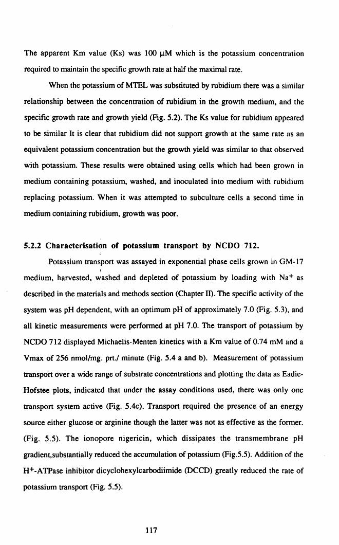
The apparent Km value (Ks) was 100 JlM which is the potassium concentration
required to maintain the specific growth rate at half the maximal rate.
When the potassium of MTEL was substituted by rubidium there was a similar
relationship between the concentration of rubidium in the growth medium, and the
sPecific growth rate and growth yield (Fig. 5.2). The Ks value for rubidium appeared
to be similar It is clear that rubidium did not support growth at the same rate as an
equivalent potassium concentration but the growth yield was similar to that observed
with potassium. These results were obtained using cells which had been grown in
medium containing potassium, washed, and inoculated into medium with rubidium
replacing potassium. When it was attempted to subculture cells a second tinle in
medium containing rubidium, growth was poor.
5.2.2 Characterisation of potassium transport by NCDO 712.
Potassium transport was assayed in exponential phase cells grown in GM-17I
medium, harvested, washed and depleted of potassium by loading with Na+ as
described in the materials and methods section (Chapter 11). The specific activity of the
system was pH dependent, with an optimum pH of approximately 7.0 (Fig. 5.3), and
all kinetic measurements were performed at pH 7.0. The transport of potassium by
NCDO 712 displayed Michaelis-Menten kinetics with a Km value of 0.74 mM and a
Vmax of 256 nmol/mg. prt./ minute (Fig. 5.4 a and b). Measurement of potassium
transport over a wide range of substrate concentrations and plotting the data as Eadie
Hofstee plots, indicated that under the assay conditions used, there was only one
transport system active (Fig. 5.4c). Transport required the presence of an energy
source either glucose or arginine though the latter was not as effective as the former.
(Fig. 5.5). The ionopore nigericin, which dissipates the transmembrane pH
gradient,substantially reduced the accumulation of potassium (Fig.5.5). Addition of the
H+-ATPase inhibitor dicyclohexylcarbodiimide (DCCD) greatly reduced the rate of
potassium transport (Fig. 5.5).
117

8.587.576.56
100 0+---...---....--.....- .....--__-_5.5
pH
Fig. 5.3 The effect of the pH of the assay buffer on potassium transport by Lc lactis
subsp. cremoris NCDO 712. The cells were harvested from exponentially growing
cultures at an OD580 of 0.5-0.7 and depleted of potassium by loading with Na + as
described in mat~rials and methods. The potassium depleted cells were assayed in
MTB-MgS04 buffer at the appropriate pH.
118

300
250 T-=~
J.
§.200
bO 150g0e 100=-> 50
00 5 10 15 20
KCI (mM)
-2 o 2 4 6 8 10
300
- 250=:€ 200B.bO 150g~ 100=-> 50
00 100 200 300 400
V/KCI (nmoVmin/mg. prt/mM)
Fig. 5.4 The kinetics of potassium ttanspon by Lc.lactis subsp. cremoris NeOO 712 cells grown in OM
17 medium, depleted of potassium and assayed in MTB-MgS04 buffer. (A) Rate of ttansport vs substrate
concentration (B) Lineweaver-Burk plot, (C) Eadie-Hofsree plot
119

1.S
-&bOg0
!):::s§~ O.S§
or;;
!§!.
00 2 3 4 S
Time (min)
Fig. 5.5 The requirement for an energy somce and the effect of nigericin and DCCD on
potassium transport by Lc.lactis subsp. cremoris NCDO 712. The cells were grown in
GM-17 medium, depleted of potasSium and assayed in MTB-MgS04 in the absence of an
energy source (. ), with 27 mM glucose ( 0), or 27 mM arginine ( 0). The effect of
adding IIJ.M nigericin ~ ) or 0.3 mM DCCD (II ) to the assay systems containing
glucose is also shown.
120

5.2.3 Inhibition of potassium transport by other monovalent cations
The results presented in the previous chapter suggest that the presence of
monovalent cations such as sodium inhibits potassium transport by NCDO 712. This
was confirmed by measuring the rate of potassium transport by NCDO 712 in the
presence and absence of different cations. In the absence of potential inhibitors,
reducing the potassium content of the assay to 0.5 mM from 20 mM resulted in a 50%
decrease in the rate of potassium transport (Fig. 5.6a). In contrast, if sodium chloride
was present in the assay, reducing the potassium concentration resulted in a substantial
further reduction in the rate of potassium transport at both potassium concentrations
(Fig. 5.6a). The cells were depleted of potassium prior to these assays by loading with
sodium ions. To verify that the inhibition was not due to inhibition of sodium efflux,
choline loaded cells were also assayed (Fig. 5.6b) and the same patterns of inhibition
were observed. In chapter IV the inhibition of growth by potassium limitation was not
observed when the aw was reduced by fructose or glucose. The influence of glucose on
transport of potassium is shown in Fig. 5.7, and it is clear that the presence of these
sugars in the assay only slightly inhibited the rate of potasssium transport.
In order to rapidly assess the inhibitory effect of other monovalent cations a
simple eXPeriment was devised to allow the effects of different cations to be compared.
A potassium concentration (2 mM) was selected with which the rate of potassium
transport was reduced substantially (~30%) in the presence of 100 mM NaCI in the
assay. The rate of potassium transport in the presence of 100 mM cesium chloride,
lithium chloride, ammonium chloride and 50 mM sodium sulphate was then
determined. LiCI was the most inhibitory of the compounds tested; the effect of Cs+
was similar to that of Na+ while Nl4+ ions were more inhibitory than Na+ but less so
than Li+. The use of Na2S04 confirmed that it was the cation rather than the anion
which was responsible for the inhibition (Table 5.2).
121
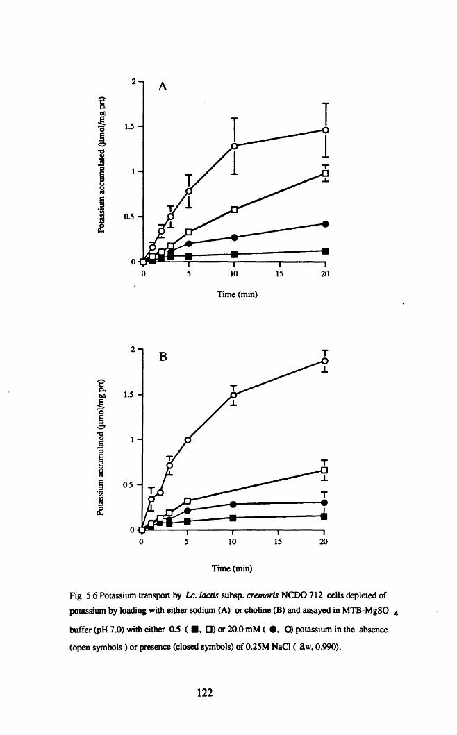
2
&~
~ loS
~"Bl§::s 1
!§.~
OoSse-
O
0 S 10 IS 20
Time (min)
2
oo 10
Time (min)
IS 20
Fig. 5.6 Potassium transport by Le.loclis subsp. cremoris NCDO 712 cells depleted of
potassium by loading with either sodium (A) or choline (8) and assayed in MTB-MgSO 4
buffer (pH 7.0) with either 0.5 ( • I []) or 20.0 mM ( ., 0) potassium in the absence
(open symbols) or presence (closed symbols) of 0.25M NaCI ( aW, 0.990).
122

2
itli>g 1.5
~'-'
'B~§8ca
.~ 0.5en
5£
0
0
Time (min)
Fig. 5.7 Potassium transport by Lc.lactis subsp. cremoris NCDO 712 cells grown in GM-17
medium and assayed in MTB-MgSO 4 with either 0.5 mM ( [J, .) or 20 mM ( ., 0) potassium in
the absence (open symbols) or presence (closed symbols) of O.44M glucose ( aw,0.99O).
123

The inhibition of potassium transport by Na+ and Nf4+ was due to competitive
inhibition; the Vmax value in the presence of either cation was unchanged whereas the
Km value increased in proportion to the inhibitor concentration (Fig. 5.8) Nl4+ was a
much more potent inhibitor than was Na+. The Ki value for each inhibitor (defined as
the inhibitor concentration required to halve the sloPe of a Lineweaver-Burk plot) was
determined by plotting the apparent Km values against the inhibitor concentrations, the
sloPe of the resulting straight-line relationship being equal to Km/Ki (Fig. 5.9). The Ki
value for Nl4+ (38 mM) was much less than that for Na+ (200 mM).
Le. lactis NCDO 712 cells depleted of potassium by loading with choline did
not accumulate sodium ions when sodium was used instead of potassium in transport
assays at concentrations up to 100 mM.
5.2.4 The effect of decreased a \IV on the activity of the potassium
transporter.
In many bacteria the activity of compatible solute transporters is enhanced if the
osmolarity of the assay buffer is increased. Our measurements of the activity of the
potassium transport system in NCDO 712 cells indicate that decreasing the aw of the
assay buffer results in a reduction in potassium transport. In buffer containing 0.88M
glucose, the rate of potassium transport by intact cells which ahad been grown in GM
17 medium containing 0.88M glucose was approx 50% of that observed in the absence
of the solute (Fig. 5.10). The inhibition observed was non-competitive. i.e the Ymax
for potassium transport ;was reduced, without any effect on the Km (Fig. 5.10).;I
5.2.5 Potassium transport was induced by pregrowth in medium with
reduced 8 w but not by potassium limitation.
We have previously observed that potassium was accumulated by Le. facris
subsp. cremoris in response to a decrease in aw (4.2.2) but that decreased aw tends to
124

Table 5.2 Inhibition of potassium transport ofNCDO 712 cells bymonovalent cations.
Solute added Cone.Rate of transport
(mM) nmoVIng. prt./min
None - 186±6
NaCl 100 132±10
Na2S04 50 137±17
CsCl 100 101±30
NH4CI 100 119±3
LiCl 100 97±1
The KCI concentration in the assay was 2 mM. The cells weredepleted of potasSium by sodium loading as described in the materialsand methods section.
125

-2 o 2
l/[KCI] (mM-I)
4
Fig. 5.8a Lineweaver-Burk plot showing inhibition of potassium transport in
Le. laetis subsp. eremoris NCOO 712 by NaCI. The NaCI concentration of
the assay was either 0 (11 ) 50 mM (A ) or 100 mM (. ).
-I..-.5
~c.cOg~S
-2 o 2
l/[KCI (mM)-1
4
Fig. 5.8b Lineweaver-Burk plot showing inhibition of potassium transport in
Le. laetis subsp. eremoris NCOO 712 by Nl4CI ions. The Nl4CI
concenttation of the assay was either 0 g ) SO mM (6 ) or 100 mM ( • ).
126

1007550250.5 .....---.....---r----.....---....
o
3.5
3
~ 2.5e-~... 2~~ 1.5
Na+ or NH4+ (mM)
Fig. 5.9 Plot of the apparent K.m for potassium transport in Le. laetis NCDO 712 against
the inhibitor concentration for Na+ (0 ) and Nl4+ (. ). The slope of the lines is
Km/Ki.
127
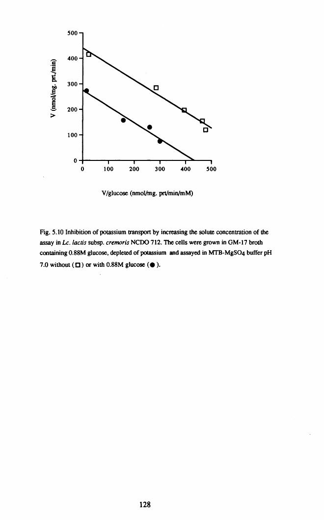
500400300200100o~--.."..--...,.--.......----,~----.
o
500
- 400c
~fi.
300bOg~S 200>
100
V/glucose (nmol/mg. prt/min/mM)
Fig. 5.10 Inhibition of potassium transport by increasing the solute concentration of the
assay in Lc.lactis subsp. cremoris NeOO 712. The cells were grown in GM-17 broth
containing 0.88M glucose. depleted of potassium and assayed in MTB-MgS04 buffer pH
7.0 without (C) or with O.88M glucose (. ).
128

400-c:
~5- 300bO
~! 200
>
100
o 100 200 300 400 soo
v/ KCI (nmoVmg. prt./min)
Fig. 5.11a Kinetics of potassiwn transport by Le. lactis subsp. cremoris
NCDO 712. The cells were grown in GM-17 medium without added solute
(0). or with glucose at 0.3 M (0 ). 0.6M (0), or 0.88M (6). The cells
were depleted of potassium and assayed in MTB-MgS04 buffer at pH 7.0
without added solute
soo400300200100oO-+---.....---..----..-.......--.r---........,
400
- 300c:
!§.bO 200g0ec:-->
100
V/KCI(nmoVmg.prt./miNmNQ
Fig. 5.11b Kinetics of potassium transport by Le lactis subsp. cremoris NCDO
712 grown in GM-17 medium (0) or GM-17 medium containing 3% w/v NaCI
(6) The cells were depleted of potassium and assayed in MTB-MgS04 buffer pH
7.0 without added solute.
129

Yo)
o
Table 5.3 The effect of reducing the potassium content of MTEL medium on the apparent Km and Vmaxfor potassium transport by Le. lactis subsp. cremoris NCDO 712.
KCI concentrationMaximal rate of transport (Vmax)
Apparent Km (roM)ofMTEL (mM) (nM/mg prt.lmin)
0.2 0.72±O.3 279±21
0.3 0.29±O.03 238±20
0.5 0.26±O.06 233±15
1.0 0.42±O.09 272±14
20 0.5±O.15 272±78

reduce the rate of transport (Fig. 5.2a). This suggests that the increased cytoplasmic
potassium levels observed may result from an increase in the amount of potassium
transporter in the cells growing at low aw which would overcome the loss in activity
due to osmotic inhibition by high concentrations of solute. As mentioned already, the
maximum rate of potassium transport by cells grown in GM-17 medium was 250
nmoVmg prtl min. Decreasing the aw of the growth medium by addition of glucose or
NaCI caused an increase in the specific activity of the potassium transport system (Fig.
5.11 a, b). Growth in medium with low aw did not alter the apparent Km for potassium
transport (Fig. 5.11 a, b) indicating that the induced transporter had the same affinity
for potassium as the uninduced transporter.
In some bacteria potassium transport systems are induced in response to
potassium limitation. Since low aw inhibited the rate of potassium transport by NCDO
712 cells it was conceivable that the increased specific activity for potassium transport
was a response to potassium limitation rather than to osmotic stress. Reducing the
potassium concentration of MTEL medium to 0.2 mM did not produce any change in
the specific activity, or the apparent Km of the potassium transport system (Table 5.3).
The rate of potassium transport predicted under these conditions (estimated from the
kinetic data obtained earlier) was less than 30% of the predicted rate expected in cells
grown and assayed in medium containing 0.88 M monosaccharide.
5.3 Discussion.
At potassium concentrations < 100 JlM approximately, the specific growth rate
and growth yield of NCDO 712 were dependent on the potassium concentration of the
growth medium. No growth occurred at potassium concentrations S 30 JlM, and the
final potassium concentration following cessation of growth was approx 30 JlM also.
This suggests that Le. lactis subsp. cremoris NCDO 712 does not possess a high
affinity potassium transport system similar to the Kdp system of Escherichia coli
(Rhoads et al. 1976) and several other bacteria (Bakker et al., 1987; Abee et al., 1992).
The Kdp system in E. coli has a Km value for potassium of about 2 JlM (Bakker,
131

1993) and allows growth to occur under conditions of low potassium availability as it
can scavenge potassium down to concentrations of around 50 nM. The yield of Le.
laetis NCDO 712 biomass per gram of potassium utilised was 6O-70g dry wt/g
potassium which is in agreement with values determined for other bacteria (Pirt, 1975).
Potassium transport by NCDO 712 had a pH optimum of 7.0 and obeyed
Michaelis-Menten kinetics. Eadie-Hofstee plots of transport assay data at potassium
concentrations from 0.1 to 20 mM were straight lines which indicated that only one
potassium transport system was present. Eadie Hofstee plots are an accepted method of
detecting transport systems with differing affinities (for an example see Pourkomailan
and Booth, 1992). Transport of potassium was substantially reduced by the presence of
DCCD which inhibits the H+-ATPase and addition of the ionopore nigericin also
inhibited potassium transport, particularly later in the assay. This suggests that proton
motive force is necessary for potassium transport. DCCD inhibits the H+ ATPase
preventing export of protons and nigericin short-circuits the proton-motive force by
allowing re-entry of exported protons. ATP production by metabolism of glucose or
arginine is also necessary for activity of the potassium transport system. The data
suggest that the transport system is similar to the Ktr I system of Ent. hirae (Bakker
and Harold, 1980) or the TrkG and TrkH systems of E. coli. (Dosch et al., 1991). It
has been suggested that potassium transport in Ent. hirae occurs via a secondary
potassium/proton symport which requires ATP and efflux of two protons via the H+
ATPase (Bakker and Harold, 1980; Kakinuma, 1993). Unlike the Ktr I and Trk
systems, potassium transport in Lc laetis was induced by high solute concentrations.
The Trk systems are constitutively expressed and controlled at the activity level by
extracellular osmolarity (Bakker, 1993). KtrI is neither induced nor activated by high
solute concentration (Kakinuma, 1993). Inhibition by high solute concentration was
observed for potassium transport by NCDO 712.
In Chapter IV we reported that the growth rate and potassium content of NCOO
712 were substantially reduced in cells grown in the presence of NaCI or NlttCI at low
potassium concentrations. The data in Fig. 5.5 support the proposition that the
132

inhibition of growth by NaCI was caused by inhibition of potassium transport. The
inhibitory effect was not confined to Na+ ions as Cs+, Li+ and NH.4+ ions also had an
inhibitory effect on potassium transport (Table 5.2). Inhibition of potassium uptake was
by comPetitive inhibition and NH4+ ions produced more substantial inhibition than did
Na+ ions (Figs.5.7 and 5.8). There was therefore a good correlation between the
degree of inhibition of potassium transport and inhibition of growth rate by Na+ and
Nl4+ ions. Apart from the observations of Macleod and Snell (1948) there have been
few reports in the literature of interference with the potassium requirements of
lactococci by other monovalent cations. In general, potassium transport systems are
regarded as having a high degree of discrimination between potassium and other
monovalent cations (Doyle et ale 1998). The degree of discrimination is greater against
the smaller cations Li+ (0.6 A) and Na+ (0.95 A) than the larger alkali cations rubidium
(1.48 A) and cesium (1.69 A). The close similarity in size of rubidium and potassium
(1.33 A) is thought to be the reason that rubidium can act as a potassium analogue
(Doyle et ale 1998). In the case of NCDO 712, discrimination by the potassium
transport system against other monovalent cations seems to be poor and it is the smaller
cations which tend to be more inhibitory. The system in NCDO 712 may be similar to
that of Ent. hirae in which a mutant with a potassium transport system more strongly
inhibited by Na+ than the wild tyPe transport has been reported. The mutation is
presumably due to an alteration in the structure of the potassium transporter giving it a
greater affinity for Na+ than its parent enzyme (Harold and Baarda, 1967).
In addition to comPetitive inhibition by monovalent cations, potassium transPOrt
in NCDO 712 was also subject to non-competitive inhibition by high solute
concentration. 0.88M glucose reduced the rate of potassium transport by approx 50%.
This inhibition is possibly due to reduced turgor pressure affecting the conformation of
the membrane bound transporter protein. The inhibitory effect of high solute
concentration on the rate of potassium transport was unexPeCted; in general, transport
systems with a role in osmotolerance are activated rather than inhibited by osmotic
stress.
133

Induction of the potassium transport system was observed when glucose or
NaCI was added to reduce the aw of the growth medium, though the extent of the
induction was at most two-fold. The possibility existed that the induction of potassium
transport activity that was observed was not due to osmotic stress per se, but rather to
the lower rate of potassium transport resulting from non-competitive inhibition by high
solute concentration. However, reducing the potassium concentration in MTEL medium
from 20 mM to 0.2 mM did not induce a greater amount of potassium transporter, even
though the rate of potassium transport (calculated from the Michaelis-Menten equation)
decreased from 240 nmol/mg prt./min to 50 nmol/mg prt/min. It therefore seems likely
that the increased rate of potassium transport observed in cells grown in reduced a w
conditions was due to osmotic induction. The fact that low aw caused by either glucose
or NaCI which are structurally different induced increased levels of the potassium
transporter supports this view.
It is clear that the potassium transport system of Le. lactis subsp. lactis NCDO
712 responds to osmotic stress. What is not clear is how the activity of the system is
controlled so as to increase the cell potassium content when required. The activity of the
E. coli Trk systems is stimulated at low turgor pressure (Bakker, 1993), whereas the
activity of the Lc. lactis system is reduced. A further factor to be taken into
consideration is the role of potassium in maintaining the pH gradient and ~ 'II.
Potassium accumulation is essential for the maintenance of a transmembrane pH
gradient in Lc. lactis, 'as potassium uptake is necessary to balance the expulsion of
protons from the cytoplasm (Kashket and Barker, 1977). Presumably, potassium
accumulation in response to proton efflux occurs electrogenically and in response to
changes in the membrane potential, ~ '1'. In contrast, osmotically mediated potassium
uptake needs to be regulated by turgor pressure. Otherwise the increased uptake
resulting from the higher specific activity of the transport system will result in efflux of
excess potassium once turgor is poised at the required level. Such futile cycling of
potassium is wasteful of energy; it is therefore likely that the potassium transpon
system is also regulated in some way by turgor pressure.
134

In conclusion, the data presented in this chapter provides an explanation for the
observations made in the previous chapter, on potassium accumulation by L. lactis
SubsP'fremoris NCDO 712 and its inhibition by other monovalent cations.
135

Chapter VI
Utilisation of glycine betaine and other compatible solutes by
Laetoeoeeus laetis: correlation between low a w sensitivity
and absence of glycine betaine transport activity.
136

Abstract
This study investigates the utilisation of compatible solutes by Le. laetis subsp.
eremoris NCDO 712. Both glycine betaine and proline relieved the inhibition of growth
rate and yield of NCDO 712 caused by growth at low water activity (aw). Proline was
much less stimulatory than was glycine betaine. The dipeptide leucyl-proline relieved
the inhibition of growth rate to a similar degree as proline. A similar degree of growth
inhibition was observed when the aw was reduced by either NaCI or glucose and
glycine betaine was equally effective in counteracting the inhibition by either. Camitine
and ectoine, which are known to act as compatible solutes in other bacteria did not
relieve the inhibition of growth of NCDO 712 at low aw • The difference between the
Le. laetis strains capable of growth at 4% w/v NaCI and those which do not grow at
this NaCI concentration was investigated. The sensitive eremoris strains have an upper
limit for growth of approximately 2% w/v NaCI. Inhibition of growth of these strains
by NaCI was not relieved by addition of glycine betaine while a slight degree of relief
was observed for one strain if the aw was reduced by glucose. The laetis strains were
capable of growth at 4% w/v NaCI and growth inhibition by either NaCI or glucose
was substantially relieved by glycine betaine. All the strains capable of growth at 4%
w/v NaCI accumulated significant amounts of glycine betaine when growing at low aw •
Very little accumulation of glycine betaine occurred during growth of the NaCI sensitive
strains. The NaCl tolerant strains had substantial levels of glycine betaine transport
activity in vitro, whereas the NaCl sensitive strains had little or no such activity. A low
aw sensitive mutant of Lc.laetis subsp. eremoris MG1363 was isolated following ISS}
insertional mutagenesis. This mutant was inhibited at the aw of 0.988 produced by
addition of 2% w/v NaCI or the equivalent glucose concentration (0.58M). The mutantI
did not accumulate glycine betaine when growing at low aw, and did not transport
glycine betaine when assayed in vitro.
137

6.1 Introduction.
Inhibition of microbial growth under conditions of hyperosmotic stress occurs
chiefly as a result of loss of cellular water. Under optimum growth conditions the
solute concentration in the cytoplasm is greater than that of the environment and there is
a tendency for water to flow from the exterior into the cytoplasm. The resulting
hydrostatic pressure, known as turgor pressure, exerts an outward force on the cell
wall (Csonka, 1989). Turgor pressure is thought to provide the force necessary for cell
expansion (Koch et al., 1981; Koch, 1995). It follows, that any decrease in the ratio
between the solute content of the cytoplasm and that of the extracellular environment
will result in an efflux of water from the cell. In bacteria, and indeed in many
eukaryotic organisms,. the most widespread means of counteracting such losses of
turgor pressure is by accumulation of 'compatible' solutes to reduce the a w of the
cytoplasm (Csonka, 1989).
Glycine betaine (N,N,N,-trimethylglycine) is probably the most widely used
organic compatible solute, not alone in bacteria but also in eukaryotic cells (Csonka,
1989). Glycine betaine accumulation occurs either as a result of transport or by
synthesis from choline; only a few bacteria are capable of de novo glycine betaine
synthesis. Bacteria subjected to osmotic stress in medium which does not contain
glycine betaine grow at substantially reduced rates (Csonka, 1989). Similarly, mutant
strains in which the transport systems for glycine betaine are inactivated grow poorly
under conditions of osmotic stress (Haardt et al., 1995; Kappes et al., 1996) and are
not stimulated by glycine betaine.
Lactococcus lactis is an industrially important microorganism, many strains of
which are used as starter cultures in the manufacture of numerous cheese varieties and
also in other dairy products. In recent years the response of L. lactis to environmental
stress such as low pH (Hartke et al., 1994; O'Sullivan and Condon, 1997), high
(Kilstrup et al., 1997; Whitaker and Batt, 1991) and low temperatures (Panoff et al.,
1995), and starvation (Hartke et al., 1994) have been studied.
138

However, there have been only a few studies (Molenaar et al., 1993; O'Sullivan and
Condon, 1997; Troller and Stinson, 1981) which examined osmotic stress in L. lactis
and to our knowlege, there has been only one report to date concerned with the
accumulation of compatible solutes by this bacterium. The accumulation of glycine
betaine by L. lactis ML3 has been reported to occur via a constitutively expressed high
affinity transport system and also via an inducible transport system which also
transports proline (Molenaar et al., 1993). There have been several reports that other
lactic acid bacteria also use glycine betaine as an osmoprotectant (Glaasker et al.,
1996a; 1996b; 1997; Hutkins et al., 1987; Kunin and Rudy, 1991; Sakaguchi, 1960).
The species Lactococcus lactis is subdivided into two subspecies, lactis and
cremoris on the basis of a number of phenotypic traits, among which is the ability to
grow in the presence of NaCI. An upper limit for growth of 2% w/v NaCI has been
used to distinguish members of the subspecies cremoris from members of the
subspecies lactis for which growth at 4% w/v salt is a general characteristic. In recent
years, DNA sequence analysis has been used to reclassify some lactis strains as
cremoris strains and vice versa (Godon et al., 1992) This reclassification has led to a
situation in which the reclassified subspecies lactis and cremoris now contain strains
with different levels of tolerance to NaCl.
The osmotolerance of Lactobacillus strains appears to be dependent on their
ability to accumulate glycine betaine (Hutkins et al., 1987). We have investigated the
possibility that the failure of L. lactis subsp. cremoris strains to grow at NaCI
concentrations in excess of 2% w/v is related to an inability to utilise glycine betaine as
an osmoprotectant. In addition, an osmotic stress sensitive mutant of Lc. lactis subsp.
cremoris MG 1363 was isolated by insertional mutagenesis, and characterised in
relation to its ability to transport glycine betaine and to grow at low aWe
139

6.2 RESULTS.
6.2.1 Inhibition of growth of Lc. lactis subsp. cremoris NCDO 712 in
complex medium at low aWe
Lactococcus lactis subsp. cremoris NCDO 712 is one of those strains formerly
classified as being a ~ember of the subspecies lactis, but which has now been
reclassified as belonging to the subspecies cremoris. An initial series of eXPeriments in
complex medium had a twofold purPOse. Firstly, to quantify the effect of low aw on the
growth rate of this strain and secondly to determine if the nature of the solute used to
reduce the aw influenced the sensitivity of NCDO 712 to osmotic stress. The specific
growth rate in OM-17 decreased gradually from a value of 1.0 h-l at an aw of 0.999 to
about 0.25 h-l at an aw of 0.977 (Fig. 6.1). , the specific growth rate in OM-17 broth
at any specific aw was the same when the aw was reduced with NaCI or glucose.
6.2.2 The effect of compatible solutes on the growth of NCDO 712 at
low awe
It was not possible to assess the effect of adding compatible solutes to OM-17
medium, which is rich in sources of these compounds. Instead, a chemically defined
medium based on that of Thomas et al. (1979) was used (MTEL medium). Olycine
betaine and proline were used initially as it has previously been shown that these are
accumulated by Le. lactis ML3 in response to osmotic stress. Orowth of NCDO 712
was strongly inhibited by addition of NaCI to MTEL medium and little growth occurred
at 4% w/v NaCI. IT 2 mM glycine betaine was added, there was a substantial relief of
inhibition of both growth rate and of growth Yield (Fig 6.2). By comparison, proline
was much less significant; there was only a slight relief of inhibition of growth rate
(Fig. 6.3 a). The Km for proline transport by the lactococci is quite high (Molenaar et
al., 1993) and as only 2 mM proline was used it was possible that the low level of
stimulation by proline (relative to that observed with glycine betaine) was due to limited
proline uptake. However, increasing the proline concentration to 20 mM only slightly
140

1.25
- 1-e::s.
! 0.75
j~~ 0.5.[tI)
0.25
00.995 0.99 0.985 0.98
aw
i I i I I0 1 2 3 4
NaCl (If> w/v)
i I I I i I0 0.2 0.4 0.6 0.8 1
Glucose(M)
Fig. 6.1 The specific growth rate, J.1 (h-l), of Lc.lactis subsp. cremoris NCOO 712 in
GM-17 medium with the aw adjusted by addition of NaCI 0 )or glucose Ci ).
141

(A)0.7
0.6
-- 0.5..C:-::s.! 0.4
l& 0.3
~
'i 0.2CI)
0.1
0
0 2 3 4
NaCI (If, w/v)
(B)
432
0.1
0.01-+---_..~--.....--_..~--_o
NaCI(lf,w/v)
Fig. 6.2 Specific growth rate, J1 (h-l), (A) and growth yield (B) of Le. lactis subsp.
cremoris NCDO 712 in MTEL medium with the aw reduced by adding NaQ, in the
presence 01 ) and absence C ) of 2 mM glycine betaine.
142

-:::-.C:-- 0.4SeoS~
~~~ 0.2fn
O~-----r-----..,...---......o 1 2
NaO (%w/v)
3
Fig. 6.3a The specific growth rate, 1.1 (h-l), ofLe. lactis subsp. cremoris NCOO 712 in
MTEL medium with the aw reduced by adding NaCI in the presence (1;1) and absence
( [J) of 2 mM proline
0.8
- 0.6-Ie~oS
~ 0.4
~S&
ron0.2
O~---p------r----r------.o
Proline (mM)
Fig. 6.3b The effect of proline concentration on the specifIC growth rate, 1.1 (h-l), of Le.
lactis subsp. cremoris NCOO 712 in MTEL medium containing 0% ([J ) or 2% w/v (6)
NaCl.
143

,increased the relief of inhibition observed (Fig. 6.3b), indicating that limitation of
proline uptake was unlikely to be the reason for the lack of stimulation by proline.
Addition of the diPePtide leucyl-proline to MTEL medium relieved the inhibition of
growth of NCDO 712 at low aw , but only to the same extent as proline (Fig. 6.4).
6.2.3 The effect of other potential osmoprotectants on the inhibition of
growth of NCDO 712 at low 8 w •
Although proline and glycine betaine are the only osmoprotectants which have
been reported to be utilised by Lc.lactis, there are a number of other compounds which
act as osmoprotectants in other bacteria. We examined the ability of two of these
compounds, camitine and ectoine, to stimulate NCDO 712 at low aw . The addition of
carnitine to MTEL medium had no effect on the SPeCific growth rate at low aw (Fig.
6.5). Ectoine had no effect on either the specific growth rate or the final biomass yield
of NCDO 712 at low awe The compound was quite eXPensive to buy, so the effect of
ectoine addition was only detennined at 3% w/v NaCI and the equivalent concentration
of glucose (O.88M) (Table 6.1).
6.4.4 The effect of glycine betaine on the growth of other Lactococcus
lactis strains
When it was attempted to extend the study of glycine betaine accumulation to
other strains, a problem was encountered in that a number of the strains were unable to
grow on MTEL. However, these strains grew satisfactorily in another chemically
defined medium (CDM) as described by Molenaar et ale (1993). It would have been
advantageous to omit proline from this medium so that glycine betaine, if added, would
be the only osmoprotectant present. Unfortunately, all the cremoris strains were proline
auxotrophs and some of the lactis strains grew at sub-optimal rates in the absence of
proline. Proline was therefore present in the CDM for all eXPeriments. To allow
comparison with previous results, the relevant eXPeriments previously done in MTEL
using NCOO 712 were rePeated using the new medium.
144

0.8
0.6::::-e::s.
! 0.4oS~
~(,)
$ 0.2~en
00 2 3 4
NaO(%w!v)
Fig. 6.4 The specific growth rate J.1 (h-1) of Le. laetis subsp. eremoris NCDO 712 inMTEL medium with the aw reduced by addition of NaCl, in the presence ( II) and absence([]) of 2 mM leucyl-proline.
145

432
NaCl (% wlv)
o-f----oro---.......---....----.;;IjDo
0.8
- 0.6-.i::'-'"~
~oS 0.4
~.g.[
0.2V)
Fig. 6.5 Specific growth ratet J.1 (h-1)t of Le. laetis subsp. eremoris NCOO 712 in MTEL
medium with the aw reduced by adding NaCl t in the presence a )and absence 0 ) of 2
mM D/L-carnitine.
146

Table 6.1 The specific growth rate. J.1 (h-l) and growth yield ofLc.
lactis subsp. cremoris NCOO 712 in MTEL medium with the awreduced by addition of NaCl or glucose in the presence and absence of 1
mM ectoine.
Culture additions J.1 (h-l) Growth Yield(00580)
None O.66±.02 1.34±O.O3
ectoine O.66±O.Ol 1.27±O.1
3% NaCl O.20±O.Ol O.25±O.O5
3% NaCI + ectoine O.22±O.Ol O.3I±O.07
O.88M Glucose O.25±O.Ol O.58±O.Ol
O.88M Glucose + O.25±O.Ol O.59±O.O3ectoine
147

The inhibition of the growth rate of the three lactis strains and the cremoris
strain NCOO 712 at NaCI concentrations >1% w/v was substantially relieved by
glycine betaine (Fig. 6.6). The relief of inhibition was most apparent at 4% w/v NaCI,
where three of the four strains (NCOO 712, BA2, and CI0) did not grow unless
glycine betaine was present. One strain (BA1) was capable of growth at 4% w/v NaCI
in the absence of glycine betaine, but at a greatly reduced rate. If the aw of the medium
was reduced by addition of glucose, the effect of betaine on the growth rate was similar
to that observed when NaCI was used (Fig. 6.6). Glycine betaine did not have as
strong an influence on growth yield as on growth rate (Fig. 6.7) The yields of strain
BA1 were similar in the presence and absence of glycine betaine. The growth yield of
strains 712, CI0 and BA2 were reduced at low aw when NaCI was used and this
reduction was prevented by glycine betaine. NCOO 712 was the only strain whose
yield was reduced at low aw by glucose and this was prevented by glycine betaine.
Addition of glycine betaine did not significantly relieve the inhibition of the
growth rate of the other cremoris strains HP, BK5 and US3 in COM medium
containing NaCI (Fig. 6.8). In the case of HP and BK5 inhibition of growth by glucose
was not relieved either; however, inhibition of the growth rate of the US3 strain by
glucose was relieved by glycine betaine. Growth of strain US3 occurred at glucose
concentrations S 0.88M (equivalent aw to 3% w/v NaCl) provided that betaine was
present. Some stimulation by glycine betaine, of the growth yield in COM when the aw
was reduced by glucose was also observed, for the cremoris strains BK5 and US3
(Fig. 6.9).
6.2.5 Glycine betaine transport by L. lactis strains.
NCOO 712 was used as a model strain to try to identify the main characteristics
of glycine transport in L. lactis. When NCOO 712 was grown in COM without added
salt or sugar the harvested cells transported glycine betaine via a high affinity system
which essentially obeyed Michaelis-Menten kinetics (Fig. 6.10). The apparent Km was
1.41lM, the Vmax was 8.0±1 nmol. mg. prt.-1. min-I, and the pH optimum was 6.0
148

- NeDO 712 ::- 1-e J::'-"
:::l 0.75 :::l
B £ 0.7S
e e~ 10.5 0.5
~ ~(.) (.)
$ 0.25 10.25
&tI) tI)
0 0
0.995 0.99 0.985 0.98 0.995 0.99 0.985 0.98
aw aw
-- -e -e:::l 0.8 :::l
~ B0.75
.c 0.6 e- -5~
~ ! 0.50.4
(.)
tJ::: ~.-~ 0.2 .- 0.25
&tI)tI)
00
0.995 . 0.99 0.985 0.980.995 0.99 0.985 0.98
aw aw
i I i I i I I I0 2 3 4 0 2 3 4
NaCl (% w/v) NaCI (% w/v)
i , I , I , I , I, I , I ,
I, I
0 0.2 0.4 0.6 0.8 0 0.2 0.4 0.6 0.8
Glucose(M) Glucose (M)
Fig. 6.6 The specific growth rates, J1 (h-l), of Le. laetis subsp. cremons NCOO 712 and
Le. lactis subsp. laetis strains CIO, BAI and BA2 in COM medium with the aw reduced
by glucose (circles) or NaCl (squares) in the presence (closed symbols) or absence (open
symbols) of 2 mM glycine betaine. The data are representative of triplicate experiments.
149

10 NCDO 712 10 CIO
- -~ i
V')V') aQQ,'0 :g"is .!a
>= 0.1>
0.1
0.01...&...--.,..--~---r--....... 0.01----.,..--.....---.-----
0.995 0.99 0.985 0.98 0.995 0.99 0.985 0.98
aw
10 BAI 10 BA2
s00 6' 1V') 00Q V')
0 a-;g.!! :g> .!t
0.1 >0.1
0.01 0.01
0.995 0.99 0.985 0.98 0.995 0.99 0.985 0.98
aw aw
i iI i i i i0 1 2 3 4 0 1 2 3 4
NaCI (% w/v) NaQ (% w/v)
I i i I i i i i0 0.2 0.4 0.6 0.8 1 0 0.2 0.4 0.6 0.8
Glucose (M) Glucose(M)
Fig.6.7 The growth yields (00580) of Le. lactis subsp. eremoris NCOO 712 and the Le.
laetis subsp.lactis strains ClOt BAI and BA2 in CDM with the aw reduced by addition
of NaCl (squares) or glucose (circles). in the presence (filled symbols) or absence (open
symbols) of 2 mM glycine betaine.
ISO

0.8--5:s. 0.68e-5
0.4
~ut=: 0.2.~
tI)
0
0.995 0.99 0.985
0.8
::-.c- 0.6:s.8e-5 0.4~
~u
0.2t=:.~
tI)
00.995 0.99 0.985
0.8:::-.c-:s. 0.68e-5
0;4~
~u
t=:0.2.~
tI)
0
0.995 0.99 0.985
aw
I I I0 1 2 3
NaCl (Ofow/v)
o 0.2 0.4 0.6 0.8
Glucose(M)
Fig. 6.8 The specific growth rates, J.l (h-l), of Le. lactis subsp. cremoris strains HP,
BK5 and US3 in COM medium with the aw reduced by addition of glucose (circles) or
NaCI (squares), in the presence (filled symbols) or absence (open symbols) of 2 mM
glycine betaine. The data are representative of triplicate experiments.
151

10- UP~In
0Q,"0~>.
-5 0.1
~Co:)
0.010.995 0.99 0.985
10-~In
0Q,:s2.~>.-5 0.1~
eCo:)
0.010.995 0.99 0.985
10
SUS3
00In
00""'-"
:s2.~ 0.1>.
-5~
eCo:) 0.01
0.995 0.99 0.985
aw
i i i i0 1 2 3
NaCl ('!ow/v)
i i i i0 0.2 0.4 0.6 0.8
Glucose(M)
Fig. 6.9 The growth yield of Le.laelis subsp. eremoris strains HP, BK and US3 in
COM with the aw reduced by addition ofNaCl (squares) or glucose (circles) in the
presence (filled symbols) or absence (open symbols) of 2 mM glycine betaine.
152

(Fig. 6.10). No transport was observed in the absence of an energy source in the assay
buffer (Fig. 6.11). When NCOO 712 cells were harvested from COM to which NaCI or
glucose was added to decrease the aw their rate of glycine betaine transport was
increased, (Table 6.2 a and b). The apparent KIn was similar to those of cells grown in
COM alone and again no transport occurred in the absence of an energy source in the
assay buffer (Fig. 6.11). However, the transport of glycine betaine by cells grown in
COM with a decreased aw, differed from that by cells grown in COM alone. The rate of
glycine betaine transport by cells grown at a low aw was stimulated by decreasing the
aw of the assay buffer, by addition of salt (Table 6.2a) or glucose (Table 6.2b) whereas
no such stimulation was detected when cells grown in COM alone were assayed with or
without solute. The accumulation of glycine betaine transport by cells grown in CDM or
COM with 1% w/v NaCI was slightly reduced by the the presence of 1 J,LM nigericin in
the assay; 1 J,LM valinomycin did not affect glycine betaine transport to the same extent
as did nigericin (Table 6.3).
The general characteristics of glycine betaine transport were similar to that of
NCOO 712 in those L.lactis subsp. lactis strains C10, BA1 and BA2 whose growth
rates in COM at low aw were stimulated by glycine betaine (Fig. 6.12). The transport
rates of these cells grown in COM with NaO were stimulated by the same concentration
of salt in the assay buffer (Fig. 6.12). Glycine betaine transport was not detected in the
L. lactis subsp. cremoris strains HP or BK5 (Fig. 6.12), neither of which could grow
in COM at aw values below 0.987 (2.5% NaCI or 0.75M glucose) and which did not
respond to glycine betaine in the growth medium. A low rate of transport was detected
in the other cremoris strain US3; this strain did respond to glycine betaine when
growing in COM containing sugar but not salt (Fig. 6.8). The low rate of transport of
US3 cells was not increased by growth in COM with 1% NaCl; activation of transport
was observed when NaCI was added to the assay buffer but the rate of transport was
less than that in NCDO 712 or the L. lactis subsp. lactis strains (Fig. 6.12).
153

100
~ 75:~~
§ 50e.~
25~
05.5 6 6.5 7
pH of assay buffer
- 10r::
I B8
bbg6
~r::-i 4~g
2'-0
~ 00 5 10 15 20
Glycine betaine (J1M)
-,r::Ibb
%es~--2 o 2 4 6 8 10
IlGlycine betaine ijiM-l)
Fig. 6.10 Transport of glycine betaine by exponential phase Le. lactis subsp. cremoris
NCOO 712 cells in phosphate buffer. (A) at different pH values with 10~ 14Cglycine
betaine and (B) at pH 6.0 at different glycine betaine concenttations. In (C) the data of (B)
are plotted in the Lineweaver-Burle fonn.
154
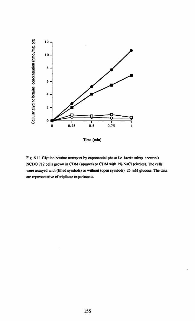
i 12bO!0 10~-.-
·1 8
8§ 6
j 4
vc'0>. 2bb
19== 0vU
0 0.25 0.5 0.75 1
Time (min)
Fig. 6.11 Glycine betaine transport by exponential phase Le. lactis subsp. cremoris
NCDO 712 cells grown in CDM (squares) or CDM with 1% NaCl (circles). The cells
were assayed with (filled symbols) or without (open symbols) 25 mM glucose. The data
are representative of triplicate experiments.
155

Table 6.2a Glycine betaine transport by exponential phase Le. lactis subsp. cremoris NCDO 712
cells grown in COM with different NaCI concentrations and asssayed with and without NaCI in the
buffer.
Rate of transport (nmoVmg prtlmin) in phosphate buffer with:
COMAdditions
0% NaCI 1% NaCI a 2% NaCI b
None 9.6±O.2 10.55±O.8 9.74±O.5
1% NaCI a 16.5±1.5 36±2 33±1
2% NaCI b 32±7 N.D. 81±14
a and b refer to similar aw values
N.D.: not determined
Table 6.2b Glycine betaine transport by exponential phase Le. lactis subsp. cremoris NCDO 712
cells grown in COM with different glucose concentrations and assayed with and without glucose
in the buffer.
Rate of transport (nmoV mg prtlmin) in phosphate buffer with:
COMAdditions 0 0.29M glucose a 0.58M glucose b
COM 9.6±O.2 N.D. N.D.
0.29M Glucose a 18.2±4 34±7 N.D.
0.58M Glucose b 27±9 N.D. 125±10
a and b refer to similar aw values
N.D.: not determined
156

-V.-J
Table 6.3 Influence of the ionopores nigericin and valinomycin on glycine betaine transport by Le. lactis
subsp. cremoris NCDO 712 cells grown in COM with and without 1% NaCI. The NaCl concentration of
the assay buffer in each case was identical to that of the COM in which the cells were grown.
NaClofCOMRate of glycine betaine transport (nmol/mg. prtJmin) in assay buffer with:
(%w/v)No additions 1 J.1.M nigericin 1 J1M valinomycin
nigericin plusvalinomycin
0% 9.0±0.7 1.0±0.1 5.6±5. O.9±O.2
1% 37.3 ±3 11.5±3 38.8±IO 5.8±O.9

80-c~ 70
~bO
60-€'0ec 50"-'
i~ 40
8..... '! 30u. .800
8.[20
'bO'S!a 10~ O.
712 C10 BA1 BA2 UP BK5 US3
Strain
Fig. 6.12 The rate of glycine betaine transport by Lc.lactis subsp.lactis strains CIO, BAI and BA2 andLe. lactis subsp. cremoris strains NCOO 712, HP, BK5 and US3. The cells were grown in COM~) orCOM with 1% NaCl (~ and assayed in phosphate buffer or grown in COM with 1% NaCl and assayed inphosphate buffer containing 1% NaCI ell).

6.2.6 Accumulation of glycine betaine during growth at low awe
NCOO 712 cells accumulated glycine betaine when growing in COM with
the aw reduced by either NaCI or glucose (6.13). A low level of glycine betaine was
present even in cells growing in COM without any added solute; addition of NaCI or
glucose caused the cellular content of glycine betaine to increase. At NaCI
concentrations between 1 and 3% w/v and glucose concentrations between 0.29M and
0.88M, there was a linear relationship between the extracellular solute concentration
and the glycine betaine content of the cells. Cells growing in COM containing glucose
accumulated higher quantities of glycine betaine than did cells growing in COM
containing NaCI at the equivalent aWe
To determine whether the remaining strains accumulated betaine during growth,
glycine betaine accumulation was measured in cells growing in COM and also in COM
containing 1.5% w/v NaCI or the equivalent glucose concentration (O.44M). These
solute concentrations were selected so as to obtain a reasonably rapid rate of growth by
the cremoris strains while being sufficiently high to elicit a reasonable level of glycine
betaine accumulation in those strains which were positive. A basal level of 20-40
nmol.mg prt- I was detected in all strains grown in COM alone. With either NaCI or
glucose as the solute added to the COM, all of the lactis strains tested accumulated
betaine to higher levels than did NCDO 712, with each strain accumulating > 200
nmol/mg protein (Fig. 6.14). Accumulation of glycine betaine in response to NaCI or
glucose by the cremoris strains was much lower than that for the lactis strains. HP
was unique in that no increase in its cellular content of glycine betaine was observed at
reduced aw whether by addition of NaCI or glucose. The glycine betaine content of the
other cremoris strains increased moderately at low aw, US3 and BK5 apPeared to
increase their betaine content in response to NaCI but not glucose.
159

1000
ibe
~-S 750ec:-8I!,g 5008.~
';b.Sle 250fI)
S~>.
U
01 0.995 0.99 0.985
aw of the medium
I0 1 2 3
NaCI(%w/v)
o 0.2 0.4 0.6
Glucose(M)
0.8
Fig. 6.13 Accumulation ofl 4<: glycine betaine by Lc.lactis subsp. cremoris NCOO 712
cells growing in COM medium with the aw adjusted by addition of glucose (e ) or NaCl
([] ).
160

500
i 400cO§0ec-- 300cB5u8..... ·s
2000\ .8......~ubtlOu
100·s.!~>.
U0 .
712 CI0 BAI BA2 HP BKS US3
Strain
Fig. 6.14 Accumulation of 14C labelled glycine betaine by Le.wctis subsp. cremoris sttains NCDO 712, HP, BK5, and US3
and Le. lactis subsp.lactis strains CIO, BAI and BA2 growing in COM (EJ), COM with 1.5% w/v sodium chloride (r.a) or
COM with O.44M glucose ( •. The NaCI and glucose concentrations each reduced the aw of the COM medium to 0.990

6.2.7 Isolation of a L lactis subsp. cremoris MG1363 mutant unable to
accumulate betaine.
NCDO 712 was unable to grow at 4% w/v NaCI unless betaine was present, and there
was a strong correlation between the ability to transport betaine and the ability to grow
at low aw • These observations suggested that it should be possible to obtain mutants of
NCDO 712 deficient in glycine betaine transport by replica plating mutagenised cells on
medium with either low or high aw in the presence of glycine betaine and selecting
colonies unable to grow at the low aw• An insertional mutagenesis system (Maguin et
al., 1996) was used to obtain a bank of mutants ofL. lactis subsp. cremoris MG 1363 a
plasmid-free derivative of NCDO 712 (Gasson, 1983). A concentration of NaCI of
3.5% w/v was selected because NCDO 712 was incapable of growth on CDM plates
containing this NaCI concentration unless glycine betaine was present. Following the
replica plating of 1,000 colonies on OM-17 and OM-17 with 3.5% NaCl, 13 colonies
incapable of growth at 3.5% w/v NaCI were identified. One of these (OSM2) was
selected for verification that it was inhibited by both NaCI and glucose, i.e that the
inhibition was due to osmotic stress rather than a specific inhibitory effect of NaCI (Fig.
6.15). A 32P-Iabelled fragment of DNA from the inserted plasmid hybridised to digested
OSM2x chromosomal DNA, but not to MG1363 chromosomal DNA (Fig. 6.16). The
mutant (OSM 2) was then treated to excise the integrated plasmid so as to create a stable
ISS] mutant (OSM 2x) which could be grown without fear of excision at 30°C. This
isolate was tested by bacteriophage typing to verify that it was the same phage type as
the MG1363 parent strain.
Several attempts were made to clone the sequences flanking the ISS] insenion
in OSM2 using the method described by Maguin et al. (1996). Unfortunately, all
attempts proved unsuccessful. As an alternative to this method, it was attempted to
clone the flanking sequences by inverse PCR. Chromosomal DNA from OSM2x was
digested with either EcoRI or HindIII, neither of which had recognition sites within
ISS]. Following religation at low DNA concentrations so as to favour intramolecular
ligation the DNA was used as a template in an inverse PCR reaction. The primers used
162

1
-- 0.75e~
~-5
~0.5
(,)
$
~(I) 0.25
o.......--....----_--_~6_-_ ...
Io
0.995
I1
0.99
aw
I2
NaCI(%w/v)
0.985
i3
0.98
I4
Io
i0.2
I0.4
I0.6
i0.8
Glucose(M)
Fig. 6.15 Specific growth rate in GM-17 of Le.laelis subsp. eremoris MG 1363 (squares)and the osmotic stress sensitive mutant OSM 2 (triangles) in GM-17 medium with theaw adjusted by addition of NaCI (filled symbols) or glucose (open symbols).
163
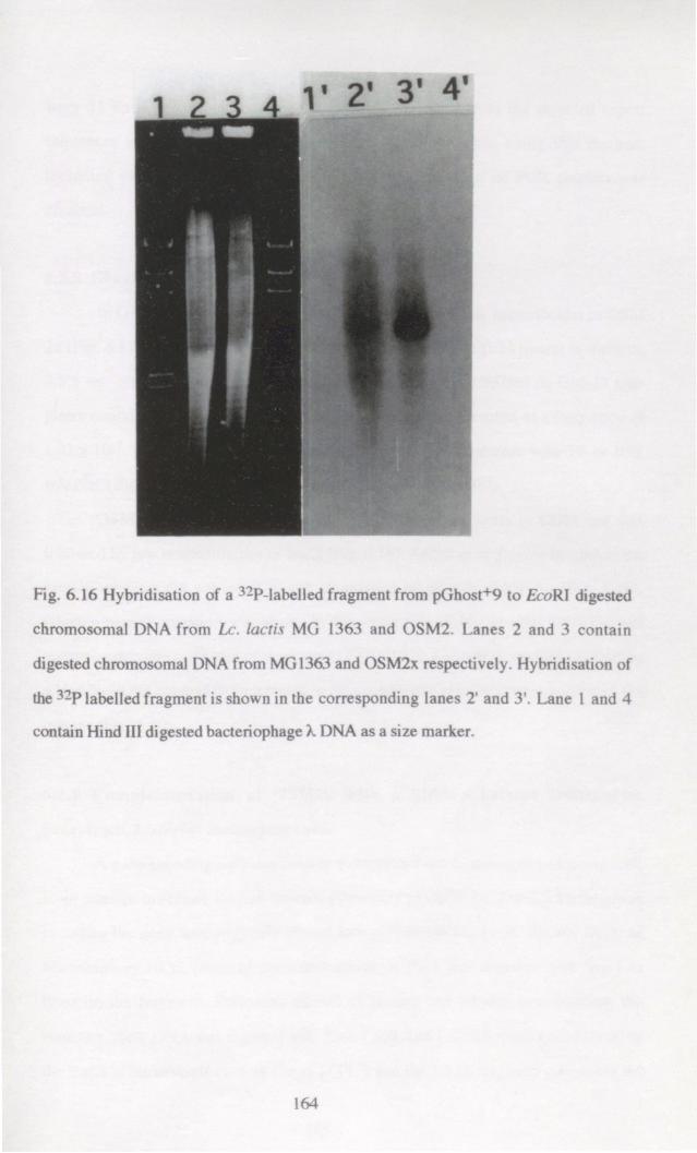
n. la 'r;ch roo mal D
di t d
n 1 nd

were 21 bp in length and designed to amplify outwards from the inverted repeat
sequences at the ends of ISS1. Several attempts were made using this method,
including variation of the primer annealing temperature but no PCR product was
obtained.
6.2.8 Characterisation of the OSM2x strain.
In GM-17 medium the presence of 3.5% w/v NaCI was bacteriocidal to OSM
2x (Fig. 6.17a). It was noticed that after prolonged incubation (>24 hours) in medium
3.5% w/v NaCI growth of OSM 2x occurred. Plating of 1()8 CFU/ml on GM-I7 agar
plates containing 3.5% w/v NaCI indicated that reversion occurred at a frequency of
1.42 x 10⁻⁷• The ability of the mutant OSM 2x to survive a challenge with 5% or 10%
w/v NaCI did not differ from that of the parent strain (Fig. 6.17b)..
OSM 2x grew somewhat slower than did its parent strain in COM and was
inhibited by low concentrations of NaCI (Fig. 6.18). Addition of glycine betaine to the
growth medium did not result in any stimulation of growth at low aw (Fig. 6.18).
Glycine betaine transport was absent in this strain (Fig. 6.19) whereas MG 1363 had
approximately the same rate of transport as NCDO 712. Growth in medium containing
added NaCI did not lead to an increase in the cytoplasmic glycine betaine content of the
mutant (Fig. 6.20).
6.2.9 Complementation of OSM2x with a glycine betaine transporter
gene from Listeria monocytogenes.
A gene encoding a glycine betaine transporter from L. monocytogenes was used
in an attempt to restore the salt tolerant phenotype in OSM 2x. The 2.5 kb fragment
encoding the gene was originally cloned into pUC18 (pCPL 1) (R. Sleator Dept. of
Microbiology UCC, personal communication). pCPL 1 was digested with Sea 1 to
linearise the fragment. Following phenol extraction and ethanol precipitation, the
resulting linear DNA was digested with Xma 1 and Xba 1 which restrict sites flanking
the insert in the multiple cloning site of pCPL 1 and the 2.5 kb fragment containing the
165

10
-~-~e5'-'"
j§c=a
0.1
0.01T
1
2520151050.001 -+---....,....--....,....--~--~--..
o
Time (hours)
Fig. 6.l7a Viable cell counts following inoculation of Le. lactis subsp. cremoris MG1363 «(J) and OSM 2x (0 ) into GM-17 broth containing 3.5% w/v NaCl.
86420.01 -+----......----.-----.-----,
o
10
0.1
100
Time (hours)
Fig. 6.17b Survival of exponential-phase Le. lactis subsp. cremoris MG1363 (circles) andOSM 2x (squares) during challenge with 5% (open symbols) and 10% (ftlled symbols)w/v NaCl in GM-17 broth. The data are representative of triplicate experiments.
166

1.2
1--5::l 0.8!i 0.6~~
°i 0.4en
0.2
00 1 2 3
NaCl (% w/v)
Fig. 6.18 The specific growth rate of Leo laetis subsp. eremoris MG1363 (squares) and
OSM2x (circles) in COM containing NaCl in the presence (filled symbols) and absence
(open symbols) of 2 mM glycine betaine. The data are representative of triplicate
experiments.
167

! 80e
l=-.-·1
60
5Co)
§40
8's.8Q,)
.5Co) 20~t:lI)
.s.!
~i 0~u 0 2 4 6 8 10
Time (min)
Fig. 6.19 Standard glycine betaine transport assay by Lc. lactis subsp. cremoris MG1363
(.) and the mutant OSM2x ( 6) cells grown in CDM medium.
168

o 0.5 1
NaCl (% wlv)
1.5 2
Fig. 6.20 Accumulation of glycine betaine by Le. lactis subsp. cremoris MG 1363 (II) and
the mutant OSM2x (IS:!) growing in CDM with its aw reduced by NaCl.
169

.....-Jo
Table 6.4 The specific growth rate, J.1 (h-l) of Le. laens subsp. eremoris MG1363 and OSM2x containing
the plasmids pCI 372 or pCPL 3a in GM-17 medium in the presence and absence of 3% w/v NaCl
GrowthSpecific growth rate J.1 (h-l) of :
Medium 1363 (pC1372) 1363 (pCPL 3) OSM 2x (pC1372) OSM 2x (pCPL 3)
GM-17 0.97 ± 0.05 0.97 ± 0.1 0.99 ± 0.07 0.94 ±0.08
GM-17 0.45 ± 0.07 0.38 ± 0.05 No growth No growth+ 3% NaCl
a Contains the glycine betaine transport gene from Listeria monoeytogenes cloned into the vector plasmidpCI372

& 60bOg~ 50c'-'
·1 40
~ 30g'5.8 20~
.5(,)>.
"EO 10(,)
'e~
~ 0>. 0 1 2 3 4 5U
Time (min)
Fig. 6.21 Glycine betaine ttanspon by Lc.lactis subsp. cremoris MG1363 (squares) and
OSM 2x (circles) containing pCI372 (open symbols) or pCPL 3 (filled symbols).
pCPL 3 consists of the glycine betaine ttanspon gene from Listeria monocytogenes
cloned into the vector plasmid pCI 372.
171

· .22
7 7.
m r' P
ith P 2
72

gene was extracted from an agarose gel and ligated into the shuttle vector pCI 372
(Hayes et al., 1990) to create pCPL 3. The ligation mix was transformed into
Escherichia coli DH5a cells by electroporation and the plasmid DNA isolated from
transformed cells. Ligation of the 2.5kb fragment containing the glycine betaine
transport gene into the vector pMG36ct was also attempted, however no transformants
were obtained following electroporation of the ligation mix into E. coli.
The strains MG1363 and OSM 2x were readily transformable with pCPL 3,by
electroporation however no increase in tolerance to low aw was observed in OSM 2x
containing pCPL 3 (Table 6.4). The presence of pCPL 3 did not restore the ability to
transport glycine betaine either (Fig. 6.21). The presence of pCPL 10 in OSM 2x and
MG1363 was confirmed by gel electrophoresis of isolated plasmid DNA (Fig. 6.22).
The Lc. lactis subsp. cremoris strain HP did not display any glycine betaine transport
activity, and this strain was also transformed with pCPI 3. As was the case with OSM
2x, the presence of the plasmid did not confer the ability to transport glycine betaine
(data not shown).
6.3 Discussion.
The data presented in this chapter suggest that glycine betaine is the most
important of the compatible solutes for lactococci. Proline, reported by Molenaar et al
(1993) to be accumulated during osmotic stress was not nearly as effective as glycine
betaine. There are two possible reasons for this. Firstly, glycine betaine may be a more
effective compatible solute than proline and its accumulation may be more beneficial to
the cell. There is a certain amount of evidence to support this proposition as glycine
betaine is preferred over proline by most bacteria, including Le. lactis (Molenaar et al.,
1993). The second possibility is that some proline accumulation occurs as a result of
synthesis and that the apparent effect of added proline is thereby reduced. The slight
stimulatory effect of added proline would then serve to augment that from synthesised
proline levels. Stimulation of growth by added leucyl-proline was also observed. What
is not known is whether intracellular peptidase activity results in the generation of free
173

proline or if the peptide itself acts as a compatible solute. 1.£ lactis strains are known to
have prolidase enzymes which hydrolyse X-pro dipeptides thus generating free proline
(Kunji et al., 1996). A similar osmoprotective effect of proline-eontaining peptides has
been reported for Listeria monocytogenes (Amezaga et al., 1995) in which stimulation
by the dipeptide proline-hydroxyproline was observed. It appeared that accumulation of
the dipeptide as well as the free amino acid residues ocurred in L. monocytogenes
(Amezaga et al., 1995). The slight stimulation observed with leucyl-proline raises the
question of whether the peptide transport system necessary for its uptake is osmotically
regulated. Le. lactis has at least two transport systems which transport di/tri peptides
one of which (DtpT) is proton motive force driven and transports hydrophilic substratesII
(Smid et al., 1989); the other (DtpP) is driven by ATP hydrolysis and transports
hydrophobic substrates (Foucaud et al., 1995). Leucyl-proline has hydrophobic
tendencies and therefore is likely to be transported by DtpP. At present there is no
infonnation available on the osmotic regulation of peptide transport systems.
Carnitine is similar to glycine betaine in that it has a tri-methylated amino group
and is utilised as a compatible solute by many Gram-positive bacteria, including
Lactobacillus plantarum (Kets et al., 1994) and Listeria monocytogenes (Beumer et al.,
1994). The data presented in Fig. 6.5 demonstrate that carnitine does not appear to
stimulate the growth of Le. lactis at low awe In Lb. plantarum, glycine betaine and
carnitine are transported by the same system (Glaasker et al., 1998b), while in L.
monocytogenes separate systems are present and the glycine betaine system does not
appear to transport carnitine (Verheul et al., 1997). It is therefore, reasonable to assume
that despite the structural similarity of both compounds, that some glycine betaine
systems are incapable of transporting carnitine and this appears to be the case for Lc.
lactis.
The low aw sensitive mutant, OSM2x, constructed by transposing the insertion
sequence ISS] into the genome of L. lactis subsp. cremoris MG1363, a plasmid free
derivative of NCDO 712, has lost the ability to take up the substantial quantities of
glycine betaine needed to grow in low aw environments. Low concentrations of glycine~
174
betaine were accumulated by the mutant growing in CDM at high aw, but these basal
levels were not increased when the aw was decreased, in contrast to the response of the
parent cultures (Fig. 6.20). When assayed for glycine betaine transport using the
conventional assay no glycine betaine transport was detected in the mutant (Fig. 6.19).
Since the mutation in OSM 2x is probably caused by a single insertion into the parent
genome (Fig. 6.16), it suggests that the parent culture has only one system for glycine
betaine transport which is capable of the response needed when the aw of the
environment is substantially reduced.
The results presented in Chapter III of this thesis show that for NCDO 712,
NaCI concentrations only slightly in excess of the upper limit for growth, are
bacteriocidal. This was also true in the case of the mutant as 3.5% w/v NaCI was lethalI
to OSM 2x even though the parent strain MG 1363 was capable of growth above this
NaCI concentration. The mutation did not appear to result in increased sensitivity to
NaCI concentrations higher than 3.5% w/v as both the parent strain and the mutant
were equally sensitive to 5 and 10% w/v NaCl. These data suggest that the mechanism
of cell death which is preventable by glycine betaine at concentrations of 3.5-4% w/v
NaCI may be different to that at higher NaCI concentrations such as 10-20% NaCI or
that glycine betaine is ineffective at these higher concentrations.
Transforming the mutant with a plasmid encoded glycine betaine transport gene
from Listeria monocytogenes did not complement the mutation in OSM 2x. The most
likely reason for this is that the promoter of the cloned gene is not recognised in Le.\
laetis. Sequence data for the gene indicates that it is under the control of the alternative
sigma factor O'B (R.Sleator, Dept of Microbiology V.C.C., personal communication). }f
It is unlikely that this promoter is recognised by Lc. laetis. Attempts to clone the gene
into the vector pMG36ct were unsuccessful; this vector contains a strong Le. laetis
promoter upstream of the cloning site which should ensure strong expression in Le.
lactis. It is possible that strong constitutive expression may be lethal, leading to failure
to obtain transformant colonies containing pMG 36ct ligated to the glycine betaine gene.
175
Molenaar et ale (1993) suggested that there were two systems for glycine betaine
uptake in L. lactis strain ML3, a close relative of NCDO 712 (Davies et al., 1981) from
which MG 1363 is derived. They suggested that the first was a high affinity transport
system which was not induced by increasing the osmolarity of the growth medium. The
second system was a low affinity proline uptake system which also transported glycine
betaine. The second system was induced by growing the cells in a chemically defined
medium at high osmolarity. Strain OSM 2x did not transport glycine betaine at high or
lowaw suggesting a complete loss of glycine betaine transport as a result of a single
insertional mutation event. This result does not support the -conclusion of Molenaar et
ale (1993) that two glycine betaine transport systems exist in lactococci.
There are several possibilities raised by this apparent difference in results. It is
possible that glycine betaine transport in ML3 differs from that in NCDO 712 even
though both strains are regarded as being very similar (Davies et al., 1981). A second
possibility is that two glycine betaine transport systems exist in NCDO 712 (and
MG1363) but that both are eliminated in the OSM 2x mutant. A third possibility is that
there is only one system for glycine betaine transport in NCDO 712 and this is
eliminated in the OSM 2x mutant. Our data fit the latter model as it is likely that the
OSM 2x mutation is due to a single insertion into the chromosome. However, there is
one important observation which does not fit this model for glycine betaine transport.
When NCDO 712 cells were grown at high aw, glycine betaine transport was not
stimulated by the presence of NaCI in the assay, whereas the transport system in cells
grown at low aw were stimulated. The Km values for glycine betaine transport for cells
grown at high or low aw were similar which is evidence for one system or two systems
with similar affinities. It is possible that the difference in glycine betaine transport
kinetics in cells grown at high and low aw is due to a fundamental change in the cells
growing at low aw which causes an activation of the glycine betaine transport protein at
low aWe Such an activation of glycine betaine transport could account for a similar
observation in lb. plantarum by Glaasker et al.(1996a). The only data to support two
transport systems is the absence of osmotic activation for cells grown at high aWe
176

Glaasker (1998) proposed that the failure of some workers (including Molenaar et al.)
to observe activation may be an artifact of the experimental conditions. For example,
osmotic activation of glycine betaine or camitine transport in L. monocytogenes was
only observed when cells contained pre accumulated compatible solutes (Verheul et aI.,
1997). On balance, glycine betaine transport in Le. lactis probably occurs via a single
system, the activity of which is osmotically regulated if the cells are grown at low aw.
The transport of glycine betaine in Gram-positive and Gram-negative bacteria
has been well studied. Multiple transport systems have been reported in many cases.
The best studied example being the ProU and ProP systems of E. coli and Salmonella
typhimurium (Caimey et al., 1985 a, b). The ProU system has a high affinity for
betaine while the ProP system has a lower affinity for glycine betaine and also
transports proline. Similar high- and low-affinity transport systems for glycine betaine
were observed in Staphylococcus aureus with the low affinity system also transporting
proline (Pourkomailan and Booth, 1992). In Bacillus subtilis, three transport systems
for glycine betaine have been reported (Kappes et al., 1996). The presence of multiple
systems of different affinities allows the cell to accumulate betaine efficiently over a
wide range of extracellular. betaine concentrations (Koo and Booth, 1994). In
Corynebacterium glutamicum (peter et al., 1995) and Lb. plantarum (Glaasker, 1998)
it appears that only one system for glycine betaine is present. Single glycine betaine
transport systems tend to have high substrate affinity, be expressed constitutively or
semi-constitutively and are strongly regulated by solute concentration at the activity
level. The characteristics of the glycine betaine transport system in NCDO 712 are
similar to those reported for Lb. plantarum and C. glutamicum in that a significant
degree of control is exerted at the level of transporter activity.
The Le.lactis strains examined formed two distinct groups with respect to their
ability to grow in the presence of added NaCI or sugar. All of the cremoris strains with
the exception of NCDO 712 were much more sensitive to low aw than were the lactis
strains. As was the case with the OSM 2x mutant, failure to grow at low aw appeared
be related to the ability of the cells to accumulate the compatible solute glycine betaine.
177

Two of the strains, did not transport glycine betaine in the assay systems used here.
Growth of these strains at low aw did not result in increased glycine betaine transport
rates. The third cremoris strain examined, US3, transported glycine betaine at a much
lower rate than did the osmotolerant strains. The low levels of betaine transport were
reflected in the glycine betaine content of exponential phase growing cells. Some
betaine accumulation was measureable for strains US3 and BK5, but only when the
solute used to reduce the aw was NaCI, unlike the lactis strains which accumulated
betaine when the aw was reduced by NaCI or sugar. It appears that glycine betaine
accumulation by the salt sensitive strains differs from that observed in the salt tolerant
strains. When the amount of glycine betaine accumulated by each strain is compared, it
is obvious that NCDO 712 accumulates much less betaine than the other salt tolerant
strains which is reflected in the fact that NCDO 712 was stimulated by glycine betaine
to a lesser extent than the other strains. This correlation between the accumulation of
glycine betaine and the ability to grow at low aw is similar to that reported for
Lactobacillus spp. A strain of Lactobacillus acidophilus which was abnormally
osmotolerant, was able to transport betaine at higher rates than less osmotolerant strains
due to the presence of a constitutively expressed transport system activated at low aw
(Hutkins et al., 1987).
The possible reasons for such a substantial difference in glycine betaine
transport between strains which are so closely related are intriguing. Strains of Lc.
lactis subsp. cremoris which have some of the traits formerly associated with Lc. lactis
subsp.lactis (growth at pH 9.2,4% w/v NaCI and at 40°C and arginine hydrolysis) are
not unusual. In one study only 26 out of 59 genotypically cremoris strains had the
classical cremoris phenotype. The remaining 33 strains had some or all of the classical
lactis traits including 25 which grew at 4% wlv NaCl (Urbach et al., 1997). Analysis of
the genome of Le. lactis strains has revealed that the overall high degree of DNA
homology between the two strains is a composite of highly conserved regions with 80
90 % homology and variable regions of <40% homology. It has been suggested, based
on studies of codon usage and base composition, that the variable regions arose as a
178

result of horizontal transfer of genes from distantly related species (Delorme et al.,
1994). Maybe gene transfer in this manner has led to differences in phenotype between
lactis and cremoris strains (Urbach et al., 1997).
179

Chapter VIIConcluding Remarks.
180

In this study the mechanisms by which Le. lactis subsp. cremoris NCDO 712
adapts to osmotic stress were investigated. Chapter I of the thesis provides a detailed
review of the literature concerning osmotic stress in bacteria and mechanisms by which
bacteria adapt to fluctuations in the aw of their environment. The role of compatible
solutes in bacterial osmotolerance was reviewed in some depth, as compatible solute
accumulation is almost universal during osmotic stress (Csonka, 1989).
In Chapter In of this thesis, the survival of Le.lactis at NaCI concentrations in
excess of those that permit growth were investigated. It has previously been shown that
the resistance of Le. lactis subsp cremoris. NCDO 712 to lethal acid challenge is
dependent on the growth phase of the culture (O'Sullivan, 1996). A similar trend was
observed, in this study, for the tolerance of NCDO 712 to 20% NaCl. There are a
number of possible explanations for the variation in tolerance. It is possible that the cell
density plays a role in triggering the development of resistance in mid-exponential
phase. Cell density dePendent regulation, also known as quorum sensing, has been
shown to have a role in induction of the lux operon in Vibrio fischeri. In addition,
bacteriocin production by lactobacilli and catalase production by Rhizobium
leguminosarum apPear to be related to the cell density of the resPective cultures. The
basic model for quorum sensing involves the production of an autoinducer molecule by
the cells; when the cell density of a culture increases there is an accelerated increase in
the inducer concentration, which accelerates the induction of the response.
Another explanation for growth phase dePendent variation in stress tolerance is
that it results from growth phase dePendent changes in gene expression. The alternative
sigma factor SS in E. coli is present only in late exponential phase/early stationary phase
cells. As SS functions in the transcription of genes involved in the response to a number
of environmental stresses (Hengge-Aronis, 1996), it is easy to see how it could
participate in growth phase dependent stress responses.
Lc.lactis subsp. cremoris NCDO 712 was capable of growth at up to 4% w/v
NaCI; increasing the NaCI concentration to 5% w/v was bacteriocidal. There is
therefore a very narrow margin between those NaCI concentrations which allow growth
181

and those which are bacteriocidal. Little is known about the mechanism by which NaCI
damages bacterial cells. However, efflux of water following osmotic stress probably
reduces the a w to a level where some damage to cell proteins would be expected to
occur. Loss of turgor pressure, if it occurred, might also inhibit the activity of
membrane bound enzymes. It has been reported that carbohydrate transport in E. coli is
inhibited by osmotic stress (Roth et al., 1995). In addition, osmotic stress inhibited
glycolysis in Clostridium pasteurianum (Walter et al., 1987). In both examples, the
generation of ATP is likely to be reduced, so that in effect, the cells are starved.
Prolonged starvation is in itself lethal to Le. laetis (Hartke et al., 1994). If transport of
carbohydrates or glycolysis were inhibited in Le.lactis then starvation stress would be
imposed, together with the other expected effects at low aw such as low turgor
pressure.
If NCDO 712 cells were exposed to 3% w/v NaCI for 1 hour prior to challenge
with lethal concentrations of NaCI they were more resistant to the challenge than cells
which were not pre-exposed to 3% w/v NaCI. The enhanced tolerance to NaCI
challenge required protein synthesis and appeared to be only effective at NaCI
concentrations S 10% w/v. The induced tolerance to NaCI appears to be analogous to
other inducible stress responses such as heat shock and the acid tolerance response
(ATR). The ATR was first reported for Escherichia coli (Goodson and Rowbury,
1989) and subsequently for a number of other Gram-positive and Gram-negative
bacteria. Induction of an ATR in Lc.lactis NCOO 712 which also induced resistance to
heat, H202, ethanol, and 20% w/v NaCI has been reported (O'Sullivan and Condon,
1997). In this study, the effect of induction of NaCI tolerance on resistance to other
stresses was not investigated, but it is known that exposure to 4% w/v NaCI for 1 hour
does not induce tolerance to pH 4.0 in NCDO 712 (O'Sullivan and Condon, 1997). It
is likely that the induced protection is due to induction of chaperone proteins. A number
of these proteins are synthesised by Le. lactis during exposure to 2.5% w/v NaCI
(Kilstrup et al., 1997). A major function of chaperone proteins is to degrade proteins
which have become damaged or malfonned, with the classic example being the action
182

of heat-shock proteins. DnaK, which is a well known heat-shock protein is known to
have a role in restoring turgor pressure in E. coli, during osmotic stress (Meury and
Kohiyama, 1991).
The mechanisms which bacteria use to survive exposure to high concentrations
of NaCl are not well understood. Survival of NCDO 712 during NaCl challenge was
enhanced by the presence of magnesium ions, as was the survival during challenge at
42°C and low pH. It is possible that magnesium has a protective effect against a number
of different stresses in Lc. lactis as it also increases survival during prolonged
starvation (Thomas and Batt, 1967). Magnesium therefore, either directly stabilises
some cell component(s) damaged by exposure to environmental stress or is essential for
the action of a stress tolerance mechanism(s). There is evidence to support both
possibilities as magnesium can act directly to stabilise ribosomes (Hui Bon Hoa et al.,
1980) and ribosomal RNA (Tolker-Nielsen and Molin 1996). Intact ribosomes may be
needed for initiation of growth when the lethal stress is removed; therefore cells
challenged in the absence of magnesium may be unable to resume growth due to lack of
functional ribosomes. Magnesium is also essential for the ATPase activity of the
chaperone DnaK (Skowyra and Wickner, 1995). The involvement of magnesium in
DnaK activity may explain its role in the tolerance of Lc. lactis to a number of stresses
as DnaK is induced in response to heat (Kilstrup et al., 1997), low pH (Hartke et al.,
1996) and NaCI (Kilstrup et al., 1997).
The role of potassium in the adaptation of Lc.lactis subsp. cremoris NCDO 712
to osmotic stress was examined in Chapters IV and V. When the concentration of
potassium was not limiting, potassium was accumulated to high cytoplasmic levels
when the aw of the medium was decreased by addition of glucose or fructose, but not
when NaCI was used. If the potassium concentration of MTEL medium was limited,
the reduction of the aw with NaCI led to a decrease in the specific growth rate,
cytoplasmic potassium content and cytoplasmic volume changes which were not
observed when potassium was not limiting. Addition of Nl4Cl instead of NaClled to
similar but slightly more severe effects on growth rate and potassium content. Similar
183

results were reported by MaCleod and Snell (1948) who observed that the potassium
requirement of lactic acid bacteria increased if other monovalent cations were present in
the medium. The degree of growth inhibition by Nl4CI and NaCI correlated well with
the inhibitory effect of each compound on potassium transport.
Growth in the presence of NaCI in MTEL medium containing 0.5 mM
potassium clearly demonstrated the role of potassium in maintaining turgor. The
volume of the cells growing in MTEL containing 0.25M NaCI decreased by 30% when
compared to cells growing in the absence of salt stress. This reduction in volume may
explain the reduction in specific growth rate observed under these conditions.
Potassium transport occurred via a single system which had a low affinity for
potassium. Potassium transport was competitively inhibited by Na+ and other cations
and non-competitively inhibited by low aw• Na+ ions were not accumulated in place of
potassium, which was confinned by the absence of Na+ transport by choline loaded
cells. The data suggest that Na+ ions bind to the potassium transporter, yet are not
transported. Nonnally, potassium transport systems are highly discriminatory in their
substrate binding (Doyle et al., 1998). The situation in Le. lactis is therefore unusual in
that the potassium transporter discriminates poorly between potassium and other
monovalent cations.
The apparent Km for potassium transport (0.75 mM) is approximately 7-fold
greater than the potassium concentration (100~) which supports growth at 50% of
the maximal rate. The cells are therefore capable of a much higher rate of potassium
transport than is needed for growth at high aw. The fact that the cell can transport
potassium at rates higher than are needed for growth suggests that the extra potassium
transport capability could be used for osmoregulation. The 2-fold increase in the
specific activity of the system in cells growing at low aw may contribute to the raised
cellular potassium content of the cells. However, the increased specific activity is
almost exactly counterbalanced by the inhibitory effect of low aw on the rate of
transport. It is therefore likely that there is another means of control of potassium
transport. The inhibition of the rate of transport by low aw seems to rule out activation
184

by reduced turgor; however, it must be remembered that the potassium transport assays
were performed using cells artificially depleted of potassium. The effect of low aw on
the activity of the system in vivo may be different. It is also possible that the
intracellular potassium concentration is controlled by the activity of efflux systems.
Potassium efflux in E. coli occurs rapidly if cells growing at low aware shifted to high
aw (Epstein and Schultz, 1965; Meury et al., 1985; Bakker, 1993; Schleyer et al.,
1993). Rapid efflux of cytoplasmic solutes in this manner is probably due to the activity
of stretch activated channels and is probably an emergency response to counteract the
risk of the cells bursting during hypo-osmotic stress. There is a requirement for tight
control of potassium movement across the cytoplasmic membrane; uncontrolled
accumulation followed by efflux amounts to futile cycling and is extremely wasteful of
energy. In E. coli, the rate of uptake of potassium when turgor is high is only 1 to 2%
of the value when turgor is low (Meury et al., 1985; Bakker, 1993). It is likely that
control of potassium levels in Le. lactis is mediated in a similar manner.
Growth of Le. lactis did not occur at potassium concentrations below 30 JlM.
Therefore Lc. lactis does not appear to possess a high affinity potassium transport
system similar to the Kdp system of E. coli or Bacillus acidocaldarius (Bakker et al.,
1987) which allows the cells to scavenge potassium efficiently at nanomole
concentrations. The relationship between the growth rate of Le. lactis NCDO 712 and
the available potassium in MTEL medium approximately obeyed Michaelis-Menten
kinetics and the apparent Km value was 100 JlM.
In Chapter VI the role of glycine betaine and other organic compatible solutes
was investigated. Of the compatible solutes tested, only glycine betaine produced
substantial stimulation of growth at low aWe Proline was also stimulatory but to a lesser
degree. Carnitine and ectoine were not stimulatory even though both have been reported
as being stimulatory in other Gram-positive bacteria. Leucyl-proline stimulated growth
to about the same extent as did proline. We cannot say whether the stimulation results
from accumulation of intact leucyl-proline as reported for other dipeptides in Listeria
185

monocytogenes (Amezaga et ai., 1995) or if leucyl-proline is hydrolysed intracellularly
to provide free proline which is then accumulated.
Glycine betaine was the only significant organic compatible solute identified.
The ability of glycine betaine to reverse the inhibition of growth rate and reduction of
volume caused by NaCI at low potasssium concentrations is a good demonstration of
its ability to restore turgor pressure. There was an excellent correlation between the
ability to accumulate glycine betaine and growth at low a w• The cremoris strains
incapable of growth at 4% w/v NaCI or the equivalent glucose concentration,
accumulated less glycine betaine than did the strains capable of growth at 4% w/v
NaCl. As would be expected, the ability to accumulate glycine betaine seemed to be
dependent on the presence of a significant degree of glycine betaine transport activity
and the NaCI sensitive strains displayed little or no glycine betaine uptake in in vitro
assays. Variation in the ability to accumulate glycine betaine within a genus is not
unknown, and has been observed in Lactobacillus (Hutkins et ai., 1987) and in
Propionibacteriium (Deborde.:J998). *The mutant strain OSM2x which was extremely sensitive to low aw did not
accumulate glycine betaine, nor did it display any measurable glycine betaine transport
activity. The abolition of glycine betaine transport in OSM 2x with what appeared to be
a single ISS} insertion suggests that there is only one transport system for glycine
betaine. This does not agree with the results of Molenaar et ai. (1993) who reported
two glycine betaine transport systems in a strain (ML3) closely related to NCDO 712.
Some of our data also suggested a second transport system, in that no activation of
transport by the aw of the assay buffer is observed for cells grown at normal aw while
activation was observed for cells grown at low aw. However, growth at low aw ~a(~~~~
several physiological changes; these could include the synthesis of an 'activatot whkh
enhances the rate of transport at low aw but not at normal aw. In such a scenario a
single glycine betaine transporter could have different rates of transport depending on
the a w of the medium in which the cells were grown and on the aw of the assay
buffer.This topic requires further study.
186
( .C~_±:_~~·,
J,,..J--

Literature Cited
187
Abee, T., J. Knol, K.J. Hellingwerf, E.P. Bakker, A. Siebers and
W.N. Konings. 1992. A kdp-like, high affinity, K+-translocating ATPase is
expressed during growth of Rhodobacter sphaeroides in low potassium media. Arch.
Microbiol. 158:374-380.
Alemayehu, D. The log-phase acid tolerance of Lactococcus lactis MG1363 during
growth at constant pH 7.0 1997. M.Sc thesis. National University of Ireland, Dublin.
Amezaga, M.-R., I. Davidson, D. McLaggan, A. Verheul, T. Abee, and
I.R. Booth. 1995. The role of peptide metabolism in the growth of Listeria
monocytogenes ATCC 23074 at high osmolarity. Microbiology. 141:41-49.
Anderson, C.B. and L.D. Witter. 1982. Glutamine and proline accumulation by
Staphylococcus aureus with reduction in water activity. J. Bacteriol. 143:1501-1503.
Anderson, D.G. and L.L. McKay. 1983. A simple and rapid method for
isolating large plasmid DNA from lactic streptococci. Appl. Environ. Microbiol.
46:549-552.
Anderson, R.R., R. Menzel, and J.M. Wood. 1980. Biochemistry and
regulation of a second L-proline transport system in Salmonella typhimurium. J.
Bacteriol. 141: 1071-1076.
Andresen, P.A., I. Kaasen, O.B. Styrvold, G. Boulnois and A. R.
Strom. 1988. Molecular cloning, Physical mapping and expression of the bet genes
governing the osmoregulatory choline-glycine betaine pathway of Escherichia coli. J.
Gen. Microbiol. 134: 1737-1746.
188

Arnold, K.W. and C.W. Kasper. 1995. Starvation- and stationary-phase induced
acid tolerance in Escherichia coli 0157:H7. Appl. Environ. Microbiol. 61:2037
2039.
Asha, H. and J. Gowrishankar. 1993. Regulation of kdp oPeron expression in
Escherichia coli: evidence against turgor as signal for transcriptional control. J.
Bacteriol. 175:4528-4537.
Badger, J.L. and V.L. Miller. 1995. Role of rpoS in survival of Yersinia
enterocolitica to a variety of environmental stresses. J. Bacteriol. 177:5370-5373.
Bae, J.H. and K.J. Miller. 1992. Identification of two proline transport systems
in Staphylococcus aureus and their possible roles in osmoregulation. Appl. Environ
Microbiol. 58:471-475.
Bakker, E.P. 1993. Cell K+ and K+ transport systems in prokaryotes. In E.P.
Bakker, (ed) Alkali cation transport systems in prokaryotes. CRC Press Inc. Boca
Raton FI USA.
Bakker, E.P. and F.M. Harold. 1980. Energy coupling to potassium transport in
Streptococcus faecalis. J. BioI. Chern. 255:433-440.
Bakker, E.P., A. Borchard, M. Michels, K. Altendorf, and A. Siebers.
1987. High-affinity potassium uptake system in Bacillus acidocaldarius showing
immunological cross-reactivity with the kdp system from Escherichia coli. J.
Bacteriol. 169:4342-4348.
189

Ball, C.A., R. Osuna, K.C. Ferguson and R.C. Johnson. 1992. Dramatic
changes in Fis levels upon nutrient upshift in Escherichia coli. J. Bacteriol.
174:8043-8056.
Becker, L.A., M.S. «;etin, R.W. Hutkins and A.K. Benson. 1998.
Identification of the gene encoding the alternative sigma factor aB from Listeria
monocytogenes and its role in osmotolerance. J. Bacteriol. 180:4547-4554.
Benjamin, M. and A.R. Datta. 1995. Acid tolerance of enterohemorrhagic
Escherichia coli. Appl. Environ. Microbiol. 61: 1669-1672.
Berkower, I. J. Leis and J. Hurwitz. 1973. Isolation and characterisation of an
endonuclease from Escherichia coli SPeCific for ribonucleic acid in ribonucleic acid
deoxyribonucleic acid hybrid structures. J. Bioi. Chem.248:5914-5921.
Bernard, T., M. Jebbar, Y. Rassouli, S. Himdi·Kabbab, J. Hamelin and
C. Blanco. 1993. Betoine accumulation and osmotic regulation in Brevibacterium
linens. J. Gen. Microbiol. 138:1629-1638.
Beumer, R.R., M.C. Te Giffel, L.J. Cox, F.M. Rombouts and T. Abee.
1994. Effect of exogenous proline, betaine, and carnitine on growth of Listeria
monocytogenes in a minimal medium. Appl. Environ. Microbiol. 60: 1359-1363.
Boch, J., B. Kempf and E. Bremer. 1994. Osmoregulation in Bacillus subtilis:
Synthesis of the osmoprotectant glycine betaine from exogenously provided choline. J.
Bacteriol. 176:5364-5371.
Boch, J., B. Kempf, R. Schmid and E. Bremer. 1996. Synthesis of the
osmoproteetant glycine betaine in Bacillus subtilis: characterisation of the gbsAB
genes. J. Bacteriol. 178:5121-5129.
190

Boch, J., G. Nau-Wagner, S. Kneip and E. Bremer. 1997. Glycine betaine
aldehyde dehydrogenase from Bacillus subtilis: characterisation of an enzyme required
for the synthesis of the osmoprotectant glycine betaine. Arch. Microbiol. 168:282
289.
Bohringer, J., D. Fischer, G. Mosler, and R. Hengge-Aronis. 1995.
UDP-glucose is a potential intracellular signal molecule in the control of expression of
SS and sS-dependent genes in Escherichia coli. J. Bacteriol. 177:413-422.
Booth, J.R., R.M. Douglas, G.P. Ferguson, A.J. Lamb, A.W. Munro
and G.Y. Ritchie. 1993. K+ Efflux systems. In Alkali cation transport systems in
prokaryotes. E.P. Bakker (Ed). CRC Press Inc. Boca Raton FL USA.
Bossemeyer, D.A., A. Borchardt, D.C. Dosch, G.C. Helmer, W.
Epstein, J.R. Booth and E.P. Bakker. 1989a. K+ transport protein TrkA of
Escherichia coli is a peripheral membrane protein that requires other trk gene products
for attachment to the cytoplasmic membrane. J. BioI. Chern. 264: 16403-1641O.s
Bossemeyer, D., A. Schlosser and E.P Bakker. 1989b. SPecific cesium
transport via the Escherichia coli Kup (TrkD) K+ uptake system. J. Bacteriol.
171:2219-2221.
Botsford, J.L. 1990. Analysis of protein expression in response to osmotic stress in
Escherichia coli. FEMS Microbiol. Lett. 72:355-360.
191

Brown, A.D. and J.R. Simpson. 1972. Water relations of sugar-tolerant yeasts;
the role of intracellular polyols. J. Gen. Microbiol. 72:589-591.
Cairney, J., J.R. Booth and C.F. Higgins. 1985a. Salmonella typhimurium
proP gene encodes a transport system for the osmoprotectant betaine. J. Bacteriol.
164: 1218-1223.
Cairney, J., J.R. Booth and C.F. Higgins. 1985b. Osmoregulation of gene
expression in Salmonella typhimurium: proU encodes an osmotically induced betaine
transport system. J. Bacteriol. 164:1218-1223.
Cheville, A.M., K.W. Arnold, C. Buchrieser, C.M. Cheng and
C.W.Kaspar. 1996. rpoS regulation of acid, heat, and salt tolerance in Escherichia
coli 0157:H7. Appl. Environ. Microbiol. 62: 1822-1824.
Clark, D. and J. Parker. 1984. Proteins induced by high osmotic pressure in
Escherichia coli. FEMS Microbiol. Lett. 25:81-83.
Cogan, T.M. 1995. Flavour production by dairy starter cultures. J. Appl. Bacteriol.
Symp. Supple 79:495-645.
Csonka, L.N, and A.D Hanson. 1991. Prokaryotic osmoregulation: genetics and
physiology. Annu. Rev. Microbiol. 45:569-606.
Csonka, L.N. 1981. Proline over-production results in enhanced osmotolerance in
Salmonella typhimurium. Mol. Gen. Genet. 182:82-86.
Csonka, L.N. 1982. A third L-proline Permease in Salmonella typhimurium which
functions in media of elevated osmotic strength. J. Bacteriol. 151: 1433-1443.
192

Csonka, L.N• 1989. Physiological and genetic responses of bacteria to osmotic
stress. Microbiol. Rev. 59:121-147.
Csonka, L.N., T.P. Ikeda, S.A. Fletcher, and S. Kustu. 1994. The
accumulation of glutamate is necessary for optimal growth of Salmonella typhimurium
in media of high osmolarity but not induction of the proU operon. J. Bacteriol.
176:6324-6333.
Dattananda, C.S., K. Rajkumari, and J. Gowrishankar. 1991. Multiple
mechanisms contribute to the osmotic inducibility of proU operon expression in
Escherichia coli: demonstration of two osmoresponsive promoters and of a negative
regulatory element within the first structural gene. J. Bacteriol. 173:7481-7490.
Davies, F.L., H.M. Underwood and M.J. Gasson. 1981. The value of
plasmid profiles for strain identification in lactic streptococci and the relationship
between Streptococcus lactis 712, ML3 and C2. J. Appl. Bacteriol. 51:325-337.
Davis, M.J., P.J. Coote and C.P. 0' Byrne. 1996. Acid tolerance in
Listeria monocytogenes: the adaptive acid tolerance response (ATR) and growth
phase-dependent acid resistance. Microbiology. 142:2975-2982.
Deborde, C. 1998. Etude du metabolisme carbone primaire de bacteries propioniques
laitieres par resonance magnetique nucleaire in vivo 13c. These Doctorat. L'Ecole
Nationale Superieure Agronomique de Rennes.
Delorme, C., J.J. Godon, S.D. Ehrlich and P. Renault. 1994. Mosaic
structure of large regions of the Lactococcus lactis subsp. cremoris chromosome.
Microbiology. 140:3053-3060.
193

Deuerling, E., B. Paeslack and W. Schumann. 1995. The ftsH gene of
Bacillus subtilis is transiently induced after osmotic and temperature upshift. J.
Bacteriol. 177:4105-4112.
Dinnbier, U. E. Limpenstiel, R. Schmid and E.P. Bakker. 1988. Transient
accumulation of potassium glutamate and its replacement by trehalose during adaptation
of growing cells of Escherichia coli K-12 to elevated sodium chloride concentrations.
Arch Microbiol. 150:348-357.
Dinsmore, P.K., D.A. Romero and T.R. Klaenhammer. 1993. Insertional
mutagenisis in Lactococcus lactis subsp.lactis mediated by 15946. FEMS Microbiol.
Lett. 107:43-48.
Dosch, D.C., G.L. Helmer, S.H. Sutton, F.F. Salvacion and W.
Epstein. 1991. Genetic analysis of potassium transport loci in Escherichia coli:
evidence for three constitutive systems mediating uptake of potassium. J. Bacteriol.
173:687-696.
Doyle, D.A., J.M. Cabral, R.A. Pfuetzner, A. Kuo, J.M. Gulbuis, S.L.
Cohen, B.T. Chait and R. MacKinnon. 1998. The structure of the potassium
channel: molecular basis of K+ conduction and selectivity. Science 280:69-77.
Dunlap, V.J., and L.N. Csonka. 1985. Osmotic regulation of L-proline transport
in Salmonella typhimurium. J. Bacteriol. 163:296-304.
Epstein, W. 1986. Osmoregulation by potassium transport in Escherichia coli.
FEMS Microbiol. Rev. 39:73-78.
Epstein, W. and B.S. Kim. 1971. Potassium transport loci in Escherichia coli. J.
Bacteriol. 108:639-644.
194

Epstein, W. and S.G. Schultz. 1965. Cation transport in Escherichia coli. V.
Regulation of cation content. J. Gen. Physiol. 49:221-234.
Epstein, W., V. Whitelaw and J. Hesse. 1978. A K+ transport ATPase in
Escherichia coli. J. BioI. Chern. 253:6666-6668.
Eshoo, M.W• 1988. lac fusion analysis of the bet genes of Escherichia coli:
regulation by osmolarity, temperature, oxygen, choline and glycine betaine. J.
Bacteriol. 170:5208-5215.
Flahaut, S., A. Benachour, J.-C. Giard, P. Boutibonnes and Y.
Auffray. 1996. Defense against lethal treatments and de novo protein synthesis
induced by NaCI in Enterococcus faecalis ATCC 19433. Arch. Microbiol. 165:317
324.
Flahaut, S., A. Hartke, J.-C. Giard and Y. Auffray. 1997. Alkaline stress
response in Enterococcus faecalis: adaptation, cross-protection, and changes in
protein synthesis. Appl. Environ. Microbiol. 63:812-814.
Foucaud, C., E.R.S. Kunji, A. Hagting, J. Richard, W.N. Konings, J.
Desmazeaud and B. Poolman. 1995. Specificity of peptide transport systems in
Lactococcus Iactis: evidence for a third system which transports hydrophobic di- and
tripeptides. J. Bacteriol. 177:4652-4657.
Free, A. and C.J. Dorman. 1997. The Escherichia coli stpA gene is transiently
expressed during growth in rich medium and is induced in minimal medium and by
stress conditions. J. Bacteriol. 179:909-918.
195

Frymier, J.S., T.R. Reed, S.A. Fletcher and L.N. Csonka. 1997.
Characterisation of transcriptional regulation of the kdp oPeron of Salmonella
typhimurium. J. Bacteriol. 179:3061-3063.
Galinski, E. and H.G. Triiper. 1982. Betaine, a compatible solute in the
extremely halophilic phototrophic bacterium Ectothiorhodospira halochloris. FEMS
Microbiol. Lett. 13:357-360.
Galinski, E.A., H.P. Pfeiffer and H.G. Truper. 1985. 1,4,5,6-tetrahydro-2
methyl-4 pyrimidinecarboxylic acid: a novel cyclic amino acid from halophilic
phototrophic bacteria of the genus Ectothiorhodospira halochloris. Eur. J. Biochem.
149:135-139.
Garvie, E.I., J.A.E. Farrow and B.A. Phillips. 1981. A taxonomic study of
some strains of streptococci which grow at l00C but not at 45°C including
Streptococcus lactis and Streptococcus cremoris. Zbl. Bakt. Hyg., I. Abt. Orig. C
2:151-165.
Gasson, M.J. 1983. Plasmid components of Streptococcus lactis NCOO 712 and
other lactic streptococci after protoplast-induced curing. J. Bacteriol. 154: 1-9.
Gauthier, M.J., G.N. Flatau, D. Le Rudulier, R.L. Clement and M-P.
Combarro. 1991. Intracellular accumulation of potassium and glutamate specifically
enhances survival of Escherichia coli in seawater. Appl. Environ. Microbiol. 57:272-
276.
Gentry, D.R., V.J. Hernandez, L.H. Nguyen, D.B. Jensen and M.
Cashel. 1993. Synthesis of the stationary-phase signa factor as is positively regulated
by ppGpp. J. Bacteriol. 175:7982-7989.
196
Georgopoulos, C. 1992. The emergence of the chaperone machines. Trends in
Biochemical Sciences 17:295-299.
Gerhardt, P.N., L.T. Smith and G.M. Smith. 1996. Sodium-driven,
osmotically activated glycine betaine transport in Listeria monocytogenes membrane
vesicles. J. Bacteriol. 178:6105-6109.
Girever, H.M., O.B. Styrvold, I. Kaasen and A.R. Strfm. 1988.
Biochemical and genetic characterisation of osmoregulatory trehalose synthesis in
Escherichia coli. J. Bacteriol. 170:2841-2849.
Glaasker, E. 1998. Osmotic regulation of transport processes in Lactobacillus
plantarum. Ph.D thesis University of Groningen, The Netherlands.
Glaasker, E., W.N. Konings and B. Poolman. 1996a. Osmotic regulation of
intracellular solute pools in Lactobacillus plantarum. J. Bacteriol. 178:575-582.
Glaasker, E., W.N. Konings and B. Poolman. 1996b. Glycine betaine fluxes
in Lactobacillus plantarum during osmostasis and hyper- and hypo-osmotic shock. J.
Biol. Chem. 271: 10060-10065.
Glasemacher, J. A. Siebers, K. Altendorf and P. Schonheit. Low-affinity
potassium uptake system in the archaeon Methanobacterium thermoautotrophicum:
overproduction of a 31-kilodalton membrane protein during growth on low potassium
medium. J. Bacteriol. 178:728-734.
197

Godon, J.J., C. Delorme, S.D. Ehrlich and P. Renault. 1992. Divergence
of genomic sequences between Lactococcus lactis subsp. lactis and Lactococcus
lactis subsp. cremoris. Appl. Environ. Microbiol. 58:4045-4047.
Goodson, M. and R.J. Rowbury. 1989. Habituation to normally lethal acidity by
prior growth of Escherichia coli at a sub-lethal acid pH value. Lett. Appl. Microbiol.
8:77-79.
Gousbet, G., A. Trautwetter, S. Bonnassie, L.F. Wu and C. Blanco.
1996. Characterisation of the Erwinia chrysanthemi osmoprotectant transporter ousA.
J. Bacteriol. 178:447-455.
Gowrishankar, J. 1985. Identification of osmoresPOnsive genes in Escherichia
coli: evidence for the participation of potassium and proline transport systems in
osmoregulation. J. Bacteriol. 164:434-445.
Graham, J.E. and B.J. Wilkinson. 1992. Staphylococcus aureus
osmoregulation: roles for choline, glycine betaine, proline, and taurine. J. Bacteriol.
175:2400-2406.
Grothe, S., R.L. Krogsrad, D.J. McLellan, J.L. Milner and J .M.
Wood. 1986. Proline transport and osmotic stress response in Escherichia coli K-12.
J. Bacteriol. 166:253-259.
Haardt, M., B. Kempf, E. Faatz and E. Bremer. 1995. The osmoprotectant
proline betaine is a major substrate for the binding protein-dependent transport syatem
ProU of Escherichia coli K-12 Mol. Gen. Genet. 246:783-786.
198
Hafer, J., A. Siebers and E.P. Bakker. 1989. The high-affinity K+
translocating ATPase complex from Bacillus acidocaldarius consists of three subunits.
Mol. Microbiol. 3:487-495.
Harold, F.M. and J.R. Baarda. 1967. Inhibition of potassium transport by
sodium in a mutant of Streptococcus faecalis. Biochemistry. 6:3107-3110.
Hartke, A. S. Bouche, X. Gansel, P. Boutibonnes and Y. Auffray. 1994.
Starvation-induced stress resistance in Lactococcus lactis subsp. lactis IL1403. Appl.
Environ. Microbiol. 60:3474-3478.
Hartke, A., J. Frere, P. Boutibonnes and Y. Auffray. 1997. Differential
induction of the chaperonin GroEL and the cO-chaperonin GroES by heat, acid and
UV-irradiation in Lactococcus lactis subsp. lactis. Curro Microbiol. 34:23-26.
Hartke, A., S. Bouche, J-C. Giard, A. Benachour, P. Boutibonnes and
Y. Auffray. 1996. The lactic acid stress response of Lactococcus lactis subsp.
lactis. Curro Microbiol. 33: 194-199.
Hartke, A., S. Bouche, J-M. Laplace, A. Benachour, P. Boutibonnes
and Y. Auffray. 1995, UV-inducible proteins and UV-induced cross-protection
against acid, ethanol, H202 or heat treatments in Lactococcus lactis subsp. lactis.
Arch. Microbiol. 163:329-336.
Hayes, F., C. Daly and G.F. Fitzgerald. 1990. Identification of the minirnaI
replicon of Lactococcus lactis subsp. lactis UC317 plasmid pCI305. Appl. Environ.
Microbiol. 56:202-209.
199

Hecker, M., C. Heim, U. Volker and L. Wolfel. 1988. Induction of stress
proteins by sodium chloride treatment in Bacillus subtilis. Arch. Microbiol. 150:564
566.
Hecker, M., W. Schumann and U. Volker. 1996. Heat shock and general
stress response in Bacillus subtilis. Mol. Microbiol. 19:417-428.
Hengge Aronis, R. 1996. Regulation of gene expression during entry into
stationary phase. In F.e. Neidhardt et ale (eds) Escherichia coli and Salmonella:
cellular and molecular biology. ASM Press, Washington DC USA.
Hengge-Aronis, R., R. Lange, N. Henneberg and D. Fischer. 1993.
Osmotic regulation of rpoS-dependent genes in Escherichia coli. J. Bacteriol.
175:259-265.
Hengge-Aronis, R., W. Klein, R. Lange, M. Rimmele and W. Boos.
1991. Trehalose synthesis genes are controlled by the putative sigma factor encoded by
rpoS and are involved in stationary phase thermotolerance in Escherichia coli. J.
Bacteriol. 173:7918-7924.
Higgins, C.F., C.J. Dorman, D.A. Stirling, L. Waddell, I.R. Booth, G.
May and E. Bremer. 1988. A physiological role for DNA supercoiling in the
osmotic regulation of gene expression in S. typhimurium and E. coli. Cell 52:569-
584.
Hill, C., C. Daly and G.F. Fitzgerald. 1987. Development of high-frequency
delivery system for transPOson Tn919 in lactic streptococci: random insertion in
Streptococcus lactis subsp. diacetylactis 18-16. Appl. Environ. Microbiol. 53:74-
78.
200
Hill, C., C. Daly and G.F. Fitzgerald. 1991. Isolation of chromosomal
mutations of Lactococcus lactis subsp. lactis biovar diacetylactis 18-16 after
introduction of Tn919. FEMS Microbiol. Lett. 81: 135-140.
Hinton, J.C.D., D.S. Santos, A. Seirafi, C.S.J. Hulton, G.D. Pavitt
and C.F. Higgins. 1992. Expression and mutational analysis of the nucleoid
associated protein H-NS of Salmonella typhimurium. Mol. Microbiol. 6:2327-2337.
Hui Bon Hoa, G., E. Begard, P. Beaudry, P. Maurel, M. Grumberg
Manago and P. Douzou. 1980. Analysis of cosolvent and divalent cation effects on
association equilibrium and activity of ribosomes. Biochemistry 19:3080-3087.
Huisman, G. and R. Kolter. 1994. Sensing starvation: a homoserine lactone
dependent signalling pathway in Escherichia coli. Science. 265:537-539.
Hutkins, R.W., W.F. Ellefson, and E.R. Kashket. 1987. Betaine transport
imparts osmotolerance on a strain of Lactobacillus acidophilus. Appl. Environ.
Microbiol. 53:2275-2281.
Imhoff, J •F. 1986. Osmoregulation and compatible solutes in eubacteria. FEMS
Microbiol. Rev. 39:57-66.
Jebbar, M., C. von Blohn and E. Bremer. 1997. Ectoine functions as an
osmoprotectant in Bacillus subtilis and is accumulated via the ABC transport system
OpuC. FEMS Microbiol. Lett. 154:325-330.
201

Jenkins, D.E., J.E. Schultz and A. Matin. 1988. Starvation-induced cross
protection against heat or H202 challenge in Escherichia coli. J. Bacteriol. 170:3910-
3914.
Jenkins, D.E., S.A. Chaisson and A. Matin. 1990. Starvation -induced cross
protection against osmotic challenge in Escherichia coli. J. Bacteriol. 172:2779
2781.
Jffrgensen, F., P.J. Stephens and S. Knffchel. 1995. The effect of osmotic
shock and subsequent adaptation on the thennotolerance and cell morphology of
Listeria monocytogenes. J. Appl. Bacteriol. 79:274-281.
Kakinuma, Y. 1993. K+ transport in Enterococcus hirae. In E.P. Bakker (ed)
Alkali cation transport systems in prokaryotes. CRC Press Inc. Boca Raton FL USA.
Kanaya, S., M. Oobatake and Y. Liu. 1996. Thermal stability of Escherichia
coli ribonuclease HI and its active site mutants in the presence and absence of the Mg2+
ion. J. Bioi. Chem. 271:32729-32736.
Kappes, R., B. Kempf and E. Bremer. 1996. Three transport systems for the
osmoprotectant glycine betaine operate in Bacillus subtilis: characterisation of OpuD.
J. Bacteriol. 178:5071-5079.
Kappes, R.M. and E. Bremer. 1998. Response of Bacillus subtilis to high
osmolarity: uptake of carnitine, crotonobetaine and y-butyrobetaine via the ABC
transport system OpuC. Microbiology 144:83-90.
202

Kashket, E.R. and S.L. Barker. 1977. Effects of potassium ions on the electrical
and pH gradients across the membrane of Streptococcus lactis cells. J. Bacteriol.
130: 1017-1023.
Kempf, B., and E. Bremer. 1995. OpuA: an osmotically regulated binding
protein dePendent transport system for the osmoprotectant glycine betaine in Bacillus
subtilis. 1995. J. Bioi. Chem. 270: 16701-16713.
Kets, E.P., P.J.M. Teunissen and J.A.M. de Bont. 1996. Effect of
compatible solutes on survival of lactic acid bacteria subjected to drying. Appl.
Environ. Microbiol. 62:259-261.
Kets, E.P.W. and J.A.M. de Bont. 1997. Effect of carnitines on Lactobacillus
plantarum subjected to osmotic stress. FEMS Microbiol. Lett. 146:205-209.
Kets, E.P.W., E.A. Galinski and J.A.M. de Bont. 1994. Carnitine: a novel
compatible solute in Lactobacillus plantarum. Arch. Microbiol. 162:243-248.
Kilstrup, M., S. Jacobsen, K. Hammer, and F.K. Vogensen. 1997.
Induction of heat shock proteins DnaK, GroEL and GroES by salt stress in
Lactococcus lactis. Appl. Environ. Microbiol. 63: 1826-1837.
Kim, S.G.K. and C.A. Batt. 1993. Cloning and sequencing of the Lactococcus
lactis groESL oPeron. Gene. 127:121-126.
Ko, R., L.T. Smith, and G.M. Smith. 1994. Glycine betaine confers enhanced
osmotolerance and cryotolerance on Listeria monocytogenes. J. Bacteriol. 176:426
431.
Koch, A.L. 1995. Bacterial growth and form. Chapman and Hall. London.
203

Koch, A., M.C. Higgins and R. Doyle. 1981. Surface tension-like forces
determine bacterial cell shapes: Streptococcusfaecium. J. Bacteriol. 117:97-100.
Koo, S.-P. and I.R. Booth. 1994. Quantitive analysis of growth stimulation by
glycine betaine in Salmonella typhimurium. Microbiology. 140:617-621.
Koo, S.P., C.F. Higgins and I.R. Booth. 1991. Regulation of compatible
solute accumulation in Salmonella typhimurium: evidence for a glycine betaine efflux
system. J. Gen. Microbiol. 137:2617-2625.
Koujima, I., H. Hayashi, K. Tomochika, A. Okabe, and Y. Kanemasa.
1978. Adaptational change in proline and water content of Staphylococcus aureus after
alteration of environmental salt concentration. Appl. Environ. Microbiol. 35:467-470.
Kroll, R.G. and I.R. Booth. 1983. The relationship between intracellular pH, the
pH gradient and potassium transport in Escherichia coli. Biochem. J. 216:709-716.
Kullik, I. and P. Giachino. 1997. The alternative sigma factor a B in
Staphylococcus aureus: regulation of the sigB operon in response to growth phase and
heat shock. Arch. Microbiol. 167:151-159.
Kunin, C.M. and J. Rudy. 1991. Effect of NaCI induced osmotic stress on
intracellular concentrations of glycine betaine and potassium in Escherichia coli,
Enterococcusfaecalis and staphylococci. J. Lab. Clin. Med. 118:217-224.
Kunji, E.R.S., I. Mierau, A. Hagting, B. Poolman and W.N. Konings.
1996. The proteolytic systems of lactic acid bacteria. Antonie van Leeuwenhoek
70: 187-221.
204

Laimins, L.A., D.B. Rhoads and W. Epstein. 1981. Osmotic control of kdp
operon expression in Escherichia coli. Proc. Natl. Acad Sci. USA. 78:464-468.
Laimins, L.A., D.B. Rhoads, K. Allendorf and W. Epstein. 1978.
Identification of the structural proteins of an ATP-driven potassium transport system in
Escherichia coli. Proc. Natl. Acad. Sci. USA. 75:3216-3219.
Lamark, T., I. Kaasen, M. Eshoo, P. Falkenberg, J. McDougall and
A.R. StrfJm. 1992. DNA sequence and analysis of the bet genes encoding the
osmoregulatory choline-glycine betaine pathway of Escherichia coli. Mol. Microbiol.
5: 1049-1064.
Lamark, T., O.B. Styrvold and A.R. Strom. 1992. Efflux of choline and
glycine betaine from osmoregulating cells of Escherichia coli. FEMS Microbiol. Lett.
96: 149-154.
Lamark, T., T.P. RfJkenes, J. McDougall and A.R. StrfJm. 1996. The
complex bet promoters of Escherichia coli: Regulation by oxygen (AreA), choline
(BetI), and osmotic stress. J. Bacteriol. 178:1655-1662.
Landfald, B. and A.R. StrfJm. 1986. Choline-glycine betaine pathway confers a
high level of osmotolerance in Escherichia coli. J. Bacteriol. 165:849-855.
Lange, R. and R. Hengge-Aronis. 1991. Identification of a single regulator of
stationary -phase gene expression in Escherichia coli. Mol Microbiol. 5:49-59.
205

Lange, R. and R. Hengge-Aronis. 1994. The cellular concentration of the as
subunit of RNA polymerase in Escherichia coli is controlled at the levels of
transcription, translation and protein stability. Genes Dev. 8: 1600-1612.
Lange, R., D. Fischer, and R. Hengge-Aronis. 1995. Identification of
transcriptional start sites and the role of ppGpp in the expression of rpoS, the structural
gene for the ss subunit of RNA polymerase in Escherichia coli. J. Bacteriol.
177:4676-4680.
Larsen, P.I., L.K. Sydnes, B. Landfald and A.R. Strffm. 1987.
Osmoregulation in Escherichia coli by accumulation of organic osmolytes: betaines,
glutamic acid and trehalose. Arch. Microbiol. 147:1-7.
Le Bourgeois, P., M. Lautier, L. Van Den Berghe, M.J. Gasson and P.
Ritzenthaler. 1995. Physical and genetic map of the Lactococcus lactis subsp.
cremoris MG1363 chromosome: comparison with that of Lactococcus lactis subsp.
lactis IL1403 reveals a large genome inversion. J. Bacteriol. 177:2840-2850.
Le Rudelier, D. and L. Bouillard. 1983. Glycine betaine, an osmotic effector in
Klebsiella pneumoniae and other members of the Enterobacteriaceae. Appl.
Environ. Microbiol. 46: 152-159.
Leenhouts, K.J., J. Kok and G. Venema. 1989. Campbell-like integration of
heterologous plasmid DNA into the chromosome of Lactococcus lactis subsp. lactis.
Appl. Environ. Microbiol. 55:394-400.
Leyer, G.J. and E.A. Johnson. 1993. Acid adaptation induces cross-protection
against environmental stresses in Salmonella typhimurium. Appl. Environ. Microbiol.
59: 1842-1847.
206

Lin, Y. and J.N. Hansen. 1995. Characterisation of a chimeric proU operon in a
subtilin-producing mutant of Bacillus subtilis 168. J. Bacteriol. 177:6874-6880.
Lou, Y. and A.E. Yousef. 1996. Resistance of Listeria monocytogenes to heat
after adaptation to environmental stresses. J. Food. Prot. 59:465-471.
Louis, P. and E.A. Galinski. 1997. Characterisation of genes for the
biosynthesis of the compatible solute ectoine from Marinococcus halophilus and
osmoregulated expression in Escherichia coli. Microbiology. 143: 1141-1149.
Lysenko, E., T. Ogura and S.M. Cutting. 1997. Characterisation of the ftsH
gene of Bacillus subtilis. Microbiology. 143:971-978.
MacLeod, R.A. and E.E. Snell. 1948 The effect of related ions on the potassium
requirement of lactic acid bacteria. J. BioI. Chern. 176:39-52.
Maguin, E., H. Prevost, S.D. Ehrlich and A. Gruss. 1996. Efficient
insertional mutagenesis in lactococci and other gram-positive bacteria. J. Bacteriol.
178:931-935.
Malin, G. and A. Lapidot. 1996. Induction of synthesis of tetrahydropyrimidine
derivitives in Streptomyces strains and their effect on Escherichia coli in response to
osmotic and heat stress. J. Bacteriol. 178:385-395.
Manna, D. and J. Gowrishankar. 1994. Evidence for involvement of proteins
HU and RpoS in transcription of the osmoresPOnsive proU operon in Escherichia coli.
J. Bacteriol. 176:5378-5384.
207

May, G., E. Faatz, M. Villarejo and E. Bremer. 1986. Binding protein
dependent transport of glycine betaine and its osmotic regulation in Escherichia coli
K12. Mol. Gen. Genet. 205:225-233.
McLaggan, D., J. Naprstek, E.T. Buurman and W. Epstein. 1994.
Interdependance of K+ and glutamate accumulation during osmotic adaptation of
Escherichia coli. J. BioI. Chem.269:1911-1917.
Measures, J.C. 1975. Role of amino acids in osmoregulation of non-halophilic
bacteria. Nature. 257:298-400.
Menzel, R. and J. Roth. 1980.Identification and mapping of a second proline
permease in Salmonella typhimurium. J. Bacterial 141: 1064-1070.
Meury, J. 1988. Glycine betaine reverses the effects of osmotic stress on DNA
replication and cellular division in Escherichia coli. Arch. Microbial. 149:232-239.
Meury, J. and M. Kohiyama. 1991. Role of heat shock protein DnaK in osmotic
adaptation of Escherichia coli. J. Bacterial. 173:4404-4410.
Meury, J., A. Robin and P. Monnier-Champeix. 1985. Turgor controlled K+
fluxes and their pathways in Escherichia coli. Eur. J. Biochem. 151:613-619.
Michels, M. and E.P. Bakker. 1987. Low-affinity potassium uptake system in
Bacillus acidocaldarius. J. Bacterial. 169:4335-4341.
Miller, K.J., S.C. Zeit and J.-H. Bae. 1991. Glycine betaine and proline are
the principle compatible solutes of Staphylococcus aureus. Curro Microbial. 23: 134
137.
208

Milner, J.L., D.J. McLellan and J.M. Wood. 1987. Factors reducing and
promoting the effectiveness of proline as an osmoprotectant in Escherichia coli K12.
J. Gen. Microbiol. 133: 1851-1860.
Minton, K.W., P. Karmin, G.M. Hahn and A.P. Minton. 1982.
Nonspecific stabilization of stress-susceptible proteins by stress-resistant proteins: a
model for the biological role of heat shock proteins. Proc. Natl. Acad. Sci. USA.
79:7107-7111.
Mojica, F.J. and C.F. Higgins. 1997 In vivo supercoiling of plasmid and
chromosomal DNA in an Escherichia coli hns mutant. J. Bacteriol. 179:3528-3533.
Molenaar, D. A. Hagting, H. Alkema, A.J.M. Driessen and W. N•
Konings. 1993. Characteristics and osmoregulatory roles of uptake systems for
proline and glycine betaine in Lactococcus lactis. J. Bacteriol. 175:5438-5444.
Mulvey, M.R., J. Switala, A. Borys and P.C. Loewen. 1990. Regulation of
transcription of katE and katF in Escherichia coli. J. Bacteriol. 172:6713-6720.
Munro, P.M., M.J. Gauthier, V.A. Breittmayer and J. Bongiovanni.
1989. Influence of osmoregulation processes on starvation survival of Escherichia coli
in seawater. Appl. Environ. Microbiol. 55:2017-2024.
Nagata, S., K. Adachi and H. Sano. 1996. NMR analyses of compatible
solutes in a halotolerant Brevibacterium sp. Microbiology 142:3355-3362.
209

Ni Bhriain, N., C.J. Dorman, and C.F. Higgins. 1989. An overlap between
osmotic and anaerobic stress responses: a potential role of DNA sUPercoiling in the
coordinate regulation of gene expression. Mol. Microbiol. 3:933-944.
O' Driscoll, B. C.G.M. Gahan and C. Hill. 1996. Adaptive acid tolerance
response in Listeria monocytogenes: isolation of an acid tolrant mutant which
demonstrates increased virulence. Appl. Environ. Microbiol. 62:1693-1698.
O' Reilly, M. and K.M. Devine. 1997. Expression of AbrB, a transition state
regulator from Bacillus subtilis is growth phase dePendent in a manner resemblng that
of Fis, the nucleoid binding protein from Escherichia coli. J. Bacteriol. 179:522-529.
O' Sullivan, E. 1996. The response of Lactococcus lactis subsp. cremoris 712 to
acid stress. Ph.D thesis. National University of Ireland, Dublin.
O'Sullivan, E. and S. Condon. 1997. Intracellular pH is a major factor in the
induction of tolerance to acid and other stresses in Lactococcus lactis. Appl. Environ.
Microbiol. 63:4210-4215.
Ohwada, T. and S. Sagisaka. 1987. An immediate and steep increase in ATP
concentration in response to reduced turgor pressure in Escherichia coli. Arch.
Biochem. Biophys.259:157-163.
Osuna, R., D. Lienau, K.T. Hughes and R.C. Johnson. 1995. Sequence,
regulation and functions oflis in Salmonella typhimurium. J. Bacteriol. 177:2021-
2052.
210

Overdier, D.G. and L.N. Csonka. 1992. A transcriptional silencer downstream
of the promoter in the osmotically controlled proU operon of Salmonella typhimurium.
Proc. Natl. Acad. Sci. USA 89:3140-3144.
Overdier, D.G., E.R. Olson, B.D. Erikson, M.M. Ederer, and L.N.
Csonka. 1989. Nucleotide sequence of the transcriptional control region of the
osmotically regulated proU operon of Salmonella typhimurium and identification of the
5' endpoint of the proU mRNA. J. Bacteriol. 171:4694-4706.
Owen-Hughes, T.A., D.D. Pavitt, D.S. Santos, J.M. Sidebotham,
C.S.J. Hulton, J.C.D. Hinton, and C.F. Higgins. 1992. The chromatin
associated protein H-NS interacts with curved DNA to influence topology and gene
expression. Cell 71:255-265.
Panoff, J .M., B. Thammavongs, J .M. Laplace, A. Hartke, P •
Boutibonnes and Y. Auffray. 1995. Cryotolerance and cold adaptation in
Lactococcus lactis subsp. lactis IL1403. Cryobiology 32:516-520.
Patchett, A., A.F. Kelly and R.G. Kroll. 1992. Effect of sodium chloride on
the intracellular solute pools of Listeria monocytogenes. Appl. Environ. Microbiol.
58:3959-3963.
Patchett, A., A.F. Kelly and R.G. Kroll. 1994. Transport of glycine betaine
by Listeria monocytogenes. Arch. Microbiol. 162:205-210.
Peddie, B.A., J. Wong-She, K. Randall, M. Lever and S.T. Chambers.
1998. Osmoprotective properties and accumulation of betaine analogues by
Staphylococcus aureus. FEMS Microbiol. Lett. 160:25-30.
211

Perego, M. and J.A. Boch. 1988. Sequence analysis and regulation of the hpr
locus, a regulatory gene for protease production and sporulation in Bacillus subtilis. J.
Bacteriol. 170:2560-2567.
Peter, B., A. Burkovski, and R. Kramer. 1996. Isolation, characterisation,
and expression of the Corynebacterium glutamicum betP gene, encoding the transport
system for the compatible solute glycine betaine. J. Bacteriol. 178:5229-5234.
Pirt, S.J. 1975. Principles of microbe and cell cultivation. Blackwell Scientific
Publications Oxford.
Polarek, J.W., G. Williams and W. Epstein. 1992. The products of the
kdpDE oPeron are required for expression of the Kdp ATPase of Escherichia coli. J.
Bacteriol. 174:2145-2151.
Pourkomailan, B. and I.R. Booth. 1992. Glycine betaine transport in
Staphylococcus aureus: evidence for two transport systems and for their possible roles
in osmoregulation. J. Gen. Microbiol. 138:2515-2518.
Pourkomailan, B. and I.R. Booth. 1994. Glycine betaine transport by
Staphylococcus aureus: evidence for feedback regulation of the activity of the two
transport systems. Microbiology 140:3131-3138.
Raefeli-Eshkol, D. and Y. Avi-Dor. 1968. Studies on halotolerance in a
moderately halophilic bacterium. Effect of betaine on salt resistance of the respiratory
system. Biochem J. 109:687-691.
212

Rallu, F., A. Gruss and E. Maguin. 1996. Lactococcus lactis and stress.
Antonie van Leeuwenhoek. 70:243-251.
Ramirez, R.M and M. Villarejo. 1991. Osmotic signal transduction to proU is
independent of DNA supercoiling in Escherichia coli. J. Bacterio/. 173:879-885.
Ramirez, R.M., W.8. Prince, E. Bremer and M. Villarejo. 1989 In vitro
reconstitution of osmoregulated expression of proU of Escherichia coli. Proc. Natl.
Acad. Sci. USA 86: 1153-1157.
Reed, R.B. and W.D.P. Stewart. 1985. Evidence for a turgor-sensitive K+
influx in the cyanobacterium Anabena variabilis ATCC 29413 and Synechocystis
PCC 6714. Biochim. Biophys. Acta 812:155-162.
Rhoads, D.B., F.B. Waters and W. Epstein. 1976. Cation transport in
Escherichia coli. VIII. Potassium transport mutants. J. Gen. Physiol. 67:325-341.
R**kenes, T.P., T. Lamark and A.R. Str**m. 1996. DNA binding properties of
the Betl repressor protein of Escherichia coli: the inducer choline stimulates BetI-DNA
complex formation. J. Bacteriol. 178:1663-1670.
Romero, D.A. and T.R. Klaenhammer. 1992. 15946 mediated integration of
heterologous DNA into the genome of Lactococcus lactis subsp. lactis. Appl.
Environ. Microbiol. 58:699-702.
Roth, W.G. M.P. Leckie and D.N. Deitzler. 1985a. Osmotic stress drastically
inhibits active transport of carbohydrates by Escherichia coli. Biochem. Biophys.
Res. Comm. 126:434-441.
213

Roth, W.G., S.E. Porter, M.P. Leckie, B.E. Porter and D.N. Deitzler.
1985b. Restoration of cell volume and the reversal of carbohydrate transport and
growth inhibition of osmotically upshocked E. coli. Biochem. Biophys. Res. Comm.
126:442-449.
Sakaguchi, K. 1960. Betaine as a growth factor for Pediococcus soyae. VIII.
Studies on the activities of bacteria in soy sauce brewing. Bull. Agric. Chem. Soc. Jpn.
24:489-496.
Salama, M., W. Sandine and S. Giovannoni. 1991. Development and
application of oligonucleotide probes for identification of Lactococcus lactis subsp.
cremoris. Appl. Environ. Microbiol. 57:1313-1318.
Salama, M.S., W.E. Sandine and S.J. Giovannoni. 1993. Isolation of
Lactococcus lactis subsp. cremoris from nature by colony hybridisation with rRNA
probes. Appl. Environ. Microbiol. 59:3941-3945.
Sambrook, J., E.F. Fritsch and T. Maniatis. 1989. Molecular cloning: a
laboratory manual. Cold Spring Harbour Laboratory Press, Cold Sp[ring Harbour,
NY.
Sandine, W.E., p.e. Radich and P.R. Elliker. 1972. Ecology of the lactic
streptococci. A review. J. Milk Food Technol. 35: 176-184.
Schleifer, K.H. and R. Kilpper-Balz. 1987. Molecular and chemotaxonomic
approaches to the classification of streptococci, enterococci and lactococci: a review.
System. Appl. Microbiol. 10: 1-19.
214

Schleifer, K.H., J. Kraus, C. Dvorak, R. Kilpper-Balz, M.D. Collins
and W. Fischer. 1985. Transfer of Streptococcus lactis and related streptococci to
the genus Lactococcus gen. nov. System. Appl. Microbiol. 6: 183-195.
Schleyer, M., R. Schmid and E.P. Bakker. 1993. Transient, specific and
extremely rapid release of osmolytes from growing cells of Escherichia coli exposed
to hypoosmotic shock. Arch. Microbiol. 160:424-431.
Schweder, T., K.H. Lee, O. Lomovskaya and A. Matin. 1996. Regulation
of Escherichia coli starvation sigma factor (as) by ClpXP protease. J. Bacteriol.
178:470-476.
Severin, J., A. Wohlfarth and E.A. Galinski. 1992. The predominant role of
recently discovered tetrahydropyrimidines for the osmoadaptation of halophilic
eubacteria. J. Gen. Microbiol. 138:1629-1638.
Siebers, A. and K.H. Altendorf. 1993. K+ translocating Kdp-ATPases and
other bacterial P-tyPe ATPases.1n Alkali cation transport systems in prokaryotes. E.P.
Bakker, (Ed). CRC press, Boca Raton Florida USA.
Skowyra, D., and S. Wickner. 1995. GrpE alters the affmity of DnaK for ATP
and Mg2+. J. Bioi. Chem.270:26282-26285.
Small, P., D. Blankenhorn, D. Welty, E. Zinser and J.L. Slonczewsi.
1994. Acid and base resistance in Escherichia coli and Shigella flexneri: role of rpoS
and growth pH. J. Bacteriol. 176:1729-1737.
215

Smid, E.J. A.J.M. Driessen, and W.N. Konings. 1989. Mechanism and
energetics of dipeptide transport in membrane vesicles of Lactococcus lactis. J.
Bacteriol. 171:292-298.
Smid, E.J., and W.N. Konings. 1990. Relationship between utilisation of
proline and proline-containing peptides and growth of Lactococcus lactis. J. Bacteriol.
172:5286-5292.
Stimeling, K., J .E. Graham, A. Kaenjenak and B.J. Wilkinson. 1994.
Evidence for feedback (trans) regulation of, and two systems for, glycine betaine
transport in Staphylococcus aureus. Microbiology 140:3139-3144.
Strauch, M.A. 1995. AbrB modulates expression and catabolite repression of a
Bacillus subtilis ribose operon. J. Bacteriol. 177:6727-6731.
Strauch, M.A. and J.A. Hoch. 1993. Transition-state regulators: sentinels of
Bacillus subtilis post-exponential gene expression. Mol. Microbiol. 7:337-342.
Styrvold, O.B., P. Falkenberg, B. Landfald, M.W. Eshoo, T. Bj~rnsen
and A.R. Str~m. 1986. Selection, mapping and characterisation of osmoregulatory
mutants of Escherichia coli blocked in the choline glycine betaine pathway. J.
Bacteriol. 165:856-863.
Sutherland, L., J. Cairney, M.J. Elmore, I.R. Booth, and C.F.
Higgins. 1986 Osmotic regulation of transcription: induction of the proU betaine
transport gene is dependent on accumulation of intracellular potassium. J. Bacteriol.
168:805-814.
216

Talibart, R., M. Jebbar, G. Gousbet, S. Himdi-Kabbab, H.
Wr6blewski, C. Blanco and T. Bernard. 1994. Osmoadaptation in Rhizobia:
ectoine-induced salt tolerance. J. Bacteriol. 176:5210-5217.
Terzaghi, B.E. and W.E. Sandine. 1975. Improved medium for lactic
streptococci and their bacteriophages. Appl. Microbiol. 29:807-813.
Teuber, M. 1995. The genus Lactococcus. In: The genera of lactic acid bacteria.
BJ.B. Wood and W.H. Holzapfel (eds) Blackie Academic and Professional, London.
Thomas, T.D. and R.D. Batt. 1968. Survival of Streptococcus lactis in
starvation conditions. J. Gen. Microbiol. 50:367-382.
Thomas, T.D., Ellwood, D.C. and M.V.C. Longyear. 1979. Change from
homo-to heterolactic fermentation by Streptococcus lactis resulting from glucose
limitation in anaerobic chemostat cultures. J. Bacteriol. 138: 109-117.
Tolker-Nielsen, T. and S. Molin. 1996. Role of ribosome degradation in the
death of heat-stressed Salmonella typhimurium. FEMS Microbiol. Lett. 142: 155
160.
Tombras-Smith, L. 1996. Role of osmolytes in adaptation of osmotically stressed
and chill-stressed Listeria monocytogenes grown in liquid media and on processed
meat surfaces. Appl. Environ. Microbiol. 62:3088-3093.
Tomoyasu, T., T. Yuki, S. Morimura, H. Mori, K. Yamakana, H. Niki,
S. Hiraga and T. Ogura. 1993. The Escherichia coli FtsH protein is a prokaryotic
member of a protein family of putative ATPases involved in membrane functions, cell
cycle control, and gene expression. J. Bacteriol. 175:1344-1351.
217

Townsend, D.E. and B.J. Wilkinson. 1992. Proline transport in
Staphylococcus aureus: a high affinity system and a low affinity system inolved in
osmoregulation. J. Bacteriol. 174:2702-2710.
Troller, J.A. and J.V. Stinson. 1981. Moisture requirements for growth and
metabolite production by lactic acid bacteria. Appl. Environ. Microbiol. 42:682-687.
Turner, K.W. and T.D. Thomas. 1980. Lactose fermentation in cheddar cheese
and the effect of salt. New Zealand J. Dairy Sci. Technol. 15:265-276.
Ueguchi, C., and T. Mizuno. 1993. The Escherichia coli nucleoid protein H-NS
functions directly as a transcriptional repressor. EMBO J. 12: 1039-1046.
Ueguchi, C., M. Kakeda and T. Mizuno. 1993. Autoregulatory expression of
the Escherichia coli hns gene encoding a nucleoid protein: H-NS functions as a
repressor of its own transcription. Mol. Gen. Genet. 236: 171-178.
Urbach, E., B. Daniels, M.S. Salama, W.E. Sandine and S .J •
Giovannoni. 1997. The ldh phylogeny for environmental isolates of Lactococcus
lactis is consistent with rRNA genotypes but not with phenotypes. Appl. Environ.
Microbiol. 63:694-702.
Verheul, A., E. Glaasker, B. Poolman and T. Abee. 1997. Betaine and L
carnitine transport by Listeria monocytogenes Scott A in response to osmotic signals.
J. Bacteriol. 179:6979-6985.
Verheul. A., F.M. Rombouts, R.R. Beumer and T. Abee. 1995. An ATP
dependent L-carnitine transporter in Listeria monocytogenes Scott A is involved in
osmoprotection. J. Bacteriol. 177:3205-3212.
218

Volker, U., H. Mach, R. Schmid and M. Hecker. 1992. Stress proteins and
cross-protection by heat and salt stress in Bacillus subtilis. J. Gen. Microbiol.
138:2125-2135.
Walderhaug, M.O., J.W. Polarek, P. Voelkner, J.M. Daniel, J .E.
Hesse, K. Altendorf and W. Epstein. 1989. KdpD and KdpE, proteins that
control expression of the kdpABC oPeron are members of the two-component sensor
effector class of regulators. J. Bacteriol. 174:2152-2159.
Walter, R.P. J.G. Morris and D.B. Kell. 1987. The roles of osmotic stress
and water activity in the inhibition of the growth, glycolysis and glucose
phosphotransferase system of Clostridium pasteurianum. J. Gen. Microbiol.
133:259-266.
Weir, J., M. Predich, E. Dubnau, G. Nair and I. Smith. 1991. Regulation
of spoOH, a gene coding for the Bacillus subtilis O'H factor. J. Bacteriol. 173:521
529.
Wengender, P.A. and K.J. Miller. 1995. Identification of a PutP proline
Permease gene homolog from Staphylococcus aureus by expression cloning of the
high-affinity proline transport system in Escherichia coli. Appl. Environ. Microbiol.
61:252-259.
Whatmore, A.M. and R.H. Reed. 1990. Determination of turgor pressure in
Bacillus subtilis: a possible role for K+ in turgor regulation. J. Gen. Microbiol.
136:2521-2526.
219

Whatmore, A.M., J.A. Chudek and R.H. Reed. 1990. The effects of osmotic
upshock on the intracellular solute pools of Bacillus subtilis: a possible role for K+ in
turgor regulation. J. Gen. Microbiol. 136:2521-2526.
Whitaker, R.D. and C.A. Batt. 1991. Characterisation of the heat shock response
in Lactococcus lactis subsp. lactis. Appl. Environ Microbiol. 57: 1408-1412.
Winson, M.K., M. Camara, A. Latifi, M. Foglino, S.R. Chhabra, M.
Bally, V. Chapon, G.P.C. Salmond, B.W. Bycroft, A. Lazdunski,
G.S.A.B. Stewart and P. Williams. Multiple N-acyl-L-homoserine lactone
signal molecules regulate production of virulence determinants and secondary
metabolites in Pseudomonas aeruginosa. Proc. Nat. Acad. Sci. USA. 92:9427-9431.
Xu, J. and R.C. Johnson. 1995. Identification of genes negatively regulated by
Fis: Fis and RpoS Comodulate growth-phase-dependent gene expression in Escherichia
coli. J. Bacteriol. 177:938-947.
Yamashino, T., C. Ueguchi and T. Mizuno. 1995. Quantative control of the
stationary phase-specific sigma factor, as in Escherichia coli: involvement of the
nucleoid protein H-NS. EMBO J. 14:594-602.
Van, D., T.P. Ikeda, A.E. Shauger and S. Kustu. 1996. Glutamate is
required to maintain the steady-state potassium pool in Salmonella ryphimurium. Proc.
Natl. Acad. Sci. USA. 93:6527-6531.
220
Yancey, P.H., M.E. Clark, S.C. Hand, R.D. Bowlus and G. N•
Somero. 1982. Living with water stress: evolution of osmolyte systems. Science
217:1214-1227.
Zeilstra-Ryalls, J., O. Fayet and C. Georgeopoulos. 1991. The universally
conserved GroE (Hsp60) chaPeronins. Annu. Rev. Microbiol. 45:301-325.
Zhang, A. and M. Belfort. 1992. Nucleotide sequence of a newly-identified
Escherichia coli gene, stpA, encoding an H-NS-like protein. Nucleic Acids. Res.
20:6735.
221

Acknowlegements.
222
I would like to thank my supervisor Professor Seamus Condon for his
advice and encouragement. In particular for his invaluable assistance in the
preparation of this thesis and for providing me with the opportunity to attend all
the scientific conferences.
Also the other staff members of the Microbiology Department, Prof Fergal
O'Gara, Prof Kevin Collins, Prof Ger Fitzgerald, Dr. Alan Dobson, Dr. Colin Hill,
Dr. John Morgan, Dr. Douwe Van Sinderen, Dr. Martina Scallan and Carmel
Shortiss The technical staff, especially Dan Walsh who always ensured that
working in lab 441 was a memorable experience, for ensuring a constant supply of
apparatus (and getting it fixed when we broke it) and especially for the Christmas
lunches. Liam for his assistance in ordering chemicals etc and for his photographic
skills, Paddy for supplying cultures, Maurice and Pat for providing assistance and
advice. Also the secretaries, Aine, Eithne, Anne and also Mary and Bernie for
their help.
My co-workers (fellow inmates) in lab 441 (past and present.) EiUs, for her
friendship, her advice on all subjects, scientific and otherwise, and for all the spins
home to Mallow. Pascal, for proof reading and all the advice over the years.
Louise, for always being willing to help out. Aisling, for her friendship. Mary, who
deserves special thanks for her willingness to proof-read this thesis, and for her
ever-cheerful nature. Also not forgetting Debebe, Pat, and Mike.
I wish to acknowlege financial assistance from the Society for General
Microbiology, in the form of travel grants to attend scientific conferences. And
also a Basic Research Grant from Forbairt .
I wish to thank Dr. Alexandra Gruss and Dr. Emmanuelle Maguin,
Genetique Microbienne, INRA, Jouy en Josas, France ,who supplied the
pGhost9:ISS1 plasmid. Also Dr. Colin Hill and Roy Sleator for providing pCPL3.
Margaret O'Brien and Mary O'Connell-Motherway for answering the
endless questions that seemed to arise whenever I did any genetic work. All the
others, too numerous to mention,who helped out along the way, especially the
inhabitants of lab 327 for the use of the printer.
Finally I would like to thank my parents and family for their support during
what must have seemed to be an interminable student career.
223