Embed Size (px)
Citation preview

Corneal Repair and Immunosuppressive Effect After Intracameral Human Amniotic
Membrane Mesenchymal Stem Cells (hAM-MSCs) Application
Alejandro Navas, MD, MSc, Fátima S. Magaña-Guerrero, BSc, Pamela Martínez-Aboytes, MD,
Rodrigo Bolanos, MD, Enrique O. Graue-Hernández, MD, MSc, Yonathan Garfias, MD, PhD
Institute of Ophthalmology “Conde de Valenciana”, Mexico City, Mexico

Financial Disclosure- AN: Carl Zeiss Meditec, Consultant (A); Alcon
Laboratories, Consultant (B) and STAAR Surgical, Consultant (B).
- FSMG, PMA, RB, EOGH & YG: No financial interest or relationship to disclose.
- Grants and support: Authors would like to thank National Council of Science and Technology (CONACYT #160286), and Institute of Ophthalmology “Conde de Valenciana” Patronage.

Purpose• To evaluate the effect of human amniotic
membrane mesenchymal stem cells (hAM-MSCs) over components of the innate immune system and their possible use in the regenerative ocular therapy.
Methods• Isolated hAM-MSCs were cultured in
DMEM/F12 with 10% fetal bovine serum (FBS). hAM-MSCs were characterized using anti-CD105, CD73, CD44, CD29 and CD45 antibodies by flow cytometry.

Methods
• In vitro tri-linage differentiation in hepatocytes, chondrocytes and neurons was performed. The xenotrasplant of hAM-MSCs was intracamerally performed in a murine corneal burn model.
• The effect of hAM-MSCs on Neutrophil Extracellular Traps (NETs) were evaluated by flow microscopy.

Figure 1: 3-millimeter diameter chemical burns were made. Different severity models were used: 1 NaOH, R-OH 99.6%
Figure 2: Simultaneous (immediately after injuries) intracameral injections of hAM-MSCs or BSS (sham) were performed.

Neutrophlis Extracellular Traps (NET’s)
Figure 3: NETs assays showing the capability of hA-MSCs to inhibit traps formation.

Results
• Cells with fibroblastic morphology and the ability to generate fibroblast-colony forming units were obtained. The phenotype of the cells was CD105+ CD73+ CD44+ CD29+ and CD45-, which suggests that these cells were mesenchymal cells.
• Their ability to differentiate into hepatocytes, chondrocytes and neurons was confirmed by the acquisition of albumin, collagen-II and nestin proteins, respectively.

Figure 5: Flow citometry characterization: CD 105, C D29, CD 73 Positive (+) and CD 34, CD45, CD 44 Negative (-).
Figure 6: hAM-MSc transdifferentiation assays. Collagen II (Insuline/chondrocytes) [A] and nestin (Tran retinoic acid/neurons [B].
A
B
Figure 4: Mesenchymal stem cells (hAM-MSCs) obtained from human amniotic membrane.

Results
• The xenotransplant of hAM-MSCs significantly improved the transparency of the murine damaged corneas and reduced the inflammatory infiltrate.
• On the other hand the interaction of peripheral blood polymorphonuclear cells (PBPMN) with the supernatant of hAM-MSCs decreased the NETs production.

Figure 7: NaOH = Sodium hydroxide 1N; BSS = Balanced Salt Solution; hA-MSCs = human Amniotic membrane-Mesenchymal Stem Cells. Burn murine model C57BL6 showing severe corneal vascularization, opacification and inflammation (A), an improvement in the intracameral hA-MSCs group was showed (B). Also severe cellular infiltration in the anterior chamber in the sham group (C) compared with mild reaction in the treated group (D). Fluoresence hA-MSCs marked are presented under fluoresence microscope (E,F). [DAPI nuclear counterstaining].
NaOH + BSS(Sham)
NaOH + intracameral hA-MSCs
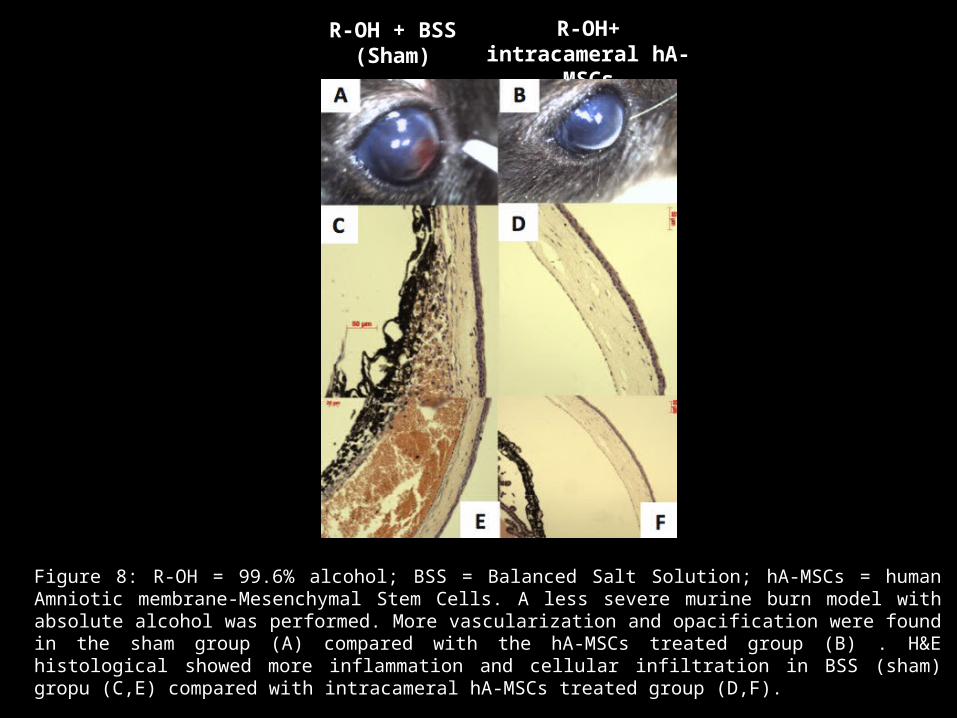
Figure 8: R-OH = 99.6% alcohol; BSS = Balanced Salt Solution; hA-MSCs = human Amniotic membrane-Mesenchymal Stem Cells. A less severe murine burn model with absolute alcohol was performed. More vascularization and opacification were found in the sham group (A) compared with the hA-MSCs treated group (B) . H&E histological showed more inflammation and cellular infiltration in BSS (sham) gropu (C,E) compared with intracameral hA-MSCs treated group (D,F).
R-OH + BSS(Sham)
R-OH+ intracameral hA-MSCs

Conclusions• These data suggest that human Amniotic
membrane-Mesenchymal Stem Cells (hA-MSCs) could be applied in the ocular regenerative therapy as an immunosuppressor treatment in ocular and inflammatory diseases.
References1) Zeng W, Li Y, Zeng G, Yang B, Zhu Y. Transplantation with cultured stem cells derived from the human amniotic
membrane for corneal alkali burns: an experimental study. Ann Clin Lab Sci 2014;44:73-81.2) Ma Y, Xu Y, Xiao Z, Yang W, Zhang C, Song E, Du Y, Li L. Reconstruction of chemically burned rat corneal surface by
bone marrow-derived human mesenchymal stem cells. Stem Cells 2006;24:315-321.3) Reinshagen H, Auw-Haedrich C, Sorg RV, Boehringer D, Eberwein P, Schwartzkopff J, Sundmacher R, Reinhard T.
Corneal surface reconstruction using adult mesenchymal stem cells in experimental limbal stem cell deficiency in rabbits. Acta Ophthalmol 2011;89:741-748.
4) Jiang TS, Cai L, Ji WY, Hui YN, Wang YS, Hu D, Zhu J. Reconstruction of the corneal epithelium with induced marrow mesenchymal stem cells in rats. Mol Vis 2010:16:1304-1316.
5) Rohaina CM, Then KY, Ng AM, Wan Abdul Halim WH, Zahidin AZ, Saim A, Idrus RB. Reconstruction of limbal stem cell deficient corneal surface with induced human bone marrow mesenchymal stem cells on amniotic membrane. Trans Res 2014;163:200-210.