Embed Size (px)
Citation preview
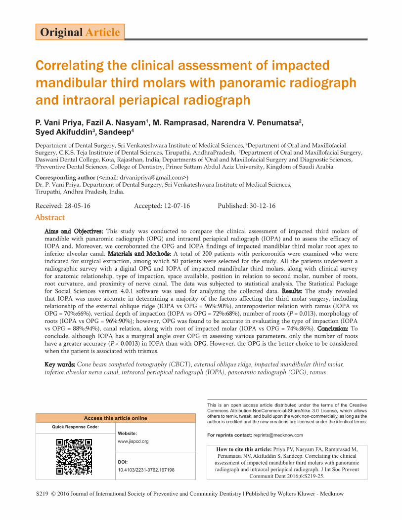
S219 © 2016 Journal of International Society of Preventive and Community Dentistry | Published by Wolters Kluwer - Medknow
Correlating the clinical assessment of impacted mandibular third molars with panoramic radiograph and intraoral periapical radiograph
Received: 28‑05‑16 Accepted: 12‑07‑16 Published: 30‑12‑16
P. Vani Priya, Fazil A. Nasyam1, M. Ramprasad, Narendra V. Penumatsa2, Syed Akifuddin3, Sandeep4
Department of Dental Surgery, Sri Venkateshwara Institute of Medical Sciences, 4Department of Oral and Maxillofacial Surgery, C.K.S. Teja Institrute of Dental Sciences, Tirupathi, AndhraPradesh, 3Department of Oral and Maxillofacial Surgery, Daswani Dental College, Kota, Rajasthan, India, Departments of 1Oral and Maxillofacial Surgery and Diagnostic Sciences, 2Preventive Dental Sciences, College of Dentistry, Prince Sattam Abdul Aziz University, Kingdom of Saudi Arabia
Corresponding author (<email: [email protected]>) Dr. P. Vani Priya, Department of Dental Surgery, Sri Venkateshwara Institute of Medical Sciences, Tirupathi, Andhra Pradesh, India.
Abstract
Aims and Objectives: This study was conducted to compare the clinical assessment of impacted third molars of mandible with panaromic radiograph (OPG) and intraoral periapical radiograph (IOPA) and to assess the efficacy of IOPA and. Moreover, we corroborated the OPG and IOPA findings of impacted mandiblar third molar root apex to inferior alveolar canal. Materials and Methods: A total of 200 patients with pericoronitis were examined who were indicated for surgical extraction, among which 50 patients were selected for the study. All the patients underwent a radiographic survey with a digital OPG and IOPA of impacted mandibular third molars, along with clinical survey for anatomic relationship, type of impaction, space available, position in relation to second molar, number of roots, root curvature, and proximity of nerve canal. The data was subjected to statistical analysis. The Statistical Package for Social Sciences version 4.0.1 software was used for analyzing the collected data. Results: The study revealed that IOPA was more accurate in determining a majority of the factors affecting the third molar surgery, including relationship of the external oblique ridge (IOPA vs OPG = 96%:90%), anteroposterior relation with ramus (IOPA vs OPG = 70%:66%), vertical depth of impaction (IOPA vs OPG = 72%:68%), number of roots (P = 0.013), morphology of roots (IOPA vs OPG = 96%:90%); however, OPG was found to be accurate in evaluating the type of impaction (IOPA vs OPG = 88%:94%), canal relation, along with root of impacted molar (IOPA vs OPG = 74%:86%). Conclusion: To conclude, although IOPA has a marginal angle over OPG in assessing various parameters, only the number of roots have a greater accuracy (P < 0.0013) in IOPA than with OPG. However, the OPG is the better choice to be considered when the patient is associated with trismus.
Key words: Cone beam computed tomography (CBCT), external oblique ridge, impacted mandibular third molar, inferior alveolar nerve canal, intraoral periapical radiograph (IOPA), panoramic radiograph (OPG), ramus
Access this article onlineQuick Response Code:
Website: www.jispcd.org
DOI: 10.4103/2231-0762.197198
How to cite this article: Priya PV, Nasyam FA, Ramprasad M, Penumatsa NV, Akifuddin S, Sandeep. Correlating the clinical
assessment of impacted mandibular third molars with panoramic radiograph and intraoral periapical radiograph. J Int Soc Prevent
Communit Dent 2016;6:S219-25.
This is an open access article distributed under the terms of the Creative Commons Attribution-NonCommercial-ShareAlike 3.0 License, which allows others to remix, tweak, and build upon the work non-commercially, as long as the author is credited and the new creations are licensed under the identical terms.
For reprints contact: [email protected]
Original Article
Priya, et al.: Correlating the clinical assessment of impacted mandibular third molars with IOPA and OPG
Journal of International Society of Preventive and Community Dentistry S220December 2016, Vol. 6, Supplement 3
INTRODUCTION
Radiographic diagnosis of impacted third molars precedes their surgical removal. The factors such as the position of tooth, the number, and morphology of roots, in particular, the relationship between the roots and mandibular canal, require assessment for an atraumatic untoward transalveolar extraction of these impacted mandibular molars. A conventional radiographic examination may consist of panoramic and/or intraoral radiography. A series of three intraoral projections has been recommended for full evaluation.[1‑4] The bucco‑lingual relation between the tooth and other anatomical structures, is better asssessed by the tube shift or buccal object rule which is traditionally employed.[3,4]
Orthopantamograph has diversified use in both general diagnosis and varied specialized clinical diagnosis, and is commonly used to assess third molars prior to transalveolar extraction.[5] During the last decade, different techniques have been developed for digital, and in recent years, the solid‑state digital X‑ray units and photostimulable phosphor plate systems which are used with conventional OPG units are available in the market. The diagnostic outcome of panoramic images is the same as that of intraoral periapical radiograph (IOPA), however, they have been evaluated only sporadically; such comparison should be done until a new diagnostic procedure is introduced for better diagnosis.
The third molar follicle first becomes apparent at an age of 6–7 years and can be diagnosed by 8–9 years. At the age of 14–16 years, the third molar follicle is apparent in radiographs. If the follicle is not present at this age, agenesis should be considered. Girls appear to be slightly ahead of boys up to the age of root formation, however, this difference disappears in the final stage of root development. Third molar agenesis is very common and is considered to have an incidence of 5–33%. When the cervical part of the root complex is formed, the tooth germ starts to move away from the mandibular canal, assuming a distally curved eruption pathway. If resistance is met during eruption, intrusive growth occurs in the proximity of the mandibular canal, resulting in indentations in the apical part of the root, apical deflections, or circumferential growth around the mandibular canal, resulting in curving of the roots and a change in the eruption pathway. These changes that occur during the developmental and the eruption stage of the mandibular third molar make meticulous preoperative radiographic evaluation an important step. This would facilitate comprehensive evaluation of the
anatomic disposition of the tooth in relation to the vital structures in the surrounding.
These types of studies have been conducted in the past comparing computed tomography and panoramic radiographs by Pecker et al. in Turkey. Though various diagnostic modalities have come up in the recent past including the use of computed tomography, IOPA and panoramic radiographs (OPG) ought to be considered the gold standards for planning transalveolar extraction of impacted third molar of mandible. However, there is sparse literature on comparison between IOPA and OPG. The aim of our study is to correlate the clinical findings with these standard radiographs and compare the efficacy of IOPA with OPG for determining the status of the anatomical factors that dictate the third molar surgery.
Aims
The aims of our study are• To confirm the clinical parameters of impacted
mandibular third molars with OPG and IOPA and to assess the efficacy between the two.
• Tocorroborate theOPGandIOPAfindingsof theroot apex of the impacted mandibular third to the inferior alveolar nerve canal.
MATERIALS AND METHODS
The study was conducted for a period of 1 year. Two hundred patients diagnosed with moderate‑to‑severe pericoronitis electing to undergo surgical extraction were choosen, out of which 50 patients were selected for the study by simple random sampling. The sample size of 50 was selected based on similar comparative studies done previously by Ishak et al.[6] We enrolled individuals who voluntarily signed an informed consent after obtaining institutional ethical committee clearance. All the patients were referred to the Department of Oral Medicine and Radiology for digital OPG and IOPA. The digital OPG was obtained using SIRONA ORTHO PHOS XG5 unit along with automatic processing of the film. IOPA was obtained using Villa India and Blue X unit in a paralleling cone technique, and manual processing was done.
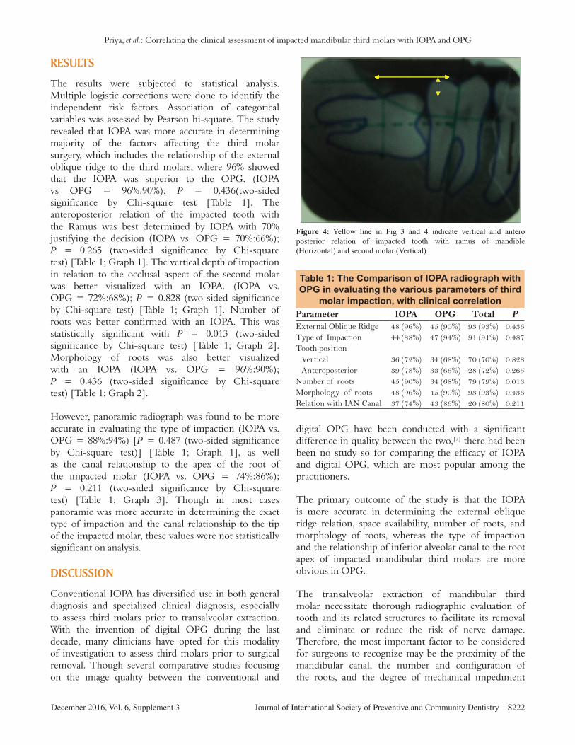
The clinical and radiographic assessments were done under standard conditions by a trained oral surgeon experienced in interpreting IOPA and OPG, as well as one observer beginning his training in Oral and Maxillofacial Surgery. Clinically, the anteroposterior width and depth of the crown exposed was measured
Priya, et al.: Correlating the clinical assessment of impacted mandibular third molars with IOPA and OPG
S221 Journal of International Society of Preventive and Community Dentistry December 2016, Vol. 6, Supplement 3
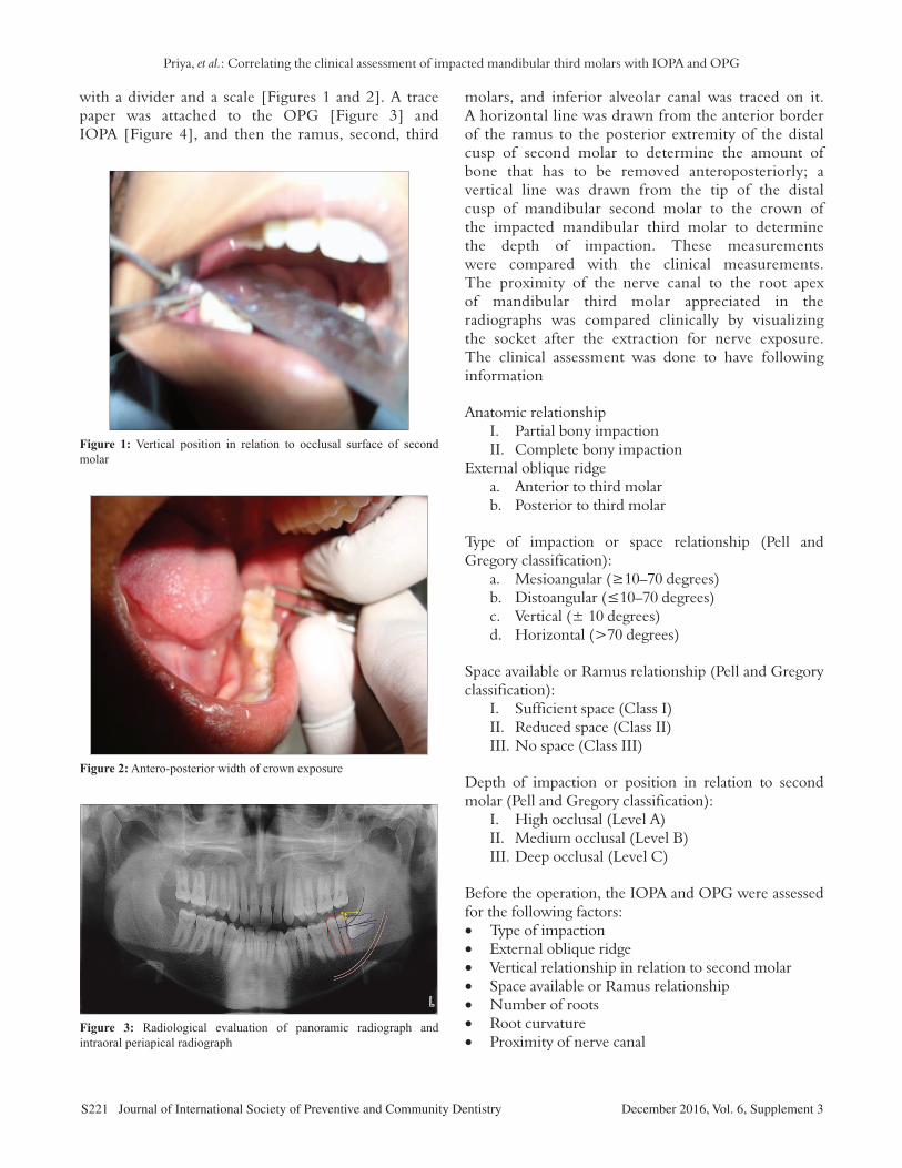
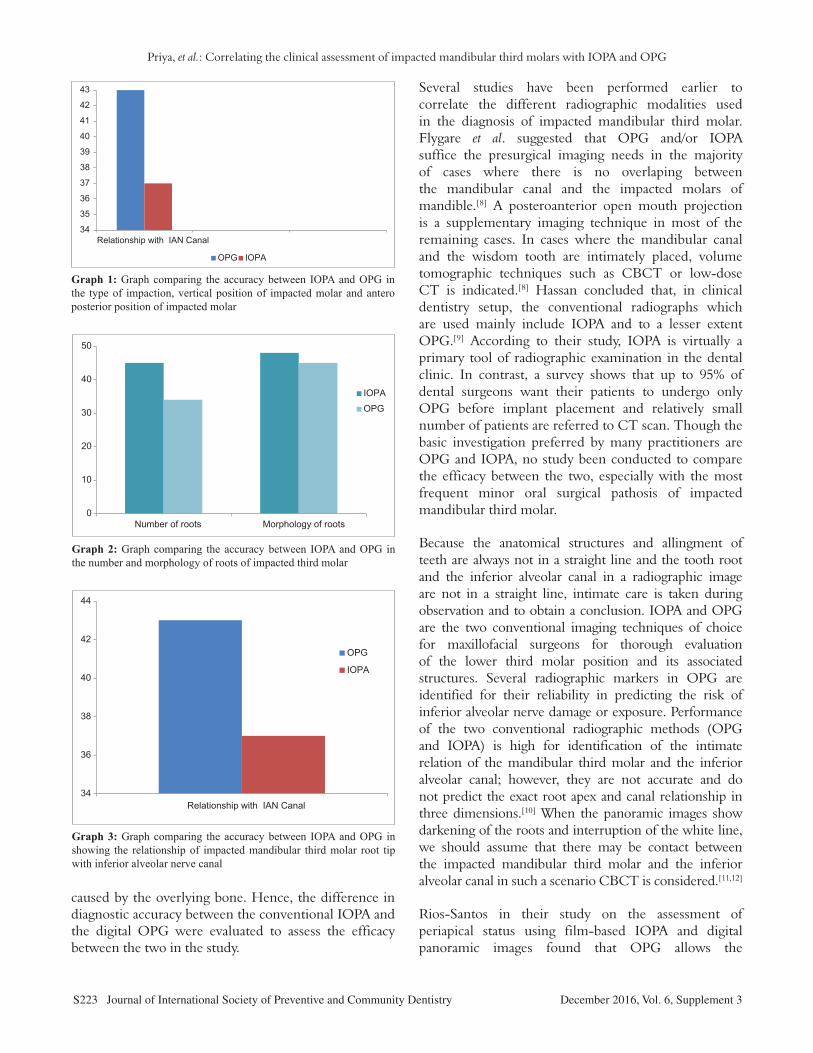
with a divider and a scale [Figures 1 and 2]. A trace paper was attached to the OPG [Figure 3] and IOPA [Figure 4], and then the ramus, second, third
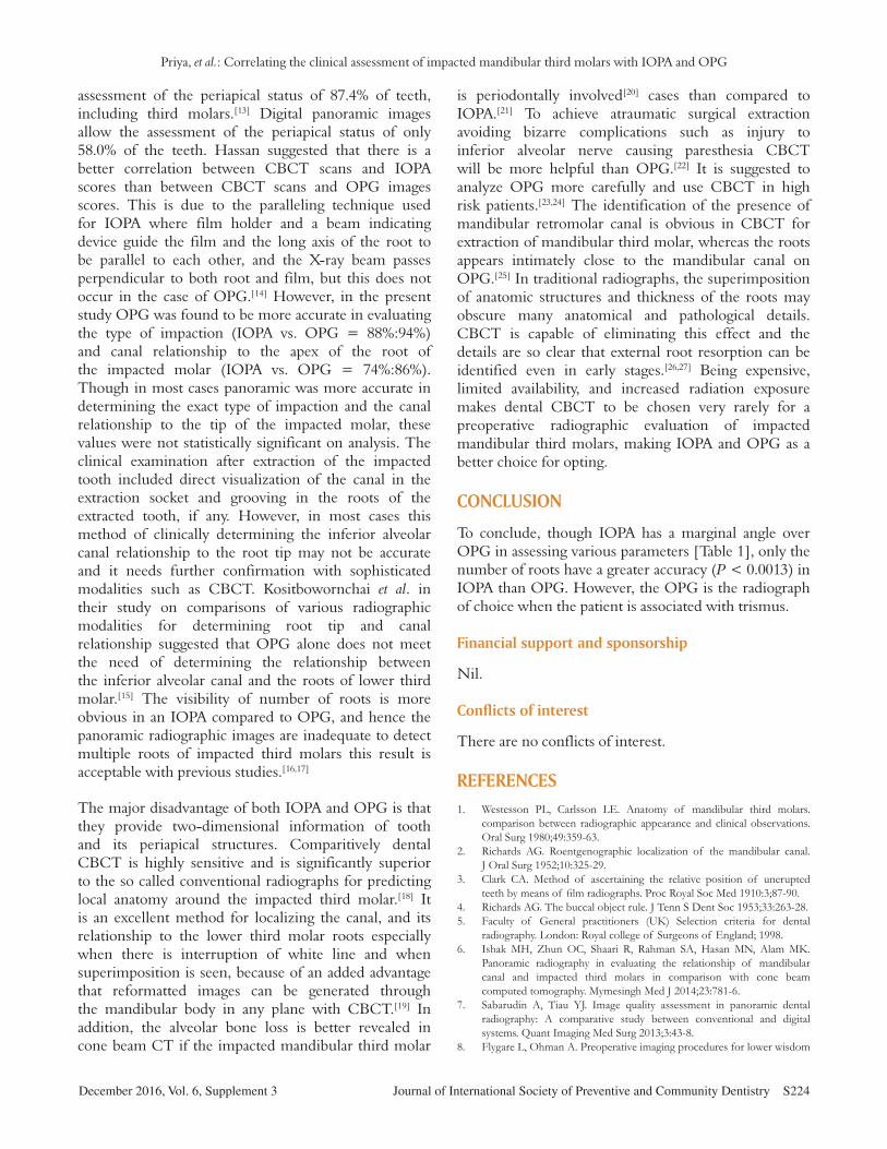
molars, and inferior alveolar canal was traced on it. A horizontal line was drawn from the anterior border of the ramus to the posterior extremity of the distal cusp of second molar to determine the amount of bone that has to be removed anteroposteriorly; a vertical line was drawn from the tip of the distal cusp of mandibular second molar to the crown of the impacted mandibular third molar to determine the depth of impaction. These measurements were compared with the clinical measurements. The proximity of the nerve canal to the root apex of mandibular third molar appreciated in the radiographs was compared clinically by visualizing the socket after the extraction for nerve exposure. The clinical assessment was done to have following information
Anatomic relationship I. Partial bony impaction II. Complete bony impactionExternal oblique ridge a. Anterior to third molar b. Posterior to third molar
Type of impaction or space relationship (Pell and Gregory classification): a. Mesioangular(≥10–70degrees) b. Distoangular(≤10–70degrees) c. Vertical (± 10 degrees) d. Horizontal (>70 degrees)
Space available or Ramus relationship (Pell and Gregory classification): I. Sufficient space (Class I) II. Reduced space (Class II) III. No space (Class III)
Depth of impaction or position in relation to second molar (Pell and Gregory classification): I. High occlusal (Level A) II. Medium occlusal (Level B) III. Deep occlusal (Level C)
Before the operation, the IOPA and OPG were assessed for the following factors:• Type of impaction• External oblique ridge• Vertical relationship in relation to second molar• Space available or Ramus relationship• Number of roots• Root curvature• Proximity of nerve canal
Figure 3: Radiological evaluation of panoramic radiograph and intraoral periapical radiograph
Figure 2: Antero-posterior width of crown exposure
Figure 1: Vertical position in relation to occlusal surface of second molar
Priya, et al.: Correlating the clinical assessment of impacted mandibular third molars with IOPA and OPG
Journal of International Society of Preventive and Community Dentistry S222December 2016, Vol. 6, Supplement 3
RESULTS
The results were subjected to statistical analysis. Multiple logistic corrections were done to identify the independent risk factors. Association of categorical variables was assessed by Pearson hi‑square. The study revealed that IOPA was more accurate in determining majority of the factors affecting the third molar surgery, which includes the relationship of the external oblique ridge to the third molars, where 96% showed that the IOPA was superior to the OPG. (IOPA vs OPG = 96%:90%); P = 0.436(two‑sided significance by Chi‑square test [Table 1]. The anteroposterior relation of the impacted tooth with the Ramus was best determined by IOPA with 70% justifying the decision (IOPA vs. OPG = 70%:66%); P = 0.265 (two‑sided significance by Chi‑square test) [Table 1; Graph 1]. The vertical depth of impaction in relation to the occlusal aspect of the second molar was better visualized with an IOPA. (IOPA vs. OPG = 72%:68%); P = 0.828 (two‑sided significance by Chi‑square test) [Table 1; Graph 1]. Number of roots was better confirmed with an IOPA. This was statistically significant with P = 0.013 (two‑sided significance by Chi‑square test) [Table 1; Graph 2]. Morphology of roots was also better visualized with an IOPA (IOPA vs. OPG = 96%:90%); P = 0.436 (two‑sided significance by Chi‑square test) [Table 1; Graph 2].
However, panoramic radiograph was found to be more accurate in evaluating the type of impaction (IOPA vs. OPG = 88%:94%) [P = 0.487 (two‑sided significance by Chi‑square test)] [Table 1; Graph 1], as well as the canal relationship to the apex of the root of the impacted molar (IOPA vs. OPG = 74%:86%); P = 0.211 (two‑sided significance by Chi‑square test) [Table 1; Graph 3]. Though in most cases panoramic was more accurate in determining the exact type of impaction and the canal relationship to the tip of the impacted molar, these values were not statistically significant on analysis.
DISCUSSION
Conventional IOPA has diversified use in both general diagnosis and specialized clinical diagnosis, especially to assess third molars prior to transalveolar extraction. With the invention of digital OPG during the last decade, many clinicians have opted for this modality of investigation to assess third molars prior to surgical removal. Though several comparative studies focusing on the image quality between the conventional and
digital OPG have been conducted with a significant difference in quality between the two,[7] there had been been no study so for comparing the efficacy of IOPA and digital OPG, which are most popular among the practitioners.
The primary outcome of the study is that the IOPA is more accurate in determining the external oblique ridge relation, space availability, number of roots, and morphology of roots, whereas the type of impaction and the relationship of inferior alveolar canal to the root apex of impacted mandibular third molars are more obvious in OPG.
The transalveolar extraction of mandibular third molar necessitate thorough radiographic evaluation of tooth and its related structures to facilitate its removal and eliminate or reduce the risk of nerve damage. Therefore, the most important factor to be considered for surgeons to recognize may be the proximity of the mandibular canal, the number and configuration of the roots, and the degree of mechanical impediment
Figure 4: Yellow line in Fig 3 and 4 indicate vertical and antero posterior relation of impacted tooth with ramus of mandible (Horizontal) and second molar (Vertical)
Table 1: The Comparison of IOPA radiograph with OPG in evaluating the various parameters of third
molar impaction, with clinical correlationParameter IOPA OPG Total PExternal Oblique Ridge 48 (96%) 45 (90%) 93 (93%) 0.436Type of Impaction 44 (88%) 47 (94%) 91 (91%) 0.487Tooth position
Vertical 36 (72%) 34 (68%) 70 (70%) 0.828Anteroposterior 39 (78%) 33 (66%) 28 (72%) 0.265
Number of roots 45 (90%) 34 (68%) 79 (79%) 0.013Morphology of roots 48 (96%) 45 (90%) 93 (93%) 0.436Relation with IAN Canal 37 (74%) 43 (86%) 20 (80%) 0.211
Priya, et al.: Correlating the clinical assessment of impacted mandibular third molars with IOPA and OPG
S223 Journal of International Society of Preventive and Community Dentistry December 2016, Vol. 6, Supplement 3
caused by the overlying bone. Hence, the difference in diagnostic accuracy between the conventional IOPA and the digital OPG were evaluated to assess the efficacy between the two in the study.
Several studies have been performed earlier to correlate the different radiographic modalities used in the diagnosis of impacted mandibular third molar. Flygare et al. suggested that OPG and/or IOPA suffice the presurgical imaging needs in the majority of cases where there is no overlaping between the mandibular canal and the impacted molars of mandible.[8] A posteroanterior open mouth projection is a supplementary imaging technique in most of the remaining cases. In cases where the mandibular canal and the wisdom tooth are intimately placed, volume tomographic techniques such as CBCT or low‑dose CT is indicated.[8] Hassan concluded that, in clinical dentistry setup, the conventional radiographs which are used mainly include IOPA and to a lesser extent OPG.[9] According to their study, IOPA is virtually a primary tool of radiographic examination in the dental clinic. In contrast, a survey shows that up to 95% of dental surgeons want their patients to undergo only OPG before implant placement and relatively small number of patients are referred to CT scan. Though the basic investigation preferred by many practitioners are OPG and IOPA, no study been conducted to compare the efficacy between the two, especially with the most frequent minor oral surgical pathosis of impacted mandibular third molar.
Because the anatomical structures and allingment of teeth are always not in a straight line and the tooth root and the inferior alveolar canal in a radiographic image are not in a straight line, intimate care is taken during observation and to obtain a conclusion. IOPA and OPG are the two conventional imaging techniques of choice for maxillofacial surgeons for thorough evaluation of the lower third molar position and its associated structures. Several radiographic markers in OPG are identified for their reliability in predicting the risk of inferior alveolar nerve damage or exposure. Performance of the two conventional radiographic methods (OPG and IOPA) is high for identification of the intimate relation of the mandibular third molar and the inferior alveolar canal; however, they are not accurate and do not predict the exact root apex and canal relationship in three dimensions.[10] When the panoramic images show darkening of the roots and interruption of the white line, we should assume that there may be contact between the impacted mandibular third molar and the inferior alveolar canal in such a scenario CBCT is considered.[11,12]
Rios‑Santos in their study on the assessment of periapical status using film‑based IOPA and digital panoramic images found that OPG allows the
34
35
36
37
38
39
40
41
42
43
Relationship with IAN Canal
OPG IOPA
Graph 1: Graph comparing the accuracy between IOPA and OPG in the type of impaction, vertical position of impacted molar and antero posterior position of impacted molar
0
10
20
30
40
50
Number of roots Morphology of roots
IOPA
OPG
Graph 2: Graph comparing the accuracy between IOPA and OPG in the number and morphology of roots of impacted third molar
34
36
38
40
42
44
Relationship with IAN Canal
OPG
IOPA
Graph 3: Graph comparing the accuracy between IOPA and OPG in showing the relationship of impacted mandibular third molar root tip with inferior alveolar nerve canal
Priya, et al.: Correlating the clinical assessment of impacted mandibular third molars with IOPA and OPG
Journal of International Society of Preventive and Community Dentistry S224December 2016, Vol. 6, Supplement 3
assessment of the periapical status of 87.4% of teeth, including third molars.[13] Digital panoramic images allow the assessment of the periapical status of only 58.0% of the teeth. Hassan suggested that there is a better correlation between CBCT scans and IOPA scores than between CBCT scans and OPG images scores. This is due to the paralleling technique used for IOPA where film holder and a beam indicating device guide the film and the long axis of the root to be parallel to each other, and the X‑ray beam passes perpendicular to both root and film, but this does not occur in the case of OPG.[14] However, in the present study OPG was found to be more accurate in evaluating the type of impaction (IOPA vs. OPG = 88%:94%) and canal relationship to the apex of the root of the impacted molar (IOPA vs. OPG = 74%:86%). Though in most cases panoramic was more accurate in determining the exact type of impaction and the canal relationship to the tip of the impacted molar, these values were not statistically significant on analysis. The clinical examination after extraction of the impacted tooth included direct visualization of the canal in the extraction socket and grooving in the roots of the extracted tooth, if any. However, in most cases this method of clinically determining the inferior alveolar canal relationship to the root tip may not be accurate and it needs further confirmation with sophisticated modalities such as CBCT. Kositbowornchai et al. in their study on comparisons of various radiographic modalities for determining root tip and canal relationship suggested that OPG alone does not meet the need of determining the relationship between the inferior alveolar canal and the roots of lower third molar.[15] The visibility of number of roots is more obvious in an IOPA compared to OPG, and hence the panoramic radiographic images are inadequate to detect multiple roots of impacted third molars this result is acceptable with previous studies.[16,17]
The major disadvantage of both IOPA and OPG is that they provide two‑dimensional information of tooth and its periapical structures. Comparitively dental CBCT is highly sensitive and is significantly superior to the so called conventional radiographs for predicting local anatomy around the impacted third molar.[18] It is an excellent method for localizing the canal, and its relationship to the lower third molar roots especially when there is interruption of white line and when superimposition is seen, because of an added advantage that reformatted images can be generated through the mandibular body in any plane with CBCT.[19] In addition, the alveolar bone loss is better revealed in cone beam CT if the impacted mandibular third molar
is periodontally involved[20] cases than compared to IOPA.[21] To achieve atraumatic surgical extraction avoiding bizarre complications such as injury to inferior alveolar nerve causing paresthesia CBCT will be more helpful than OPG.[22] It is suggested to analyze OPG more carefully and use CBCT in high risk patients.[23,24] The identification of the presence of mandibular retromolar canal is obvious in CBCT for extraction of mandibular third molar, whereas the roots appears intimately close to the mandibular canal on OPG.[25] In traditional radiographs, the superimposition of anatomic structures and thickness of the roots may obscure many anatomical and pathological details. CBCT is capable of eliminating this effect and the details are so clear that external root resorption can be identified even in early stages.[26,27] Being expensive, limited availability, and increased radiation exposure makes dental CBCT to be chosen very rarely for a preoperative radiographic evaluation of impacted mandibular third molars, making IOPA and OPG as a better choice for opting.
CONCLUSION
To conclude, though IOPA has a marginal angle over OPG in assessing various parameters [Table 1], only the number of roots have a greater accuracy (P < 0.0013) in IOPA than OPG. However, the OPG is the radiograph of choice when the patient is associated with trismus.
Financial support and sponsorship
Nil.
Conflicts of interest
There are no conflicts of interest.
REFERENCES1. Westesson PL, Carlsson LE. Anatomy of mandibular third molars.
comparison between radiographic appearance and clinical observations. Oral Surg 1980;49:359‑63.
2. Richards AG. Roentgenographic localization of the mandibular canal. J Oral Surg 1952;10:325‑29.
3. Clark CA. Method of ascertaining the relative position of unerupted teeth by means of film radiographs. Proc Royal Soc Med 1910:3;87‑90.
4. Richards AG. The buccal object rule. J Tenn S Dent Soc 1953;33:263‑28.5. Faculty of General practitioners (UK) Selection criteria for dental
radiography. London: Royal college of Surgeons of England; 1998.6. Ishak MH, Zhun OC, Shaari R, Rahman SA, Hasan MN, Alam MK.
Panoramic radiography in evaluating the relationship of mandibular canal and impacted third molars in comparison with cone beam computed tomography. Mymesingh Med J 2014;23:781‑6.
7. Sabarudin A, Tiau YJ. Image quality assessment in panoramic dental radiography: A comparative study between conventional and digital systems. Quant Imaging Med Surg 2013;3:43‑8.
8. Flygare L, Ohman A. Preoperative imaging procedures for lower wisdom
Priya, et al.: Correlating the clinical assessment of impacted mandibular third molars with IOPA and OPG
S225 Journal of International Society of Preventive and Community Dentistry December 2016, Vol. 6, Supplement 3
teeth removal. Clin Oral Investig 2008;12:291‑302.9. Hassan BA. Reliability of Periapical Radiographs and
Orthopantomograms in Detection of Tooth Root Protrusion in the Maxillary Sinus: Correlation Results with Cone Beam Computed Tomography. J Oral Maxillofac Res 2010;1:e6.
10. Xu GZ, Yang C, Fan XD, Yu CQ, Cai XY, Wang Y, et al. Anatomic relationship between impacted third mandibular molar and the mandibular canal as the risk factor of inferior alveolar nerve injury. Br J Oral Maxillofac Surg 2013;51:e215‑9.
11. Peker I, Sarikir C, Alkurt MT, Zor ZF. Panoramic radiography and cone‑beam computed tomography findings in preoperative examination of impacted mandibular third molars. BMC Oral Health 2014;14:71.
12. Ishak MH, Zhun OC, Shaari R, Rahman SA, Hasan MN, Alam MK. Panoramic radiography in evaluating the relationship of mandibularcanal and impacted third molars in comparison with Cone beam computed tomography. Mymesingh Med J 2014; 23:781‑6.
13. Ríos‑Santos JV, Ridao‑Sacie C, Bullón P, Fernández‑Palacín A, Segura‑Egea JJ. Assessment of periapical status: A comparative study using film‑based periapical radiographs and digital panoramic images. Med Oral Patol Oral Cir Bucal 2010;15:e952‑6.
14. Suomalainen A, Ventä I, Mattila M, Turtola L, Vehmas T, Peltola JS. Reliability of CBCT and other radiographic methods in preoperative evaluation of lower third molars. Oral Surg Oral Med Oral Pathol Oral Radiol Endod 2010;109:276‑84.
15. Kositbowornchai S, Densiri‑aksorn W, Piumthanaroj P. Ability of two radiographic methods to identify the closeness between the mandibular third molar root and the inferior alveolar canal: A pilot study. Dentomaxillofac Radiol 2010;39:79‑84.
16. Ghaeminia H, Meijer GJ, Soehardi A, Borstlap WA, Mulder J, Vlijmen OJ, et al. The use of cone beam CT for the removal of wisdom teeth changes the surgical approach compared with panoramic radiography: A pilot study. Int J Oral Maxillofac Surg 2011;40:834‑9.
17. Tantanapornkul W, Okouchi K, Fujiwara Y, Yamashiro M, Maruoka Y, Ohbayashi N, et al. A comparative study of cone‑beam computed tomography and conventional panoramic radiography in assessing the topographic relationship between the mandibular canal and impacted third molars. Oral Surg Oral Med Oral Pathol Oral Radiol Endod 2007;103:253‑9.
18. Nakagawa Y, Kobayashi K, Ishii H, Mishima A, Ishii K, Asada K,
et al. Preoperative application of limited cone beam computerized tomography as an assessment tool before minor oral surgery. Int J Oral Maxillofac Surg 2002;31:322‑7.
19. Lee B, Park Y, Ahn J, Chun J, Park S, Kim M, et al. Assesment of the proximity between the mandibular third molar and inferior alveolar canal using preoperative 3D‑CT to prevent inferior alveolar nerve damage. Maxillofac Plast Reconst Surg 2015;37:30.
20. Takeshita WM, Iwaki LC, Da Silva MC, Tonin RH. Evaluation of diagnostic accuracy of conventional and digital periapical radiography, panoramic radiography, and cone‑beam computed tomography in the assessment of alveolar bone loss. Comtemp Clin Dent 2014;5:318‑23.
21. Ashwinirani SR, Suragimath G, Jaishankar HP, Kulkarni P, Bijjaragi SC, Sangle VA. Comparison of Diagnostic Accuracy of Conventional Intraoral Periapical and Direct Digital Radiographs in Detecting Interdental Bone Loss. J Clin Diagn Res 2015;9:ZC35‑8.
22. Vujanovic‑Eskenazi A, Valero‑James JM, Sánchez‑Garcés MA, Gay‑Escoda C. A retrospective radiographic evaluation of the anterior loop of the mental nerve: Comparison between panoramic radiography and cone beam computerized tomography. Bucal Med Oral Patol Oral Cir Bucal 2015;20:e239‑45.
23. Muinelo‑Lorenzo J, Suárez‑Quintanilla JA, Fernández‑Alonso A, Marsillas‑Rascado S, Suárez‑Cunqueiro MM. Descriptive study of the bifid mandibular canals and retromolar foramina: Cone beam CT vs panoramic radiography. Dentomaxillofac Radiol 2014;43:20140090.
24. Nemati S, Moghadam AA, Kajan ZD, Mohtavipour ST, Amouzad H. An analysis of visibility and anatomic variations of mandibular canal in digital panoramic radiographs of dentulous and edentulous patients in Northern Iran populations. J Dent 2016;17:112‑20.
25. Kubilius M, Kubilius R, Varinauskas V, Žalinkevičius R, Tözüm TF, Juodžbalys G. Descriptive study of mandibular canal visibility: Morphometric and desitometric analysis for digital panoramic radiographs. Dentomaxillofac Radiol 2016;45:20160079.
26. Sisman Y, Ercan‑Sekerci A, Payveren‑Arıkan M, Sahman H. Diagnostic accuracy of cone‑beam CT compared with panoramic images in predicting retromolar canal during extraction of impacted mandibular third molars. Med Oral Patol Oral Cir Bucal 2015;20:e74‑e81.
27. Creanga AG, Geha H, Sankar V, Teixeira FB, McMahan CA, Noujeim M. Accuracy of digital periapical radiography and cone‑beam computed tomography in detecting external root resorption. Imaging Sci Dent 2015;45:153‑8.


![Orthodontic Treatment of Bilateral Impacted Mandibular ...downloads.hindawi.com/journals/crid/2019/7638959.pdf · than unilateral impaction [7–10]. Even though lower canine impaction](https://img.pdfslide.net/doc/110x75/5f0a053a7e708231d429a0ad/orthodontic-treatment-of-bilateral-impacted-mandibular-than-unilateral-impaction.jpg)