Embed Size (px)
Citation preview

1
MQP-BIO-DSA-1615
MQP-BIO-DSA-6822
CORRELATIONS BETWEEN SEVERE NEONATAL
INTRAVENTRICULAR HEMORRHAGE AND
NEURODEVELOPMENTAL COMPLICATIONS
A Major Qualifying Project Report
Submitted to the Faculty of the
WORCESTER POLYTECHNIC INSTITUTE
in partial fulfillment of the requirements for the
Degree of Bachelor of Science
in
Biology and Biotechnology
by
_________________________ _________________________
Hannah Israel Kristin Newell
April 28, 2011
APPROVED:
_________________________ _________________________
Robin Adair, Ph.D. David Adams, Ph.D.
Developmental/Behavioral Pediatrics Biology and Biotechnology
UMASS Medical Center WPI Project Advisor
Major Advisor

2
ABSTRACT
Intraventricular hemorrhaging (IVH) is surprisingly common in premature infants, and
may cause neurodevelopmental problems. However, demonstration of this correlation has been
difficult to obtain at UMass Memorial Medical Center due to a communication disconnect
between the Neonatal Intensive Care Unit where IVH is usually first diagnosed, and the Follow-
up Clinic where subsequent developmental problems would be identified. To bridge this
disconnect, standardized forms were developed to allow physicians to obtain medical follow-up
data on infants. The results of a sample analysis of severe IVH show that severe IVH in
premature infants born under 1000g correlates with neurodevelopmental problems such as
cerebral palsy, cognitive impairment, and various sensory impairments.

3
TABLE OF CONTENTS
Signature Page ………………………………….……………………………………. 1
Abstract ………………………………………….…………………………………… 2
Table of Contents ………………………………………….…………………….…… 3
Acknowledgements ………………………………………………………………….. 4
Background ………………………………………………………………………….. 5
Project Purpose ………………………………………….…………………...………. 25
Methods …………………………………………….…………………………...…… 26
Results …………………………………………………………………………...….. 30
Discussion …………………………………………….…………………………...… 39
Bibliography ………………………………………………………………………… 42
Appendix ……………………………………..……………………………………... 53

4
ACKNOWLEDGEMENTS
We would like to thank UMass Memorial Medical Center, especially Robin Adair, MD
and Alan Picarillo, MD, for sponsoring this project and for their help and guidance throughout
our entire project. We would also like to thank Mary Naples for providing us with IS support
throughout this project. Lastly, we would like to thank Professor David Adams for advising us
on this project, and for all of his help in initiating and editing this project.

5
BACKGROUND
Vermont Oxford Network
The Vermont Oxford Network (VON) is a worldwide non-profit organization, consisting
of 850 neonatal intensive care units. Yearly, VON acquires data on approximately 50,000
infants. VON is a voluntary service of health care professionals working to improve the safety
and quality of newborn infants' medical care. The network holds an Annual Quality Congress of
Neonatology for all participating institutions (Vermont Oxford Network, 2011).
These health care professionals conduct numerous clinical trials, long-term follow-up
studies, as well as outcome and epidemiologic research. This data is contained in a network
database and is used to determine information about how newborn infant care influences their
outcomes. The results of these studies are published by the network in scientific articles, peer
reviewed medical journals, network publications, and on their website (Vermont Oxford
Network, 2011).
The VON database contains information on very low birth weight (VLBW) infants as
well as others that fit their requirements. VON is able to take information from the database from
the participating hospitals and provide an analysis and report confidentially on those hospitals.
This allows hospitals to see which areas can be improved for quality assurance purposes
(Vermont Oxford Network, 2011). Quality assurance opportunities are also available through
collaboration with other hospitals through face-to-face contact or through web-based conferences
(Vermont Oxford Network, 2011).

6
Prematurity
Infants born before 37 weeks gestation are considered premature. The more preterm an
infant is born, the greater the risk that the infant will experience complications of prematurity.
High rates of morbidity and mortality in preterm infants can be attributed to complications
associated with prematurity. Approximately one third of infant deaths can be associated with
prematurity. Extremely premature infants have a mortality rate around 50 percent (the highest of
any gestational age group), as well as having the greatest risk of morbidity in the long-term.
Prematurity accounts for 25 percent of children with hearing or cognitive impairments, 35
percent of those with visual impairments, and 45 percent of children with cerebral palsy
(Eichenwald & Stark, 2008).
Three standard subdivisions classify underweight infants and three are designated for the
degree of immaturity (approximate gestational age at birth). Infants born weighing less than 1000
g are considered to be extremely low birth weight (ELBW). An infant born weighing between
1000 g and 1500 g is considered to be very low birth weight (VLBW). Infants born weighing
between 1500 g and 2500 g are considered to be low birth weight (LBW) (WHO, 2011). An
infant born before 25 weeks gestation is referred to as being extremely preterm (Nicolas et al.,
2000). Infants born between 25 and 32 weeks gestation is referred to as being very preterm. An
infant born from 32 to less than 37 weeks gestation is referred to as being late preterm (Pamela et
al., 2004).
Major NICU Advancements
Advancements in antenatal medicine and neonatal care have improved the rates of
preterm infant survival. However, this improved rate of survival was not followed by
7
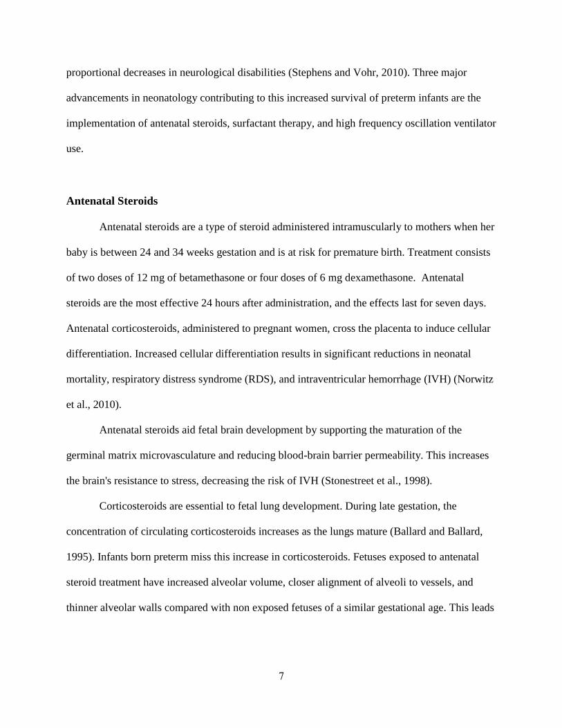
proportional decreases in neurological disabilities (Stephens and Vohr, 2010). Three major
advancements in neonatology contributing to this increased survival of preterm infants are the
implementation of antenatal steroids, surfactant therapy, and high frequency oscillation ventilator
use.
Antenatal Steroids
Antenatal steroids are a type of steroid administered intramuscularly to mothers when her
baby is between 24 and 34 weeks gestation and is at risk for premature birth. Treatment consists
of two doses of 12 mg of betamethasone or four doses of 6 mg dexamethasone. Antenatal
steroids are the most effective 24 hours after administration, and the effects last for seven days.
Antenatal corticosteroids, administered to pregnant women, cross the placenta to induce cellular
differentiation. Increased cellular differentiation results in significant reductions in neonatal
mortality, respiratory distress syndrome (RDS), and intraventricular hemorrhage (IVH) (Norwitz
et al., 2010).
Antenatal steroids aid fetal brain development by supporting the maturation of the
germinal matrix microvasculature and reducing blood-brain barrier permeability. This increases
the brain's resistance to stress, decreasing the risk of IVH (Stonestreet et al., 1998).
Corticosteroids are essential to fetal lung development. During late gestation, the
concentration of circulating corticosteroids increases as the lungs mature (Ballard and Ballard,
1995). Infants born preterm miss this increase in corticosteroids. Fetuses exposed to antenatal
steroid treatment have increased alveolar volume, closer alignment of alveoli to vessels, and
thinner alveolar walls compared with non exposed fetuses of a similar gestational age. This leads

8
to better gas exchange and an enhanced response to postnatal surfactant treatment (Bonanno and
Wapner, 2009).
Surfactant Therapy
Surfactant is a lipoprotein complex, found in the lungs, which reduces alveolar collapse
by forming a layer between the alveolar surface and the alveolar gas in the lungs, reducing
surface tension (Berry, 1991). Very premature infants are often not able to produce their own
surfactant because their type II alveolar epithelial cells, where surfactant is produced, have not
matured. Without surfactant, the alveoli may not inflate or collapse on expiration, which can lead
to respiratory distress syndrome (RDS) (Berry, 1991). Exogenous surfactant is the primary life-
saving therapy for RDS in preterm infants (Ramanathan, 2009). There are two strategies for
surfactant administration; prophylactic (or preventative) treatment, and rescue or therapeutic
treatment (Ramanathan, 2009).
High Frequency Oscillation Ventilator
High frequency oscillatory ventilation (HFOV) is a mechanical ventilator that uses
constant distending pressure, with pressure variations oscillating around the mean airway
pressure. This ventilation strategy produces small tidal volumes, in contrast to conventional
ventilators, which induces large pressure changes and gas volumes, and is associated with
ventilator induced injury. HFOV uses alternative mechanisms of gas exchange, such as
molecular diffusion. Consequently, HFOV has become the most accepted mode of ventilation
support for RDS in VLBW infants, and has been shown to improve survival without an increase

9
in the incident of chronic lung disease (Moriette et al., 2001; Johnson et al., 2002; Kessel et al.,
2010).
Short Term Complications of Prematurity
It is important to quickly stabilize infants in the delivery room to reduce their risk of
developing short term complications (Lemons et al., 2001). Short term complications of
prematurity are defined as those occurring during the neonatal period (Eichenwald & Stark,
2008). Premature infants are increasingly susceptible to developing short term complications
with decreasing birth weight and gestational age, and are the result of anatomical or functional
immaturity (Faranoff et al., 2007).
Retinopathy of prematurity (ROP) is a condition occurring around 34 weeks
postmenstrual age, and occurs when the retina of premature infants is incompletely vascularized.
ROP generally spontaneously resolves, but requires treatment when ROP is severe and does not
resolve spontaneously (Palmer et al., 1991). Infants with ROP are more likely to have vision
impairment or poor ocular outcome (Trese and Droste, 1998; Moshfeghi et al., 2004; Prenner et
al., 2004; Lakhanpal et al., 2005; Repka et al., 2006).
Many VLBW infants need resuscitation at birth, most requiring endotracheal intubation
and others only need resuscitation medications. Prophylactic administration of surfactant reduces
the risk of respiratory complications, such as respiratory distress syndrome (RDS), pulmonary
interstitial emphysema, and pneumothorax (Lemons et al., 2001). Other complications include
bronchopulmonary dysplasia (BPD), and apnea of prematurity (Henderson-Smart, 1981; Frank
and Sosenko, 1987). Respiratory distress syndrome (RDS) results from insufficient surfactant
production prior to birth (Frank and Sosenko, 1987) and occurs about 93 percent of the time

10
(Stoll et al., 2010). A symptomatic patent ductus anteriosus (PDA) occurs in about 46 percent of
VLBW infants (Stoll et al., 2010). BPD is a chronic lung disease defined as dependence upon
oxygen at 36 weeks postmenstrual age, occurring late in the neonatal period (Shennan et al.,
1988; Marshal et al., 1999) in approximately 42 percent of infants (Stoll et al., 2010). Apnea of
prematurity occurs in about 25 percent of premature infants (Henderson-Smart, 1981).
Necrotizing enterocolitis (NEC), a gastrointestinal complication, increases the risk of
neurodevelopmental disabilities (Rees et al., 2007; Schulzke et al., 2007) and growth delay later
in life (Hintz et al., 2005).
Late-onset sepsis, or a positive blood culture occurring after three days of age, occurs in
about 21 percent of VLBW infants (Stoll et al., 2002). Neonatal sepsis is also associated with an
increase in growth impairment and poor neurodevelopmental outcomes later in life (Stoll et al.,
2004).
Short term cardiovascular complications include a patent ductus anteriosus (PDA) and
systemic hypotension (Faranroff et al., 2007; Seri and Noori, 2005). PDA causes increased blood
flow through pulmonary circulation and decreased blood flow through systemic circulation by
shunting the blood from left to the right side of the heart (Rudolph, 1970). If a significant amount
of shunting occurs, symptoms such as respiratory distress, apnea, or heart failure may present
(Thibeault et al., 1975; Cotton et al., 1978; Jacob et al., 1980; Mahony et al., 1982; Cassady et
al., 1989; Schmidt et al., 2001). Systemic hypotension, occurring immediately after birth, can
lead to the development of IVH (Seri and Noori, 2005; Osborn et al., 2007; Miletin and
Dampsey, 2008).
Overall, the more short term complications seen in an infant, the greater the chances that
the child will experience long term complications of prematurity (Eichenwald and Stark, 2008).

11
Preterm Brain Complications
A child born very preterm uses different regions of the brain to process information than
those regions a term infant uses. When an infant is born prematurely, the brain compensates for
being underdeveloped to function properly in its new environment. These changes can have
detrimental effects in long term (Jobe, 2010). The preterm infant born at 24 weeks gestational
age has a brain weight around 100g with a smooth surface with no external architecture (gyri).
While at full term, an infant’s brain weighs about 350g and has a convoluted surface and great
complexity (Ment et al., 2009). The brain of an ELBW neonate grows, but the surface structure
is less complex than the full term brain (Ajayi-Obe et al., 2000). A preterm brain has a lower
volume of deep nuclear grey matter, which can be further damaged by white matter injury (Inder
et al., 2005).
New born brain injury occurs as often as 1 in 4000 live births. Greater than 95% of
infants who survive a brain injury survive until adulthood, but many suffer motor and cognitive
disabilities (Nelson and Lynch, 2004). Therefore, it is important to examine the brain
complications of preterm infants whose susceptibility for brain injury is higher than that of term
infants. Neonatal brain injury is difficult to detect in VLBW infants due to the absence of some
common signs of brain injury, including lethargy, hyperexcitability, and stupor (Mercuri et al.,
2003).
Preterm infants are predisposed to brain injury due to factors including hypoxia,
ischemia, hyperoxia, and maternal-fetal infection. Perinatal impacts to the brain can result in
inflammation, excitotoxicity, and oxidative stress. Genetic factors cause some infants to be more

12
susceptible to these complications. These factors contribute to encephalopathy of prematurity,
which is white and grey matter damage of the premature brain (Kaindl et al., 2009).
The most common brain injury in preterm infants is periventricular white-matter injury.
Periventricular white matter injury is the primary cause of chronic neurological morbidity (Deng
et al., 2008). Periventricular leukomalacia (PVL), the most common type of white matter injury,
is marked by microglial activation and depletion of premyelinationg oligodendrocytes (Kaindl et
al., 2009). Neuropathological studies have found that diffuse white matter damage is
characterized by a lack of white matter, thinning of the corpus callosum, and delayed
myelination. This is caused by death of late oligodendrocyte progenitor cells (Back et al. 2001).
White-matter damage is accompanied by neuronal loss and impaired neuronal guidance.
Some preterm infant complications result from reduced connectivity between areas of the brain
needed for integrating information (Kadhim et al., 2003; Kesler et al., 2006; Leviton and
Gressens, 2007; Okoshi et al., 2007).
The neonatal brain is vulnerable to oxidative damage because of its high concentrations
of unsaturated fatty acids, high rates of oxygen consumption, low concentrations of antioxidants,
and availability of redox-active iron (Halliwell, 1992). In the immature brain, oligodendrocyte
progenitor cells are susceptible to the depletion of antioxidants and exposure to free radicals,
while mature oligodendrocytes are extremely resistant to this stress (Baud et al., 2004). This
vulnerability gives reason to white matter injury occurring more often in preterm infants.
Oxidative stress can lead to ischemic damage to the neonatal brain. Ischemia is a decrease in the
blood supply caused by constriction or obstruction of blood vessels. This leads to tissue damage
because of a lack of oxygen and nutrients (Kanold et al., 2003).

13
Excitotoxicity is also a factor in ischemic damage to the preterm brain. Excitotoxicity is
the excessive activation of glutamanergic neurotransmitters leading to cell death (Olney, 2003).
This cell death in the neonate brain may be triggered by the impairment of the uptake of
glutamate by glia causing overactive receptors (McDonald and Johnston 1990). The expressions
of these glutamate receptors dictate the reaction of a newborn to brain injury. Blocking these
receptors protects against hypoxic-ischemic injury to the white matter (Deng et al., 2004).
Maternal infection is another factor associated with white matter disease in the premature
brain. Chorioamnionitis, inflammation of the amnion and chorion due to bacterial infection, is a
risk factor for preterm infants (Wu et al., 2003). The problem with this association is that it is
difficult to define chorioamnionitis, as it is rare to document it by histological examination of the
placenta. This condition can be as vague as maternal fever (Khong et al., 2000).
Neonatal strokes often originate arterialy, and are caused by ischemic damage, but about
30 percent are caused by sinovenous thrombosis (deVeber et al., 2001; Wu et al., 2004). Risk
factors of neonates with stroke due to cerebral venous thrombosis include coagulation
abnormalities, certain genetic mutations and polymorphisms (Mercuri et al., 2001). The risk of
reoccurring neonatal stroke is low at 5 percent, and is associated with complications of systemic
disorders (Kurnik et al., 2003).
Intraventricular Hemorrhage
A decline in the incidence of intraventricular hemorrhage (IVH) has been seen since the
1980s where IVH occurred 50 to 80 percent of the time. It now occurs at the rate of 10 to 15
percent. The increased survival of extremely premature infants ensures that IVH remains a

14
significant problem in infants. Intraventricular hemorrhage, in premature infants, is a leading
cause of brain injury (Volpe, 2001).
Intraventricular hemorrhage, as its name implies, is bleeding in or around the ventricles
of the brain, which function to store cerebral spinal fluid. There are four grades of IVH. In Grade
1 IVH bleeding occurs on the edge of the ventricles but does not extend into the ventricles. In
IVH Grade 2 bleeding has progressed into the ventricles. In Grade 3 IVH the ventricles have
become enlarged due to the bleed. Grade 4 IVH, the most severe, is present when bleeding is so
severe that blood is forced into the tissue surrounding ventricles. IVH Grades 1 and 2 typically
do not result in further complications. IVH Grades 3 and 4 (severe IVH) are less common, and
can result in permanent damage to the brain (Lucile Packard Children's Hospital at Standford,
2011). Infants with severe IVH face a mortality rate near 20 percent, with more than 50 percent
developing progressive ventricular dilation (Volpe, 2001).
IVH most commonly occurs in infants less than 1500 g or less than 32 weeks gestation.
Ninety percent of IVH occurs within the first three days of life (Volpe, 2001). IVH is rarely
isolated, and is often accompanied by periventricular leukomalacia (PVL) (Armstrong et al.,
1987) a contributing factor of IVH (Guzetta et al., 1986). Other contributing factors include
periventricular hemorrhagic infarction, and parenchymal injury (Guzetta et al., 1986).
IVH typically occurs in the frail germinal matrix (Fanaroff et al., 2007; Stoll et al., 2010).
The germinal matrix is the richly vascularized and highly cellular layer of the subependymal
subventricular zone, region of the brain that gives rise to glia and neurons during infant
development (Sidman et al., 1982). Infants are predisposed to hemorrhage if the structural
support of the germinal matrix is insufficient (Grunnet, 1989). Astrocytic support of blood

15
vessels in the germinal matrix at 27 weeks gestation is minimal, and at 31 weeks is much more
prominent (Gould and Howard, 1987).
The best method for preventing IVH would be to prevent a premature birth, but antenatal
corticosteroid administration significantly reduces the risk of the IVH when premature birth
cannot be prevented (Crowley, 2000). Some common preventative measures include maintaining
hemodynamic stability, efforts to prevent conditions that disrupt cerebral autoregulation, and
appropriate and timely resuscitation (Jim et al., 2005). Clamping the umbilical cord after thirty
seconds following birth is also associated with a decrease in the incidence of IVH (Rabe et al.,
2004; Mercer et al., 2006).
Risk factors for IVH include prolonged resuscitation, respiratory distress syndrome (Palta
et al., 2008), pneumothorax, seizures, and necrotizing enterocolitis (Goddard-Finegold et al.,
1997; Jen et al., 2006). Additionally, infants younger than 33 weeks gestation, whose mother had
chorioamnionitis show an increased risk of developing severe IVH (Soriasham et al., 2009).
Infants born prematurely are less able to regulate cerebral blood flow which results in a
pressure passive circulation in which a rise in systemic blood pressure results in an increase in
cerebral blood flow, damaging the delicate germinal matrix (Papile et al., 1978; Perlman et al.,
1981; Wallin et al., 1990). As infants mature, the range of blood pressures over which they are
able to autoregulate increases (Papile et al., 1985). Autoregulation can be impaired due to
asphyxia (Pryds et al., 1990), hypoxia, hypocarbia, hyperoxia, and hypercarbia (Jim et al., 2005).
Activities such as movement, feeding, and crying, medical interventions such as
suctioning and endotracheal intubation as well as pathologic states including seizures (Goddard-
Finegold et al., 1997; Volpe, 2001) and pneumothorax can all induce hypertension (Goddard-
Finegold et al., 1997) resulting in an increased chance of developing IVH. Premature infants

16
who have spontaneous motor activity or undergo intensive care procedures resulting in an
increase in their systemic blood pressure are more likely to develop IVH.
It is uncommon for severe IVH to occur in term infants, occurring in them most
frequently with the rupture of a vascular malformation (Heafner et al., 1985), alloimmune
thrombocytopenia (Mao et al., 1999), sinvenous thrombosis (Wu et al., 2003), trauma, such as
abdominal compression (Wehberg et al., 1992), and a diagnosis of hemophilia (Tarantino et al.,
2007).
IVH presentation can be catastrophic, saltatory, or (in 25-50 percent of cases) clinically
silent. Catastrophic presentation of IVH is the least common, and is characterized by
inappropriate antidiuretic hormone (ADH) secretion, bradycardia, hypotension, falling
hematocrit levels, and a bulging anterior fontanel. Other signs of catastrophic IVH include
cranial nerve abnormalities, such as the pupils being fixed to light, generalized tonic seizures,
decerebrate posturing, flaccid weakness, irregular respirations such as, apnea or hypoventilation,
and coma or stupor. In a saltatory presentation of IVH, respiratory function is sometimes affected
as well as the presence of hypotonia, an altered level of consciousness, and a decrease in subtle,
spontaneous, or elicited eye movements. Saltatory presentation of IVH typically occurs within
hours to several days (Tarby and Volpe, 1982).
Diagnosis of IVH is most commonly done via cranial ultrasound. Cranial ultrasounds of
IVH Grades 1, 2, 3 are shown in Figures 1, 2, 3 and 4, respectively below. Cranial ultrasound
screening for IVH is routinely performed on premature infants because nearly half of all
incidences of IVH are clinically silent (Ment et al., 2002). Another diagnostic measure used to
detect IVH is a lumbar puncture. With a lumbar puncture, the cerebral spinal fluid (CSF) of an

17
individual with IVH would contain high protein concentrations and red blood cells (Volpe,
2001).
Figure 1: Cranial ultrasound of Grade 1 IVH (coronal view). The ultrasound
shows bleeding in the germinal matrix but does not extend into the ventricles. (©
Auckland. http://www.adhb.govt.nz/newborn/TeachingResources/Radiology/HUSS/Images/IVH/Grade1/Grade
%201%20coronal.jpg)
Figure 2: Cranial ultrasound of Grade 2 IVH (coronal view). The ultrasound
shows bleeding extending into the ventricles, but does not result in extension of
the ventricles. (© Auckland
http://www.adhb.govt.nz/newborn/TeachingResources/Radiology/HUSS/Images/IVH/Grade2/Grade
2Coronal2.jpg)

18
Figure 3: Cranial ultrasound of Grade 3 IVH (coronal view). The ultrasound
shows a bilateral bleed causing extension of the ventricles. (© Auckland
http://www.adhb.govt.nz/newborn/TeachingResources/Radiology/HUSS/Images/IVH/Grade3/Day%202b.jpg)
Figure 4: Cranial ultrasound of Grade 4 IVH (coronal view). The ultrasound
shows a bilateral bleed extending past the ventricles into the brain tissue. (©
Auckland
http://www.adhb.govt.nz/newborn/TeachingResources/Radiology/HUSS/Images/IVH/Grade3/Day%204a.jpg)
Supportive treatment to limit the damage of IVH includes aiming to minimize further
complications, as well as preserving cerebral perfusion. Treatment can include providing
nutritional, fluid and metabolic support, as well as maintaining systemic blood pressure to
prevent hypertension or hypotension, and the use of supportive oxygenation and ventilation
techniques (Volpe, 2001).

19
The severity of the long term outcomes increases with the severity of IVH as well as
decreasing gestational age and birth weights (Sherlock et al., 2005). Some of the long term
complications of IVH include cognitive dysfunction, cerebral palsy, major neurosensory
disabilities (Sherlock et al., 2005), and intellectual disability (formerly reported as mental
retardation) (Luu et al., 2009). Other complications include posthemorrhagic hydrocephalus, and
major cognitive impairments, as well as other developmental disabilities (Pinto-Martin et al.,
1999; Murphy et al., 2002). Many children with these complications require special education
services in school (Vhor et al., 2003). Adverse neurodevelopmental outcomes are greatest among
those ELBW infants having severe IVH (Adams-Chapman et al., 2008; Brouwer et al., 2008).
Long Term Complications of Prematurity
Premature children generally have a lower body mass index, are shorter, lighter, and have
a smaller head circumference than those born full-term due to reduced growth (Bracewell et al.,
2008). Children born preterm have increased prevalence of chronic medical conditions such as
gastroesophageal reflux (Omari et al., 1998), bronchopulmonary dysplasia (Jobe & Bancalari,
2001), as well as having an increased risk of hearing (Thompson et al., 2001) and vision
impairments (Knight-Nanan & O'Keefe, 1996; Hebbandi et al., 1997; Repka et al., 1998; Quin et
al., 1998; Holmstrom et al., 1999) and sudden infant death syndrome (Verma & Sridhar, 2003).
Premature infants are more likely to be rehospitalized for recurrent illnesses including
feeding problems (Korvenranta et al., 2009), surgical issues (Harper et al., 1975; Peevy et al.,
1986; Powell et al., 1986; Rajput et al., 1992; McCourt & Griffin, 2000), infections, notably
respiratory syncytial virus infection (Nachman et al., 1997; McCormick & Tubman, 2002), and
respiratory problems including asthma compared to infants born full-term (Koivisto, 2005;

20
Underwood et al., 2007). Premature children also have an increased risk of having impaired lung
function, which may result in an increase in respiratory symptoms, and a reduced exercise and
lung capacity (Smith et al., 2008).
In ELBW children, neurodevelopmental complications result in more functional
limitations such as developmental, growth and motor delay, as well as decreased social skills,
limited physical ability, and sensorineural deficits. Children born ELBW are also more likely to
require equipment or help for activities of daily life such as washing, dressing and feeding, as
well as increased medication use. In addition, ELBW children are at increased risk of requiring
additional services such as educational programs individualized to their needs, other special
school arrangements, and acute care visits to specialized health care professionals than children
born at a normal birth weight (Stein et al., 2006).
As adults, those born prematurely seem to be more likely to have higher blood pressure
and an increased resistance to insulin compared to adults born full-term (Hovi et al., 2007;
Rotteveel et al., 2008). During their late teens, ELBW adults score higher on measures of
inattention, anxiety, depression, withdrawn behavior, and social problems. In addition, VLBW
adults in the same age group report lower rates of alcohol and drug use, sexual activity, and
pregnancy than adults born at normal birth weight (Hack et al. 2004). An association can be
made between decreased reproduction in adulthood and prematurity compared to the
reproductive rates of full-term adults. Additionally, women who were preterm also have a higher
risk of having a preterm child. However, men born prematurely have no increased risk of their
children being born prematurely (Swamy et al., 2008).

21
Neurodevelopmental Outcomes of Premature Infants
Common neurological impairments associated with prematurity include mental
retardation (cognitive impairment), cerebral palsy (CP), blindness, and hearing impairments.
These are the highest in ELBW infants (Hack and Farnaroff, 2000). Research centers and
hospitals have reported a range of occurrences of neurological impairment. The variability of this
data can be contributed to rates of neonatal morbidities and differences in treatment management
style (Vohr et al., 2004).
The most common neurological impairment is cognitive impairment, which is defined as
a score more than two standard deviations below the mean on standardized cognitive tests
(Bayley, 1993). Rates of cognitive impairment are inversely proportional to gestational age and
birth weight. While testing of cognition is done during infancy, it is not always predictive of
cognitive function later in life (Jobe, 2010).
ELBW and VLBW school age children have lower Intelligence Quotient (IQ) scores and
higher rates of cognitive impairment compared with their normal peers (Marlow et al., 2005).
Compared with their normal peers, VLBW and ELBW children have impairments of executive
functioning, visual-motor skills, and memory. Infants born LBW are also more likely to develop
learning disabilities such as attention deficit disorder or attention deficit hyperactivity disorder
(Hack et al., 2002).
Other neurological impairments of prematurity affect motor functions. The main concern
here is cerebral palsy (CP). Cerebral Palsy is defined as “a disorder of movement and posture
that involves abnormalities in tone, reflexes, coordination and movement, delay in motor
milestone achievement, and aberration in primitive reflexes (Vohr et al., 2005).” The most
common form of CP is spastic diplegi: spastic quadriplegia, and hemiplegia (Bracewell and

22
Marlow, 2002; Vohr et al., 2005; Stephens and Vohr, 2010). Some LBW infants develop soft
neurological signs of motor impairment. Soft signs include deviations in speech, balance,
coordination, gait, tone, and fine motor or visual motor tasks that do not signify localized brain
dysfunction (Breslau et al., 2000). Standard evaluations of motor function include muscle tone,
strength, reflexes, joint angles, and posture.
Neurosensory disabilities are not as common as cognitive and motor impairments, but are
prevalent in ELBW infants. Visual impairments include unilateral or bilateral blindness, myopia,
and strabismus. Hearing impairments requiring amplification occur in 1% to 9% of ELBW
infants (Vohr et al., 2004). Mild hearing impairments are more common (Hack et al., 2005).
Prematurity, especially VLBW infants, has been associated with many behavioral and
psychological diagnoses and disabilities. Evaluation of behavior is often obtained by parents and
teachers and measures behavior, attention, adaptive skills, and depression. There is concern that
low birth weight and gestational age presents a risk for autism spectrum disorders, but the true
risk is unknown (Schendel and Bhasin, 2008).
Diagnostic Developmental Tests
The Bayley Scales of Infant Development - Second Edition (BSID-II) is a developmental
test of both cognitive and motor skills for infants one month to 42 months (Bayley Scale, 2011).
BSID-II has three characterized scales evaluating the mental (mental scale), motor (motor scale),
and behavioral development (behavioral scale) of a child (BSID-II, 2011). The mental scale
gives a normalized Mental Developmental Index (MDI) and Psychomotor Development Index
(PDI) standard score (BSID-II, 2011). The motor scale tests large muscle coordination, degree of
body control, fine manipulatory skills involving he fingers and hands as well as stereognosis,

23
dynamic movement, dynamic praxis, and postural imitation (BSID-II, 2011). The behavioral
scale is an assessment of the child's ability to perform the mental and motor tests looking at
motor quality, orientation/engagement, attention/arousal, as well as emotional regulation and is
used as a supplementary scale to the mental and motor scales (BSID-II, 2011).
The BSID-II was normalized to a sample of 1,700 children randomly selected infants
between the ages of one month to 42 months (BSID-II, 2011). The manual for the BSID-II
provides information about specific groups as reference, including children who have the HIV
antibody, Down's syndrome, are developmentally delayed, are autistic, have frequent ottis media,
were asphyxiated at birth, who were prenatally exposed to drugs, or were born prematurely
(BSID-II, 2011).
The Bayley Scales of Infant and Toddler Development --Third Edition (Bayley-III) also
tests children from one month to 42 months of age (Bayley-III, 2011). The Bayley-III is similar
to the BSID-II in the fact that they are both testing the same basic aspects of development;
however the Bayley-III is a bit more comprehensive. The Bayley-III has five scales
corresponding to the five developmental domains in which the children are evaluated: social-
emotional, adaptive behavior, cognitive, motor, and language development (Bayley-III, 2011).
The Bayley-III focuses on the developmental skills the child possesses, but also has scores
children with a scaled and composite score in the fields of cognition, motor (fine and gross
motor), and language (both receptive and expressive language) (Bayley, 2006).
The social-emotional domain of the Bayley-III is meant to monitor emotional and social
functioning, the progress of early intervention, as well as determining if the child has mastered
the early aspects of their social-emotional growth, age related milestones and detecting
developmental social-emotional problems or deficits (Bayley-III, 2011).

24
The adaptive behavior domain of the Bayley-III includes self-care, self-direction, health
and safety, home living, leisure, functional pre-academics, social, motor, communication, and
community use (Bayley-III, 2011).
The cognitive domain of the Bayley-III includes exploration and manipulation,
habituation, memory, concept formation, object relatedness, sensorimotor development, visual
preference, visual acuity, as well as object permanence and other cognitive processing abilities,
as well as age-appropriate cognitive skills (Bayley-III, 2011). The motor domain of the Bayley-
III includes gross motor and fine motor skills on which the children are evaluated (Bayley-III,
2011). The language domain of the Bayley-III is comprised of two communication groups:
expressive communication and receptive communication (Bayley-III, 2011).
The manual for the Bayley-III includes reference material on children who are premature,
small for gestational age, at-risk, have Down's syndrome, pervasive developmental disorder,
cerebral palsy, language impairment, and FAS/polysubstance use (Bayley-III, 2011). The scores
generated by the Bayley-III include information on percentiles, age equivalents, T score, and cut
scores (Bayley-III, 2011).

25
PROJECT PURPOSE
The purpose of this project was to bridge the information disconnection between the
neonatal intensive care unit (NICU) at the UMass Memorial Medical Center (where IVH is first
likely to be diagnosed) and their Developmental and Behavioral Pediatric Follow-Up clinic
(where subsequent developmental outcomes are first identified). Physicians from the NICU at
UMass Memorial Medical Center require information for parents facing the decision as to
whether to continue life sustaining care premature infant when he/she has a particular neonatal
complication. Standardized health and developmental follow-up forms are needed for physicians
in the NICU to attain follow-up data on children who suffered from similar conditions in the
NICU. With this information parents can understand what complications their premature infant
my face later in life and will be better able to make an informed decision on whether to continue
to provide life-sustaining care to their infants with severe IVH who may be neurologically
devastated. In this project, an analysis of infants with severe IVH born under 1000g was
performed to demonstrate potential correlations with neurodevelopmental follow-up statistics.
Physicians can use this information to show parents the neurodevelopmental outcomes of severe
IVH infants at UMass and the chances of their Infant with IVH developing neurological
impairments.

26
METHODS
Pediatric Follow-Up Form Development
Standardized Follow-up Health Status and Developmental Status forms were created for
use by the UMass Pediatric Developmental and Behavioral Follow-up Clinic and neonatal
intensive care unit (NICU) to track the development of children after discharge from the NICU.
These forms were based on the Vermont Oxford Network (VON) Forms (Appendicies 1a & b)
and altered to fit the needs of UMass Memorial Medical Center (UMMC). VON evaluates
infants between 18 and 24 months but the follow-up clinic sees children of all ages, thus there is
a wider range of information to collect.
Dr. Robin Adair of the UMass follow-up clinic and Dr. Alan Picarillo of the UMass
NICU were interviewed to identify what information needed to be captured during follow-up
visits. These discussions provided insight on which data the follow-up clinic captures for
children at different ages, and what information is necessary for neurodevelopmental diagnosis.
Developmental and Behavioral Pediatric Follow-up appointments were observed to better
understand the process of diagnosis.
The UMMC Follow-up Health Status and Developmental Status forms were designed for
a relatively new database system at the follow-up clinic, AllScripts. The forms were designed
with Microsoft Word, and numerically coded for electronic entry. A total of seven forms were
developed and are listed in Table 1.

27
Table 1: UMass follow-up Clinic Forms
Age Range
Form (Months . Days)
Developmental Status 0.0 – 15.29
Developmental Status 16.0 – 26.0
Developmental Status 26.1 – 42.0
Developmental Status >42.1
Health Status 0.0 – 15.29
Health Status 16.0 – 26.0
Health Status >26.1
Severe IVH Database Setup
Infants born under 1000g with severe IVH (grades 3 and 4) were studied to determine
trends in neurodevelopment. An Excel database was set up including all infants with severe IVH,
born under 1000 grams, at the UMMC NICU from 2003 to 2009 (n = 17). Because past
information was not recorded on the newly developed follow-up forms, data was extracted from
paper files and was recorded in the excel database. Independent variables included all perinatal
conditions. Dependent variables were all neurodevelopmental follow-up conditions.
Neurodevelopment conditions were determined by the physicians at the UMMC Pediatric
Follow-up Clinic. A list of the independent and dependent variables used in data analysis are
provided in Table 2.
Table 2: Severe IVH Independent and Dependent Variables
Independent Variables
(Perinatal Conditions)
Dependent variables
(Neurodevelopmental)
Grade of IVH
Early Intervention
Mode of Delivery Cerebral Palsy (CP)
Antenatal Steroids Rehospitalization
Bayley II MDI scores
Bayley III scores
Prescription Glasses
Hearing Impairment

28
A comprehensive list of variables collected can be found in Appendix 2.
Data Analysis
The data collected in the severe IVH database (n=17) was compared to infants born at
UMMC between 2003 and 2009 that were under 1000g but did not have severe IVH (n = 179)
and infants born between 2003 and 2007, under 1000g in the VON data base (n = 85175). A two
tailed Fisher’s Exact Test, (Quick Calcs, 2005), was used to compare the occurrence of the
neurodevelopmental outcomes in the different populations. The two tailed Fisher’s Exact Tests
are listed in Table 3.
Table 3: Two Tailed Fisher’s Exact Tests
1. Cerebral Palsy: Severe IVH Infants vs. No Severe IVH Infants (UMass)
2. Severe Cognitive Impairment vs. No Severe Cognitive Impairment: Severe IVH Infants
vs. No Severe IVH Infants (UMass)
3. Moderate Cognitive Impairment vs. No Cognitive Impairment: Severe IVH Infants vs. No
Severe IVH Infants (UMass)
4. Eyeglasses: Severe IVH Infants vs. No Severe IVH Infants (UMass)
5. Hearing Impairment: Severe IVH Infants vs. No Severe IVH Infants (UMass)
6. Rehospitalization: Severe IVH Infants vs. No Severe IVH Infants (UMass)
7. Grade of Severe IVH (3 or 4): Use of Antenatal Steroids
8. Grade of Severe IVH (3 or 4): Mode of Delivers (Vaginal Delivery or Cesarean Section)
The occurrence of severe IVH and cerebral palsy in infants born under 1000g at UMMC
and in VON was compared. The occurrence of severe IVH was compared to see how UMMC
compares to the average NICU that submits data to VON. The occurrence of cerebral palsy was
used as a positive control in which to compare UMMC and VON. A Two Tailed Fisher’s Exact
Test could not be used for these comparisons because of the large number of infants, so a Chi
Squared Analysis with a Yates Correction was used for these comparisons.

29
The cognition levels of these children were determined by examining their Bayley II and
III test scores completed between ages 14 and 26 months. Some children were given either the
Bayley II or III. If both test were administered the Bayley III was used for analysis. The Bayley
II has one index score for cognition termed the Mental Development Index (MDI) score. Scores
of 85 or above are considered normal, scores 70 to 84 are considered moderately impaired and
scores below 70 are considered severely impaired. The Bayley III test produces multiple scores
so as advised by Dr. Robin Adair and Dr. Alan Picarillo, the Cognitive Index scores and
Language Index scores were averaged to determine an equivalent MDI score. The average of
these scores was used to determine the degree of cognitive impairment. Again, scores of 85 or
above are considered normal, 70 to 84 are considered moderately impaired and scores below 70
are considered severely impaired. All other neurodevelopmental outcomes were found directly
in the patients’ paper charts.
The Fisher’s Exact Test was used to compare the occurrence of neurodevelopmental
outcomes in different infant populations, calculate the probability that a difference in two
categories is a significant difference or a coincidence (not significant). The Fisher’s Exact Test
calculated P values for each comparison. The P value is the probability that a difference will be
observed that is as large as or larger than observed if the null hypothesis were true. The Null
hypothesis for each of these comparisons was that there is no difference between the groups. A
P value of 0.05 or less is considered to suggest a significant difference between two groups.
Meaning there is only a 5% chance that the difference is insignificant.

30
RESULTS
This project helped bridge the information disconnection between the neonatal intensive
care unit (NICU) at the UMass Memorial Medical Center (UMMC) and their Developmental and
Behavioral Pediatric Follow-UP Clinic. Physicians from the NICU will now be able to more
efficiently acquire information from the Follow-Up Clinic from the developed Health and
Developmental Status Forms, which will be used in the AllScripts database. The new Health and
Developmental Status Forms can be found in Appendices 3a-g. This information will enable
parents to understand what complications their premature infant may face later in life.
An analysis if infants with severe intraventricular hemorrhage (IVH) under 1000g was
conducted to demonstrate the advantages of the Health and Developmental Status Forms in the
attainment of neurodevelopmental follow-up statistics. The results show that severe IVH in
premature infants born under 1000g correlates with certain neurodevelopmental complications.
All but one of the severe IVH infants received Early Intervention to assist in their
neurodevelopment.
A positive control was performed to ensure that UMass and the Vermont Oxford
Network (VON) had accurate follow up data. As a positive control, the occurrence of cerebral
palsy (CP) in ELBW infants, from 2003 to 2007, was compared between UMMC and VON. The
occurrence of CP was used because it is a possible neurodevelopmental outcome of any ELBW
infant. A Two-tailed Fisher’s Exact Test indicated no difference in the occurrences of CP in
either population. Because there is no significant difference between the two populations, the
positive control indicates that these two populations are comparable, and therefore the results of

31
the statistical analysis between the groups are reliable. A Graph of the Occurrence of CP in
ELBW infants at both UMass and Von is displayed in Figure 5.
Figure 5: Occurrence of Cerebral Palsy in ELBW Infants: UMass vs. VON.
The occurrence of cerebral palsy at UMass compared to the VON Network. This functions as a
positive control showing no statistical difference in the overall occurrence of cerebral palsy at
UMass (n=187) compared to the VON Network (n=4007) with a P value of 0.0692.
To determine how well UMMC is stabilizing ELBW infants and preventing severe IVH,
The occurrence of severe IVH in ELBW infants, between 2003 and 2007, was compared
between UMMC and VON using a Chi Squared analysis with Yates correction. The analysis
showed that the occurrence of severe IVH in ELBW infants at UMass was statistically
significantly lower than infants reported to the national Vermont Oxford Network database. A
Graph of the occurrence of severe IVH at UMass and VON is displayed in Figure 6.
4.5% 8.0%
0%
5%
10%
15%
20%
25%
30%
35%
40%
45%
50%
UMMC VON Network
Occ
urr
en
ce (
%)
Occurence of Cerebral Palsy UMMC vs. VON Network
CerebralPalsy

32
Figure 6: Occurrence of Severe IVH: UMass vs. Von.
The occurrence of severe IVH in preterm infants at UMass (n=218) was compared to the
occurrence in preterm infants in the Vermont Oxford Network (n=85175). The occurrence of IVH
was extremely statistically significantly lower (***) at UMass than in the Vermont Oxford
Network with a P value of 0.0004.
One possible neurodevelopmental complication is cerebral palsy. To determine if severe
IVH correlates with cerebral palsy, a Two-tailed Fisher’s Exact Test was performed for ELBW
infants from the UMMC NICU with and without severe IVH, from 2003 to 2009. ELBW infants
with severe IVH were 22% more likely to develop cerebral palsy than ELBW infants without
IVH. A Graph of the Occurrence of Cerebral Palsy in Infants with Severe IVH and Infants
without severe IVH is displayed in Figure 7.
7.8%
17%
0%
5%
10%
15%
20%
25%
30%
35%
40%
45%
50%
UMMC VON Network
Occ
urr
en
ce (
%)
Occurence of Severe IVH UMMC vs. VON
Occurrence of IVH
***

33
Figure 7: Occurrence of Cerebral Palsy: Severe IVH vs. No Severe IVH.
The occurrence of cerebral palsy in preterm infants with severe IVH (n=17) was s found to be very
statistically significantly higher (**) in the severe IVH infants than in the no severe IVH infants
with a P value of 0.0032.
Another possible neurodevelopmental complication is cognitive impairment. To
determine if severe IVH correlates with moderate or severe cognitive impairment a Two-tailed
Fisher’s Exact Test was performed for ELBW infants from the UMMC NICU with and without
severe IVH, from 2003 to 2009. ELBW infants with severe IVH were 38% more likely to
become moderately cognitively impaired than ELBW infants without IVH. Severe IVH infants
are no more likely to develop severe cognitive impairment than infants without severe IVH. A
Graph of the Occurrence of cognitive impairment in Infants with Severe IVH and Infants without
severe IVH is displayed in Figure 8.
24%
2%
0%
5%
10%
15%
20%
25%
30%
35%
40%
45%
50%
Severe IVH No Severe IVH
Occ
urr
en
ce (
%)
Occurence Cerebral Palsy Severe IVH vs. No Severe IVH
Cerebral Palsy
**

34
Figure 8: Cognitive Impairment: Severe IVH vs. No Severe IVH.
The Cognition of preterm infants with severe IVH (n=17) was compared with the cognition of
preterm infants without severe IVH (n=158). All Cognition tests were performed between 14 and
26 months corrected age. There was no statistical significant difference in the occurrence of
severe cognitive impairment in either the severe IVH group or the no severe IVH group with a P
value of 1. The infants with severe IVH had a very statistically significantly (**) higher
occurrence of moderate cognitive impairment than infant without IVH with a P value of 0.0036.
To determine if there is a correlation between sensory impairment, particularly sight and
vision, a Two-tailed Fisher’s Exact Test was run to compare the need for eyeglasses and any
hearing impairment in ELBW infants with and without severe IVH between 2003 and 2009.
These sensory impairments were found to correlate with severe IVH. ELBW infants with severe
IVH were 30% more likely to need eyeglasses than infants without severe IVH. ELBW infants
with severe IVH were also 13% more likely to have a hearing impairment. A graph of the
occurrence of sensory impairments in infants with and without severe IVH is displayed in
Figure-9.
42%
58%
0%
75%
20%
5%
0%
20%
40%
60%
80%
100%
Normal Cognition Moderate CognitiveImpairment
Severe CognitiveImpairment
Occ
ure
nce
(%)
Cognitive Impairment Severe IVH vs. No Severe IVH
(2003-2009)
Severe IVH
No Severe IVH
**

35
Figure 9: Sensory Impairment: Severe IVH vs. No Severe IVH.
The occurrence of sensory impairments, the need for eyeglasses and any hearing impairment, in
premature infants with severe IVH (n=17) was compared to the occurrence in infants without
severe IVH (n=165). Premature infants with severe IVH had a very statistically significantly (**)
higher occurrence of the need for eyeglasses with a P value of 0.0027. Severe IVH Infants also
had a statistically significantly (*) higher occurrence of hearing impairment than did infants
without severe IVH.
The occurrence of rehospitalization of ELBW infants with and without severe IVH,
between 2003 and 2009, was compared using a Two-tailed Fisher’s Exact Test. There was no
significant difference between the rehospitalization of infants with and without severe IVH. No
correlation between severe IVH and rehospitalization was found. A graph of the occurrence of
rehospitalization is displayed as Figure 10.
36%
14%
6%
1%
0%
5%
10%
15%
20%
25%
30%
35%
40%
45%
50%
Eyeglasses Hearing Impairment
Occ
urr
en
ce (
%)
Sensory Impairment Severe IVH vs. No Severe IVH
Severe IVH
No Severe IVH
*
**

36
Figure 10: Occurrence of Rehospitalization: No Severe IVH vs. Severe IVH.
The occurrence of rehospitalization after discharge from the NICU of preterm infants with severe
IVH (n=17) was compared to its occurrence for preterm infants without IVH (n=162). The
Occurrence of rehospitalization was found not to be statistically different in the severe IVH infants
than in the no severe IVH infants with a P value of 0.1213.
To determine whether mode of delivery, either vaginal delivery or cesarian section,
correlates with the grade of severe IVH, a Two-tailed Fisher’s Exact Test was performed. There
was no significant difference between the grade of IVH with vaginal delivery or cesarian section.
Thus, no correlation was found between mode of delivery and severity of IVH. A graph of this
comparison can be found in Figure 11.
42.0%
64.7%
0%
10%
20%
30%
40%
50%
60%
70%
80%
90%
100%
No Severe IVH Severe IVH
Occ
ure
nce
(%
)
Occurrence of Rehospitalization No Severe IVH vs. Severe IVH
Rehospitalization

37
Figure 11: Severity of IVH Compared to Mode of Delivery.
The severity of IVH, grade 3 (n=8) or grade 4 (n=9), was compared to the mode of delivery,
vaginal or cesarian section. No statistically significant difference in grade of severe IVH was
found based on the mode of delivery, with a P value of 0.6199.
The severity of IVH was compared to the administration of antenatal steroids with a
Two-tailed Fisher’s Exact Test. No significant difference in severity of IVH was found with or
without the use of antenatal steroids. These results can be seen in Figure 12.
Figure 12: Severity of IVH Compared to Use of Antenatal Steroids.
The severity of IVH, grade 3 (n=8) or grade 4 (n=9), was compared to the use of antenatal
steroids. No statistically significant difference in grade of severe IVH was found based on the use
of antenatal steroids, with a P value of 0.6199.
60%
40% 42%
58%
0
20
40
60
80
100
Vaginal Delivery C-Section
Occ
urr
en
ce (
%)
Grade of Severe IVH Vagenal Delivery vs. Cesarian Section
IVH Grade 3
IVH Grade 4
33%
67%
55% 45%
0
20
40
60
80
100
No Antenatal Steroids Antenatal Steroids
Occ
urr
en
ce (
%)
Grade of Severe IVH Antenatal Steroids vs. No Antenatal Steroids
IVH Grade 3
IVH Grade 4

38
Severe IVH was found to have correlations with cerebral palsy, moderate cognitive
impairments, as well as impairments but not with rehospitalization or severe cognitive
impairments at UMMC. No correlation was found between the severity of IVH and the mode of
delivery or use of antenatal steroids at UMMC. This information can be used to inform parents
and aid in the difficult decision of resuscitation and continuation of medical care for their ELBW
infant.

39
DISCUSSION
To compare different variables recorded in this severe IVH study, two statistical analytic
tests were performed, including two-tailed Fisher’s exact test and Chi Squared analysis with
Yates correction. A Chi Squared analysis with Yates correction shows that there are significantly
fewer cases of severe IVH at UMass Memorial Medical Center (UMMC) compared with the
VON Network as a whole. This data suggests that the NICU at UMMC is doing a better job
stabilizing babies born <1000g than the average VON Network center. The chi squared analysis
with Yates correction was performed for this comparison because the Fisher’s exact analysis
could not be performed because of the large population size. Fisher’s exact tests were used for all
other statistical analyses. Statistical analysis shows a very strong correlation between babies with
severe IVH and the development of moderate cognitive impairment later in life. However, no
correlation was found with severe cognitive impairment. At UMMC, there is a very strong
correlation between babies who had severe IVH and the development of cerebral palsy as an
infant. In addition, a very strong correlation was found between babies who had severe IVH
needing eyeglasses later in life due to visual impairments. A strong correlation was found
between babies, at UMMC, who had severe IVH and the occurrence of hearing impairment.
These correlations indicate that severe IVH increases the risk of poor neurodevelopmental, motor
and sensory outcomes.
According to the Fischer’s exact test there is no statistical significance between the
incidence of CP at UMMC compared to the VON network, which acts as a positive control,
showing that UMass Memorial Medical Center is comparable to the VON Network. There was
also no significant difference in rehospitalization in children who had severe IVH compared to

40
those who did not have severe IVH at UMMC. This indicates that infants at UMMC with severe
IVH are not at an increase risk of rehospitalization compared to those who did not have severe
IVH. Other analyses showing no statistical significance were the administration of antenatal
steroids compared to the grade of severe IVH (grade 3 vs. grade 4), the mode of delivery
compared to the grade of severe IVH, and the occurrence of cerebral palsy compared to the grade
of severe IVH. These comparisons likely did not show statistical significance due to small
sample sizes (Grade 3 IVH, n=8; Grade 4 IVH, n=9). If the populations were larger, likely there
would be a greater chance of these differences being significant.
Had the sample sizes been larger for the comparisons of the grade of severe IVH and
mode of delivery or antenatal steroid use a correlation would likely have been found since these
correlations have been found with larger populations. The lack of a correlation between severe
IVH and increased incidence of rehospitalization is also interesting because the expectation
would be that a correlation would exist between the two, based on previous research. This begs
the question of whether trends found from analyzing single hospitals with a small population size
of interest are reliable. If data is pooled over 30 years in order to get a large enough populations,
size the results of analysis will likely be unreliable due to advances in patient care during those
30 years. It would be most beneficial to pool data from multiple hospitals for analyzing trends in
order to have a large enough sample size to obtain reliable correlations over a shorter time
period.
The forms created for UMass will allow physicians in the NICU to find these types of
correlations for babies with various NICU complications born at UMMC. This would allow
physicians in the NICU to give parents more accurate information regarding the possible
sensory, motor, neurodevelopmental or other outcomes their child faces in order to make a better

41
educated decision about whether to continue to provide life-sustaining care to their infants who
may be neurologically devastated.

42
BIBLIOGRAPHY
Adams-Chapman I, Hansen NI, Stoll BJ, Higgins R (2008) NICHD Research Network.
Neurodevelopmental outcome of extremely low birth weight infants with posthemorrhagic
hydrocephalus requiring shunt insertion. Pediatrics 121: e1167.
Ajayi-Obe M, Saeed N, Cowan FM, Rutherford MA, Edwards AD (2000) Reduced development
of cerebral cortex in extremely preterm infants. Lancet 356: 1162–1163.
Armstrong DL, Sauls CD, Goddard-Finegold J (1987) Neuropathologic findings in short-term
survivors of intraventricular hemorrhage. Am J Dis Child 141: 617.
Back SA, Luo NL, Borenstein NS, Levine JM, Volpe JJ, Kinney HC (2001) Late
oligodendrocyte progenitors coincide with the developmental window of vulnerability for human
perinatal white matter injury. Journal of Neuroscience. 21(4): 1302-1312.
Ballard PL, Ballard RA (1995) Scientific basis and therapeutic regimens for use of antenatal
glucocorticoids. Am J Obstet Gynecol 173: 254-262.
Baud O, Greene AE, Li J, Wang H, Volpe JJ, Rosenberg PA (2004) Glutathione
peroxidasecatalase cooperativity is required for resistance to hydrogen peroxide by mature rat
oligodendrocytes. Journal of Neuroscience 24: 1531-40.
Bayley N (1993) Bayley Scales of Infant Development-II. San Antonio (TX): Psychological
Corporation.
Bayley Scale of Infant and Toddler Development -- Third Edition (Bayley-III) Product Summary
(2011) Pearson Education Inc. http://www.pearsonassessments.com/HAIWEB/ Cultures/en-
us/Productdetail.htm?Pid=015-8027-23X&Mode=summary.
Bayley Scales of Infant Development -- Second Edition (BSID-II) Product Summary (2011)
Pearson Education Inc. http://www.pearsonassessments.com/HAIWEB/Cultures/en
us/Productdetail.htm?Pid=015-8028-007.
Bayley, Nancy (2006) Bayley Scales of Infant and Toddler Development, Third Edition. San
Antonio, TX: NCS Pearson, Inc.
Berry D (1991) Neonatology in the 1990's: surfactant replacement therapy becomes a reality.
Clin Pediatr 30(3): 167-170.
Bonanno C, Wapner RJ (2009) Antenatal Corticosteroid Treatment: What’s happened since Drs
Liggins and Howie. American Journal of Obstetrics and Gynecology 200(4): 448-457.
Bracewell M, Marlow N (2002) Patterns of Motor Disability in Very Preterm Children. Ment
Retard Dev Disabil Res Rev 8(4): 241-248.

43
Bracewell MA, Hennessy EM, Wolke D, Marlow N (2008) The EPICure study: growth and
blood pressure at 6 years of age following extremely preterm birth. Arch Dis Child Fetal
Neonatal Ed 93: F108.
Breslau N, Chilcoat HD, Johnson EO, et al. (2000) Neurologic Soft Signs and Low Birth
Weight: Their Association and Neuropsychiatric implications. Biological Psychiatry 47(1): 71-
79.
Brouwer A, Groenendaal F, van Haastert IL, et al. (2008) Neurodevelopmental outcome of
preterm infants with severe intraventricular hemorrhage and therapy for post-hemorrhagic
ventricular dilatation. J Pediatr 152: 648.
Cassady G, Crouse DT, Kirklin JW, et al. (1989) A randomized, controlled trial of very early
prophylactic ligation of the ductus arteriosus in babies who weighed 1000 g or less at birth. N
Engl J Med 320: 1511.
Cotton RB, Stahlman MT, Kovar I, Catterton WZ (1978) Medical management of small preterm
infants with symptomatic patent ductus arteriosus. J Pediatr 92: 467.
Crowley P (2000) Prophylactic corticosteroids for preterm birth. Cochrane Database Syst
Rev:CD000065.
Deng W, Pleasure J, Pleasure D (2008) Progress in periventricular leukomalacia. Arch Neurol.
65(10): 1291-1295.
Deng W, Wang H, Rosenberg PA, Volpe JJ, Jensen FE (2004) Role of Metabotropic glutamate
receptors in oligodendrocyte excitotoxicity and oxidative stress. Proc Natl Acad Sci USA 101:
7751-7756.
deVeber G, Andrew M, Adams C, et al. (2001) Cerebral sinovenous thrombosis in children. New
England Journal of Medicine 345: 417-423.
Eichenwald EC, Stark AR (2008) Management and outcomes of very low birth weight. N Engl J
Med 358: 1700.
Fanaroff AA, Stoll BJ, Wright LL, et al. (2007) Trends in neonatal morbidity and mortality for
very low birthweight infants. Am J Obstet Gynecol 196:147.e1.
Frank L, Sosenko IR (1987) Development of lung antioxidant enzyme system in late gestation:
possible implications for the prematurely born infant. J Pediatr 110: 9.
Goddard-Finegold J, Hansen TN, McIntosh N, WB Saunders (1997) Pharmacologic prevention
of intraventricular hemorrhage. In: Current Topics in Neonatology, p.170.
Gould SJ, Howard S (1987) An immunohistochemical study of the germinal layer in the late
gestation human fetal brain. Neuropathol Appl Neurobiol 13: 421.

44
Grunnet, ML (1989) Morphometry of blood vessels in the cortex and germinal plate of
premature neonates. Pediatr Neurol 5: 12.
Guzzetta F, Shackelford GD, Volpe S, et al. (1986) Periventricular intraparenchymal
echodensities in the premature newborn: critical determinant of neurologic outcome. Pediatrics
78: 995.
Hack M, Farnaroff AA (2000) Outcomes of children of extreamly low birthweight and
gestational age in the 1990’s. Seminars in Neonatology 5(2): 89-106.
Hack M, Flannery DJ, Schluchter M et al. (2002) Outcomes in Young Adulthood for Very-Low
Birth Weight infants. New England Journal of Medicine 246(3): 149-157.
Hack M, Taylor HG, Drotar D, et al. (2005) Poor predictive Validity of the Baley Scales of
infant development for cognitive function of extremely low birth weight children at school age.
Pediatrics 116(2): 333-341.
Hack M, Youngstrom EA, Carter L, et al. (2004) Behavioral Outcomes and evidence of
Psychopathology among Very Low Birth Weight Infants at age 20 years. Pediatrics 114(4): 932-
940.
Hack M, Taylor HG, Drotar D, et al. (2005) Chronic conditions, functional limitations, and
special health care needs of school-aged children born with extremely low-birth-weight in the
1990s. JAMA 294: 318.
Halliwell B (1992) Reactive oxygen species and the central nervous system. Journal of
Neurochemistry 59: 1609-1623.
Harper RG, Garcia A, Sia C (1975) Inguinal hernia: a common problem of premature infants
weighing 1,000 grams or less at birth. Pediatrics 56: 112.
Heafner MD, Duncan CC, Kier EL, et al. (1985) Intraventricular hemorrhage in a term neonate
secondary to a third ventricular AVM. Case report. J Neurosurg 63: 640.
Hebbandi SB, Bowen JR, Hipwell GC, et al. (1997) Ocular sequelae in extremely premature
infants at 5 years of age. J Paediatr Child Health 33: 339.
Henderson-Smart, DJ (1981) The effect of gestational age on the incidence and duration of
recurrent apnoea in newborn babies. Aust Paediatr J, 17: 273.
Hintz, SR, Kendrick, DE, Stoll, BJ, et al (2005) Neurodevelopmental and growth outcomes of
extremely low birth weight infants after necrotizing enterocolitis. Pediatrics; 115: 696.
Holmström G, el Azazi M, Kugelberg U (1999) Ophthalmological follow up of preterm infants:
a population based, prospective study of visual acuity and strabismus. Br J Ophthalmol 83:143.

45
Hovi P, Andersson S, Eriksson JG, et al. (2007) Glucose regulation in young adults with very
low birth weight. N Engl J Med 356: 2053.
Inder TE, Warfield SK, Wang H, Huppi PS, Volpe JJ (2005) Abnormal Cerebral Structure is
Present at Term in Premature Infants. Pediatrics 115: 286-294.
Inder TE, Volpe JJ (2000) Mechanisms of perinatal brain injury. Semin Neonatol 5: 3.
Jacob J, Gluck L, DiSessa T, et al. (1980) The contribution of PDA in the neonate with severe
RDS. J Pediatr 96: 79.
Jen HC, Graber JJ, Hil JL, et al. (2006) Surgical necrotizing enterocolitis and intraventricular
hemorrhage in premature infants below 1000 g. J Pediatr Surg 41: 1425.
Jim WT, Chiu NC, Chen MR, et al. (2005) Cerebral hemodynamic change and intraventricular
hemorrhage in very low birth weight infants with patent ductus arteriosus. Ultrasound Med Biol
31: 197.
Jobe AH (2010) “Miracle” Extremely Low Birth Weight Neonates. Obstetrics and Gynecology
116: 1184-1190.
Jobe AH (2001) Bancalari, E. Bronchopulmonary dysplasia. Am J Respir Crit Care Med 163:
1723.
Johnson AH, Peacock JL, Greenough A, et al. (2002) High-frequency oscillatory ventilation for
the prevention of chronic lung disease of prematurity. New England Journal of Medicine 347(9):
633-642.
Kadhim H, Tabarki B, De Prez C, Sebire G (2003) Cytokine immunoreactivity in cortical and
subcortical neurons in periventricular leukomalacia: are cytokines implicated in neuronal
dysfunction in cerebral palsy Acta Neuropathology. 105(3): 209-216.
Kaindl A.M., Favrais G, Gressens P (2009) Molecular Mechanisms Involved in Injury to the
Preterm Brain. Journal of Child Neurology 24: 1112-1118.
Kanold PO, Kara P, Reid RC, Shatz CJ (2003) Role of subplate neurons in functional maturation
of visual cortical columns. Science 301: 521-525.
Kesler SR, Vohr B, Schneider KC, et al. (2006) Increased temporal lobe gyrification in preterm
children. Neuropsychologia 44(3): 445-453.
Kessel I, Waisman D, Branet-Grinnes O, Ben Ari TZ, Rotschild A (2010) Benefits of High
Frequency Oscillatory Ventilation for Premature Infants. Original Articles 12: 144-149.

46
Khong TY, Bendon RW, Qureshi F, et al. (2000) Chronic deciduitis in the placental basal plate:
definition and interobserver reliability. Human Pathology 31: 292-5.
Knight-Nanan DM, O'Keefe M (1996) Refractive outcome in eyes with retinopathy of
prematurity treated with cryotherapy or diode laser: 3 year follow up. Br J Ophthalmol 80: 998.
Koivisto M, Marttila R, Saarela T, et al. (2005) Wheezing illness and re-hospitalization in the
first two years of life after neonatal respiratory distress syndrome. J Pediatr 147: 486.
Korvenranta E, Lehtonen L, Peltola M, et al. (2009) Morbidities and hospital resource use during
the first 3 years of life among very preterm infants. Pediatrics 124: 128.
Kurnik K, Kosch A, Strater R, Schobess R, Heller C, Nowak-Gottl U (2003) Recurrent
thromboembolism in infants and children suffering from symptomatic neonatal arterial stroke: a
prospective follow-up study. Stroke 34: 2887-2892.
Lakhanpal RR, Sun RL, Albini TA, Holz ER (2005) Anatomic success rate after 3-port lens-
sparing vitrectomy in stage 4A or 4B retinopathy of prematurity. Ophthalmology 112: 1569.
Lemons JA, Bauer CR, Oh W, et al. (2001) Very low birth weight outcomes of the National
Institute of Child health and human development neonatal research network, January 1995
through December 1996. NICHD Neonatal Research Network. Pediatrics 107: E1.
Leviton A, Gressens P (2007) Neuronal damage accompanies perinatal white-matter damage.
Trends in Neuroscience. 30(9): 473-478.
Lucile Packard Children's Hospital at Stanford (2011) Intraventricular Hemorrhage.
http://www.lpch.org/DiseaseHealthInfo/HealthLibrary/hrnewborn/ivh.html
Luu TM, Ment LR, Schneider KC, et al. (2009) Lasting effects of preterm birth and neonatal
brain hemorrhage at 12 years of age. Pediatrics 123: 1037.
Mahony L, Carnero V, Brett C, et al. (1982) Prophylactic indomethacin therapy for patent ductus
arteriosus in very-low-birth-weight infants. N Engl J Med 306: 506.
Mao C, Guo J, Chituwo BM. (1999) Intraventricular haemorrhage and its prognosis, prevention
and treatment in term infants. J Trop Pediatr 45: 237.
Marlow N, Wolke D, Bracewell MA, et al. (2005) Neurological and Developmental disabilities
at six years of age after extremely preterm birth. New England Journal of Medicine 352(2): 9-19.
Marshall DD, Kotelchuck M, Young TE, et al. (1999) Risk factors for chronic lung disease in the
surfactant era: a North Carolina population-based study of very low birth weight infants. North
Carolina Neonatologists Association. Pediatrics 104: 1345.

47
McCormick J, Tubman R (2002) Readmission with respiratory syncytial virus (RSV) infection
among graduates from a neonatal intensive care unit. Pediatr Pulmonol 34: 262.
McCourt MF, Griffin CM (2000) Comprehensive primary care follow-up for premature infants. J
Pediatr Health Care 14: 270.
McDonald JW, Johnston MV (1990) Physiological and pathophysiological roles of excitatory
amino acids during central nervous system development. Brain Res Brain Res Rev 15: 41-70.
Ment LR, Hirtz D, Huppi PS (2009) Imaging biomarkers of outcome in the developing preterm
brain. Lancet Neurolology 8: 1042–1055.
Ment LR, Bada HS, Barnes P, et al. (2002) Practice parameter: neuroimaging of the neonate:
report of the Quality Standards Subcommittee of the American Academy of Neurology and the
Practice Committee of the Child Neurology Society. Neurology 58: 1726.
Mercer JS, Vohr BR, McGrath MM, et al. (2006) Delayed cord clamping in very preterm infants
reduces the incidence of intraventricular hemorrhage and late-onset sepsis: a randomized,
controlled trial. Pediatrics 117: 1235.
Mercuri E, Cowan F, Gupte G, et al. (2001) Prothrombotic disorders and abnormal
neurodevelopmental outcome in infants with neonatal cerebral infarction. Pediatrics 107: 1400-
1404.
Mercuri E, Guzzetta A, Laroche S, et al. (2003) Neurologic examination of preterm infants at
term age: comparison with term infants. Journal of Pediatrics 142: 647-655.
Miletin J, Dempsey EM (2008) Low superior vena cava flow on day 1 and adverse outcome in
the very low birthweight infant. Arch Dis Child Fetal Neonatal Ed 93: F368.
Moriette G, Paris-Llado J, Walti H, et al. (2001) Prospective randomized multicenter comparison
of high frequency oscillatory ventilation and conventional ventilation in preterm infants of less
than 30 weeks with respiratory distress syndrome. Pediatrics 107(2): 363-372.
Moshfeghi AA, Banach MJ, Salam GA, Ferrone PJ (2004) Lens-sparing vitrectomy for
progressive tractional retinal detachments associated with stage 4A retinopathy of prematurity.
Arch Ophthalmol 122: 1816.
Murphy BP, Inder TE, Rooks V, Taylor GA, Anderson NJ, Mogridge N, Horwood LJ, Volpe JJ
(2002) Posthaemorrhagic ventricular dilation in the premature infant: natural history and
predictors of outcome. Arch Dis Child Fetal Neonatal Ed 87: F37-F41.
Nachman SA, Navaie-Waliser M, Qureshi MZ (1997) Rehospitalization with respiratory
syncytial virus after neonatal intensive care unit discharge: A 3-year follow-up. Pediatrics 100:
E8.

48
Nelson KB, Lynch JK (2004) Stroke in newborn infants. Lancet Neurology 3: 150-158.
Nicholas S. Wood, M.B., Ch.B., Neil Marlow, D.M., Kate Costeloe, M.B., B.Chir., Alan T.
Gibson, Ph.D., and Andrew R. Wilkinson, M.B., Ch.B. (2000) Neurologic and Developmental
Disability after Extremely Preterm Birth. N Engl J Med; 343: 378-384.
Norwitz ER, Phaneuf LE, Greenberg JA (2010) Beyond Antenatal Corticosteroids: What Did
Mont Liggins Teach Us? Rev Obstet Gynecol 3(3): 79-80.
Okoshi Y, Mizuguchi M, Itoh M, et al. (2007) Altered nestin expression in the cerebrum with
periventricular leukomalacia. Pediatcis Neurology. 36(3): 170-174.
Olney JW (2003) Excitotoxicity, apoptosis and neuropsychiatric disorders. Current Opinion in
Pharmacology 3: 101-109.
Omari TI, Barnett C, Snel A, et al. (1998) Mechanisms of gastroesophageal reflux in healthy
premature infants. J Pediatr 133: 650.
Osborn, DA, Evans, N, Kluckow, M, et al. (2007) Low superior vena cava flow and effect of
inotropes on neurodevelopment to 3 years in preterm infants. Pediatrics 120: 372.
Palmer EA, Flynn JT, Hardy RJ, et al. (1991) Incidence and early course of retinopathy of
prematurity. The Cryotherapy for Retinopathy of Prematurity Cooperative Group.
Ophthalmology 98: 1628.
Palta M, Sadek-Badawi M, Carlton DP (2008) Association of BPD and IVH with early
neutrophil and white counts in VLBW neonates with gestational age <32 weeks. J Perinatol 28:
604.
Pamela J. Surkan, M.S., Olof Stephansson, M.D., Ph.D., Paul W. Dickman, Ph.D., and Sven
Cnattingius, M.D., Ph.D. (2004) Previous and Small-for-Gestational-Age Births and the
Subsequent risk of Stillbirth. N Engl J Med; 350: 777-785.
Papile LA, Burstein J, Burstein R, et al. (1978) Relationship of intravenous sodium bicarbonate
infusions and cerebral intraventricular hemorrhage. J Pediatr 93: 834.
Papile LA, Rudolph AM, Heymann MA (1985) Autoregulation of cerebral blood flow in the
preterm fetal lamb. Pediatr Res 19: 159.
Peevy KJ, Speed FA, Hoff CJ (1986) Epidemiology of inguinal hernia in preterm neonates.
Pediatrics 77: 246.
Perlman JM, Hill A, Volpe JJ (1981) The effect of patent ductus arteriosus on flow velocity in
the anterior cerebral arteries: ductal steal in the premature newborn infant. J Pediatr 99: 767.

49
Pinto-Martin JA,Whitaker AH, Feldman JF, Van Rossem R, Paneth N (1999) Relation of cranial
ultrasound abnormalities in low-birthweight infants to motor or cognitive performance at ages 2,
6, and 9 years. Dev Med Child Neurol 41: 826-833.
Powell TG, Hallows JA, Cooke RW, Pharoah PO (1986) Why do so many small infants develop
an inguinal hernia? Arch Dis Child 61: 991.
Prenner JL, Capone A Jr, Trese MT (2004) Visual outcomes after lens-sparing vitrectomy for
stage 4A retinopathy of prematurity. Ophthalmology 111: 2271.
Pryds O, Greisen G, Lou H, Friis-Hansen B (1990) Vasoparalysis associated with brain damage
in asphyxiated term infants. J Pediatr. 117: 119.
Quick Calcs (2005) Analyze a 2x2 contingency table.
http://graphpad.com/quickcalcs/contingency1.cfm
Quinn GE, Dobson V, Kivlin J, et al. (1998) Prevalence of myopia between 3 months and 5 1/2
years in preterm infants with and without retinopathy of prematurity. Cryotherapy for
Retinopathy of Prematurity Cooperative Group. Ophthalmology 105: 1292.
Rabe H, Reynolds G, Diaz-Rossello J (2004) Early versus delayed umbilical cord clamping in
preterm infants. Cochrane Database Syst Rev: CD003248.
Rajput A, Gauderer MW, Hack M (1992) Inguinal hernias in very low birth weight infants:
incidence and timing of repair. J Pediatr Surg 27: 1322.
Ramanathan R (2009) Choosing a Right Surfactant for Respiratory Distress Syndrome
Treatment. Neonatology 95: 1-5.
Rees, CM, Pierro, A, Eaton, S (2007) Neurodevelopmental outcomes of neonates with medically
and surgically treated necrotizing enterocolitis. Arch Dis Child Fetal Neonatal Ed; 92: F193.
Repka MX, Summers CG, Palmer EA, et al. (1998) The incidence of ophthalmologic
interventions in children with birth weights less than 1251 grams. Results through 5 1/2 years.
Cryotherapy for Retinopathy of Prematurity Cooperative Group. Ophthalmology 105: 1621.
Repka MX, Tung B, Good WV, et al. (2006) Outcome of eyes developing retinal detachment
during the Early Treatment for Retinopathy of Prematurity Study (ETROP). Arch Ophthalmol
124: 24.
Rotteveel J, van Weissenbruch MM, Twisk JW, Delemarre-Van de Waal HA (2008) Infant and
childhood growth patterns, insulin sensitivity, and blood pressure in prematurely born young
adults. Pediatrics 122: 313.
Rudolph AM (1970) The changes in the circulation after birth. Their importance in congenital
heart disease. Circulation 41: 343.

50
Schendel D, Bhasin TK (2008) Birth Weight and Gestational age Characteristics of Children
with Autism, Including Comparison with other Developmental Disibilities. Pediatrics 121(6):
1155-1164.
Schmidt, B, Davis, P, Moddemann, D, et al. (2001) Long-term effects of indomethacin
prophylaxis in extremely-low-birth-weight infants. N Engl J Med 344: 1966.
Schulzke, SM, Deshpande, GC, Patole, SK (2007) Neurodevelopmental outcomes of very low-
birth-weight infants with necrotizing enterocolitis: a systematic review of observational studies.
Arch Pediatr Adolesc Med; 161: 583.
Seri I, Noori S (2005) Diagnosis and treatment of neonatal hypotension outside the transitional
period. Early Hum Dev 81: 405.
Shennan AT, Dunn MS, Ohlsson A, et al. (1988) Abnormal pulmonary outcomes in premature
infants: prediction from oxygen requirement in the neonatal period. Pediatrics 82: 527.
Sherlock RL, Anderson PJ, Doyle, LW (2005) Victorian Infant Collaborative Study Group.
Neurodevelopmental sequelae of intraventricular haemorrhage at 8 years of age in a regional
cohort of ELBW/very preterm infants. Early Hum Dev 81: 909.
Sidman, RL, Rakic, P. (1982) Development of the human central nervous system. In: Histology
and histopathology of the nervous system, Haymaker, W, Adams, RD (Eds), CC Thomas,
Springfield, IL. p.1.
Smith LJ, van Asperen PP, McKay KO, et al. (2008) Reduced exercise capacity in children born
very preterm. Pediatrics 122: e287.
Soraisham AS, Singhal N, McMillan DD, et al. (2009) A multicenter study on the clinical
outcome of chorioamnionitis in preterm infants. Am J Obstet Gynecol 200: 372.e1.
Stein RE, Siegel MJ, Bauman LJ (2006) Are children of moderately low birth weight at
increased risk for poor health? A new look at an old question. Pediatrics 118: 217.
Stephens BE, Vohr BR (2010) Neurodevelopmental Outcome of the Premature Infant. Pediatric
Clinics N Am 56: 631-646.
Stoll BJ, Hansen N, Fanaroff AA, et al. (2002) Late-onset sepsis in very low birth weight
neonates: the experience of the NICHD Neonatal Research Network. Pediatrics 110: 285.
Stoll BJ, Hansen NI, Adams-Chapman I, et al. (2004) Neurodevelopmental and growth
impairment among extremely low-birth-weight infants with neonatal infection. JAMA 292(19):
2357-2365.

51
Stoll BJ, Hansen NI, Bell EF, et al. (2010) Neonatal outcomes of extremely preterm infants from
the NICHD Neonatal Research Network. Pediatrics 126: 443.
Stonestreet BS, Petersson GB, Pettigrew KD, Patlak CS (1998) Antenatal steroids decrease
blood brain barrier permeability in the ovine fetus. American Physiological Society 276(2):
R283-R289.
Swamy GK, Ostbye T, Skjaerven R (2008) Association of preterm birth with long-term survival,
reproduction, and next-generation preterm birth. JAMA 299: 1429.
Tarantino MD, Gupta SL, Brusky RM (2007) The incidence and outcome of intracranial
haemorrhage in newborns with haemophilia: analysis of the Nationwide Inpatient Sample
database. Haemophilia 13: 380.
Tarby TJ, Volpe JJ (1982) Intraventricular hemorrhage in the premature infant. Pediatr Clin
North Am 29: 1077.
Thibeault DW, Emmanouilides GC, Nelson RJ, et al. (1975) Patent ductus arteriosus
complicating the respiratory distress syndrome in preterm infants. J Pediatr 86: 120.
Thompson DC, McPhillips H, Davis RL, et al. (2001) Universal newborn hearing screening:
summary of evidence. JAMA 286: 2000.
Trese, MT, Droste, PJ (1998) Long-term postoperative results of a consecutive series of stages 4
and 5 retinopathy of prematurity. Ophthalmology 105: 992.
Underwood MA, Danielsen B, Gilbert WM (2007) Cost, causes and rates of rehospitalization of
preterm infants. J Perinatol 27: 614.
Verma RP, Sridhar S, Spitzer AR (2003) Continuing care of NICU graduates. Clin Pediatr
(Phila) 42: 299.
Vermont Oxford Network (2011) Improving Care for Infants and Their Families.
http://www.vtoxford.org/
Vohr BR, Allan WC, Westerveld M, et al. (2003) School-age outcomes of very low birth weight
infants in the indomethacin intraventricular hemorrhage prevention trial. Pediatrics 111: e340-
346.
Vohr BR, Allan WC, Westerveld M, Schneider KC, Katz KH, Makuch RW, Ment LR (2003)
School-age outcomes of very low birth weight infants in the indomethacin intraventricular
hemorrhage prevention trial. Pediatrics 111: e340-e346.
Vohr BR, Msall ME, Wilson D, et al. (2005) Spectrum of Gross Motor Function in Extremely
Low Birth Weight Children with Cerebral Palsy at 18 months of age. Pediatrics 116(1): 123-129.

52
Vohr BR, Wright LL, Dusick AM, et al. (2004) Center differences and outcomes of extremely
low birth weight infants. Pediatrics 113(4): 781-789.
Volpe JJ (2001) Neurology of the newborn. Philadelphia: W.B. Saunders, 4: 912.
Volpe, JJ (2001) Intracranial hemorrhage: Germinal matrix-intraventricular hemorrhage. In:
Neurology of the Newborn, WB Saunders, Philadelphia, 4: 428.
Wallin, LA, Rosenfeld, CR, Laptook, AR, et al. (1990) Neonatal intracranial hemorrhage: II.
Risk factor analysis in an inborn population. Early Hum Dev; 23: 129.
Wehberg K, Vincent M, Garrison B, et al. (1992) Intraventricular hemorrhage in the full-term
neonate associated with abdominal compression. Pediatrics 89: 327.
Wu YW, Escobar GJ, Grether JK, Croen LA, Greene JD, Newman TB (2003) Chorioamnionitis
and cerebral palsy in term and near term infants. JAMA 290: 2677-2684.
Wu YW, March WM, Croen LA, et al. (2004) Perinatal stroke in children with motor
impairment: a population-based study. Pediatrics 114: 612-619.
Wu YW, Hamrick SE, Miller SP, et al. (2003) Intraventricular hemorrhage in term neonates
caused by sinovenous thrombosis. Ann Neurol 54: 123.
(2011) Indicator definitions and metadata. WHO Statistical Information System (WHOSIS).
World Health Organization (WHO). April 12, 2011.http://www.who.int/whosis/indicators/2007
LBW/en/index.html

53
APPENDIX
Appendix 1a: VON Developmental Status Report

54
Appendix 1b: VON Health Status Report

55

56
Appendix 2: Comprehensive List of Variables
Independent Variables Gestational age (GA) (criteria: days from LMP (last menstrual period) documented by OB, or
ultrasound dating
Birth weight
Weight for gestational age 1. Small for gestational age (SGA)
2. Average for gestational age (AGA)
3. Large for gestational age (LGA)
Apgar, 5 minute 1. Not applicable (N/A)
2. Score
AP5 1-10
9. Unknown
Mode of delivery 1. C-section (CS)
2. Vaginal
VAGDEL
0. No (C-section)
1. Yes
9. Unknown
Chronic Lung Disease
1. Yes
0. No
7. Not applicable
9. Unknown
Ox36
0. No
1. Yes
7. Not applicable
9. Unknown
Intraventricular Hemorrhage (IVH)
(highest grade, unilateral or bilateral) 1. Grade 3
2. Grade 4
Periventricular Leukomalacia 1. None
2. Cystic
3. Diffuse
UGRADE 0-4
7. Not applicable
9. Unknown
PVL 0. No
1. Yes (Cystic)
7. Not applicable
9. Unknown
Retinopathy of prematurity (ROP) 1. None
2. < Grade 2+
3. Grade 2+ (plus disease)
4. Grade 3

57
5. Grade 4 ISTAGE
0-4
7. Not applicable
9. Unknown
Retinopathy of prematurity (ROP) laser
treatment 1. Not applicable (N/A) 2. Yes 3. No
ROPSURG 0. No
1. Yes
7. Not applicable
9. Unknow
Hearing (BAER) 1. Pass bilaterally 2. Referred one ear 3. Referred both ears 4. Not recorded
Sepsis 1. None
2. Symptoms & blood culture (cx) positive
3. Symptoms and urine culture (cx)
positive
4. Symptoms and cerebral spinal fluid (csf)
culture (cx) positive
SEPSIS 0. No
1. Yes
9. Unknown
Antenatal steroids 1. None
2. Betamethasone (BMZ) or Celestone
3. Other (please note)
ASTER
0. No
1. Yes
9. Unknown
Antenatal Steroid Doses 1. Not eligible (criteria: 23-5/7 to 34-0/7 wks gest)
2. None
3. 1 dose
4. 2 doses
5. Not known
Chorioamnionitis evidence (maternal
fever, other mention) 1. Yes
2. No
CHORIO
0. No
1. Yes
9. Unknown
Dependent Variables Cerebral palsy (CP)
1. Yes
2. No
Cpalsy
1. Yes
0. No
CPImp
1. Diplegia
2. Hemiplegia
3. Paraplegia

58
Seizures/Epilepsy 1. Yes
2. No
SEIZURE 0. No
1. Yes
7. Not applicable
9. Unknown
SeizAdm
1. Yes
0. No
Hearing impaired 1. Yes
2. No
HearImp
0. Not impaired
1. One ear
2. Both ears
9. Unsure
HearingTest 0. No
1. Yes
Vision impaired 1. Yes
2. No
Blindness
0. Not blind
1. One eye
2. Both eyes
9. Unsure
Hospitalizations since NICU discharge 1. 0
2. 1
3. 2
4. 3
5. 4+
Surgery since NICU discharge 1. 0
2. 1
3. 2
4. 3
5. 4+
Surgery 0. No
1. Yes
9. Unknown
Bayley Scales of
Infant
Development II:
MDI 1. Not
applicable
(N/A)
2. MDI score
MDI 0. No
1. Yes
MDIIndex

59
MDIRaw
Bayley Scales of
Infant
Development II:
PDI 1. Not
applicable
(N/A)
2. PDI score
PDI 0. No
1. Yes
PDIIndex PDIRaw
Bayley Scales of
Infant and
Toddler
Development III:
Cognitive 1. Not
applicable
(N/A)
2. Composite
score
CogComp 0. No
1. Yes
CogCompIndex CogCompRaw
Bayley Scales of
Infant and
Toddler
Development III:
Language 1. Not
applicable
(N/A)
2. Composite
score
LangComp 0. No
1. Yes
LangCompIndex LangCompRaw
Bayley Scales of
Infant and
Toddler
Development III:
Motor 1. Not
applicable
(N/A)
2. Composite
score
MotorComp
0. No
1. Yes
MotorCompIndex
MotorCompRaw

Appendix 3a: UMass Memorial Medical Center Developmental Status Report (0 to 15-29/30
months)
Date Last Seen
(MM/DD/YYYY) ___/___/_____
SECTION A: GROWTH PARAMETERS
Corrected Age Growth Parameters Were Obtained
(months/days): __ __ months __ __ days
Weight: __ __ . __ __ kg
Head
Circumference: __ __ . __ __ cm
Length: __ __ . __ __ cm or __ __ . __ __ in
Early Intervention Received: □ Yes □ No
SECTION B: VISION AND HEARING
Formal Ophthalmologic Exam: □ Yes □ No
Blindness: □ One eye □ Both eyes □ Not blind □ Unsure
Prescription Glasses: □ Yes □ No
Formal Hearing Test: □ Yes □ No
Hearing Impairment: □ One ear □ Both ears □ Not deaf □ Unsure
Amplification: □ Yes □ No
SECTION C: CEREBRAL PALSY
Cerebral Palsy: □ Yes □ No
If Yes, a. Impairment: □ Diplegia □ Hemiplegia □ Quadriplegia
If No, b. Muscle tone: □ Hypotonia □ Hypertonia □ Both (hypo- & hypertonia) □ Normal
SECTION D: GROSS MOTOR MILESTONES Sits Independently: □ Yes □ No
If No, a. Sits with support: □ Yes □ No
Walks ten (10) steps independently: □ Yes □ No
If No, a. Walks ten (10) steps with support: □ Yes □ No

61
SECTION E: DEVELOPMENTAL TESTING
Bayley Scales of Infant Development: □ Completed □ Partly Completed □ Not Performed
If completed or partially completed,
a. Corrected age used in scoring: __ __ months __ __ days
b. Results: Check all sections that apply.
□ BSID-II
MDI: Raw Score: __ __ __ Index Score for Corrected Age: __ __ __
□ BSID-II
PDI: Raw Score: __ __ __ Index Score for Corrected Age: __ __ __
□ BSID-III
Cognitive: Scaled Score: __ __ __ Composite Score: __ __ __
□ BSID-III
Language:
Sum Scaled
Score: __ __ __ Composite Score: __ __ __
□ BSID-III
Motor:
Sum Scaled
Score: __ __ __ Composite Score: __ __ __
BINS: □ L
□ M
□ H
IF partly completed or not performed,
Check why: □ Neurosensory impairment (blind or deaf) □ Too severely delayed
□ Uncooperative □ Other reason: ____________________________
Other Developmental Test Performed: □ Yes □ No
If Yes, Abnormal results: □ Yes □ No
SECTION F: OVERALL CLINICAL APPRAISAL
Clinical Appraisal: Cognitive Function: □ Normal □ Suspect □ Impaired
Language: □ Normal □ Suspect □ Impaired
Motor Function: □ Normal □ Suspect □ Impaired

62
Appendix 3b: UMass Memorial Medical Center Health Status Report (0 to 15-29/30
months)
Date Last Seen
(MM/DD/YYYY) ___/___/_____
Birth Weight (g): □ <1000 □ 1000-1250 □ 1251-2000 □ 2001-2500 □ >2500
Gestational Age (weeks): □ <28 □ 28 – 38 □ 30 1/7-34
□ 34 1/7 -36 6/7 □ 37 – 42 □ >42
SECTION A: HEALTH STATUS
Corrected Age at the Follow-Up Visit:
(months/days)
__ __ months __ __ days
SECTION B: LIVING SITUATION
Maternal Age at Infant Birth: __ __ Years □ Unknown
Home Child Resides: □ Parent/Family Member □ Foster Care □ Chronic care facility
Caregiver(s):
(Check only one) □ Single Parent □
Single parent
extended Family □ Institutional
□ Two Parents □ Two parent extended family
Primary Caregiver
Education: □
Some high school
or less □ Some college/university
(Check only one) □
High school
degree/GED □ College /university degree
□ Not applicable □ Unknown
Caregiver(s) Primary Language: □ English □ Spanish □ Other: __________
SECTION C: SUPPORT AFTER DISCHARGE
Support After NICU Discharge: □ Yes □ No □ Unsure
If Yes, Check all that apply
□ 1. Tracheostomy □ 4. Gastrostomy
□ 2. Ventilator □ 5. Nasogastric Feeds
□ 3. Oxygen □ 6. Apnea or Cardio Respiratory Monitor

63
SECTION D: MEDICAL HOSPITALIZATION & SURGERIES
Medical Hospitalization After NICU Discharge: □ Yes □ No □ Unsure
If Yes, Category:
Check all that apply
Number of
Admissions
Number of
Admissions
□ 1. Respiratory Illness __ __ □ 5. Infections (not respiratory
of shunt infections):
__ __
□ 2. Nutrition/Failure to Thrive __ __ □ a. Meningitis __ __
□ 3. Seizure Complications __ __ □ b. Urinary Tract Infection __ __
□ 4. Shunt Complication __ __ □ c. Gastrointestinal Infection __ __
□ d. Other Infection __ __
(Specify)_______________________
□ 6. Other Medical Hospitalizations: (Specify)______________________________________
SURGICAL PROCEDURE CODES (P-CODES)
CODE PROCEDURE
Central Nervous System
P-101 Shunt or shunt revision for hydrocephalus
P-102 Other neurosurgical procedure
Congenital Heart Defect Surgery
P-201 Cardiac surgery
Gastrointestinal Surgery
P-301 Gastrostomy tube
P-302 Inguinal hernia repair
P-303 Other gastrointestinal surgical procedure
Genitourinary Surgery
P-401 Circumcision
P-402 Other genitourinary surgical procedure
Otolaryngology
P-501 Tracheostomy
P-502 Tympanostomy tubes
P-503 Other ENT surgical procedures
Ophthalmologic Surgery
P-601 Retinal cryosurgery or laser surgery: single eye
P-602 Retinal cryosurgery or laser surgery: both eye
P-603 Strabismus surgery
P-604 Other ophthalmological surgical procedure
P-900 Other Surgical Procedures
Surgical Procedures After NICU Discharge: □ Yes □ No □ Unsure
Number of Procedures
(P-Codes) _________________________ __ __
_________________________ __ __

64
Appendix 3c: UMass Memorial Medical Center Developmental Status Report (VON: 16 to
26 months)
Center Number: ____________ Center Name: ___________________________________________
Network ID
Number: ______________
Year of Birth:
(YYYY) __________
Date Last Seen:
(MM/DD/YYYY) ___/___/_____
SECTION A: GROWTH PARAMETERS
Corrected Age Growth Parameters Were Obtained
(months/days): __ __ months __ __ days
Weight: __ __ . __ __ kg
Head
Circumference: __ __ . __ __ cm
Height: __ __ . __ __ cm or __ __ . __ __ in
Early Intervention Received: □ Yes □ No
SECTION B: VISION AND HEARING
Formal Ophthalmologic Exam: □ Yes □ No
Blindness: □ One eye □ Both eyes □ Not blind □ Unsure
Prescription Glasses: □ Yes □ No
Formal Hearing Test: □ Yes □ No
Hearing Impairment: □ One ear □ Both ears □ Not deaf □ Unsure
Amplification: □ Yes □ No
SECTION C: CEREBRAL PALSY
Cerebral Palsy: □ Yes □ No
If Yes, a. Impairment: □ Diplegia □ Hemiplegia □ Quadriplegia
If No, b. Muscle tone: □ Hypotonia □ Hypertonia □ Both (hypo- & hypertonia) □ Normal
SECTION D: GROSS MOTOR MILESTONES Sits Independently: □ Yes □ No
If No, a. Sits with support: □ Yes □ No
Walks ten (10) steps independently: □ Yes □ No
If No, a. Walks ten (10) steps with support: □ Yes □ No

65
SECTION E: DEVELOPMENTAL TESTING
Bayley Scales of Infant Development: □ Completed □ Partly Completed □ Not Performed
If completed or partially completed,
a. Corrected age used in scoring: __ __ months __ __ days
b. Results: Check all sections that apply.
□ BSID-II
MDI: Raw Score: __ __ __ Index Score for Corrected Age: __ __ __
□ BSID-II
PDI: Raw Score: __ __ __ Index Score for Corrected Age: __ __ __
□ BSID-III
Cognitive:
Scaled
Score: __ __ __ Composite Score: __ __ __
□ BSID-III
Language:
Sum Scaled
Score: __ __ __ Composite Score: __ __ __
□ BSID-III
Motor:
Sum Scaled
Score: __ __ __ Composite Score: __ __ __
Mullen:
Domain
Age
Equivalent Descriptive Category
Gains from
last visit
(months) (months)
GM __ __ □ very
low □
below
average □ average □
above
average □
very
high __ __
VR __ __ □ very
low □
below
average □ average
□ above
average □
very
high __ __
FM __ __ □ very
low □
below
average □ average
□ above
average □
very
high __ __
RL __ __ □ very
low □
below
average □ average
□ above
average □
very
high __ __
EL __ __ □ very
low □
below
average □ average
□ above
average □
very
high __ __
ADOS: □ No BINS: □ L
□ ASC □ M
□ AC □ H
IF partly completed or not performed,
Check why: □ Neurosensory impairment (blind or deaf) □ Too severely delayed
□ Uncooperative □ Other reason: ___________________________
Other Developmental Test Performed: □ Yes □ No
If Yes, Abnormal results: □ Yes □ No
SECTION F: OVERALL CLINICAL APPRAISAL
Clinical
Appraisal:
Cognitive
Function: □ Normal
□ Suspect
□ Impaired
Language: □ Normal □ Suspect □ Impaired
Motor Function: □ Normal □ Suspect □ Impaired

66
Appendix 3d: UMass Memorial Medical Center Health Status Report (VON: 16 to 26
months)
Center Number: __________ Center Name: ________________________________________
Network ID Number: __________ Year of Birth
(YYYY): ______
Date Last Seen
(MM/DD/YYYY) ___/___/_____
Birth Weight (g): □ <1000 □ 1000-1250 □ 1251-2000 □ 2001-2500 □ >2500
Gestational Age (weeks): □ <28 □ 28 – 38 □ 30 1/7-34
□ 34 1/7 -36 6/7 □ 37 – 42 □ >42
SECTION A: HEALTH STATUS
Status at 18-24 Months Corrected Age: □ Alive □ Expired □ Unknown
Consent Obtained at the Follow-Up-Visit: □ Yes □ No
Corrected Age at the Follow-Up Visit:
(months/days)
__ __ months __ __ days
SECTION B: LIVING SITUATION
Maternal Age at Infant Birth: __ __ Years □ Unknown
Home Child Resides: □ Parent/Family Member □ Foster Care □ Chronic care facility
Caregiver(s):
(Check only one) □ Single Parent □
Single parent
extended Family □ Institutional
□ Two Parents □ Two parent extended family
Primary Caregiver
Education: □ Some high school or less □ Some college/university
(Check only one) □ High school degree/GED □ College /university degree
□ Not applicable □ Unknown
Caregiver(s) Primary Language: □ English □ Spanish □ Other: __________
SECTION C: SUPPORT AFTER DISCHARGE
Support After NICU Discharge: □ Yes □ No □ Unsure
If Yes, Check all that apply
□ 1. Tracheostomy □ 4. Gastrostomy
□ 2. Ventilator □ 5. Nasogastric Feeds
□ 3. Oxygen □ 6. Apnea or Cardio Respiratory Monitor
SECTION D: MEDICAL HOSPITALIZATION & SURGERIES

67
Medical re-hospitalization after ultimate discharge: □ Yes □ No □ Unsure
If Yes, Category:
Check all that apply
Number of
Admissions
Number of
Admissions
□ 1. Respiratory Illness __ __ □ 5. Infections (not respiratory
of shunt infections):
__ __
□ 2. Nutrition/Failure to Thrive __ __ □ a. Meningitis __ __
□ 3. Seizure Complications __ __ □ b. Urinary Tract Infection __ __
□ 4. Shunt Complication __ __ □ c. Gastrointestinal Infection __ __
□ d. Other Infection __ __
(Specify)_______________________
□ 6. Other Medical Hospitalizations: (Specify)______________________________________
Surgical procedures after discharge: □ Yes □ No □ Unsure
Number of Procedures
(P-Codes) _________________________ __ __
_________________________ __ __
SURGICAL PROCEDURE CODES (P-CODES)
CODE PROCEDURE
Central Nervous System
P-101 Shunt or shunt revision for hydrocephalus
P-102 Other neurosurgical procedure
Congenital Heart Defect Surgery
P-201 Cardiac surgery
Gastrointestinal Surgery
P-301 Gastrostomy tube
P-302 Inguinal hernia repair
P-303 Other gastrointestinal surgical procedure
Genitourinary Surgery
P-401 Circumcision
P-402 Other genitourinary surgical procedure
Otolaryngology
P-501 Tracheostomy
P-502 Tympanostomy tubes
P-503 Other ENT surgical procedures
Ophthamologic Surgery
P-601 Retinal cryosurgery or laser surgery: single eye
P-602 Retinal cryosurgery or laser surgery: both eye
P-603 Strabismus surgery
P-604 Other ophthamological surgical procedure
P-900 Other Surgical Procedures
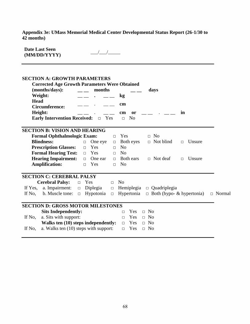
68
Appendix 3e: UMass Memorial Medical Center Developmental Status Report (26-1/30 to
42 months)
Date Last Seen
(MM/DD/YYYY) ___/___/_____
SECTION A: GROWTH PARAMETERS
Corrected Age Growth Parameters Were Obtained
(months/days): __ __ months __ __ days
Weight: __ __ . __ __ kg
Head
Circumference: __ __ . __ __ cm
Height: __ __ . __ __ cm or __ __ . __ __ in
Early Intervention Received: □ Yes □ No
SECTION B: VISION AND HEARING
Formal Ophthalmologic Exam: □ Yes □ No
Blindness: □ One eye □ Both eyes □ Not blind □ Unsure
Prescription Glasses: □ Yes □ No
Formal Hearing Test: □ Yes □ No
Hearing Impairment: □ One ear □ Both ears □ Not deaf □ Unsure
Amplification: □ Yes □ No
SECTION C: CEREBRAL PALSY
Cerebral Palsy: □ Yes □ No
If Yes, a. Impairment: □ Diplegia □ Hemiplegia □ Quadriplegia
If No, b. Muscle tone: □ Hypotonia □ Hypertonia □ Both (hypo- & hypertonia) □ Normal
SECTION D: GROSS MOTOR MILESTONES Sits Independently: □ Yes □ No
If No, a. Sits with support: □ Yes □ No
Walks ten (10) steps independently: □ Yes □ No
If No, a. Walks ten (10) steps with support: □ Yes □ No

69
SECTION E: DEVELOPMENTAL TESTING
Bayley Scales of Infant Development: □ Completed □ Partly Completed □ Not Performed
If completed or partially completed:
a. Corrected age used in scoring: __ __ months __ __ days
b. Results: Check and complete all sections that apply:
□ BSID-II
MDI: Raw Score: __ __ __ Index Score for Corrected Age: __ __ __
□ BSID-II
PDI: Raw Score: __ __ __ Index Score for Corrected Age: __ __ __
□ BSID-III
Cognitive:
Scaled
Score: __ __ __ Composite Score: __ __ __
□ BSID-III
Language:
Sum Scaled
Score: __ __ __ Composite Score: __ __ __
□ BSID-III
Motor:
Sum Scaled
Score: __ __ __ Composite Score: __ __ __
Mullen:
Domain
Age
Equivalent Descriptive Category
Gains from
last visit
(months) (months)
GM __ __ □ very
low □
below
average □ average □
above
average □
very
high __ __
VR __ __ □ very
low □
below
average □ average
□ above
average □
very
high __ __
FM __ __ □ very
low □
below
average □ average
□ above
average □
very
high __ __
RL __ __ □ very
low □
below
average □ average
□ above
average □
very
high __ __
EL __ __ □ very
low □
below
average □ average
□ above
average □
very
high __ __
IF partly completed or not performed check why:
Check why: □ Neurosensory impairment (blind or deaf) □ Too severely delayed
□ Uncooperative □ Other reason: ___________________________
Other Developmental Test Performed: □ Yes □ No
If Yes, Abnormal results: □ Yes □ No
SECTION F: OVERALL CLINICAL APPRAISAL
Clinical Appraisal: Cognitive Function: □ Normal □ Suspect □ Impaired
Language: □ Normal □ Suspect □ Impaired
Motor Function: □ Normal □ Suspect □ Impaired
ADOS: □ No module □ 1 □ 3
□ ASC □ 2 □ 4
□ AC

70
Appendix 3f: UMass Memorial Medical Center Health Status Report (26-1/30 months and
older)
Date Last Seen
(MM/DD/YYYY) ___/___/_____
Birth Weight (g): □ <1000 □ 1000-1250 □ 1251-2000 □ 2001-2500 □ >2500
Gestational Age (weeks): □ <28 □ 28 – 38 □ 30 1/7-34
□ 34 1/7 -36 6/7 □ 37 – 42 □ >42
SECTION A: HEALTH STATUS
Corrected Age at the Follow-Up Visit:
(months/days)
__ __ months __ __ days
SECTION B: LIVING SITUATION
Maternal Age at Infant Birth: __ __ Years □ Unknown
Home Child Resides: □ Parent/Family Member □ Foster Care □ Chronic care
facility
Caregiver(s):
(Check only one) □ Single Parent □
Single parent
extended Family □ Institutional
□ Two Parents □ Tw0 parent extended family
Primary Caregiver
Education: □ Some high school or less □ Some college/university
(Check only one) □ High school degree/GED □ College /university degree
□ Not applicable □ Unknown
Caregiver(s) Primary Language: □ English □ Spanish □ Other: __________
SECTION C: SUPPORT AFTER DISCHARGE
Support After NICU Discharge: □ Yes □ No □ Unsure
If Yes, Check all that apply
□ 1. Tracheostomy □ 4. Gastrostomy
□ 2. Ventilator □ 5. Nasogastric Feeds
□ 3. Oxygen □ 6. Apnea or Cardio Respiratory Monitor

71
SECTION D: MEDICAL HOSPITALIZATION & SURGERIES
Medical re-hospitalization since last seen: □ Yes □ No □ Unsure
If Yes, Category: Check all that
apply
Number of
Admissions
Number of
Admissions
□ 1. Respiratory Illness __ __ □ 5. Infections (not respiratory
of shunt infections):
__ __
□ 2. Nutrition/Failure to Thrive __ __ □ a. Meningitis __ __
□ 3. Seizure Complications __ __ □ b. Urinary Tract Infection __ __
□ 4. Shunt Complication __ __ □ c. Gastrointestinal Infection __ __
□ d. Other Infection __ __
(Specify)_______________________
□ 6. Other Medical Hospitalizations: (Specify)______________________________________
Surgical procedures since last seen: □ Yes □ No □ Unsure
Number of Procedures
(P-Codes) _________________________ __ __
_________________________ __ __
SURGICAL PROCEDURE CODES (P-CODES)
CODE PROCEDURE
Central Nervous System
P-101 Shunt or shunt revision for hydrocephalus
P-102 Other neurosurgical procedure
Congenital Heart Defect Surgery
P-201 Cardiac surgery
Gastrointestinal Surgery
P-301 Gastrostomy tube
P-302 Inguinal hernia repair
P-303 Other gastrointestinal surgical procedure
Genitourinary Surgery
P-401 Circumcision
P-402 Other genitourinary surgical procedure
Otolaryngology
P-501 Tracheostomy
P-502 Tympanostomy tubes
P-503 Other ENT surgical procedures
Ophthalmologic Surgery
P-601 Retinal cryosurgery or laser surgery: single eye
P-602 Retinal cryosurgery or laser surgery: both eye
P-603 Strabismus surgery
P-604 Other ophthalmological surgical procedure
P-900 Other Surgical Procedures

72
Appendix 3g: UMass Memorial Medical Center Developmental Status Report (42.1 months
and older)
Date Last Seen
(MM/DD/YYYY) ___/___/_____
SECTION A: GROWTH PARAMETERS
Corrected Age Growth Parameters Were Obtained
(months/days): __ __ months __ __ days
Weight: __ __ . __ __ kg
Head
Circumference: __ __ . __ __ cm
Height: __ __ . __ __ cm or __ __ . ____ in
Early Intervention Received: □ Yes □ No
SECTION B: VISION AND HEARING
Formal Ophthalmologic Exam: □ Yes □ No
Blindness: □ One eye □ Both eyes □ Not blind □ Unsure
Prescription Glasses: □ Yes □ No
Formal Hearing Test: □ Yes □ No
Hearing Impairment: □ One ear □ Both ears □ Not deaf □ Unsure
Amplification: □ Yes □ No
SECTION C: CEREBRAL PALSY
Cerebral Palsy: □ Yes □ No
If Yes, a. Impairment: □ Diplegia □ Hemiplegia □ Quadriplegia
If No, b. Muscle tone: □ Hypotonia □ Hypertonia □ Both (hypo- & hypertonia) □ Normal
SECTION D: GROSS MOTOR MILESTONES Sits Independently: □ Yes □ No
If No, a. Sits with support: □ Yes □ No
Walks ten (10) steps independently: □ Yes □ No
If No, a. Walks ten (10) steps with support: □ Yes □ No

73
SECTION E: DEVELOPMENTAL TESTING
Mullen:
Domain
Age
Equivalent Descriptive Category
Gains from
last visit
(months) (months)
GM __ __ □ very
low □
below
average □ average □
above
average □
very
high __ __
VR __ __ □ very
low □
below
average □ average
□ above
average □
very
high __ __
FM __ __ □ very
low □
below
average □ average
□ above
average □
very
high __ __
RL __ __ □ very
low □
below
average □ average
□ above
average □
very
high __ __
EL __ __ □ very
low □
below
average □ average
□ above
average □
very
high __ __
ADOS: □ No module □ 1 □ 3
□ ASC □ 2 □ 4
□ AC
KBIT-2: __ __ __ V (verbal)
__ __ __ N (non-verbal)
WRAT: __ __ __ WR (word reading)
__ __ __ SC (sentence
comprehension)
__ __ __ S (spelling)
Einstein: □ A (Acceptable)
□ U (Unacceptable)
IF partly completed or not performed, check why:
□ Neurosensory impairment (blind or deaf) □ Too severely delayed
□ Uncooperative □ Other reason: ___________________________
Other Developmental Test Performed: □ Yes □ No
If Yes, Abnormal results: □ Yes □ No
SECTION F: OVERALL CLINICAL APPRAISAL
Clinical Appraisal: Cognitive Function: □ Normal □ Suspect □ Impaired
Language: □ Normal □ Suspect □ Impaired
Motor Function: □ Normal □ Suspect □ Impaired