Embed Size (px)
Citation preview

INVESTIGACION Revista Mexicana de Fısica58 (2012) 328–334 AGOSTO 2012
Corrosion resistance and biocompatibility of zirconium oxynitridethin film growth by rf sputtering
G.I. CubillosDepartamento de Quımica, Universidad Nacional de Colombia, AA 14490, Bogota- Colombia.
J.J. OlayaFacultad de Ingenierıa, Universidad Nacional de Colombia, AA 14490, Bogota- Colombia.
D. ClavijoFacultad de Medicina, Universidad Nacional de Colombia, AA 14490, Bogota- Colombia.
Facultad de Medicina Fundacion Universitaria Sanitas, Bogota-Colombia.
J.E. AlfonsoDepartamento de Fısica Universidad Nacional de Colombia, AA 14490, Bogota- Colombia.
e-mail: [email protected]
M. BethencourtDepartamento de Ciencia de los Materiales e Ingenierıa Metalurgica y Quımica Inorganica.
Universidad de Cadiz. Centro Andaluz de Ciencia y Tecnologıa Marinas. Avda Republica de Saharaui.Puerto real, E-11510. Cadiz- Spain.
Recibido el 23 de noviembre de 2011; aceptado el 16 de mayo de 2012
Thin films of zirconium oxynitride were grown on common glass, silicon (100) and stainless steel 316 L substrates using the reactive rfmagnetron sputtering technique. The films were analyzed through structural, morphological, and biocompatibility studies. The structuralanalysis was carried out using X-ray diffraction (XRD), and the morphological analysis was carried out using scanning electron microscopy(SEM) and atomic force microscopy (AFM). These studies were done as a function of growth parameters, such as power applied to thetarget, substrate temperature, and flow ratios. The corrosion resistance studies were made on samples of stainless steel 316 L coated anduncoated with ZrxNyO films, through of polarization curves. The studies of biocompatibility were carried out on zirconium oxynitride filmsdeposited on stainless steel 316L through proliferation and cellular adhesion. The XRD analysis shows that films deposited at 623 K, witha flow ratioΦN2/ΦO2 of 1.25 and a total deposit time of 30 minutes grew preferentially oriented along the (111) plane of the zirconiumoxyitride monoclinic phase. The SEM analyses showed that the films grew homogenously, and the AFM studies indicated that the averagerugosity of the film was 5.9 nm and the average particle size was 150 nm. The analysis of the corrosion resistant, shows that the stainlesssteel coated with the film was increased a factor 10. Finally; through the analysis of the biocompatibility we established that the films have abetter surface than the substrate (stainless steel 316 L) in terms of the adhesion and proliferation of bone cells.
Keywords:Ziconium oxynitride; thin films; bone cells; biocompatibility.
PACS: 81.15.Cd; -87.68.+z; -68.43.-h
1. Introduction
Biomaterials are important in the development of biomedi-cal devices and implants. The surface of the biomaterial isthe first thing to contact the living tissue when the materialis placed in the body. Therefore, the initial response of theliving tissue to the biomaterial depends on the surface prop-erties. Currently, it is rare that a biomaterial with good bulkproperties also possess the surface characteristics suitable forsome clinical applications. For that reason, over the last fewyears, the development of surface modification techniquesfor biomaterials has been expanding rapidly. In this way, itis possible to make ideal biomaterials with surface attributesthat are decoupled from the bulk properties. For instance, byaltering the surface functionality through the deposition of athin film, the optimal surface, chemical, and physical proper-ties can be attained.
For the foregoing reasons, thin films of metal such as Tiand alloys (Ti6Al4V and AISI 316 L steel) have been used in
biomedical applications [1-3]. In these biomedical applica-tions, during the last few decades Ti and its oxides have beenthe materials most widely used, because they have a high an-ticorrosive resistance within the aggressive environment ofthe body; however, nowadays research is being carried outthat seeks to increase the lifespan of the prosthesis throughprocesses of proliferation and adhesion of bone cells on thinfilms of, for example, niobium, zirconium and tantalum andits oxides [3].
Taking advantage of the fact that the surface characteris-tics are relevant for the interaction between the implant andthe living tissue [4–5], we proposed to make use of thin filmtechnology to modify any base material with a biocompati-ble coating. The coating material would be ceramic, such aszirconium oxynitride (ZrOxNy), a material that can be eas-ily and inexpensively produced through thin film depositiontechniques.
The growth of oxynitride films is strongly influenced bythe amount of oxygen molecules and nitrogen present in the
CORROSION RESISTANCE AND BIOCOMPATIBILITY OF ZIRCONIUM OXYNITRIDE THIN FILM GROWTH BY RF SPUTTERING 329
deposition chamber. This influence has been studied by Mar-tin et al. [6], who deposited TiNxOy using the pulsing gastechnique and found that the chemical composition, struc-tural evolution, and electrical response varies as a functionthe values ofx andy. The techniques most commonly usedto prepare oxynitride films are reactive rf, dc magnetron sput-tering, and cathodic reactive arc evaporation [7].
The purpose of the present study is to evaluate the bio-compatibility of zirconium oxynitride thin films deposited onstainless steel, using reactive rf magnetron sputtering throughthe analysis of the proliferation and adhesion of bone cells.
2. Experimental techniques
2.1. Growth of the ZrNxOy films
The equipment used to grow the ZrNxOy films was anAlcatel HS 2000 described in previous papers [8]. TheZrNxOy films were obtained from a 4”×1/4” Zr (99.9%)target (CERAC, Inc.). The parameter set used during de-position process was: base pressure (2.0×10⁻³ Pa), totalworking pressure (7.4×10⁻¹Pa), and deposition time (halfhour), target–substrate distance (5 cm), argon (99.999%) flow(20 sccm). We studied the influence of several depositionparameters such as: power supplied to the target (from 200to 350 W), substrate temperature (which varied from 287 to623 K), and flow ratios (1.0, 1.25 and 1.50) of N2 (99.99 %)and O2 (99.99 %) in same ratios inside the deposit chamber.The final working pressure was maintained using a valve con-troller for all the ratio flow values given above. The temper-ature of the substrate was measured with thermocouple typeK, and the argon, nitrogen and oxygen flows were controlledwith mass flow controllers. The structural characterization ofthe films was performed through XRD with a Philips diffrac-tometer operated at 30 kV and 20 mA, working in Bragg-Brentano configuration and using Cu Kα radiation. The aver-age crystalline size was calculated from s=0.9 k/Bcosθ whereB = Bm – Bi, Bm being the broadening of the diffraction line,measured full width at half maximum (FWHM) and Bi the in-strument peak width[9]. Surface morphology was character-ized by imaging the secondary electrons with a Quanta 2000scanning electron microscope operating at 15 kV and 10 mAand non-contact AFM Autoprobe cp Park Scientific instru-ment with study area of 25µm2 and frequency of 10 Hz.
2.2. Corrosion Tests
The corrosion resistance of zirconium oxynitride coatingsand stainless steel 316 L was evaluated through means ofelectrochemical techniques of potentiodynamic polarizationand polarization resistance in a solution of NaCl at 3.5%(pH=6.5). Measurements were made in a K 235 flat cell ofParc EG&G using a Solartron SI 1287 potentiostat the ref-erence electrode used was a Ag/AgCl electrode of Crison0.207 mV/SHE with exposed area of 1.00 cm2.
The activity of the samples of stainless steel 316 L andzirconium oxynitride coatings in the solution of NaCl was an-alyzed from the value of the polarization resistance, Rp. Thedegree of protection was evaluated by comparing the value ofthis resistance with that corresponding to a bare sample.
In the corrosion test, the polarization in the anodic regionwas evaluated in order to determine the corrosion resistanceof the ZrNxOy film, and the polarization resistance (Rp) wascalculated through the Simonds and Larson method [16-17],where Rp is the slope of the obtained curve of the graphicvoltage vs density current; the equation that allows calculat-ing Rp is that of Stern-Geary [18-19].
Rp =B
icorr=
BnF
νcorr(1)
where B is the proportionality constant between polarizationresistance and the current density, icorr is the current corro-sion, n is the number of electrons transferred during the cor-rosion process, F is Faraday constant, and vcorr is the potentialcorrosion.
2.3. Biological Tests
Bone lineage cells from nursing mice were suspended in a 1:1solution of DMEM (Dulbecco’s Modified Eagle’s Medium)supplemented with fetal bovine serum 10%, 100 U/mLpenicillin, streptomycin 100 mg/mL, 0.25 mg/mL ampho-tericin B, 10 mM beta-glycerophosphate, L-ascorbic acid-2-phosphate 100 uM, and dexamethasone 10 nM. This wascentrifuged at 1200 rpm for 10 minutes, and the precipitatewas suspended in 1 mL of the medium described. The solu-tion was transferred to a well of 35 mm2 with an additional1 mL of medium and transferred to the incubator at 310 Kand an atmosphere of 5% CO2 in order to allow cell adhe-sion. After 48 hours, there was 100% confluence, and thefirst cell passage was made. Stainless steel samples with andwithout coating were sterilized by autoclaving for 30 minutesat 423 K and placed in plates of six wells, and 10,000 cellswere added, obtained from the first passage, suspended in themedium described. These samples were incubated at 310 Kand an atmosphere of 5% CO2. When cell confluence wasobtained, samples were extracted from the culture mediumand washed three times with PBS (Phosphate Buffer Solu-tion) at 310 K. The PBS solution was discarded, and the sam-ples were immersed in 2 mL of 2.6-diamidine-2-phenylindole(DAPI) (SIGMA D9542) at a concentration of 1 mg/mL at277 K. After 12 hours, the samples were removed and washedthree times with PBS at 277 K in order to be deposited onglass slides.
DAPI-stained cells on coated and uncoated stainlesssteel were observed using fluorescence confocal microscopy(NIKON C1-Plus) and were captured in photographs. Forimage analysis, a series of algorithms was used, which weremerged into the program Matlab (image toolbox) in order totransform the original color images to grayscale and apply al-gorithms and image processing (segmentation, threshold andfiltering) in order to reduce the noise.
Rev. Mex. Fis.58 (2012) 328–334
330 G.I. CUBILLOS, J.J. OLAYA, D. CLAVIJO, J.E. ALFONSO, AND M. BETHENCOURT
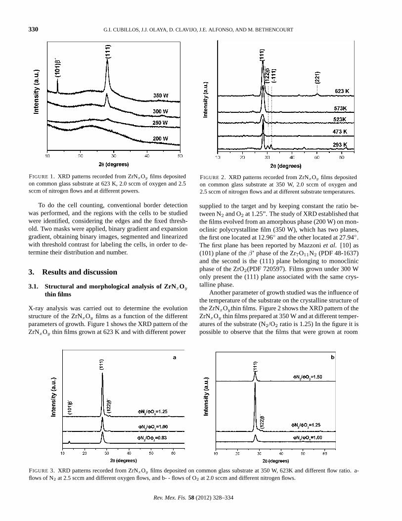
FIGURE 1. XRD patterns recorded from ZrNxOy films depositedon common glass substrate at 623 K, 2.0 sccm of oxygen and 2.5sccm of nitrogen flows and at different powers.
To do the cell counting, conventional border detectionwas performed, and the regions with the cells to be studiedwere identified, considering the edges and the fixed thresh-old. Two masks were applied, binary gradient and expansiongradient, obtaining binary images, segmented and linearizedwith threshold contrast for labeling the cells, in order to de-termine their distribution and number.
3. Results and discussion
3.1. Structural and morphological analysis of ZrNxOy
thin films
X-ray analysis was carried out to determine the evolutionstructure of the ZrNxOy films as a function of the differentparameters of growth. Figure 1 shows the XRD pattern of theZrNxOy thin films grown at 623 K and with different power
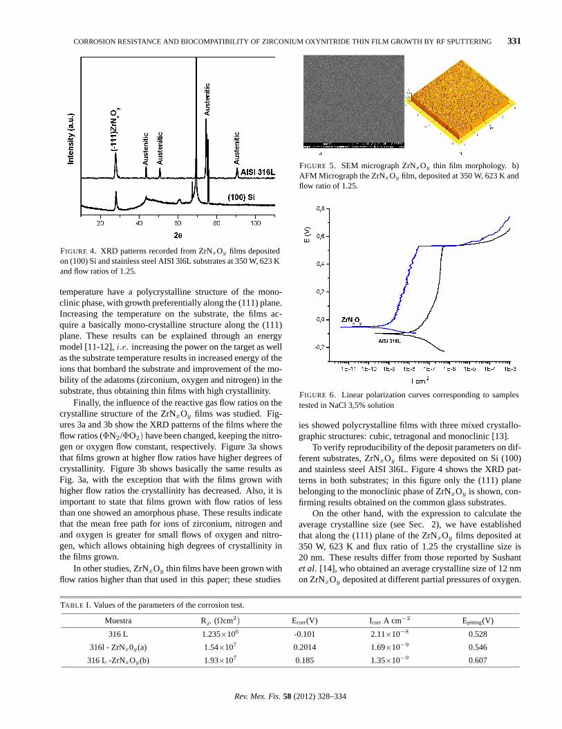
FIGURE 2. XRD patterns recorded from ZrNxOy films depositedon common glass substrate at 350 W, 2.0 sccm of oxygen and2.5 sccm of nitrogen flows and at different substrate temperatures.
supplied to the target and by keeping constant the ratio be-tween N2 and O2 at 1.25”. The study of XRD established thatthe films evolved from an amorphous phase (200 W) on mon-oclinic polycrystalline film (350 W), which has two planes,the first one located at 12.96 and the other located at 27.94.The first plane has been reported by Mazzoniet al. [10] as(101) plane of theβ’ phase of the Zr7O11N2 (PDF 48-1637)and the second is the (111) plane belonging to monoclinicphase of the ZrO2(PDF 720597). Films grown under 300 Wonly present the (111) plane associated with the same crys-talline phase.
Another parameter of growth studied was the influence ofthe temperature of the substrate on the crystalline structure ofthe ZrNxOythin films. Figure 2 shows the XRD pattern of theZrNxOy thin films prepared at 350 W and at different temper-atures of the substrate (N2/O2 ratio is 1.25) In the figure it ispossible to observe that the films that were grown at room
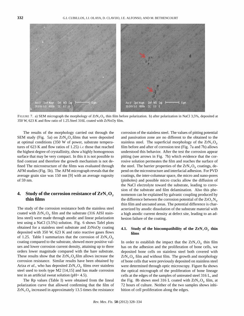
FIGURE 3. XRD patterns recorded from ZrNxOy films deposited on common glass substrate at 350 W, 623K and different flow ratio. a-flows of N2 at 2.5 sccm and different oxygen flows, and b- - flows of O2 at 2.0 sccm and different nitrogen flows.
Rev. Mex. Fis.58 (2012) 328–334
CORROSION RESISTANCE AND BIOCOMPATIBILITY OF ZIRCONIUM OXYNITRIDE THIN FILM GROWTH BY RF SPUTTERING 331
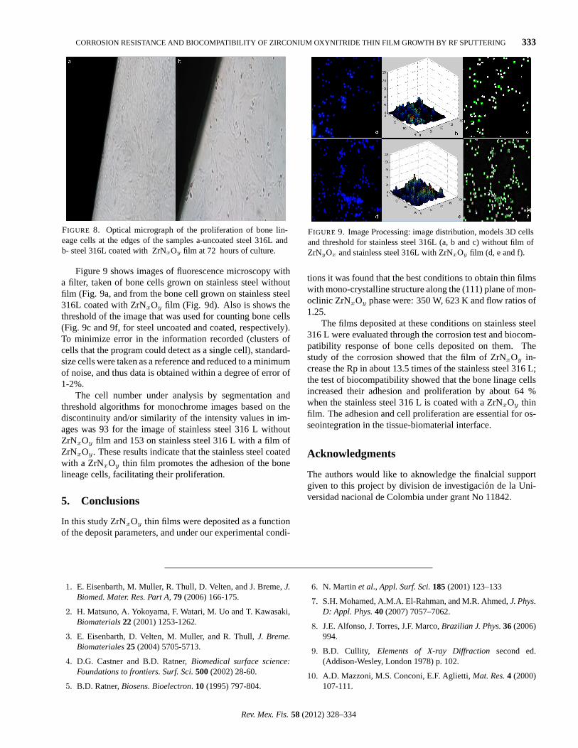
FIGURE 4. XRD patterns recorded from ZrNxOy films depositedon (100) Si and stainless steel AISI 3l6L substrates at 350 W, 623 Kand flow ratios of 1.25.
temperature have a polycrystalline structure of the mono-clinic phase, with growth preferentially along the (111) plane.Increasing the temperature on the substrate, the films ac-quire a basically mono-crystalline structure along the (111)plane. These results can be explained through an energymodel [11-12],i.e. increasing the power on the target as wellas the substrate temperature results in increased energy of theions that bombard the substrate and improvement of the mo-bility of the adatoms (zirconium, oxygen and nitrogen) in thesubstrate, thus obtaining thin films with high crystallinity.
Finally, the influence of the reactive gas flow ratios on thecrystalline structure of the ZrNxOy films was studied. Fig-ures 3a and 3b show the XRD patterns of the films where theflow ratios (ΦN2/ΦO2) have been changed, keeping the nitro-gen or oxygen flow constant, respectively. Figure 3a showsthat films grown at higher flow ratios have higher degrees ofcrystallinity. Figure 3b shows basically the same results asFig. 3a, with the exception that with the films grown withhigher flow ratios the crystallinity has decreased. Also, it isimportant to state that films grown with flow ratios of lessthan one showed an amorphous phase. These results indicatethat the mean free path for ions of zirconium, nitrogen andand oxygen is greater for small flows of oxygen and nitro-gen, which allows obtaining high degrees of crystallinity inthe films grown.
In other studies, ZrNxOy thin films have been grown withflow ratios higher than that used in this paper; these studies
FIGURE 5. SEM micrograph ZrNxOy thin film morphology. b)AFM Micrograph the ZrNxOy film, deposited at 350 W, 623 K andflow ratio of 1.25.
FIGURE 6. Linear polarization curves corresponding to samplestested in NaCl 3,5% solution
ies showed polycrystalline films with three mixed crystallo-graphic structures: cubic, tetragonal and monoclinic [13].
To verify reproducibility of the deposit parameters on dif-ferent substrates, ZrNxOy films were deposited on Si (100)and stainless steel AISI 3l6L. Figure 4 shows the XRD pat-terns in both substrates; in this figure only the (111) planebelonging to the monoclinic phase of ZrNxOy is shown, con-firming results obtained on the common glass substrates.
On the other hand, with the expression to calculate theaverage crystalline size (see Sec. 2), we have establishedthat along the (111) plane of the ZrNxOy films deposited at350 W, 623 K and flux ratio of 1.25 the crystalline size is20 nm. These results differ from those reported by Sushantet al. [14], who obtained an average crystalline size of 12 nmon ZrNxOy deposited at different partial pressures of oxygen.
TABLE I. Values of the parameters of the corrosion test.
Muestra RP (Ωcm2) Ecorr(V) I corr A cm−2 Epitting(V)
316 L 1.235×10⁶ -0.101 2.11×10⁻⁸ 0.528
316l - ZrNx0y(a) 1.54×10⁷ 0.2014 1.69×10⁻⁹ 0.546
316 L -ZrNxOy(b) 1.93×10⁷ 0.185 1.35×10⁻⁹ 0.607
Rev. Mex. Fis.58 (2012) 328–334
332 G.I. CUBILLOS, J.J. OLAYA, D. CLAVIJO, J.E. ALFONSO, AND M. BETHENCOURT
FIGURE 7. a) SEM micrograph the morphology of ZrNxOy thin film before polarization. b) after polarization in NaCl 3,5%, deposited at350 W, 623 K and flow ratio of 1.25.Steel 316L coated with ZrNxOy film.
The results of the morphology carried out through theSEM study (Fig. 5a) on ZrNyOxfilms that were depositedat optimal conditions (350 W of power, substrate tempera-tures of 623 K and flow ratios of 1.25)i.e those that reachedthe highest degree of crystallinity, show a highly homogenoussurface that may be very compact. In this it is not possible tofind contrast and therefore the growth mechanism is not de-fined The microstructure of the films was evaluated throughAFM studies (Fig. 5b). The AFM micrograph reveals that theaverage grain size was 150 nm [9] with an average rugosityof 59 nm.
4. Study of the corrosion resistance of ZrNxOy
thin films
The study of the corrosion resistance both the stainless steelcoated with ZrNxOy film and the substrate (316 AISI stain-less steel) were made through anodic and linear polarizationtest using a NaCl (3.5%) solution. Fig. 6 shows Tafel plotsobtained for a stainless steel substrate and ZrNxOy coatingdeposited with 350 W, 623 K and ratio reactive gases flowsof 1.25. Table I summarizes that the corrosion of ZrNxOy
coating compared to the substrate, showed more positive val-ues and lower corrosion current density, attaining up to threeorders lower magnitude compared with the bare substrate.These results show that the ZrNxOyfilm allows increase thecorrosion resistance. Similar results have been obtained byAriza et al., who has deposited ZrNxOy films over stainlesssteel used to tools type M2 [14,15] and has made corrosiontest in an artificial sweat solution (pH= 4.5).
The Rp values (Table I) were obtained from the linealpolarization curve that allowed confirming that the film ofZrNxOy increased in approximately 13.5 times the resistance
corrosion of the stainless steel. The values of pitting potentialand passivation zone are no different to the obtained to thestainless steel. The superficial morphology of the ZrNxOy
film before and after of corrosion test (Fig. 7a and 7b) allowsunderstood this behavior. After the test the corrosion appearpitting (see arrows in Fig. 7b) which evidence that the cor-rosive solution permeates the film and reaches the surface ofthe steel. The barrier properties of the ZrNxOy coatings, de-pend on the microstructure and interfacial adhesion. For PVDcoatings, the inter-columnar space, the micro and nano-pores(pinholes) and possible micro cracks allow the diffusion ofthe NaCl electrolyte toward the substrate, leading to corro-sion of the substrate and film delamination. Also this phe-nomenon can be explained by galvanic coupling produced bythe difference between the corrosion potential of the ZrOxNy
thin film and uncoated areas. The potential difference is char-acterized by anodic dissolution of the substrate material witha high anodic current density at defect site, leading to an ad-hesion failure of the coating.
4.1. Study of the biocompatibility of the ZrNxOy thinfilms
In order to establish the impact that the ZrNxOy thin filmhas on the adhesion and the proliferation of bone cells, wedeposited bone cells on stainless steel both covered withZrNxOy film and without film. The growth and morphologyof bone cells that were previously deposited on stainless steelwere determined through optic microscopy. Figure 8a showsthe optical micrograph of the proliferation of bone lineagecells at the edges of the samples of untreated steel 316 L, andthe Fig. 8b shows steel 316 L coated with ZrNxOy film, at72 hours of culture. Neither of the two samples shows inhi-bition of cell proliferation along the edges.
Rev. Mex. Fis.58 (2012) 328–334
CORROSION RESISTANCE AND BIOCOMPATIBILITY OF ZIRCONIUM OXYNITRIDE THIN FILM GROWTH BY RF SPUTTERING 333
FIGURE 8. Optical micrograph of the proliferation of bone lin-eage cells at the edges of the samples a-uncoated steel 316L andb- steel 316L coated with ZrNxOy film at 72 hours of culture.
Figure 9 shows images of fluorescence microscopy witha filter, taken of bone cells grown on stainless steel withoutfilm (Fig. 9a, and from the bone cell grown on stainless steel316L coated with ZrNxOy film (Fig. 9d). Also is shows thethreshold of the image that was used for counting bone cells(Fig. 9c and 9f, for steel uncoated and coated, respectively).To minimize error in the information recorded (clusters ofcells that the program could detect as a single cell), standard-size cells were taken as a reference and reduced to a minimumof noise, and thus data is obtained within a degree of error of1-2%.
The cell number under analysis by segmentation andthreshold algorithms for monochrome images based on thediscontinuity and/or similarity of the intensity values in im-ages was 93 for the image of stainless steel 316 L withoutZrNxOy film and 153 on stainless steel 316 L with a film ofZrNxOy. These results indicate that the stainless steel coatedwith a ZrNxOy thin film promotes the adhesion of the bonelineage cells, facilitating their proliferation.
5. Conclusions
In this study ZrNxOy thin films were deposited as a functionof the deposit parameters, and under our experimental condi-
FIGURE 9. Image Processing: image distribution, models 3D cellsand threshold for stainless steel 316L (a, b and c) without film ofZrNyOx and stainless steel 316L with ZrNxOy film (d, e and f).
tions it was found that the best conditions to obtain thin filmswith mono-crystalline structure along the (111) plane of mon-oclinic ZrNxOy phase were: 350 W, 623 K and flow ratios of1.25.
The films deposited at these conditions on stainless steel316 L were evaluated through the corrosion test and biocom-patibility response of bone cells deposited on them. Thestudy of the corrosion showed that the film of ZrNxOy in-crease the Rp in about 13.5 times of the stainless steel 316 L;the test of biocompatibility showed that the bone linage cellsincreased their adhesion and proliferation by about 64 %when the stainless steel 316 L is coated with a ZrNxOy thinfilm. The adhesion and cell proliferation are essential for os-seointegration in the tissue-biomaterial interface.
Acknowledgments
The authors would like to aknowledge the finalcial supportgiven to this project by division de investigacion de la Uni-versidad nacional de Colombia under grant No 11842.
1. E. Eisenbarth, M. Muller, R. Thull, D. Velten, and J. Breme,J.Biomed. Mater. Res. Part A,79 (2006) 166-175.
2. H. Matsuno, A. Yokoyama, F. Watari, M. Uo and T. Kawasaki,Biomaterials22 (2001) 1253-1262.
3. E. Eisenbarth, D. Velten, M. Muller, and R. Thull,J. Breme.Biomateriales25 (2004) 5705-5713.
4. D.G. Castner and B.D. Ratner,Biomedical surface science:Foundations to frontiers. Surf. Sci.500(2002) 28-60.
5. B.D. Ratner,Biosens. Bioelectron. 10 (1995) 797-804.
6. N. Martin et al., Appl. Surf. Sci.185(2001) 123–133
7. S.H. Mohamed, A.M.A. El-Rahman, and M.R. Ahmed,J. Phys.D: Appl. Phys.40 (2007) 7057–7062.
8. J.E. Alfonso, J. Torres, J.F. Marco,Brazilian J. Phys.36 (2006)994.
9. B.D. Cullity, Elements of X-ray Diffractionsecond ed.(Addison-Wesley, London 1978) p. 102.
10. A.D. Mazzoni, M.S. Conconi, E.F. Aglietti,Mat. Res.4 (2000)107-111.
Rev. Mex. Fis.58 (2012) 328–334

334 G.I. CUBILLOS, J.J. OLAYA, D. CLAVIJO, J.E. ALFONSO, AND M. BETHENCOURT
11. B.A. Movchan and A.V. Demchishin,Fiz. Met. Metalloved.28(1969) 83.
12. R. Messier, A.P. Giri, R.A. Roy,J. Vac. Sci. and Tech. A.2(2,Pt. 1), 500-3.
13. M. Laurikaitis, S. Burinskas, J. Dudonis, and D. Mileius,J. ofPhys.: Conference Series 100(2008) 08205.
14. K. Rawal Sushant, A. Kumar Chawla, V. Chawla, R. Jayagan-than, and R. Chandra,Mat. Sci. and Eng. B172 (2010) 259–266.
15. S.C. Ferreiraet al., Surface & Coatings Technology200(2006)6634–6639.
16. E. Arizaet al., Thin Solid Films469–470(2004) 274–281.
17. E.J. Simmons,Corrosion11 (1955) 255.
18. R.V. Skold and T.E. Larson,Corrosion13 (1964) 139.
19. M. Stern,Corrosion14 (1958) 329.
20. M. Stern,Corrosion14 (1958) 440.
Rev. Mex. Fis.58 (2012) 328–334


![Anodic dissolution behavior of zirconium in Bu NBr ... · acids and strong alkalis [21]. Thus, zirconium corrosion in non-aqueous solutions would be different from titanium and deserve](https://img.pdfslide.net/doc/110x75/5e51370660b2355520448e26/anodic-dissolution-behavior-of-zirconium-in-bu-nbr-acids-and-strong-alkalis.jpg)


![Effects of Occupational Environmental Controls on the ... · biocompatibility, high strength, adequate tarnish and . corrosion resistance [1], [3], [4]. Despite high value of the](https://img.pdfslide.net/doc/110x75/5f8987e7308caf3191710e76/effects-of-occupational-environmental-controls-on-the-biocompatibility-high.jpg)