Embed Size (px)
Citation preview
VOL. 103 NO. 6 I JUNE 2019 357WWW.MDEDGE.COM/DERMATOLOGY
CASE REPORT
Erythema gyratum repens (EGR) is a rare and poorly understood derma-tosis. The relationship of superficial dermatophytic infection to EGR-like eruptions in mycosis fungoides (MF) is unclear. We present a case of an EGR-like eruption in a patient with Sézary syndrome (SS). Histopatho-logic examination revealed both a superficial dermatophyte (Trichophyton rubrum) and cutaneous T-cell lymphoma (CTCL) in biopsies of the skin, regardless of whether those biopsies showed EGR-like lesions or erythroderma clinically. On 2 occasions, treatment of the superficial dermatophytic infection led to resolution of the EGR-like eruption and associated pruritus but not to resolution of the erythroderma. This case supports a role for dermatophytic superinfection in an EGR-like eruption in SS. Further investigation is necessary to fully understand the impact of dermatophytic infection in this clinical setting.
Cutis. 2019;103:357-360.
Case ReportA 65-year-old woman presented with stage IVA2 mycosis fungoides (MF)(T4N3M0B2)/Sézary syndrome (SS). A peripheral blood count contained 6000 Sézary cells with
cerebriform nuclei, a CD2+/−CD3+CD4+CD5+/−CD7+CD8−
CD26− immunophenotype, and a highly abnormal CD4 to CD8 ratio (70:1). Positron emission tomography and com-puted tomography demonstrated hypermetabolic subcu-taneous nodules in the base of the neck and generalized lymphadenopathy. Lymph node biopsy showed involve-ment by T-cell lymphoma and dominant T-cell receptor γ clonality by polymerase chain reaction.
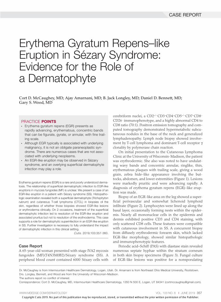
On initial presentation to the Cutaneous Lymphoma Clinic at the University of Wisconsin-Madison, the patient was erythrodermic. She also was noted to have undulat-ing wavy bands and concentric annular, ringlike, thin, erythematous plaques with trailing scale, giving a wood grain, zebra hide–like appearance involving the but-tocks, abdomen, and lower extremities (Figure 1). Lesions were markedly pruritic and were advancing rapidly. A diagnosis of erythema gyratum repens (EGR)–like erup-tion was made.
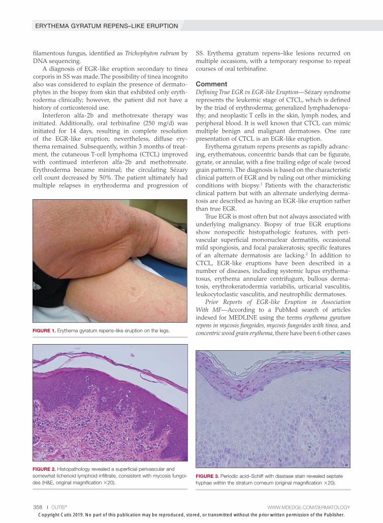
Biopsy of an EGR-like area on the leg showed a super-ficial perivascular and somewhat lichenoid lymphoid infiltrate (Figure 2). Lymphocytes were lined up along the basal layer, occasionally forming nests within the epider-mis. Nearly all mononuclear cells in the epidermis and dermis exhibited positive CD3 and CD4 staining, with only scattered CD8 cells. These features were compatible with cutaneous involvement in SS. A concurrent biopsy from diffusely erythrodermic forearm skin, which lacked EGR-like morphology, showed similar histopathologic and immunophenotypic features.
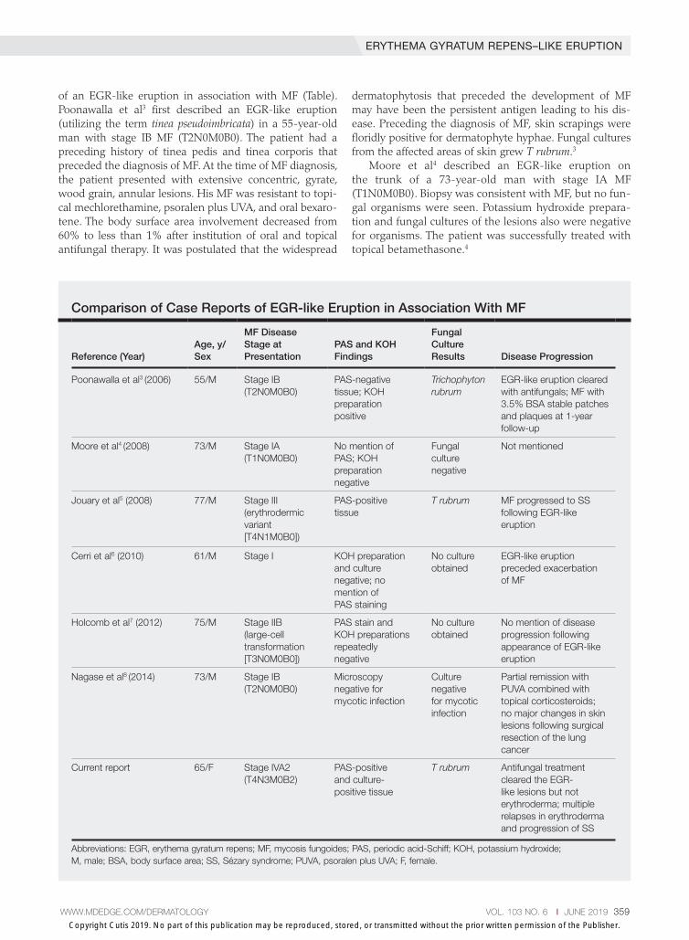
Periodic acid–Schiff (PAS) with diastase stain revealed numerous septate hyphae within the stratum corneum in both skin biopsy specimens (Figure 3). Fungal culture of EGR-like lesions was positive for a nonsporulating
Erythema Gyratum Repens–like Eruption in Sézary Syndrome: Evidence for the Role of a Dermatophyte
Cort D. McCaughey, MD; Ajay Amarnani, MD; B. Jack Longley, MD; Daniel D. Bennett, MD; Gary S. Wood, MD
Dr. McCaughey is from Intermountain Healthcare Dermatology, Logan, Utah. Dr. Amarnani is from Northeast Ohio Medical University, Rootstown. Drs. Longley, Bennett, and Wood are from the University of Wisconsin-Madison. The authors report no conflict of interest. Correspondence: Cort D. McCaughey, MD, Intermountain Healthcare Dermatology, 1350 N 500 E, Logan, UT 84341 ([email protected]).
PRACTICE POINTS• Erythema gyratum repens (EGR) presents as
rapidly advancing, erythematous, concentric bands that can be figurate, gyrate, or annular, with fine trail-ing scale.
• Although EGR typically is associated with underlying malignancy, it is not an obligate paraneoplastic syn-drome. There are numerous cases that are not asso-ciated with underlying neoplasms.
• An EGR-like eruption may be observed in Sézary syndrome, and an overlying superficial dermatophyte infection may play a role.
Copyright Cutis 2019. No part of this publication may be reproduced, stored, or transmitted without the prior written permission of the Publisher.
CUTIS
Do
not c
opy
ERYTHEMA GYRATUM REPENS–LIKE ERUPTION
358 I CUTIS® WWW.MDEDGE.COM/DERMATOLOGY
filamentous fungus, identified as Trichophyton rubrum by DNA sequencing.
A diagnosis of EGR-like eruption secondary to tinea corporis in SS was made. The possibility of tinea incognito also was considered to explain the presence of dermato-phytes in the biopsy from skin that exhibited only eryth-roderma clinically; however, the patient did not have a history of corticosteroid use.
Interferon alfa-2b and methotrexate therapy was initiated. Additionally, oral terbinafine (250 mg/d) was initiated for 14 days, resulting in complete resolution of the EGR-like eruption; nevertheless, diffuse ery-thema remained. Subsequently, within 3 months of treat-ment, the cutaneous T-cell lymphoma (CTCL) improved with continued interferon alfa-2b and methotrexate. Erythroderma became minimal; the circulating Sézary cell count decreased by 50%. The patient ultimately had multiple relapses in erythroderma and progression of
SS. Erythema gyratum repens–like lesions recurred on multiple occasions, with a temporary response to repeat courses of oral terbinafine.
CommentDefining True EGR vs EGR-like Eruption—Sézary syndrome represents the leukemic stage of CTCL, which is defined by the triad of erythroderma; generalized lymphadenopa-thy; and neoplastic T cells in the skin, lymph nodes, and peripheral blood. It is well known that CTCL can mimic multiple benign and malignant dermatoses. One rare presentation of CTCL is an EGR-like eruption.
Erythema gyratum repens presents as rapidly advanc-ing, erythematous, concentric bands that can be figurate, gyrate, or annular, with a fine trailing edge of scale (wood grain pattern). The diagnosis is based on the characteristic clinical pattern of EGR and by ruling out other mimicking conditions with biopsy.1 Patients with the characteristic clinical pattern but with an alternate underlying derma-tosis are described as having an EGR-like eruption rather than true EGR.
True EGR is most often but not always associated with underlying malignancy. Biopsy of true EGR eruptions show nonspecific histopathologic features, with peri-vascular superficial mononuclear dermatitis, occasional mild spongiosis, and focal parakeratosis; specific features of an alternate dermatosis are lacking.2 In addition to CTCL, EGR-like eruptions have been described in a number of diseases, including systemic lupus erythema-tosus, erythema annulare centrifugum, bullous derma-tosis, erythrokeratodermia variabilis, urticarial vasculitis, leukocytoclastic vasculitis, and neutrophilic dermatoses.
Prior Reports of EGR-like Eruption in Association With MF—According to a PubMed search of articles indexed for MEDLINE using the terms erythema gyratum repens in mycosis fungoides, mycosis fungoides with tinea, and concentric wood grain erythema, there have been 6 other cases FIGURE 1. Erythema gyratum repens–like eruption on the legs.
FIGURE 2. Histopathology revealed a superficial perivascular and somewhat lichenoid lymphoid infiltrate, consistent with mycosis fungoi-des (H&E, original magnification ×20).
FIGURE 3. Periodic acid–Schiff with diastase stain revealed septate hyphae within the stratum corneum (original magnification ×20).
Copyright Cutis 2019. No part of this publication may be reproduced, stored, or transmitted without the prior written permission of the Publisher.
CUTIS
Do
not c
opy
ERYTHEMA GYRATUM REPENS–LIKE ERUPTION
VOL. 103 NO. 6 I JUNE 2019 359WWW.MDEDGE.COM/DERMATOLOGY
of an EGR-like eruption in association with MF (Table). Poonawalla et al3 first described an EGR-like eruption (utilizing the term tinea pseudoimbricata) in a 55-year-old man with stage IB MF (T2N0M0B0). The patient had a preceding history of tinea pedis and tinea corporis that preceded the diagnosis of MF. At the time of MF diagnosis, the patient presented with extensive concentric, gyrate, wood grain, annular lesions. His MF was resistant to topi-cal mechlorethamine, psoralen plus UVA, and oral bexaro-tene. The body surface area involvement decreased from 60% to less than 1% after institution of oral and topical antifungal therapy. It was postulated that the widespread
dermatophytosis that preceded the development of MF may have been the persistent antigen leading to his dis-ease. Preceding the diagnosis of MF, skin scrapings were floridly positive for dermatophyte hyphae. Fungal cultures from the affected areas of skin grew T rubrum.3
Moore et al4 described an EGR-like eruption on the trunk of a 73-year-old man with stage IA MF (T1N0M0B0). Biopsy was consistent with MF, but no fun-gal organisms were seen. Potassium hydroxide prepara-tion and fungal cultures of the lesions also were negative for organisms. The patient was successfully treated with topical betamethasone.4
Comparison of Case Reports of EGR-like Eruption in Association With MF
Reference (Year)Age, y/Sex
MF Disease Stage at Presentation
PAS and KOH Findings
Fungal Culture Results Disease Progression
Poonawalla et al3 (2006) 55/M Stage IB (T2N0M0B0)
PAS-negative tissue; KOH preparation positive
Trichophyton rubrum
EGR-like eruption cleared with antifungals; MF with 3.5% BSA stable patches and plaques at 1-year follow-up
Moore et al4 (2008) 73/M Stage IA (T1N0M0B0)
No mention of PAS; KOH preparation negative
Fungal culture negative
Not mentioned
Jouary et al5 (2008) 77/M Stage III (erythrodermic variant [T4N1M0B0])
PAS-positive tissue
T rubrum MF progressed to SS following EGR-like eruption
Cerri et al6 (2010) 61/M Stage I KOH preparation and culture negative; no mention of PAS staining
No culture obtained
EGR-like eruption preceded exacerbation of MF
Holcomb et al7 (2012) 75/M Stage IIB (large-cell transformation [T3N0M0B0])
PAS stain and KOH preparations repeatedly negative
No culture obtained
No mention of disease progression following appearance of EGR-like eruption
Nagase et al8 (2014) 73/M Stage IB (T2N0M0B0)
Microscopy negative for mycotic infection
Culture negative for mycotic infection
Partial remission with PUVA combined with topical corticosteroids; no major changes in skin lesions following surgical resection of the lung cancer
Current report 65/F Stage IVA2 (T4N3M0B2)
PAS-positive and culture-positive tissue
T rubrum Antifungal treatment cleared the EGR-like lesions but not erythroderma; multiple relapses in erythroderma and progression of SS
Abbreviations: EGR, erythema gyratum repens; MF, mycosis fungoides; PAS, periodic acid-Schiff; KOH, potassium hydroxide; M, male; BSA, body surface area; SS, Sézary syndrome; PUVA, psoralen plus UVA; F, female.
Copyright Cutis 2019. No part of this publication may be reproduced, stored, or transmitted without the prior written permission of the Publisher.
CUTIS
Do
not c
opy
ERYTHEMA GYRATUM REPENS–LIKE ERUPTION
360 I CUTIS® WWW.MDEDGE.COM/DERMATOLOGY
Jouary et al5 described an EGR-like eruption in a 77-year-old man with stage III erythrodermic MF (T4N1M0B0). Biopsy showed mycelia on PAS stain. Subsequent culture isolated T rubrum. Terbinafine (250 mg/d) and ketoconazole cream 2% daily were initi-ated and the patient’s EGR-like rash quickly cleared, while MF progressed to SS.5
Cerri et al6 later described a case of EGR-like eruption in a 61-year-old man with stage I MF and an EGR-like eruption. Microscopic examination of potassium hydrox-ide (KOH) preparations and fungal culture of the lesions failed to demonstrate mycotic infection. There was no mention of PAS stain of skin biopsy specimens. In this case, the authors mentioned that EGR-like lesions pre-ceded exacerbation of MF and questioned the prognostic significance of the EGR-like eruption in relation to MF.6
Holcomb et al7 reported the next case of a 75-year-old man with stage IIB MF (T3N0M0B0) with CD25+ and CD30+ large cell transformation who presented with an EGR-like eruption. In this case, PAS stain and KOH preparations were repeatedly negative for mycotic infec-tion. Disease progression was not mentioned following the appearance of the EGR-like eruption.7
Nagase et al8 most recently described a case of a 73-year-old Japanese man with stage IB (T2N0M0B0) CD4−CD8− MF and lung cancer who developed a cuta-neous eruption mimicking EGR. Microscopy and culture excluded the presence of a mycotic infection. The patient achieved partial remission with photochemotherapy (psoralen plus UVA) combined with topical corticoste-roids. No major changes in the patient’s skin lesions were noted following surgical resection of the lung cancer.8
Dermatophyte Infection—It is known that conventional tinea corporis can occur in the setting of CTCL. However, EGR-like eruptions in CTCL can be distinguished from standard tinea corporis by the classic morphology of EGR and clinical history of rapid migration of these character-istic lesions.
Tinea imbricata is known to have a clinical appear-ance that is similar to EGR, but the infection is caused by Tinea concentricum, which is limited to southwest Polynesia, Melanesia, Southeast Asia, India, and Central America. Although T rubrum was the dermatophyte iso-lated by Poonawalla et al,3 Jouary et al,5 and in our case, whether T rubrum infection in the setting of CTCL has any impact on prognosis needs further study.
Our case of an EGR-like eruption presented in a patient with SS and tinea corporis. Biopsy specimens showed CTCL and concomitant dermatophytic infection that was confirmed with PAS stain and identified as T rubrum. Interestingly, our patient’s EGR-like eruption cleared with oral terbinafine therapy, consistent with findings described by Poonawalla et al3 and Jouary et al5 in which treatment of the dermatophytic infection led to resolution of the EGR-like eruption, suggesting a causative role.
However, testing for dermatophytes was negative in the other reported cases of EGR-like eruptions in patients
with MF, despite screening for the presence of fungal microorganisms using KOH preparation, PAS staining, or fungal culture, or a combination of these methods,3-8 which raises the question: Do the cases reported without dermatophytic infection represent false-negative test results, or can the distinct clinical appearance of EGR indeed be seen in patients with CTCL who lack super-imposed dermatophytosis? In 3 prior reported cases of EGR-like eruptions in MF, the eruption was preceded by immunosuppressive therapy.5-7
Further investigation is needed to correlate the role of dermatophytic infection in EGR-like eruptions. Our case and the Jouary et al5 case reported dermatophyte-positive EGR-like eruptions in MF and SS detected with histopathologic analysis and PAS stain. This low-cost screening method should be considered in future cases. If the test result is dermatophyte positive, a 14-day course of oral terbinafine (250 mg/d) might induce resolution of the EGR-like eruption.
ConclusionThe role of dermatophyte-induced EGR or EGR-like eruptions in other settings also warrants further investigation to shed light on this poorly understood yet striking dermatologic condition. Our patient showed both MF and dermatophytes in skin biopsy results, regardless of whether those sites showed erythroderma or EGR-like features clinically. On 3 occasions, antifungal treatment cleared the EGR-like lesions and associated pruritus but not erythroderma. Therefore, it appears that the mere presence of dermatophytes was necessary but not sufficient to produce the EGR-like lesions observed in our case.
REFERENCES 1. Rongioletti F, Fausti V, Parodi A. Erythema gyratum repens is not
an obligate paraneoplastic disease: a systematic review of the literature and personal experience. J Eur Acad Dermatol Venereol. 2012;28:112-115.
2. Albers SE, Fenske NA, Glass LF. Erythema gyratum repens: direct immunofluorescence microscopic findings. J Am Acad Dermatol. 1993;29:493-494.
3. Poonawalla T, Chen W, Duvic M. Mycosis fungoides with tinea pseu-doimbricata owing to Trichophyton rubrum infection. J Cutan Med Surg. 2006;10:52-56.
4. Moore E, McFarlane R, Olerud J. Concentric wood grain erythema on the trunk. Arch Dermatol. 2008;144:673-678.
5. Jouary T, Lalanne N, Stanislas S, et al. Erythema gyratum repens-like eruption in mycosis fungoides: is dermatophyte superinfection under-diagnosed in cutaneous T-cell lymphomas? J Eur Acad Dermatol Venereol. 2008;22:1276-1278.
6. Cerri A, Vezzoli P, Serini SM, et al. Mycosis fungoides mimicking erythema gyratum repens: an additional variant? Eur J Dermatol. 2010;20:540-541.
7. Holcomb M, Duvic M, Cutlan J. Erythema gyratum repens-like erup-tions with large cell transformation in a patient with mycosis fungoides. Int J Dermatol. 2012;51:1231-1233.
8. Nagase K, Shirai R, Okawa T, et al. CD4/CD8 double-negative mycosis fungoides mimicking erythema gyratum repens in a patient with underlying lung cancer. Acta Derm Venereol. 2014;94:89-90.
Copyright Cutis 2019. No part of this publication may be reproduced, stored, or transmitted without the prior written permission of the Publisher.
CUTIS
Do
not c
opy


![Photoshop Presentation - Longley [March 4th,2015]](https://img.pdfslide.net/doc/110x75/55c59c16bb61ebdc6a8b468c/photoshop-presentation-longley-march-4th2015.jpg)