Embed Size (px)
Citation preview
1312 Biophysical Journal Volume 98 April 2010 1312–1320
Cotranslational Folding Increases GFP Folding Yield
Krastyu G. Ugrinov and Patricia L. Clark*Department of Chemistry and Biochemistry, University of Notre Dame, Notre Dame, Indiana
ABSTRACT Protein sequences evolved to fold in cells, including cotranslational folding of nascent polypeptide chains duringtheir synthesis by the ribosome. The vectorial (N- to C-terminal) nature of cotranslational folding constrains the conformations ofthe nascent polypeptide chain in a manner not experienced by full-length chains diluted out of denaturant. We are still discoveringto what extent these constraints affect later, posttranslational folding events. Here we directly address whether conformationalconstraints imposed by cotranslational folding affect the partitioning between productive folding to the native structure versusaggregation. We isolated polyribosomes from Escherichia coli cells expressing GFP, analyzed the nascent chain length distri-bution to determine the number of nascent chains that were long enough to fold to the native fluorescent structure, and calculatedthe folding yield for these nascent chains upon ribosome release versus the folding yield of an equivalent concentration of full-length, chemically denatured GFP polypeptide chains. We find that the yield of native fluorescent GFP is dramatically higherupon ribosome release of nascent chains versus dilution of full-length chains from denaturant. For kinetically trapped nativestructures such as GFP, folding correctly the first time, immediately after release from the ribosome, can lead to lifelong popu-lation of the native structure, as opposed to aggregation.
INTRODUCTION
Newly synthesized polypeptide chains face the challenge of
folding efficiently to form their native structures in the
complex and crowded environment of the cell (1). For cyto-
solic proteins, protein folding can be initiated cotranslation-
ally while the polypeptide chain is still tethered to the
translating ribosome (2–5). In contrast to protein refolding,
where full-length polypeptide chains are typically diluted
away from a chemical denaturant, long-range interactions
between distal portions of the sequence cannot be established
during early stages of protein biosynthesis. Yet, many protein
structural topologies, particularly those rich in b-sheet struc-
ture, contain such long-range interactions. The amino acid
residues that make contacts in a b-sheet are typically located
farther apart in the primary protein sequence than the amino
acids in an a-helical structure, and many b-sheets are formed
from b-strands that are not contiguous in the protein primary
structure. Hence, during polypeptide chain synthesis by the
ribosome, the formation of native-like contacts in the
N-terminal portions of a b-sheet protein may be delayed
because the C-terminal contacting residues have not yet
been synthesized or are sterically inaccessible in the 100 A-
long ribosome exit tunnel.
Submitted July 13, 2009, and accepted for publication December 4, 2009.
*Correspondence: [email protected]
Abbreviations used: aa, amino acid; ATP, adenosine triphosphate; BCIP,
bromo-chloro-indolyl-phosphate; EDTA, ethylenediaminetetraacetic acid;
GdHCl, guanidine hydrochloride; GFP, green fluorescent protein; GFPib,
unfolded green fluorescent protein isolated from inclusion bodies; GTP,
guanosine triphosphate; IBs, inclusion bodies; IPTG, isopropyl b-D-1-thio-
galactopyranoside; MW, molecular weight; nt, nucleotide; NBT, nitro blue
tetrazolium chloride; RNase, ribonuclease; rRNA, ribosomal RNA; SDS-
PAGE, sodium dodecyl sulfate polyacrylamide gel electrophoresis; TCEP,
tris (2-carboxyethyl) phosphine; TF, trigger factor.
Editor: George I. Makhatadze.
� 2010 by the Biophysical Society
0006-3495/10/04/1312/9 $2.00
Although the nonvectorial topology of many protein struc-
tures raises questions regarding how much native-like struc-
ture can be formed cotranslationally, it is undeniable that this
is the environment and selective pressure under which pro-
tein sequence evolution has occurred. Hence, a reasonable
question to ask is whether protein sequences have evolved
to stabilize intermediates formed under these cotranslational
conditions, even though these intermediates may not be pref-
erentially populated when the full-length polypeptide chain
is diluted all at once out of a chemical denaturant (6).
Of course, some protein native structures are only margin-
ally stable, and these polypeptide chains are likely to spend
their lifetime sampling partially folded or even highly dena-
tured conformations in addition to the native structure.
However, a growing number of proteins are being character-
ized as ‘‘kinetically stable’’, meaning they have an unusually
high energy barrier for unfolding (7). For these proteins,
getting the process of folding to work correctly the first
time, immediately after synthesis, might ensure lifelong pop-
ulation of the native structure. Intriguingly, b-sheets and
other complex structural topologies appear to be overrepre-
sented among kinetically trapped proteins (8).
Indeed, past studies have demonstrated that cotransla-
tional folding of nascent chains can occur more quickly
than in vitro refolding (9,10). Investigations of the impact
of cotranslational folding on folding yield, however, have
focused more on the contributions of molecular chaperones
(11,12) or translation on ribosomes from different organisms
(9,13,14) to folding yield, rather than directly comparing the
yields of correctly folded protein produced in vivo versus
in vitro.
We investigated the effect of cotranslational folding on the
overall folding efficiency of GFP (15), a protein with
a complex b-sheet-rich structural topology. In native GFP,
doi: 10.1016/j.bpj.2009.12.4291
FIGURE 1 GFP and the GFP-polysome complex. (A)
Crystal structure of cycle3 GFP (PDB ID: 1b9c (20)). (B)
Native GFP b-strand topology. The b-strands are shown
as wide green arrows; a-helices are shown as blue cylin-
ders; chromophore location is shown as a yellow square
in the longest a-helix; thin black arrows highlight native
structure contacts between noncontiguous b-strands. (C)
Schematic of the GFP and GFPex constructs used in this
study. The GFP sequence is shown as an open green box.
The C-terminal SecM stall sequence (S) and extended stall
sequence (ExS) are shown as a red solid box. The black
filled stars indicate the position of cycle3 GFP residue
229 in each construct. The black dashed line represents
the approximate boundary of the ribosomal surface at the
opening of the ribosome exit tunnel. (D) Schematic repre-
sentation of a GFP-polysome complex. The ribosomes on
the mRNA molecule are numbered from 1 to 8, where ribo-
some 1 is the ribosome closest to the 30 end of the mRNA
and bears a GFP nascent chain synthesized up to the point
of translational stalling at the C-terminus of the SecM stall
sequence. The ribosome exit tunnel, spanning the entire
50S ribosome subunit, is denoted with light gray dotted
lines. The solid green line represents the GFP sequence
from residues 1–229, necessary for complete folding and
maturation of cycle3 GFP (27). The black filled star repre-
sents the relative position of GFP residue 229 in the ribo-
some tunnel. The synthesis of residue 229 by ribosome 4
is possible but depends on the precise packing of ribosomes
1–3, and hence is denoted with an open star. The dotted line
represents the reminder of the cycle3 GFP sequence, which
is not required for complete folding and maturation of GFP.
The darker solid line represents the SecM stall sequence.
This GFP-polysome complex consists of eight ribosomes
(the maximum number of nascent chain lengths detected
in Fig. 2); however, not all GFP polysomes necessarily
consist of eight ribosomes per mRNA.
Cotranslational Folding of GFP 1313
the polypeptide chain forms an 11-stranded b-barrel that
wraps around the central a-helix, enabling the autocatalytic
formation of the central imidazolinone chromophore
(Fig. 1, A and B). GFP folding requires the formation of
more than 200 sequence-distant contact pairs (R12 residues
apart, %6 A maximum distance) (16,17), and is a prerequisite
for the formation of a functional chromophore (15).
Although the GFP native structure is quite stable and
exhibits some properties of kinetically trapped proteins,
overexpression of wild-type GFP in Escherichia coli leads
to misfolding and aggregation (15). Intriguingly, the
majority of GFP polypeptide chains isolated from IBs do
not contain a chromophore, which suggests that the bulk of
GFP misfolding occurs before an initial round of correct
folding (18). Because GFP is so broadly used as a fluorescent
reporter for cell-based assays, its high tendency to aggregate
has produced intense interest in the GFP folding mechanism
(18–22). Typical in vitro refolding yields for GFP hover
around 50–60% (18–20). However, the contribution of co-
translational folding to the overall efficiency of GFP folding
has not been addressed. Here we report the de novo folding
efficiency of newly synthesized GFP polypeptide chains
released from polysomes, versus folding of full-length GFP
upon dilution from denaturant, and present evidence that
the vectorial synthesis of GFP contributes to GFP folding
efficiency.
MATERIALS AND METHODS
Plasmids
The cycle3 GFP gene (23), plus a sequence encoding a 6 aa N-terminal affinity
sequence (not use for the purposes of this work), was cloned into a pET21b-
derived plasmid encoding the SecM stall sequence (24). The GFPDSecM
construct was created by site-directed mutagenesis, with the first codon of
the SecM stall sequence (TTC) changed to a stop codon (TAA).
Protein expression
Competent E. coli BL21(DE3)pLysS cells (Promega, Madison, WI) trans-
formed with either pET21b/GFP, pET21b/GFPDSecM, or empty pET21b
were grown for 3.5 h (OD600 of 0.4–0.5) at 30�C. Protein expression was
induced by the addition of IPTG (Ambion, Foster City, CA) to a final concen-
tration of 0.5 mM. After a 30 min induction, protein expression was halted by
adding two 8 mL R buffer (50 mM Tris, pH 7.5; 10 mM MgCl2; 150 mM KCl)
ice cubes to the cell suspensions and transferring the flasks to ice (24).
Isolation of ribosomes
Ribosomes and polysomes were isolated as previously described (24), with
minor changes. Cells were pelleted, resuspended in 500 mL cold R buffer,
Biophysical Journal 98(7) 1312–1320
1314 Ugrinov and Clark
and frozen at �80�C for 30 min. Next, the resuspended cells were thawed
and treated with lysozyme (1 mg/mL) and subjected to a second freeze/
thaw cycle. The thawed lysate was supplemented with 50 mM MgSO4,
treated with DNase I (Ambion) for 15 min at 4�C, and spun at 14,000 � g,
4�C until a solid pellet formed. The supernatant was removed and layered
onto a 35% sucrose cushion and spun at 437,000 � g for 15 min at 4�C(70.1 Ti rotor; Beckman Coulter, Fullerton, CA). The supernatant and
sucrose were removed, and the ribosome/polysome pellet was gently washed
and resuspended in freshly prepared R buffer supplemented with 1 mM
TCEP (Pierce, Rockford, IL).
Separation of polysomes and 70S ribosomesusing size exclusion chromatography
Ribosomes and polysomes were separated using a Sepharose 6B (Sigma-
Aldrich, St. Louis, MO) size exclusion column equilibrated with R buffer.
Fractions were eluted with R buffer and the ribosome/polysome distribution
was analyzed via sucrose density gradient centrifugation (10–50% sucrose
density, w/v; 44,300 � g; 18 h; Beckmann SW 28.1 rotor).
SDS-PAGE and Western blotting
Each sample was pretreated with RNase (0.5 mg/mL; Roche, Indianapolis,
IN) for 30 min at room temperature to disrupt residual tRNA-nascent chain
complexes. Samples were separated by SDS-PAGE, bands were transferred
to PVDF membrane (Bio-Rad, Hercules, CA), and the immunoblots were
developed with the appropriate antibodies. Rabbit polyclonal antibody
against GFP (Novus Biologicals, Littleton, CO) or rabbit anti-TF antibody
(a kind gift of B. Bukau) were used as the primary antibody. Goat anti-rabbit
AP-conjugated antibody (Novus Biologicals) was used as a secondary anti-
body. All washes and incubations were performed in phosphate-buffered
saline. The GFP bands were visualized after development of an alkaline
phosphatase reaction using NBT and BCIP (Promega) as substrates. The
intensity of the GFP bands was analyzed with ImageJ software (National
Institutes of Health).
Determination of GFP nascent chain lengthdistribution in polysomes
The relative MW of each GFP nascent chain detected on a Western blot was
calculated from a standard curve based on the migration distance of the MW
markers. The GFP band with the largest MW corresponded to the full-length
protein synthesized by ribosome 1, stalled at residue 17 of the SecM stall
sequence (corresponding to residue 166 in wild-type SecM, and residue
number 260 in the GFP construct used here; Fig. 1, C and D). Nascent chain
lengths were determined according the relative MW and the corresponding
GFP amino acid sequence.
GFP chromophore absorbance spectra
Equivalent amounts (according the absorption at 280 nm) of native purified
GFP and unfolded GFP isolated from IBs were used. The absorbance spectra
were collected between 250 nm and 500 nm (18) with a Beckman Coulter
DU 530 spectrophotometer.
Fluorescence measurements
All fluorescence measurements were performed with a QM-6 fluorescence
spectrophotometer (PTI, Birmingham, NJ) equipped with a temperature-
equilibrated cuvette holder. Samples were excited at 397 nm and the fluores-
cence emission signal was recorded between 470 and 550 nm with an
integration time of 1 s using 5 nm slit widths. Measurements were performed
in a 10 mm cuvette at 20�C.
Biophysical Journal 98(7) 1312–1320
Purification of native GFP
Native cycle3 GFP was purified according to published procedures (21),
with some modifications. The method used is capable of separating native
GFP from nonnative, nonfluorescent (but still soluble) GFP. The supernatant
collected from the top of the 35% sucrose cushion (see above) was used as
a source of soluble GFP. This supernatant contains ribosome-released
nascent chains (24,25) and therefore contains GFP chains with the same
origin as stalled GFP nascent chains tethered to ribosome. The supernatant
was separated on a size exclusion column packed with Sephadex G-75/
Superfine resin (Sigma) and equilibrated with GFP purification buffer
(GP: 20 mM Tris, pH 7.5; 1 mM EDTA; 1 mM TCEP). The sample was
eluted with GP buffer and the fractions were screened for GFP fluorescence.
The fractions with the highest specific GFP fluorescence (according to
Western blot analysis) were combined, concentrated, loaded onto a DEAE
ion exchange column (DEAE Sepharose resin; Sigma-Aldrich), and eluted
with a step gradient of 0–0.2 M NaCl in GP buffer (0.02 M NaCl incre-
ments). The fractions were collected and analyzed for GFP chromophore
fluorescence and protein content. Fractions containing native GFP were
pooled and reseparated by size exclusion chromatography. The final GFP
concentration was calculated using the GFP molar extinction coefficient at
397 nm (30,000 Lmol�1/cm�1) (26).
Purification of GFP from IBs
Escerichia coli BL21(DE3)pLysS cells transformed with pET21b/
GFPDSecM were grown at 37�C for 3.5 h. Upon induction with IPTG,
cultures were incubated at 39�C for 4 h. Protein expression was halted
and the cultures were frozen and lysed, as described above. The lysate
was spun for 30 min at 14,000 x g, 4�C. The supernatant was removed
and the pellet, including cell debris and GFP aggregates, was washed twice
with R buffer containing 1% Triton X-100 (Sigma). The final, washed
pellet was resuspended in R buffer containing 6 M GdHCl. The GFP concen-
tration was calculated using a standard curve derived from a Western blot
prepared with a purified GFP.
Folding experiments with ribosome-released GFP
Folding reactions were initiated by adding 100 mM EDTA (Fisher Scien-
tific, Hanover Park, IL) and 75 mL RNase to a GFP-polysome sample.
Each sample originated from an independent ribosome preparation. The
average total protein concentration (determined by Bradford assay; see
below) and RNA concentration (determined by the absorbance at 260 nm)
in each folding reaction was 220 5 35 mg/mL and 346 5 63 mg/mL, respec-
tively. The concentration of GFP nascent chains that were long enough to
fold and fluoresce (350 5 43 nM) was calculated from quantification of
GFP bands from Western blots using ImageJ software and comparison
with a standard curve of purified GFP. The GFP fluorescence spectrum
was taken immediately after the addition of EDTA/RNase and monitored
until the signal plateaued (~90 h). Acquisition of GFP fluorescence was
orders of magnitude slower than the release of the nascent chains, as
measured by the decrease in GFPex anisotropy after EDTA/RNase addition
(not shown). The GFP folding yield was calculated as the ratio between the
concentration of folded GFP molecules and the total concentration of GFP
chains with sufficient length for complete folding and fluorescence
(R229 aa) (27). The concentration of folded, native GFP was calculated
by comparing the GFP fluorescence of released chains with the fluorescence
of a standard curve of purified native GFP.
Protein assay
The total protein concentration of the folding reactions was determined via
Bradford assay (Bio-Rad). The Bradford reaction was developed for 30 min
at room temperature. Each sample was prepared in triplicate and the protein
concentration was calculated from a standard curve produced with bovine
serum albumin (Bio-Rad).
Cotranslational Folding of GFP 1315
Folding experiments with GFPib
Folding reactions were initiated by 100-fold dilution of GdHCl-denatured
GFPib into R buffer containing 100 mM EDTA, 75 mL RNase, and ribo-
somes purified from cells transformed with empty pET21b vector. The
folding reactions were performed in the presence or absence of 1 mM
TCEP as a reducing agent. The fluorescence measurements and the GFP
folding yield calculations were performed as described for the GFP-poly-
some complexes.
GFP solubility in vivo
Lysates from cells expressing GFP, GFPDSecM, or empty vector were
prepared as described above for purification of GFP polysomes. The cell
lysate was divided in half. One half was untreated and represents the samples
denoted as ‘‘whole lysate’’ in Fig. S5 of the Supporting Material. The other
half was treated with DNase I (in the presence of 50 mM MgCl2) for 30 min
and spun at 14,000 x g for 40 min at 4�C, and the supernatant obtained repre-
sents the samples denoted as ‘‘supernatant’’ in Fig. S5. The solubility for
each construct was calculated as the ratio between the GFP in the superna-
tant and the GFP in the lysate.
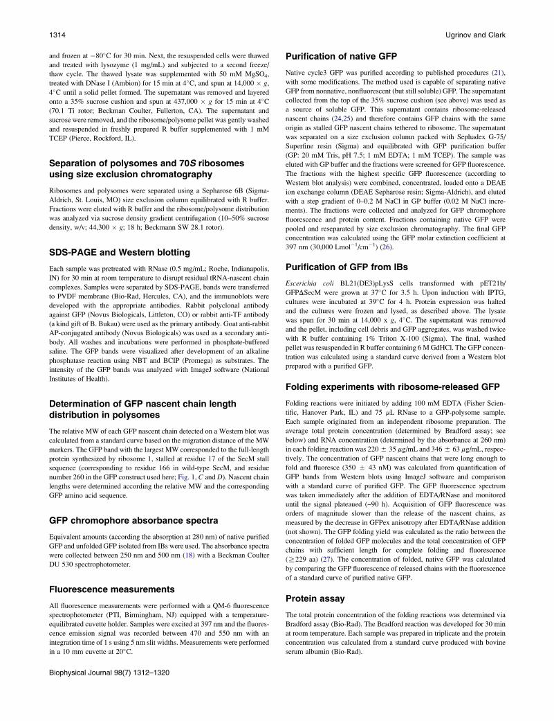
FIGURE 2 Detection of GFP nascent chains. (A) Sucrose density gradient
profile of purified GFP-polysome complexes before (solid line) and after
(dashed line) treatment with EDTA and RNase; 1x corresponds to 70Smonosomes, 2x corresponds to dimer polysomes, etc., and 4xþ corresponds
to polysomes consisting of four or more ribosomes. (B) Anti-GFP Western
blot analysis of purified polysome complexes. Lanes 1–6 represent indepen-
dent GFP-polysome preparations; e.v. represents polysomes prepared from
cells transformed with the empty vector. The numbered arrows denote
GFP nascent chain bands and correspond to the ribosome numbering
in Fig. 1 D. The calculated relative MW of each GFP band is shown in
Table 1.
RESULTS
Purification and characterization of polysomesbearing GFP nascent chains
During active protein synthesis, ~80% of translating E. coliribosomes are engaged in poly-ribosomes (polysomes) (28),
meaning that the earliest cotranslational folding processes
can occur while a nascent polypeptide chain is a component
of a polysome complex. To most closely reproduce cellular
cotranslational folding conditions, we isolated polysomes
from actively translating cells expressing polypeptide chains
consisting of full-length cycle3 GFP (23) followed by a
C-terminal linker encoding the 17-aa SecM translation stall
sequence (24,29,30) (Fig. 1 C). The presence of the SecM stall
sequence generates GFP nascent chains with progressively
shorter chain lengths on polysomes (Fig. S1), but does not
skew the overall cellular distribution of ribosomes and poly-
somes (Fig. S2) (31).
To determine the length distribution of GFP nascent
chains on E. coli polysomes, polysomes were separated by
size exclusion chromatography from 70S ribosomes and
released GFP chains, and analyzed by gel electrophoresis.
Immunoblotting revealed eight distinct GFP bands with sizes
ranging from 19 to 30 kDa, indicating the existence of up to
eight ribosomes per GFP mRNA (Figs. 1 D and 2 B, and
Table 1). The calculated MW of the largest GFP band
(30.8 kDa) corresponds to the MW of the full-length
SecM-stalled GFP construct (260 aa, including an N-ter-
minal affinity tag not used in this study). The calculated
MW of each GFP band (Table 1) and the amino acid content
of the corresponding GFP constructs revealed an average
length difference of 12 aa (36 nucleotides) between nascent
chains on adjacent ribosomes (Table 1). This calculated
difference in GFP nascent chain lengths and the ribosome
distribution on the GFP mRNA are remarkably similar to
previous findings regarding the distribution of ribosomes
on bovine preprolactin and arg-2 mRNAs after translational
pausing (32,33). Of interest, the smallest detected GFP band
had a calculated MW of ~19 kDa, corresponding to a differ-
ence of 29 aa in length between the nascent chains of ribo-
somes 7 and 8, equivalent to 87 nt between ribosome centers
(Fig. 2 B, band 8, and Table 1). Such ribosome spacing is
more characteristic of polysomes formed in vivo in the
absence of a translational pause (28), and suggests that ribo-
some 8, which is most distant from the SecM stall point, is
not part of the ribosome cluster formed as a result of transla-
tional stalling (Fig. 1 D).
Calculation of GFP de novo folding yield requires
discrimination between GFP nascent chains that have at-
tained the minimum length necessary for formation of native
fluorescent protein (229 residues (27)) from shorter, incom-
plete GFP nascent chains tethered to the upstream ribosomes
(Fig. 1, C and D). The calculated lengths of the GFP nascent
Biophysical Journal 98(7) 1312–1320
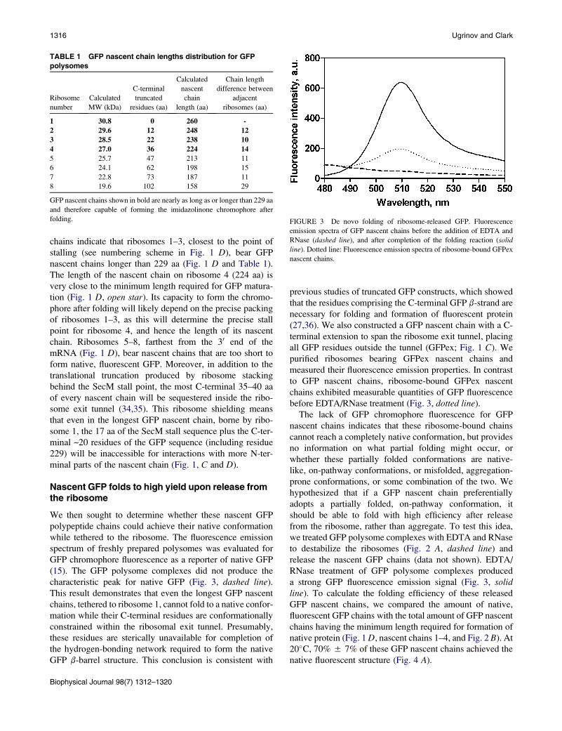
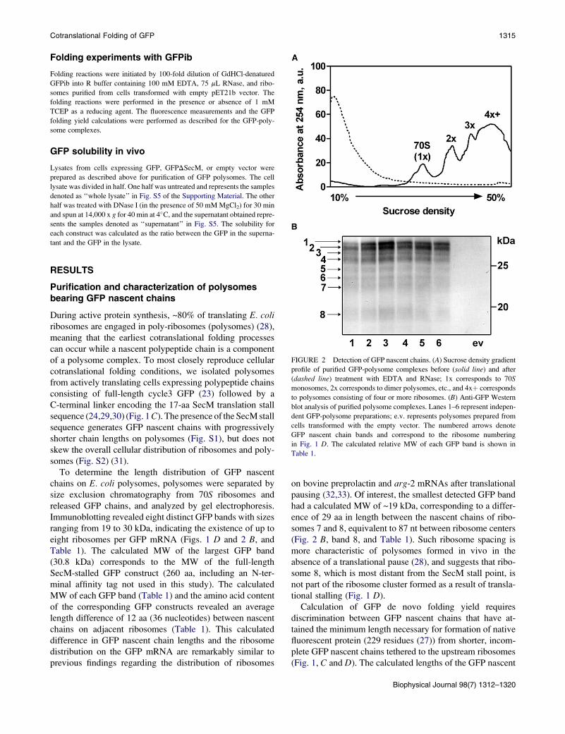
FIGURE 3 De novo folding of ribosome-released GFP. Fluorescence
emission spectra of GFP nascent chains before the addition of EDTA and
RNase (dashed line), and after completion of the folding reaction (solid
line). Dotted line: Fluorescence emission spectra of ribosome-bound GFPex
nascent chains.
TABLE 1 GFP nascent chain lengths distribution for GFP
polysomes
Ribosome
number
Calculated
MW (kDa)
C-terminal
truncated
residues (aa)
Calculated
nascent
chain
length (aa)
Chain length
difference between
adjacent
ribosomes (aa)
1 30.8 0 260 -
2 29.6 12 248 12
3 28.5 22 238 10
4 27.0 36 224 14
5 25.7 47 213 11
6 24.1 62 198 15
7 22.8 73 187 11
8 19.6 102 158 29
GFP nascent chains shown in bold are nearly as long as or longer than 229 aa
and therefore capable of forming the imidazolinone chromophore after
folding.
1316 Ugrinov and Clark
chains indicate that ribosomes 1–3, closest to the point of
stalling (see numbering scheme in Fig. 1 D), bear GFP
nascent chains longer than 229 aa (Fig. 1 D and Table 1).
The length of the nascent chain on ribosome 4 (224 aa) is
very close to the minimum length required for GFP matura-
tion (Fig. 1 D, open star). Its capacity to form the chromo-
phore after folding will likely depend on the precise packing
of ribosomes 1–3, as this will determine the precise stall
point for ribosome 4, and hence the length of its nascent
chain. Ribosomes 5–8, farthest from the 30 end of the
mRNA (Fig. 1 D), bear nascent chains that are too short to
form native, fluorescent GFP. Moreover, in addition to the
translational truncation produced by ribosome stacking
behind the SecM stall point, the most C-terminal 35–40 aa
of every nascent chain will be sequestered inside the ribo-
some exit tunnel (34,35). This ribosome shielding means
that even in the longest GFP nascent chain, borne by ribo-
some 1, the 17 aa of the SecM stall sequence plus the C-ter-
minal ~20 residues of the GFP sequence (including residue
229) will be inaccessible for interactions with more N-ter-
minal parts of the nascent chain (Fig. 1, C and D).
Nascent GFP folds to high yield upon release fromthe ribosome
We then sought to determine whether these nascent GFP
polypeptide chains could achieve their native conformation
while tethered to the ribosome. The fluorescence emission
spectrum of freshly prepared polysomes was evaluated for
GFP chromophore fluorescence as a reporter of native GFP
(15). The GFP polysome complexes did not produce the
characteristic peak for native GFP (Fig. 3, dashed line).
This result demonstrates that even the longest GFP nascent
chains, tethered to ribosome 1, cannot fold to a native confor-
mation while their C-terminal residues are conformationally
constrained within the ribosomal exit tunnel. Presumably,
these residues are sterically unavailable for completion of
the hydrogen-bonding network required to form the native
GFP b-barrel structure. This conclusion is consistent with
Biophysical Journal 98(7) 1312–1320
previous studies of truncated GFP constructs, which showed
that the residues comprising the C-terminal GFP b-strand are
necessary for folding and formation of fluorescent protein
(27,36). We also constructed a GFP nascent chain with a C-
terminal extension to span the ribosome exit tunnel, placing
all GFP residues outside the tunnel (GFPex; Fig. 1 C). We
purified ribosomes bearing GFPex nascent chains and
measured their fluorescence emission properties. In contrast
to GFP nascent chains, ribosome-bound GFPex nascent
chains exhibited measurable quantities of GFP fluorescence
before EDTA/RNase treatment (Fig. 3, dotted line).
The lack of GFP chromophore fluorescence for GFP
nascent chains indicates that these ribosome-bound chains
cannot reach a completely native conformation, but provides
no information on what partial folding might occur, or
whether these partially folded conformations are native-
like, on-pathway conformations, or misfolded, aggregation-
prone conformations, or some combination of the two. We
hypothesized that if a GFP nascent chain preferentially
adopts a partially folded, on-pathway conformation, it
should be able to fold with high efficiency after release
from the ribosome, rather than aggregate. To test this idea,
we treated GFP polysome complexes with EDTA and RNase
to destabilize the ribosomes (Fig. 2 A, dashed line) and
release the nascent GFP chains (data not shown). EDTA/
RNase treatment of GFP polysome complexes produced
a strong GFP fluorescence emission signal (Fig. 3, solidline). To calculate the folding efficiency of these released
GFP nascent chains, we compared the amount of native,
fluorescent GFP chains with the total amount of GFP nascent
chains having the minimum length required for formation of
native protein (Fig. 1 D, nascent chains 1–4, and Fig. 2 B). At
20�C, 70% 5 7% of these GFP nascent chains achieved the
native fluorescent structure (Fig. 4 A).
FIGURE 4 GFP folding efficiency and solubility. (A) Folding efficiency
of ribosome-released GFP (nascent chains) or unfolded GFP from IBs in
the absence or presence of reducing agent (TCEP). All reaction mixtures
contained ribosomes, EDTA, RNase, and TCEP if denoted. The calculated
folding efficiency represents the ratio of the amount of native fluorescent
GFP to the total amount of GFP chains that are long enough to form a native
protein structure (from ribosomes 1–4; see text). Precise quantification of the
folding yield of GFPib at concentrations of 350 nM was hindered by the
formation of macroscopic aggregates; hence, the reported value is approxi-
mate. (B) Effect of SecM stall sequence on GFP solubility. GFPDSecM
lacks the SecM stall sequence at the C-terminus of the GFP construct. The
calculated solubility represents the ratio of the amount of soluble protein
(in the supernatant) to the amount of total expressed protein (in the cell
lysate). In all cases, error bars represent the standard error calculated from
a minimum of three independent samples.
Cotranslational Folding of GFP 1317
Folding of full-length GFP diluted from denaturantis less efficient
Next, we compared the inherent de novo folding efficiency
of ribosome-released GFP nascent chains with the folding
efficiency of full-length GFP containing the SecM stall
sequence (Fig. 1 C) that was forced into IBs by expression
at 39�C. In vivo, after ribosome release, these GFP chains
are pulled into IBs before chromophore formation occurs
(Fig. S4). Because these chains lack a preformed chromo-
phore, their folding can be directly compared with de novo
folding of newly synthesized GFP chains, which also lack
the chromophore (18,37). Full-length GFPib chains were
solubilized in GdHCl, and folding was initiated by dilution
out of denaturant into conditions analogous to the folding
experiments described above for ribosome-released GFP
chains. These folding reactions included polysomes purified
from E. coli transformed with the empty vector as a substitute
for the ribosomal component of GFP polysome complexes,
as previous studies have suggested a direct contribution of
ribosome components to in vitro protein refolding yields
(38). Yet, in contrast to the folding of ribosome-release
nascent GFP chains, folding of GFPib diluted from GdHCl
was dramatically less efficient. Folding experiments using
GFPib concentrations analogous to the concentrations of
nascent GFP in polysome complexes (350 5 43 nM) re-
sulted in the formation of substantial aggregates and no
significant folding yield (Fig. 4 A, the bar without error
bars). At lower GFPib concentrations (<100 nM), the calcu-
lated folding efficiency for GFPib was 13.0% 5 0.4%, still
significantly lower than the folding efficiency of ribosome-
released GFP nascent chains (Fig. 4 A).
GFP has two cysteine residues, both of which are reduced
in the native structure (20). To estimate the maximum
possible folding efficiency for GFPib in the presence of de-
stabilized polysomes, we supplemented the GFPib folding
buffer with 1 mM TCEP as a reducing agent. The folding
efficiency of these free, denatured chains increased to
38% 5 3%, still significantly lower than the folding effi-
ciency of ribosome-released GFP nascent chains (Fig. 4 A).
Nascent GFP folding and molecular chaperones
The high folding efficiency of ribosome-released GFP
nascent chains relative to chemically denatured GFP chains
isolated from IBs indicates that the nascent GFP polypeptide
chains populate a distribution of partially folded conforma-
tions that is different from the distribution of conformations
populated by free GFP polypeptide chains upon dilution
from denaturant. Cotranslational formation of foldable, on-
pathway conformations has been reported for nascent chains
of other proteins, including firefly luciferase, ricin, rhoda-
nese, and P22 tailspike protein (11,25,39–41). For efficient
folding, however, the ribosome-released polypeptide chains
of many proteins required the presence of ATP and/or
GTP as an energy source for ATP-dependent molecular
Biophysical Journal 98(7) 1312–1320
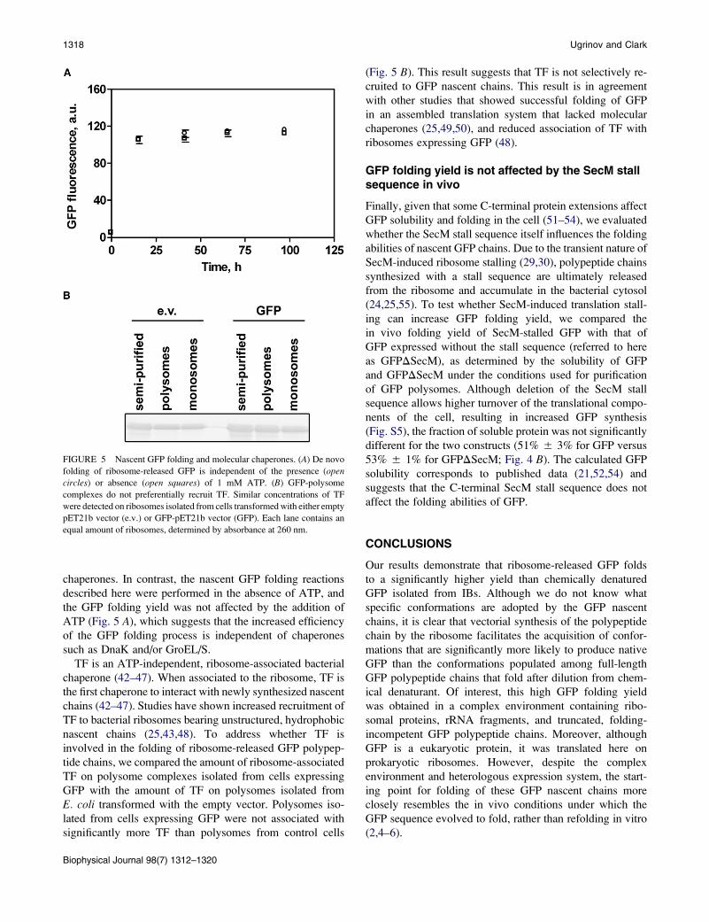
FIGURE 5 Nascent GFP folding and molecular chaperones. (A) De novo
folding of ribosome-released GFP is independent of the presence (opencircles) or absence (open squares) of 1 mM ATP. (B) GFP-polysome
complexes do not preferentially recruit TF. Similar concentrations of TF
were detected on ribosomes isolated from cells transformed with either empty
pET21b vector (e.v.) or GFP-pET21b vector (GFP). Each lane contains an
equal amount of ribosomes, determined by absorbance at 260 nm.
1318 Ugrinov and Clark
chaperones. In contrast, the nascent GFP folding reactions
described here were performed in the absence of ATP, and
the GFP folding yield was not affected by the addition of
ATP (Fig. 5 A), which suggests that the increased efficiency
of the GFP folding process is independent of chaperones
such as DnaK and/or GroEL/S.
TF is an ATP-independent, ribosome-associated bacterial
chaperone (42–47). When associated to the ribosome, TF is
the first chaperone to interact with newly synthesized nascent
chains (42–47). Studies have shown increased recruitment of
TF to bacterial ribosomes bearing unstructured, hydrophobic
nascent chains (25,43,48). To address whether TF is
involved in the folding of ribosome-released GFP polypep-
tide chains, we compared the amount of ribosome-associated
TF on polysome complexes isolated from cells expressing
GFP with the amount of TF on polysomes isolated from
E. coli transformed with the empty vector. Polysomes iso-
lated from cells expressing GFP were not associated with
significantly more TF than polysomes from control cells
Biophysical Journal 98(7) 1312–1320
(Fig. 5 B). This result suggests that TF is not selectively re-
cruited to GFP nascent chains. This result is in agreement
with other studies that showed successful folding of GFP
in an assembled translation system that lacked molecular
chaperones (25,49,50), and reduced association of TF with
ribosomes expressing GFP (48).
GFP folding yield is not affected by the SecM stallsequence in vivo
Finally, given that some C-terminal protein extensions affect
GFP solubility and folding in the cell (51–54), we evaluated
whether the SecM stall sequence itself influences the folding
abilities of nascent GFP chains. Due to the transient nature of
SecM-induced ribosome stalling (29,30), polypeptide chains
synthesized with a stall sequence are ultimately released
from the ribosome and accumulate in the bacterial cytosol
(24,25,55). To test whether SecM-induced translation stall-
ing can increase GFP folding yield, we compared the
in vivo folding yield of SecM-stalled GFP with that of
GFP expressed without the stall sequence (referred to here
as GFPDSecM), as determined by the solubility of GFP
and GFPDSecM under the conditions used for purification
of GFP polysomes. Although deletion of the SecM stall
sequence allows higher turnover of the translational compo-
nents of the cell, resulting in increased GFP synthesis
(Fig. S5), the fraction of soluble protein was not significantly
different for the two constructs (51% 5 3% for GFP versus
53% 5 1% for GFPDSecM; Fig. 4 B). The calculated GFP
solubility corresponds to published data (21,52,54) and
suggests that the C-terminal SecM stall sequence does not
affect the folding abilities of GFP.
CONCLUSIONS
Our results demonstrate that ribosome-released GFP folds
to a significantly higher yield than chemically denatured
GFP isolated from IBs. Although we do not know what
specific conformations are adopted by the GFP nascent
chains, it is clear that vectorial synthesis of the polypeptide
chain by the ribosome facilitates the acquisition of confor-
mations that are significantly more likely to produce native
GFP than the conformations populated among full-length
GFP polypeptide chains that fold after dilution from chem-
ical denaturant. Of interest, this high GFP folding yield
was obtained in a complex environment containing ribo-
somal proteins, rRNA fragments, and truncated, folding-
incompetent GFP polypeptide chains. Moreover, although
GFP is a eukaryotic protein, it was translated here on
prokaryotic ribosomes. However, despite the complex
environment and heterologous expression system, the start-
ing point for folding of these GFP nascent chains more
closely resembles the in vivo conditions under which the
GFP sequence evolved to fold, rather than refolding in vitro
(2,4–6).
Cotranslational Folding of GFP 1319
SUPPORTING MATERIAL
Five figures are available at http://www.biophysj.org/biophysj/supplemental/
S0006-3495(09)06140-2.
We thank Kay Finn for technical assistance, and Ian Sander and the other
members of the Clark group for helpful discussions. The anti-TF antibody
was a generous gift from Bernd Bukau.
This work was supported by an award from the National Institutes of Health
(GM 74807).
REFERENCES
1. Ellis, R. J. 2001. Macromolecular crowding: obvious but underappreci-ated. Trends Biochem. Sci. 26:597–604.
2. Fedorov, A. N., and T. O. Baldwin. 1997. Cotranslational proteinfolding. J. Biol. Chem. 272:32715–32718.
3. Komar, A. A. 2009. A pause for thought along the co-translationalfolding pathway. Trends Biochem. Sci. 34:16–24.
4. Kramer, G., V. Ramachandiran, and B. Hardesty. 2001. Cotranslationalfolding—omnia mea mecum porto? Int. J. Biochem. Cell Biol. 33:541–553.
5. Evans, M. S., T. F. Clarke 4th, and P. L. Clark. 2005. Conformations ofco-translational folding intermediates. Protein Pept. Lett. 12:189–195.
6. Clark, P. L. 2004. Protein folding in the cell: reshaping the folding fun-nel. Trends Biochem. Sci. 29:527–534.
7. Xia, K., M. Manning, ., W. Colon. 2007. Identifying the subproteomeof kinetically stable proteins via diagonal 2D SDS/PAGE. Proc. Natl.Acad. Sci. USA. 104:17329–17334.
8. Manning, M., and W. Colon. 2004. Structural basis of protein kineticstability: resistance to sodium dodecyl sulfate suggests a central rolefor rigidity and a bias toward beta-sheet structure. Biochemistry.43:11248–11254.
9. Kolb, V. A., E. V. Makeyev, and A. S. Spirin. 2000. Co-translationalfolding of an eukaryotic multidomain protein in a prokaryotic transla-tion system. J. Biol. Chem. 275:16597–16601.
10. Fedorov, A. N., and T. O. Baldwin. 1999. Process of biosyntheticprotein folding determines the rapid formation of native structure.J. Mol. Biol. 294:579–586.
11. Frydman, J., E. Nimmesgern, ., F. U. Hartl. 1994. Folding of nascentpolypeptide chains in a high molecular mass assembly with molecularchaperones. Nature. 370:111–117.
12. Svetlov, M. S., A. Kommer, ., A. S. Spirin. 2006. Effective cotransla-tional folding of firefly luciferase without chaperones of the Hsp70family. Protein Sci. 15:242–247.
13. Nicola, A. V., W. Chen, and A. Helenius. 1999. Co-translational foldingof an alphavirus capsid protein in the cytosol of living cells. Nat. CellBiol. 1:341–345.
14. Netzer, W. J., and F. U. Hartl. 1997. Recombination of protein domainsfacilitated by co-translational folding in eukaryotes. Nature. 388:343–349.
15. Tsien, R. Y. 1998. The green fluorescent protein. Annu. Rev. Biochem.67:509–544.
16. Kamagata, K., M. Arai, and K. Kuwajima. 2004. Unification of thefolding mechanisms of non-two-state and two-state proteins. J. Mol.Biol. 339:951–965.
17. Andrews, B. T., A. R. Schoenfish, ., P. A. Jennings. 2007. The roughenergy landscape of superfolder GFP is linked to the chromophore.J. Mol. Biol. 373:476–490.
18. Reid, B. G., and G. C. Flynn. 1997. Chromophore formation in greenfluorescent protein. Biochemistry. 36:6786–6791.
19. Ward, W. W., and S. H. Bokman. 1982. Reversible denaturation ofAequorea green-fluorescent protein: physical separation and character-ization of the renatured protein. Biochemistry. 21:4535–4540.
20. Battistutta, R., A. Negro, and G. Zanotti. 2000. Crystal structure and
refolding properties of the mutant F99S/M153T/V163A of the green
fluorescent protein. Proteins. 41:429–437.
21. Fukuda, H., M. Arai, and K. Kuwajima. 2000. Folding of green fluores-
cent protein and the cycle3 mutant. Biochemistry. 39:12025–12032.
22. Jackson, S. E., T. D. Craggs, and J. R. Huang. 2006. Understanding the
folding of GFP using biophysical techniques. Expert Rev. Proteomics.3:545–559.
23. Crameri, A., E. A. Whitehorn, ., W. P. Stemmer. 1996. Improved
green fluorescent protein by molecular evolution using DNA shuffling.
Nat. Biotechnol. 14:315–319.
24. Evans, M. S., K. G. Ugrinov, ., P. L. Clark. 2005. Homogeneous
stalled ribosome nascent chain complexes produced in vivo or in vitro.
Nat. Methods. 2:757–762.
25. Evans, M. S., I. M. Sander, and P. L. Clark. 2008. Cotranslational
folding promotes b-helix formation and avoids aggregation in vivo.
J. Mol. Biol. 383:683–692.
26. Chalfie, M., and S. Kain. 1998. Green Fluorescent Protein: Properties,
Applications, and Protocols. Wiley-Liss, New York.
27. Li, X., G. Zhang, ., C. C. Huang. 1997. Deletions of the Aequoreavictoria green fluorescent protein define the minimal domain required
for fluorescence. J. Biol. Chem. 272:28545–28549.
28. Neidhardt, F. C., and R. Curtiss. 1996. Escherichia coli and Salmonella:
Cellular and Molecular Biology. ASM Press, Washington, DC.
29. Nakatogawa, H., and K. Ito. 2002. The ribosomal exit tunnel functions
as a discriminating gate. Cell. 108:629–636.
30. Nakatogawa, H., and K. Ito. 2001. Secretion monitor, SecM, undergoes
self-translation arrest in the cytosol. Mol. Cell. 7:185–192.
31. Brandt, F., S. A. Etchells, ., W. Baumeister. 2009. The native 3D orga-
nization of bacterial polysomes. Cell. 136:261–271.
32. Wolin, S. L., and P. Walter. 1988. Ribosome pausing and stacking
during translation of a eukaryotic mRNA. EMBO J. 7:3559–3569.
33. Sachs, M. S., Z. Wang, ., A. Jacobson. 2002. Toeprint analysis of the
positioning of translation apparatus components at initiation and termi-
nation codons of fungal mRNAs. Methods. 26:105–114.
34. Nissen, P., J. Hansen, ., T. A. Steitz. 2000. The structural basis of ribo-
some activity in peptide bond synthesis. Science. 289:920–930.
35. Lu, J., and C. Deutsch. 2005. Folding zones inside the ribosomal exit
tunnel. Nat. Struct. Mol. Biol. 12:1123–1129.
36. Cabantous, S., T. C. Terwilliger, and G. S. Waldo. 2005. Protein
tagging and detection with engineered self-assembling fragments of
green fluorescent protein. Nat. Biotechnol. 23:102–107.
37. Siemering, K. R., R. Golbik, ., J. Haseloff. 1996. Mutations that
suppress the thermosensitivity of green fluorescent protein. Curr.Biol. 6:1653–1663.
38. Das, B., S. Chattopadhyay, ., C. Dasgupta. 1996. In vitro protein
folding by ribosomes from Escherichia coli, wheat germ and rat liver:
the role of the 50S particle and its 23S rRNA. Eur. J. Biochem.235:613–621.
39. Kudlicki, W., Y. Kitaoka, ., B. Hardesty. 1995. Elongation and
folding of nascent ricin chains as peptidyl-tRNA on ribosomes: the
effect of amino acid deletions on these processes. J. Mol. Biol.252:203–212.
40. Clark, P. L., and J. King. 2001. A newly synthesized, ribosome-bound
polypeptide chain adopts conformations dissimilar from early in vitrorefolding intermediates. J. Biol. Chem. 276:25411–25420.
41. Kudlicki, W., O. W. Odom, ., B. Hardesty. 1994. Activation and
release of enzymatically inactive, full-length rhodanese that is bound
to ribosomes as peptidyl-tRNA. J. Biol. Chem. 269:16549–16553.
42. Buskiewicz, I., E. Deuerling, ., W. Wintermeyer. 2004. Trigger factor
binds to ribosome-signal-recognition particle (SRP) complexes and is
excluded by binding of the SRP receptor. Proc. Natl. Acad. Sci. USA.101:7902–7906.
Biophysical Journal 98(7) 1312–1320
1320 Ugrinov and Clark
43. Kaiser, C. M., H. C. Chang, ., J. M. Barral. 2006. Real-time observa-
tion of trigger factor function on translating ribosomes. Nature. 444:
455–460.
44. Merz, F., D. Boehringer, ., E. Deuerling. 2008. Molecular mechanism
and structure of trigger factor bound to the translating ribosome. EMBO
J. 27:1622–1632.
45. Rutkowska, A., M. P. Mayer, ., B. Bukau. 2008. Dynamics of trigger
factor interaction with translating ribosomes. J. Biol. Chem. 283:
4124–4132.
46. Ferbitz, L., T. Maier, ., N. Ban. 2004. Trigger factor in complex with
the ribosome forms a molecular cradle for nascent proteins. Nature.431:590–596.
47. Tomic, S., A. E. Johnson, ., S. A. Etchells. 2006. Exploring the
capacity of trigger factor to function as a shield for ribosome bound
polypeptide chains. FEBS Lett. 580:72–76.
48. Agashe, V. R., S. Guha, ., J. M. Barral. 2004. Function of trigger
factor and DnaK in multidomain protein folding: increase in yield at
the expense of folding speed. Cell. 117:199–209.
Biophysical Journal 98(7) 1312–1320
49. Uemura, S., R. Iizuka, ., T. Funatsu. 2008. Single-molecule imagingof full protein synthesis by immobilized ribosomes. Nucleic AcidsRes. 36:e70.
50. Shimizu, Y., A. Inoue, ., T. Ueda. 2001. Cell-free translation reconsti-tuted with purified components. Nat. Biotechnol. 19:751–755.
51. Waldo, G. S., B. M. Standish, ., T. C. Terwilliger. 1999. Rapidprotein-folding assay using green fluorescent protein. Nat. Biotechnol.17:691–695.
52. Sacchetti, A., V. Cappetti, ., S. Alberti. 2001. Green fluorescentprotein variants fold differentially in prokaryotic and eukaryotic cells.J. Cell. Biochem. 81:117–128.
53. Sacchetti, A., and S. Alberti. 1999. Protein tags enhance GFP folding ineukaryotic cells. Nat. Biotechnol. 17:1046.
54. Chang, H. C., C. M. Kaiser, ., J. M. Barral. 2005. De novo folding ofGFP fusion proteins: high efficiency in eukaryotes but not in bacteria.J. Mol. Biol. 353:397–409.
55. Muto, H., H. Nakatogawa, and K. Ito. 2006. Genetically encoded butnonpolypeptide prolyl-tRNA functions in the A site for SecM-mediatedribosomal stall. Mol. Cell. 22:545–552.