Embed Size (px)
Citation preview
Washington County EMS
Protocols1

Table of ContentsINTRODUCTION............................................................................................................................................................5
ASTHMA/COPD (ADULT).............................................................................................................................................7
Agitated and Combative Patients................................................................................................................................9
Appendix B (Adult).....................................................................................................................................................11
Appendix C (Pediatric)................................................................................................................................................12
Allergy & Anaphylaxis.................................................................................................................................................13
Altered Level of Consciousness (ALOC)...................................................................................................................15
Asystole & Pulseless Electrical Activity (Adult)............................................................................................................16
Bradycardia (Adult)....................................................................................................................................................18
Burns..........................................................................................................................................................................20
Cardiac Arrest -Adult (CCR).........................................................................................................................................23
Chest Pain..................................................................................................................................................................26
Patient with Concealed Carry Weapon........................................................................................................................28
Congestive Heart Failure / Cardiogenic Shock.............................................................................................................30
Eclampsia...................................................................................................................................................................33
GUIDELINES FOR TRAUMA DEFINITION.......................................................................................................................35
Heat Emergencies.......................................................................................................................................................37
Hospital Bypass Protocol............................................................................................................................................40
Hyperglycemia............................................................................................................................................................42
Hypoglycemia.............................................................................................................................................................432
Hypothermia & Frostbite............................................................................................................................................45
Hypovolemia and Shock.............................................................................................................................................48
Delivery of Newborn - Procedure................................................................................................................................50
Narrow Complex Tachycardia’s (Adult).......................................................................................................................53
Nausea and Vomiting.................................................................................................................................................56
Pain Management......................................................................................................................................................58
Allergy & Anaphylaxis (Pediatric)................................................................................................................................62
Asthma (Pediatric)......................................................................................................................................................64
(Includes Reactive Airways Disease and Bronchospasm).............................................................................................64
Asystole/PEA (Pediatric).............................................................................................................................................66
Bradycardia (Pediatric)...............................................................................................................................................68
Burns (Pediatric).........................................................................................................................................................70
Cardiac Arrest (Pediatric)............................................................................................................................................73
Hyperglycemia (Pediatric)...........................................................................................................................................76
Hypoglycemia (Pediatric)............................................................................................................................................78
Hypovolemia and Shock (Pediatric).............................................................................................................................80
Respiratory Distress (Pediatric)...................................................................................................................................82
Seizure (Pediatric)......................................................................................................................................................85
SVT (Pediatric)............................................................................................................................................................87
Toxic Exposure/Overdose (Pediatric)..........................................................................................................................89
Ventricular Fibrillation & Pulseless Ventricular Tachycardia (Pediatric).......................................................................92
Wide Complex Tachycardia (Pediatric)........................................................................................................................95
Pregnant Female in Labor...........................................................................................................................................97
Rapid Atrial Fibrillation (Adult)...................................................................................................................................99
Refusal of Care.........................................................................................................................................................102
3

Respiratory Distress (Adult)......................................................................................................................................104
Routine Medical Care (Pediatric)...............................................................................................................................106
Routine Medical Care (Adult)....................................................................................................................................108
Routine Trauma Care (Adult)....................................................................................................................................110
Routine Trauma Care (Pediatric)...............................................................................................................................113
RSA (Adult)...............................................................................................................................................................115
Rapid Sequence Airway............................................................................................................................................115
Seizure.....................................................................................................................................................................117
Stroke.......................................................................................................................................................................119
Submersion (Adult)...................................................................................................................................................120
Syncope....................................................................................................................................................................122
Toxic Exposure/Overdose.........................................................................................................................................123
Hospital Destination / Selection................................................................................................................................127
Vaginal Bleeding After Delivery.................................................................................................................................128
Vaginal Bleeding Before Delivery..............................................................................................................................130
Vertigo.....................................................................................................................................................................132
Wide Complex Tachycardia’s (Adult).........................................................................................................................134
Withholding or Withdrawing of Resuscitative Efforts................................................................................................138
Index........................................................................................................................................................................141
4
INTRODUCTION
This document contains the protocols, guidelines, and instructions for emergent out-of –hospital care for EMERGENCY MEDICAL RESPONDER (EMR), EMERGENCY MEDICAL TECHNICIAN BASIC (EMT), and EMERGENCY MEDICAL INTERMEDIATE TECHNICIAN, EMERGENCY MEDICAL INTERMEDIATE, EMERGENCY MEDICAL TECHNICIAN PARAMEDIC under the medical control of Aurora Medical Center of Washington County and Froedtert Health St Joseph’s Hospital. It establishes standards of care that conform to the current guidelines of the State of Wisconsin.
The practice of out-of-hospital medicine requires a relative degree of flexibility to adequately address the great variability of situations that are part and parcel of working in a relatively uncontrolled environment. As such, circumstances may require occasional deviation from these instructions. The specific goals of any treatment must always be improvement in the patient’s condition.
The practice of out-of-hospital medicine is also continually changing. As more and more research in this field is performed, guidelines for care will change. New technology, both in this particular arena and in hospital medicine, will likewise change the manner in which patients and their problems are managed. It is fully anticipated that this document will go through several modifications over time, to make the most of new knowledge and advances in technology for the benefit of the patient.
The protocols are subdivided by the interventions available to each provider. Those of more advanced training are expected to ensure that the interventions of the previous levels have been performed.
It is expected that each level will request appropriate additional resources (ALS Intercept, Helicopter, etc.) based on dispatch information and mechanism of patient injury or condition. Resource requests should be made as early as possible to maximize potential interventions and prevent delays in transport.
These protocols shall be utilized: As written, standing orders of a physician and treatment guidelines to be
administered by pre-hospital caregivers, as circumstances allow, for thetreatment of the ill or injured patient and orders to be used by MedicalControl when directing pre-hospital care.
Medical care orders can only be initiated through these protocols orauthorized Medical Center. EMS personnel who are uncertain of a specificprotocol or drug dosage to be used must contact Medical Controlimmediately.
In disaster situations, if the usual and customary forms of communicationsare not available, that patient care will be given in accordance with theresponse areas disaster plans.
It is recognized that hospice patients, patients with a valid DNR order, patients who have not responded to ALS procedures, or patients involved in a mass casualty incident (MCI) present unique circumstances that may, in the medical opinion of the physician directing the call, justify deviation from these protocols, including bypassing the nearest hospital.
The orders that constitute each protocol are listed in the general order in which they are to be performed, with full awareness that in any given situation the order in which interventions are performed may change to adapt to the circumstance. It is recognized that these
5
pre-hospital standing medical orders are intended to stabilize most patient care situations, but can never anticipate all possible circumstances. All levels of providers may operate under these protocols without on-line medical control, unless specifically mentioned. In the event that communications cannot be established with Medical Control, pre-hospital personnel shall continue to provide treatment to the degree authorized by the Medical Director in these protocols. In these situations the “medical control permission to administer” requirements are waived.
An EMS provider may enact and follow any of these protocols until they reach CONTACT MEDICAL CONTROL. At this point in the protocol, the EMT must make verbal contact via radio (or cellular phone) to advise online Medical Control of the situation. Based on the information provided, Medical Control may order an intervention listed after the “CONTACT MEDICAL CONTROL” line in the protocol. At his/her discretion the Medical Control physician may also deny any further intervention or order another intervention. Even if ordered by Medical Control, at no time may an EMT exceed the level of his/her training or licensure. At any time, the EMT is unsure about the application of skills or medications to a patient, contact medical control for assistance and clarification.
The physician’s orders are to be obtained from the Emergency Physician staffing the hospital in the Emergency Department. Any orders received should be read back to Medical Control to assure accuracy and carried out exactly as ordered. Document the time, order, and reason for refusal. In all cases, the EMT shall document all orders received from Medical Control and any circumstances which required the EMT to deviate from these protocols in an effort to improve the patient’s condition.
6
Washington County
Suggested Medical GuidelinesASTHMA/COPD (ADULT)
(Includes Reactive Airways Disease, Bronchospasm, Emphysema and Chronic Bronchitis)
Note: All hypoxic patients should be given enough oxygen therapy to reverse their hypoxia (SpO2 ≥ 90%), even if they have COPD,
but all COPD patients must be closely monitored for signs of respiratory depression due to oxygen therapy. Look for: somnolence, lethargy, decreased rate or depth of breaths. If these appear, back off on the rate of flow and prepare to assist ventilations.
Patients with COPD are usually older adults with a long and heavy smoking history. This includes patients with emphysema (“pink puffers”) and chronic bronchitis (“blue bloaters”). Exacerbations are often triggered by infections.
Asthma is usually a disease of childhood, but may occur or re-occur later in life. There is usually an identifiable trigger, like infection, weather changes or exposure to certain allergens (e.g., dogs, pollen, etc.). The so-called classic triad of dyspnea, cough and wheezing may not always be present.
Patients with a history of near fatal asthma are at increased risk of recurrent severe attacks and asthma-related death. Remember: “All that wheezes is not asthma!” Always consider the possibility of Congestive Heart Failure in older adults
with wheezing. The absence of wheezing may be indicative of extreme airflow obstruction.
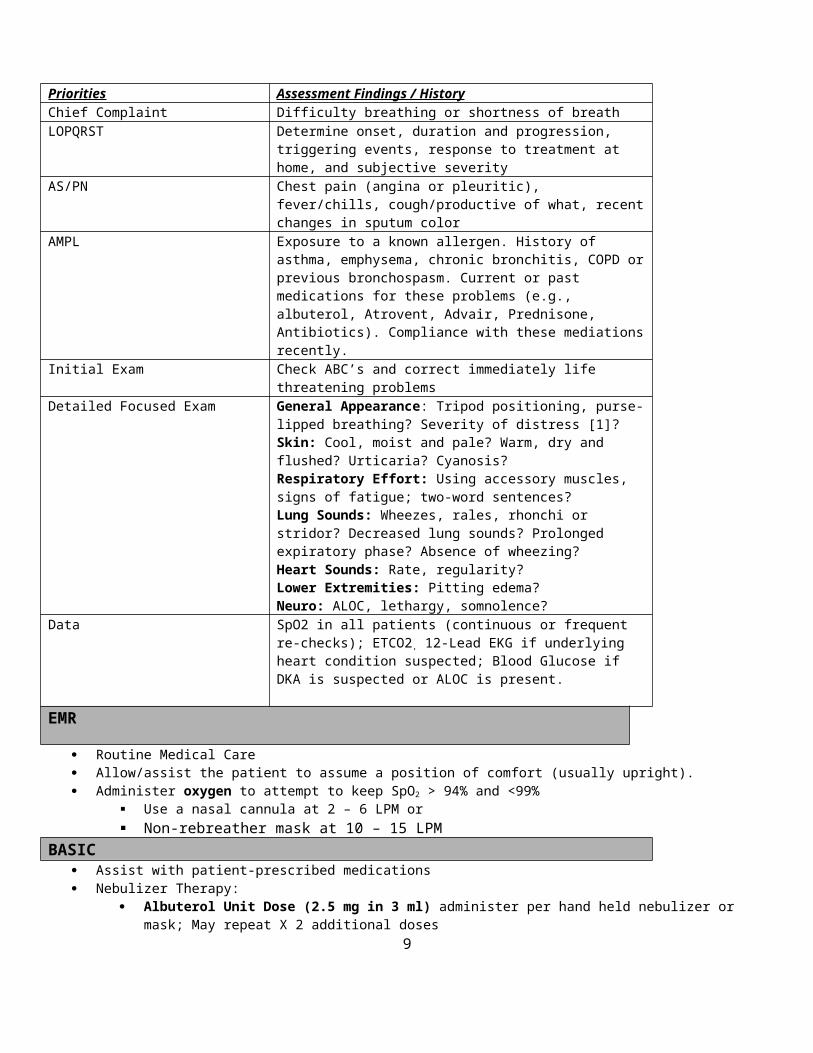
Priorities Assessment Findings / HistoryChief Complaint Difficulty breathing or shortness of breathLOPQRST Determine onset, duration and progression, triggering events,
response to treatment at home, and subjective severityAS/PN Chest pain (angina or pleuritic), fever/chills, cough/productive of
what, recent changes in sputum colorAMPL Exposure to a known allergen. History of asthma, emphysema,
chronic bronchitis, COPD or previous bronchospasm. Current or past medications for these problems (e.g., albuterol, Atrovent, Advair, Prednisone, Antibiotics). Compliance with these mediations recently.
Initial Exam Check ABC’s and correct immediately life threatening problemsDetailed Focused Exam General Appearance: Tripod positioning, purse-lipped breathing?
Severity of distress [1]?Skin: Cool, moist and pale? Warm, dry and flushed? Urticaria? Cyanosis?Respiratory Effort: Using accessory muscles, signs of fatigue; two-word sentences?Lung Sounds: Wheezes, rales, rhonchi or stridor? Decreased lung sounds? Prolonged expiratory phase? Absence of wheezing?Heart Sounds: Rate, regularity?Lower Extremities: Pitting edema?Neuro: ALOC, lethargy, somnolence?
Data SpO2 in all patients (continuous or frequent re-checks); ETCO2, 12-
7

Lead EKG if underlying heart condition suspected; Blood Glucose if DKA is suspected or ALOC is present.
EMR
Routine Medical Care Allow/assist the patient to assume a position of comfort (usually upright). Administer oxygen to attempt to keep SpO2 > 94% and <99%
Use a nasal cannula at 2 – 6 LPM or Non-rebreather mask at 10 – 15 LPM
BASIC Assist with patient-prescribed medications Nebulizer Therapy:
Albuterol Unit Dose (2.5 mg in 3 ml) administer per hand held nebulizer or mask; May repeat X 2 additional doses Atrovent Unit Dose (0.5mg in 2.5 ml) administer per hand held nebulizer or mask May Mix Albuterol and Atrovent in same nebulizer or give them separately. Do not repeat Atrovent alone or in combination without an order from Medical Control.
Assisted Ventilation: Consider assisting breathing with gentle synchronous ventilations with bag-valve mask (BVM); Support ventilation with BVM if apnea or hypopnea occurs.
Airway Adjuncts: If there is loss of consciousness and loss of gag reflex, insert an oropharyngeal or nasopharyngeal airway; Insert a non-visualized airway in unconscious patients without a gag reflex (see Respiratory Distress Guideline) Initiate CPAP at 5- 10 cm H2O. (See Respiratory Distress Guideline) To be used only by EMT’s with proper training and
certification by the medical director or authorized representative. INTERMEDIATE TECHNICIAN
IV 0.9% NS @ KVO; If signs of dehydration or hypovolemia are present, administer 250 ml bolus, and then decrease to KVO. (Check lung sounds)
Contact Medical Control for the following: For severe asthma, consider Epinephrine 1:1,000 0.3 – 0.5 mg Sub-Q/IM
INTERMEDIATE
Epinephrine 1:10,000 0.5 mg slow IV push every 5 minutes as needed for severe asthmaContact medical control for the following:
Repeat subcutaneous epinephrine if the signs of severe distress continue after 20 minutes.
PARAMEDIC Epinephrine 1:1,00 0.3 mg Sub-Q/IM For severe asthma or COPD, administer Solu-Medrol 125 mg IV
Contact Medical Control for the following: Additional doses of these medications appear to be needed. Magnesium Sulfate 2 gm slow IV push (over 10 minutes) for severe asthma Epinephrine 1:10,000 0.5 mg slow IV push every 5 minutes as needed for severe asthma
FOOTNOTES:[1] Severity of Respiratory Distress:
Mild = RR<20 + minimal additional breathing effort + speaking in complete sentences + minimal subjective distress, No ALOC
Moderate = RR 20 to 25 + moderate additional breathing effort + difficult to complete a sentence + moderate subjective distress + No ALOC
Severe = RR> 25 + marked additional breathing effort + 2 or 3 word sentences + marked subjective distress + possible ALOC
8
Date of Origin: 11/10/11
Updated 3/15/12
Washington County
Suggested Medical Guidelines
Agitated and Combative PatientsNote:
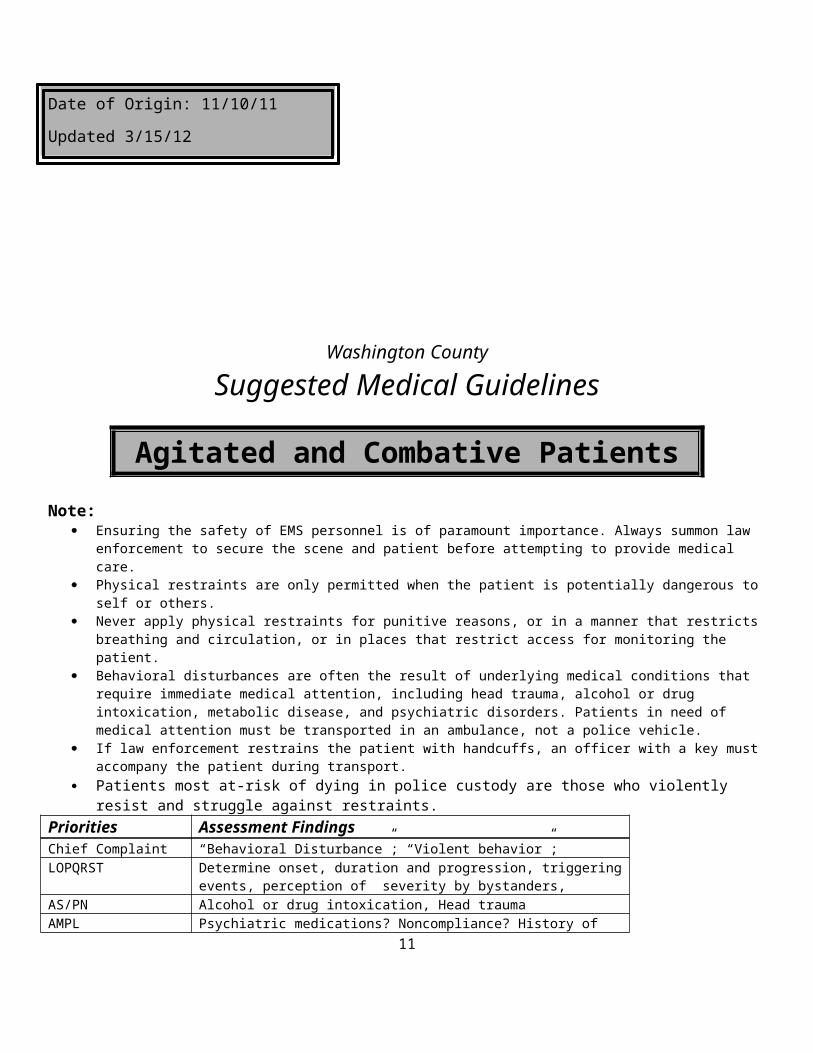
Ensuring the safety of EMS personnel is of paramount importance. Always summon law enforcement to secure the scene and patient before attempting to provide medical care.
Physical restraints are only permitted when the patient is potentially dangerous to self or others. Never apply physical restraints for punitive reasons, or in a manner that restricts breathing and circulation, or in places that
restrict access for monitoring the patient. Behavioral disturbances are often the result of underlying medical conditions that require immediate medical attention,
including head trauma, alcohol or drug intoxication, metabolic disease, and psychiatric disorders. Patients in need of medical attention must be transported in an ambulance, not a police vehicle.
If law enforcement restrains the patient with handcuffs, an officer with a key must accompany the patient during transport. Patients most at-risk of dying in police custody are those who violently resist and struggle against restraints.
Priorities Assessment FindingsChief Complaint “Behavioral Disturbance”; “Violent behavior”; LOPQRST Determine onset, duration and progression, triggering events, perception of
severity by bystanders, AS/PN Alcohol or drug intoxication, Head traumaAMPL Psychiatric medications? Noncompliance? History of schizophrenia or bipolar
disorder? History of drug or alcohol abuse? Initial Exam Check ABCs and correct immediately life-threatening problems.Detailed Focused Exam General Appearance: Bizarre behavior, violent, aggressive, combative, loud,
obnoxious, agitated; partial or complete undressing? Uncooperative (Does not respond to verbal commands to desist)?Skin: Diaphoresis? Cool, moist and pale? Warm, dry and flushed?Respiratory Effort: Labored breathing? Heavy breathing?Lung Sounds: Wheezes, rales, rhonchi or stridor? Decreased lung sounds? Cardiovascular: Hypertensive and tachycardia?Extremities: Trauma?Neuro: Excited, agitated, increased activity and increased intensity of activityPsych: Bizarre thoughts and actions; Paranoia, delusional, confused, clouded consciousness?
Data SpO2 in all patients (continuous or frequent re-checks);
9

End Tidal CO2
12-Lead EKG as soon as it becomes practical to obtain one; Blood Glucose to rule out hypoglycemia as a cause of the behavioral disturbance.
Goals of Therapy Physically or chemically restrain the patient to reduce the threat to self and others, especially emergency responders (law enforcement and EMS)
Monitoring BP, HR, RR, EKG, SpO2.ETCO2
EMR Scene size-up Do not approach an agitated and combative patient before law enforcement has gained control of the situation. It is reasonable to attempt verbal de-escalation, but do not persist if it appears to be futile or making the situation worse. Initiate Routine Medical Care once it is safe and practical. Monitor vital signs every 5 minutes
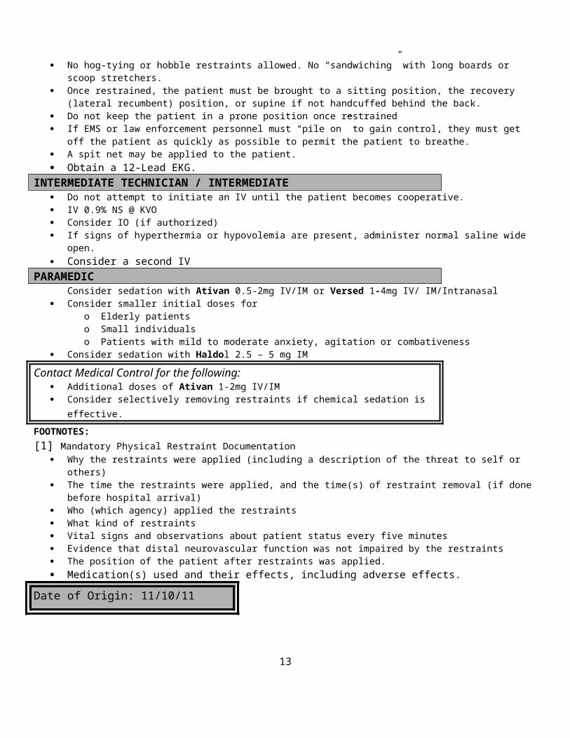
BASIC Consider physical restraints [1] as a last resort when verbal control is ineffective Soft restraints or padded hard restraints are preferred for use by EMS personnel. No hog-tying or hobble restraints allowed. No “sandwiching” with long boards or scoop stretchers. Once restrained, the patient must be brought to a sitting position, the recovery (lateral recumbent) position, or supine if not
handcuffed behind the back. Do not keep the patient in a prone position once restrained If EMS or law enforcement personnel must “pile on” to gain control, they must get off the patient as quickly as possible to
permit the patient to breathe. A spit net may be applied to the patient. Obtain a 12-Lead EKG.
INTERMEDIATE TECHNICIAN / INTERMEDIATE Do not attempt to initiate an IV until the patient becomes cooperative. IV 0.9% NS @ KVO Consider IO (if authorized) If signs of hyperthermia or hypovolemia are present, administer normal saline wide open. Consider a second IV
PARAMEDIC Consider sedation with Ativan 0.5-2mg IV/IM or Versed 1-4mg IV/ IM/Intranasal
Consider smaller initial doses foro Elderly patientso Small individualso Patients with mild to moderate anxiety, agitation or combativeness
Consider sedation with Haldol 2.5 – 5 mg IM
Contact Medical Control for the following: Additional doses of Ativan 1-2mg IV/IM Consider selectively removing restraints if chemical sedation is effective.
FOOTNOTES:[1] Mandatory Physical Restraint Documentation
Why the restraints were applied (including a description of the threat to self or others) The time the restraints were applied, and the time(s) of restraint removal (if done before hospital arrival) Who (which agency) applied the restraints What kind of restraints Vital signs and observations about patient status every five minutes Evidence that distal neurovascular function was not impaired by the restraints The position of the patient after restraints was applied.
10
Medication(s) used and their effects, including adverse effects.
Date of Origin: 11/10/11
11

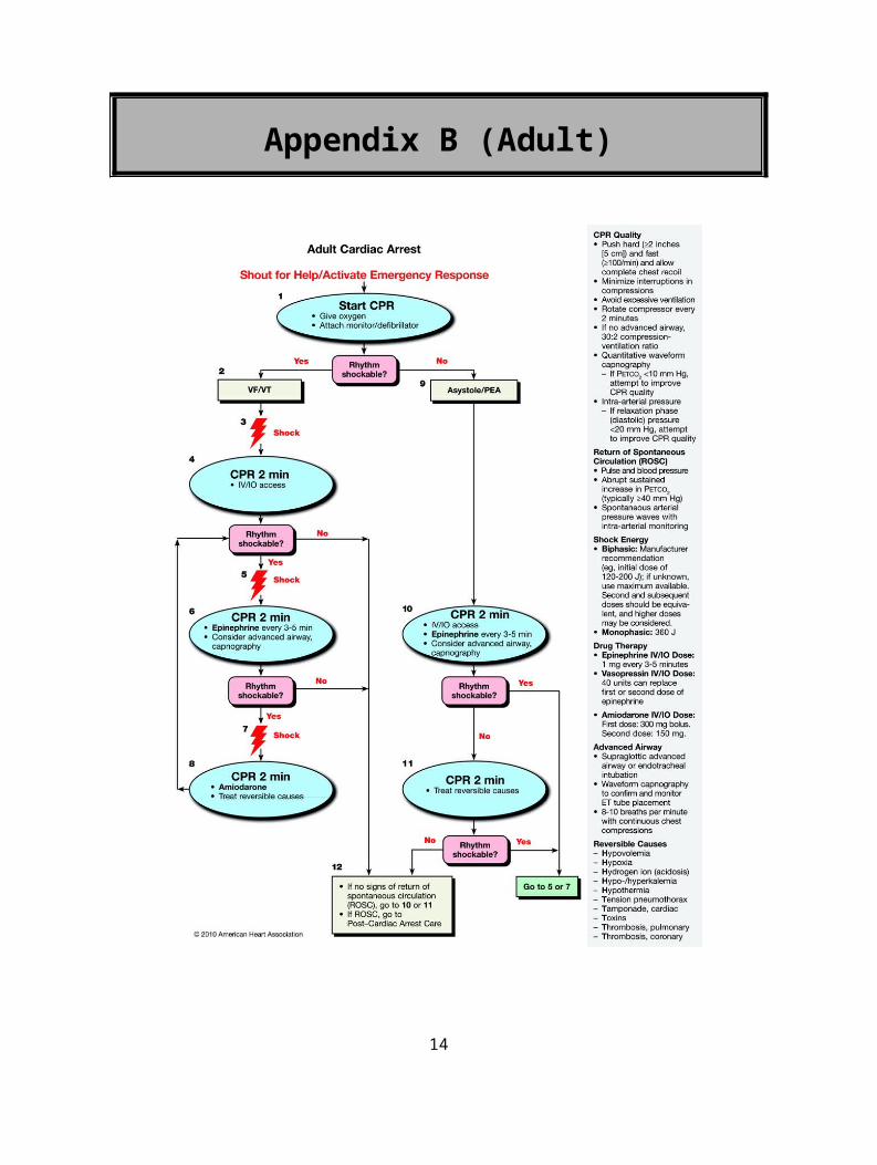
Appendix B (Adult)
12
Appendix C (Pediatric)
13
Washington County
Suggested Medical Guidelines
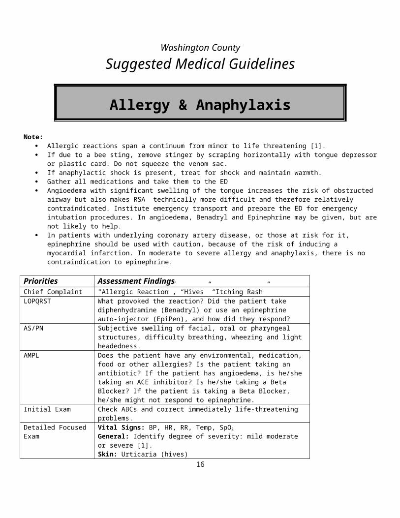
Allergy & Anaphylaxis
Note: Allergic reactions span a continuum from minor to life threatening [1]. If due to a bee sting, remove stinger by scraping horizontally with tongue depressor or plastic card. Do not squeeze the
venom sac. If anaphylactic shock is present, treat for shock and maintain warmth. Gather all medications and take them to the ED Angioedema with significant swelling of the tongue increases the risk of obstructed airway but also makes RSA technically
more difficult and therefore relatively contraindicated. Institute emergency transport and prepare the ED for emergency intubation procedures. In angioedema, Benadryl and Epinephrine may be given, but are not likely to help.
In patients with underlying coronary artery disease, or those at risk for it, epinephrine should be used with caution, because of the risk of inducing a myocardial infarction. In moderate to severe allergy and anaphylaxis, there is no contraindication to epinephrine.
Priorities Assessment FindingsChief Complaint “Allergic Reaction”, “Hives” “Itching Rash”LOPQRST What provoked the reaction? Did the patient take diphenhydramine (Benadryl)
or use an epinephrine auto-injector (EpiPen), and how did they respond?AS/PN Subjective swelling of facial, oral or pharyngeal structures, difficulty
breathing, wheezing and light headedness.AMPL Does the patient have any environmental, medication, food or other allergies?
Is the patient taking an antibiotic? If the patient has angioedema, is he/she taking an ACE inhibitor? Is he/she taking a Beta Blocker? If the patient is taking a Beta Blocker, he/she might not respond to epinephrine.
Initial Exam Check ABCs and correct immediately life-threatening problems.Detailed Focused Exam Vital Signs: BP, HR, RR, Temp, SpO2
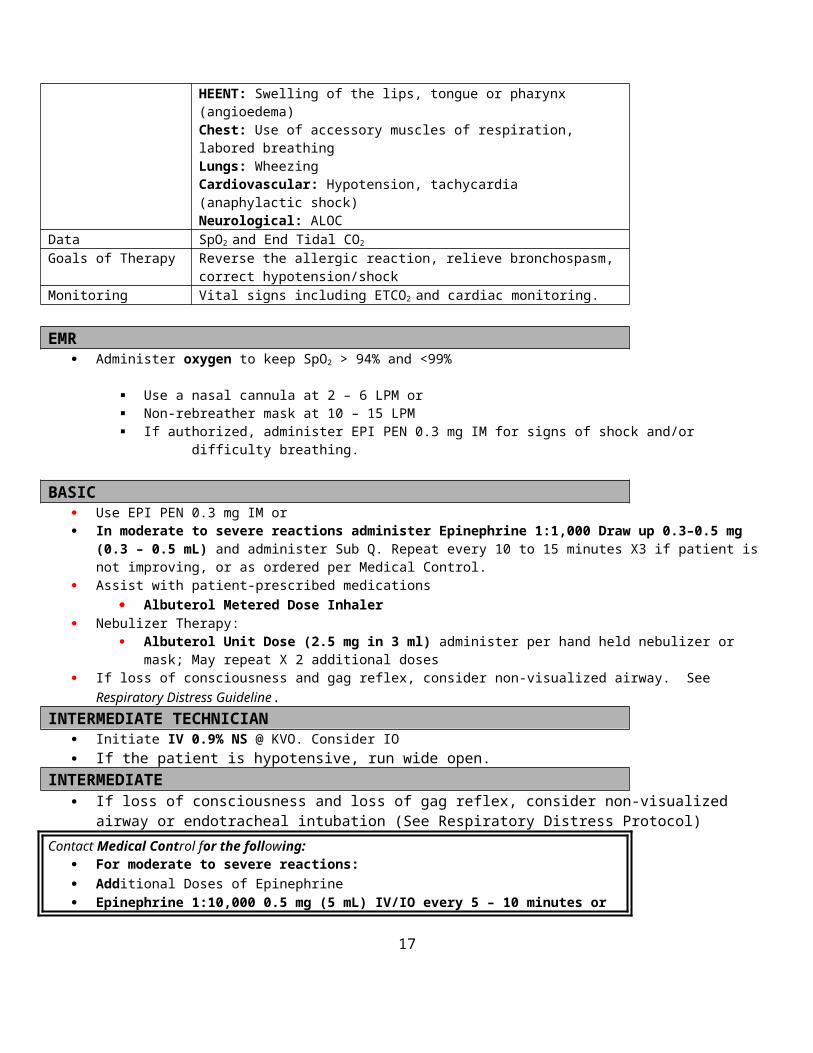
General: Identify degree of severity: mild moderate or severe [1].Skin: Urticaria (hives)HEENT: Swelling of the lips, tongue or pharynx (angioedema)Chest: Use of accessory muscles of respiration, labored breathingLungs: WheezingCardiovascular: Hypotension, tachycardia (anaphylactic shock)Neurological: ALOC
Data SpO2 and End Tidal CO2
Goals of Therapy Reverse the allergic reaction, relieve bronchospasm, correct hypotension/shockMonitoring Vital signs including ETCO2 and cardiac monitoring.
EMR Administer oxygen to keep SpO2 > 94% and <99%
Use a nasal cannula at 2 – 6 LPM or Non-rebreather mask at 10 – 15 LPM If authorized, administer EPI PEN 0.3 mg IM for signs of shock and/or difficulty breathing.
14

BASIC Use EPI PEN 0.3 mg IM or In moderate to severe reactions administer Epinephrine 1:1,000 Draw up 0.3–0.5 mg (0.3 – 0.5 mL) and administer Sub
Q. Repeat every 10 to 15 minutes X3 if patient is not improving, or as ordered per Medical Control. Assist with patient-prescribed medications
Albuterol Metered Dose Inhaler Nebulizer Therapy:
Albuterol Unit Dose (2.5 mg in 3 ml) administer per hand held nebulizer or mask; May repeat X 2 additional doses If loss of consciousness and gag reflex, consider non-visualized airway. See Respiratory Distress Guideline.
INTERMEDIATE TECHNICIAN Initiate IV 0.9% NS @ KVO. Consider IO If the patient is hypotensive, run wide open.
INTERMEDIATE If loss of consciousness and loss of gag reflex, consider non-visualized airway or endotracheal intubation (See
Respiratory Distress Protocol)Contact Medical Control for the following:
For moderate to severe reactions: Additional Doses of Epinephrine Epinephrine 1:10,000 0.5 mg (5 mL) IV/IO every 5 – 10 minutes or as ordered by Medical
Control. Consider Epinephrine 1:10,000 0.3-0.5mg (3 - 5 mL) ET, if you cannot obtain peripheral IV
access. Flush with 2 ml NS.
PARAMEDIC Benadryl 50 mg IV/IO for mild, moderate or severe reactions or IM if IV cannot be established. Solu-Medrol 125 mg IV/IO for moderate to severe reactions
Contact Medical Control for the following: For moderate to severe reactions: Epinephrine 1:10,000 0.5 mg (5 mL) IV/IO every 5 – 10 minutes or as ordered by Medical
Control. Glucagon 1 mg IV if the patient is taking Beta Blockers and is not responding to Epinephrine. Additional orders
FOOTNOTES:[1] Severity of Allergy/Anaphylaxis
Mild Allergic reaction : localized or generalized urticaria, without swelling of oral or pharyngeal structures, difficulty breathing, hypotension or ALOC;
Moderate Allergic Reaction : oral or pharyngeal swelling is present, mild to moderate difficulty breathing and wheezing are present.
Severe Allergic Reaction (Anaphylaxis) : moderate to severe difficulty breathing is present, hypotension is present and ALOC may occur.
Date of Origin: 11/10/11
15
Washington County
Suggested Medical Guidelines
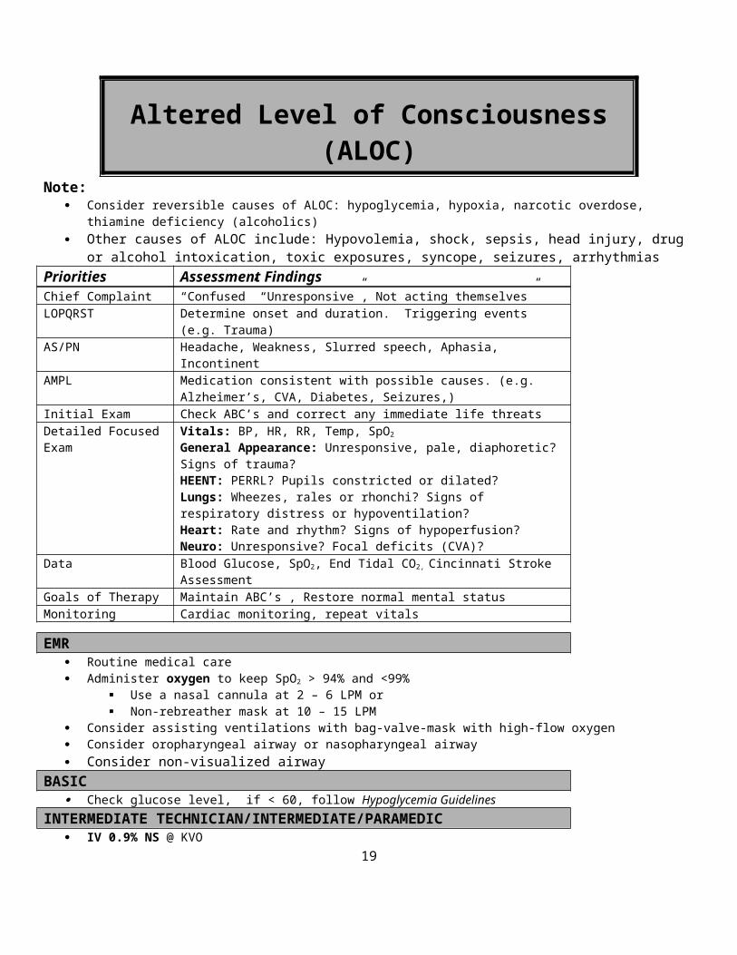
Altered Level of Consciousness (ALOC)Note:
Consider reversible causes of ALOC: hypoglycemia, hypoxia, narcotic overdose, thiamine deficiency (alcoholics) Other causes of ALOC include: Hypovolemia, shock, sepsis, head injury, drug or alcohol intoxication, toxic
exposures, syncope, seizures, arrhythmiasPriorities Assessment FindingsChief Complaint “Confused” “Unresponsive”, Not acting themselves”LOPQRST Determine onset and duration. Triggering events (e.g. Trauma)AS/PN Headache, Weakness, Slurred speech, Aphasia, IncontinentAMPL Medication consistent with possible causes. (e.g. Alzheimer’s, CVA, Diabetes,
Seizures,)Initial Exam Check ABC’s and correct any immediate life threatsDetailed Focused Exam Vitals: BP, HR, RR, Temp, SpO2
General Appearance: Unresponsive, pale, diaphoretic? Signs of trauma?HEENT: PERRL? Pupils constricted or dilated?Lungs: Wheezes, rales or rhonchi? Signs of respiratory distress or hypoventilation?Heart: Rate and rhythm? Signs of hypoperfusion? Neuro: Unresponsive? Focal deficits (CVA)?
Data Blood Glucose, SpO2, End Tidal CO2, Cincinnati Stroke AssessmentGoals of Therapy Maintain ABC’s , Restore normal mental statusMonitoring Cardiac monitoring, repeat vitals
EMR Routine medical care Administer oxygen to keep SpO2 > 94% and <99%
Use a nasal cannula at 2 – 6 LPM or Non-rebreather mask at 10 – 15 LPM
Consider assisting ventilations with bag-valve-mask with high-flow oxygen Consider oropharyngeal airway or nasopharyngeal airway Consider non-visualized airway
BASIC Check glucose level, if < 60, follow Hypoglycemia Guidelines
INTERMEDIATE TECHNICIAN/INTERMEDIATE/PARAMEDIC IV 0.9% NS @ KVO Consider IO (if Authorized) If SBP < 100 mmHg, initiate a fluid bolus of 250 ml Normal Saline. If a narcotic overdose is suspected, consider Narcan 0.4mg – 2mg (May be given intranasal) Refer to Toxic
Exposure/Overdose Guidelines. Suspected toxic overdose, refer to Toxic Exposure/Overdose Guidelines
Date of Origin: 11/10/10
16
Washington County
Suggested Medical Guidelines
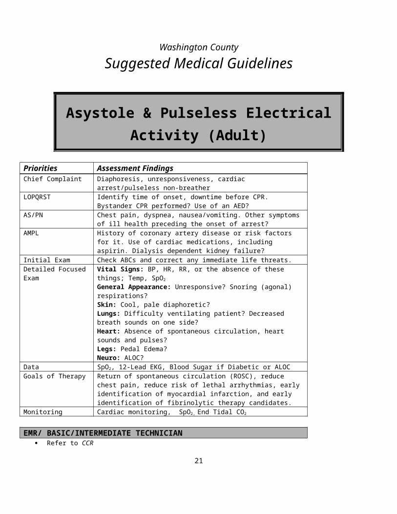
Asystole & Pulseless Electrical Activity (Adult)
Priorities Assessment FindingsChief Complaint Diaphoresis, unresponsiveness, cardiac arrest/pulseless non-breatherLOPQRST Identify time of onset, downtime before CPR. Bystander CPR performed? Use
of an AED?AS/PN Chest pain, dyspnea, nausea/vomiting. Other symptoms of ill health preceding
the onset of arrest?AMPL History of coronary artery disease or risk factors for it. Use of cardiac
medications, including aspirin. Dialysis dependent kidney failure?Initial Exam Check ABCs and correct any immediate life threats.Detailed Focused Exam Vital Signs: BP, HR, RR, or the absence of these things; Temp, SpO2
General Appearance: Unresponsive? Snoring (agonal) respirations? Skin: Cool, pale diaphoretic?Lungs: Difficulty ventilating patient? Decreased breath sounds on one side?Heart: Absence of spontaneous circulation, heart sounds and pulses?Legs: Pedal Edema?Neuro: ALOC?
Data SpO2, 12-Lead EKG, Blood Sugar if Diabetic or ALOCGoals of Therapy Return of spontaneous circulation (ROSC), reduce chest pain, reduce risk of
lethal arrhythmias, early identification of myocardial infarction, and early identification of fibrinolytic therapy candidates.
Monitoring Cardiac monitoring, SpO2, End Tidal CO2
EMR/ BASIC/INTERMEDIATE TECHNICIAN Refer to CCR
INTERMEDIATE/ PARAMEDIC Apply a cardiac monitor If Asystole appears on the monitor, confirm true asystole
o Check on/off switcheso Check leadso Check gain and sensitivity settingso Confirm asystole in 2 or 3 leads
Resume CPR immediately for 5 cycles When IV/IO is available, give vasopressor
o Vasopressin 40 units oro Epinephrine (1:10,000) 1 mg IV/IO every 3-5 minutes -or-o If IV/IO is not available administer Epinephrine (1:10,000) 2.0 – 2.5 mg ET in 10cc saline every 3 – 5 minutes
17

Identify and correct reversible causes: The Seven H’s and the Five T’so This applies mostly to PEA, but to a lesser extent, asystole, as well.o “The Seven H’s” (treatment orders are in parentheses)
Hypovolemia (Infuse Normal Saline wide open) Hypoxia (Administer high-flow oxygen and perform ventilation [1]) Hydrogen Ion, i.e. acidosis (Perform ventilation [1]) (EMT-P: Give Sodium Bicarbonate 1 amp IV) Hyperkalemia [2]
(EMT-P: Give 10 ml Calcium Chloride 10% IV over 2 – 5 minutes. May repeat X 1) (EMT-P: Give Sodium Bicarbonate 1 amp IV) (EMT-I/P: Albuterol nebulizer treatment with 1 – 2 Unit Doses)
Hypokalemia (Even if hypokalemia is suspected, it is not treated in the field.) Hypothermia (See Hypothermia and Frostbite Guidelines) Hypoglycemia (See Hypoglycemia Guidelines)
o “The Five T’s” (treatment orders are in parentheses) Tablets (See Toxic Exposure and Overdose Guidelines) Tamponade Tension pneumothorax (Perform bilateral needle decompression) Thrombosis, cardiac i.e. myocardial infarction (No specific prehospital treatment available) Thrombosis, pulmonary i.e. pulmonary embolism (No specific prehospital treatment available)
Initiate transcutaneous pacing (TCP) if patient is in Asystole. Set the HR at 70 – 80 beats/min Set the voltage at 40 mA initially and watch for the pacer spikes on the monitor Increase voltage by 20 mA every 3-5 seconds until there is 100% capture:
a wide QRS complex appears on the monitor after every pacer spike a pulse can be felt in the femoral or carotid artery after every QRS complex
Then increase the voltage by 10%
PARAMEDIC For suspected tricyclic antidepressant overdose administer Sodium Bicarb 1 AMP IVP Suspected Beta blocker overdose administer Glucagon 1mg IVP
Contact Medical Control for the following: Additional orders Consider termination of resuscitation efforts
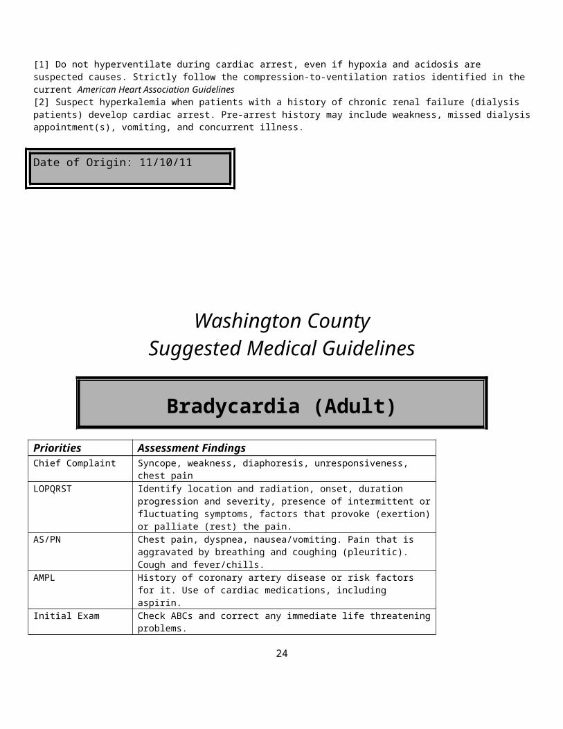
FOOTNOTES:[1] Do not hyperventilate during cardiac arrest, even if hypoxia and acidosis are suspected causes. Strictly follow the compression-to-ventilation ratios identified in the current American Heart Association Guidelines[2] Suspect hyperkalemia when patients with a history of chronic renal failure (dialysis patients) develop cardiac arrest. Pre-arrest history may include weakness, missed dialysis appointment(s), vomiting, and concurrent illness.
Date of Origin: 11/10/11
18
Washington CountySuggested Medical Guidelines
Bradycardia (Adult)
Priorities Assessment FindingsChief Complaint Syncope, weakness, diaphoresis, unresponsiveness, chest painLOPQRST Identify location and radiation, onset, duration progression and severity,
presence of intermittent or fluctuating symptoms, factors that provoke (exertion) or palliate (rest) the pain.
AS/PN Chest pain, dyspnea, nausea/vomiting. Pain that is aggravated by breathing and coughing (pleuritic). Cough and fever/chills.
AMPL History of coronary artery disease or risk factors for it. Use of cardiac medications, including aspirin.
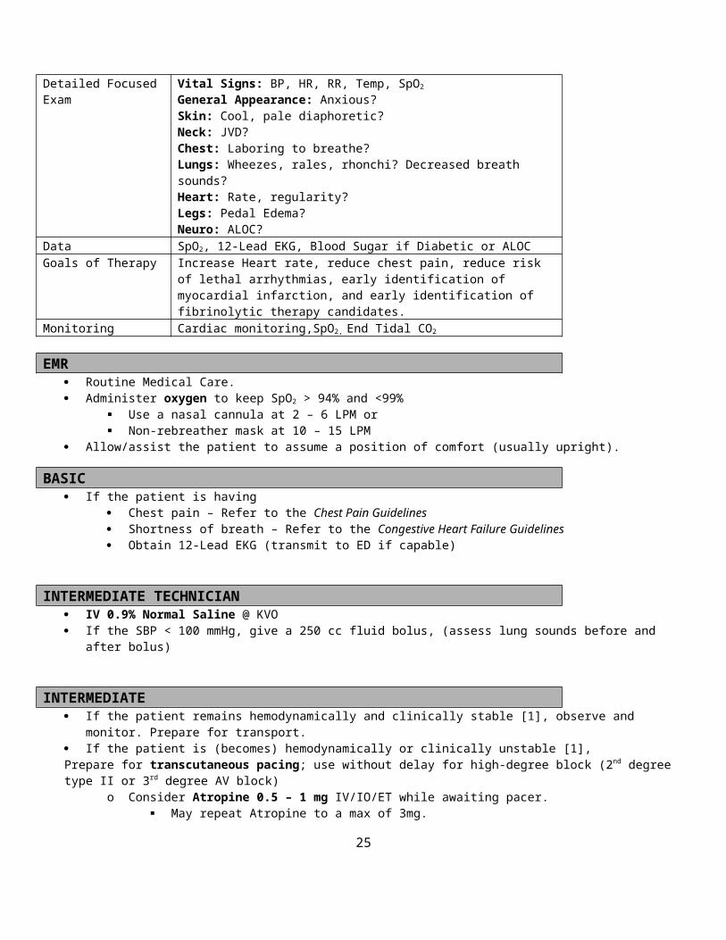
Initial Exam Check ABCs and correct any immediate life threatening problems.Detailed Focused Exam Vital Signs: BP, HR, RR, Temp, SpO2
General Appearance: Anxious?Skin: Cool, pale diaphoretic?Neck: JVD?Chest: Laboring to breathe?Lungs: Wheezes, rales, rhonchi? Decreased breath sounds?Heart: Rate, regularity?Legs: Pedal Edema?Neuro: ALOC?
Data SpO2, 12-Lead EKG, Blood Sugar if Diabetic or ALOCGoals of Therapy Increase Heart rate, reduce chest pain, reduce risk of lethal arrhythmias, early
identification of myocardial infarction, and early identification of fibrinolytic therapy candidates.
Monitoring Cardiac monitoring,SpO2, End Tidal CO2
EMR Routine Medical Care. Administer oxygen to keep SpO2 > 94% and <99%
Use a nasal cannula at 2 – 6 LPM or Non-rebreather mask at 10 – 15 LPM
Allow/assist the patient to assume a position of comfort (usually upright).
BASIC If the patient is having
Chest pain – Refer to the Chest Pain Guidelines Shortness of breath – Refer to the Congestive Heart Failure Guidelines Obtain 12-Lead EKG (transmit to ED if capable)
INTERMEDIATE TECHNICIAN IV 0.9% Normal Saline @ KVO If the SBP < 100 mmHg, give a 250 cc fluid bolus, (assess lung sounds before and after bolus)
19

INTERMEDIATE If the patient remains hemodynamically and clinically stable [1], observe and monitor. Prepare for transport. If the patient is (becomes) hemodynamically or clinically unstable [1],Prepare for transcutaneous pacing; use without delay for high-degree block (2nd degree type II or 3rd degree AV block)
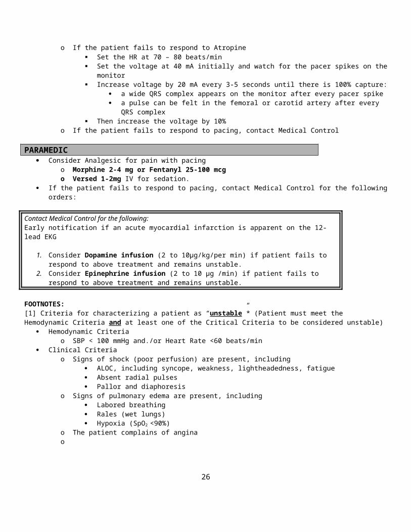
o Consider Atropine 0.5 – 1 mg IV/IO/ET while awaiting pacer. May repeat Atropine to a max of 3mg.
o If the patient fails to respond to Atropine Set the HR at 70 – 80 beats/min Set the voltage at 40 mA initially and watch for the pacer spikes on the monitor Increase voltage by 20 mA every 3-5 seconds until there is 100% capture:
a wide QRS complex appears on the monitor after every pacer spike a pulse can be felt in the femoral or carotid artery after every QRS complex
Then increase the voltage by 10%o If the patient fails to respond to pacing, contact Medical Control
PARAMEDIC Consider Analgesic for pain with pacing
o Morphine 2-4 mg or Fentanyl 25-100 mcg o Versed 1-2mg IV for sedation.
If the patient fails to respond to pacing, contact Medical Control for the following orders:
Contact Medical Control for the following:Early notification if an acute myocardial infarction is apparent on the 12-lead EKG
1. Consider Dopamine infusion (2 to 10µg/kg/per min) if patient fails to respond to above treatment and remains unstable.
2. Consider Epinephrine infusion (2 to 10 µg /min) if patient fails to respond to above treatment and remains unstable.
FOOTNOTES:[1] Criteria for characterizing a patient as “unstable”* (Patient must meet the Hemodynamic Criteria and at least one of the Critical Criteria to be considered unstable)
Hemodynamic Criteriao SBP < 100 mmHg and./or Heart Rate <60 beats/min
Clinical Criteriao Signs of shock (poor perfusion) are present, including
ALOC, including syncope, weakness, lightheadedness, fatigue Absent radial pulses Pallor and diaphoresis
o Signs of pulmonary edema are present, including Labored breathing Rales (wet lungs) Hypoxia (SpO2 <90%)
o The patient complains of anginao
Date of Origin: 11/10/11
20
Washington County
Suggested Medical Guidelines
Burns
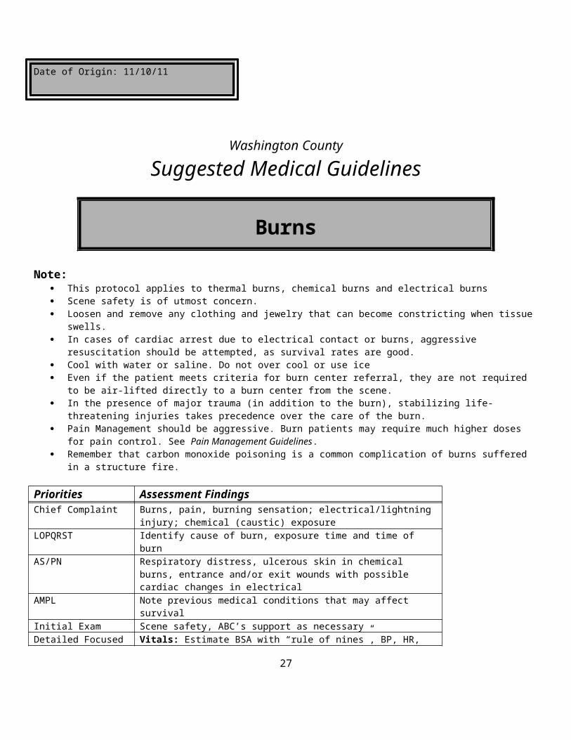
Note: This protocol applies to thermal burns, chemical burns and electrical burns Scene safety is of utmost concern. Loosen and remove any clothing and jewelry that can become constricting when tissue swells. In cases of cardiac arrest due to electrical contact or burns, aggressive resuscitation should be attempted, as survival rates are
good. Cool with water or saline. Do not over cool or use ice Even if the patient meets criteria for burn center referral, they are not required to be air-lifted directly to a burn center from
the scene. In the presence of major trauma (in addition to the burn), stabilizing life-threatening injuries takes precedence over the care
of the burn. Pain Management should be aggressive. Burn patients may require much higher doses for pain control. See Pain
Management Guidelines. Remember that carbon monoxide poisoning is a common complication of burns suffered in a structure fire.
Priorities Assessment FindingsChief Complaint Burns, pain, burning sensation; electrical/lightning injury; chemical (caustic)
exposureLOPQRST Identify cause of burn, exposure time and time of burnAS/PN Respiratory distress, ulcerous skin in chemical burns, entrance and/or exit
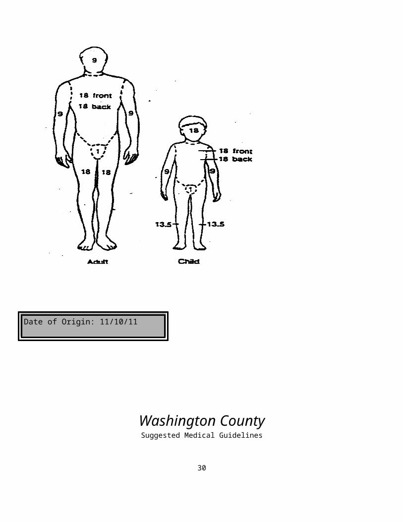
wounds with possible cardiac changes in electricalAMPL Note previous medical conditions that may affect survivalInitial Exam Scene safety, ABC’s support as necessaryDetailed Focused Exam Vitals: Estimate BSA with “rule of nines”, BP, HR, RR, Temp, SpO2,
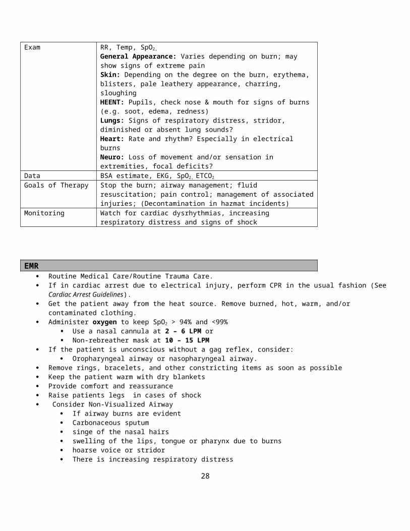
General Appearance: Varies depending on burn; may show signs of extreme painSkin: Depending on the degree on the burn, erythema, blisters, pale leathery appearance, charring, sloughingHEENT: Pupils, check nose & mouth for signs of burns (e.g. soot, edema, redness)Lungs: Signs of respiratory distress, stridor, diminished or absent lung sounds?Heart: Rate and rhythm? Especially in electrical burns Neuro: Loss of movement and/or sensation in extremities, focal deficits?
Data BSA estimate, EKG, SpO2, ETCO2
Goals of Therapy Stop the burn; airway management; fluid resuscitation; pain control; management of associated injuries; (Decontamination in hazmat incidents)
Monitoring Watch for cardiac dysrhythmias, increasing respiratory distress and signs of shock
EMR Routine Medical Care/Routine Trauma Care.
21

If in cardiac arrest due to electrical injury, perform CPR in the usual fashion (See Cardiac Arrest Guidelines). Get the patient away from the heat source. Remove burned, hot, warm, and/or contaminated clothing. Administer oxygen to keep SpO2 > 94% and <99%
Use a nasal cannula at 2 – 6 LPM or Non-rebreather mask at 10 – 15 LPM
If the patient is unconscious without a gag reflex, consider: Oropharyngeal airway or nasopharyngeal airway.
Remove rings, bracelets, and other constricting items as soon as possible Keep the patient warm with dry blankets Provide comfort and reassurance Raise patients legs in cases of shock Consider Non-Visualized Airway
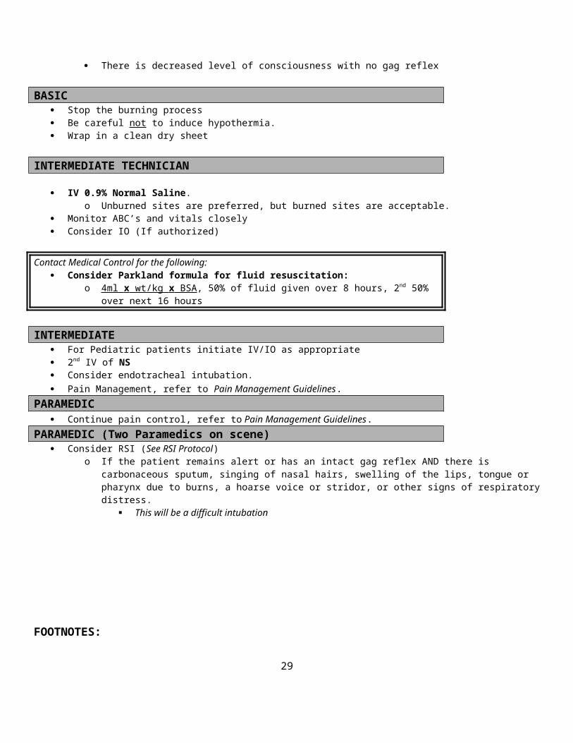
If airway burns are evident Carbonaceous sputum singe of the nasal hairs swelling of the lips, tongue or pharynx due to burns hoarse voice or stridor There is increasing respiratory distress There is decreased level of consciousness with no gag reflex
BASIC Stop the burning process Be careful not to induce hypothermia. Wrap in a clean dry sheet
INTERMEDIATE TECHNICIAN
IV 0.9% Normal Saline.o Unburned sites are preferred, but burned sites are acceptable.
Monitor ABC’s and vitals closely Consider IO (If authorized)
Contact Medical Control for the following: Consider Parkland formula for fluid resuscitation:
o 4ml x wt/kg x BSA , 50% of fluid given over 8 hours, 2nd 50% over next 16 hours
INTERMEDIATE For Pediatric patients initiate IV/IO as appropriate 2nd IV of NS Consider endotracheal intubation. Pain Management, refer to Pain Management Guidelines.
PARAMEDIC Continue pain control, refer to Pain Management Guidelines.
PARAMEDIC (Two Paramedics on scene) Consider RSI (See RSI Protocol)
o If the patient remains alert or has an intact gag reflex AND there is carbonaceous sputum, singing of nasal hairs, swelling of the lips, tongue or pharynx due to burns, a hoarse voice or stridor, or other signs of respiratory distress.
This will be a difficult intubation
22

FOOTNOTES:
Date of Origin: 11/10/11
Washington County23
Suggested Medical Guidelines
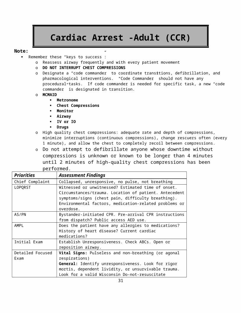
Cardiac Arrest -Adult (CCR)Note:
Remember these “keys to success”: o Reassess airway frequently and with every patient movemento DO NOT INTERRUPT CHEST COMPRESSIONSo Designate a “code commander” to coordinate transitions, defibrillation, and pharmacological interventions. “Code
Commander” should not have any procedural tasks. If code commander is needed for specific task, a new “code commander” is designated in transition.
o MCMAID Metronome Chest Compressions Monitor Airway IV or IO Drugs
o High quality chest compressions: adequate rate and depth of compressions, minimize interruptions (continuous compressions), change rescuers often (every 1 minute), and allow the chest to completely recoil between compressions.
o Do not attempt to defibrillate anyone whose downtime without compressions is unknown or known to be longer than 4 minutes until 2 minutes of high-quality chest compressions has been performed.
Priorities Assessment FindingsChief Complaint Collapsed, unresponsive, no pulse, not breathing LOPQRST Witnessed or unwitnessed? Estimated time of onset. Circumstances/trauma.
Location of patient. Antecedent symptoms/signs (chest pain, difficulty breathing). Environmental factors, medication-related problems or overdose.
AS/PN Bystander-initiated CPR. Pre-arrival CPR instructions from dispatch? Public access AED use.
AMPL Does the patient have any allergies to medications? History of heart disease? Current cardiac medications?
Initial Exam Establish Unresponsiveness. Check ABCs. Open or reposition airway.Detailed Focused Exam Vital Signs: Pulseless and non-breathing (or agonal respirations)
General: Identify unresponsiveness. Look for rigor mortis, dependent lividity, or unsurvivable trauma. Look for a valid Wisconsin Do-not-resuscitate bracelet.Skin: Warm/cold, dependent lividity, rash, ecchymosis?HEENT: Airway patent, foreign bodies (e.g. dentures), neck swelling or trauma, trachea in midline?Chest: Spontaneous respirations, subcutaneous air or crepitation, or deformity?Lungs: Equal breath sounds, difficulty bagging or ventilating?Cardiovascular: Absence of heart sounds, carotid or femoral pulses?Abdomen: Distended?Extremities: Rigor mortis, edema, deformity?Neurological: Unresponsive to verbal and painful stimulation?
Data Cardiac rhythm analysis, blood sugar, SpO2
Goals of Therapy Return of spontaneous circulation (ROSC)Monitoring BP, HR, RR, EKG, SPO2 ETCO2
24
EMR/ BASIC Establish that the patient is unresponsive, without a pulse, and not breathing Rule out DNR status, dependent lividity, rigor mortis: Place an oral airway and O2 via non-rebreather mask at 15 LPM while compressions ongoing First Priority: Perform High-Quality Chest Compressions
o Perform a head-tilt-chin-lift maneuver. If suspected C-spine injury, use the jaw thrust, instead.
o Insert an OPA. Ensure airway is patent and document.o Apply O2 via NRB at 15 lpmo Compress at a rate of 100 times per minute.
“Push hard and push fast”. In adults compress the chest at least 2 inches. Allow the chest to recoil completely between compressions. Change compressors every 1minute
Second Priority: Defibrillate (Repeated every 2 minutes)o Paramedic with Manual Defibrillator must communicate with compressors.
At 1 minute and 50 seconds charge the defibrillator to the recommended power setting for defibrillation. At 2 minutes tell the compressor to stop and analyze the rhythm
V-Fib or pulseless V-Tach deliver the shock. If the rhythm is not a shockable rhythm “dump” the charge.
Instruct the compressor to resume compressions. o Interruption of chest compressions should not exceed 5 secondso If using an AED follow the prompts from the AED
INTERMEDIATE TECHNICIAN Third Priority: Establish venous access
o Initiate IV 0.9% Normal Saline If pt. does not have a good IV site go directly to IOo Run the IV wide open or appropriate rate, if IO then use pressure infuser or BP cuff
Fourth Priority: Non-visualized/Visualized airwayDO NOT interrupt compressions to place an airway.
o After 6 minutes of CCR insert an appropriate airway (Non-visualized for appropriately trained and certified EMRs or Basics) (Visualized for appropriately trained and certified Intermediate Technicians) (Visualized for Intermediates or Paramedics). This may be done sooner if the initial cardiac rhythm is Asystole, there is a known respiratory problem or there is a specific need to protect the airway (e.g., a large volume of gastric contents in the pharynx)
o Do not interrupt compressions to place an airway.o After one failed ETT attempt go to non-visualized airway o After the visualized airway is in place…
Continue compressions uninterrupted at least 100 times per minute. Give a single rescue breath 6 times per minute (every 10 seconds). Or titrate respirations to maintain
ETCO2 between 30 and 50 mmHG Attach End Tidal CO2 monitor
INTERMEDIATE/PARAMEDIC Ensure basic rescuer interventions are continued. Initiate cardiac rhythm monitoring and analysis Initiate manual defibrillation after every two minutes of chest compressions, if indicated. (As noted above) If an advanced airway is not already in place, insert an advanced airway.(As noted above) Consider venous access, if not already done
o Drug administration routes in order of preference: IV – IO Lack of venous access is not an acceptable indication for converting a non-visualized airway that is
functioning well for ventilations to an ET tube.25
Rather, use IO access. Fifth Priority: ACLS resuscitation medications
o Vasopressors should be given as soon as IV/IO is establishedo Give 1mg Epinephrine 1:10,000 first unless the pt. has had excellent chest compressions and short down time as
well as agonal respirations, then give 40 units of Vasopressin.o 2nd dose of a pressor agent after 4 minutes should be 40 units of Vasopressin unless already used as noted above,
continue Epinephrine every 4 minutes afterwardo If persistent or re-current V-Fib or Pulseless V-Tach then give 300 mg of Amiodarone (give amiodarone after
seeing v-fib or pulseless v-tach at 2 rhythm checks)o After 4 minutes of chest compressions and there is persistent V-Fib or Pulseless V-Tach then give 150 mg of
Amiodaroneo Consider Renal Failure or Tricyclic antidepressant overdose and give 1mEq/kg of Sodium Bicarbonate (Paramedic
only) and 10mg/kg of Calcium Chloride.(Paramedic Only)o If persistent V-Fib or Pulseless V-Tach then give 1-2 grams of Magnesium Sulfate (Paramedic only)
If there is Return of Spontaneous Circulation(ROSC), o Amiodorone 150mg(1amp), over 10 min (15mg/min)o Amiodorone drip at 1mg/min (Paramedic only)
Combine (1) amp of Amiodarone (150 mg) to 150 ml of NS in burette vessel. Administer Amiodarone solution at 1 mg per minute (60ml/hr or 1 drip every second)
o Reassess the need for airway devices Maintain the advanced airway if tolerated by the patient If the patient regains consciousness consider sedation to allow the advanced airway to remain in place. If removal of the advanced airway is necessary refer to the procedures for removing advanced airway
devices in the Respiratory Distress Guidelines.o Monitor patient
Consider treatment for STEMI if documented Consider nasogastric tube (NGT) for gastric decompression (paramedic only)
Contact Medical Control for the following: Additional medication orders To discuss termination of resuscitation in the absence of a valid Wisconsin DNR Bracelet
FOOTNOTES:
Date of Origin: 11/10/11
26
Washington County
Suggested Medical Guidelines
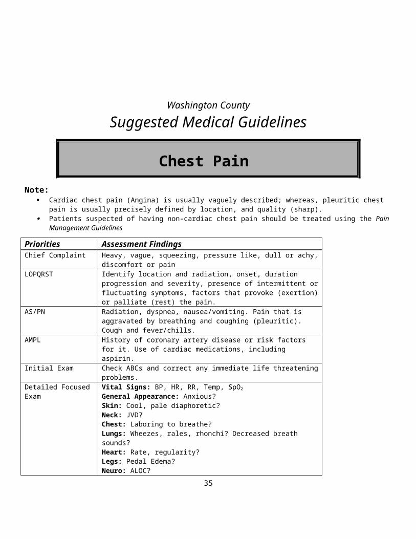
Chest Pain Note:
Cardiac chest pain (Angina) is usually vaguely described; whereas, pleuritic chest pain is usually precisely defined by location, and quality (sharp).
Patients suspected of having non-cardiac chest pain should be treated using the Pain Management Guidelines
Priorities Assessment FindingsChief Complaint Heavy, vague, squeezing, pressure like, dull or achy, discomfort or painLOPQRST Identify location and radiation, onset, duration progression and severity,
presence of intermittent or fluctuating symptoms, factors that provoke (exertion) or palliate (rest) the pain.
AS/PN Radiation, dyspnea, nausea/vomiting. Pain that is aggravated by breathing and coughing (pleuritic). Cough and fever/chills.
AMPL History of coronary artery disease or risk factors for it. Use of cardiac medications, including aspirin.
Initial Exam Check ABCs and correct any immediate life threatening problems.Detailed Focused Exam Vital Signs: BP, HR, RR, Temp, SpO2
General Appearance: Anxious?Skin: Cool, pale diaphoretic?Neck: JVD?Chest: Laboring to breathe?Lungs: Wheezes, rales, rhonchi? Decreased breath sounds?Heart: Rate, regularity?Legs: Pedal Edema?Neuro: ALOC?
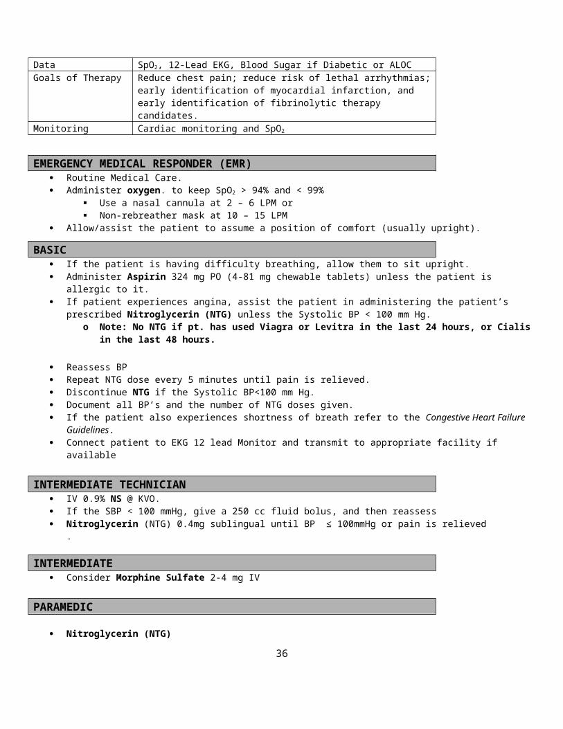
Data SpO2, 12-Lead EKG, Blood Sugar if Diabetic or ALOCGoals of Therapy Reduce chest pain; reduce risk of lethal arrhythmias; early identification of
myocardial infarction, and early identification of fibrinolytic therapy candidates.
Monitoring Cardiac monitoring and SpO2
EMERGENCY MEDICAL RESPONDER (EMR) Routine Medical Care. Administer oxygen. to keep SpO2 > 94% and < 99%
Use a nasal cannula at 2 – 6 LPM or Non-rebreather mask at 10 – 15 LPM
Allow/assist the patient to assume a position of comfort (usually upright).
BASIC If the patient is having difficulty breathing, allow them to sit upright. Administer Aspirin 324 mg PO (4-81 mg chewable tablets) unless the patient is allergic to it. If patient experiences angina, assist the patient in administering the patient’s prescribed Nitroglycerin (NTG) unless the
Systolic BP < 100 mm Hg.o Note: No NTG if pt. has used Viagra or Levitra in the last 24 hours, or Cialis in the last 48 hours.
27
Reassess BP Repeat NTG dose every 5 minutes until pain is relieved. Discontinue NTG if the Systolic BP<100 mm Hg. Document all BP’s and the number of NTG doses given. If the patient also experiences shortness of breath refer to the Congestive Heart Failure Guidelines. Connect patient to EKG 12 lead Monitor and transmit to appropriate facility if available
INTERMEDIATE TECHNICIAN IV 0.9% NS @ KVO. If the SBP < 100 mmHg, give a 250 cc fluid bolus, and then reassess Nitroglycerin (NTG) 0.4mg sublingual until BP ≤ 100mmHg or pain is relieved
.
INTERMEDIATE Consider Morphine Sulfate 2-4 mg IV
PARAMEDIC
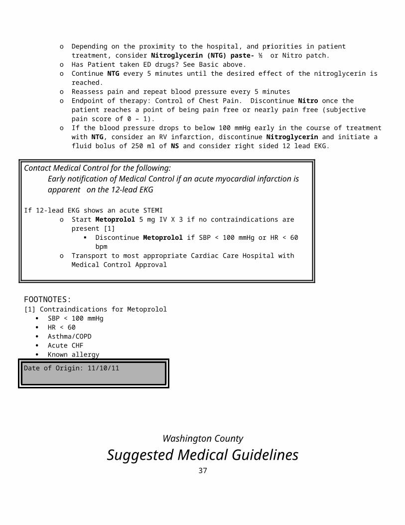
Nitroglycerin (NTG) o Depending on the proximity to the hospital, and priorities in patient treatment, consider Nitroglycerin (NTG) paste-
½” or Nitro patch.o Has Patient taken ED drugs? See Basic above. o Continue NTG every 5 minutes until the desired effect of the nitroglycerin is reached. o Reassess pain and repeat blood pressure every 5 minutes o Endpoint of therapy: Control of Chest Pain. Discontinue Nitro once the patient reaches a point of being pain free or
nearly pain free (subjective pain score of 0 – 1). o If the blood pressure drops to below 100 mmHg early in the course of treatment with NTG, consider an RV
infarction, discontinue Nitroglycerin and initiate a fluid bolus of 250 ml of NS and consider right sided 12 lead EKG.
Contact Medical Control for the following:Early notification of Medical Control if an acute myocardial infarction is apparent on the 12-lead EKG
If 12-lead EKG shows an acute STEMIo Start Metoprolol 5 mg IV X 3 if no contraindications are present [1]
Discontinue Metoprolol if SBP < 100 mmHg or HR < 60 bpm o Transport to most appropriate Cardiac Care Hospital with Medical Control Approval
FOOTNOTES:[1] Contraindications for Metoprolol
SBP < 100 mmHg HR < 60 Asthma/COPD Acute CHF Known allergy
Date of Origin: 11/10/11
28
Washington County
Suggested Medical Guidelines
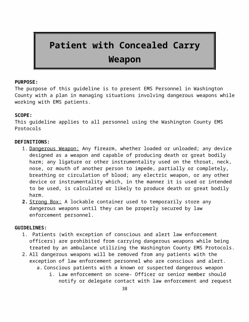
Patient with Concealed Carry Weapon
PURPOSE:The purpose of this guideline is to present EMS Personnel in Washington County with a plan in managing situations involving dangerous weapons while working with EMS patients.
SCOPE:This guideline applies to all personnel using the Washington County EMS Protocols
DEFINITIONS:1. Dangerous Weapon: Any firearm, whether loaded or unloaded; any device designed as a weapon and
capable of producing death or great bodily harm; any ligature or other instrumentality used on the throat, neck, nose, or mouth of another person to impede, partially or completely, breathing or circulation of blood; any electric weapon, or any other device or instrumentality which, in the manner it is used or intended to be used, is calculated or likely to produce death or great bodily harm.
2. Strong Box: A lockable container used to temporarily store any dangerous weapons until they can be properly secured by law enforcement personnel.
GUIDELINES:1. Patients (with exception of conscious and alert law enforcement officers) are prohibited from carrying
dangerous weapons while being treated by an ambulance utilizing the Washington County EMS Protocols.
2. All dangerous weapons will be removed from any patients with the exception of law enforcement personnel who are conscious and alert.
a. Conscious patients with a known or suspected dangerous weaponi. Law enforcement on scene- Officer or senior member should notify or delegate contact
with law enforcement and request that they remove the weapon. Law enforcement will be responsible for making arrangements to secure and store the weapon with the patient if necessary.
ii. Law enforcement not on scene – Officer or senior member should notify patient that dangerous weapons are not allowed in EMS transport vehicles. Request patient to remove holster, sheath, case, etc. with weapon stored. The weapon should be secured by patient in own residence, transferred to a family member or placed by patient into strongbox and secured. If the individual refuses, it will be considered a refusal for medical care. If the individual becomes belligerent or threatening, all EMS personnel
29
will evacuate the scene to a safe area and contact dispatch to request immediate response from law enforcement. EMS personnel can return to the scene once it has been secured by law enforcement.
b. Unconscious patient with a known or suspected dangerous weaponi. Law enforcement on scene – Officer or senior member should notify or delegate contact
with law enforcement to secure and store the weapon.ii. Law enforcement not on scene – Officer of senior member should delegate the most
“weapon experienced” EMS individual on scene to remove the holster, sheath, case, etc. with the weapon secured for placement into the strongbox. The weapon will not be handled any more than is necessary to be placed in the strongbox.
3. If a dangerous weapon is found while transporting, the vehicle will be pulled over and come to a complete stop while addressing the situation based on guidelines above for conscious and unconscious patients.
4. When a dangerous weapon is secured in a strongbox, the Officer or senior member will notify the hospital loss prevention/security as soon as possible. Upon arrival to the hospital they will turn the weapon over to the loss prevention/security in the strongbox and receive an empty strongbox back from the hospital.
Date of Origin: 11/10/11
30
Washington County
Suggested Medical Guidelines
Congestive Heart Failure / Cardiogenic Shock
Note: Remember that acute myocardial infarction may present with shortness of breath (alone) and new onset acute congestive
heart failure!
Priorities Assessment FindingsChief Complaint “Difficulty breathing”; “Shortness of breath”LOPQRST Assess onset, duration, progression, subjective severity, possible triggering
events, and response to treatments before EMS arrival.AS/PN Cardiac chest pain, frothy sputum, blood tinged sputum AMPL Check past history of CHF or heart disease; medications for CHF (e.g.,
furosemide, digoxin, ACE inhibitors, long acting nitrates, etc.), and compliance with these medications.
Initial Exam Check ABCs and correct immediately life-threatening problems.Detailed Focused Exam General Appearance: Tripod positioning; Severity of distress [1]?
Skin: Cool, moist and pale? Warm, dry and flushed? Cyanotic?Neck: JVD?Respiratory Effort: Using accessory muscles, signs of fatigue; two-word sentences?Lung Sounds: The presence of rales (wet lungs) is a strong indication of CHF. Wheezing is also common in CHF.Heart Sounds: Rate, regularity.Peripheral Edema: Pitting edema of the ankles is common in CHF, but its absence does not rule out CHF.Neuro: ALOC? Lethargy? Somnolence?
Data SpO2, 12-Lead EKGGoals of Therapy Differentiate CHF from other causes of dyspnea, reduce the work of breathing,
improve pump function, and improve oxygenation and ventilation.Monitoring Carefully monitor blood pressure, respiratory effort, level of consciousness,
and SpO2 and End-tidal CO2
EMR
Routine Medical Care Allow/assist the patient to assume a position of comfort (usually upright). Administer oxygen to keep SpO2 > 94% and <99%
Use a nasal cannula at 2 – 6 LPM or Non-rebreather mask at 10 – 15 LPM
Assisted Ventilation: Consider assisting breathing with gentle synchronous ventilations with bag-valve mask (BVM); Support ventilation with BVM if apnea or hypopnea occurs.
Airway Adjuncts: If there is loss of consciousness and loss of gag reflex, insert an oropharyngeal or nasopharyngeal airway. Remember breathing may get worse when lying on their backs.
31

BASIC If the patient complains of chest pain (angina),
o Consider Aspirin 324 mg chewed and swallowed (except in aspirin allergic patients)o If the patient is prescribed nitroglycerin consider assisting them in taking providing systolic blood pressure >100mmHg
o Note: No NTG if pt has used Viagra or Levitra in the last 24 Hours, Cialis in the last 48 Hours If the patient is wheezing, assist with use of metered dose inhalers or give Albuterol Unit Dose (2.5 mg in 3 ml) administer
per hand held nebulizer or mask; Initiate CPAP at 5- 10 cm H2O. (See Respiratory Distress Guideline) To be used only by EMT’s with proper training and
certification by the medical director or authorized representative. Airway Adjuncts: If there is loss of consciousness and loss of gag reflex, insert an Advanced Airway
INTERMEDIATE TECHICIAN IV 0.9% NS @ KVO Give Nitroglycerin 0.4 mg (1) sublingual every 3-5 minutes. No maximum dose.Consider IV fluid if the patient in congestive heart failure is also hypotensive (SPB ≤ 100 mmHg).
INTERMEDIATE If SBP ≤ 100 mmHg, do not give Nitroglycerin, Furosemide or Morphine. Give Furosemide 40 mg IV or double the patient’s single dose. Consider Morphine 2 mg IV, if chest pain (angina) is present, may repeat once. Consider endotracheal intubation if CPAP fails, if there is ALOC, or if respiratory failure is imminent.
If the patient becomes anxious from the CPAP mask, consider o Valium 2.5-5 mg IV o Or, Ativan 0.5-2 mg IV/IM
Give Nitroglycerin 0.4 mg (1) every 3-5 minutes until SBP 110 mmHg. No maximum dose. If SBP ≤ 100 mmHg, do not give Nitroglycerin. Consider Nitro Paste- ½” or patch
Contact Medical Control for the following:o If SBP is less than or equal to 90, orders to administer a Dopamine infusion. Dopamine is
provided in premixed (400mg) in 250 ml D5W solution using 60 gtts tubing. This yields a concentration of 1600 mcg/ml. The initial rate of infusion is 2-20 mcg/kg/minute, which can be obtained with 24gtts/minute-infusion rate. Begin infusion at 24gtts/min and increase by 12gtts/min every 2 minutes and maintain a systolic BP of at least 100mmHG. Closely monitor vital signs.
o If patient has a cardiac dysrhythmia, treat the underlying rhythm disturbance according to the appropriate procedure.
o Transport as soon as possible. Transportation can be initiated at any time during this sequence.
Additional orders are needed Provide patient report ASAP
PARAMEDIC (Two Paramedics on scene) Consider RSI if any of the following indications is met: (See RSI protocol)
o A trial of CPAP fails to improve the work of breathing or oxygenation, or if the patient shows signs of deterioration on it
32
PARAMEDIC
o There is ALOC and the gag reflex is intacto Respiratory failure is imminent (e.g., severe fatigue)
FOOTNOTES:[1] Severity of Respiratory Distress:
Mild = RR<20 + minimal additional breathing effort + speaking in complete sentences + minimal subjective distress, No ALOC
Moderate = RR 20 to 25 + moderate additional breathing effort + difficult to complete a sentence + moderate subjective distress + No ALOC
Severe = RR> 25 + marked additional breathing effort + 2 or 3 word sentences + marked subjective distress + possible ALOC
Date of Origin: 11/10/11
33
Washington County
Suggested Medical Guidelines
EclampsiaNote:
Eclampsia occurs in pregnant patients with “preeclampsia”. Preeclampsia is a syndrome that involves hypertension [2] and output of protein in the urine.
o Preeclampsia occurs most often (but not exclusively) in non-white first-time mothers in their teens or early twenties from low socioeconomic backgrounds, or in mothers over the age of 35. Other risk factors include previous pregnancy with preeclampsia, multiple fetuses, a family history of preeclampsia and obesity.
Eclampsia occurs when seizures and/or coma develop between the 20th week of pregnancy and the 4th week after delivery There is a significant associated risk of death for the mother and the baby. Maternal complications of eclampsia include:
placental abruption, hemorrhagic stroke, pulmonary edema, cardiac arrest, and postpartum hemorrhage. Magnesium sulfate is the drug of choice for treating seizures in eclampsia.
Priorities Assessment FindingsChief Complaint Seizure/coma in mid to late pregnancy or within a month after deliveryLOPQRST Generalized, tonic-clonic type seizures (usually), lasting (3-4 minutes) that
resolve spontaneouslyAS/PN Has the doctor told her that she has developed high blood pressure during this
pregnancy with protein in the urine and swelling of the ankles? Has she had adequate prenatal care? Other symptoms of preeclampsia may be present [1].
AMPL If the mother has had adequate prenatal care, she may already know that she has a diagnosis of “preeclampsia”, or “pregnancy-induced hypertension”, but not always.
Initial Exam ABCs and correct any immediately life-threatening problemsDetailed Focused Exam Vital Signs: BP [2], HR, RR, Temp, SpO2
General Appearance: Seizing or postictal? Incontinent (bowel, bladder)?Skin: Pale, cool, moist? Cyanotic?Eyes: Dilated pupils during the seizure?Mouth: Frothy salivation? Tongue biting?Legs: Pedal edema?Neuro: Focal deficits? ALOC?
Data SpO2, Blood Sugar (to rule out hypoglycemia as a cause of the seizure)Goals of Therapy Protect the mother from injury during seizures, stop recurrences of the seizures.
Do not attempt to treat maternal hypertension in the field.Monitoring Blood pressure, HR, SpO2, Neuro Status; Cardiac Monitor for possible rhythm
disturbances
EMRRoutine Medical CareIf unconscious with a stable airway, pregnant patients should be placed in the recovery position on their left side
O If the airway is not stable, insert a nasopharyngeal airway, an oropharyngeal airway or a non-visualized airway. Administer oxygen to keep SpO2 > 94% and <99%
Use a nasal cannula at 2 – 6 LPM or Non-rebreather mask at 10 – 15 LPM
Assisted Ventilation: Consider assisting breathing with gentle synchronous ventilations with bag-valve mask (BVM); Support ventilation with BVM if apnea or hypopnea occurs.
34
Airway Adjuncts: If there is loss of consciousness and loss of gag reflex, insert an oropharyngeal or nasopharyngeal airway. Remember breathing may get worse when lying on their backs.
Provide comfort and reassurance
BASICIf glucose is <60, administer Glucagon, 1 mg IM.
INTERMEDIATE TECHNICIAN IV 0.9% NS @ KVO Initiate a 250 ml bolus if there are signs of hypotension or shock (check lung sounds)
INTERMEDIATE Ativan 0.5-2 mg IM or IV over 1 min
o Monitor patient closely for hypotension, sedation and respiratory depression
Contact Medical Control for the following: Additional doses of Valium or Ativan
PARAMEDIC Magnesium Sulfate (MgSO4) 2 grams IV over 5 min X 2 (4 grams over 10 min)
o If IV access unobtainable, give MgSO4 5 grams IM in each buttock (10 gm total)o Monitor patient closely for hypotension, muscle weakness (including respiratory muscle paralysis), and heart rhythm
disturbances Valium 5 mg IV over 1 min; repeat as needed X 3 (Max dose 20 mg)
o Alternative to Valium: Ativan 0.5-2 mg IM or IV; repeat as needed X 3 (Max dose 8 mg)o Monitor patient closely for hypotension, sedation and respiratory depressiono If the fetus delivers after a benzodiazepine is given to the mother, monitor the newborn for signs of respiratory
depression. Be prepared to assist ventilations and provide oxygen.
FOOTNOTES:
[1] Other symptoms of preeclampsia include: headache, blurred vision, epigastric abdominal pain, nausea, swelling of the hands, feet and face (generalized edema).[2] Hypertension during pregnancy is defined by a systolic pressure over 140 mmHg and a diastolic pressure over 90 mmHg. Pregnancy usually lowers the blood pressure. A rise in the blood pressure after the 20th week of gestation is worrisome for preeclampsia. Eclampsia sometimes occurs even in women with blood pressures below 140/90 mmHg.
Date of Origin: 11/10/11
35
GUIDELINES FOR TRAUMA DEFINITION
Definition of major Trauma Activate local trauma plan
1. Unresponsive to voice commands
2. Unstable blood pressure:
a. Adult: Systolic Blood Pressure < 90 mmHg b. Pediatric: Infant < 2 years < 65 mmHg c. Child 2 – 5 years < 70 mmHg d. Child 6 – 12 years < 80 mmHg
3. Respiratory rate: a. Adult: Less than 10 or greater than 30 breaths per minute b. Pediatrics under 12: Less than 10 and greater than 60 breaths per minute c. Ineffective breathing, grunting or stridor in a child
4. Penetrating injury to head, neck, torso or proximal extremity 5. Flail chest 6. Trauma with concurrent burns greater than 15% Body Surface Area 7. Distended, rigid abdomen with signs of shock 8. Two or more proximal long bone fractures 9. Depressed or open skull fracture 10. Unstable pelvic fracture 11. New onset paralysis 12. Amputation proximal to wrist or ankle
Indicators of possible Major Trauma Have high suspicion. Consider trauma plan activation.
1. Ejection from automobile during crash 2. Death of another occupant of same vehicle in a crash 3. Extrication time in excess of 20 minutes 4. Falls from higher than 20 feet. Use 10 feet for a child 5. Victim of a roll over motor vehicle crash 6. Victim of a high speed vehicle crash; >40mph, >20mph for a child 7. Major auto deformity, intrusion of damage into passenger compartment 8. Auto vs. pedestrian or bicycle 9. Pedestrian thrown or run over 10. Any motorcycle crash 11. Trauma patient with extremes of age <5 or >55 12. Injured patient with underlying lung or cardiac disease 13. Injured patient who is pregnant 14. Injured patient who is immunosuppressed 15. Injured patient with bleeding disorder or who is on anticoagulation medication
36
37
Washington County
Suggested Medical Guidelines
Heat Emergencies
Note: Heat emergencies can afflict any age patient, with or without underlying health problems, in a variety of ambient
temperatures High temperatures, high humidity, and high exertion are often factors that lead to a heat emergency Heat emergencies are most common in elderly patients, infants and young children, morbidly obese patients, athletes, and
other patients with underlying health problems Heat exhaustion is a circulatory system problem. It presents as hypovolemia. The patient has a normal or slightly elevated
core temperature problem. . Heat stroke is a life threatening neurological problem. The patient has an extremely high core temperature problem. 50% of heat stroke patients have hot, red, dry skin. 50% of heat stroke patients have hot, red, moist skin. Hyperthermia may be a result of illegal drug use Many medications and illnesses compromise body’s ability to thermo regulate Water intake and urination frequency are key history findings to differentiate hyponatremia and heat exhaustion
Problem CauseCore
Temperature Clinical Findings and History
Heat CrampsDehydrationElectrolyte imbalances
99-101.3 F
Most common in children and athletesSevere localized cramps in abdomen or extremitiesNormal vital signsUsually occur suddenly during or after strenuous physical activity
Heat Exhaustion
Inadequate fluid intake
and excessive fluid loss
99-104 F
General: fatigue, weakness, anxiety, intense headaches, profuse sweating, nausea and vomiting, and limited to no urine outputCompensated: Altered mental status--lethargy or irritability, Elevated pulse and respirations, Normal blood pressureDecompensated: Decreased level of consciousness, Decreased blood pressure, elevated pulse and respirations
Heat StrokeDangerous
Core Temperature
> 105 F
Altered mental status, decreased level of consciousness, skin color, temperature and moisture is not a reliable finding, increased pulse and respirations, hypotension,
HyponatremiaElectrolyte depletion or
dilution
Inadequate food or electrolyte intake, excessive water intake, frequent urination, altered mental status, ataxia, nausea and vomiting, headache
38
Priorities Assessment FindingsChief Complaint “Person hot, lethargic, acting funny, lethargic in a hot environment”LOPQRST What led up to this? Where was the patient found?AS/PN Consider other causes of altered mental status—i.e. drug use, hypoglycemia,
head injury, toxin inhalation or ingestion. AMPL Check for medications that could be contributory (beta blockers, psychiatric
medications, sedatives, narcotics or barbiturates).Inquire about fluid consumption and frequency of urination
Initial Exam Check ABCs and correct immediately life-threatening problems.Detailed Focused Exam Vital Signs: BP, HR, RR, Temp, SpO2, ETCO2
If possible, obtain an oral or rectal temperature in the field with a digital thermometer.General Appearance: overdressed for environment, sweating, evidence of trauma?Skin: pale, cool clammy OR hot, red, dry OR hot, red, moistLungs: breath soundsHeart: Rate and rhythm?Neuro: Loss of coordination, impaired judgment, altered mental status, decreased level of consciousness
Data SpO2, End Tidal CO2, Blood glucose, 12-Lead EKGGoals of Therapy 1. End the heat challenge and increase heat loss from conduction,
convection, radiation, and evaporation2. Support ABCs
Monitoring SpO2, ETCO2, Cardiac Monitoring
EMR/BASIC
End the heat challenge. Remove the patient from the hot environment into an area with shade, air conditioning, air movement, etc.
Protect the patient from hot surfaces, i.e. running track or asphalt road Remove excessive clothing No food or fluids if the patient has altered consciousness, nausea, vomiting, or is otherwise not in control of his/her own
airway Administer oxygen to keep SpO2 > 94% and <99%
Use a nasal cannula at 2 – 6 LPM or Non-rebreather mask at 10 – 15 LPM
Begin rapid cooling If possible, aggressively mist patient with tepid water and fan (Preferred method) Apply ice packs in neck, armpits, and groin As a last resort, cover patients with cool, wet sheets Prepare for rapid transport
Additional Heat Stroke Considerations Do not delay transport to begin cooling patient on-scene Start cooling enroute to the hospital
INTERMEDIATE TECHNICIAN39
IV 0.9% NS.o 250 – 1000 mL bolus for heat exhaustion or heat stroke patients (check lung sounds)o Call medical control before administering additional fluid
Consider a second IV. Consider IO (If authorized) Do not delay transport to initiate an IV. Peripheral IVs may be quite difficult to start in a diaphoretic patient.
INTERMEDIATE Consider endotracheal intubation, if the patient is unresponsive without a gag reflex Control seizures with Ativan 0.5mg-2mg IV
PARAMEDIC Or Valium 2mg-5mg IV or Intranasal Versed 2mg-5mg. See seizure protocol.
FOOTNOTES:
Date of Origin: 11/10/11
40
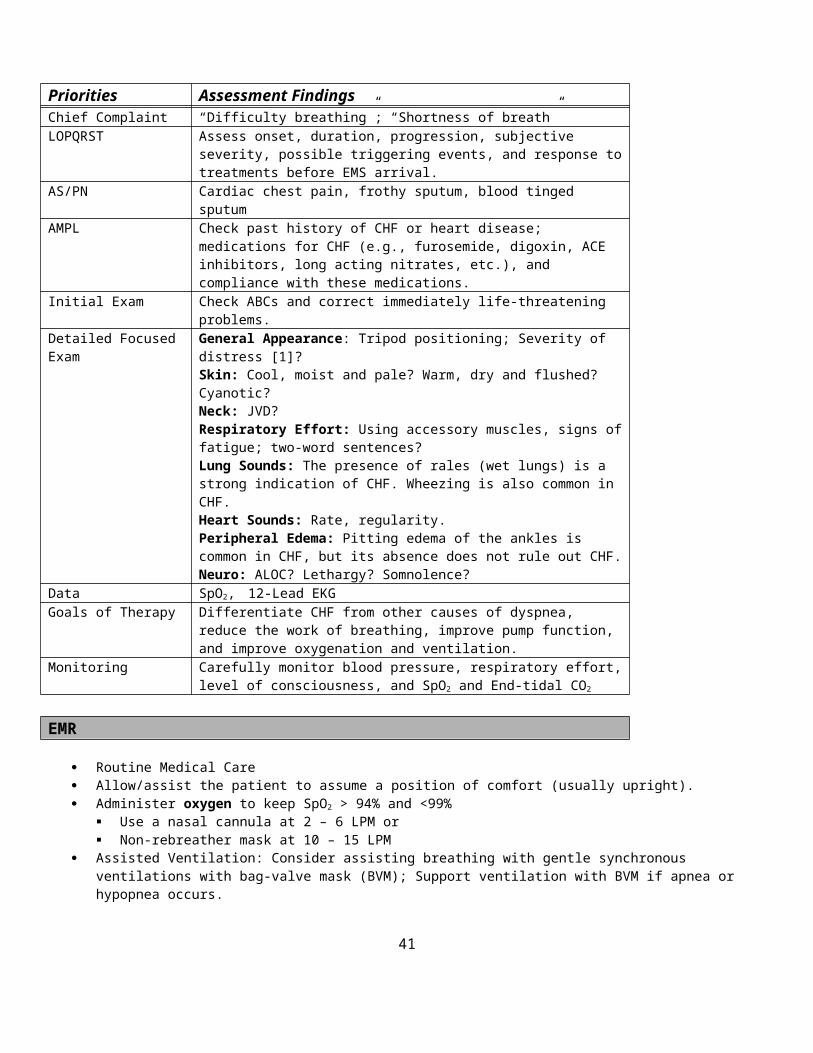
Hospital Bypass Protocol
Objective: To transport patients to the most accessible medical facility which is staffed, equipped, and prepared to administer
emergency care appropriate to the needs of the patient. To have a mechanism where hospitals can have ambulance patients diverted away from their emergency
departments when it has been determined that the hospital is not staffed, equipped, and/or prepared to provide emergency care for additional patients.
Provide a means of communicating hospital status and capabilities to EMS providers.Eligibility:
A condition exists where the emergency department is unable to safely care for additional patients and the hospital has already implemented its own internal procedures for activating backup staff and resources.
A condition exists where the hospital inpatient status or ICU is full (and the hospital has already implemented its own internal procedures for expanding capacity) – preventing additional admissions to the hospital through the emergency department or direct admissions from ambulance services
Notification: Emergency Departments changing status should contact area dispatch centers by phone as well as update
WITRAC. “WITRAC” internet posting is the location for official hospital status posting Hospital Status will be listed as one of the following in WITRAC:
Open: Open to all patientsDivert: Not accepting patients
_ Comments (qualifiers are not limited to)Open to OBOpen to TraumaOpen to ECG confirmed AMI (Acute Myocardial Infarction)Open to BurnOpen to TPA candidate CVA (Cerebral Vascular Accident)Diverting Psychiatric only
If the Emergency Department (ED) is completely closed due to plant failure (i.e., power, fire, etc...) The Emergency Department can accept no patients under any circumstances. This will be listed in the comments section of WITRAC and the status will be set at Divert. Hospitals closed for this reason cannot be “forced open”.
Diversion Reason (hospitals will choose a reason) and list in WITRAC:Diagnostic services unavailable (CT scanner down, Cath Lab down, etc...)No ED Beds / ED at capacity
No Inpatient Beds: Critical Care (the hospital has no critical care beds) No Inpatient Beds: Telemetry/Floor (the hospital has no general medical beds)
No Inpatient Beds: Other (explain)Physical Plant Problems: (i.e. Power outage)
Forced Open : Hospital can be forced open once all Washington County Emergency Departments are diverted Control of “forced open” status is linked to communication and cooperation between hospital administrators
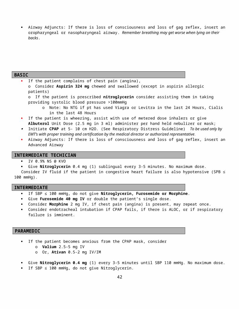
Procedure: When a hospital is diverting patients, ambulances transporting the type of patient being diverted, will bypass that
hospital and transport to an “open” facility. If in a EMTs judgment, the patient has not been stabilized to the extent that the extra transport time would be life
or limb threatening, they should transport to the diverting hospital (override diversion). The EMT must also consider the delay that may occur once the patient has arrived at an already overwhelmed facility.
Some possible examples (not limited to):PNBFailed airwaySymptomatic hypotension resistant to pre-hospital treatment (BPs <
90mmHg & symptomatic)Severe trauma where the diverting hospital is the preferred and proper
facility based on area trauma triage criteria.Status Epilepticus (continuous seizure activity lasting longer than 30
min. resistant to treatment)Pregnant patients in active laborCerebral Vascular Accident who is a TPA candidate (unless diversion based on CT scan
nonfunctional)EKG documented Acute Myocardial Infarction
All cases where hospital diversion has been overridden will be reviewed by the service medical director for quality assurance.
A hospital may not change their status while in the midst of receiving an ambulance radio report.“In-Route” diversions are not acceptable.
If a patient demands transport to a diverting facility, the patient may still be transported to that facility however the EMT must explain to the patient all of the possible delays and dangers associated with entering an overwhelmed emergency department and discourage this destination choice. The EMT should document this carefully. EMTs must understand that they could incur liability regarding their destination choice if a patient is released to a knowingly overwhelmed facility unless they stay in attendance with the patient until an equal or greater level of care can be provided at that facility.
A diverting hospital may not refuse a patient who has presented to them. In cases of Mass Casualty incidents, each hospital should be in communication with incident command on scene
to give active capacity updates allowing fluid transport decisions. These communications may supersede the above process.
Field triage must be considered where incidents involve multiple victims. Avoid overwhelming any one facility and transport the most appropriate patient to the closest most appropriate hospital.
The above policy may not apply to direct admission patients if the admission has been arranged before the diversion status. Agencies transporting direct admission patients should clarify the destination if needed before starting the transfer.
Washington County Suggested Medical Guidelines

HyperglycemiaNote:
Some patients with hyperglycemia have diabetic ketoacidosis (DKA), which is a life-threatening complication of diabetes that includes severe dehydration and metabolic acidosis.
Patients with DKA may have Kussmaul’s respirations, which are rapid and deep. Priorities Assessment FindingsChief Complaint “High blood sugar”, “Diabetic Coma”LOPQRST Check onset/duration. Identify possible contributing factors [1]. Using sliding
scale to adjust insulin doses to rising glucose levels? AS/PN Fever/Chills. Signs/Symptoms of infection. Adequate food and water intake?
Increasing thirst? Increasing urine output?AMPL Medications for diabetes.Initial Exam ABCs and correct any immediately life-threatening problems.Detailed Focused Exam Vital Signs: BP, HR, RR, Temp, SpO2
General Appearance: Appears sick? Dehydrated? Kussmaul’s Respirations?Heart: Tachycardia? Hypotension?Skin: Cool, pale, diaphoretic? Warm, dry, flushed?Neuro: ALOC? Focal deficits (CVA)?
Data Blood GlucoseGoals of Therapy Use IV fluids to reduce glucose level, improve hydration, normalize acid-base
balanceMonitoring Repeat blood glucose
EMR Routine Medical Care. Administer oxygen to keep SpO2 > 94% and <99%
Use a nasal cannula at 2 – 6 LPM or Non-rebreather mask at 10 – 15 LPM
Monitor vitals.BASIC
Check blood glucose
INTERMEDIATE TECHNICIAN / INTERMEDIATE / PARAMEDIC If Blood glucose > 250 mg/dl initiate IV 0.9% NS and run wide open, assess vitals and lung sounds every 250ml to avoid
fluid overload Contact Medical Control
FOOTNOTES:[1] Contributing factors
Illness, especially infections Lack or relative lack of insulin Dehydration Illness Positive for skin turgor
Date of Origin: 11/10/11
Washington County
Suggested Medical Guidelines

Hypoglycemia
Note: Hypoglycemia can become a life threatening emergency in a matter of minutes.
Priorities Assessment FindingsChief Complaint “Low blood sugar” “Altered Level of Consciousness”LOPQRST Check onset/duration. Identify possible contributing factors [1]. Recent history
of frequent episodes. AS/PN Fever/Chills. Signs/Symptoms of infection.AMPL Medications for diabetes.Initial Exam ABCs and correct any immediately life-threatening problems.Detailed Focused Exam Vital Signs: BP, HR, RR, Temp, SpO2
General Appearance: Unresponsive? Agitated and combative?Skin: Cool, pale, diaphoretic?Neuro: ALOC? Focal deficits (CVA)?
Data Blood GlucoseGoals of Therapy Restore normal mental statusMonitoring Repeat blood glucose
EMR
Routine Medical Care. Administer oxygen to keep SpO2 > 94% and <99%
Use a nasal cannula at 2 – 6 LPM or Non-rebreather mask at 10 – 15 LPM
Monitor vitals. May administer oral glucose 15G, if pt. is a known diabetic and is conscious with a patent airway.
BASIC Check glucose level.
o If Blood Sugar > 60, monitor for hypoglycemia.o If Blood Sugar < 60, if conscious and patent airway, administers one dose (15 grams) of oral glucose or juice if
available.o If Blood Sugar < 60, unconscious, give Glucagon 1 mg IM.
INTERMEDIATE TECHNICIAN / INTERMEDIATE / PARAMEDIC
Blood sugar < 60 and patient has altered mental status or is unconscious: Initiate IV 0.9% NS at TKO and give 25 g D50 1 amp (50ml).
After treatment Pt. may wish to not be transported. Make sure Pt. has someone to watch over them for the next few hours and to recheck blood glucose as necessary.
Have patient sign a refusal form if they choose to refuse transport.
Contact Medical Control for the following: Additional orders
FOOTNOTES:[1] Contributing factors
Too much insulin?
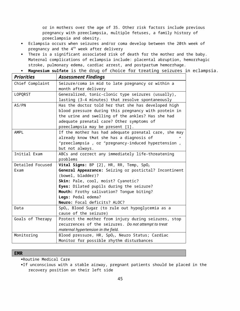
Not enough food? Overexertion Illness
[2] Ensuring Patient Safety Adequate social support is available The patient has access to food or money to buy food The patient is not ill or in need of immediate medical attention
Date of Origin: 11/10/11

Washington County
Suggested Medical Guidelines
Hypothermia & Frostbite
Note: In the hypothermic patient, rough handling can precipitate ventricular fibrillation. When checking pulses and respiratory rates, check for 60 seconds, because bradycardia and bradypnea are common in
moderate to severe hypothermia. Look for signs of trauma in all patients with hypothermia. Hypothermia may be categorized by mild, moderate and severe. The following table may be used to estimate the degree of
hypothermia based on clinical findings.
Severity Temperature Clinical FindingsMild > 93 oF Shivering, impaired judgment; Tachycardia and hypertension may be
presentModerate 86 – 93 oF Consciousness clouded to stuporous; Shivering stops. Blood pressure
becomes difficult to obtain.Severe < 86 oF Bradycardia, hypotension and slow respirations; Arrhythmias may
develop; Consciousness is lost.
Priorities Assessment FindingsChief Complaint “Person found down in a cold environment”LOPQRST What led up to this? Where was the patient found?AS/PN Associated trauma and MOI? Drug or alcohol use?AMPL Check for medications that could be contributory (beta blockers, psychiatric
medications, sedatives, narcotics or barbiturates).Initial Exam Check ABCs and correct immediately life-threatening problems.Detailed Focused Exam Vital Signs: BP, HR, RR, Temp, SpO2
General Appearance: Shivering, paradoxical undressing, evidence of trauma?Skin: Signs of frostbite (pallor, blisters)?Lungs: pulmonary edema?Heart: Rate and rhythm?Neuro: Loss of coordination, impaired judgment, ALOC?
Data SpO2, End Tidal CO2, Blood glucose, 12-Lead EKG (tachycardia, bradycardia, atrial fibrillation, junctional rhythm, ventricular fibrillation?)
Goals of Therapy Above all, avoid rough handling! Initiate Active and passive external rewarming measures in the field. Support airway, breathing and circulation. Do not attempt to thaw frozen limbs in the field.
Monitoring SpO2, ETCO2, Cardiac Monitoring
EMR Attempt to remove the patient from the cold environment if it can be done gently. Rough handling must be avoided. Do not attempt to rewarm frostbitten or frozen parts by rubbing them Remove wet clothing and gently dry the skin by patting, not rubbing, with dry towels Initiate passive rewarming with blankets on top of and underneath the patient; insulate the patient from the cold ground;
shield them from the cold wind or helicopter rotor wash. If the patient is shivering, support thermogenesis by giving the patient fluids and calories (e.g. oral glucose). Initiate active external rewarming with warm blankets and hot packs in the axillae and groin. Administer oxygen to keep SpO2 > 94% and <99%

Use a nasal cannula at 2 – 6 LPM or Non-rebreather mask at 10 – 15 LPM Attempt to administer warmed humidified oxygen by mask [1].
If there is a pulse, no matter how slow, do not initiate chest compressions. If there is no pulse, perform rescue breathing for three minutes, before beginning CPR; Continue CPR until directed by a
physician to discontinue. If the chest is frozen solid, or ice blocks the airway, CPR will be futile and should be discontinued (or not even
started) in the field. If the patient was submerged for more than 90 minutes, do not initiate CPR.
Apply an AED and analyze. If shocks are indicated, attempt defibrillation. The shock should be given no matter what the core temperature is. Do not delay defibrillation to measure a core temperature. Do not attempt to defibrillate more than one time until the core temperature is documented to be > 86 oF.
If frozen limbs are fractured and angulated, splint in the position found. Do not attempt to straighten until they are completely thawed.
BASIC Turn up heat in ambulance
INTERMEDIATE TECHNICIAN IV 0.9% NS. Consider IO (If authorized) If warm saline [2] is available, run at 250 ml/hr. Consider a second IV of warm saline. Do not delay transport to initiate an IV. Peripheral IVs may be quite difficult to start in a hypothermic patient.
INTERMEDIATE/PARAMEDIC Consider endotracheal intubation, if the patient is unresponsive without a gag reflex
o There is no evidence that laryngoscopy or tracheal intubation increase the risk of ventricular fibrillationo Administer warm humidified oxygen [1].
If cardiac arrest is present, attempt defibrillation, if not already doneo If the core temperature is > 86 oF, additional shocks may be attempted in the field (see Cardiac Arrest Guidelines)o If the core temperature is < 86 oF, no more than one shock should be attempted in the field (see Cardiac Arrest
Guidelines)o If the core temperature is unknown, continue CPR and transport emergently to the hospital.
Defibrillation is likely to be futile if the core temperature < 86 oF.o Further attempts at defibrillation should be delayed until the core temperature rises above 86 oFo If ventricular fibrillation is detected, a single dose of Amiodarone 300 mg IV/IO is acceptable. Or Lidocaine 1.0 –
1.5 mg/kg (or 100 mg) IV is acceptable.o Antiarrhythmic is not likely to be effective.o It may be harmful in higher doses.
All other resuscitation medications should be withheld until the core temperature is > 86 oF.o If the core temperature is between 86 and 93 oF, double or triple the dosing interval for all medications given,
because hypothermia slows metabolism. If bradycardia is present,
o Pacing will not be effective, if the core temperature is < 86 oF.o Pacing should be withheld until the core temperature is > 86 oF.
FOOTNOTES:[1] Technique for warming and humidifying oxygen
Place saline in a nebulizer Wrap a hot pack around the nebulizer Start oxygen flow Administer by mask
[2] Technique for warming IV Fluids in the field Wrap the IV tubing around a hot pack several times
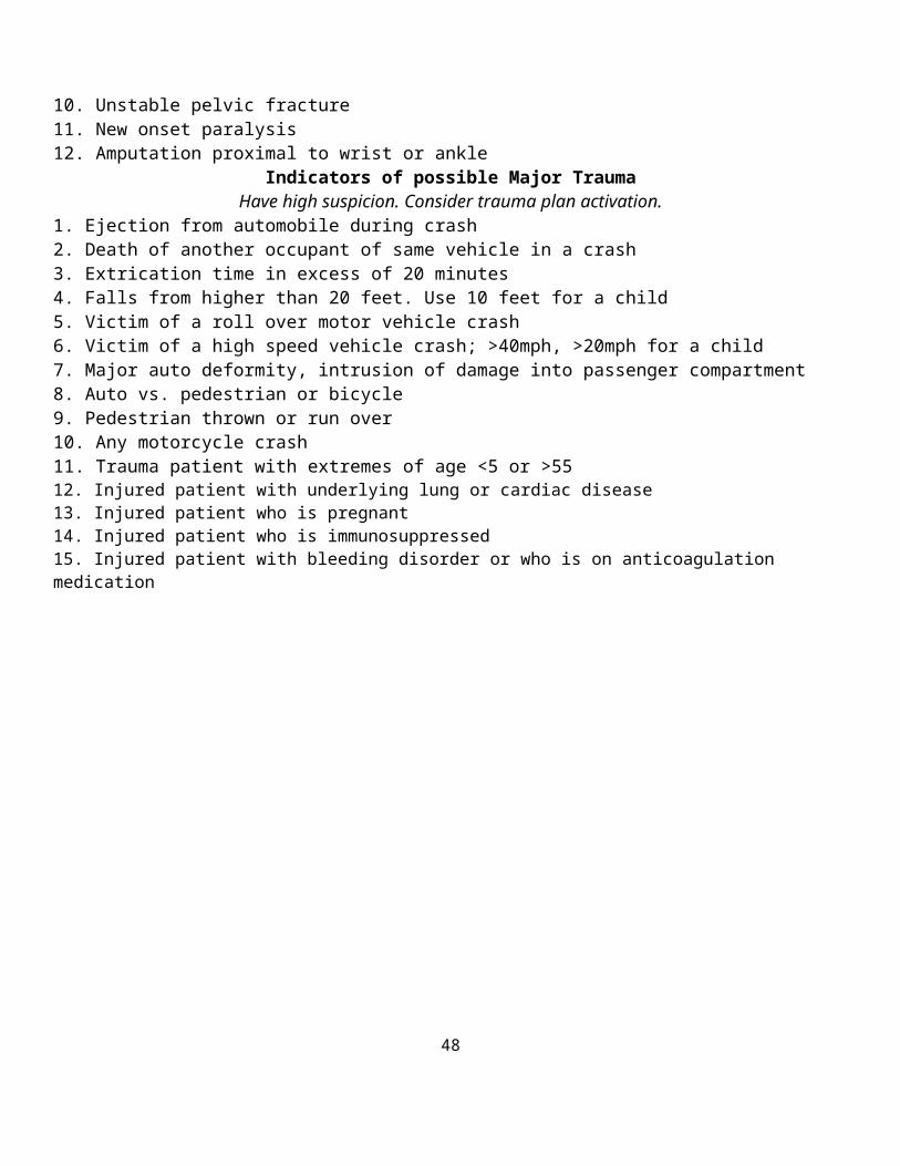
Date of Origin: 11/10/11

Washington County
Suggested Medical Guidelines
Hypovolemia and Shock
Note: Potential causes of hypovolemia and shock include:
o Infections/sepsiso Traumao Hemorrhage (Internal, External)o Spinal cord injuryo Pump Failureo Heart Rhythm Disturbanceso Dehydrationo Drugs and Toxinso Vasovagal Syncopeo Metabolic Disturbanceso Anaphylaxiso Pulmonary Embolism
Shock is defined as inadequate perfusion of vital organs, not merely hypotension. Clinical evidence of shock includes altered mental status.
Priorities Assessment FindingsChief Complaint “Altered Level of Consciousness”LOPQRST Identify onset, duration, progression and provocation.AS/PN Fever/Chills, Chest Pain (Angina), TraumaAMPL Pertinent past history and medications may provide important clues.Initial Exam ABCs and correct immediately life-threatening problems.Detailed Focused Exam Vital Signs: BP, HR, RR, Temp, SpO2, ETCO2
General Appearance: Does the patient appear ill? External Hemorrhage? Skin: Pale, cool, and moist? Flushed, warm and dry?Chest: Labored breathing?Lungs: Wheezes, rales or rhonchi?Heart: Rate and Rhythm?Abdomen: Internal hemorrhage? Tender? Distended? GI Blood loss?Extremities: Trauma? Edema?Neuro: ALOC?
Data SpO2, 12-Lead EKG, Blood Sugar, End Tidal CO2
Goals of Therapy Restore volume and support blood pressureMonitoring Blood pressure, heart rate and cardiac rhythm
EMR/BASIC
Routine Medical -or- Trauma Care. Secure and maintain airway. Perform non-visualized airway, if indicated. Administer oxygen to keep SpO2 > 94% and <99%
Use a nasal cannula at 2 – 6 LPM or Non-rebreather mask at 10 – 15 LPM
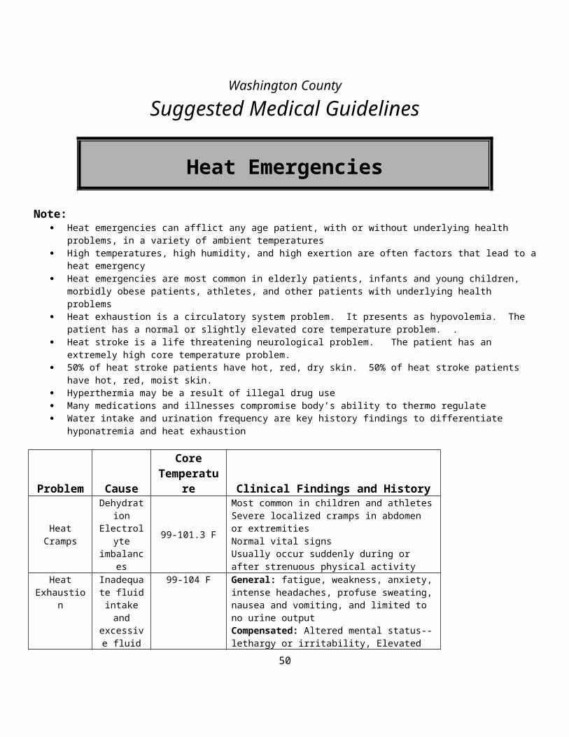
Control external hemorrhage Keep patient flat with lower extremities elevated (if possible). Splint fractures Conserve body temperature, and reassure patient.
INTERMEDIATE TECHNICIAN/INTERMEDIATE
IV 0.9% NS (Large Bore) IO (If authorized) Consider 2nd IV 0.9% NS Initiate a 500 - 1000 ml fluid bolus wide open (Check lung sounds every 250ml) Identify underlying problem and refer to the appropriate protocol.
PARAMEDIC
Consider Dopamine infusion (2 to 10µg/kg/per min) if patient fails to respond to above treatment and remains unstable. Consider RSA (Refer to RSA Protocol)
FOOTNOTES:
Date of Origin: 11/10/11
Washington County
Suggested Medical Guidelines

Delivery of Newborn - Procedure
Priorities Assessment FindingsChief Complaint Uterine contractions, “in labor”LOPQRST Location of pain, radiation of pain, time of onset of contractions, interval
between contractions, quality of contractions, severity of contractions, events surrounding onset of contractions, due date
Associated Symptoms’/Pertinent Negatives
Vaginal bleeding (presence, quantity, and character), , “bloody show”, leakage of fluid or discharge, need to “push”, “bear down” or have a bowel movement, presence of fetal movement, RUQ pain, vomiting, visual changes
SAMPLE Allergies Medications Past medical history, past surgical history, number of previous
pregnancies, previous Cesarean delivery, prenatal care Previous pregnancy or delivery complications (eclampsia, precipitous
delivery, etc.) Recent infectious diseases Complications of current pregnancy (i.e. preeclampsia, placenta previa,
gestational diabetes, premature labor, ultrasound showing abnormal fetal position etc.)
Last mealInitial Exam ABCsDetailed Focused Exam Vital Signs: BP, HR, RR, Temp, SpO2, ETCO2
General Appearance, HEENT: Cracked lips, sunken eyes or cheeks indicating dehydrationSkin: Cool, pale diaphoretic Chest: Labored breathingHeart: Tachycardia Abdomen: Scars, Tenderness, masses, uterine size/location, distention,
deformityLegs: EdemaNeuro: Mental statusGyn: Vaginal bleeding, infant head crowning, prolapsed cord, presenting part, meconium staining
Goals of Therapy Atraumatically deliver newborn with maintenance of normal vital signs for both mother and newborn
Monitoring Frequency of contractions, SpO2, ETCO2
EMR Routine Medical Care Administer oxygen to keep SpO2 > 94% and <99%
Use a nasal cannula at 2 – 6 LPM or Non-rebreather mask at 10 – 15 LPM
Maintain patient in position of comfort, preferably on left side, but typically delivery will require supine position with knees flexed and legs apart.
Check for crowning If delivery appears imminent, open OB pack, apply sterile gloves, and drape abdomen. If there is a Breech presentation, coach the mother to perform shallow breathing and avoid pushing. If there is a Prolapsed umbilical cord, place gloved fingers into the vagina to hold the vaginal wall away from the cord. If there is massive hemorrhage, hypotension, or shock, place the mother in the left lateral recumbent position In the absence of a breech presentation or prolapsed umbilical cord, do not attempt to prevent or delay delivery. Control rate of delivery of head using palm of your hand, applying gentle pressure to protect perineum When head is delivered, compress bulb suction device and place into mouth to suction mouth then repeat for nose.

Limit suction to ten (10) seconds Check to see if cord is wrapped around baby’s neck
If so, gently attempt to slip cord over the baby’s head if cord is semi-loose If cord cannot be slipped over head or cord is tight, clamp two sites on the cord and cut between clamps – Use of scissors is
preferred over scalpel Gently guide head and neck down to allow delivery of upper shoulder. Then guide head and neck up to deliver lower shoulder and body If baby delivers, grasp ankles in one hand and hold head with the other. Prevent heat loss - Provide warm environment, dry baby, and wrap baby in clean blanket Slightly extend head to facilitate patent airway. Suction mouth then nose as needed Continue to maintain an open airway and assess breathing rate and effort. If <20 breaths per minute, refer to Neonatal Resuscitation protocol Provide tactile (tapping of the feet) stimulation as needed to facilitate normal respiratory effort, continually reassessing airway
patency. Assess circulation. If heart rate <100 beats per minute, refer to Current AHA Guideline Place baby lower than placenta and assess cord pulsations. After pulsations have ceased double clamp cord at approximately 7” and 10” from baby and cut between clamps. Record time of birth Assess baby for APGAR scoring at 1 and 5 minutes after recorded time of birth. If baby is premature (<36 weeks gestation), prepare for neonatal resuscitation and early transport. Allow for delivery of placenta. Massage fundus abdominally to stimulate uterine contraction and/or allow baby to breast-feed. If perineum is torn/bleeding, apply direct pressure with gauze. Notify ED early on to allow OB and ED to prepare for arrival.
BASIC Evaluate for imminent delivery
o History of precipitous deliveryo Contractions 2 or less minutes aparto Mother feels need to push or have bowel movemento Visually inspect perineum for crowning
If signs of imminent delivery, prepare for delivery of baby on-site If no signs of imminent delivery, transport mother on left side. Check for signs of complicated childbirth and initiate Emergent BLS Transport if any of the following are present:
o Breech presentation (buttocks or extremities presenting first)o Prolapsed umbilical cordo Massive hemorrhageo Hypotension/Shocko Hypertensiono Seizureso Altered Mental Status
INTERMEDIATE TECH Establish IV NS @ TKO, if time permits (for Mother) Fluid bolus 20ml/kg up to 500ml for hypotension/shock. Reassess and repeat. Consider IO access if no IV is available. (If authorized)
INTERMEDIATE If Mother is hypertensive and experiences seizure, refer to eclampsia protocol. If during delivery meconium staining is present do not suction nose and mouth immediately after delivery of infant’s head.
Allow completion of delivery and if infant does not cry vigorously immediately, place baby in a supine position WITHOUT stimulation and visualize the airway with a laryngoscope.
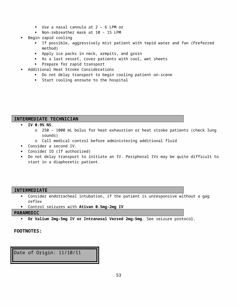
Advance ETT, with meconium aspirator attached to suction, 1-2 cm past vocal cordsApply suction and slowly remove ETTRepeat procedure with a new ETT with each insertion in rapid succession until clear fluid is presentThis procedure is not needed if the infant is immediately crying vigorously without stimulation after birth.
PARAMEDIC Valium 5 mg IV for refractory seizures.
NOTE:APGAR Scores are performed at one minute and 5 minutes after birth according to the following table:
SCORE 0 1 2APPEARANCE Blue/pale Pink Body/Blue
ExtremitiesPink
PULSE Absent Slow (< 100/minute) > 100/minuteGRIMACE No response to suction Grimace to suction Cough or Sneeze to
suctionACTIVITY Limp Some Flexion Active MotionRESPIRATIONS Absent Slow/Irregular Good/Crying
Date of Origin: 11/10/11
Updated : 10/2/12
Washington County
Suggested Medical Guidelines
Narrow Complex Tachycardia’s (Adult)
Note: Narrow complex rhythms have a QRS duration < 0.12 sec
Priorities Assessment FindingsChief Complaint Palpitations, fast heart rate, shortness of breath, chest pain, weakness

LOPQRST Onset and duration, precipitating factors and circumstances, associated symptoms, stroke symptoms, nausea vomiting
AS/PN Chest pain, shortness of breath, weakness, anxiety, leg swellingAMPL Previous history, history of thyroid disease, CAD, Cardiac
Medications Obtain history of previous episodes of tachycardia, including diagnoses
if known. Pay particular attention to whether there is an underlying history of pre-excitation, including the Wolff-Parkinson-White (WPW) Syndrome.
Obtain history of what medications have been used to treat previous arrhythmias, if known.
Obtain history of any previous complications from previous arrhythmia treatments, if known.
Obtain history of the duration of the current episode of tachycardia, if known.
Initial Exam Check ABCs and correct any immediate life threatening problems.Detailed Focused Exam Vitals Signs: BP, HR, RR, Temp, SpO2
General Appearance: Anxious?Skin: Cool, pale diaphoretic Neck: JVD?Chest: Labored breathingLungs: Wheezes, rales, rhonchi? Decreased breath sounds?Heart: Regular, rate fast or slow, murmur Legs: EdemaNeuro: ALOC?
Data SpO2, 12-Lead EKG, Blood Sugar if Diabetic or ALOCGoals of Therapy Decrease Rate, treat chest pain, treat CHFMonitoring Cardiac Monitoring and SpO2
EMR Routine Medical Care Administer oxygen to keep SpO2 > 94% and <99%
Use a nasal cannula at 2 – 6 LPM or Non-rebreather mask at 10 – 15 LPM
If the patient is having difficulty breathing allow them to sit upright
Basic If patient experiences angina, administer patient’s prescribed Nitroglycerin 0.4 mg sublingually, unless the Systolic BP <
100 mm Hg.o Note: No NTG if pt. has used Viagra or Levitra in the last 24 Hours, Cialis in the last 48 Hourso Give NTG every 5 minutes until pain is relievedo Repeat BP before each Nitroglycerin dose.o Discontinue Nitroglycerin if the Systolic BP drops below 100 mm Hg.o Document all BP’s and the number of Nitroglycerin doses given.
If the patient also experiences shortness of breath, consider the Congestive Heart Failure Guidelines Obtain 12 lead EKG and transmit (if capable)
INTERMEDIATE TECHNICIAN IV 0.9% Normal Saline. An antecubital site is preferred for the administration of Adenosine.
If SBP < 100 mmHg give 250cc fluid bolus, and then reassess
INTERMEDIATE If the patient is hemodynamically or clinically unstable [1] Contact Medical Control. Narrow Complex Tachycardia’s include:
o Sinus Tachycardiao Rapid Atrial Fibrillation/Flutter (RAF)o Paroxysmal Supraventricular Tachycardia (PSVT) with or without pre-excitation, e.g. Wolf-Parkinson-White
(WPW) Syndrome If sinus tachycardia is present, the underlying causes include: pain, dehydration, hypotension, shock, hypoglycemia,
hypoxemia, anxiety, fever, sepsis, drug induced, recent heavy exertion, hyperthyroidism and anemia.o The causes listed in bold-face print are within the scope of prehospital providers to identify and treat. Refer to the
appropriate guidelines.o Do not treat sinus tachycardia with medications or Cardioversion
If RAF is present, refer to the Rapid Atrial Fibrillation Guidelines. If the patient is hemodynamically and clinically stable, transport the patient to the hospital. Attempt vagal maneuvers If patient is clinically stable with a HR >150 and signs or symptoms of Narrow Complex Tachycardia, a trial of adenosine is
acceptable.o Adenosine 6 mg IV over 1-2 seconds. If unsuccessful, repeat dose with 12 mg IV over 1-2 seconds. If unsuccessful,
repeat dose with 12 mg IV over 1-2 seconds. Follow all doses with a 10 cc saline flush by rapid IV push.o Warn patient about brief but unpleasant side effects of adenosine: including flushing, lightheadedness, slowing of
heart rate, anxiety and chest pain. Print a rhythm strip during Adenosine administration.
Paramedic If identified rhythm (either initially or during adenosine administration) is A-fib or A-flutter refer to the rapid A-fib
protocol. If the patient is hemodynamically unstable and meets one or more of the clinical criteria [1]
o Prepare to perform synchronized Cardioversion.o Perform first synchronized Cardioversion @ 50joules.o If unsuccessful, increase by 50 joules for each subsequent attempt.
Consider Analgesic for paino Morphine 2-4 mg or Fentanyl 25-100 mcg
AndVersed 2-4mg IV for sedation
FOOTNOTES:[1] Criteria for characterizing a patient as “unstable” (Patient must meet the Hemodynamic Criteria and at least one of the Critical Criteria to be considered unstable)
Hemodynamic Criteriao SBP < 100 mmHg AND Heart Rate > 150 beats/min
Clinical Criteriao Signs of shock (poor perfusion) are present, including
ALOC Absent radial pulses Pallor and diaphoresis
o Signs of pulmonary edema are present, including Labored breathing Rales (wet lungs) Hypoxia (SpO2 <90%)
o The patient complains of anginao Nausea
Date of Origin: 11/10/11
Washington County
Suggested Medical Guidelines
Nausea and Vomiting
Note: Think of potential causes
Infectious diseases Food borne illness Drug or alcohol intoxication Adverse reaction to medication Head injury Diabetic problems Heart problems (angina, CHF) Abdominal Problems (bowel obstruction, pancreatitis)

Vertigo
Priorities Assessment FindingsChief Complaint Nausea and/or vomitingLOPQRST Onset, number of episodes of vomitingAS/PN Associated diarrhea? Bloody emesis or diarrhea?AMPL Recent travel, exposure to others with similar problem, contaminated food?
Alcohol excess? Drugs or other toxins?Initial Exam ABCs and correct immediately life-threatening problems.Detailed Focused Exam Vital Signs: BP, HR, RR, Temp, SpO2, End Tidal CO2
General: Ill appearing? Dehydrated?Abdomen: Soft? Tender? Distended?Neuro: ALOC?
Data SpO2, ETCO2
Goals of Therapy Stop vomiting, relieve nausea, correct dehydrationMonitoring Response to medications.
EMR/BASIC
Supportive Care Administer oxygen to keep SpO2 > 94% and <99%
Use a nasal cannula at 2 – 6 LPM or Non-rebreather mask at 10 – 15 LPM
Do not use an oxygen mask if patient is actively vomiting.
IV TECH/ INTERMEDIATE
IV 0.9% NS @ KVO Give a 250 ml bolus if signs of dehydration are present (check lung sounds)
PARAMEDIC Before administration of anti-nausea medication ensure there is no hx of adverse side effects to Phenergan, Tigan,
Compazine etc., and if so contact medical control prior to administration. Ondansetron (Zofran) 4mg SLOW IV If extra pyramidal side effects (EPS) develop [1], give Benadryl 25mg IV
o If no venous access is available give 50mg IMContact Medical Control for the following:
Additional doses of Ondansetron (Zofran)
FOOTNOTES:[1] Extra pyramidal Side Effects (EPS) of Ondansetron (Zofran)
Akathisia (restlessness) Torticollis (head turned) Opisthotonus (back arched) Oculogyric crisis (eye movement disorder) Buccolingual crisis (tongue movement disorder)
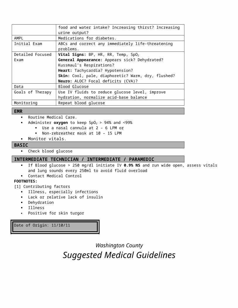
Date of Origin: 11/10/11
Washington County
Suggested Medical Guidelines
Pain Management
Priorities Assessment FindingsChief Complaint “Pain”LOPQRST Location, onset, provocation, palliation, quality, radiation, severity (subjective
pain score on a 0-10 scale or mild, moderate, severe), time (intermittent or continuous; steady vs. improving or worsening)
AS/PN Associated symptoms/pertinent negativesAMPL Allergies, medications, pertinent past history, last mealInitial Exam Check ABCs and correct immediately life-threatening problems.Detailed Focused Exam Vital Signs: BP, HR, RR, Temp, SpO2, ETCO2
General Appearance: Writhing in pain, facial grimacing, moaning, screaming or crying? Assess objectively how severe the pain appears to you (mild, moderate or severe).Skin: Pale, cool, diaphoretic?Source of pain (chest, abdomen, back, extremities, etc.): Swelling, ecchymosis or deformity? Tenderness on palpation? CMS?
Data SpO2. EKG for chest pain., End Tidal CO2
Goals of Therapy Reduce pain to a tolerable level. Monitoring BP, HR, RR, EKG, SpO2. ETCO2
EMR/BASIC Display a calm and compassionate attitude Acknowledge and assess the patient’s pain by obtaining a thorough history Identify and treat the cause
o Musculoskeletal injuries: Realign angulated fractures, if possible, being cautious not to aggravate the injury or pain Reposition (not reduce) dislocated joints to improve comfort, circulation, sensation, and motion Apply a well-padded splint that immobilizes the long bone above and below the injury or the joint above
and below the injury Do not compromise distal circulation
Immobilize joints in mid-range position Elevate the injured extremity if no fracture or dislocation is found Apply ice or cold packs to the injured area Apply a compression bandage or ace wrap if a splint is not needed
o Consider spinal immobilization, if needed Pad the backboard with a blanket(s)(consider for geriatric patients) Pad voids between the patient and backboard—behind knees, and small of back Pad the straps Keep the patient warm and protected from rain/snow, ambulance exhaust etc.
o Administer oxygen to keep SpO2 > 94% and <99% Use a nasal cannula at 2 – 6 LPM or Non-rebreather mask at 10 – 15 LPM
Reassure and comfort the patient; Use a calm and soothing voice. Distract them or encourage them not to focus on their injury, but to think about something more pleasant Eliminate stress inducing distractions—i.e. family, police and bystanders Coach the patient’s breathing—calm, deep full inhalations, and relaxed slow exhalations. Explain to the patient what is happening and what will happen next. Adjust the ambient temperature of the treatment area to a comfortable level for the patient

Reassess pain after all interventions
IV TECH
IV 0.9% Normal Saline @ KVO Consider a bolus of 250 ml if signs of hypovolemia are present
INTERMEDIATE
Morphine Sulfate 2 – 4 mg IV/IM/IO Reassess patient’s pain after each injection Recheck blood pressure before each additional dose; withhold Morphine, if SBP < 100 mmHg Repeat Morphine every 5 minutes up to 10 mg, as needed to reduce pain to a tolerable level
PARAMEDIC
Use the “Pain Management Decision Matrix” to select which medication to give Use the “Pain Management Dosing Matrix” to select how much to give, how often, and to what maximum dose. All medications may be given by IV, IM or IO routes. (Versed and Fentanyl may be given Intranasal) Reassess patient’s pain before each additional dose. Recheck blood pressure before each additional dose; withhold Dilaudid and Morphine, if SBP < 100 mmHg. Consider Zofran (Ondansetron) 2 - 4 mg IM/IV/IO for medication induced nausea and/or vomiting For severe burn patients who meet criteria for RSA, deeper sedation is necessary and should be achieved prior to intubation if
possible.o This is strictly a two-Paramedic provider procedure.o It should be done in consultation with medical control, except when time is of the essence in securing the airway
(threat of imminent airway obstruction).o This only applies to severely burned patients.o Higher maximal narcotic and benzodiazepine doses listed in the table below do not always require an order from
medical control, if the threat of losing the airway is your foremost consideration.o Monitor blood pressure closely and increase fluids if necessary to maintain it.
For patients suffering from severe burn pain, titrate the narcotic and benzodiazepine to the point of minimal pain and light sedation
Contact Medical Control for the following: When you’ve reached the maximum dose, contact medical control for additional orders. Abdominal Pain
FOOTNOTES:
Pain Management Decision Matrix1
Source of Pain Mild2 Moderate2 Severe2 Special Considerations
Isolated Narcotic Narcotic Narcotic Moderate pain
Date of Origin: 11/10/11
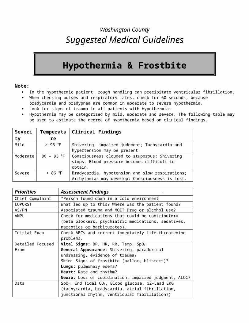
Orthopedic Trauma
relief desired
Multiple Trauma
Narcotic Narcotic Narcotic Moderate pain relief desired
Renal Colic(Kidney Stones)
Toradol4Toradol4 Narcotic
Moderate pain relief desired
Non-cardiac Chest Pain
Toradol4 Toradol4 Toradol4/Narcotic Moderate pain relief desired
Abdominal Pain
Fentanyl Only Fentanyl Only Fentanyl Only Moderate pain relief desired
Migraine Toradol4 Toradol4/Narcotic Toradol4/Narcotic Moderate pain relief desired
Notes:
1. Narcotic (Choose Morphine, Dilaudid and/or Fentanyl); 2. Evaluate the degree of severity on both the patient’s subjective rating (0 – 10) and your
objective assessment (mild – severe).3. Benzodiazepines should be used as an adjunct to pain control for patients with severe burn
pain (Choose either Versed, Valium or Ativan)4. When using Toradol be cautious of possible intracranial bleed or any new bleeds
Pain Medication Dosing MatrixMedication Initial Interval Additional Max1 Ped Cautions
Morphine Sulfate
Mild: 2 – 4 mg Moderate: 4 – 6 mgSevere: 6 – 10 mg
5 min. 2 – 4 mg 10 mg(20 mg) 3
0.1-0.2 mg/kgIV, IO, IM, SQ
AllergyHistamine reactionOver sedation
Dilaudid (Paramedic only)
Mild: 0.5 mg Moderate: 1 mgSevere: 1.5 mg
10 min 0.25 – 0.5 mg
2 mg(4 mg)3
10 kg-50kg0.01mg/kg-0.15mg/kg>50kg to adult is 0.2-0.6mg/kg
AllergyOver sedation
Fentanyl (Paramedic only)
25-100mcg SLOW IVPPed dose: 0.5-1.0 mcg/kg
5 min 25-50 mcg 200 mcg
2-10 mcg/kg up to 0.2 mg single doseMax total dose 1 mg
Allergy, Hemorrhage, Shock, Dizziness, Altered LOC
Toradol (Paramedic only)
Mild: 15 mg Moderate: 15 – 30 mgSevere: 30 mg2
10 min 15 mg 30 mg2
1 mg/kg IM with a max of 30mg0.5mg/kg IV max of 15 mg
NSAID AllergyContraindicated in pregnancy
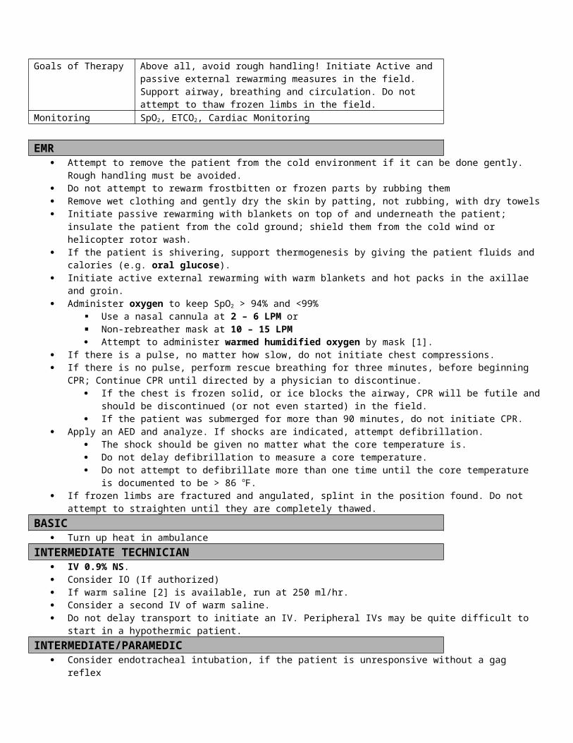
Ondansetron (Paramedic only) 4mg SLOW IV N/A 0 4 mg
0.1 mg/kg slow IV, or IMMax 4 mg
0.1 mg/kg for patients under 40kg
Ativan
Mild/Moderate: N/A SLOW IVPSevere: 1 – 2 mg SLOW IVP
5 min 0.5 – 1 mg 4 mg(8 mg)3
0.05-0.2 mg/kg IV,IO or IMIV,IO must be diluted in 1:1 NS
Respiratory depression
Valium (Paramedic only)
Mild/Moderate: N/A Severe: 5 mg
5 min 5 mg 10 mg(20 mg)3
0.3 mg/kg/ dose IV up to 6-10 mg. Respiratory
depression
Versed (Paramedic only)
2-5 mg SLOW IVPPed dose: 0.1 mg/kg
5 min 2-5 mg 10 mg
0.1 mg/kg IV, Max 2.5 mg/dose
Allergy, Pregnancy, Drowsiness, Hypotension, Respiratory distress, Apnea,
Notes: 1. Maximum dose; More may be given as ordered by Medical Control2. If Toradol is given by the IM route 60 mg may be given IM, but not
IV3. Maximum doses are doubled only for severe burn pain patients who
meet criteria for RSA. See protocol, above.
Washington County
Suggested Medical Guidelines

Allergy & Anaphylaxis (Pediatric)
Note: Allergic reactions span a continuum from minor to life threatening [1]. If due to a bee sting, remove stinger by scraping horizontally with tongue depressor or plastic card. Do not squeeze the
venom sac. If anaphylactic shock is present, treat for shock and maintain warmth. Angioedema with significant swelling of the tongue increases the risk of obstructed airway. Institute emergent transport and
notify ED as soon as possible. In angioedema, Benadryl and Epinephrine may be given, but are not likely to help. IV epinephrine should be used with caution, because of the risk of inducing dysrhythmias. In moderate to severe allergy and
anaphylaxis, there is no contraindication to epinephrine.
Priorities Assessment FindingsChief Complaint “Allergic Reaction”, “Hives” “Itching Rash”LOPQRST What provoked the reaction? Has the patient taken or been given
diphenhydramine (Benadryl) or used an epinephrine auto-injector (EpiPen), and how did they respond?
AS/PN Subjective swelling of facial, oral or pharyngeal structures, difficulty breathing, wheezing and light headedness.
AMPL Does the patient have any environmental, medication, food or other allergies? Is the patient taking an antibiotic or other medication?Has the patient had a reaction in the past? If so, how severe?
Initial Exam Check ABCs and correct immediately life-threatening problems.Detailed Focused Exam Vital Signs: BP, HR, RR, Temp, SpO2
General: Identify degree of severity: mild moderate or severe [1].Skin: Urticaria (hives), Cyanosis?HEENT: Swelling of the lips, tongue or pharynx (angioedema)Chest: Sternal, subcostal or intercostal retractions? Use of accessory muscles of respiration, labored breathing?Lungs: WheezingCardiovascular: Hypotension, tachycardia (anaphylactic shock)Neurological: ALOC
Data SpO2
Goals of Therapy Reverse the allergic reaction, relieve bronchospasm, correct hypotension/shockMonitoring Vital signs and cardiac monitoring.
EMR Administer oxygen to keep SpO2 > 94% and <99%
Use a nasal cannula at 2 – 6 LPM or Non-rebreather mask at 10 – 15 LPM
If loss of consciousness and gag reflex occurs, consider airway adjuncts. See Respiratory Distress Guideline. Epinephrine- Epi-Pen Jr (0.01 mg/kg (1:1,000) SubQ; max dose 0.3-0.5mg) for less than 60lbs
BASIC
Nebulizer Therapy: Albuterol Unit Dose (2.5 mg in 3 ml) administer per hand held nebulizer or mask; May repeat X 2 additional dose
INTERMEDIATE TECHNICIAN Consider IV/IO, Normal Saline @ KVO rate
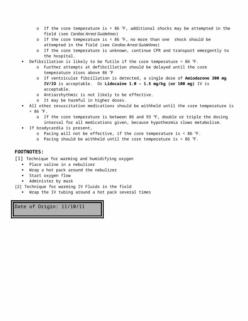
If signs of hypovolemia or inadequate perfusion are present, administer fluid bolus at 20mL/kg (Neonates less than 30 days old use 10ml/kg).
Continue with Albuterol Nebulizer as directed Repeat Epinephrine as directed
Contact Medical Control for the following: Additional doses of Albuterol Additional doses of Epinephrine
INTERMEDIATE If airway unmaintainable consider non-visualized airway or endotracheal intubation (See Respiratory Distress Protocol) Consider additional Epinephrine 0.01mg/kg (1:1000) max 0.3 - 0.5 mg SQ/IM for moderate to severe reactions.
Repeat every 10 – 15 minutes X3 if patient is not improving, or as ordered per Medical Control.Contact Medical Control for the following:
For moderate to severe reactions: Additional Doses of SubQ Epinephrine Epinephrine 0.01mg/kg (1:10,000) max single dose 0.1mg IV every 5 – 10 minutes as ordered
by Medical Control.
PARAMEDIC Benadryl 1 mg/kg IM/IV for mild, moderate or severe reactions Solu-Medrol 2 mg/kg IV/IO for moderate to severe reactions
Contact Medical Control for the following: For moderate to severe reactions: Epinephrine 0.01mg/kg (1:10,000) max single dose 0.1mg IV every 5 – 10 minutes as ordered
by Medical Control.
FOOTNOTES:
[1] Severity of Allergy/Anaphylaxis Mild Allergic reaction : localized or generalized urticaria, without swelling of oral or pharyngeal structures, difficulty
breathing, hypotension or ALOC; Moderate Allergic Reaction : oral or pharyngeal swelling is present, mild to moderate difficulty breathing and wheezing are
present. Severe Allergic Reaction (Anaphylaxis) : moderate to severe difficulty breathing is present, hypotension is present and ALOC
may occur.
Date of Origin: 11/10/11
Washington County
Suggested Medical Guidelines
Note:
Asthma (Pediatric)
(Includes Reactive Airways Disease and Bronchospasm)
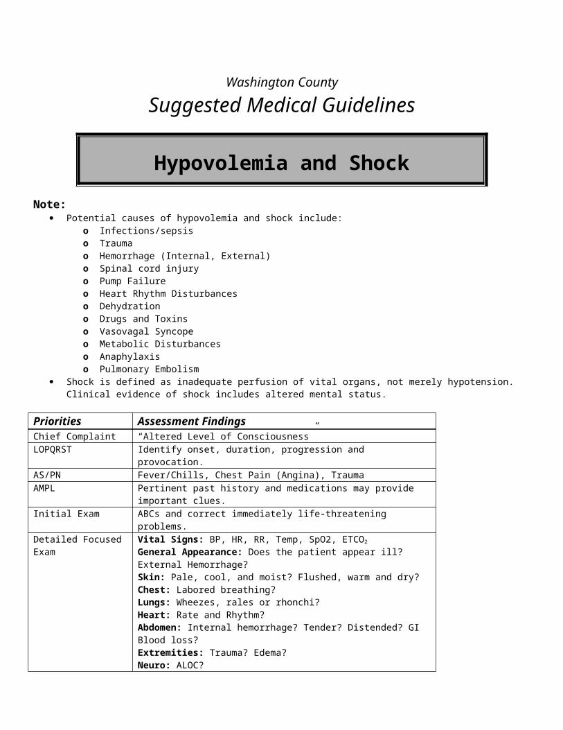
Asthma is usually a disease that develops in childhood. There is usually an identifiable trigger, like infection, weather changes or exposure to certain allergens (e.g., dogs, pollen, etc.).
The so-called classic triad of dyspnea, cough and wheezing may not always be present. Patients with a history of near fatal asthma or have been hospitalized on ventilatory support are at increased risk of recurrent
severe attacks and asthma-related death. Remember: “All that wheezes is not asthma!” The absence of wheezing may be indicative of extreme airflow obstruction.
Priorities Assessment Findings / HistoryChief Complaint Difficulty breathing or shortness of breathLOPQRST Determine onset, duration and progression, triggering events,
response to treatment at home, and subjective severityAS/PN Chest pain (angina or pleuritic), fever/chills, cough/productive of
what, recent changes in sputum colorAMPL Exposure to a known allergen. History of asthma, recent infection or
previous bronchospasm. Current or past medications for these problems (e.g., albuterol, Atrovent, Advair, Prednisone, Antibiotics).
Initial Exam Check ABC’s and correct immediately life threatening problemsDetailed Focused Exam General Appearance: Severity of distress [1]? May be lethargic;
minimal or no verbal response (age appropriate)?Skin: Cool, moist and pale? Warm, dry and flushed? Urticaria? Cyanosis?HEENT: Nasal flaring?Chest: Sternal, subcostal or intercostal retractions? Using accessory muscles, signs of fatigue; Lung Sounds: Wheezes, rales, rhonchi or stridor? Decreased lung sounds? Prolonged expiratory phase? Absence of wheezing?Heart Sounds: Rate, regularity?Lower Extremities: Pale, mottledNeuro: ALOC, lethargy, somnolence?
Data SpO2 in all patients (continuous or frequent re-checks); Blood Glucose if DKA is suspected or ALOC is present.
EMR Routine Medical Care Allow/assist the patient to assume a position of comfort (usually upright). Administer oxygen to keep SpO2 > 94% and <99%
Use a nasal cannula at 2 – 6 LPM or Non-rebreather mask at 10 – 15 LPM
Assisted Ventilation: Consider assisting breathing with gentle synchronous ventilations with bag-valve mask (BVM); Support ventilation with BVM if apnea or hypopnea occurs.
BASIC Airway Adjuncts: If there is loss of consciousness and loss of gag reflex, insert an oropharyngeal or nasopharyngeal airway. Nebulizer Therapy:
Albuterol Unit Dose (2.5 mg in 3 ml) administer per hand held nebulizer or mask; May repeat X 2 additional doses In unconscious without a gag reflex, consider Non-Visualized Airway (See Pediatric Respiratory Distress Guidelines)
Contact medical control for the following:
For severe asthma, consider Epinephrine 0.01mg/kg (1:1000) max dose 0.3 – 0.5mg , administer SubQ (if authorized to draw up Epi) (or Epi-Pen Jr for under 60lbs)
More doses of Albuterol Epinephrine 0.01mg/kg (1:1000) max dose 0.3-0.5mg administer SubQ
INTERMEDIATE TECHNICIAN Consider use of Atrovent (Ipratropium)
o Atrovent Unit Dose (0.25mg ) [footnote 2] with Albuterol per hand held nebulizer or masko Do not repeat Atrovent without an order from Medical Control.
Establish IV/ normal saline, run at appropriate rate
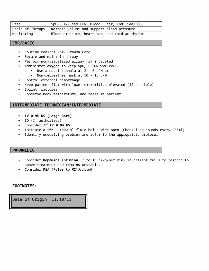
Consider IO (If authorized)Continue with airway support and medications as directed
Contact medical control for the following: Epinephrine 0.01mg/kg (1:1000) max dose 0.3-0.5mg administer SubQ (or Epi-Pen Jr for under 60lbs)
INTERMEDIATEContact medical control for the following:
Repeat subcutaneous epinephrine if the signs of severe distress continue after 15 minutes.
If airway can’t be controlled, consider endotracheal intubation Solu-Medrol 2 mg/kg IV/IO
Contact Medical Control for the following: Epinephrine 0.01mg/kg (1:10,000) max single dose 0.1mg slow IV push every five minutes as
needed for severe asthma.
Footnotes:[1] Normal respiratory rates for children per AHA:[2] If patient does not have a peanut allergy
Date of Origin: 11/10/11
Washington County
Suggested Medical Guidelines
Asystole/PEA (Pediatric)
Note: It is extremely important to understand that PEA is a condition in which the EKG shows a rhythm that would be
expected to generate a pulse. PEA should not be considered or treated as a dysrhythmia. Utilization of the AHA – 4 H’s & 4 T’s is highly suggested
o Hypoxemia (oxygen therapy)o Hypovolemia (fluid therapy)o Hypothermia (refer to protocol)o Hyperkalemia (Sodium Bicarbonate), Hypokalemia, metabolic disorders (contact MC)o Tamponade (Contact MC)o Tension pneumothorax (needle decompression)o Toxins/poisons/drugs (refer to protocol)
PARAMEDIC
Infant 30-60Toddler 24-40Preschooler 22-34School age 18-30Adolescent 12-16
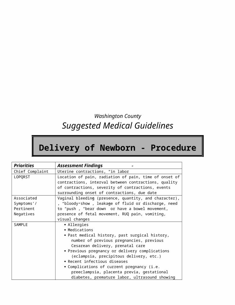
o Thrombosis(contact MC)Priorities Assessment FindingsChief Complaint None by patientLOPQRST Determine onset of symptoms along with possible causes. AS/PN Respiratory arrest and pulselessnessAMPL Recent traumatic injury or metabolic disorders. Developmental and / or
congenital disordersInitial Exam Assess ABC’s, support airway and breathing as necessary. Begin Chest
compressions at a rate >100Detailed Focused Exam General Appearance: Unresponsive, limp
Skin: Cool / cold, pale, gray or cyanotic.Respiratory Effort: Agonal early, none laterLung Sounds: None (unless assisting)Heart Sounds: NoneExtremities: No muscle tone or pulses, mottled, no capillary refillNeuro: Unresponsive
Data SPO2, EKG in all cases, Core temperature, blood glucose, continuous pulse checks (especially during compressions), End Tidal CO2
Goals of Therapy Restore heart function and normal perfusionMonitoring Closely watch heart and respiratory rates, SPO2, EKG (especially in cases of
actual cardiac involvement), ETCO2
EMR Routine Medical Care, maintain normal body temperature. Ascertain patent airway and begin ventilatory assist with adjuncts as needed Begin chest compressions and follow per current AHA guidelines. Apply AED (Use Pediatric Pads if available) If poisoning is suspected, refer to poisoning protocol If hypothermic, see hypothermia protocol If hypoglycemia is suspected refer to hypoglycemia protocol Continue Airway support as needed
o May use non-visualized airways (combitube) if child is within manufacturers limits and airway is unable to be maintained with OPA or NPA
CPR until child’s pulse returns at a rate >100 and adequate perfusion is evident.BASIC
Attach 4 lead EKG as available and appropriate Assess blood glucose level. If glucose <60 see hypoglycemia protocol
INTERMEDIATE TECHNICIAN Establish IV Normal Saline @ KVO rate Consider IO (If authorized) If signs of hypovolemia or inadequate perfusion are present, administer fluid bolus at 20mL/kg (Neonates less than 30 days
old use 10ml/kg). Continue support of airway and chest compressions Cardiac monitor
INTERMEDIATE Consider Endotracheal intubation if unable to maintain airway Administer Epinephrine 10,000 0.01 mg/kg IV/IO may repeat Q 3-5 min (Double dose for ET tube)
Contact Medical Control for the following:Repeat doses of Epinephrine
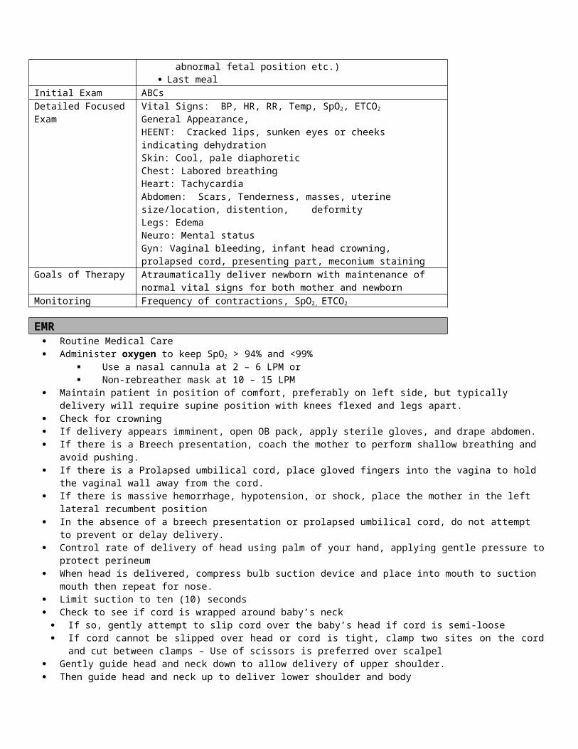
PARAMEDIC If Bradycardia transcutaneous pacing using peds pads, set rate at 100 If unable to secure airway with ETT may consider surgical or needle cricothyrotomy. If Tension Pneumothorax suspected perform needle decompression
Contact Medical Control for the following: Orogastric tube placement for stomach decompression Cricothyrotomy Needle Chest Decompression
Date of Origin: 11/10/11
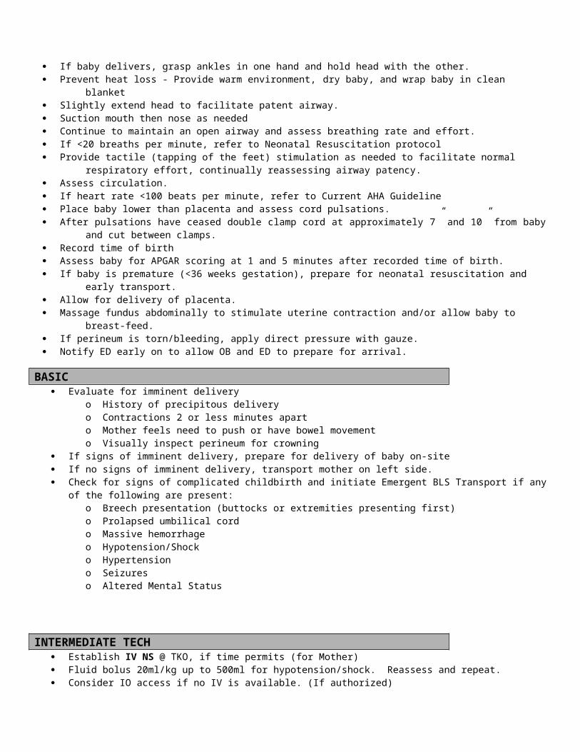
Washington County
Suggested Medical Guidelines
Bradycardia (Pediatric)Note:
Hypoxia is the # 1 cause of bradycardia in children primarily < 8 years of age. Maintain adequate oxygenation and ventilation Bradycardia may also be caused by allergic reactions, hypothermia, and medication ingestion or poisoning.
Priorities Assessment FindingsChief Complaint Weakness, unresponsiveness, signs of inadequate perfusionLOPQRST Determine onset of symptoms along with possible causes. AS/PN Respiratory distress, chest pain, nausea, vomitingAMPL Patient with past respiratory or cardiac event history, current medications and
compliance. Initial Exam Assess ABC’s. Check for/relieve foreign body obstruction, support airway and
breathing as necessary. Detailed Focused Exam General Appearance: Somnolent, flaccid or poor muscle tone. Look for signs
of poisoning.Skin: Cool, pale, gray, mottled, or cyanotic?Respiratory Effort: May be normal or signs of distress, i.e.: subcostal and intercostal retractions, sternal retraction, nasal flaring, etc. Lung Sounds: May be diminished or sound “wet”Heart Sounds: Normal, except in some cases of cardiac diseaseExtremities: Poor muscle tone, weak or no pulses, poor capillary refillNeuro: ALOC, unresponsive
Data SpO2, EKG in all cases, core temperature, blood glucose and capillary refill checks, B/P in older children, End Tidal CO2
Goals of Therapy Increase heart rate and restore normal perfusion, oxygenation and ventilationMonitoring Closely watch heart and respiratory rates, SpO2, EKG , ETCO2
EMR Routine Medical Care Maintain normal body temperature Administer oxygen to keep SpO2 > 94% and <99%
If signs of hypoventilation are present, assist ventilation with a BVM and high-flow oxygen or Use a nasal cannula at 2 – 6 LPM or Non-rebreather mask at 10 – 15 LPM Use airway adjuncts as needed.
Begin chest compressions if bradycardia along with signs of inadequate perfusion persists after oxygen therapy Patient must exhibit signs of hemodynamic instability and also one or more clinical signs of poor perfusion as listed
below. heart rate < 60 in children 1 – 8 years old heart rate < 100 in children < 1 year old
Clinical Criteria Signs of shock (poor perfusion) are present, including
ALOC, including syncope, weakness, lightheadedness, fatigue Absent radial pulses Pallor and diaphoresis Hypoxia (SpO2 <90%)
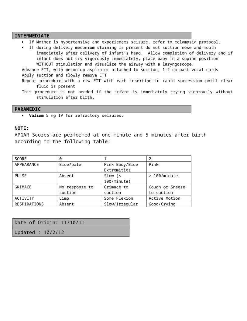
If poisoning is suspected, refer to poisoning protocol If hypothermic, see hypothermia protocol Continue Airway support as needed; avoid use of non-visualized airways CPR as needed until heart rate maintains >100 and adequate perfusion evident.
BASIC Assess blood glucose level. If glucose <60 see Pediatric Hypoglycemia Guidelines
INTERMEDIATE TECHNICIAN Establish IV Normal Saline @ KVO rate Consider IO (If authorized)
If signs of hypovolemia or inadequate perfusion are present, administer fluid bolus at 20mL/kg (Neonates less than 30 days old use 10ml/kg).
Cardiac monitor as needed
INTERMEDIATE Intubation as needed If airway and breathing are adequate, and temperature is normal, but heart rate is still <60
o Give Epinephrine 0.01 mg/kg (0.1 ml/kg 1:10,000) IV/IO (preferred)o may repeat every 3-5 mino Maximum single dose 1 mg
Contact Medical Control for the following:Consider Atropine (0.1 mg/ml)
o 0.02 mg/kg; minimum dose 0.1mg (1 ml) o Max dose 0.5mg (5 ml) every 5 minuteso Max dose 1 mg from 6 months to 8 years; 2 mg over 8 yearso Atropine is contraindicated in patients under 6 months of age
Repeat doses of Epinephrine and/or Atropine If bradycardia persists, despite the above measures, initiate external transcutaneous pacing at 100
per minute. Medication administration by ET
o 0.1 mg/kg (0.1 ml/kg Epinephrine 1:1,000) Additional pacing orders
PARAMEDIC Transcutaneous Pacing using Peds Pads, set rate at 100 Consider Atropine (0.1 mg/ml)
o 0.02 mg/kg; minimum dose 0.1mg (1 ml) o Max dose 0.5mg (5 ml) every 5 minuteso Max dose 1 mg from 6 months to 8 years; 2 mg over 8 yearso Atropine is contraindicated in patients under 6 months of age
Date of Origin: 11/10/11
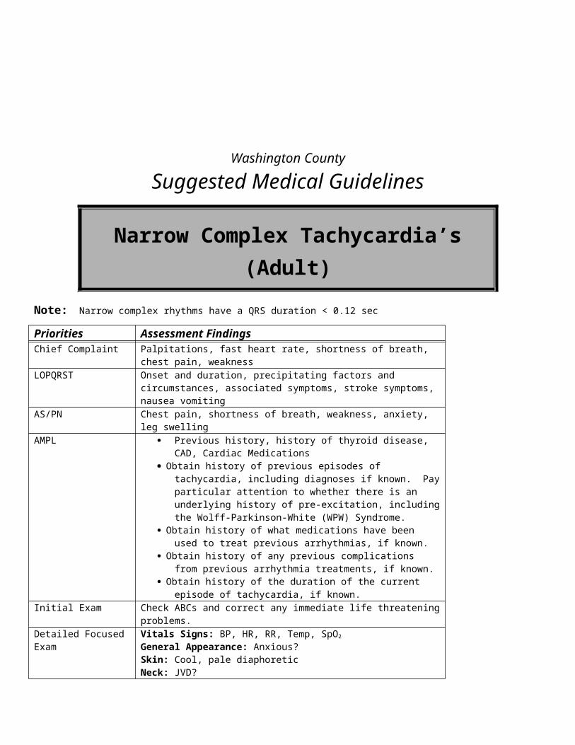
Washington County
Suggested Medical Guidelines
Burns (Pediatric)
Note: This protocol applies to thermal burns, chemical burns and electrical burns Scene safety is of utmost concern. Loosen and remove any clothing and jewelry that can become constricting when tissue swells. In cases of cardiac arrest due to electrical contact or burns, aggressive resuscitation should be attempted, as survival rates are
good. Burns over 10% should not be cooled with water due to possibility of causing hypothermia. Even if the patient meets criteria for burn center referral, they are not required to be air-lifted directly to a burn center from
the scene. In the presence of major trauma (in addition to the burn), stabilizing life-threatening injuries takes precedence over the care
of the burn. Pain Management should be aggressive. Burn patients may require much higher doses for pain control. See Pain
Management Guidelines. Remember that carbon monoxide poisoning and cyanide poisoning is a common complication of burns suffered in a structure
fire.
Priorities Assessment FindingsChief Complaint Burns, pain, burning sensation; electrical/lightning injury; chemical (caustic)
exposureLOPQRST Identify cause of burn, exposure time and time of burnAS/PN Respiratory distress, ulcerous skin in chemical burns, entrance and/or exit
wounds with possible cardiac changes in electricalAMPL Note previous medical conditions that may affect survivalInitial Exam Scene safety, ABC’s support as necessaryDetailed Focused Exam Vitals: Estimate BSA with “rule of nines” [1], BP, HR, RR, Temp, SpO2,
ETCO2
General Appearance: Varies depending on burn; may show signs of extreme painSkin: Depending on the degree on the burn, erythema, blisters, pale leathery appearance, charring, sloughingHEENT: Pupils, check nose & mouth for signs of burns (e.g. soot, edema, redness)Lungs: Signs of respiratory distress, stridor, diminished or absent lung sounds?Heart: Rate and rhythm? Especially in electrical burns Neuro: Loss of movement and/or sensation in extremities, focal deficits?
Data BSA estimate, EKG, SpO2, ETCO2
Goals of Therapy Stop the burn; airway management; fluid resuscitation; pain control; management of associated injuries; (Decontamination in hazmat incidents)
Monitoring Watch for cardiac dysrhythmias, increasing respiratory distress and signs of shock
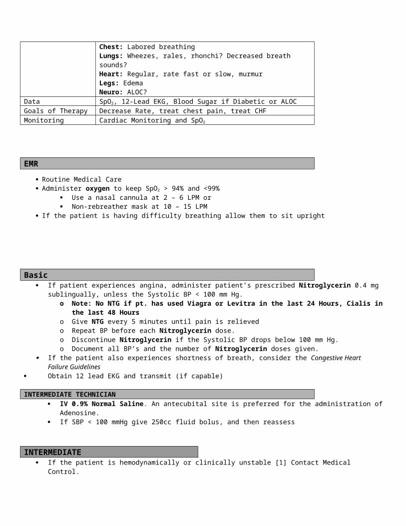
EMR/BASIC Routine Medical Care/Routine Trauma Care. If in cardiac arrest due to electrical injury, perform CPR in the usual fashion (See Cardiac Arrest Protocol). Get the patient away from the heat source. Remove burned, hot, warm, and/or contaminated clothing. Administer oxygen to keep SpO2 > 94% and <99%
Use a nasal cannula at 2 – 6 LPM or Non-rebreather mask at 10 – 15 LPM
If the patient is unconscious without a gag reflex, consider: Oropharyngeal airway or nasopharyngeal airway.
Remove rings, bracelets, and other constricting items as soon as possible Keep the patient warm with dry blankets Provide comfort and reassurance Raise patients legs in cases of shock If less than 10% body surface area (BSA), dress burns with wet saline dressings.
Be careful not to induce hypothermia. If burns are more than 10% BSA, use dry dressings or clean sheets. Consider Advanced Airway,
If airway burns are evident carbonaceous sputum singeing of nasal hairs swelling of the lips, tongue or pharynx due to burns hoarse voice or stridor
There is increasing respiratory distress There is decreased level of consciousness with no gag reflex
INTERMEDIATE TECHNICIAN
IV 0.9% Normal Saline.o Unburned sites are preferred, but burned sites are acceptable.
Consider IO (If authorized) Monitor ABC’s and vitals closely
Contact Medical Control for the following: Consider parkland formula for fluid resuscitation:
o 4ml x wt/kg x BSA , 50% of fluid given over 8 hours, 2nd 50% over next 16 hours
INTERMEDIATE
Consider endotracheal intubation. Pain Management, refer to Pain Management Guidelines.
PARAMEDIC
Continue pain control, refer to Pain Management Guidelines.
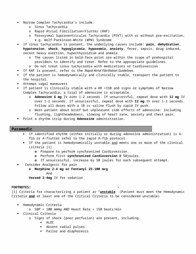
FOOTNOTES:[1]
Date of Origin: 11/10/11
Washington County
Suggested Medical Guidelines
Cardiac Arrest (Pediatric)
Note: These guidelines are based on (or adapted from) the 2010 American Heart Association Guidelines for Cardiopulmonary
Resuscitation and Emergency Cardiovascular Care Child is defined as before the onset of puberty Unlike adult cardiac arrest, which is usually due to a primary cardiac abnormality, pediatric cardiac arrest most often occurs
as a result of asphyxia. The most common reasons for this include progressive respiratory failure and shock.
Priorities Assessment FindingsChief Complaint Collapsed, unresponsive, no pulse, not breathing LOPQRST Witnessed or unwitnessed? Estimated time of onset. Circumstances/trauma.
Location of patient. Antecedent symptoms/signs (difficulty breathing, serious illness) and their duration.
AS/PN Bystander-initiated CPR. Pre-arrival CPR instructions from dispatch? Public access AED use.
AMPL Does the patient have any allergies to medications? History of heart disease? Current cardiac medications?
Initial Exam Establish Unresponsiveness. Check ABCs. Open and reposition airway.Detailed Focused Exam Vitals Signs: Pulseless and non-breathing (or agonal respirations)
General: Identify unresponsiveness. Look for rigor mortis, dependent lividity, or unsurvivable trauma. Look for a valid Wisconsin Do-not-resuscitate bracelet.Skin: Mottled, Warm/cold, dependent lividity, rash, ecchymosis?HEENT: Airway patient, foreign bodies, dry mucus membranes, furrowed lips?Chest: Spontaneous respirations?Lungs: Equal breath sounds, difficulty bagging or ventilating?Cardiovascular: Absence of heart sounds, carotid or femoral pulses?Abdomen: Distended?Extremities: Rigor mortis, edema, deformity?Neurological: Unresponsive to verbal and painful stimulation?
Data Cardiac rhythm analysis, blood sugar, SpO2, End Tidal CO2
Goals of Therapy Return of spontaneous circulation (ROSC)Monitoring Cardiac Monitoring, Vital Signs, and SpO2, ETCO2
EMR/BASIC Establish that the patient is unresponsive, without a pulse, and not breathing
o Infants: Check brachial pulseo Children: Check carotid or femoral pulseo Pulse checks require at least 10 seconds to perform
Rule out DNR status, dependent lividity, rigor mortis, or other contraindications to initiating resuscitation.o If there is any uncertainty about what to do, initiate resuscitation.
First Priority: Defibrillateo If you actually witness the arrest, or if the downtime is definitely < 4 minutes, apply AED, analyze and, if a shock is
indicated, defibrillate immediately. Defibrillation is indicated for children 1 year old or older Use pediatric defibrillation pads for children less than 15 kg, if available; otherwise use adult pads.
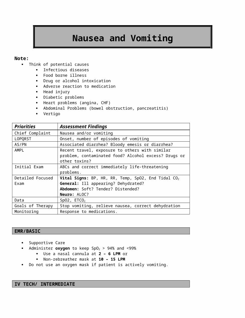
Deliver 1 (one) shock with an AED If no shock is indicated, perform CPR for 2 minutes, and then reanalyze.
o If you are uncertain of the downtime, or the defibrillator is not available/ready, perform two minutes of CPR.o Defibrillate once, and then begin CPR immediately.
Do not delay CPR while waiting for an AED Do not delay CPR to perform another pulse check. Do not delay CPR to reanalyze the rhythm.
o If the downtime is > 4 minutes, perform CPR for 2 minutes, and then attempt defibrillation. Second Priority: Perform Effective CPR
o Perform a head-tilt-chin-lift maneuver. In suspected C-spine injury, use the jaw thrust, instead.
o Deliver 2 rescue breaths that make the chest rise.o Compress at a rate of 100 times per minute.
“Push hard and push fast”. In children (ages 1 – onset of puberty) compress the chest 1/3 – ½ the depth of the chest.
Use the heel of one or two hands on the lower half of the sternum In infants (< 1 year old), compress about 1/3 to ½ the depth of the chest
Place two thumbs on the lower half of the sternum with your hands encircling the chest (preferred technique for health care providers with 2 rescuers present); Compress with thumbs, use hands/fingers for counter pressure.
Allow the chest to recoil completely between compressions. Minimize the frequency and duration (10 seconds at most) of interruptions in chest compressions. Do not pause compressions for ventilations. Change rescuers every 2 minutes. (This is not an option!)
o Perform rescue breathing. Compression to Ventilation Ratio: 15 to 2 (Two rescuers); 30:2 (One rescuer) Insert an oropharyngeal or nasopharyngeal airway. Use Bag-Valve-Mask and high-flow oxygen. Deliver each breath over 1 second with enough volume to cause chest rise.
o After 2 minutes, analyze for a shockable rhythmo Shock, if indicated, or…
Resume CPR immediately, if there is no pulse, or… Proceed to supportive measures, if spontaneous circulation returns.
o Repeat this cycle until spontaneous circulation returns, the patient is transported, or resuscitation is terminated. Third Priority: Perform an advanced airway procedure
o Insert a non-visualized airway (First Responder or Basic) or an endotracheal tube (Intermediate or Paramedic). No non-visualized airways if child is < 4 ft. tall.
o Do not interrupt compressions to do this, unless absolutely necessary.o After the advanced airway is in place…
Discontinue cycles of 15:2 Continue compressions uninterrupted at 100 times per minute. Give a single rescue breath 8 – 10 times per minute (every 6 – 8 seconds).
If there is ROSC, provide the following supportive interventions:o Oxygen 10 – 15 LPM by non-rebreather mask or bag-valve-mask or assist ventilations with a BVM as neededo Keep the patient warm.
INTERMEDIATE TECHNICIAN Fourth Priority: Establish venous access Establish IV/IO, Normal Saline @ KVO rate If signs of hypovolemia or inadequate perfusion are present, administer fluid bolus at 20mL/kg Neonates (less than 30 days
old) use 10ml/kg.
INTERMEDIATE/PARAMEDIC Direct First Responders and EMTs to continue basic rescuer interventions. If an advanced airway is not already in place, insert an endotracheal tube [1].
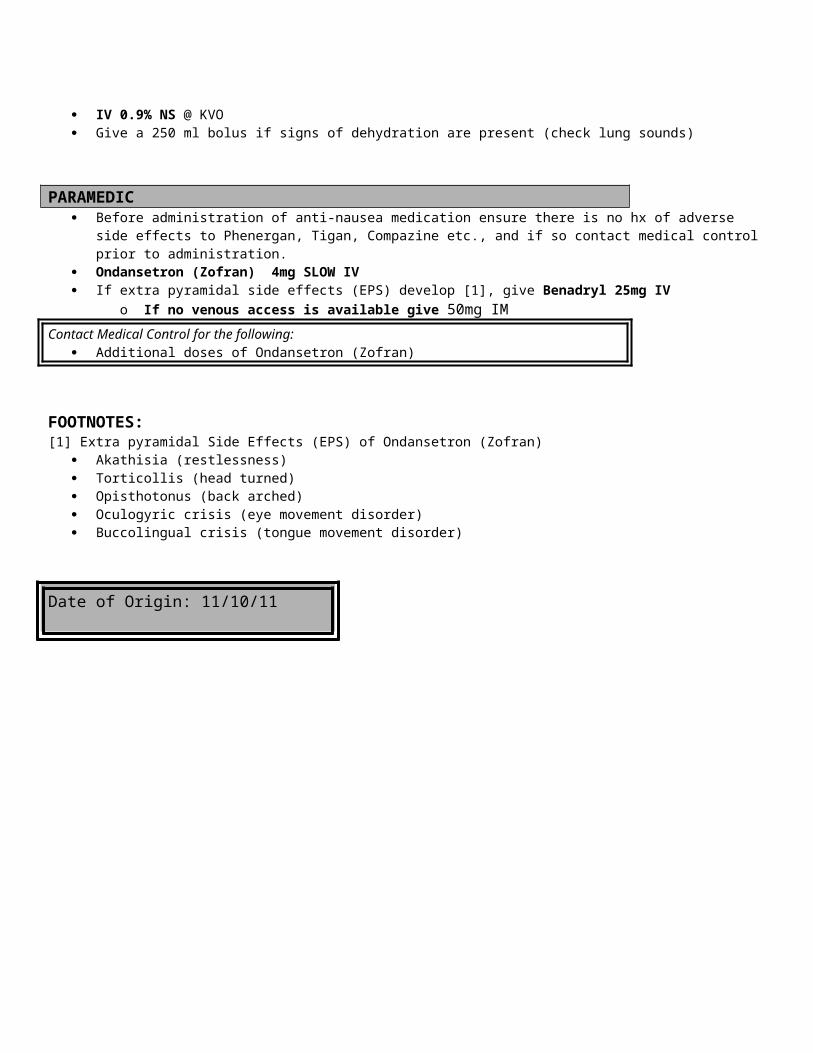
Initiate cardiac rhythm monitoring and analysis. Consider venous access, if not already done
o Drug administration routes in order of preference: IV – IO – ETo Do not attempt to administer medications via a non-visualized airway
Lack of venous access is not an acceptable indication for converting a non-visualized airway that is functioning well for ventilations to an ET tube.
Rather, use IO access. Fifth Priority: Proceed to ACLS resuscitation medications according to the respective protocols for:
o Asystole/Pulseless Electrical Activityo Ventricular Fibrillation/Pulseless Ventricular Tachycardia (VF/PVT)
If unsuccessful, consider Amiodarone 5mg/kg over 20-60 min or Lidocaine 1 mg/kg IV/IO.o Symptomatic Bradycardiao Symptomatic Tachycardia
If there is ROSC o Reassess the need for airway devices
Maintain advanced airway, if the patient remains unconscious If the patient wakes up, the airway may be removed. Use the procedures for removing advanced airway
devices in the Respiratory Distress Guidelines.o Monitor patient
Consider treatment for STEMI (Chest Pain Guidelines), if documentedContact Medical Control for the following:
To discuss termination of resuscitation in the absence of a valid Wisconsin DNR Bracelet
FOOTNOTES:[1] Endotracheal Tube Size Selection & Chart
Tube size = (age + 16) / 4.Tube size = Size of the patient’s little finger or nostril.Select tube size from the following table:
Newborn 3.0
Newborn – 6 months 3.5
6 – 18 months 3.5 – 4.0
18 months – 3 years 4.0 – 4.5
3 – 5 years 4.5
5 – 6 years 5.0
6 – 8 years 5.5 – 6.0
8 – 10 years 6.0
10 – 12 years 6.0 – 6.5
12 – 14 years 6.5 – 7.0
Date of Origin: 11/10/11
Washington CountySuggested Medical Guidelines
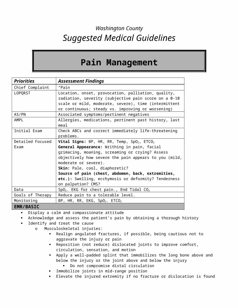
Hyperglycemia (Pediatric)
Note: Most pediatric patients with hyperglycemia have Diabetic ketoacidosis (DKA), which is a life-threatening complication of
diabetes that includes severe dehydration and metabolic acidosis. Patients with DKA have Kussmaul’s respirations, which are rapid and deep. In children, the parents may not know the child has diabetes before the first episode of DKA. The first clues of new onset diabetes may be excessive thirst or urination, including inappropriate wetting (e.g. wetting the
bed or wetting pants). The patient may be an unreliable historian. Parents or older siblings may provide a better history. In the pediatric population, diabetes is most likely due to failure of insulin production by the pancreas (a.k.a.
Type I Diabetes Mellitus or Insulin Dependent Diabetes Mellitus or Juvenile Onset Diabetes Mellitus).Priorities Assessment FindingsChief Complaint “High blood sugar”, Breathing Fast, Vomiting, Abdominal Pain, “Diabetic
Coma”LOPQRST Check onset/duration of symptoms. Identify possible contributing factors [1].
Use of sliding scale to adjust insulin doses to rising glucose levels? AS/PN Fever/Chills. Signs/Symptoms of infection. Polyuria, Polydipsia, Polyphagia,
Adequate food and water intake? Increasing thirst? Increasing urine output?AMPL Known history of Diabetes. Medications for diabetes.Initial Exam ABCs and correct any immediately life-threatening problems.Detailed Focused Exam Vital Signs: BP, HR, RR, Temp, SpO2, ETCO2, Patient’s Weight
General Appearance: Appears sick? Dehydrated? Kussmaul’s Respirations? Smell of Acetone on Breath.Heart: Tachycardia? Hypotension?Resp: Rapid RespirationGI: Diffuse Abdominal TendernessSkin: Cool, pale, diaphoretic? Warm, dry, flushed? TentingNeuro: ALOC? Focal deficits (CVA)?
Data Blood Glucose, Patients Weight, SPO2, ETCO2
Goals of Therapy Use IV fluids to reduce glucose level, improve hydration, normalize acid-base balance
Monitoring Repeat blood glucose
EMR Routine Medical Care. Administer oxygen to keep SpO2 > 94% and <99%
Use a nasal cannula at 2 – 6 LPM or Non-rebreather mask at 10 – 15 LPM
Monitor vitals.
BASIC Check glucose level.
INTERMEDIATE TECH / INTERMEDIATE / PARAMEDIC Consider IV/ IO (If authorized) If Blood glucose > 250 mg/dl initiate IV 0.9% NS give 20 ml/kg fluid bolus. Neonates (less than 30 days old) use 10ml/kg. If BP < 80 systolic give additional 10ml/kg fluid bolus.
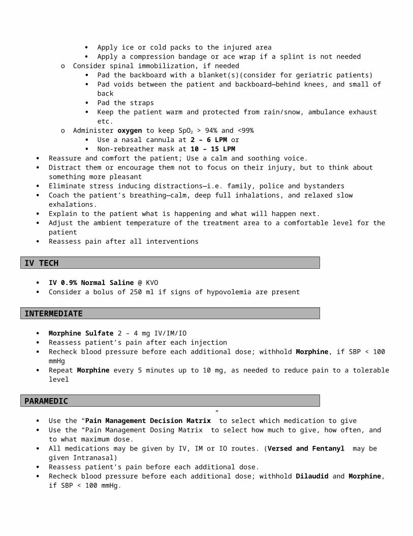
Contact Medical Control for the following: If the patient appears very sick or dehydrated, consider a second 10ml/kg fluid bolus 0.9% NS.
FOOTNOTES:[1] Contributing factors
Illness, especially infections Lack or relative lack of insulin Dehydration Illness
Date of Origin: 11/10/11
Washington County
Suggested Medical Guidelines
Hypoglycemia (Pediatric)Note:
The patient may be an unreliable historian. Parents or older siblings may provide a better history.

In the pediatric population, diabetes is most likely due to failure of insulin production by the pancreas (a.k.a. Type I Diabetes Mellitus or Insulin Dependent Diabetes Mellitus or Juvenile Onset Diabetes Mellitus).
Priorities Assessment FindingsChief Complaint “Low blood sugar” “Altered Level of Consciousness”LOPQRST Check onset/duration. Identify possible contributing factors [1]. Recent history
of frequent episodes, Last Meal. AS/PN Fever/Chills. Signs/Symptoms of infection, Possible Ingestions,
Nausea/Vomiting.AMPL Medications for diabetes, Time and dose of last Insulin, received.Initial Exam ABCs and correct any immediately life-threatening problems.Detailed Focused Exam Vital Signs: BP, HR, RR, Temp, SpO2 , ETCO2, Patient’s Weight
General Appearance: Unresponsive? Agitated and combative?Skin: Cool, pale, diaphoretic?Neuro: ALOC? Focal deficits (CVA)?
Data Blood Glucose, obtain MedicationsGoals of Therapy Restore normal mental statusMonitoring Repeat blood glucose
EMR Routine Medical Care. Administer oxygen to keep SpO2 > 94% and <99%
Use a nasal cannula at 2 – 6 LPM or Non-rebreather mask at 10 – 15 LPM
Monitor vitals.
BASIC Check glucose level.
o Sugar > 60, monitor for hypoglycemia.o Blood Sugar < 60, conscious, administers oral glucose (15 grams oral glucose) or juice if available.
Closely monitor airwayo Blood Sugar < 60, unconscious:
Age < 5 yrs. give Glucagon 0.5 mg IM. Age > 5 yrs. give Glucagon 1 mg IM. If patient is unconscious oral glucose can carefully be administered in the cheek of the patient (thin layer
smeared inside cheek or gums)o Recheck blood sugar 5 – 10 minutes after oral glucose or Glucagon.
INTERMEDIATE TECHNICIAN / INTERMEDIATE / PARAMEDIC Consider IV/IO (If authorized) Blood sugar < 60 and patient unconscious: Initiate IV 0.9% NS at KVO and administer:
Less than age 2 years: Dextrose 25% 2 ml/kg IVP. Note response [3]Age 2 years or older: Dextrose 50% 1ml/kg IVP, Up to 25g in 50ml (1 AMP) Note: patients over 110 pounds/50 Kg receive the adult dose
Contact Medical Control for the following: If the patient remains unconscious, call Medical Control to request a second dose of D25. If the patient’s guardian wants to refuse transport, ensure patient safety [2], notify medical control
and get release signed.
FOOTNOTES:[1] Contributing factors
Too much insulin? Not enough food? Overexertion
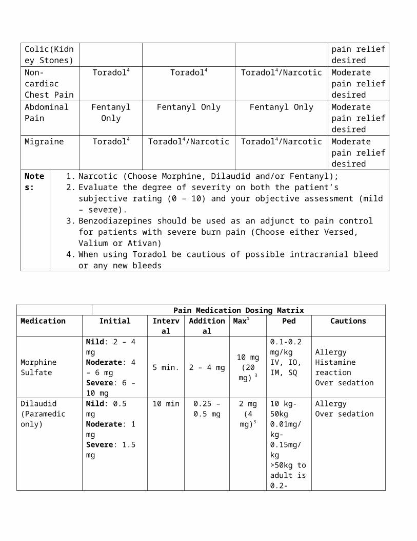
Illness[2] Ensuring Patient Safety
Adequate family and social support is available The patient’s family has access to food or money to buy food The patient is not ill, unable to eat or in need of immediate medical attention
[3] Creating Dextrose 25% (from Dextrose 50%) Dispose of 25ml (half an amp) of dextrose, replace with normal saline. Mix well before administration.
Date of Origin: 11/10/11
Washington County
Suggested Medical Guidelines
Hypovolemia and Shock (Pediatric)
Note: Potential causes of hypovolemia and shock include:
o Infections/sepsis

o Trauma/Burnso Hemorrhage (Internal, External)o Spinal cord injuryo Pump Failureo Heart Rhythm Disturbanceso Dehydration/Heat emergencieso Drugs and Toxinso Vasovagal Syncopeo Metabolic Disturbanceso Anaphylaxiso Pulmonary Embolism
Shock is defined as inadequate perfusion of vital organs, not merely hypotension. Clinical evidence of shock includes altered mental status.
Priorities Assessment FindingsChief Complaint “Altered Level of Consciousness”, Weakness, PaleLOPQRST Identify onset, duration, progression and provocation.AS/PN Fever/Chills, Nausea/Vomiting, Trauma, New MedicationsAMPL Pertinent past history and medications may provide important clues.Initial Exam ABCs and correct immediately life-threatening problems.Detailed Focused Exam Vital Signs: BP, HR, RR, Temp, SpO2, Patient’s Weight
General Appearance: Does the patient appear ill? External Hemorrhage? Skin: Pale, cool, and moist? Flushed, warm and dry?, Cyanosis, Rash? Petechiae, Purpura, Bruising? Insect bite or sting?Chest: Labored breathing?Lungs: Wheezes, rales or rhonchiHeart: Rate and Rhythm?Abdomen: Internal hemorrhage? Tender? Distended? GI Blood loss?Extremities: Trauma? Edema?Neuro: ALOC?
Data SpO2, 12-Lead EKG, Blood SugarGoals of Therapy Restore volume and support blood pressureMonitoring Blood pressure, heart rate and cardiac rhythm
EMR/BASIC
Routine Medical -or- Trauma Care. Secure and maintain airway. Oropharyngeal or nasopharyngeal airway, if indicated Administer oxygen to keep SpO2 > 94% and <99%
Use a nasal cannula at 2 – 6 LPM or Non-rebreather mask at 10 – 15 LPM
Control external hemorrhage Keep patient flat with lower extremities elevated (if possible). Splint fractures Conserve body temperature, and reassure patient.
INTERMEDIATE TECHNICIAN
Establish IV Normal Saline @ KVO rate If signs of hypovolemia or inadequate perfusion are present, administer fluid bolus at 20mL/kg (Neonates less than 30
days old use 10ml/kg). Consider IO (If authorized)
INTERMEDIATE/ PARAMEDIC
Identify underlying problem and refer to the appropriate protocol.
FOOTNOTES:
Date of Origin: 11/10/11
Washington County
Suggested Medical Guidelines
Respiratory Distress (Pediatric)
Note: This protocol may apply to the following conditions:
o Asthma/Bronchospasm/Reactive Airways Diseaseo Allergy/Anaphylaxiso Pulmonary Infectionso Spontaneous Pneumothoraxo Upper Airway Obstructiono Anxiety and Hyperventilation Syndrome
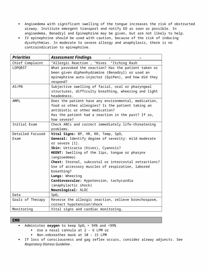
Priorities Assessment FindingsChief Complaint “Difficulty breathing”; “Shortness of breath”LOPQRST Assess onset, duration, progression, subjective severity, possible triggering
events, and response to treatments before EMS arrival.AS/PN Chest pain (what kind?), fever/chills, productive (of what?) cough AMPL Check for possible exposure to known allergens. Check past history,
medications and compliance for clues to cause of present illness. Initial Exam Check ABCs and correct immediately life-threatening problems.Detailed Focused Exam Vital Signs: BP, HR, RR, Temp, SpO2, ETCO2
General Appearance: Severity of distress [1]? Lethargic?Skin: Cool, moist and pale? Warm, dry and flushed? Urticaria? Cyanosis?HEENT: Nasal flaring?Chest: Sternal, subcostal or intercostal retractions?Respiratory Effort: Using accessory muscles, signs of fatigue; reduced or no verbal response (age appropriate)?Lung Sounds: Wheezes, rales, rhonchi or stridor?Heart Sounds: Rate, regularity.Lower Extremities: Pale, mottled?Neuro: ALOC, lethargy, somnolence?
Data SpO2
Blood Sugar, if DKA is suspected or if there is ALOCGoals of Therapy Improve oxygenation and ventilation, reduce the work of breathing, and treat
underlying conditions.Monitoring SPO2 frequently or continuously. ETCO2
Heart Rate and Blood Pressure frequently.
EMR Routine Medical Care Allow/assist the patient to assume a position of comfort (usually upright). Administer oxygen to keep SpO2 > 94% and <99%
Use a nasal cannula at 2 – 6 LPM or Non-rebreather mask at 10 – 15 LPM
Assisted Ventilation: Consider assisting breathing with gentle synchronous ventilations with bag-valve mask (BVM); Support ventilation with BVM if apnea or hypopnea occurs.
Airway Adjuncts: If airway can’t be maintained and there is an altered level of consciousness and loss of gag reflex, insert an
oropharyngeal or nasopharyngeal airway.
BASIC/ INTERMEDIATE TECHNICIAN Assist with patient-prescribed medications or initiate the following therapies:
Albuterol as indicated for Asthma (See specific protocols). Epinephrine0.01mg/kg (1:1,000) or Epi-Pen Jr for less than 60lbs may be needed in allergy, anaphylaxis or severe
asthma. (see specific protocol) If airway can’t be maintained and there is an altered level of consciousness and loss of gag reflex, for children at least 4 ft.
tall, consider a non-visualized airway
INTERMEDIATE
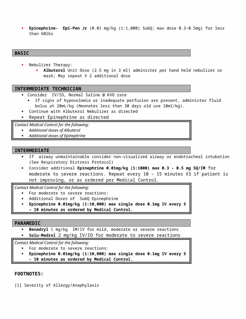
CROUPSTABLE: No cyanosis, mild respiratory distresso 5 ml Saline in nebulizer by mask or aim mist at child's face with oxygen at 6-8 L.
If wheezing: see Asthma protocol UNSTABLE: Cyanosis, marked stridor or respiratory distress.
o Consider possibility of epiglottitis and treat as below if airway obstruction progresses.o Epinephrine (1:1000) 3 ml (3 mg) via nebulizer mask or aim mist at child's face with Oxygen at 6-8 L.
EPIGLOTTITIS STABLE: No cyanosis, effective air exchange.o Epinephrine (1:1000) 3 ml (3 mg) via nebulizer mask or aim mist at child's face with oxygen at 6-8L.UNSTABLE: Bradycardic, altered mental status, marked stridor/ventilatory distress, retractions, ineffective air exchange, and/or
actual or impending respiratory arrest.o Position supine in sniffing position. Ventilate with 100% oxygen/Peds BVM using slow compressions of bag. If unable to
ventilate; temporarily stop ambulance and attempt one oral endotracheal intubation. Be prepared for airway status to worsen after intubation attempt if unsuccessful. (Do not use RSA)
o If a spontaneous tension pneumothorax is suspected, perform a needle decompression on the affected side.o Continue expeditious transport.
PARAMEDIC Airway Adjuncts:
If airway can’t be maintained and there is an altered level of consciousness and loss of gag reflex, consider endotracheal intubation
In respiratory arrest or cardiopulmonary arrest, insert an endotracheal tube. Consider Nasal intubation
Contact Medical Control or the paramedics en route regarding the following: Nature and severity of the problem Treatment or procedures proposed or performed Results of treatment Changes in patient status
FOOTNOTES:[1] Severity of Respiratory Distress:
Mild = RR<20 + minimal additional breathing effort + speaking in complete sentences + minimal or no accessory muscle use, No ALOC
Moderate = RR 20 to 30 + moderate additional breathing effort + difficult to speak + moderate accessory muscle use + possible ALOC
Severe = RR> 30 + marked additional breathing effort + minimal or no verbal response + major accessory muscle use + probable ALOC
[2] For the purposes of endotracheal intubation, one “attempt” is counted when the laryngoscope is removed from the mouth, even if there has not been an attempt to pass the tube. [3] Endotracheal Tube Size Selection & Chart
Tube size = (age + 16) / 4.Tube size = Size of the patient’s little finger or nostril.
Date of Origin: 11/10/11
Updated 10/2/12
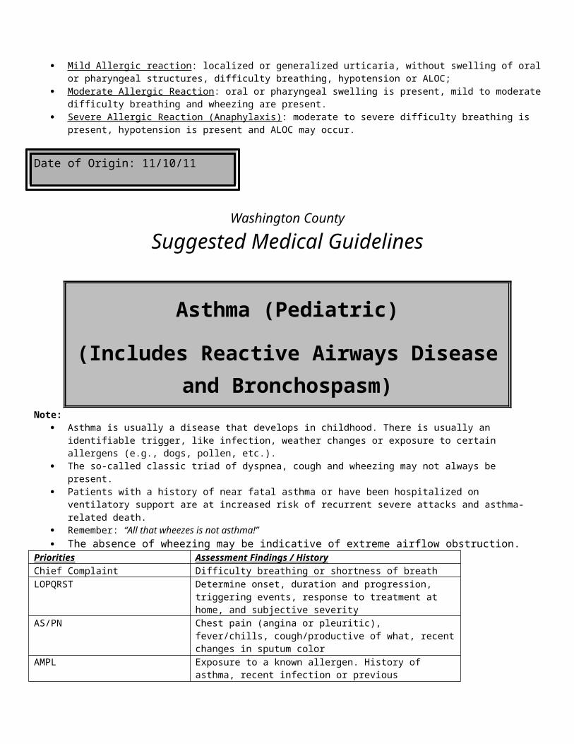
Washington County
Suggested Medical Guidelines
Seizure (Pediatric)Note:
Seizures usually last from 1-3 minutes and involve a loss of consciousness and convulsions. Not uncommonly, the patient is incontinent and may bite their tongue or be injured in other ways because of the convulsions.
When the seizure is over, the patient enters a postictal state, characterized by a gradual return to full consciousness over about 20 – 30 minutes, with initial confusion eventually giving way to normal alertness and orientation.
Whenever seizures occur, look for an underlying cause and treat it especially fever in pediatrics. This is especially important if there is no previous history of epilepsy.
Febrile Seizure is defined as a seizure with a fever >100.6 F. Status Epilepticus is defined as a seizure lasting longer than 30 minutes or frequently recurring seizures without clearing the
postictal state or return to baseline neurological status. This is a life-threatening emergency! Pseudo seizures look like seizures, but are actually a behavioral disturbance characterized by intermittent spells of
non-epileptic convulsions that are usually involuntary. They are frequently misdiagnosed as epilepsy and often treated with anti-epilepsy drugs for a long time, before the true nature of the attacks is revealed. Careful assessment may reveal telltale clues [2].
Priorities Assessment Findings
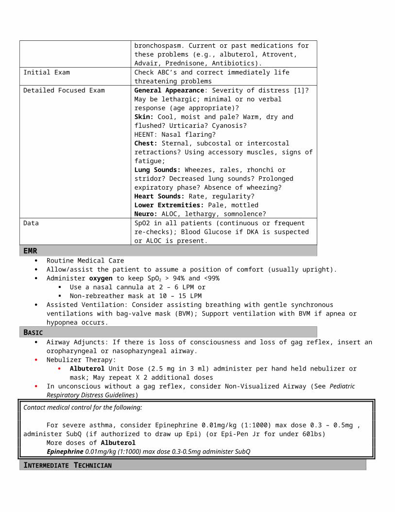
Chief Complaint “Seizure” “Unresponsive” “Convulsions” “Passed Out” “Fever”LOPQRST How long did it last? History of seizures? Fever? Possible contributing factors
[1]AS/PN Unresponsive, Postictal, IncontinentAMPL History of seizures, Seizure medications?Initial Exam ABC’s and correct any immediate life threatsDetailed Focused Exam Scene size-up: Is there a significant mechanism of injury?
General Appearance: Pt. currently seizing? Unresponsive? Postictal? Vitals: BP, HR, RR, Temp, SpO2, Patient’s WeightSkin: Flushed, warm, Rash, PetechiaeHEENT: Neck StiffResp: Airway Patent? Breathing? Neuro: ALOC?, Focal deficits (CVA)
Data Rectal Temperature, Blood Glucose, SpO2, ETCO2
Goals of Therapy Stop the seizureTreat the underline causeMonitor and maintain airway.
Monitoring Vitals, Cardiac monitoring, SpO2, ETCO2
EMR Routine medical care Consider oropharyngeal or nasopharyngeal airway, if the patient is unable to maintain a patent airway Protect the patient with ongoing seizures from harming themselves by clearing away potential hazards and placing a pillow or
padding under the head. Administer oxygen to keep SpO2 > 94% and <99%
Use a nasal cannula at 2 – 6 LPM or Non-rebreather mask at 10 – 15 LPM
BASIC Obtain blood glucose. If < 60 refer to hypoglycemia protocol. Check Temperature. If >100.6 F, report fever to Medical Control.
INTERMEDIATE TECHNICIAN IV 0.9% NS @ KVO Consider IO (If authorized)
INTERMEDIATE / PARAMEDIC If the patient is still seizing
o IV access give; Ativan 0.05-0.1mg/kg/dose up to 1-2mg.Or Valium (Paramedic Only) 0.3 mg/kg/dose IV up to 5 mg. May repeat in 5-10 minutes for continued seizure
o No IV Access give; Intranasal Versed (Paramedic Only) 0.3 mg/kg max dose of 1ml May repeat in 5-10 minutes for continued seizure Or Valium (Paramedic Only) 0.3 mg/kg IM. Max dose of 5mg May repeat in 10 minutes for continued seizure give 0.25 mg/kg.
Or Valium (Paramedic Only) 0.5 mg/kg/dose rectally. Max dose of 20 mg.
Contact Medical Control for the following: If pseudo seizures are suspected, withhold Ativan and Valium until you speak with Medical
Control. If IV access unavailable consider IO administration of Ativan or Valium at the same doses. If seizures persist, repeat doses of Ativan or Valium as stated above until seizures stop. Maximum
doses: Ativan 1-2 mg, Valium 5 mg.
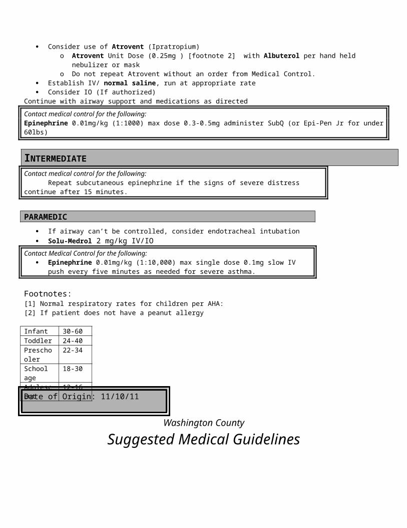
Do not give Ativan or Valium if patient is not seizing. Do not give Ativan or Valium for Febrile Seizures Persistent Seizure
FOOTNOTES:[1] The causes of seizures include: fever (in children up to about 6 yrs.), epilepsy, hypoglycemia, hypoxia, medications, drug withdrawal, drug overdose, head trauma.[2] Characteristics of pseudo seizures are listed below:
Identifiable trigger (emotional stress, crisis or grief) The patient usually has an audience Asynchronous or asymmetric motion during the seizure (“bicycling” or head turning from side to side) Mid-range and reactive pupils during the convulsion (they’re widely dilated in a real seizure) Lack of tongue biting or incontinence Apparent purposeful movements Remaining conscious, or even speaking, during the convulsion
Date of Origin: 11/10/11
Washington County
Suggested Medical Guidelines
SVT (Pediatric)
Note: This protocol pertains to unstable children or those suspected of deterioration at any time. Stable children should be
transported for evaluation preferably by a Pediatric Cardiologist. Remember children have higher underlying heart rates, a thorough history of events is very important AHA suggests pulse rates for SVT are >180 for ages 1-8 years and >220 for ages newborn – 1 year. History of the complaint is the key. SVT symptoms usually show normal children suddenly deteriorating with minimal or no
history of other recent illness or injury
Priorities Assessment FindingsChief Complaint Weakness, any altered LOC, signs of inadequate perfusionLOPQRST Determine onset of symptoms along with possible causes. AS/PN Respiratory distress, chest pain, nausea, vomiting, poor capillary refill AMPL Patient with known congenital heart defects, respiratory insufficiencies and
current medications. Initial Exam Assess ABC’s, support airway and breathing as necessary.Detailed Focused Exam General Appearance: Somnolent, flaccid or poor muscle tone.
Skin: Cool, pale or gray.

Respiratory Effort: May be normal or signs of distress, i.e.: sub and intercostal retractions, nasal flaring, etc. Lung Sounds: May be diminished or sound “wet” due to poor cardiac output.Heart Sounds: may be distant or difficult to hear due to poor cardiac output, except in some cases of cardiac hx.Extremities: Poor muscle tone, weak or no pulses, mottled, poor capillary refillNeuro: ALOC, unresponsive
Data SPO2, EKG in all cases, Core temperature, blood glucose and capillary refill checks, B/P in older children
Goals of Therapy Decrease heart rate and restore normal perfusionMonitoring Closely watch heart and respiratory rates, SPO2, EKG (especially in cases of
actual cardiac involvement)
EMR Routine Medical Care Maintain normal body temperature Administer oxygen to keep SpO2 > 94% and <99%
Use a nasal cannula at 2 – 6 LPM or Non-rebreather mask at 10 – 15 LPM
Vitals signs as appropriate Continue Airway support as needed; avoid use of non-visualized airways (combitube)
BASIC Apply ECG if available Obtain 12 lead ECG if available Assess blood glucose level. If glucose <60 see hypoglycemia protocol
INTERMEDIATE TECHNICIAN Establish IV Normal Saline KVO or 20ml/kg Consider IO (If authorized) Continue airway support
INTERMEDIATE Attempt valsalva maneuvers as able by child, i.e.; forceful cough, cold packs on face (must touch cheek bones) and neck,
or blow through a straw Intubation as needed May try Adenosine at 0.1mg/kg IV or IO max dose of 6 mg, be sure to administer rapidly and follow with a rapid fluid
flush. Repeat Adenosine at 0.2 mg/kg IV max dose of 12 mg.
PARAMEDIC If child is hemodynamically and clinically [1] unstable, perform immediate synchronized Cardioversion at 0.5-1.0
Joules/kg. If unsuccessful, increase to 2 Joules/kg for each subsequent attempt. Sedation prior to Cardioversion with Versed 0.1mg/kg slow IV / IO push.
FOOTNOTES: Hemodynamic Criteria
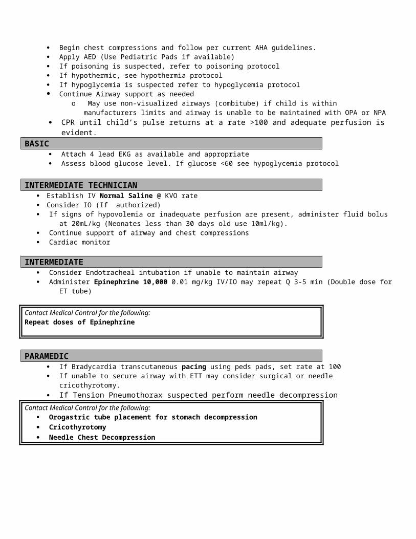
o Pulse rates for SVT are >180 for ages 1-8 years and >220 for ages newborn-1 yearEvaluating perfusion status in children
Signs of compensated shock include:o Tachycardiao Cool extremitieso Prolonged capillary refill (despite warm ambient temperature)o Weak peripheral pulses compared with central pulseso Normal blood pressure
Signs of uncompensated shock in children include:o ALOCo Tachypneao Weak central pulseso Hypotension
Date of Origin: 11/10/11
Washington CountySuggested Medical Guidelines
Toxic Exposure/Overdose (Pediatric)
Note: Perform scene size-up and ensure crew safety. In a hazardous materials incident, stage up wind of the incident, and do not
attempt to treat any patients who have not been decontaminated. Be especially suspicious of scenes in which many people or animals appear to be affected.
Beware of the potential for the patient to vomit spontaneously. Following any form of cyanide ingestion, emesis may off-gas toxic hydrogen cyanide, placing rescuers and health care workers at risk.
Beware of the potential for seizures or altered level of consciousness due to toxic exposures. Beware of the potential for cardiovascular collapse and respiratory compromise due to toxic exposures.
Priorities Assessment FindingsChief Complaint “Overdose” “Ingestion” “Exposure to chemicals” “Unresponsive”LOPQRST Determine type and kind of ingestion. Determine time of exposure/ingestion,
Determine amount/length of exposure.AS/PN Dyspnea, nausea/vomiting, abdominal pain, unresponsive; Suicidal ideation or
suicide attempt. Accidental or intentional exposure.AMPL Medication history and medications, exposure to chemicals.Initial Exam Check ABC’s, and correct any immediate life threatsDetailed Focused Exam Vitals: BP, HR, RR, Temp, SpO2, ETCO2, Patient Weight
General Appearance: level of alertness, signs of agitation, willingness to cooperate with authorities, signs of ingestion?Skin: Cool, pale and diaphoretic? Warm, dry and flushed? Rash?HEENT: Are the pupils constricted or dilated? Nystagmus?Lungs: Wheezes, rales or rhonchi?Heart: Rate, regularity, BP, peripheral perfusion?GI: Abdominal Distention

Neuro: Signs of intoxication? Ataxia? Slurred speech? Psych: Depressed affect? Bizarre thoughts? Signs of suicidal ideation or intent?
Data Blood glucose, Identify possible toxic substances ingested/exposed too.Goals of Therapy Reduce amount of substance absorbed into the body; Treat with antidotes if
possible; Correct toxic effects on the CNS, cardiovascular and respiratory systems.
Monitoring Cardiac monitoring
Poison Control Center Phone # 1-800-222-1222
EMR Routine medical care Administer oxygen to keep SpO2 > 94% and <99%
Use a nasal cannula at 2 – 6 LPM or Non-rebreather mask at 10 – 15 LPM
If the patient is unconscious, place him/her in the recovery position. Follow the ALOC Guidelines. Consider use of oropharyngeal or nasopharyngeal airway.
BASIC If the patient is unconscious, check blood glucose. If < 60, follow the Pediatric Hypoglycemia Guidelines.
INTERMEDIATE TECHNICIAN Establish IV Normal Saline @ KVO rate Consider IO (If authorized) If signs of hypovolemia or inadequate perfusion are present, administer fluid bolus at 20mL/kg (Neonates less than 30 days
old use 10ml/kg). If the patient has an altered level of consciousness and a narcotic overdose is suspected, consider Narcan
o < 20 kg 0.1mg/kg/doseo 20 kg or > 5 years old give 0.4 – 2 mg/dose IV/IM/Sub-Q/Intranasal and repeat every 5 minutes X 3 total
doses. If there is no response to Narcan, consider an alternative explanation or contact medical control. Contact Medical Control for the following:
Additional Narcan orders
INTERMEDIATE/ PARAMEDIC Specific overdose therapies are contained in the table below
o If you do not recognize what class the drug or toxin belongs to, contact Medical Control.
Class of drugs Treatment Indications Specific Treatment(s)Narcotics [1] Narcan may be used in cases of over
sedation due to narcotic administration, or in suspected narcotics overdoses in patients without a history of long-term use, chronic abuse or addiction. Signs of narcotic overdose or over sedation include: decreased level of consciousness, pinpoint pupils (except Demerol), and respiratory depression.
Caveat: Giving Narcan to a long-term narcotic user, chronic abuser or addict can induce narcotic withdrawal, which
In the setting of an accidental or intentional overdose, if the patient has ALOC – with or without a gag reflex, or shows signs of respiratory depression, airway management takes precedence over reversing the overdose with Narcan.
For other patients with narcotic overdose or over sedation give:Narcan (naloxone) follow dosing protocol above until the patient
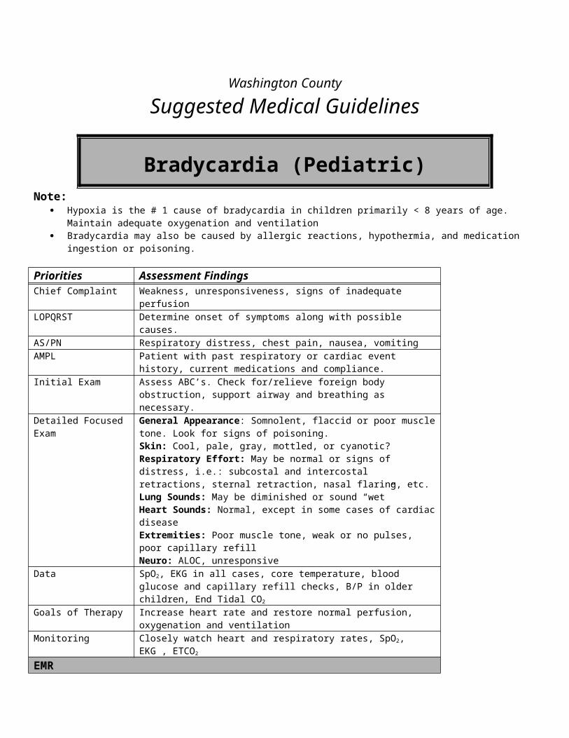
creates a new set of difficult problems. Airway management and supportive care is the preferred approach.
reaches a desirable level of consciousness and respiratory function, or as directed by Medical Control. If no response, reconsider diagnosis. Contact medial control.
Tricyclic Antidepressants (TCA)
Decreased level of consciousness; hypotension, seizures, malignant arrhythmias (e.g. Torsades de Pointes, VT), prolongation of the QT or QRS intervals.
Caveat: Patients with TCA overdoses are prone to deteriorating very quickly.
Note: Sodium containing solutions act like antidotes, because they protect the heart against the toxic effects of the TCA. Induced alkalosis from bicarbonate and hyperventilation also protect against the toxic effects of TCAs.
Give 20ml/kg Normal Saline Bolus. May Repeat.Sodium Bicarbonate (Paramedic Only)1 mEq/ml/kg IV bolus. Repeat as directed by medical control. Consider RSA with End-tidal CO2 of 25 – 30.
Treat arrhythmias according to the appropriate protocol.Treat seizures according to the Pediatric Seizure Guidelines.
Beta Blockers Profound bradycardia, hypotension or conduction defects
Contact medical control. Consider Glucagon 0.03-0.1 mg/kg/dose every 20 minutes as needed slow IVP. Max Dose: 1 mg/dose.
Calcium Channel Blockers
Profound bradycardia, hypotension or conduction defects
Contact medial control.
Amphetamines Agitation, psychosis, or ventricular arrhythmias
Caveat: For patients with Excited Delirium, refer to the Agitated and Combative Patients Guidelines.
Contact Medical control.
Cocaine Agitation, seizures, or ventricular arrhythmias
Caveat: For patients with Excited Delirium, refer to the Agitated and Combative Patients Guideline.
Contact Medical control.
Organophosphate Poisoning (Pesticides and Nerve Agents)
Profound bradycardia, seizures, abnormal (wet) lung sounds
The organophosphate toxidrome:S – Salivation, SeizuresL – LacrimationU – UrinationG – GI vomiting and diarrheaB – Bradycardia*, bronchorrhea,
bronchospasmA – ArrhythmiasM – Miosis (small pupils)** Tachycardia and mydriasis (dilated pupils) are also possibleCaveat: Organophosphates are highly toxic in very small quantities and pose a significant risk to EMS and health
Atropine 0.02mg/kg IV or IM every 3-5 min until lung sounds clear to auscultation. Use atropine in the initial treatment of bradycardia and seizures. Contact Medical Control.
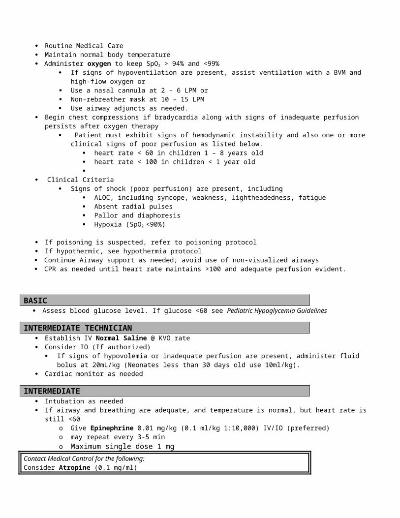
care workers through secondary contamination.
FOOTNOTES:
[1] The CNS depressant effects of narcotics and benzodiazepines are potentiated by ingestion of other CNS depressants, especially alcohol.
Date of Origin: 11/10/11
Washington County
Suggested Medical Guidelines
Ventricular Fibrillation & Pulseless Ventricular Tachycardia (Pediatric)
Note: These guidelines are based on (or adapted from) the 2010 American Heart Association Guidelines for Cardiopulmonary
Resuscitation and Emergency Cardiovascular Care Unlike adult cardiac arrest, which is usually due to a primary cardiac abnormality, pediatric cardiac arrest most often occurs
as a result of asphyxia. The most common reasons for this include progressive respiratory failure and shock.
Priorities Assessment FindingsChief Complaint Collapsed, unresponsive, no pulse, not breathing LOPQRST Witnessed or unwitnessed? Estimated time of onset. Circumstances/trauma.
Location of patient. Antecedent symptoms/signs (difficulty breathing, serious illness) and their duration.
AS/PN Bystander-initiated CPR. Pre-arrival CPR instructions from dispatch? Public access AED use.
AMPL Does the patient have any allergies to medications? History of heart disease? Current cardiac medications?
Initial Exam Establish Unresponsiveness. Check ABCs. Open and reposition airway.Detailed Focused Exam Vitals Signs: Pulseless and non-breathing (or agonal respirations)
General: Identify unresponsiveness. Look for rigor mortis, dependent lividity, or unsurvivable trauma. Look for a valid Wisconsin Do-not-resuscitate bracelet.Skin: Mottled, Warm/cold, dependent lividity, rash, ecchymosis?HEENT: Airway patent, foreign bodies, dry mucus membranes, furrowed lips?Chest: Spontaneous respirations?Lungs: Equal breath sounds, difficulty bagging or ventilating?Cardiovascular: Absence of heart sounds, carotid or femoral pulses?
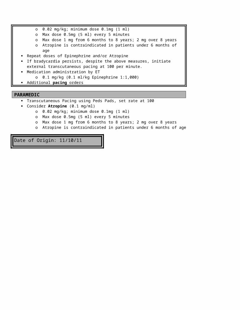
Abdomen: Distended?Extremities: Rigor mortis, edema, deformity?Neurological: Unresponsive to verbal and painful stimulation?
Data Cardiac rhythm analysis, blood sugar, SpO2, ETCO2
Goals of Therapy Return of spontaneous circulation (ROSC)Monitoring Cardiac Monitoring, Vital Signs, ETCO2 and SpO2
EMR/BASIC/IV TECH Refer to the Pediatric Cardiac Arrest Guidelines
INTERMEDIATE/PARAMEDIC Direct First Responders and EMTs to continue basic rescuer interventions. Follow Pediatric Cardiac Arrest Guidelines. Attach a cardiac monitor and evaluate the rhythm First Priority: Defibrillate
o First defibrillation at 2 J (Joules)/kg All subsequent shocks at 4 J/kg If defibrillation is successful, and spontaneous circulation returns (ROSC), refer to reassessment section for
Lidocaine administration Second Priority: Perform Effective CPR
o Perform a head-tilt-chin-lift maneuver. In suspected C-spine injury, use the jaw thrust, instead. Consider a nasopharyngeal or oropharyngeal airway
o Follow American Heart Association Guidelines for CPR in childreno Continue until spontaneous circulation returns, the patient is transported, or resuscitation is terminated.
Third Priority: Establish an Advanced Airwayo If an advanced airway is not already in place, insert an endotracheal tube [1].o If a non-visualized airway is in place, leave it there, unless it is malfunctioning.
Fourth Priority: Establish Venous Access Consider venous access, if not already done
o Establish a peripheral IV (IV Tech/Int/Paramedic)o Consider IO (If authorized) (IV Tech/Int/Paramedic)o Drug administration routes in order of preference: IV – IO – ETo Do not attempt to administer medications via a non-visualized airway
Lack of venous access is not an acceptable indication for converting a non-visualized airway that is functioning well for ventilations to an ET tube.
Rather, use IO access. Fifth Priority: Proceed to ACLS resuscitation medications:
o Epinephrine 1:10,000 – give 0.01 mg/kg IV/IO every 3 – 5 minutes Via ET route: Epinephrine 1:1,000 0.1 mg/kg every 3 – 5 minutes
o Amiodarone 5mg/kg IV or IO may repeat up to 2 timeso ORo Lidocaine 1 mg/kg IV/IO Bolus
Via ET route: 2 – 3 mg/kg ET followed by 5 ml of Normal Saline and 5 ventilations (EMT-P Only): Lidocaine drip(Paramedic Only): 0.02 – 0.05 mg/kg/min
o Magnesium Sulfate (Paramedic Only) 25 – 50 mg/kg IV/IO/IM (Max 2 gm) slowly over 2 minutes, if Torsades de Pointes or polymorphic VT is detected on the monitor.
Reassessmento If there is return of spontaneous circulation transport, monitor, and administer Lidocaine if used to convert.
Lidocaine 1 mg/kg IV/IO Boluso Via ET route: 2 – 3 mg/kg ET followed by 5 ml of Normal Saline and 5 ventilationso (EMT-P Only): Lidocaine drip(Paramedic Only): 0.02 – 0.05 mg/kg/min
Maintain advanced airway, if the patient remains unconscious. 8 – 10 rescue breaths per minute
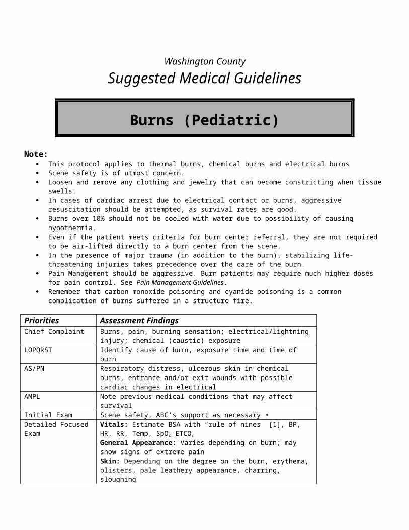
High flow oxygen via BVM Monitor SpO2 and ETCO2
If the patient wakes up, the airway may be removed. Use the procedures for removing advanced airway devices in the Respiratory Distress Guidelines.
o After three rounds of shocks and drugs without ROSC, re-evaluate interventions. Contact medical control.o If Asystole or PEA develops, refer to the Pediatric Asystole and PEA Guidelines.o Transport and continue resuscitative efforts.
Infrequently, prehospital termination of pediatric resuscitation may be indicated. If this appears to be the wishes of the family, contact medical control.
Contact Medical Control for the following: To discuss termination of resuscitation in the absence of a valid Wisconsin DNR Bracelet
FOOTNOTES:
[1] Endotracheal Tube Size Selection & ChartTube size = (age + 16) / 4.Tube size = Size of the patient’s little finger or nostril.Select tube size from the following table:
Newborn 3.0
Newborn – 6 months 3.5
6 – 18 months 3.5 – 4.0
18 months – 3 years 4.0 – 4.5
3 – 5 years 4.5
5 – 6 years 5.0
6 – 8 years 5.5 – 6.0
8 – 10 years 6.0
10 – 12 years 6.0 – 6.5
12 – 14 years 6.5 – 7.0
Date of Origin: 11/10/11
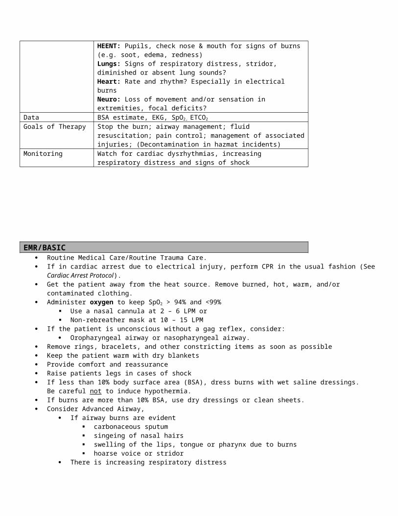
Washington County
Suggested Medical Guidelines
Wide Complex Tachycardia (Pediatric) Note:
Wide complex rhythms have a QRS duration > 0.08 sec Although some wide complex tachycardia develop from Supraventricular tachycardia’s, prehospital providers should
always assume that wide complex rhythms are ventricular tachycardia (VT), particularly if the patient is unstable [1].
Priorities Assessment FindingsChief Complaint Palpitations, fast heart rate, shortness of breath, chest pain, weakness, syncope,
cardiac arrest/pulseless non-breatherLOPQRST Onset and duration, precipitating factors and circumstances, associated
symptoms, stroke symptoms, nausea vomitingAS/PN Chest pain, shortness of breath, weakness, anxiety, leg swellingAMPL Previous history, history of thyroid disease, CAD, Cardiac Medications
Obtain history of previous episodes of tachycardia, including diagnoses if known. Pay particular attention to whether there is an underlying history of pre-excitation, including the Wolff-Parkinson-White (WPW) Syndrome.
Obtain history of what medications have been used to treat previous arrhythmias, if known.
Obtain history of any previous complications from previous arrhythmia treatments, if known.
Obtain history of the duration of the current episode of tachycardia, if known.
Initial Exam Check ABCs and correct any immediate life threatening problems.Detailed Focused Exam Vitals Signs: BP, HR, RR, Temp, SpO2, ETCO2
General Appearance:Skin: Cool, pale diaphoretic Neck: JVD?Chest: Labored breathingLungs: Wheezes, rales, rhonchi? Decreased breath sounds?Heart: Regular, rate fast or slow, murmur Legs: EdemaNeuro: ALOC?
Data SpO2, 12-Lead EKG, Blood Sugar if Diabetic or ALOCGoals of Therapy Decrease Rate, treat chest pain, treat CHFMonitoring Cardiac Monitoring and ETCO2, SpO2
EMR/BASIC Routine Medical Care Ensure an adequate airway.
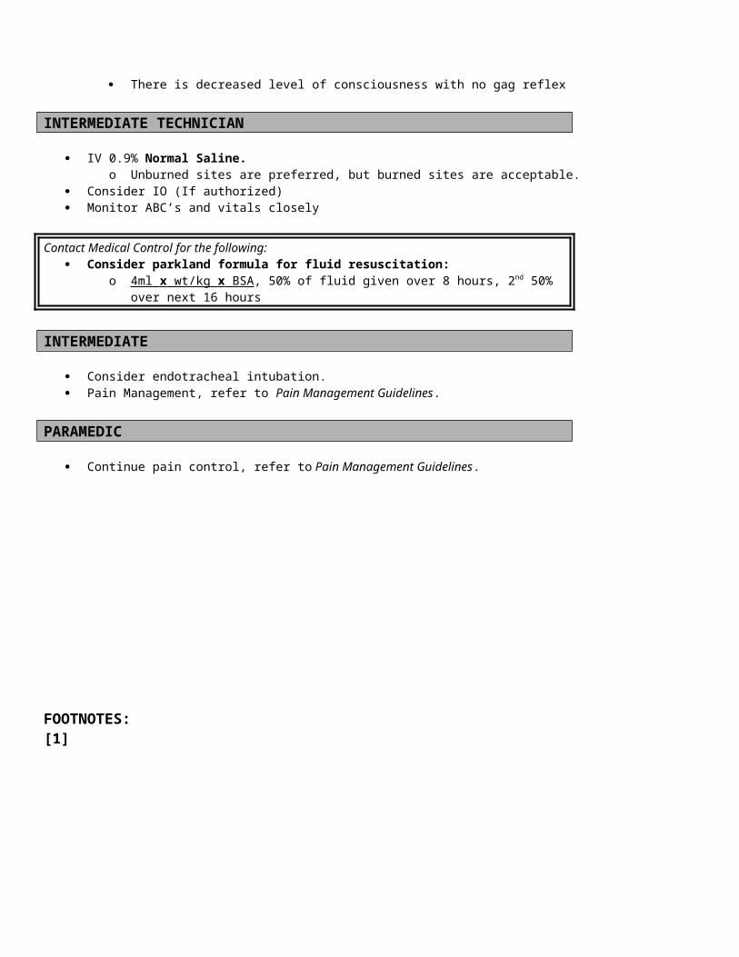
Administer oxygen to keep SpO2 > 94% and <99% Use a nasal cannula at 2 – 6 LPM or Non-rebreather mask at 10 – 15 LPM
If the patient is having difficulty breathing allow them to sit upright If the patient becomes unresponsive, pulseless and non-breathing, follow the Pediatric Cardiac Arrest Guidelines. If the patient also experiences signs of respiratory distress or inadequate breathing, follow the Pediatric Respiratory Distress
Guidelines
INTERMEDIATE TECHNICIAN Establish IV Normal Saline @ KVO rate Consider IO (If authorized)
If signs of hypovolemia or inadequate perfusion are present, administer fluid bolus at 20mL/kg (Neonates less than 30 days old use 10ml/kg).
Do not delay transport to start an IV, if the patient has signs of poor perfusion
INTERMEDIATE/PARAMEDIC Monitor the heart rhythm
o Identify a wide complex rhythm (QRS > 0.08 sec) If the patient has no palpable pulses, proceed according to the Pediatric Ventricular Fibrillation and Pulseless Ventricular
Tachycardia Guidelines Evaluate the perfusion status [1]
o Contact Medical Controlo If the patient has good perfusion [1] prepare for transport
If the patient has signs of poor perfusion [1] o Prepare to perform synchronized Cardioversion o Perform first synchronized Cardioversion @ 0.5 – 1 J/kgo If unsuccessful, increase to 2 J/kg for the second shocko If unsuccessful consider Amiodarone 5mg/kg IV max dose of 150 mg over 20 minuteso OR
If unsuccessful, consider Lidocaine 1 mg/kg IV/IO. If the patient maintains adequate perfusion, further treatment can be safely delayed until the patient arrives in the emergency
department.o Consider the use of Adenosine 0.1 mg/kg up to 6 mg rapid IV/IO push with saline flusho May repeat Adenosine 0.2 mg/kg up to 12 mg rapid IV/IO push with saline flusho Adenosine may be used in pediatric patients with poor perfusion, if doing so will not delay electrical
CardioversionContact Medical Control for the following:
Consultation about rhythm analysis
FOOTNOTES:[1] Evaluating perfusion status in children
Signs of compensated shock include:o Tachycardiao Cool extremitieso Prolonged capillary refill (despite warm ambient temperature)o Weak peripheral pulses compared with central pulseso Normal blood pressure
Signs of uncompensated shock in children include:o ALOCo Tachypneao Weak central pulseso Hypotension

Date of Origin: 11/10/11
Washington County
Suggested Medical Guidelines
Pregnant Female in LaborPriorities Assessment FindingsChief Complaint Uterine contractions, “in labor”LOPQRST Location of pain, radiation of pain, time of onset of contractions, interval
between contractions, quality of contractions, severity of contractions, events surrounding onset of contractions, due date
Associated Symptoms/Pertinent Negatives
Vaginal bleeding (presence, quantity, and character), , “bloody show,” leakage of fluid or discharge, need to “push,” “bear down” or have a bowel movement, presence of fetal movement, RUQ pain, vomiting, visual changes
SAMPLE Allergies and Medications Past medical history, past surgical history, number of previous
pregnancies, previous Cesarean delivery, prenatal care Previous pregnancy or delivery complications (eclampsia, precipitous
delivery, etc.) Recent infectious diseases Complications of current pregnancy (i.e. preeclampsia, placenta previa,
gestational diabetes, premature labor, ultrasound showing abnormal fetal position etc.)
Last mealInitial Exam ABCsDetailed Focused Exam Vital Signs: BP, HR, RR, Temp, SpO2
General Appearance, HEENT: Cracked lips, sunken eyes or cheeks indicating dehydrationSkin: Cool, pale diaphoretic Chest: Labored breathingHeart: Tachycardia Abdomen: Scars, Tenderness, masses, uterine size/location, distention,
deformityLegs: EdemaNeuro: Mental statusGyn: Vaginal bleeding, infant head crowning, prolapsed cord, presenting part, meconium staining
Goals of Therapy Atraumatically deliver newborn with maintenance of normal vital signs for both mother and newborn
Monitoring Frequency of contractions, SpO2,
EMR/BASIC Routine Medical Care Administer oxygen to keep SpO2 > 94% and <99%
Use a nasal cannula at 2 – 6 LPM or Non-rebreather mask at 10 – 15 LPM
Refer to Delivery of Newborn Procedure Protocol, if delivery is imminent. Place patient in position of comfort, preferably on left side.
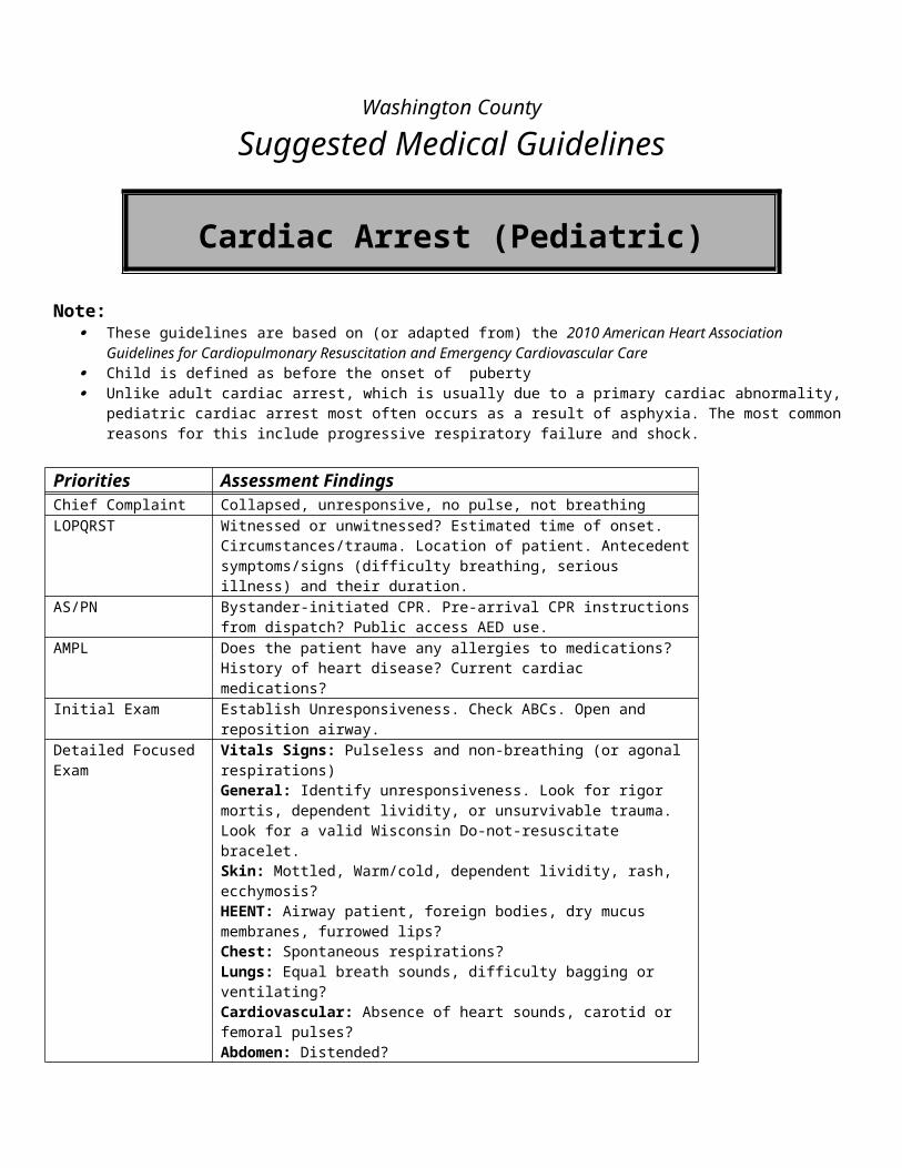
INTERMEDIATE TECH/INTERMEDIATE/PARAMEDIC Establish IV NS @ TKO if time permits before delivery If patient seizes, refer to Eclampsia Protocol
Date of Origin: 11/10/11

Washington County
Suggested Medical Guidelines
Rapid Atrial Fibrillation (Adult)
Note:
Priorities Assessment FindingsChief Complaint Palpitations, fast heart rate, shortness of breath, chest pain, weaknessLOPQRST Onset and duration, precipitating factors and circumstances, associated
symptoms, stroke symptoms, nausea vomitingAS/PN Chest pain, shortness of breath, weakness, anxiety, leg swellingAMPL Previous history, history of thyroid disease, CAD, cardiac medicationsInitial Exam Check ABCs and correct any immediate life threatening problems.Detailed Focused Exam Vitals Signs: BP, HR, RR, Temp, SpO2
General Appearance:Skin: Cool, pale diaphoretic Neck: JVD?Chest: Labored breathingLungs: Wheezes, rales, rhonchi? Decreased breath sounds?Heart: Regular, rate fast or slow, murmur Legs: Edema? Signs of an acute arterial occlusion (embolism)?Neuro: ALOC? Signs of stroke?
Data SpO2, 12-Lead EKG, Blood Sugar if Diabetic or ALOCGoals of Therapy Decrease Rate, treat chest pain, treat CHFMonitoring Cardiac Monitoring, SpO2, End Tidal CO2
EMERGENCY MEDICAL RESPONDER (EMR) Routine Medical Care. Administer oxygen. Allow/assist the patient to assume a position of comfort (usually upright).
Basic Routine Medical Care
Administer oxygen to keep SpO2 > 92% and <100% Use a nasal cannula at 2 – 6 LPM or Non-rebreather mask at 10 – 15 LPM
If the patient is having difficulty breathing allow them to sit upright Obtain a 12-Lead EKG If patient experiences angina give Aspirin 324 mg PO (Four(4)-81 mg chewable tablets)
o Do not give Aspirin if the patient is allergic to it.o Aspirin is optional if the patient has already taken a full dose todayo Coumadin (warfarin) use is not a contraindication to Aspirin in the patient with Angina
Administer patient’s prescribed Nitroglycerin (NTG) sublingually, unless the Systolic BP < 100 mm Hg.o Note: No NTG if pt has used Viagra or Levitra in the last 24 hours, or Cialis in the last 48 hours.o Repeat BP (before and after ) Nitroglycerin dose every 5 minutes until pain is relieved.o Discontinue nitroglycerine if the Systolic BP drops below 100 mm Hg.o Document all BP’s and the number of Nitroglycerin doses given.
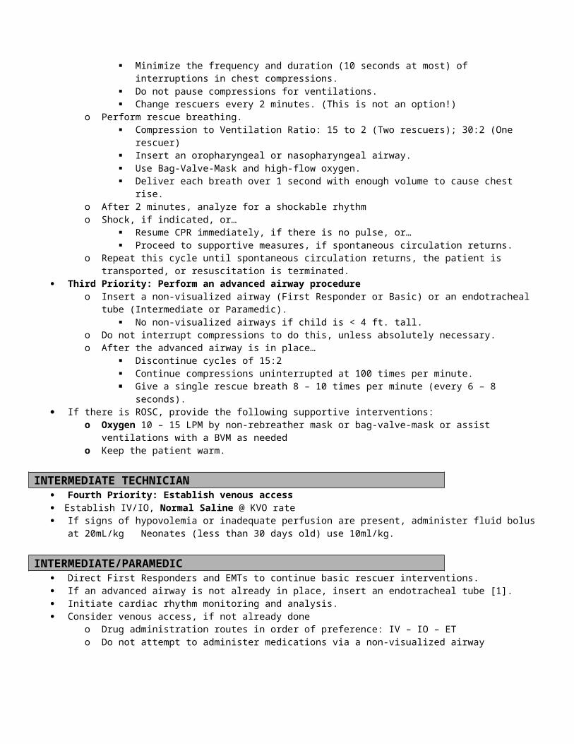
If the patient experiences shortness of breath, follow the Congestive Heart Failure Guidelines If the patient has suffered a stroke, follow the Stroke Guidelines.
INTERMEDIATE TECHNICIAN IV 0.9% Normal Saline If SBP < 100 mmHg give 250cc fluid bolus, and then reassess
o Consider Trendelenburg position, if there is no dyspnea
INTERMEDIATE If the patient does not have a history of atrial fibrillation, consider the potential causes that may have a bearing on prehospital
care, including:o Acute coronary syndromes o Pulmonary embolismo Alcohol useo Stimulant drug abuse (e.g. cocaine and amphetamines)
If the patient has a history of atrial fibrillation, consider possible causes of tachycardia, including: pain, dehydration, hypotension, shock, hypoglycemia, hypoxemia, anxiety, fever, sepsis, drug induced, recent heavy exertion, hyperthyroidism and anemia.
o The causes listed in bold-face print are within the scope of prehospital providers to identify and treat. Refer to the appropriate guidelines or contact medical control.
If the patient is hemodynamically and clinically stable, observe and monitor.
PARAMEDIC
If the patient is hemodynamically and clinically stable, transport, observe and monitor. Efforts to reduce the heart rate will add little benefit in the absence of adverse effects from RAF.
If the patient is hemodynamically unstable and meets one or more of the clinical criteria [1], consider synchronized Cardioversion.
o Perform first Synchronized Cardioversion @ 50 Joules.o If unsuccessful, increase by 100 joules for each subsequent attempt.
Consider Morphine Sulfate 2-5 mg IV/IO or Fentanyl 50 – 100 mcg IV/IO and Versed 1-2 mg IV/IO for sedation and pain control before Cardioversion..
If the patient is hemodynamically and clinically stable, transport, observe and monitor. Efforts to reduce the heart rate will add little benefit in the absence of adverse effects from RAF.
o Indications for pre-hospital treatment with Cardizem: RAF is causing mild hypotension (SBP 80 – 100 mmHg) but no other serious signs or symptoms are
present RAF is causing an acute exacerbation of CHF and the SBP is normal to high RAF is causing rate-related chest pain
o If the patient is mildly hypotensive, give a bolus of normal saline 250ml prior to treatment with Cardizem (check breath sounds)
Consider pre-medicating with Calcium Chloride 100 – 200 mg (1 – 2 ml of 10% Calcium Chloride) to prevent the hypotensive effect of Cardizem
o If the patient is experiencing CHF, also follow the Congestive Heart Failure Guidelines.o If the patient is experiencing rate-related chest pain without other serious signs or symptoms, treat with Cardizem to
reduce the rate. Then re-assess the patient for symptoms of ongoing chest pain.o Cardizem 0.25 mg/kg IV slowly over 5 min.
Use caution with patients currently taking beta blockers and/or digoxino May repeat Cardizem if response inadequate after 15 min. 0.35 mg/kg IV slowly over 5 min.
FOOTNOTES:[1] Criteria for characterizing a patient as “unstable”* (Patient must meet the Hemodynamic Criteria and at least one of the Critical Criteria to be considered unstable)
Hemodynamic Criteria
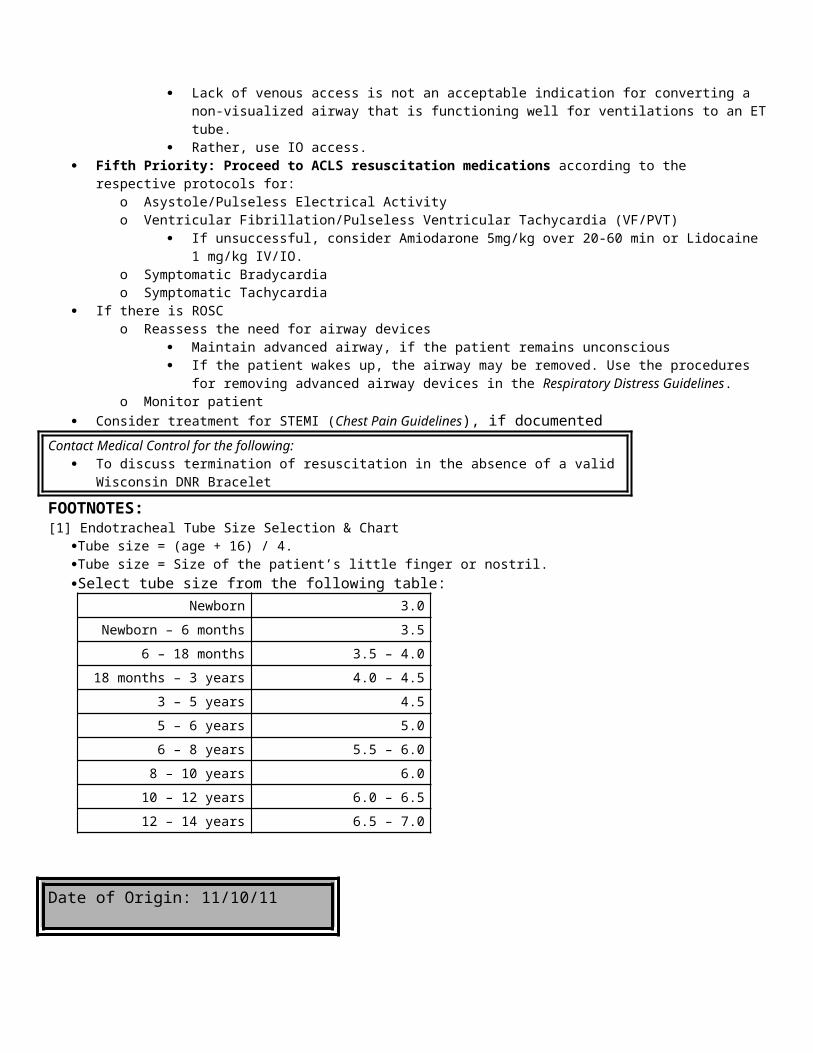
o SBP <100 mmHg and Heart Rate > 150 beats/min Clinical Criteria
o Signs of shock (poor perfusion) are present, including ALOC Absent radial pulses Pallor and diaphoresis
o Signs of pulmonary edema are present, including Labored breathing Rales (wet lungs) Hypoxia (SpO2 <90%)
o The patient complains of anginao Nausea
Date of Origin: 11/10/11
Refusal of CareThe following content will be considered the Guideline/Standard of care for any patient who is a candidate to sign a release or refusal of treatment form:
REFUSAL OF CARE
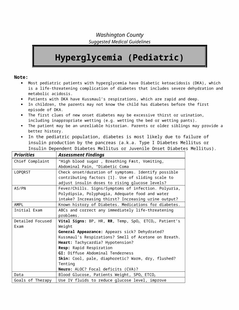
Assure scene safety and observe universal precautions Complete the history and physical assessment Assess the patient’s level of consciousness, level of activity, body language and affect.In order to sign a release or refuse care, the patient must be able to demonstrate decision- making capacity:
Demonstrate the ability to communicate and understand informationDemonstrate the ability to reason and deliberateAppreciate the current situation and its risks and consequences if treatment is refusedBe logically consistentNot be homicidal or suicidal
For the patient, who is refusing care and/or transport, ascertain:There is no history of or present altered level of consciousness.There is no significant or suspected head injury.The patient is presently oriented to time, place and person.There is no suspected alcohol or drug ingestion by exam or history.The patient can ambulate without difficulty.
There is an obligation to treat and transport, if possible, any patient who shows an altered level of consciousness secondary to an injury, hypoxia, hypoglycemia, a mind-altering substance or psychosis. These individuals should not be considered able to sign a release.Only an adult patient (age 18 years or older), guardian, adult caregiver (relative, friend, police officer or EMT), agent (durable power of attorney for health care), or legally emancipated minor may sign a release form. The EMT must be comfortable with this arrangement. If the EMT has reservations about the responsible party, law enforcement can be notified to assist.Information must be given to the person refusing care about the consequences, including (they must receive discharge instructions):
They have the right to refuse treatment and/or transport.They are aware of the potential consequences of refusal of care.Medical examination and/or treatment by a physician are highly recommended.Ambulance transport is strongly recommended.The condition may worsen or further injury may occur.Disability or death could occur from the illness or injury.Transport by means other than ambulance could be hazardous.If treatment or transport if refused but later desired, the patient should immediately call 9-1-1 and the EMS
system will respond to render care and transport.Contact should be made with medical control if there is any question about the patient’s ability to understand the consequences of his/her decision.Complete a patient care report, documenting all pertinent information given to the patient, situation of the original response, information given to the patient (all patients must receive discharge instructions), and conditions under which the patient was left.Two sets of vital signs are preferred to document a stable trendA signature of the patient and witness (es) on a refusal of care form may be appropriate.
Special CircumstancesUninjured Minor – Adult guardian telephone approval for release is acceptable as long as there is a responsible adult present to whom the minor can be released and both the patient’s guardian and the EMT feel comfortable with the arrangement. If the patent’s adult guardian is not available, an adult caregiver may substitute.Uninjured adult with possible mind-altering substance – If the patient shows no altered level of consciousness, has normal speech and stable gait and can demonstrate decision-making capacity (as above), they may be signed out to a responsible adult who is not under the influence of a mind-altering substance. Often time, Law enforcement is able to assist in convincing these patients to allow transport to the hospital

Washington CountySuggested Medical Guidelines
Respiratory Distress (Adult) Note:
This protocol may apply to the following conditions:o Congestive Heart Failure (CHF)o Asthma/COPDo Allergy/Anaphylaxiso Pulmonary Infectionso Spontaneous Pneumothorax
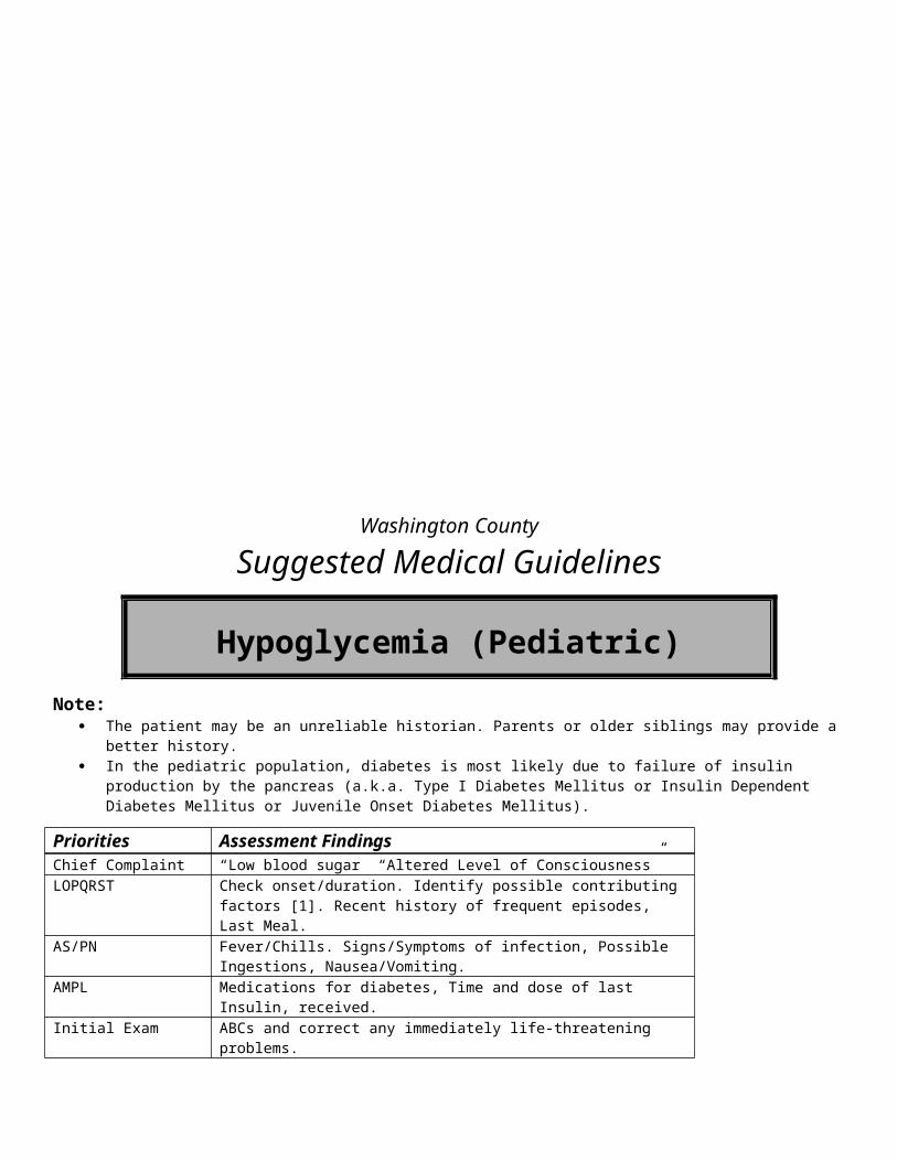
o Upper Airway Obstructiono Anxiety and Hyperventilation Syndromeo Acute Coronary Syndromes
Priorities Assessment FindingsChief Complaint “Difficulty breathing”; “Shortness of breath”LOPQRST Assess onset, duration, progression, subjective severity, possible triggering
events, and response to treatments before EMS arrival.AS/PN Chest pain (what kind?), fever/chills, productive (of what?) cough AMPL Check for possible exposure to known allergens. Check past history,
medications and compliance for clues to cause of present illness. Initial Exam Check ABCs and correct immediately life-threatening problems.Detailed Focused Exam Vital Signs: BP, HR, RR, Temp, SpO2
General Appearance: Tripod positioning; Purse-lipped breathing. Severity of distress [1]?Neck: JVD?Skin: Cool, moist and pale? Warm, dry and flushed? Urticaria? Cyanosis?Respiratory Effort: Using accessory muscles, signs of fatigue; two-word sentences?Lung Sounds: Wheezes, rales, rhonchi or stridor?Heart Sounds: Rate, regularity.Lower Extremities: Pitting edema of the ankles?Neuro: ALOC, lethargy, somnolence?
Data SpO2, on room air or home O2
EKG, if an acute coronary syndrome is suspectedBlood Sugar, if DKA is suspected or if there is ALOCEnd Tidal CO2
Goals of Therapy Improve oxygenation and ventilation, reduce the work of breathing, and treat underlying conditions.
Monitoring SPO2 frequently or continuously.ETCO2
Cardiac rhythm monitoring.Heart Rate and Blood Pressure frequently.
EMR Routine Medical Care Allow/assist the patient to assume a position of comfort (usually upright).
Administer oxygen to keep SpO2 > 94% and <99% Use a nasal cannula at 2 – 6 LPM or Non-rebreather mask at 10 – 15 LPM
Assisted Ventilation: Consider assisting breathing with gentle synchronous ventilations with bag-valve mask (BVM); Support ventilation with BVM if apnea or hypopnea occurs.
Airway Adjuncts: If there is altered level of consciousness and loss of gag reflex, insert an oropharyngeal or nasopharyngeal airway or
consider a non-visualized airway In respiratory arrest or cardiopulmonary arrest, insert a non-visualized airway
BASIC Assist with patient-prescribed medications or initiate the following therapies:
Nitroglycerin is indicated for acute coronary syndromes and CHF (See specific protocols). Albuterol and Atrovent are indicated for Asthma and COPD (See specific protocols). Aspirin is indicated for acute coronary syndromes (See specific protocols). Initiate CPAP at 5- 10 cm H2O. (See Respiratory Distress Guideline) To be used only by EMT’s with proper
training and certification by the medical director or authorized representative and State of WI EMS Office. INTERMEDIATE TECHNICIAN
IV 0.9% NS @ KVO Consider IO if no IV access Initiate a 250 ml fluid bolus, if hypotension or dehydration is present.(Check lung sounds)
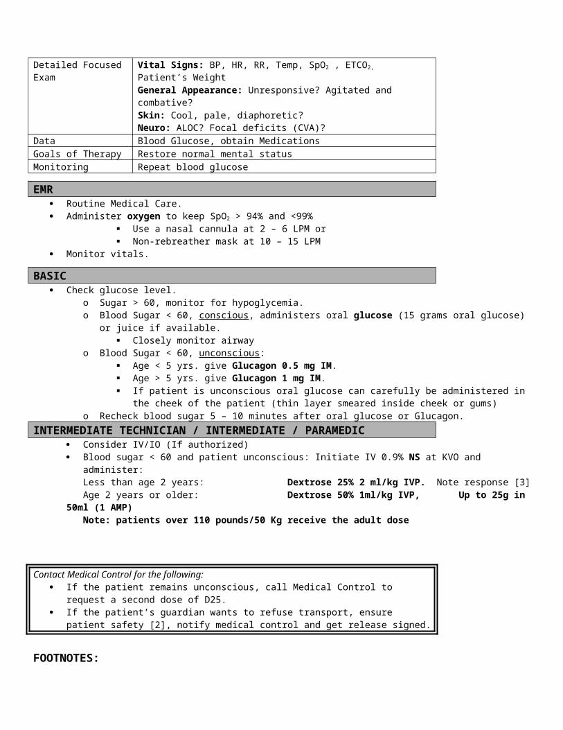
INTERMEDIATE Airway Adjuncts:
If there is altered level of consciousness and loss of gag reflex, insert an oropharyngeal or nasopharyngeal airway or consider endotracheal intubation
In respiratory arrest or cardiopulmonary arrest, insert an advanced airway If a spontaneous tension pneumothorax is suspected, perform a needle decompression on the affected side.
Contact Medical Control for the following: If the patient becomes anxious from the CPAP mask, consider Valium 5 mg or Ativan 1-2 mg IV
PARAMEDIC Airway Adjuncts:
If there is altered level of consciousness and loss of gag reflex, insert an oropharyngeal or nasopharyngeal airway or consider endotracheal intubation
In respiratory arrest or cardiopulmonary arrest, insert an endotracheal tube Consider Nasal intubation
If the patient becomes anxious from the CPAP mask, consider Valium 5 mg or Ativan 1-2 mg IVContact Medical Control for the following:
Nature and severity of the problem Treatment or procedures proposed or performed Results of treatment Changes in patient status
FOOTNOTES:[1] Severity of Respiratory Distress:
Mild = RR<20 + minimal additional breathing effort + speaking in complete sentences + minimal subjective distress, No ALOC
Moderate = RR 20 to 25 + moderate additional breathing effort + difficult to complete a sentence + moderate subjective distress + No ALOC
Severe = RR> 25 + marked additional breathing effort + 2 or 3 word sentences + marked subjective distress + possible ALOC
[2] For the purposes of endotracheal intubation, one “attempt” is counted when the laryngoscope is removed from the mouth, even if there has not been an attempt to pass the tube.
Date of Origin: 11/10/11
Updated 3/15/12
Washington County
Suggested Medical Guidelines
Routine Medical Care (Pediatric)
Priorities Assessment FindingsChief Complaint Refer to specific protocols for the kinds of chief complaints patients are likely
to make.LOPQRST Location, Onset, Provocation/Palliation, Quality, Radiation, Severity, Time
(duration, progression)As an alternative: Use “Onset, Duration, Progression and Severity”

AS/PN Associated Symptoms/Pertinent NegativesAMPL Allergies, Medications, Pertinent Past Medical History, Last MealInitial Exam Check ABCs and correct immediately life-threatening problems.Detailed Focused Exam Vital Signs: BP, HR, RR, Temp, Data Follow specific protocol. Consider SpO2, Blood Sugar, EKGGoals of Therapy Follow specific protocol.Monitoring Follow specific protocol.
EMR Scene size-up Body-Substance Isolation (BSI) Obtain History Airway Management – Perform the following, if indicated:
o Head tilt – chin lifto Oropharyngeal or nasopharyngeal airwayo Non-visualized airway
Breathing Management – Perform the following, if indicated:o Check pulse oximetry (SpO2)o Administer oxygen to keep SpO2 > 94% and <99%
Use a nasal cannula at 2 – 6 LPM or Non-rebreather mask at 10 – 15 LPM
o May assist patient with prescribed Albuterol and Atrovent inhalers; See Ped Asthma/COPD Guidelines and Allergy & Anaphylaxis Guidelines
o Ventilate or assist ventilations with a bag-valve-mask connected to high-flow oxygen Circulation Management – Perform the following, if indicated:
o Control external hemorrhage with direct pressure, or pressure points, and bandageso Cardiopulmonary resuscitation (CPR); see Cardiac Arrest Guidelineso Automated External Defibrillator (AED); see Cardiac Arrest Guidelineso Raise patients legs; see Hypovolemia and Shock Guidelines
BASIC After checking ABCs, correct any immediate life threats, if indicated:
o Obstructed airway maneuvers per American Heart Association Guidelineso Use a laryngoscope and Magill Forceps to remove an upper airway foreign body.o Suction the airway using soft or rigid suction devices as needed to remove secretions or foreign material.o Check blood glucose if there is an altered level of consciousness (ALOC); see Altered Level of Consciousness
Guidelines and Hypoglycemia Guidelines. Obtain Vital Signs
o Blood Pressure (BP), Heart Rate (HR), Respiratory Rate (RR), Temperature (Temp)* and Pulse Oximetry (SpO2), End Tidal CO2 (ETCO2)
o * Temperature is optional. Use a digital thermometer, if available. Perform a focused physical exam Initiate additional EMT-Basic treatments as directed in specific guidelines.
INTERMEDIATE TECHNICIAN Establish IV/IO, Normal Saline @ KVO rate
If signs of hypovolemia or inadequate perfusion are present, administer fluid bolus at 20mL/kg Neonates (less than 30 days old) use 10ml/kg.
After checking ABCs, correct any immediate life threats, if indicated: 50 Blood sugar < 60 and patient unconscious: Initiate IV 0.9% NS at KVO and administer:
Less than age 2 years: Dextrose 25% 2 ml/kg IVP. Note response [3]

Age 2 years or older: Dextrose 50% 1ml/kg IVP, Up to 25g in 50ml (1 AMP) o Note: patients over 110 pounds/50 Kg receive the adult doseo ; see Hypoglycemia Guidelines
Initiate additional EMT-Basic IV Tech treatments as directed in specific guidelines.
INTERMEDIATE Airway Management – Perform the following, if indicated:
o Endotracheal Intubation Breathing Management – Perform the following, if indicated:
o Needle decompression of a tension pneumothorax; see Non-Cardiac Chest Pain Guidelines and Respiratory Distress Guidelines
After checking ABCs, correct any immediate life threats, if indicated:o Synchronized Cardioversion in unstable patients; see Wide Complex Tachycardia Guidelines and Narrow Complex
Tachycardia Guidelines.o Transcutaneous Pacing in unstable patients; see Bradycardia Guidelines and Asystole & Pulseless Electrical Activity
Guidelines. Initiate additional EMT- Intermediate treatments as directed in specific guidelines.
PARAMEDIC Airway Management – Perform the following, if indicated:
o Perform a cricothyrotomy (needle) if an upper airway obstruction cannot be relieved by non-invasive means Initiate additional EMT- Paramedic treatments as directed in specific guidelines.
FOOTNOTES:
Date of Origin 11/10/11
Washington County
Suggested Medical Guidelines
Routine Medical Care (Adult)
Note:
Priorities Assessment FindingsChief Complaint Refer to specific protocols for the kinds of chief complaints patients are likely
to make.LOPQRST Location, Onset, Provocation/Palliation, Quality, Radiation, Severity, Time
(duration, progression)As an alternative: Use “Onset, Duration, Progression and Severity”
AS/PN Associated Symptoms/Pertinent NegativesAMPL Allergies, Medications, Pertinent Past Medical History, Last MealInitial Exam Check ABCs and correct immediately life-threatening problems.Detailed Focused Exam Vital Signs: BP, HR, RR, Temp, Blood glucose
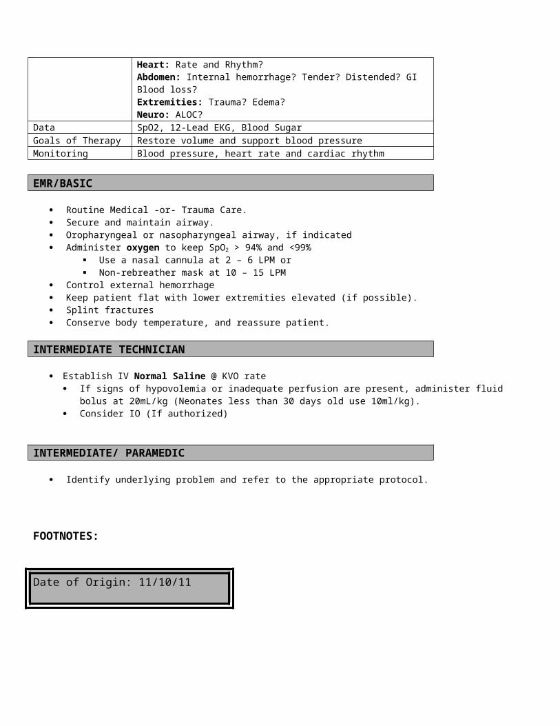
Data Follow specific protocol. Consider SpO2, Blood Sugar, EKG, ETCO2
Goals of Therapy Follow specific protocol.Monitoring Follow specific protocol.
EMR Scene size-up Body-Substance Isolation (BSI) Obtain History Airway Management – Perform the following, if indicated:
o Head tilt – chin lifto Oropharyngeal or nasopharyngeal airwayo Non-visualized airway
Breathing Management – Perform the following, if indicated:o Check pulse oximetry (SpO2)o Administer oxygen to keep SpO2 > 94% and <99%
Use a nasal cannula at 2 – 6 LPM or Non-rebreather mask at 10 – 15 LPM
o Ventilate or assist ventilations with a bag-valve-mask connected to high-flow oxygen Circulation Management – Perform the following, if indicated:
o Control external hemorrhage with direct pressure, or pressure points, and bandageso Cardiopulmonary resuscitation (CPR); see Cardiac Arrest Guidelineso Automated External Defibrillator (AED); see Cardiac Arrest Guidelineso Place the patient in Trendelenburg Position; see Hypovolemia and Shock Guidelines
BASIC After checking ABCs, correct any immediate life threats, if indicated:
o Obstructed airway maneuvers per American Heart Association Guidelineo Suction the airway using soft or rigid suction devices as needed to remove secretions or foreign material.o Use a laryngoscope and Magill Forceps to remove an upper airway foreign body.o May use Epinephrine 1:1,000 SQ for anaphylaxis; see Allergy & Anaphylaxis Guidelineso May administer Albuterol and Atrovent nebulized Unit Doses; (If authorized) (or assist with patients own) See
Adult Asthma/COPD Guidelines and Allergy & Anaphylaxis Guidelineso Check blood glucose if there is an altered level of consciousness (ALOC); see Altered Level of Consciousness
Guidelines and Hypoglycemia Guidelines. Obtain Vital Signs (minimum of 2 sets of vitals for all patients who are transported)
o Blood Pressure (BP), Heart Rate (HR), Respiratory Rate (RR), Temperature (Temp)* and Pulse Oximetry (SpO2), End Tidal CO2 (ETCO2)(If authorized)
o * Temperature is optional. Use a digital thermometer, if available. Perform a focused physical exam Initiate additional EMT-B treatments as directed in specific guidelines.
INTERMEDIATE TECHNICIAN Circulation Management – Perform the following, if indicated:
o IV Normal Saline @ KVOo If the patient is hypotensive, give a 250 ml Normal Saline bolus
After checking ABCs, correct any immediate life threats, if indicated:o 50% Dextrose for hypoglycemia; see Hypoglycemia Guidelines
Initiate additional EMT-Basic IV Tech treatments as directed in specific guidelines.
INTERMEDIATE Airway Management – Perform the following, if indicated:
o Endotracheal Intubation Breathing Management – Perform the following, if indicated:
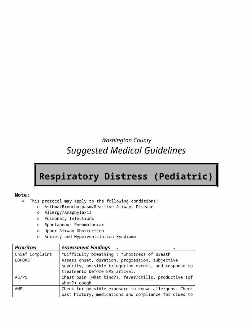
o Needle decompression of a tension pneumothorax Circulation Management – Perform the following, if indicated:
o Intraosseous (IO) infusion and drug administration After checking ABCs, correct any immediate life threats, if indicated: Transcutaneous Pacing in unstable patients; see Bradycardia Guidelines Initiate additional EMT- Intermediate treatments as directed in specific guidelines.
PARAMEDIC
Airway Management – Perform the following, if indicated:o Perform a cricothyroidotomy (surgical/needle) if an upper airway obstruction cannot be relieved by non-invasive
means Circulation Management – Perform the following, if indicated:
o Initiate an IV at a indwelling central line port (if trained and authorized) After checking ABCs, correct any immediate life threats, if indicated:
o Synchronized Cardioversion in unstable patients; see Wide Complex Tachycardia Guidelines and Narrow Complex Tachycardia Guidelines.
Initiate additional EMT- Paramedic treatments as directed in specific guidelines.
FOOTNOTES:
Date of Origin: 11/10/11
Washington County
Suggested Medical Guidelines
Routine Trauma Care (Adult)Note:
This protocol may be used as a general guide for trauma in both Adults and Pediatrics. Follow appropriate protocol and/or procedure for specific trauma care.
Priorities Assessment FindingsChief Complaint Various depending on incident. LOPQRST Identify specific cause of traumatic injuryAS/PN Significant mechanism, loss or altered level of consciousness. Evidence of
intoxicant use.AMPL Identify medical conditions that may have led to the event (e.g. Alzheimer’s,
CVA, Diabetes, Seizures,)Initial Exam – Rapid Trauma Assessment
Check ABC’s and corrects any immediate life threats. Manual C-spine stabilization. Perform rapid trauma assessment as appropriate.
Detailed Focused Exam Vitals: BP, HR, RR, Temp, SpO2
General Appearance: Unresponsive, pale, diaphoretic? Signs of trauma?HEENT: PERRL? Pupils constricted or dilated? Discharge from ears or nose?Lungs: Signs of respiratory distress, hypoventilation, diminished or absent lung sounds?
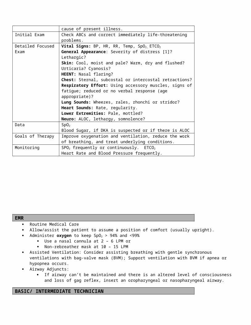
Heart: Rate and rhythm? Signs of hypoperfusion? Neuro: Loss of movement and/or sensation in extremities, Unresponsive? Focal deficits?
Data Blood Glucose, SpO2, EKGGoals of Therapy Maintain ABC’s, restore adequate respiratory and circulatory conditions,
reduce painMonitoring SpO2, Cardiac monitoring, repeat vitals EMR
Ensure “Scene Safety” and Body Substance Isolation (BSI) Determine need for additional resources (e.g. helicopters, additional ambulances, heavy rescue). Airway: Relieve airway obstruction, if present
Open the airway with a jaw-thrust (No head tilt – Chin lift in trauma patients) Remove foreign material, emesis and blood Suction the airway If no gag reflex consider oropharyngeal airway or nasopharyngeal airway (nasopharyngeal airway not recommended
in facial trauma) Breathing:
o Administer Oxygen to keep SpO2 > 94% and <99% Use a nasal cannula at 2 – 6 LPM or Non-rebreather mask at 10 – 15 LPM
Assist ventilations with bag-valve-mask and high-flow oxygen, as needed Cover sucking chest wounds with a three-sided flap valve
Circulation: Control external hemorrhage with direct pressure or pressure points. If the patient arrests,
Re-assess the airway and oxygen delivery Consider initiating the Cardiac Arrest Guidelines. Prolonged efforts to restore spontaneous circulation in a traumatic arrest should not be made CPR should not be attempted if:
Blunt trauma caused the arrest There are other injured survivors with urgent needs for help
C-Spine: Manual stabilization Splint obvious extremity fractures Refer to Pain Management Guidelines Begin other interventions as needed according to specific guidelines
BASIC If there is ALOC
Check Blood Glucose Follow Hypoglycemia Guidelines if < 60
Spinal Immobilization, as indicated. Nexus research may be used to “clear” potential C-spine injuries in the field Patient must meet all the following: (If Authorized and trained)
a. No complaint of neck tenderness or painb. Normal mental statusc. No evidence of any alcohol or drug use (i.e. odor or alcohol, slurred speech...)d. No Neurologic abnormality (i.e. numbness, tingling....)e. No other painful distracting injury (i.e. long bone fracture, facial injuries, abdominal pain...)
If patient meets the above listed criteria, perform physical assessment of the cervical spine: a. Manually palpate upper thoracic spine and adjacent muscles b. Ask patient to turn head slowly from side to side c. Extend head slowly d. Flex chin to chest
o Document all findings Refer to the Triage & Transport Guidelines to consider possible transport to a Regional Trauma Center for the following:
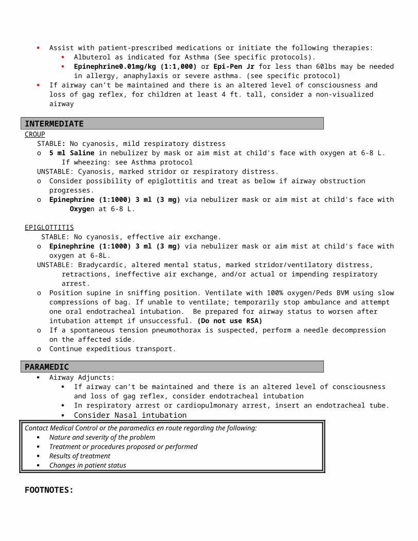
Criteria List A (Definition of Major Trauma)1. Glasgow Coma Scale of 13 or less2. Clinical signs of shock: pale, cold, weak pulses, prolonged capillary refill3. Unstable blood pressure
a. Adult: Systolic blood pressure <90 mmHgb. Pediatric: Infant<6 months: BP <60 mmHg
Child 2 months-5 years: <70 mmHgChild 6-12 years: <80 mmHg
4. Respiratory rate a. Adult: Less than 10 or greater than 30 breaths per minuteb. Pediatrics under 12: Infants <6 months: <20 breaths per minute
6 months-12 years: <16 breaths per minuteAll ages: >60 breaths per minute
5. Penetrating injury to head, neck, torso or proximal extremity6. Flail chest7. Trauma in a patient with burns to face or airway or with burns of 15% or greater of the total body surface area8. Distended, rigid abdomen9. Two or more long-bone fractures (humerus, femur)10. Depressed or open skull fracture11. Unstable pelvic fracture12. New onset paralysis13. Amputation above the wrist or ankle
Criteria List B (Indicators of possible major trauma)1. Crashes in which the patient was ejected from the vehicle2. Crashes in which another occupant of the vehicle was killed3. Extrication time in excess of 20 minutes4. Falls of 20 feet or greater for adults, 10 feet or greater for children5. Victim of a roll-over motor vehicle crash6. Estimated crash speed was 40 mph or greater for adults, <20 mph for children7. Passenger compartment intrusion >12 inches is present8. Auto vs. pedestrian or bicycle9. Motorcycle crashes
Criteria List C: Trauma patients whose injuries may be significantly impacted by other factors1. Whose age is <5 or >55 2. Who have known cardiac or respiratory disease or3. Who are pregnant4. Who are immunosuppressed5. Who have a bleeding disorder
INTERMEDIATE TECHNICIAN IV (18ga or larger) 0.9% Normal Saline @ KVO or an appropriate rate. Consider 2nd IV where hypovolemia is suspected (Adult only) If SBP < 100 mmHg, initiate a fluid bolus of Normal Saline: 250 ml (Adult) or 20 ml/kg (Peds).
INTERMEDIATE Consider endotracheal intubation:
o Respiratory arrest or apnea If tension pneumothorax is suspected perform needle decompression. Consider IV, external jugular (EJ), or Intraosseous (IO) access. . Selective Spinal Immobilization – In the presence of a mechanism of injury for spinal trauma:
Extrication/Immobilization: apply appropriately sized rigid C-collar and full spinal immobilization per Procedure of Practical Skills Manual.
o Unstable scenes or patients require appropriate rapid extricationo Consider extrication using a KED if indicated: stable MVA patients still in the vehicle complaining of, or presenting
with, head or neck pain or tenderness upon palpation; head injuries; any neurovascular impairment; spinal deformity upon palpation.
o Ambulatory patients complaining of neck pain or tenderness upon palpation should be immobilized using standing
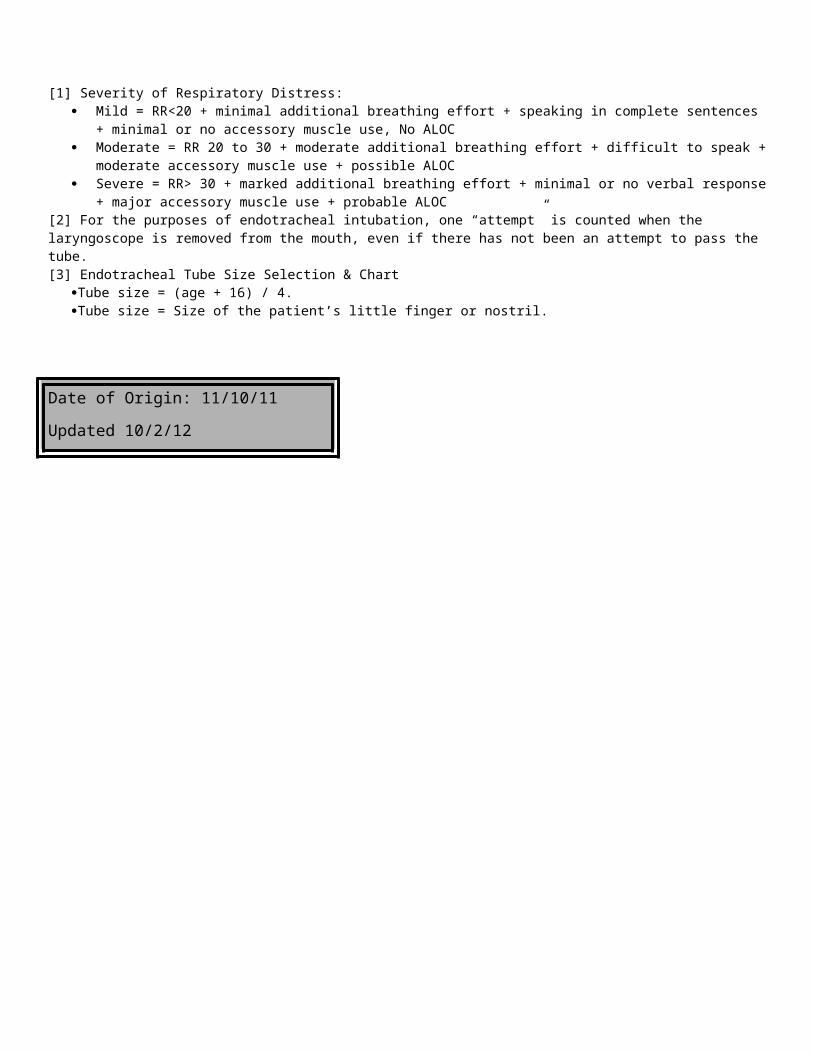
backboard techniqueo Protect paralyzed limbs by securing patient to backboardo Do not remove a C-collar or long board already applied. You may open the C-collar briefly to examine the neck or
to intubate.
Contact Medical Control for the following: Additional pain orders
PARAMEDIC If the airway is obstructed or obstruction is imminent and 3 attempts to intubate the trachea have failed, perform surgical or
needle cricothyroidotomy. Consider gastric decompression with nasogastric tube, unless contraindicated by facial trauma or skull fracture Consider sedation for combative patients, refer to Chemical Restraint Guidelines
Contact Medical Control for the following: Surgical or needle cricothyroidotomy
FOOTNOTES:* Cushing's triad is the triad of hypertension, bradycardia and irregular respirations. It is sign of increased intracranial pressure
Date of Origin:11/10/11
Washington County
Suggested Medical Guidelines
Routine Trauma Care (Pediatric)Note:
This protocol may be used as a general guide for trauma in both Adults and Pediatrics. Follow appropriate protocol and/or procedure for specific trauma care.
Priorities Assessment FindingsChief Complaint Various depending on incident. LOPQRST Identify specific cause of traumatic injuryAS/PN Significant mechanism, loss or altered level of consciousness. Evidence of
intoxicant use.AMPL Identify medical conditions that may have led to the event (e.g. Alzheimer’s,
CVA, Diabetes, Seizures,)Initial Exam – Rapid Trauma Assessment
Checks ABC’s and correct any immediate life threats. Manual C-spine stabilization. Perform rapid trauma assessment as appropriate.
Detailed Focused Exam Vitals: BP, HR, RR, Temp, SpO2
General Appearance: Unresponsive, pale, diaphoretic? Signs of trauma?HEENT: PERRL? Pupils constricted or dilated? Discharge from ears or nose?Lungs: Signs of respiratory distress, hypoventilation, diminished or absent lung sounds?Heart: Rate and rhythm? Signs of hypoperfusion? Neuro: Loss of movement and/or sensation in extremities, Unresponsive? Focal deficits?
Data Blood Glucose, SpO2, EKGGoals of Therapy Maintain ABC’s, restore adequate respiratory and circulatory conditions,
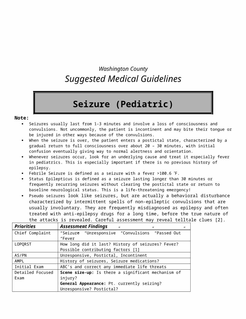
reduce painMonitoring SpO2, Cardiac monitoring, repeat vitalsEMR
Ensure “Scene Safety” and Body Substance Isolation (BSI) Determine need for additional resources (e.g. helicopters, additional ambulances, heavy rescue). Airway: Relieve airway obstruction, if present
Open the airway with a jaw-thrust (No head tilt – Chin lift in trauma patients) Remove foreign material, emesis and blood Suction the airway Consider oropharyngeal airway or nasopharyngeal airway (use caution in facial trauma)
Administer oxygen to keep SpO2 > 94% and <99% Use a nasal cannula at 2 – 6 LPM or Non-rebreather mask at 10 – 15 LPM
Breathing: Assist ventilations with bag-valve-mask and high-flow oxygen, as needed Cover sucking chest wounds with a three-sided flap valve
Circulation: Control major external hemorrhage with direct pressure or pressure points. If the patient arrests,
Re-assess the airway and oxygen delivery Consider initiating the CCR Guidelines. Prolonged efforts to restore spontaneous circulation in a traumatic arrest should not be made, unless
It is due to a penetrating injury The nearest hospital is < 5 minutes away.
BASIC
C-Spine: Manual stabilization Splint obvious extremity fractures Refer to Pain Management Guidelines Begin other interventions as needed according to specific protocols If there is ALOC
Check Blood Glucose Follow Hypoglycemia Guidelines if < 60
Spinal Immobilization, as indicated. See Appendix A for state trauma definition.
INTERMEDIATE TECHNICIAN
Establish IV/IO, Normal Saline @ KVO rate If signs of hypovolemia or inadequate perfusion are present, administer fluid bolus at 20mL/kg (Neonates
less than 30 days old use 10ml/kg).INTERMEDIATE
Consider endotracheal intubation:o Respiratory arrest or apnea
If tension pneumothorax is suspected perform needle decompression. Consider external jugular (EJ) IV if one cannot be established in the extremities. Consider Intraosseous (IO) access if an EJ cannot be established. Selective Spinal Immobilization – In the presence of a mechanism of injury for spinal trauma:
o Unstable scenes or patients require appropriate rapid extricationo Consider extrication using a KED if indicated: stable MVA patients still in the vehicle complaining of, or presenting
with, head or neck pain or tenderness to palpation; head injuries; any neurovascular impairment; spinal deformity on palpation.
o Ambulatory patients complaining of neck pain or tenderness to palpation should be immobilized using standing backboard technique
o Protect paralyzed limbs by securing patient to backboard

o Do not remove a C-collar or long board already applied. You may open the C-collar briefly to examine the neck or to intubate.
Contact Medical Control for the following: Additional pain orders
PARAMEDIC If the airway is obstructed or obstruction is imminent and 3 attempts to intubate the trachea have failed, perform surgical or
needle cricothyroidotomy. Consider gastric decompression with nasogastric tube, unless contraindicated by facial trauma or skull fracture (if trained and
authorized) Consider sedation for combative patients, refer to Chemical Restraint Guidelines
Contact Medical Control for the following: Surgical or needle cricothyroidotomy
FOOTNOTES:* Cushing's triad is the triad of hypertension, bradycardia and irregular respirations. It is sign of increased intracranial pressure
Date of Origin: 11/10/11
Washington County
Suggested Medical Guidelines
RSA (Adult)
Rapid Sequence Airway
NOTE: TWO (2) Paramedics MUST be bedside to initiate RSA and must remain beside throughout the transport of a patient that has been chemically sedated and paralyzed for the purposes of RSA
Indications:o Severe respiratory distress or failureo Persistent hypoxia after high-flow O2
o Airway management in a combative patiento Altered mental status with need to protect/secure airwayo Airway compromise in the multiple-trauma patient (See Routine Trauma Care Guidelines for more details)
Contraindications:o Known allergy to RSA medicationso Suspected epiglottitiso Severe oral, mandibular, or anterior neck traumao Anatomic abnormalities that increase the risk of failed intubation (e.g. severe rheumatoid arthritis, micrognathia,
morbid obesity, etc.)Prepare:
o Initiate IV 0.9% NS or consider IOo Draw up RSA medications.o Organize equipment.o Assure suction capabilities are available.
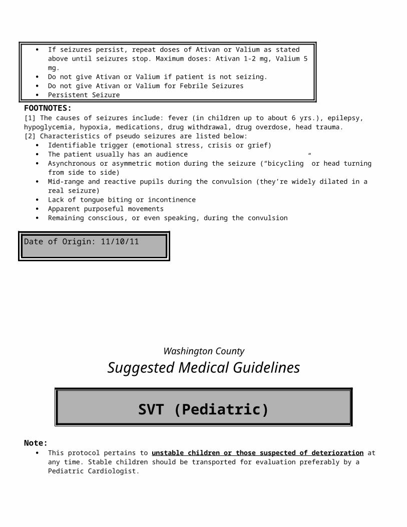
o Select and prepare ET tube/stylet.Pre-Oxygenate:
o Continuous pulse oximetryo High-flow oxygen for 3 - 5 minutes prior to intubation or 4 vital capacity breaths
Pre-Treat:o Consider Atropine 0.5 mg - 1.0 mg IV to decrease respiratory secretions (antisialogogue).o Consider Lidocaine 1.0 - 1.5 mg/kg IV to blunt adverse reflexes due to laryngoscopy and endotracheal intubation.o Consider Dilaudid (hydromorphone) 0.5 - 1.5 mg IV/IO, or Morphine Sulfate 2.0 - 4.0 mg IV/IO or Fentanyl
50 mcg IV/IO for pain control or sedation.
o Versed 5-10 mg IV (Must Administer)o ORo Etomidate 0.3 mg/kg IV
Paralyze (with induction):o Succinylcholine 1mg/kg IVP To initiate paralysis. Watch for notable fasciculation and wait approximately 1
minute prior to ETT attempt. o OR o Vecuronium 0.1mg/kg over 5-15 seconds repeat q 15 minutes as needed ORo Rocuronium 0.6-1.2 mg/kg
Protect and Position:o After the patient is paralyzed, continually apply Sellick’s Maneuver.o DO NOT release Sellick’s Maneuver until the ET tube is properly positioned.o If you must bag the patient, maintain Sellick’s Maneuver.o Position the head and neck for intubation
Placement:o Insert the ETT until the cuff passes the vocal cords.
Make two attempts [2] If unsuccessful, proceed to the Difficult Airway Procedure below
o Inflate the cuffo Auscultate for gastric insufflations and bilateral breath sounds.o Check for condensation in the tube.o Use a colorimetric or electronic Capnographyo Monitor ETCO2
o Monitor SpO2
o Secure ET tube.
Post-Intubation Management:o Vecuronium 0.1 mg/kg over 5 - 15 seconds.o May repeat dose at 15 minute intervals as needed. . ORo Rocuronium 1 mg/kg
Removing the ETT in the fieldo In general, an ETT should not be removed in the fieldo If IV access is unobtainable, use IO access or the ETT access for cardiac medications. o Indications:
The patient wakes up The ETT is not performing adequately
o Procedure for removing an ETT Place the patient in the recovery position (left side)
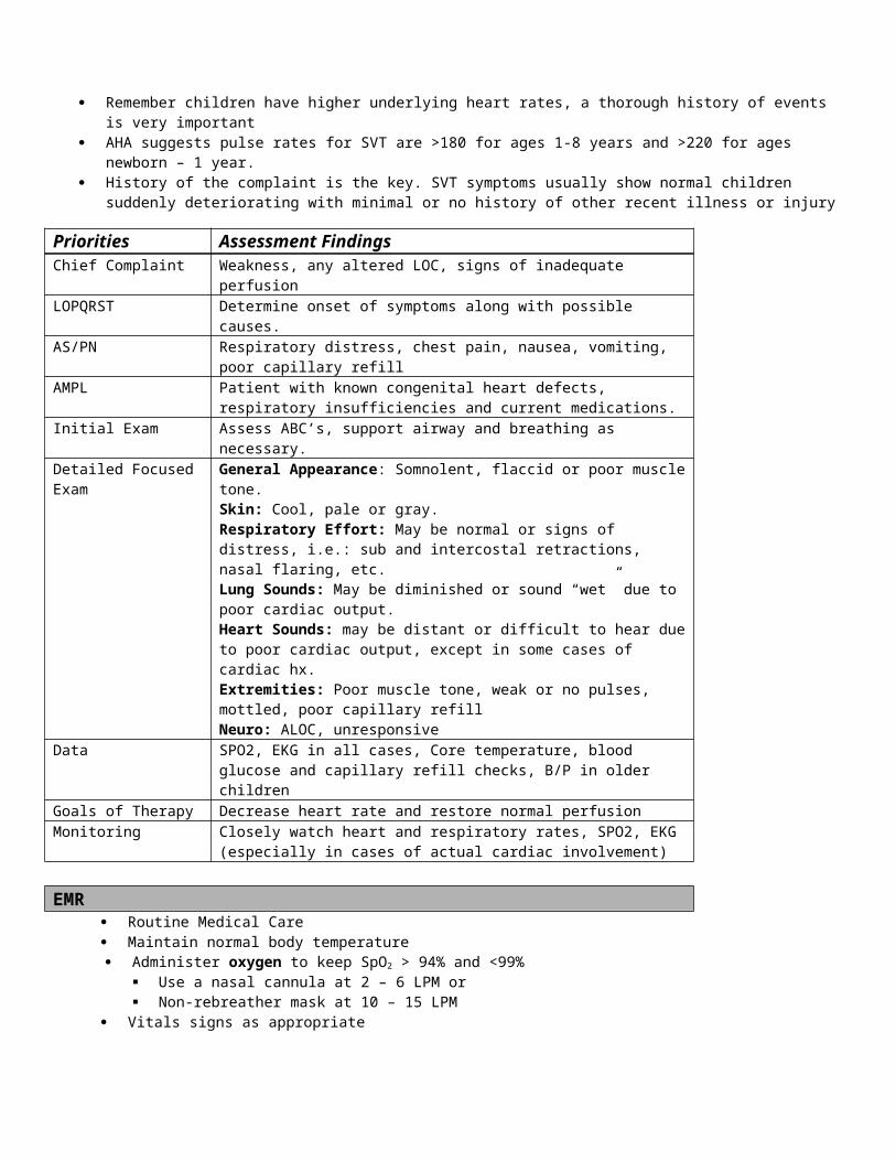
Deflate cuff and remove tube Be prepared to suction the pharynx
o Continue to monitor and re-assess the patient
Difficult Airway Procedureo If one rescuer cannot intubate the trachea after two attempts [2], a second experienced rescuer may attempt
intubation once. Failed Intubation: If the second rescuer cannot intubate with one attempt [1], proceed immediately to a non-visualized
airway
o If the non-visualized airway fails consider the following options: Let the medications wear off, while bagging the patient Consider a surgical airway
o Consult Medical Control for assistance in managing all difficult airways
Date of Origin: 11/10/11
Updated 10/2/12
Washington County
Suggested Medical Guidelines
SeizureNote:
Seizures usually last from 1-3 minutes and involve a loss of consciousness and convulsions. Not uncommonly, the patient is incontinent and may bite his tongue or be injured in other ways because of the convulsions.
When the seizure is over, the patient enters a postictal state, characterized by a gradual return to full consciousness over about 20 – 30 minutes, with initial confusion eventually giving way to normal alertness and orientation.
Whenever seizures occur, look for an underlying cause and treat it. This is especially important if there is no previous history of epilepsy.
If the patient is more than 20 weeks pregnant, refer to the Eclampsia Guidelines. Status Epilepticus is defined as a seizure lasting longer than 30 minutes or frequently recurring seizures without clearing the
postictal state or return to baseline neurological status. This is a life-threatening emergency! Pseudo seizures look like seizures, but are actually a behavioral disturbance characterized by intermittent spells of non-
epileptic convulsions that are usually voluntary. They are frequently misdiagnosed as epilepsy and often treated with anti-epilepsy drugs for a long time, before the true nature of the attacks is revealed. Careful assessment may reveal telltale clues [2].
Priorities Assessment FindingsChief Complaint “Seizure” “Unresponsive” “Convulsions”LOPQRST How long did it last? History of seizures? Possible contributing factors [1]AS/PN Unresponsive, Postictal, IncontinentAMPL History of seizures, Seizure medications?Initial Exam ABC’s and correct any immediate life threatsDetailed Focused Exam Scene size-up: Is there a significant mechanism of injury?
General Appearance: Pt. currently seizing? Unresponsive? Postictal?

Vitals: BP, HR, RR, Temp, SpO2Skin: Flushed, warmNeuro: ALOC?, Focal deficits (CVA)
Data Blood Glucose, SpO2, End Tidal CO2
Goals of Therapy Stop the seizureTreat the underlying causeMonitor and maintain airway.
Monitoring Vitals, Cardiac monitoring, SpO2,ETCO2
EMR Routine medical care Consider oropharyngeal or nasopharyngeal airway, if the patient is unable to maintain a patent airway Protect the patient with ongoing seizures from harming themselves by clearing away potential hazards and placing a pillow or
padding under the head. Administer oxygen to keep SpO2 > 94% and <99%
Use a nasal cannula at 2 – 6 LPM or Non-rebreather mask at 10 – 15 LPM
BASIC Obtain blood glucose. If < 60 refer to hypoglycemia protocol.
INTERMEDIATE TECHNICIAN IV 0.9% NS @ KVO Consider IO (If authorized)
INTERMEDIATE/PARAMEDIC
If the patient is still seizing, give Ativan 1-2mg IV or IM or Valium (Paramedic Only) 5mg IV or Versed 2-5mg IV, Intranasal
If the patient is still seizing, give Ativan 1-2mg IV or IM or Valium (Paramedic Only) 5mg IV. May repeat every 5 min until seizures stop. Maximum doses: Ativan 8mg, Valium 20mg(Paramedic Only)
If IV/IO access unavailable administer IM medications. (same dose as IV)
Contact Medical Control for the following: If pseudo seizures are suspected, withhold Ativan and Valium until you speak with Medical
Control. If IV access unavailable consider IO administration of Ativan or Valium at the same doses. If seizures persist, repeat doses of Ativan or Valium every 5 min until seizures stop. Maximum
doses: Ativan 8mg, Valium 20mg Persistent seizures There appears to be a need for RSI (See RSI Protocol). Note: Once the patient is paralyzed, muscular convulsions will cease, but occult CNS seizure
activity may persist. Therefore, you must repeat doses of Valium or Ativan every 5 minutes under the assumption of ongoing seizure activity.
FOOTNOTES:
[1] The causes of seizures include: epilepsy, eclampsia, hypoglycemia, hypoxia, drug or alcohol withdrawal, drug overdose, stroke and head trauma.[2] Characteristics of pseudo seizures are listed below:
Identifiable trigger (emotional stress, crisis or grief) The patient usually has an audience Asynchronous or asymmetric motion during the seizure (“bicycling” or head turning from side to side)
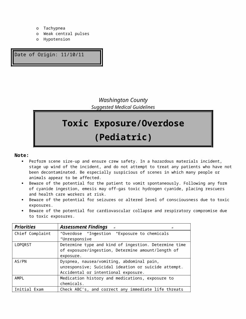
Mid-range and reactive pupils during the convulsion (they’re widely dilated in a real seizure) Lack of tongue biting or incontinence Apparent purposeful movements Remaining consciousness, or even speaking, during the convulsion
Date of Origin: 11/10/11
Washington County
Suggested Medical Guidelines
Stroke
Priorities Assessment FindingsChief Complaint “Weakness”, “Confusion”, “Slurred Speech”, “Unresponsive”LOPQRST When did it start? Was it witnessed?AS/PN Headache, weakness, pupil dilation, slurred speech, aphasia, incontinentAMPL Medication consistent with history of stroke or TIAInitial Exam ABC’s and correct any immediate life threatsDetailed Focused Exam Vital signs: BP, HR, RR, Temp, SpO2
General Appearance: Unresponsive?, noticeable facial droop, drooling, slouched postureNeuro: Cincinnati pre-hospital stroke scale (speech, facial symmetry, motor)
Data Blood GlucoseGoals of Therapy Maintain ABC’s and adequate vital signsMonitoring Cardiac rhythm strip
Heart rate and blood pressure
EMR Routine medical care Administer oxygen to keep SpO2 > 94% and <99%
Use a nasal cannula at 2 – 6 LPM or Non-rebreather mask at 10 – 15 LPM
Support airway as needed Ascertain time of onset and the last time patient felt well Notify responding ambulance of results of Cincinnati Prehospital Stroke Scale. (Have the patient smile note any irregularity.
Have the patient extend both arms in front of them and with their eyes closed and note any arm “drift” and have the patient say the following statement “You can’t teach an old dog new tricks”.)
BASIC
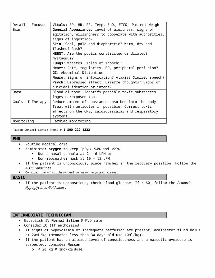
Check Blood glucose, If <60 mg/dl refer to Hypoglycemic Guidelines Do not delay transport to the closest facility with 24/7 CT and TPA availability (if known) Rapid transport is indicated if S/S onset less than 24 hours
INTERMEDIATE TECHNICIAN IV 0.9% NS @ KVO Consider IO (If authorized)
INTERMEDIATE/ PARAMEDIC Consider intubation if unresponsive without gag reflex or Nasal intubation for patients with decreased LOC with
an intact gag reflex.
Date of Origin: 11/10/11
Washington County
Suggested Medical Guidelines
Submersion (Adult)
Notes: RESCUER SAFETY IS #1. Many well-intentioned volunteer and professional rescuers have been injured or killed
attempting to save a drowning victim. If the victim is still in the water dispatch local water rescue resources Submersion is a loss of consciousness under water Submersion is primarily a respiratory problem When delivering ventilations and chest compressions assume the patient will vomit. Be prepared to suction. Secure the
patient’s airway as soon as possible. Any patient successfully resuscitated after a loss of consciousness underwater needs transport to the hospital
and physician evaluationPriorities Assessment FindingsChief Complaint “Drowning”, “Near Drowning”LOPQRST Onset. Duration of time under water. Water temperature, if known. Bystander
CPR performed? AED Used? AS/PN Alcohol involved? Trauma involved?AMPL Allergies? Medications?Initial Exam Check ABCs and correct immediately life-threatening problems.Detailed Focused Exam Vital Signs: BP, HR, RR, Temp, SpO2
General Appearance: lifelessSkin: pale, cool, mottledLungs: wet or clear?Heart: Rate and regularity? Absent heart sounds?Neuro: Unresponsive?
Data Blood sugar, EKG, SpO2.Goals of Therapy Return of spontaneous circulation (ROSC)Monitoring BP, HR, RR, EKG, SpO2.ETCO2
EMR Routine C-spine stabilization of all submersion patients is not indicated.
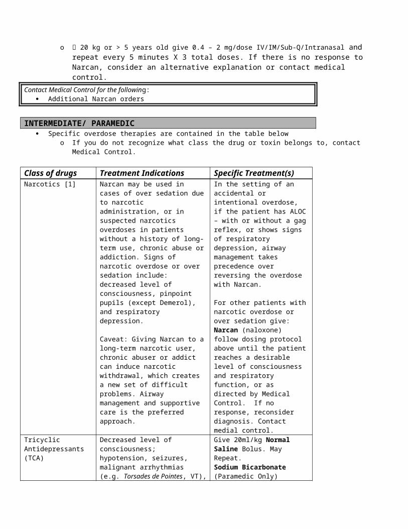
When a mechanism of injury (e.g. diving accidents), or obvious signs of trauma, is present:o C-spine stabilization is indicated.o Open the airway with a jaw-thrust maneuver.o Ventilate the patient while maintaining C-spine stabilization.o Remove the patient from the water on a long-spine board.
Always assume that hypothermia is present and follow the Hypothermia and Frostbite Guidelines. Do NOT start CPR if the patient has been submerged for more than 90 min If the patient is pulseless and not breathing, follow the Cardiac Arrest Guidelines.
o Remove the patient from standing watero Dry the chest with towelso Attach an AED and defibrillate if shocks are indicated.o If hypothermia is suspected, follow the resuscitation procedure outline in the Hypothermia Guidelines.
If an upper airway obstruction is suspected follow American Heart Association Guidelines
BASIC Check blood glucose, if hypoglycemia is suspected
o Follow Hypoglycemia Guidelines if the blood glucose is < 60
INTERMEDIATE TECHNICIAN
IV Normal Saline @ KVO If hypotension is suspected, initiate a Normal Saline Bolus of 250 ml Warm the IV fluids and run at 250 ml/hr according to the Hypothermia and Frostbite Guidelines If Hypoglycemia is present, follow Hypoglycemia Guidelines. If a narcotic overdose is suspected, follow the Toxic Exposure and Overdose Guidelines.
INTERMEDIATE/PARAMEDIC
Consider endotracheal intubation
FOOTNOTES:
Date of Origin: 11/10/11
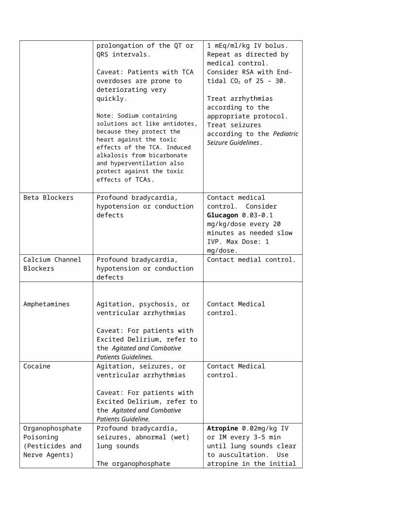
Washington County
Suggested Medical Guidelines
SyncopeNote:
Common causes of syncope include dehydration and vasovagal reflexes; less commonly syncope may result from arrhythmias and stroke
Syncope and seizures both result in loss of consciousness. Both may occur with or without convulsions. In syncope, the convulsions are brief. Unlike seizures, in syncope the patient regains consciousness quickly and without the usual postictal confusion.
Priorities Assessment FindingsChief Complaint “Passed Out”; “Fainted”LOPQRST Determine onset, duration and triggering events AS/PN Headache, dizziness, confusion, vomiting, diarrhea, dehydration, incontinence,
seizure, lack of food or waterAMPL Exposure to known allergen. History of heart disease or stroke. Current or past
medication for these problems. Compliance with these medications recently.Initial Exam Check ABCs and correct any immediately life threatening problems.Detailed Focused Exam Vital Signs: BP, HR, RR, Temp, SpO2, End Tidal CO2
General Appearance: may be normal or ill appearingSkin: Pale, cool, diaphoreticHeart: Hypotension, tachycardia, weak pulses, poor capillary refill?Neuro: May be A&OX3; ALOC? Focal deficits, signs of trauma due to falling?
Data Blood glucose. EKGGoals of Therapy Treat symptomatic bradycardia/hypotension.Monitoring BP,HR, RR, EKG, SpO2, ETCO2
EMR Routine Medical Care Gently lower the patient to a supine position or Trendelenburg position if hypotensive. Administer oxygen to keep SpO2 > 94% and <99%
Use a nasal cannula at 2 – 6 LPM or Non-rebreather mask at 10 – 15 LPM
BASIC Check blood glucose. If <60 refer to hypoglycemia protocol Cardiac monitor
INTERMEDIATE TECHNICIAN Initiate IV 0.9%NS @ KVO Consider IO (If authorized)
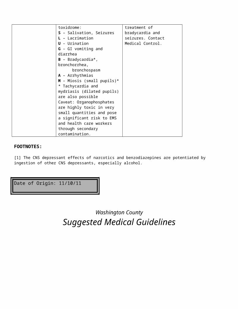
If patient is hypotensive, or shows signs of dehydration administer 250 ml fluid bolus of NSINTERMEDIATE / PARAMEDIC
Consider Atropine .5mg-1mg IV (and refer to Bradycardia protocol)
Date of Origin: 11/10/11
Washington County
Suggested Medical Guidelines
Toxic Exposure/Overdose
Note: Perform scene size-up and ensure crew safety. In a hazardous materials incident, stage up wind of the incident, and do not
attempt to treat any patients who have not been decontaminated. Be especially suspicious of scenes in which many people or animals appear to be affected.
Beware of the potential for the patient to vomit spontaneously. Following any form of cyanide ingestion, emesis may off-gas toxic hydrogen cyanide, placing rescuers and health care workers at risk.
Beware of the potential for seizures or altered level of consciousness due to toxic exposures. Beware of the potential for cardiovascular collapse and respiratory compromise due to toxic exposures.
Priorities Assessment FindingsChief Complaint “Overdose” “Exposure to chemicals” “Unresponsive”LOPQRST Determine time of exposure/ingestion, Determine amount/length of exposureAS/PN Dyspnea, nausea/vomiting, abdominal pain, unresponsive; Suicidal ideation or
suicide attempt. Accidental or intentional exposure.AMPL Psychiatric history and medications, exposure to chemicalsInitial Exam Check ABC’s, and correct any immediate life threatsDetailed Focused Exam Vitals: BP, HR, RR, Temp, SpO2, ETCO2
General Appearance: level of alertness, signs of agitation, willingness to cooperate with authoritiesSkin: Cool, pale and diaphoretic? Warm, dry and flushed?HEENT: Are the pupils constricted or dilated? Nystagmus?Lungs: Wheezes, rales or rhonchi?Heart: Rate, regularity, BP, peripheral perfusion?Neuro: Signs of intoxication? Ataxia? Slurred speech? Psych: Depressed affect? Bizarre thoughts? Signs of suicidal ideation or intent?
Data Blood glucose, Identify possible toxic substances ingested/exposed too.Goals of Therapy Reduce amount of substance absorbed into the body; Treat with antidotes if
possible; Correct toxic effects on the CNS, cardiovascular and respiratory systems.
Monitoring Cardiac monitoring
EMR Routine medical care Administer oxygen to keep SpO2 > 94% and <99%
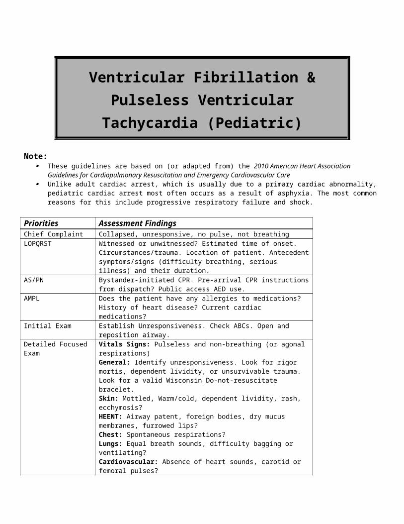
Use a nasal cannula at 2 – 6 LPM or Non-rebreather mask at 10 – 15 LPM
If the patient is unconscious, place him/her in the recovery position. Follow the ALOC Guidelines. Consider use of a non-visualized airway.
BASIC If the patient is unconscious, check blood glucose. If < 60, follow the Hypoglycemia Guidelines
IV TECH IV 0.9% NS @ KVO Initiate a bolus of 250 ml Normal Saline, if the patient is hypotensive or tachycardia. (check lung sounds) If the patient has an altered level of consciousness and a narcotic overdose is suspected, consider Narcan 0.4-2.0 mg
IV/IM/SubQ/Intranasal and repeat every 5 minutes X 3 total doses titrated to respirations. If there is no response to Narcan, consider an alternative explanation or contact medical control.
Contact Medical Control for the following: Additional Narcan orders
INTERMEDIATE/ PARAMEDIC Specific overdose therapies are contained in the table below
o If you do not recognize what class the drug or toxin belongs to, contact Medical Control.Class of drugs Treatment Indications Specific Treatment(s)Narcotics [1] Narcan may be used in cases of over
sedation due to narcotic administration, or in suspected narcotics overdoses in patients without a history of long-term use, chronic abuse or addiction. Signs of narcotic overdose or over sedation include: decreased level of consciousness, pinpoint pupils (except Demerol), and respiratory depression.
Caveat: Giving Narcan to a long-term narcotic user, chronic abuser or addict can induce narcotic withdrawal, which creates a new set of difficult problems. Airway management and supportive care is the preferred approach.
In the setting of an intentional overdose, if the patient has ALOC – with or without a gag reflex, or shows signs of respiratory depression, airway management takes precedence over reversing the overdose with Narcan. EMT-P considers RSI and withholding Narcan.
For other patients with narcotic overdose or over sedation give:Narcan (naloxone) 0.5mg IVP up to 2 mg every 5 minutes until the patient reaches a desirable level of consciousness and respiratory function, or as directed by Medical Control. If no response, reconsider diagnosis.
Benzodiazepines(BZD) [1]
Pure benzodiazepine overdoses are rarely life threatening.
Airway management and supportive care is the preferred approach.
Treatment consists of primarily of aggressive airway support
Tricyclic Antidepressants (TCA)
Amitriptyline, Amoxapine, Ascendin, Desipramine, Desyrel, Elavil, Endep,
Decreased level of consciousness; hypotension, seizures, malignant arrhythmias (e.g. Torsades de Pointes, VT), prolongation of the QT or QRS intervals.
Caveat: Patients with TCA overdoses are prone to deteriorating very quickly.
Run 1 or 2 IVs of Normal Saline wide open.Treat arrhythmias according to the appropriate protocol.Treat seizures according to the Seizure GuidelinesEMT-P (only): Sodium Bicarbonate (Paramedic Only) 1-

Imipramine, Ludiomil, Norpramine, Pamelor, Sinequan, Triavil, Tofranil and others
Note: Sodium containing solutions act like antidotes, because they protect the heart against the toxic effects of the TCA. Induced alkalosis from bicarbonate and hyperventilation also protect against the toxic effects of TCAs.
2 Amps IV bolus. Repeat as directed by medical control. For long transports, consider a Sodium Bicarbonate (Paramedic Only) drip with 3 amps in a liter of NS @ 250 ml/hr after the initial boluses are in.Consider RSI
Beta Blockers Profound bradycardia, hypotension or conduction defects
Contact medical control. EMT-P: Consider Glucagon 1 mg slow IVP. May repeat up to 3 mg total.
Calcium Channel Blockers
Profound bradycardia, hypotension or conduction defects
Contact medial control. Consider Calcium Chloride (Paramedic Only) 10 ml of 10% solution IV over 20 minutes.
Amphetamines Agitation, psychosis, or ventricular arrhythmias
Caveat: For patients with Excited Delirium, refer to the Agitated and Combative Patients Guidelines.
EMT-I may treat with Ativan or Valium (Paramedic Only), if seizures develop (see doses below).EMT-P may consider the following medications for sedation: Versed 2-5mg IV/IM/IO/Intranasal or seizures: Valium (Paramedic Only) 5mg IVP or Ativan 1-2 mg IV or IM. May repeat once. Haldol (Paramedic Only) 5.0 mg IM for agitation or combativeness.Contact Medical control.
Cocaine Agitation, seizures, ventricular arrhythmias, or coronary vasospasm
Caveat: For patients with Excited Delirium, refer to the Agitated and Combative Patients Guideline.
EMT-I may treat with Ativan or Valium (Paramedic Only) , if seizures develop (see doses below).EMT-P may consider the following medication:Valium (Paramedic Only) 5mg IVP or Ativan 1-2 mg IV or IM. Haldol (Paramedic Only) 5.0 mg IM May repeat once. Contact Medical control.
Organophosphate Poisoning (Pesticides and Nerve Agents)
Profound bradycardia, seizures, abnormal (wet) lung sounds
The organophosphate toxidrome:S – Salivation, SeizuresL – LacrimationU – UrinationG – GI vomiting and diarrheaB – Bradycardia*, bronchorrhea,
bronchospasmA – ArrhythmiasM – Miosis (small pupils)** Tachycardia and mydriasis (dilated
Atropine 2mg IV or IM every 3-5 min until lung sounds clear to auscultation. Use atropine in the initial treatment of bradycardia. Contact Medical Control.EMT-I and EMT-P may treat with Ativan or Valium (Paramedic Only), if seizures develop. Valium (Paramedic Only) 5mg IVP or Ativan 1-2 mg IV or IM. May repeat once.

pupils) are also possibleCaveat: Organophosphates are highly toxic in very small quantities and pose a significant risk to EMS and health care workers through secondary contamination.
FOOTNOTES:Poison Control Center: 1-800-222-1222[1] The CNS depressant effects of narcotics and benzodiazepines are potentiated by ingestion of other CNS depressants, especially alcohol.
Request dispatch of the Washington Co Haz-mat team if decontamination is thought to be needed
Date of Origin: 11/10/11

Hospital Destination / Selection
The following content will be considered the Guideline/Standard of care for the patient in need of:
Hospital Destination / Transport / Bypass
Selecting a transport destination should be based on the following factors:
The presence of a medical condition where the loss of life or limb is imminent (i.e. inability to effectively ventilate a failed airway)
Patient hospital preference Proximity to hospitals Specialty medical care available (i.e.. Trauma care based on trauma level designation, Stroke center, Advanced
cardiac care – cardiac Cath lab, Burn management, Neonatal intensive care, Pediatric intensive care, Hyperbaric therapy)
Current available bed status at hospital or emergency department (diversionary status) Available resources at area hospitals affected by other transports from current emergency or mass casualty Local rules or policies limiting destination choices Current level of service (scope of practice) on scene or available Patient preference is typically the leading factor in determining a hospital destination for patients in Washington
County. EMTs should assist patients in making this decision based on consideration of the above factors. An emergency department should not be bypassed if an EMT is unable to affectively ventilate a patient or if a
patient has a critically low blood pressure that is not responding to all available treatments. Additional options include ALS intercept before approaching the closest hospital or utilizing aero medical
services. An emergency department should not be bypassed if an EMT is concerned that a patient’s condition will
significantly worsen during the delay caused by driving to the alternate facility.Exceptions:
Upon occasion, patients, or their legal representatives, will insist on being transported to a facility that is not the closest most appropriate facility when in the opinion of the EMT, the closest most appropriate facility is medically indicated.
o Under such circumstances, it is advisable to discuss the situation with medical control and have the patient or their legal representative sign an appropriate waver indicating their desire to deviate from the medical plan of care proposed.
Contact on-line medical control with any questions
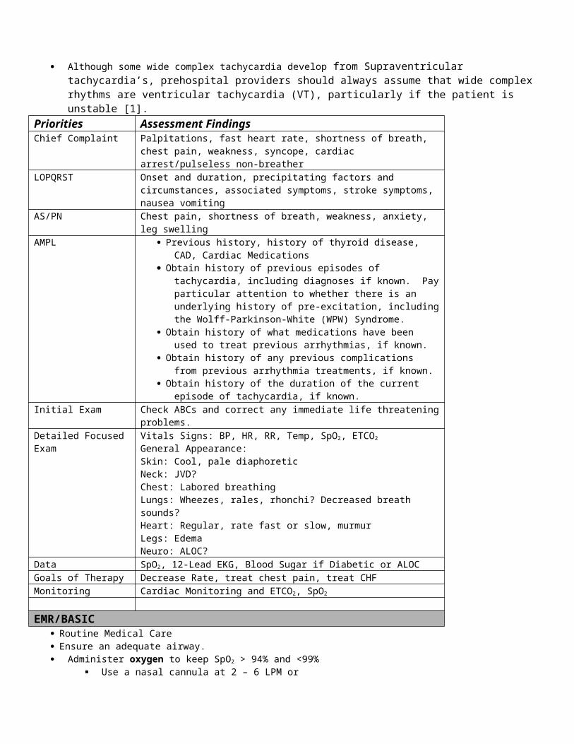
Washington County
Suggested Medical Guidelines
Vaginal Bleeding After Delivery
Note: Pregnancy usually lowers a woman’s blood pressure. If you get systolic readings between 80 – 100 mmHg, ask the mother
what her most recent blood pressure was in her doctor’s office.
Priorities Assessment FindingsChief Complaint “Vaginal bleeding and pregnant”LOPQRST Onset. Attempt to quantify the amount of blood lostAS/PN Is the patient having severe crampy pains? Has any fetal tissue passed?AMPL Has there been any prenatal care? An ultrasound? Was it normal?Initial Exam ABCs and correct any immediately life-threatening problems.Detailed Focused Exam Vital Signs: BP, HR, RR, Temp, SpO2
General Appearance: Pain or anxiety-related distress? External Hemorrhage? Skin: Pale, cool, and moist? Chest: Labored breathing?Heart: Rate and Rhythm?Abdomen: Internal hemorrhage? Tender? Distended? GU Blood loss?Neuro: ALOC?
Data SpO2
Goals of Therapy Identify potentially life-threatening hemorrhage. Treat for shock. Display sensitivity to the emotional needs of the parents. Reduce pain to the “comfortable” range.
Monitoring Monitor blood pressure, heart rate and mental status for signs of shock.
EMR/BASIC Administer oxygen to keep SpO2 > 94% and <99%
Use a nasal cannula at 2 – 6 LPM or Non-rebreather mask at 10 – 15 LPM
Massage fundus vigorously while applying suprapubic pressure with your other hand to prevent the uterus from expelling. This may cause discomfort to the mother
Place the baby to breast or chest level Perform any necessary neonatal care and assessment/resuscitation
Keep the mother warm and offer comfort measures. Transport in head down, left lateral recumbent position. Loose bulky dressings (do not pack)
IV TECH IV 0.9% NS @ KVO Give 250 ml bolus if SBP < 100 mmHg and lungs are clear Treat for shock as needed (See hypovolemia Protocol)
Contact Medical Control for the following: Additional fluid boluses are needed.
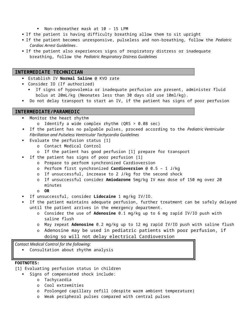
INTERMEDIATE/ PARAMEDIC Attempt to preserve any products of conception that pass and take them to the ED.
Contact Medical Control for the following: Additional orders for pain medications or fluids
FOOTNOTES:
Date of Origin:11/10/11
Washington County
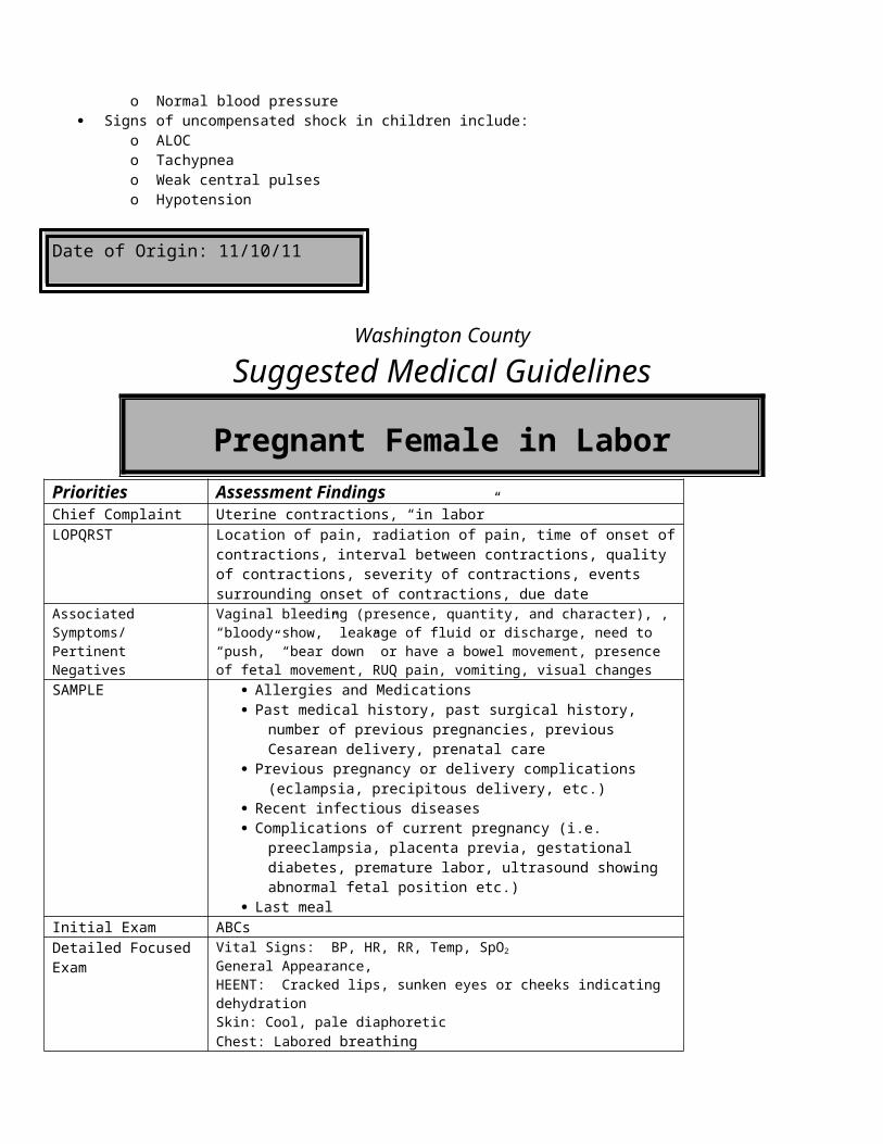
Suggested Medical Guidelines
Vaginal Bleeding Before Delivery
Note: Vaginal bleeding and severe lower abdominal pain in the first trimester of pregnancy should be considered a ruptured ectopic
pregnancy until proven otherwise. This is a true medical emergency! Bleeding at any point in pregnancy can be associated with loss of the fetus, and parents know this. You must be sensitive to
their sense of potential loss. After about 20 weeks of pregnancy, when the mother is in a supine position, the gravid uterus can compress the inferior vena
cava, which decreases preload and causes hypotension. Pregnancy usually lowers a woman’s blood pressure. If you get systolic readings between 80 – 100 mmHg, ask the mother
what her most recent blood pressure was in her doctor’s office.
Priorities Assessment FindingsChief Complaint “Vaginal bleeding and pregnant”LOPQRST Onset. Attempt to quantify the amount of blood lost (note number of pads)AS/PN Is the patient having severe crampy pains? Has any fetal tissue passed?AMPL Has there been any prenatal care? An ultrasound? Was it normal?Initial Exam ABCs and correct any immediately life-threatening problems.Detailed Focused Exam Vital Signs: BP, HR, RR, Temp, SpO2
General Appearance: Pain or anxiety-related distress? External Hemorrhage? Skin: Pale, cool, and moist? Chest: Labored breathing?Heart: Rate and Rhythm?Abdomen: Internal hemorrhage? Tender? Distended? GU Blood loss?Neuro: ALOC?
Data SpO2, ETCO2
Goals of Therapy Identify potentially life-threatening hemorrhage. Treat for shock. Display sensitivity to the emotional needs of the parents. Reduce pain to the “comfortable” range.
Monitoring Monitor blood pressure, heart rate and mental status for signs of shock.
EMR/BASIC
Administer oxygen to keep SpO2 > 94% and <99% Use a nasal cannula at 2 – 6 LPM or Non-rebreather mask at 10 – 15 LPM
Place in supine position with legs elevated. If > 20 weeks pregnant, place on left lateral side, recumbent, for transport. Keep the mother warm and offer comfort measures. Attempt to preserve any products of conception that pass and take them to the ED.
INTERMEDIATE TECHNICIAN
IV 0.9% NS @ KVO Give 250 ml bolus if SBP < 100 mmHg
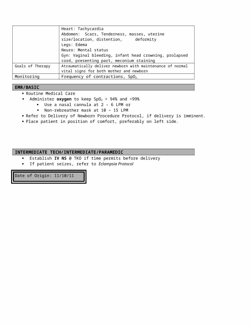
INTERMEDIATE/PARAMEDIC
Treat the pain. Morphine Sulfate 2-4 mg IV every 10 minutes X 3. Carefully monitor blood pressure response to Morphine Do not give Morphine, if SBP < 90 mm Hg May place the patient in left lateral Trendelenburg Position, if possible
Contact Medical Control for the following: Additional orders for pain medications.
FOOTNOTES: After about 20 weeks of pregnancy, when the mother is in a supine position, the gravid uterus can compress the inferior vena
cava, which decreases preload and causes hypotension. Be sure to transport all OB patients on left side if at all possible for this reason
Date of Origin: 11/10/11
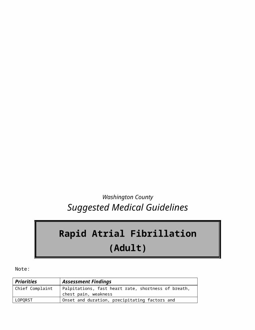
Washington County
Suggested Medical Guidelines
Vertigo
Note: Vertigo ranges from mild to severe – severe enough to become incapacitating and require EMS
help. Most patients complain about “dizziness”. The provider must differentiate the spinning or falling
feeling associated with vertigo from lightheadedness, which is another common reason for patients to complain of “dizziness”, but should not be treated according to this protocol.
Vertigo can occasionally be a symptom of stroke, but is most commonly a disturbance of spatial orientation and motion sense in the inner ear.
Vertigo is commonly associated with nausea and vomitingPriorities Assessment FindingsChief Complaint “Dizziness”, “Spinning or falling sensation”LOPQRST Determine onset and duration. Provoked by changing position or turning head.AS/PN Dizziness, nausea, vomiting, ataxia, and falls (with or without injury)AMPL Exposure to known allergen. History of CVA/TIA.Initial Exam ABC’s and correct any life threatsDetailed Focused Exam Vital Signs: BP, HR, RR, SpO2
General Appearance: Eyes closed, PaleNeuro: ALOC?, Focal deficits, PupilsEyes: Nystagmus with lateral gaze
Data Blood Glucose
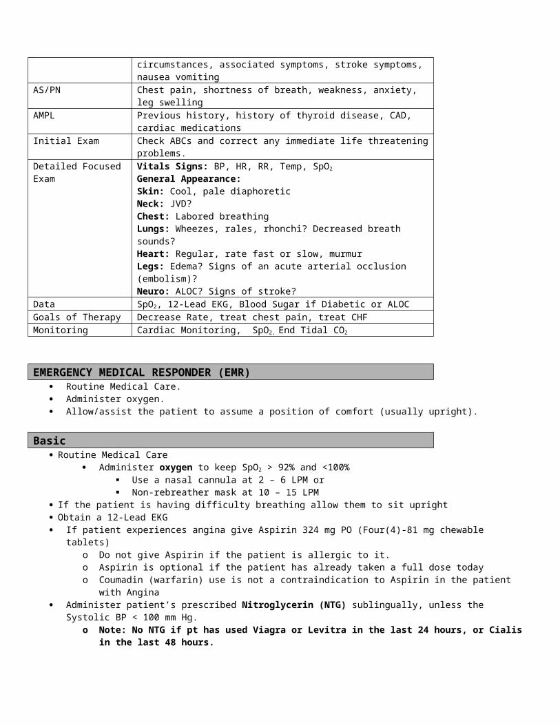
Goals of Therapy Maintain ABC’s and vitalsMonitoring Cardiac rhythm strip
Heart rate and blood pressure
EMR/BASIC Routine medical care Allow the patient to assume the position that minimizes or eliminates the symptoms – usually the
supine position. Beware that asking the patient to turn their head or sit up, or testing the movement of their extra
ocular muscles can trigger vertigo and result in vomiting. Administer Oxygen to keep SpO2 > 94% and <99%
Use a nasal cannula at 2 – 6 LPM or Non-rebreather mask at 10 – 15 LPM
INTERMEDIATE TECH / INTERMEDIATE Consider IV 0.9% NS @ KVO If the patient appears to be dehydrated, consider a 250 ml bolus of Normal Saline Consider IO (If authorized)
PARAMEDIC Use the following medication for treatment of nausea and vomiting associated with vertigo Ondansetron (Zofran) 4mg SLOW IV Consider Benadryl (Diphenhydramine) 25 mg IV or 50mg IM to correct extra pyramidal side
effects
Contact Medical Control for the following: Notify medical control if extrapyramidal side effects develop. These include:
a) Akathisia (uncontrollable restlessness)b) Torticollis (twisted neck)c) Opisthotonus (arched back)d) Oculogyric crisis (rapid eye movement)e) Buccolingual crisis (sputtering from the mouth)
FOOTNOTES:
Date of Origin: 11/10/11
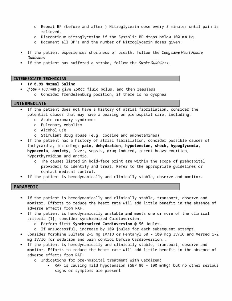
Washington County
Suggested Medical Guidelines
Wide Complex Tachycardia’s (Adult)
Note: Wide complex rhythms have a QRS duration > 0.12 sec Although some wide complex tachycardia develops from supraventricular tachycardia, prehospital
providers should always assume that wide complex rhythms are ventricular tachycardia (VT), particularly if the patient is unstable [1].
Treating wide complex tachycardia with medications used to treat supraventricular tachycardia is fraught with danger and must be avoided in the prehospital setting [2].
Priorities Assessment FindingsChief Complaint Palpitations, fast heart rate, shortness of breath, chest pain, weakness, syncope,
cardiac arrest/pulseless non-breatherLOPQRST Onset and duration, precipitating factors and circumstances, associated
symptoms, stroke symptoms, nausea vomitingAS/PN Chest pain, shortness of breath, weakness, anxiety, leg swellingAMPL Previous history, history of thyroid disease, CAD, Cardiac Medications
Obtain history of previous episodes of tachycardia, including diagnoses if known. Pay particular attention to whether there is an underlying history of pre-excitation, including the Wolff-Parkinson-White (WPW) Syndrome.
Obtain history of what medications have been used to treat previous arrhythmias, if known.
Obtain history of any previous complications from previous arrhythmia treatments, if known.
Obtain history of the duration of the current episode of tachycardia, if
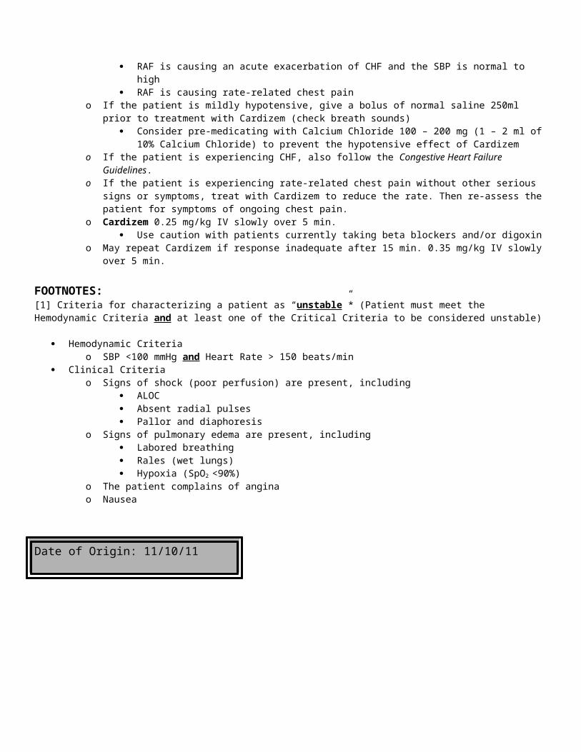
known.Initial Exam Check ABCs and correct any immediate life threatening problems.Detailed Focused Exam Vitals Signs: BP, HR, RR, Temp, SpO2
General Appearance:Skin: Cool, pale diaphoretic Neck: JVD?Chest: Labored breathingLungs: Wheezes, rales, rhonchi? Decreased breath sounds?Heart: Regular, rate fast or slow, murmur Legs: EdemaNeuro: ALOC?
Data SpO2, ETCO2, 12-Lead EKG, Blood Sugar if Diabetic or ALOCGoals of Therapy Decrease Rate, treat chest pain, treat CHFMonitoring Cardiac Monitoring End Tidal CO2 and SpO2
EMR Routine Medical Care Administer oxygen to keep SpO2 > 94% and <99%
Use a nasal cannula at 2 – 6 LPM or Non-rebreather mask at 10 – 15 LPM
If the patient is having difficulty breathing allow them to sit uprightBASIC
If the patient becomes unresponsive, pulseless and non-breathing, follow the CCR Guidelines.
If patient experiences angina, administer patient’s prescribed Nitroglycerin 0.4 mg sublingually, unless the Systolic BP < 100 mm Hg.
o Note: No NTG if pt. has used Viagra or Levitra in the last 24 Hours, Cialis in the last 48 Hours
o Give NTG every 5 minutes until pain is relievedo Repeat BP before each Nitroglycerin dose.o Discontinue nitroglycerine if the Systolic BP drops below 100 mm Hg.o Document all BP’s and the number of Nitroglycerin doses given.
If the patient also experiences shortness of breath, consider the Congestive Heart Failure Guidelines
Perform a 12-Lead EKG (transmit to ED if capable)
INTERMEDIATE TECHNICIAN IV 0.9% Normal Saline If SBP < 100 mmHg give 250cc fluid bolus the reassess
INTERMEDIATE/PARAMEDIC Monitor the heart rhythm If the patient is hemodynamically and clinically stable,
o Differentiate between monomorphic and polymorphic ventricular tachycardiao Differentiate between regular and irregular rhythms
Regular rhythms are monomorphic VT until proven otherwise (e.g. PSVT with aberrancy).
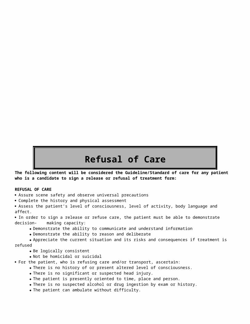
Irregular rhythms are polymorphic VT (including Torsades de Pointes) until proven otherwise (e.g. Atrial Fibrillation with aberrancy or pre-excitation).
For stable patients with monomorphic VT
o Consider Amiodorone 150mg(1amp), over 10 min (15mg/min)o Amiodorone drip at 1mg/min (Paramedic only) – Contact Medical Control
Combine (1) amp of Amiodarone (150mg) to 150 mL of NS in the burette vessel. Administer the Amiodarone solution at 1 mg per minute (60mL/hr or 1 drip every second)
-Or-o Consider Lidocaine 1-1.5 mg/kg IV Pusho Rebolus Lidocaine 0.5- 0.75 mg/kg IV push every 5-10 minutes up to 3mg/kgo Initiate Lidocaine drip (Paramedic only) – Contact Medical Control
Drip 1-4 mg/min. Premixed bags with mirco drip tubingLido Drip (4mg/ml) 1mg 2mg 3mg 4mgµdrops/min (ml/hr) 15gtt 30gtt 45gtt 60gtt
For patients with symptomatic PVC’so Lidocaine 1.0-1.5 mg/kg IV Pusho Rebolus Lidocaine 0.5- .75 mg/kg IV push every 5-10 min. up to 3mg/kgo Initiate Lidocaine drip (Paramedic only and medical control permission)
Use table above Transport emergently Monitor the patient very closely for signs of deterioration
Contact Medical Control for the following: Unstable patients Failure to respond to treatment Initiation of Lidocaine drip for VT or PVC’s For stable polymorphic VT that does not respond to Magnesium or Lidocaine, consider
Cardioversion or defibrillation under the direction of medical control For all other stable wide-complex tachycardia, prepare the patient for transport and contact
Medical Control.o Treating stable patients with antiarrhythmic therapy for wide complex rhythms that are
suspected to be of Supraventricular origin is not allowed in the prehospital setting [2].o Provide supportive care and monitor the patient closely during transport.
Mag Sulfate for polymorphic(Torsades) VT 1-2 gm over 5-60min loading dose (Paramedic only)
Consultation about rhythm analysis If the patient is hemodynamically or clinically unstable [1] with monomorphic VT
o Prepare to perform synchronized Cardioversion.o Perform first synchronized Cardioversion @ 50 Joules.o If unsuccessful, increase by 50 joules for each subsequent attempt.
If the patient is hemodynamically or clinically unstable [1] with polymorphic VT, or if the patient develops pulseless VT
o Defibrillate (i.e. unsynchronized Cardioversion) at device-specified dose in a biphasic defibrillator)
Consider Versed1-2mg IV for sedation and pain control before Cardioversion or defibrillation (Paramedic only).

If the patient remains hemodynamically and clinically stable, further treatment can be safely delayed until the patient arrives in the emergency department.
FOOTNOTES:[1] Criteria for characterizing a patient as “unstable” (Patient must meet the Hemodynamic Criteria and at least one of the Critical Criteria to be considered unstable)
Hemodynamic Criteriao SBP < 100 mmHg and Heart Rate > 150 beats/min
Clinical Criteriao Signs of shock (poor perfusion) are present, including
ALOC, including syncope Absent radial pulses Pallor and diaphoresis
o Signs of pulmonary edema are present, including Labored breathing Rales (wet lungs) Hypoxia (SpO2 <90%)
o The patient complains of anginao Nausea
[2] Caution: Treating wide complex rhythms of Supraventricular origin Although PSVT and Rapid Atrial Fibrillation/Flutter can result in wide-complex tachycardia in the
presence of a bundle branch block or pre-excitation due to an accessory pathway (e.g. the Wolf-Parkinson-White Syndrome), the potential adverse consequences of treating these rhythms with antiarrhythmic drugs (Adenosine, Diltiazem, Metoprolol) outweighs the potential benefit to a stable patient.
o Identifying a Supraventricular rhythm as the origin of a wide-complex tachycardia is beyond the scope of practice of all prehospital providers.
o Even if the wide complex rhythm is intermittent, do not give Adenosine for PSVT, if you have seen a wide complex rhythm on the monitor, rhythm strip or 12-lead EKG.
o Even if the wide complex rhythm is intermittent, do not give Diltiazem for rapid atrial fibrillation/flutter, if you have seen a wide complex rhythm on the monitor, rhythm strip or 12-lead EKG.
o Even if the wide complex rhythm occurs in the presence of an acute coronary syndrome, do not give Metoprolol.
o Even if you receive an order from on-line medical control to give Adenosine, Diltiazem or Metoprolol in the context of a wide-complex tachycardia, do not do it without the express written consent of your EMS Medical Director.
For stable patients, observe and monitor patient while enroute to Hospital
Date of Origin: 11/10/11

Withholding or Withdrawing of Resuscitative Efforts
1. If at any time you are not certain which of these policies applies, begin treatment and contact medical control for orders.
2. Emotional support should be provided to significant others.
3. Disposition of the patient will be handled according to local and county requirements.
4. Use of this protocol MUST be guided by Medical Control. Contact should be established via appropriate communication option.
St Joseph’s ER Charge Nurse cell phone (262) 836-1764
St. Joseph’s ER Doctors cell phone number: (262) 836-1757
St Joseph’s Nurses’ Station (262) 836-7892
St Joseph’s EMS phone (262) 836-7893
Aurora Medical Center Washington County (262) 670-7201
DNR Orders:
Patients with DNR orders who are NOT in cardiac arrest should receive supportive care enroute to the hospital. Do not withhold oxygen and medications. DNR orders are not in effect until the patient is in cardiac/respiratory arrest.
Valid DNR status exists if a patient has a state approved DNR bracelet, a metal medic-alert bracelet, or a valid DNR order. A valid DNR order must be a written document containing:
o Name of the patiento Name and signature of the attending physiciano Effective date. Renewal periods have not been determined for DNR orders. Their validity will not
expire unless modified or revoked at any time by the maker.o The words "Do Not Resuscitate"o Evidence of consent either:
a. signature of the patient or,
b. signature of legal guardian or,
c. signature of Durable Power of Attorney for Health Care agent or
d. attached Living Will or other advance directive prepared by or on behalf of the patient
Obviously Dead Patients: "Triple Zero"
Obviously dead patients are those found to be non-breathing, pulseless and have one or more of the following long term indications of death:
o Decapitationo Rigor mortis without hypothermiao Profound dependent lividityo Decompositiono Mummification/putrificationo Incinerationo Massive Trauma with asystole
No resuscitative efforts are to be initiated for the patients listed above.
Contact Medical Examiner.
Law Enforcement or EMS must remain with the deceased until the Medical Examiner arrives at the scene, as per usual protocol for home deaths.
If there is any doubt the patient shall be assessed by a minimum of 2 EMS personnel
Those 2 EMS personnel shall attach a cardiac monitor and verify asystole in 2 different leads
Those 2 EMS personnel will use extreme caution to not disrupt the scene as it may be a crime scene
Be sure to document the scene thoroughly. Include body position, clothing, room contents and placement and what you did to the body
Hospice Patients not in arrest
If patients are registered in a hospice program, initiate BLS and immediately contact medical control for orders on treatment and disposition. Inform medical control of the presence of written treatment orders and/or valid DNR orders.
Power of Attorney for Healthcare/Living Wills
If someone represents themselves as having a power of attorney to direct medical care of the patient and/or a document referred to as a living will is presented; follow these procedures:
Begin or continue medical treatment.
Contact Medical Control; explain the situation and follow any orders received.
Living wills may not be honored by field personnel.
Bring any documents received to the hospital.
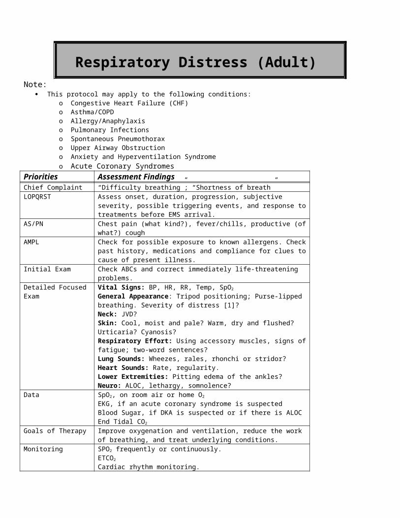
PATIENTS IN PERSISTENT ASYSTOLE WHO DO NOT RESPOND TO TREATMENT:
Note: An order of a physician is required before stopping treatment under this protocol.
1. Provide patient care, per protocol, based on the patient’s condition.
2. Contact a medical control physician and explain the events of the call. Report any response to ALS treatment.
3. Provide Medical Control; all of the following information:
o Patient historyo The patient is an adult, is normothermic, and experienced an arrest unwitnessed by EMT'so The patient remains in asystoleo Advanced airway and IV are correctly placed, ando Drug therapy, defibrillation and CPR attempts have been carried out according to protocol
4. Continue resuscitative efforts, unless otherwise directed by Medical Control.
5. Under the direction of Medical Control, resuscitative attempts can be terminated in the field. Physician name, date, and time of terminated attempts should be reported on the Ambulance Report Form. Note: This is not legal pronouncement of death.
6. If resuscitative attempts are terminated in the ambulance or en route to the hospital, contact Dispatch to have the Medical Examiner paged. (Make sure the Medical Control Physician is aware that the person had to be moved into the ambulance.)
7. For Ambulance Providers who use St. Joseph’s Community Hospital and Aurora Medical Center-Hartford as Medical Control, the body can be taken to the hospital and the ED staff will direct EMS personnel to a private area to take the body. If family arrives in the ED, they can be directed to be with the body.
8. Law Enforcement or EMS must remain with the deceased until the Medical Examiner arrives at the Hospital, as per usual protocol for home deaths.
9. The Medical Examiner will pronounce death and complete the death investigation in the same manner as a home death.
10. If the physician gives the order to continue resuscitative efforts until you reach the hospital, resuscitative efforts, per protocol, are to be carried out. Transport the patient to the Emergency Department.
11. If unable to establish communication with medical control, resuscitative efforts should be continued until the patient reaches the hospital.
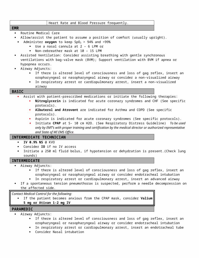
Index
Adenosine..................................................................................................................54, 88, 96
Agitated and Combative Patients...........................................................................................9
Albuterol.............................................................................................8, 14, 17, 31, 63, 65, 105
Allergic reactions....................................................................................................See allergy
Allergy...................................................................................................................................13
Allergy & Anaphylaxis (Pediatric)...........................................................................................62
Altered Level of Consciousness.............................................................................................15
Amiodorone...................................................................................................................25, 135
Amphetamines....................................................................................................................125
Anaphylaxis...........................................................................................................................13
Anaphylaxis (Pediatric)..........................................................................................................62
APGAR.................................................................................................................................52
Aspirin............................................................................................................................26, 31
ASTHMA..................................................................................................................................7
Asthma (Pediatric).................................................................................................................64
Asystole.................................................................................................................................16
Asystole/PEA (Pediatric)........................................................................................................66
Ativan......................................................................................10, 34, 39, 61, 86, 105, 118, 125
Atrial Fibrillation....................................................................................................................99
Atropine...........................................................................................................19, 69, 115, 125
Atrovent.....................................................................................................................8, 65, 105
Benadryl.......................................................................................................13, 14, 57, 63, 133
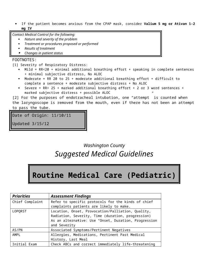
Beta Blockers......................................................................................................................125
Bradycardia...........................................................................................................................18
Bradycardia (Pediatric)...........................................................................................................68
Breech presentation..............................................................................................................51
Bronchospasm.............................................See Respiratory Distress (Pediatric), See Asthma
Burns.....................................................................................................................................20
Burns (Pediatric)....................................................................................................................70
Calcium Channel Blockers.................................................................................................125
Calcium Chloride..........................................................................................................17, 125
Cardiac Arrest........................................................................................................................23
Cardiac Arrest (Pediatric).......................................................................................................73
Cardiac Chest Pain.................................................................................................................26
Cardiogenic Shock..................................................................................................................30
Cardioversion......................................................................................54, 88, 96, 100, 107, 136
Child birth........................................................................................See Delivery of Newborn
Chronic Bronchitis.................................................................................................................7
Cincinnati Prehospital Stoke Scale.....................................................................................119
Cocaine...............................................................................................................................125
Combative Patients.................................................................................................................9
Congestive Heart Failure........................................................................................................30
COPD.....................................................................................................................................7
CPAP.........................................................................................................................8, 31, 105
Cricothyrotomy.............................................................................................................67, 107
Croup...............................................................................See Respiratory Distress (Pediatric)
C-spine injuries.....................................................................See Routine Trauma Care
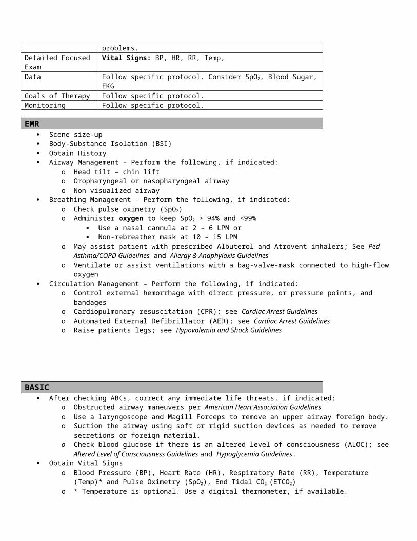
Cushing's triad....................................................................................................................114
D50.......................................................................................................................See Dextrose
Delivery of Newborn..............................................................................................................50
Dextrose........................................................................................................................78, 107
Diabetic ketoacidosis..................................................................................See Hyperglycemia
Dilaudid.........................................................................................................................60, 115
Diphenhydramine................................................................................................See Benadryl
Dizziness.................................................................................................................See Vertigo
Dopamine..................................................................................................................19, 31, 49
Drowning........................................................................................................See Submersion
Eclampsia...............................................................................................................................33
Emphysema............................................................................................................................7
Endotracheal Tube Size Selection & Chart..........................................................................75
Epiglottitis......................................................................See Respiratory Distress (Pediatric)
Epinephrine....................................................................8, 13, 14, 16, 19, 62, 65, 67, 69, 83, 93
Fentanyl........................................................................................................................60, 115
Frostbite................................................................................................................................45
Furosemide...........................................................................................................................31
Glucagon......................................................................................................17, 34, 43, 78, 125
Glucose............................................................................................................................43, 78
Heat Emergencies..................................................................................................................37
Hyperglycemia.......................................................................................................................42
Hyperglycemia (Pediatric)......................................................................................................76
Hypoglycemia........................................................................................................................43
Hypoglycemia (Pediatric).......................................................................................................78
Hypothermia..........................................................................................................................45
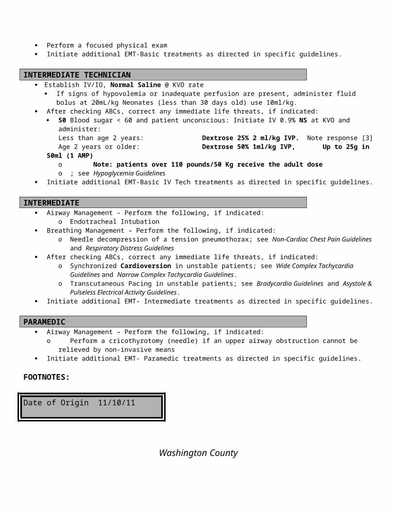
Hypovolemia and Shock.........................................................................................................48
Hypovolemia and Shock (Pediatric)........................................................................................80
Intranasal Versed...............................................................................................................118
Ipratropium.........................................................................................................See Atrovent
Labor.....................................................................................................................................97
Level of Consciousness..........................................................................................................15
Lidocaine..........................................................................................................46, 93, 115, 135
Magnesium Sulfate................................................................................................8, 33, 34, 93
Medical Care (Adult)............................................................................................................108
Medical Care (Pediatric).......................................................................................................106
Metoprolol............................................................................................................................27
Morphine.......................................................................................................................31, 115
Morphine Sulfate...............................................................................................27, 59, 60, 131
Narcan.....................................................................................................................15, 90, 124
Narcotics.............................................................................................................................124
Narrow Complex Tachycardias...............................................................................................53
Nausea..................................................................................................................................56
Near Drowning................................................................................................See Submersion
Needle decompression..............................................................................................17, 67, 107
Nexus research......................................................................See Routine Trauma Care
Nitroglycerin...................................................................................26, 27, 31, 54, 99, 105, 135
NTG.............................................................................................................See Nitroglycerin
Ondansetron (Zofran)...................................................................................................57, 133
Organophosphate Poisoning...............................................................................................125
Overdose.............................................................................................................................123
Overdose (Pediatric)..............................................................................................................89
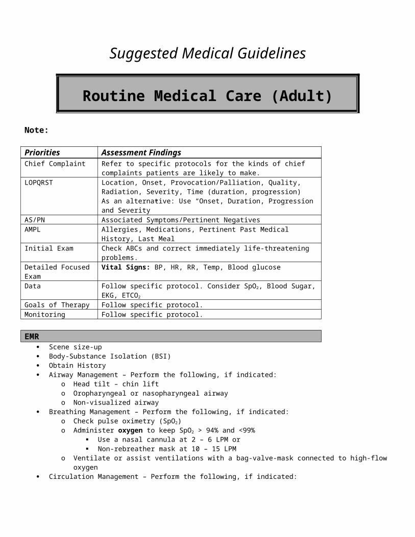
Pacing.................................................................................................................19, 67, 69, 107
Pain Management..................................................................................................................58
Paroxysmal Supraventricular Tachycardia...................................See Narrow complex Tach
PEA (Pediatric).......................................................................................................................66
Pediatric Wide Complex Tachycardia......................................................................................95
Poison Control Center........................................................................................................126
Pregnant Female....................................................................................................................97
Prolapsed umbilical cord......................................................................................................51
Pulmonary Infections......................................................See Respiratory Distress (Pediatric)
Pulseless Electrical Activity.....................................................................................................16
Rapid Atrial Fibrillation..........................................................................................................99
Rapid Sequence Airway........................................................................................................115
Respiratory Distress.............................................................................................................104
Respiratory Distress (Pediatric)..............................................................................................82
Resuscitative Efforts.............................................................................................................138
Routine Medical Care (Adult)...............................................................................................108
Routine Medical Care (Pediatric)..........................................................................................106
Routine Trauma Care (Adult)................................................................................................110
Routine Trauma Care (Pediatric)..........................................................................................113
Seizure.................................................................................................................................117
Seizure (Pediatric)..................................................................................................................85
Selective Spinal Immobilization......................................................See Routine Trauma Care
Shock.....................................................................................................................................48
Shock (Pediatric)....................................................................................................................80
Sodium Bicarbonate......................................................................................................17, 124
Solu-Medrol...........................................................................................................8, 14, 63, 65

Spontaneous Pneumothorax............................................See Respiratory Distress (Pediatric)
STEMI....................................................................................................................................26
Stroke..................................................................................................................................119
Submersion..........................................................................................................................120
Succinylcholine...................................................................................................................115
Supraventricular Tachycardia........................................................See Narrow complex tach
SVT (Pediatric).......................................................................................................................87
Synchronized Cardioversion..................................................................96, See Cardioversion
Syncope...............................................................................................................................122
Tachycardias...................................................................................................................53, 134
Toradol.................................................................................................................................61
Toxic Exposure.....................................................................................................................123
Toxic Exposure/Overdose (Pediatric).....................................................................11, 12, 35, 89
Trauma Care (Adult).............................................................................................................110
Trauma Care (Pediatric).......................................................................................................113
Tricyclic Antidepressants...................................................................................................124
Upper Airway Obstruction..............................................See Respiratory Distress (Pediatric)
Vaginal Bleeding After Delivery............................................................................................128
Vaginal Bleeding Before Delivery.........................................................................................130
Valium.................................................................................................39, 61, 86, 105, 118, 125
Vecuronium........................................................................................................................116
Ventricular Fibrillation & Pulseless Ventricular Tachycardia (Pediatric)..................................92
Ventricular Tachycardia (Pediatric).........................................................................................92
Versed........................................................................................10, 19, 54, 61, 86, 88, 115, 118
Vertigo.................................................................................................................................132
Vomiting................................................................................................................................56
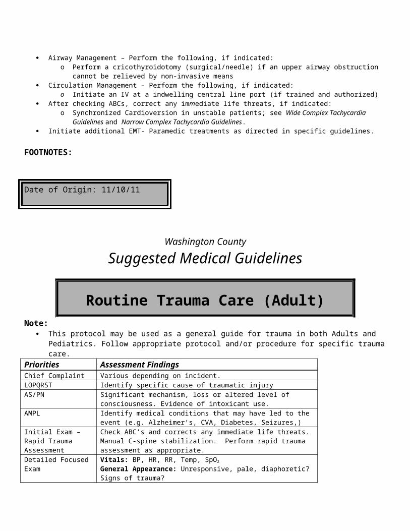
Water rescue...................................................................................................See Submersion
Wide Complex Tachycardia
Pediatric...........................................................................................................................95
Wide Complex Tachycardias.....................................................................................................................134