Embed Size (px)
Citation preview

EHS 207 GENERAL BIOCHEMISTRY FORENVIRONMENTAL HEALTH
Course Team Dr. O. A. Saliu (Course Writer) - NOUNDr. O. A. Idowu (Co-writer) - UniIlorinProf. A. T. Oladiji (Course Editor) - UniIlorinProf. G. C. Okoli-Nnabuenyi (ProgrammeLeader/Coordinator) - NOUN
NATIONAL OPEN UNIVERSITY OF NIGERIA
COURSEGUIDE
EHS 207 COURSE GUIDE
ii
© 2020 by NOUN PressNational Open University of NigeriaHeadquartersUniversity VillagePlot 91, Cadastral ZoneNnamdi Azikiwe ExpresswayJabi, Abuja
Lagos Office14/16 Ahmadu Bello WayVictoria Island, Lagos
e-mail: [email protected]: www.nou.edu.ng
All rights reserved. No part of this book may be reproduced, in anyform or by any means, without permission in writing from the publisher.
Printed 2020
ISBN: 978-978-970-151-3
EHS 207 COURSE GUIDE
iii
CONTENTS PAGE
Introduction……………………………………………. ivWhat you will Learn in the Course………………….. ivCourse Aim……………………………………………. vCourse Objective………………………………………. vWorking through the Course…………………………. vThe Course Materials………………………………….. vStudy Units…………………………………………….. vPresentation Schedule…………………………………. viAssessment……………………………………………. viTutor-Marked Assignment……………………………. viiCourse Marking Scheme……………………………… viiiFacilitators/Tutors and Tutorials……………………… viiiSummary……………………………………………… viii

EHS 207 COURSE GUIDE
iv
INTRODUCTION
EHS 207 title “General Biochemistry” is a three (3) Unit course withfour (4) Modules and fifteen (15) Units. Biochemistry is the study ofbiomolecules. It can also be defined as the application of chemistry tothe study of biological processes in living organisms. Biochemistry isboth a life science and a chemistry science; it explores the chemistry ofliving organisms and the molecular basis for the changes occurring inliving cells.
Millions of complex chemical reactions are going on in the human bodyat any given time, ranging from the balance of the endocrine system tothe storage and utilisation of fuel molecules such as glucose. Bystudying and understanding this highly complex reaction, biochemistshave found better ways to fight infections and diseases not just at themolecular level but also at the cellular level. Since an Engineer cannotrepair a vehicle if he does not understand how it works, so a biochemistmust understand how the human body functions and the variousmechanisms involved in process.
Much of biochemistry also deals with the structures and functions ofcellular components such as proteins, carbohydrates, lipids and nucleicacids collectively known as biomolecules. The main purpose of all theefforts of Biochemistry is to benefit humans in all forms particularly inthe diagnosis and treatment of different diseases. For example,investigation of diabetes mellitus is completely based upon thelaboratory test in Biochemistry laboratories, where the presence of sugarin urine is tested by Benedict’s test. Similarly, investigations of otherdisorders such as albuminuria, lactosuria, etc are a few of so manyailments that are investigated in Biochemistry laboratories.
WHAT YOU WILL LEARN IN THE COURSE
In this course, you will learn about the branches of biochemistry and itsrelevance to other life sciences, biochemistry of living cells, biologicaloxidation and electron transport chain, buffer, acidity, alkalinity, pH,pKa values and their roles in cellular metabolism. You will learn aboutthe metabolism of biomolecules such as carbohydrate, proteins as wellas lipids. The structure of the DNA will also be discussed. Theknowledge that will be acquired in this course will assist you inunderstanding the various biochemical reactions that takes place in theliving system.
EHS 207 COURSE GUIDE
v
COURSE AIM
The aim of this course is to build your foundation in the knowledge ofbiochemistry as it relates to the proper physiological functioning of thehuman system.
COURSE OBJECTIVES
At the completion of this course, you should be able to:
explain the branches and relevance of biochemistry, biochemistryof living cells, biological oxidation and electron transport chain,buffer, acidity, alkalinity, pH, pKa values and their roles incellular metabolism.
understand the metabolism of major biomolecules such ascarbohydrate, proteins, lipids as well as nucleic acid.
WORKING THROUGH THE COURSE
As a student of this course you are expected to register for this courseonline which is available at the NOUN website before you can haveaccess to all the materials. You will be expected to read every modulealong with all assigned readings to prepare you for assessment.
Reading the reference materials will enhance your understanding of thecourse.
Note that each unit has self-assessment exercises which you are advisedto do and at certain periods during the course, you will be expected tosubmit your assignment for the purpose of assessment. There will befinal examination
THE COURSE MATERIALS
The main components of the course are:1. The Course Guide2. Study Unit3. References/Further Reading4. Assignments
STUDY UNITS
There are four (4) modules and fourteen (14) study units in this courseThey are as follows:
EHS 207 COURSE GUIDE
vi
Module 1 Introduction to General Biochemistry
Unit 1 Definition of BiochemistryUnit 2 Cell Structure, Cell Components and their FunctionsUnit 3 Biochemistry of the Plasma Membrane
Module 2 Water, Acids, Bases, Buffer and Macromolecules
Unit 1 Water, Acids, Bases and BufferUnit 2 Chemistry of CarbohydratesUnit 3 Chemistry of Amino Acids and ProteinUnit 4 Chemistry of LipidsUnit 5 Chemistry of Nucleic Acids
Module 3 Metabolism of Biomolecules
Unit 1 Metabolism of CarbohydratesUnit 2 Krebs Cycle and Oxidative PhosphorylationUnit 3 Metabolism of ProteinsUnit 4 Metabolism of Lipids
Module 4 Micronutrients (Vitamins and Minerals) andDetoxification
Unit 1 Water Soluble VitaminsUnit 2 Trace ElementsUnit 3 Detoxification
There are activities related to the lecture in each unit which will helpyour progress and comprehension of the unit. You are required to workon these exercises which together with the TMAs will enable you toachieve the objectives of each unit.
PRESENTATION SCHEDULE
There is a time-table prepared for the early and timely completion andsubmissions of your TMAs as well as attending the tutorial classes. Youare required to submit all your assignments by the stipulated time anddate. Avoid falling behind the schedule time.
ASSESSMENT
There are three aspects to the assessment of this course.The first one is the self-assessment exercises. The second is the tutormarked assignments and the third is the written examination or theexamination to be taken at the end of the course.
EHS 207 COURSE GUIDE
vii
Do the exercises or activities in the unit by applying the information andknowledge you acquired during the course. The tutor-markedassignments must be submitted to your facilitator for formal assessmentin accordance with the deadlines stated in the presentation schedule andthe assignment file.
The work submitted to your tutor for assessment will count for 30% ofyour total course work.
At the end of this course, you have to sit for a final or end of courseexamination of about a three-hour duration which will count for 70% ofyour total course mark.
TUTOR-MARKED ASSIGNMENT
This is the continuous assessment component of this course and itaccounts for 30% of the total score. You will be given four (4) TMAs byyour facilitator to answer. Three of which must be answered before youare allowed to sit for the end of course examination.
These answered assignments are to be returned to your facilitator.
You’re expected to complete the assignments by using the informationand material in your readings, references and study units.
Reading and researching into your references will give you a betterdeeper understanding of the subject.
1. It is important that each assignment reaches your facilitator on orbefore the deadline given in the presentation schedule andassignment file. If for any reason you are not able to completeyour assignment, make sure you contact your facilitator beforethe assignment is due to discuss the possibility of an extension.Request for extension will not be granted after the due dateexcept for exceptional circumstances.
2. You will need to revise the whole course content before sittingfor the examination. The self-assessment activities and TMAswill be useful for this purpose. The examination concludes theassessment for the course and constitutes 70% of the wholecourse. All areas of the course will be examined and you will beinformed the time for the examination.
EHS 207 COURSE GUIDE
viii
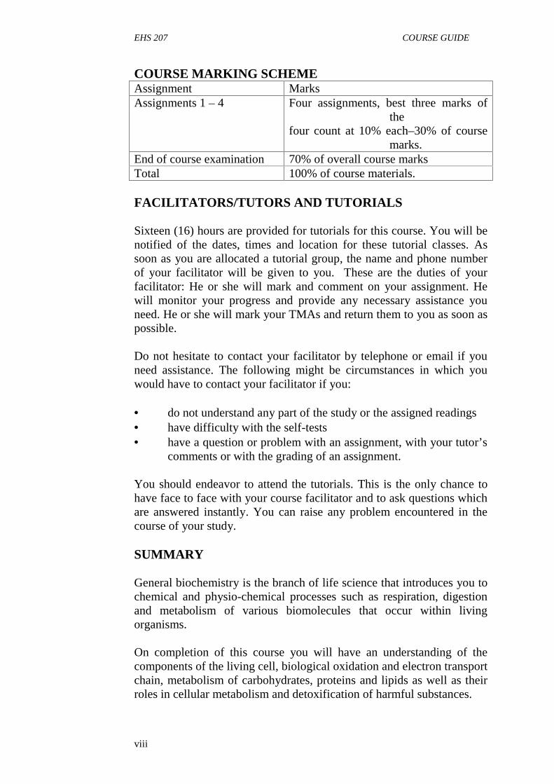
COURSE MARKING SCHEMEAssignment MarksAssignments 1 – 4 Four assignments, best three marks of
thefour count at 10% each–30% of course
marks.End of course examination 70% of overall course marksTotal 100% of course materials.
FACILITATORS/TUTORS AND TUTORIALS
Sixteen (16) hours are provided for tutorials for this course. You will benotified of the dates, times and location for these tutorial classes. Assoon as you are allocated a tutorial group, the name and phone numberof your facilitator will be given to you. These are the duties of yourfacilitator: He or she will mark and comment on your assignment. Hewill monitor your progress and provide any necessary assistance youneed. He or she will mark your TMAs and return them to you as soon aspossible.
Do not hesitate to contact your facilitator by telephone or email if youneed assistance. The following might be circumstances in which youwould have to contact your facilitator if you:
do not understand any part of the study or the assigned readings have difficulty with the self-tests have a question or problem with an assignment, with your tutor’s
comments or with the grading of an assignment.
You should endeavor to attend the tutorials. This is the only chance tohave face to face with your course facilitator and to ask questions whichare answered instantly. You can raise any problem encountered in thecourse of your study.
SUMMARY
General biochemistry is the branch of life science that introduces you tochemical and physio-chemical processes such as respiration, digestionand metabolism of various biomolecules that occur within livingorganisms.
On completion of this course you will have an understanding of thecomponents of the living cell, biological oxidation and electron transportchain, metabolism of carbohydrates, proteins and lipids as well as theirroles in cellular metabolism and detoxification of harmful substances.
EHS 207 COURSE GUIDE
ix
You are expected to apply the knowledge you have acquired during thiscourse to your practical life.
Furthermore, you should be able to answer the following questions:
define biochemistry discuss the metabolism of carbohydrates, proteins and lipids give an account of oxidative phosphorylation and electron
transport chain explain the detoxification processes of harmful substances.
We wish you success in this course.
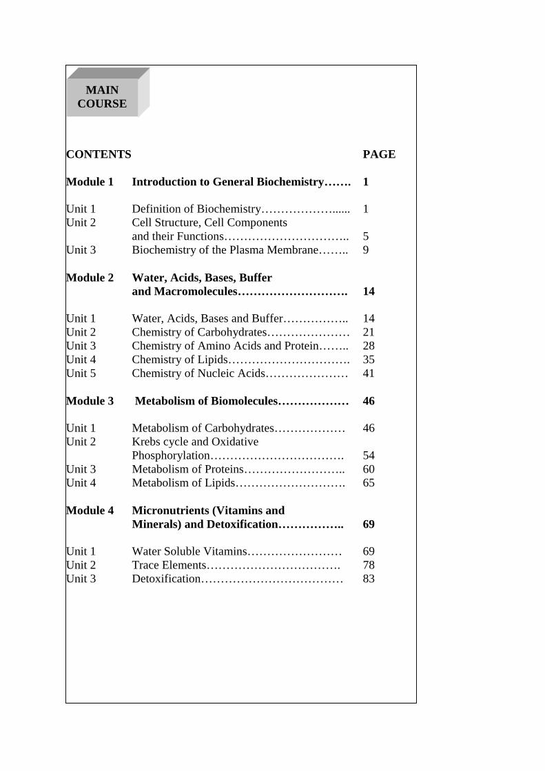
CONTENTS PAGE
Module 1 Introduction to General Biochemistry……. 1
Unit 1 Definition of Biochemistry………………...... 1Unit 2 Cell Structure, Cell Components
and their Functions………………………….. 5Unit 3 Biochemistry of the Plasma Membrane…….. 9
Module 2 Water, Acids, Bases, Bufferand Macromolecules………………………. 14
Unit 1 Water, Acids, Bases and Buffer…………….. 14Unit 2 Chemistry of Carbohydrates………………… 21Unit 3 Chemistry of Amino Acids and Protein…….. 28Unit 4 Chemistry of Lipids…………………………. 35Unit 5 Chemistry of Nucleic Acids………………… 41
Module 3 Metabolism of Biomolecules……………… 46
Unit 1 Metabolism of Carbohydrates……………… 46Unit 2 Krebs cycle and Oxidative
Phosphorylation……………………………. 54Unit 3 Metabolism of Proteins…………………….. 60Unit 4 Metabolism of Lipids………………………. 65
Module 4 Micronutrients (Vitamins andMinerals) and Detoxification…………….. 69
Unit 1 Water Soluble Vitamins…………………… 69Unit 2 Trace Elements……………………………. 78Unit 3 Detoxification……………………………… 83
MAINCOURSE
EHS 207 MODULE 1
1
MODULE 1 INTRODUCTION TO GENERALBIOCHEMISTRY
Unit 1 Definition of BiochemistryUnit 2 Cell Structure, Cell Components and their FunctionsUnit 3 Biochemistry of the Plasma Membrane
UNIT 1 DEFINITION OF BIOCHEMISTRY
CONTENTS
1.0 Introduction2.0 Objectives3.0 Main Content
3.1 Definition of Biochemistry3.2 Relevance of Biochemistry as a Life Science3.3 Branches of Biochemistry
4.0 Conclusion5.0 Summary6.0 Tutor-Marked Assignment7.0 References/Further Reading
1.0 INTRODUCTION
The essence of studying biochemistry is for the purpose ofunderstanding the various chemical reactions that occur in livingorganisms at both the cellular and molecular levels. Biochemistry as alife science is applicable and relevant in different fields of study such asmedicine, agriculture, pharmacy, nursing etc.
2.0 OBJECTIVES
By the end of this unit, you will be able to:
define biochemistry state the relevance of biochemistry to other life sciences (nursing,
medicine, pharmacy) describe different branches of biochemistry.
EHS 207 GENERAL BIOCHEMISTRY FOR ENVIRONMENTAL HEALTH
2
3.0 MAIN CONTENT
3.1 Definition of Biochemistry
Biochemistry is the study of biomolecules. It can also be defined as theapplication of chemistry to the study of biological processes in livingorganisms. Biochemistry is both a life science and a chemical science; itexplores the chemistry of living organisms and the molecular basis forthe changes occurring in living cells.
Millions of complex chemical reactions are going on in the human bodyat any given time, ranging from the balance of the endocrine system tothe storage and utilisation of fuel molecules such as glucose. Bystudying and understanding these highly complex reactions, biochemistshave found better ways to fight infections and diseases at the molecularlevel. Since an Engineer cannot repair a vehicle if he does notunderstand how it works, so a biochemist must understand how theliving system works in order to proffer solutions in disease states. Thusmuch of biochemistry deals with the structures and functions of cellularcomponents such as proteins, carbohydrates, lipids and nucleic acidscollectively known as biomolecules. The main focus of biochemistry isin understanding how biological molecules give rise to the processesthat occur within living cells, which in turn relates greatly to the studyand understanding of the whole organism (human being).
3.2 Relevance of Biochemistry to other life Sciences
Biochemistry provides foundation for other life sciences such asmedicine, nursing, pharmacy, zoology, microbiology etc. as well asagriculture.
In pharmacy biochemistry provides an
understanding of the constitution of drugs, the half-life of drugsand drug metabolism.
In agriculture, the knowledge of biochemistry plays a valuable role infarming, fishery, poultry, sericulture etc as it
helps in the prevention and treatment of diseases gives an idea of how the use of fertilizers can increase plants
growth, their yield and quality of food can help to evaluate pesticide residues or other toxic waste in
plants, food grain and seed through biochemical test help in the monitoring of the quality of milk in animal husbandry
which can be checked by biochemical test.
EHS 207 MODULE 1
3
In medicine, biochemistry gives an insight into the changes andphysiological alterations that take place in the body. It also assists inclinical diagnosis of diseases.
Biochemists have contributed greatly to the discovery of new drugs totreat chronic diseases such as cancer, viral infections and metabolicdisorders. They are able to do this because they have thoroughunderstanding of what happens at the molecular and cellular levels ofliving organisms.
3.3 Branches of Biochemistry
i. Toxicology: This field studies the adverse effects of toxic orforeign chemical substances on the organisms. Environmentaland food toxicology also fall under this branch ofbiochemistry.
ii. Enzymology: This is the study of enzymes, their functions,deficiency and the consequence of such deficiency indiseases.
iii. Molecular biology and Biotechnology: This field evolveddirectly from Nucleic acid biochemistry and it involvesmanipulation of DNA to improve drug research and solve healthproblems. It has wide applications in other fields of sciencewhich includes cancerresearch.
iv. Lipid and Carbohydrate biochemistry: This field studies thebiochemical basis of metabolic disorders such as diabetes,obesity and Cardiovasculardiseases.
v. Natural products biochemistry: This is a new area of researchin biochemistry; it evolved as a result of interest of scientistsacross the world in searching for new drugs from plants. Quinineand Artesunate (antimalarial drugs) were isolated fromplants.
4.0 CONCLUSION
In this introductory unit, biochemistry has been introduced a life sciencewith important contribution to the field of nursing, medicine, agricultureetc. The five main branches of biochemistry discussed above havedifferent dimensions of improving human life.
5.0 SUMMARY
The primary aim of this unit is to enlighten you about biochemistry as afield of science, its branches as well as its relevance in the field ofnursing, medicine, pharmacy and agriculture.
EHS 207 GENERAL BIOCHEMISTRY FOR ENVIRONMENTAL HEALTH
4
6.0 TUTOR-MARKED ASSIGNMENT
1. Define biochemistry.2. Name and explain three (3) branches of biochemistry.3. Discuss the relevance of biochemistry to other life sciences.
7.0 REFERENCES/FURTHER READING
Amanullah, M. (2011). Medical Biochemistry and Biotechnology.(1st
ed.).
Murray, R.K., Bender, D. A., Botham, K., M., Kennelly, P.J., RodwellV.W. & Well, P.A. (2012).Harper’s Illustrated Biochemistry(29thed.).McGraw-HillMedical.
Nelson, D.L. & Cox M. M. (2009). Lehninger Principles ofBiochemistry (4thed.).
EHS 207 MODULE 1
5
UNIT 2 CELL STRUCTURE, COMPONENTS OF THECELL AND THEIR FUNCTIONS
CONTENTS
1.0 Introduction2.0 Objectives3.0 Main Content
3.1 Structure of Animal Cell3.2 Components of the Cell and their Functions
4.0 Conclusion5.0 Summary6.0 Tutor-Marked Assignment7.0 References/Further Reading
1.0 INTRODUCTION
The living cell we are to discuss here is not different from the cell youlearnt in Biology when you were in secondary school. Cells are the basicbuilding blocks of all living organism. The human body is composed oftrillion of cells. Cells have many parts, each with a different function.Some of these parts called organelles are specialized structures thatperform certain tasks within the cell. Biochemical arrangement of cellsand how these cells interact to perform various functions in man are notonly fascinating but also very interesting. Imagine the sensitivity of cellsresponsible for taste; different region of your tongue detects differenttaste.
Some cells are replaced every 72 hours in our body while some spendup to ten years before they die. Also, some cells remain in our bodythroughout our lifetime. There are two basic types of cells in nature andthese are prokaryotic and eukaryotic cells. Prokaryotic cells are thesimplest cells and are without a nucleus and cell organelles whileeukaryotic cells are sophisticated cells with a well-defined nucleus andcell organelles. A group of cells forms tissue, various tissues forms anorgan and different organs make up the body.
It is important to understand compartmentalization and the functions ofvarious organelles present in the cells. Most biochemical reactions takeplace inside the cell but in different organelles; for example, energygeneration takes place inside the mitochondria. Thorough understandingof cell structure will help you to understand the root causes of manydiseases and the biochemical mechanisms of their treatment.
EHS 207 GENERAL BIOCHEMISTRY FOR ENVIRONMENTAL HEALTH
6
2.0 OBJECTIVE
By the end of this unit, you will be able to:
discuss the cell, structure of the animal cell, cell organelles andtheir functions.
3.0 MAIN CONTENT
3.1 Structure of Animal Cell
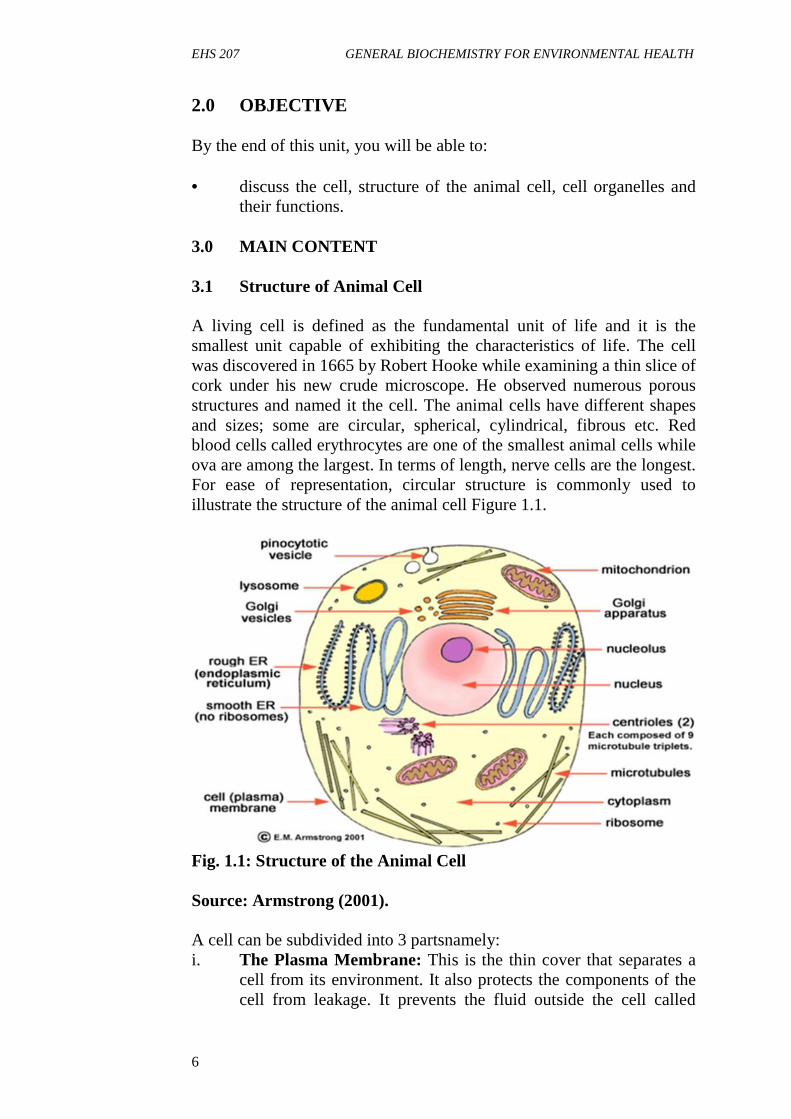
A living cell is defined as the fundamental unit of life and it is thesmallest unit capable of exhibiting the characteristics of life. The cellwas discovered in 1665 by Robert Hooke while examining a thin slice ofcork under his new crude microscope. He observed numerous porousstructures and named it the cell. The animal cells have different shapesand sizes; some are circular, spherical, cylindrical, fibrous etc. Redblood cells called erythrocytes are one of the smallest animal cells whileova are among the largest. In terms of length, nerve cells are the longest.For ease of representation, circular structure is commonly used toillustrate the structure of the animal cell Figure 1.1.
Fig. 1.1: Structure of the Animal Cell
Source: Armstrong (2001).
A cell can be subdivided into 3 partsnamely:i. The Plasma Membrane: This is the thin cover that separates a
cell from its environment. It also protects the components of thecell from leakage. It prevents the fluid outside the cell called
EHS 207 MODULE 1
7
extracellular fluid (ECF) from mixing with the fluid inside thecell called intracellular fluid (ICF). Plasma membrane regulatesthe materials that enters or leaves the cell, for this reason, it issaid to be semi-permeable. In addition, the plasma membrane hassome glycoproteins and glycolipids on its surface; thesemolecules serve as signal molecule between cells.
ii. The Cytoplasm: This is the fluid-like space between the plasmaand nuclear membrane. Cytoplasm is the cavity where theorganelles are found. It providesspace for the movement ofsynthesised products from one compartment to another for furtherprocessing. The organelles are suspended in the cytoplasm bycytoskeleton network that resemble nets.
iii. Nucleus: This is the most important part of the cell, the nucleus isalways centrally located. It has its own membrane called nuclearmembrane which protects the content of the nucleus. Nucleus isvery important to the cell because it contains the genetic materials(DNA and RNA) that control all the activities of the cell. Nucleusregulates the rate and time of cell division. It also determines thematerials that enter or exit thecell.
3.2 Functions of Cell Organelles
i. Rough Endoplasmic Reticulum: It is a vast system ofinterconnected, membranous, sacks that are located in the cell’scytoplasm and it is responsible for the synthesis of protein (due tothe presence of ribosomes attached to it) and degradation of wornoutorganelles.
ii. Smooth Endoplasmic Reticulum: It is located in the cell’scytoplasm and transports materials throughout the cell. Itcontains enzymes which produce and digest lipids (fats) andmembrane proteins. The smooth endoplasmic reticulum istherefore responsible for the synthesis of lipids and steroids,storage and metabolism of calcium and detoxification oftoxicsubstances.
iii. Golgi Apparatus: It is a flattened sac-like organelle that looks likea stack of pancakes. It is located near the nucleus. It produces themembranes that surround the lysosomes. The golgi apparatuspackages proteins and carbohydrates into membrane-boundvesicles for export from the cell.
iv. Lysosome: These are round organelles surrounded by amembrane where the digestion of cell nutrients takes place due to
EHS 207 GENERAL BIOCHEMISTRY FOR ENVIRONMENTAL HEALTH
8
presence of the digestive enzymes. It contains more than 40different hydrolytic enzymes and they are collectively known asLYSOZYMES which are actively involved in the degradation ofmacromolecules, worn out organelles and the removal of excesssecretory products. Lysosome has the thickest membrane toprevent the leakage of hydrolytic enzymes.
v. Peroxisomes: These are single membrane spherical organelles,also called micro-bodies. They contain antioxidant enzymes suchas catalase and peroxidases which are involved in thedetoxification of hydrogen peroxide and other radicals.
vi. Mitochondria: The mitochondrion is known as the power houseof the cell as it generates energy in form of ATP (adenosinetriphosphate), the energy currency of all living cells. It isspherical in shape and has double membrane i.e. inner and outermitochondrion membrane.
vii. Ribosomes: These are small organelles rich in ribonucleic acid(RNA) and are active in the synthesis ofproteins.
ix. Vacuole: The vacuole is a fluid-filled, membrane-surroundedcavity inside a cell. The vacuole fills with food being digestedand waste material that is on its way out of the cell. There arespecialized vacuoles which function to store fat as fat droplets.
4.0 CONCLUSION
The cell is the structural and functional basic unit of life. The humanbody contains several billions of cells that perform various functions.There are specialized structures called cell organelles present within thecell through which the cell performs these various functions.
5.0 SUMMARY
In this unit, you have learnt about the cell, types of cells, structure of theanimal cell, cell organelles and their functions.
6.0 TUTOR-MARKEDASSIGNMENT
1. Draw a well labeled structure of the animalcell2. List four cell organelles and their functions.
EHS 207 MODULE 1
9
7.0 REFERENCES/FURTHER READING
Amanullah, M. (2011). Medical Biochemistry and Biotechnology. (1st
ed.).
Devlin T.M. (2010). Textbook of Biochemistry with ClinicalCorrelation 7th Edition.John Wiley & Sons Inc.
EHS 207 GENERAL BIOCHEMISTRY FOR ENVIRONMENTAL HEALTH
10
UNIT3 BIOCHEMISTRY OF THE PLASMAMEMBRANE
CONTENTS
1.0 Introduction2.0 Objectives3.0 Main Content
3.1 Components of the Plasma Membrane3.2 The Functions of Plasma Membranes3.3 Transportation of Materials across the Plasma Membrane
4.0 Conclusion5.0 Summary6.0 Tutor-Marked Assignment7.0 References/Further Reading
1.0 INTRODUCTION
The cell or plasma membrane can be referred to as ‘the wall of a city. Itprotects the components of the cell and also regulates what enters orleaves the cell. The plasma membrane is very important to all cells sincethe cell owes its survival to an intact and functional cell membrane. Ifthere is any injury to the cell membrane, the whole cell may bedestroyed. Technically, the cell membrane is a liquid. At roomtemperature, it has about the same consistency as vegetables oil. Lipids,proteins and carbohydrates in the plasma membrane can diffuse freelythroughout the cell membrane; they are essentially floating across itssurface. This process is known as the fluid mosaic model, which wascoined by S. J. Singer and G. L. Nicolson in 1972.
2.0 OBJECTIVES
By the end of this unit, you will be able to:
describe the plasma membrane list and discuss various mechanism of transport of materials
across the plasma membrane.
3.0 MAIN CONTENT
3.1 Components of the Plasma Membrane
Plasma membrane mainly consists of phospholipids, cholesterol andproteins. There is a wide variation in lipid- protein ratio for different cellmembranes. The functions performed by the cell and the location

EHS 207 MODULE 1
11
determine the quantity of proteins and lipids present in the plasmamembrane.
Membrane Lipids
There are several types of membrane lipids. The fundamental buildingblocks of cell membranes are the phospholipids. Membrane lipids areamphipathic molecules (they have both hydrophilic and hydrophobicends, hydrophilic means “water loving”; this part readily associates withwater while hydrophobic ends means “water hating”; they tend to moveaway from water). When cellular membranes form, phospholipidsassemble into two layers because of their hydrophilic and hydrophobicproperties. The phosphate heads in each layer face the aqueous orwatery environment on both side, and the tails hide away from the waterbetween the layers of heads, because they are hydrophobic (Figure 1.2).
Fig. 1.2: Structure of Plasma Membrane BilayerSource: Sadler (2004).
Cholesterol
Cholesterol is another important membrane lipid found exclusively inthe plasma membrane of mammalian cells. The cholesterol moleculesare randomly distributed across the phospholipid bilayer, helping thebilayer stay fluid in different environmental conditions. It holds thephospholipids together so that they don’t separate too far thereby lettingunwanted substances in, or compact too tightly, restricting movementacross the membrane. Without cholesterol, the phospholipids in theplasma membrane will start to get closer together when exposed to cold,making it more difficult for small molecules, like gases to squeeze inbetween the phospholipids like they normally do. Also the presence ofcholesterol prevents phospholipids from separating from each otherwhich could have resulted in large gaps.
Membrane Proteins
Membrane proteins can be classified as being either peripheral orintegral on the basis of their association with the membrane lipids.
EHS 207 GENERAL BIOCHEMISTRY FOR ENVIRONMENTAL HEALTH
12
Integral membrane proteins interact extensively with the hydrocarbonchains of membrane lipids. Most of these integral proteins span the lipidbilayer, protruding at both ends. They have high percentage of non-polaramino acids and represent about 70% of total membrane proteins.Examples are membrane enzymes, hormone receptors, pumps andchannels. Integral proteins are helpful for transporting larger moleculeslike glucose across the cell membrane. In contrast, peripheral proteinsare bound to the surface of lipid bilayer primarily by electrostatic andhydrogen bonds. Many peripheral membrane proteins are also bound tothe surfaces of integral proteins, on either the cytosolic or extra cellularside of the membrane. Examples include cytochrome c and acetylcholineesterase.
3.2 Functions of the Plasma Membrane
i. Protection: The primary function of the plasma membrane is toprotect the cytoplasm and the organelles present in the cell. It isresponsible for the maintenance of shape and size of cells.
ii. Transportation: The cell membrane act as semi permeablemembrane which allows only some substances to pass through itthereby acting as a barrier for other substances. For example,small hydrophobic molecules such as CO2, O2 and small lipidsdissolve in the membrane and pass through readily. Ions andmost nutrient molecules do not move freely through themembrane, but are often carried by the transport protein channels,either with or without the use of energy.
3.3 Mechanism of Transportation of Materials across thePlasma Membrane
Passive Transport: Passive transport in cells involves the process ofdiffusion; the diffusion can be simple or facilitated.
Simple diffusion – In terms of cellular activity, the rate of simplediffusion can be affected by temperature, molecular size, concentrationof the gradient. Materials that are moved through membranes by simplediffusion include: water, carbon dioxide, oxygen, some lipid solublemolecules such as alcohol.
Facilitated diffusion – Most molecules cannot move freely through themembrane, but do cross membranes with the help of membranetransport proteins, which temporarily bind to the substance to be movedthrough the membrane, a process called facilitated diffusion. No energyis involved in the process since both carrier proteins and channelproteins are involved. Materials that pass through membranes by
EHS 207 MODULE 1
13
facilitated diffusion include glucose, amino acids and many small ions.The movement of water through membranes also involves facilitateddiffusion. The special protein channel used for this is called aquaporins,and it facilitates the movement of water at a rate needed for cellactivities. Facilitated diffusion process may be coupled to the movementof other molecules in the same direction or opposite direction. In co-transport, the transport of one molecule depends on sequential transferof another molecule. Co-transport may be symport or antiport. Asymport moves two molecules in the same direction e.g. sodium-glucosetransporter. Antiport system moves two molecules in opposite direction.It is also known as counter transport e.g. sodium-potassium transporter.
Active transport – Energy requiring transport across membranes.Activetransport is involved in the movement of molecules across the cellmembrane from a region of lower concentration to a region of higherconcentration i.e. against the concentration gradient. Generally, mostcells need to move substances through the membrane in a directioncounter to the gradient or move substances that are too large or bulkywith the use of energy. Some transport proteins (carrier proteins) canmove substances through the membrane against the concentrationgradient. Active transport typically requires two carrier protein activesites. One recognizes the substances to be carried while the otherreleases ATP to provide energy for the protein carrier. In some cases,concentration gradients of ions typically (H+) protons or (Na+) sodiumions can be used to provide the energy needed to move moleculesthrough the membranes.
Active transport is classified into two types according to the source ofenergy used. Primary active transport derives its energy directly fromthe hydrolysis of ATP while the secondary active transport uses anindirect energy of an electrochemical gradient or membrane potentialproduced originally by primary active transport. An example of primaryactive transport is sodium-potassium pump (Na+ - K+ ATPase). It is theprotein or enzyme responsible for the transportation of Na+ and K+
across the cell membrane. The enzyme is known as sodium-potassiumAdenosine triphosphatase.
The energy required for the transportation of sodium and potassium ionsare derived from the hydrolysis of ATP. For every three Na+ pumpedout of the cell, two K+ are released into the cytosol.

EHS 207 GENERAL BIOCHEMISTRY FOR ENVIRONMENTAL HEALTH
14
Fig. 1.3: Active and Passive Transport across the Plasma MembraneSource: Delvin (2010).
4.0 CONCLUSION
The plasma membrane is a semi-permeable membrane that controls theentry and exit of molecules within the cell. The chemical structure of theplasma membrane explains the process of transportation across differentgradients of the cell.
5.0 SUMMARY
In this unit, you have learnt about the lipids and protein components ofthe cell membrane. We also discussed the different mechanism oftransportation of material across the cell membrane with explanation ofthe factors that drive such exchange.
6.0 TUTOR-MARKED ASSIGNMENT
1. Write short notes on the lipids and protein components of theplasma membrane
2. Explain the passive mechanism of transportation across the cellmembrane, draw diagrams to illustrate your answers.
3. Differentiate between the active and passive methods oftransportation.

EHS 207 MODULE 1
15
7.0 REFERENCES/FURTHER READING
Amanullah, M. (2011).Medical Biochemistry and Biotechnology. (1st
ed.).
Devlin T.M. (2010). Textbook of Biochemistry with ClinicalCorrelation. (7thed.). John Wiley & SonsInc.
Murray, R. K., Bender, D. A., Botham, K., M., Kennelly, P.J., RodwellV. W. and Well, P. A. (2012). Harper’s IllustratedBiochemistry (29th Edition) McGraw-HillMedical.
Nelson, D.L. and Cox M.M., (2009).Lehninger Principles ofBiochemistry 4thedition
Sadler, T. W. (2004). Langman’s Medical Embryology 9th edition.
EHS 207 GENERAL BIOCHEMISTRY FOR ENVIRONMENTAL HEALTH
16
MODULE 2 WATER, ACIDS, BASES, BUFFER ANDMACROMOLECULES
Unit 1 Water, Acids, Bases and BufferUnit 2 Chemistry of CarbohydratesUnit 3 Chemistry of Amino Acids and ProteinUnit 4 Chemistry of LipidsUnit 5 Chemistry of Nucleic Acids
UNIT 1 WATER, ACIDS, BASES AND BUFFER
1.0 Introduction2.0 Objectives3.0 Main Content
3.1 The Properties of Water3.2 Biological Importance of Water3.3 Acid, Base and Buffer3.4 Biological Importance of buffer
4.0 Conclusion5.0 Summary6.0 Tutor-marked Assignment7.0 References/Further reading
1.0 INTRODUCTION
Water is the most abundant matter on earth and also a major componentof the body. Typically, organisms are constituted of 70 to 90 % water. Itmust be present before any metabolic activity can take place in the celland it is referred to as a weak electrolyte because it can undergo partialdissociation into a proton (H+) and hydroxyl ion (OH-).
Water is made up of oxygen and two hydrogen atoms. Oxygen has atendency to pull the electrons more towards itself, thereby becomingelectronegative and leaving the hydrogens electropositive. This results inthe creation of a dipole due to the fact that each water molecule issurrounded by four other water molecules. The bond between ‘H’ of onewater molecule and ‘O’ of the other is known as hydrogen bond. In thisunit we shall also be discussing acids and bases which are defined withrespect to their ability to gain or lose protons and the importance ofbuffers in biological system.
2.0 OBJECTIVES
At the end of this unit, you should understand the properties of water,the importance of water as the major component of living organisms and
EHS 207 MODULE 2
17
be able to define acid, base and a buffer. You will also learn how tocalculate pH, pOH and pKa of a given solution.
3.0 MAIN CONTENT
3.1 The properties of water
i. Water is the predominant chemical component of all livingorganisms.
ii. Most chemical reactions in the cell take place in aqueousenvironment.
iii. It has a high boiling and melting points when compared to otherliquids.
iv. It has a specific heat of vaporizationv. Hydrogen bonds hold the oxygen and hydrogen atoms together in
a water molecule.vi. The oxygen of water is very electronegative, while hydrogen is
electropositive; as a result water is dipolar and exhibit tendencyto dissociate.
3.2 Biological importance of water
i. Water helps in the digestion of food: It helps to break downfood so that the body can absorb the nutrients.
ii. It regulates the body temperature: Water that is stored in themiddle layers of the skin comes to the skin’s surface as sweatwhen the body heats up. As it evaporates, it cools the body.
iii. It cushions the brain, spinal cord and other sensitive tissues:Dehydration can affect brain structure and function. It is alsoinvolved in the production of hormones and neurotransmitters.Prolonged dehydration can lead to problems with thinking andreasoning.
iv. It boosts skin health and beauty: With dehydration, the skincan become more vulnerable to skin disorders and prematurewrinkling.
v. It delivers oxygen throughout the body: Blood is more than 90percent, and blood carries oxygen to different parts of the body.
vi. It lubricates the joint: Cartilage, found in joints and the disks ofspine contains around 80 percent water. Long-term dehydrationcan reduce the joints shock-absorbing ability, leading to jointpain.
vii. The strong dipole and high dielectric constant of waterenables it to dissolve large quantities of charged compounds.
viii. The presence of hydrogen bond also enables water to dissolvemany organic molecules that contain functional groups.

EHS 207 GENERAL BIOCHEMISTRY FOR ENVIRONMENTAL HEALTH
18
ix. Water provides environment for macromolecules to achievestable structure in solution
3.3 Acid, Base and Buffer
An acid is a proton donor. It is also a compound that dissociates inaqueous solution to produce (H+) proton and a conjugate base (A-).
HA H+ + A-
Acid may dissociate partially (called weak acid e.g. ethanoic acid,water) or completely (called strong acid e.g. hydrochloric acid) insolution. In solution, weak acid establishes equilibrium between theproton and its conjugate base. Weak acids are those which have a slighttendency to give up protons e.g. acetic acid. On the other hand, strongacids give up protons readily e.g. HCl.
HCl H+ + Cl-
CH3COOH CH3COO- + H+
A Base is a compound that accepts proton in aqueous environment. Justlike an acid, there are strong bases and weak bases. For example, sodiumhydroxide is a strong base which releases hydroxyl ions very easily, andwater is a weak base as it is a poor source of hydroxyl ions.
NaOH Na+ + OH-
H2O H+ + OH-
pH of a solution is simply defined as the negative logarithm of thehydrogen ion concentration in a media. In simple terms it is a value thatgives the amount of hydrogen ions present in a solution. This value isexpressed in a reverse or negative form i.e. higher the pH value lower isthe hydrogen ion concentration and lower the pH value higher is thehydrogen ion concentration. The pH of all solutions ranges between 0and 14 only. pH of value 7.0 is neutral e.g. water and pH ranging from 0to 0.69 is acidic and 7.1 to 14 is basic or alkaline.
The normal pH of the blood plasma ranges between 7.35 and 7.45,average being 7.4. The intracellular pH of the tissues is 7.25 to 7.35averaging to 7.30 and pH of extracellular fluid is 7.30 to 7.40 with anaverage of 7.35. A decrease in the pH of blood is termed as acidosis andan increase in the pH of blood is termed as alkalosis. Alkalosis is morefatal than acidosis.
EHS 207 MODULE 2
19
Mathematically,pH = - log [H+] and
pOH = - log [OH-]
The equilibrium constant is called the acid dissociation constant and it isrepresented as:
Ka = = [H+] [A-][HA]
Where K is the equilibrium constant and a is the acid.
Calculation of pH, pOH and pKa
Example 1: If the H+ concentration of a solution is 4.2 x 10⁻³ calculatethe pH of the solution.
Solution:
pH = - log [H+], log[4.2 x 10⁻³] = log 4.2 + log 10-3 = 0.62 -3 = -2.38.Substitute for log [H+] in the equation.pH = -(-2.38), the two negative values canceled out, pH = 2.38
Example 2: Calculate the [H+], [OH-] and pH of 0.01M ethanoic acid,given that (Ka = 1.76 x 10⁻⁵).
Solution:
Note that ethanoic acid is a weak acid, it dissociates partially in solution,therefore HA = H+ + A- , if the conjugates are represented by x, then HA= 0.1- x ≈ 0.1 (value of x is negligible)Ka = [H+] [A-]
[HA]
Ka = 1.76 x 10-5 = X2/0.1X2 = 1.76 x 10⁻⁶, X is equal to the square root of 1.76 x 10⁻⁶
This is equal to 1.33 x 10⁻³, therefore,
[H+] = 1.33 x 10⁻³
pH = -log 1.33 x 10-3 = 0.12-3, if you take away 3 from 0.12, this willgive you a negative value (-2.88).
pH = 2.88 .
EHS 207 GENERAL BIOCHEMISTRY FOR ENVIRONMENTAL HEALTH
20
To calculate the pOH, the dissociation of pure water will be considered.H3O = H+ + OH-,
[H+] + [OH-] = 1.0 x10⁻¹⁴,
[OH-] = 1.0 x 10⁻¹⁴/ [H+]
= 1.0 x 10⁻¹⁴/1.33 x 10-3,
[OH-] =7.52 x 10⁻¹².
But pOH = -log 7.52 x 10⁻¹². This is equal to 0.88 – 12,pOH = 11.12
Buffer
A buffer is a solution that resists changes in pH (hydrogen ionconcentration) when an acid or a base is added. A buffer contains aweak acid and its conjugate base. Examples of buffer solutions areAcetate buffer (acetic acid and acetate salt), Bicarbonate buffer(carbonic acid and bicarbonate salt), phosphate buffer (sodium hydrogenphosphate and potassium hydrogen phosphate) etc.
Regulation of pH solution by buffer
If (H+) hydrogen ions are added to a buffer solution, the conjugate basereacts with the excess hydrogen ions to form the acid. On the otherhand, if (OH-) hydroxyl ions are added, they react with the acid presentin the buffer to produce water and conjugate base.
Preparation of buffer
To prepare buffer, Henderson-Hasselbalch equation is usually used tocalculate the concentrations of acid and base components of the buffer tobe prepared.
The equation is pH = pKa + log [A-][HA]
3.4 Biological importance of buffer
Buffers are chemical substances that help to maintain a relativelyconstant pH in a solution. Buffering is important in living systems as ameans of maintaining a fairly constant internal environment also knownas homeostasis. The following are examples of buffers in biological
EHS 207 MODULE 2
21
system and their importance:
i. The maintenance of blood pH is regulated via the bicarbonatebuffer. This system consists of carbonic acid and bicarbonateions. When the blood pH drops into the acidic range, this bufferacts to form carbon dioxide gas. The lungs expel this gas out ofthe body during the process of respiration. During alkalineconditions, this buffer brings the pH back to neutral by causingexcretion of the bicarbonate ions through the urine.
ii. The phosphate buffer system acts in a manner similar to thebicarbonate buffer, but only that it has a much stronger action.The internal environment of all cells contains this buffercomprising hydrogen phosphate ions and dihydrogen phosphateions. Under conditions when excess hydrogen enters the cell, itreacts with the hydrogen phosphate ions, which accepts them.Under alkaline conditions, the dihydrogen phosphate ions acceptthe excess hydroxide ions that enter the cell.
4.0 CONCLUSION
Water is life and a core component of all other fluids that make chemicalreactions possible in the body. Acids, bases and buffers serve differentbiological purposes in the living system.
5.0 SUMMARY
In this study unit, you have learnt about the properties of water and itsbiological importance, acid, base and buffers. You are also introduced tothe Henderson-Hasselbalch equation
6.0 TUTOR-MARKED ASSIGNMENT
1. What are the properties of water?2. Define an acid and a base.3. Give examples of two buffers and explain how they regulate pH
in the living system.4. Calculate the pH of a solution of weak acid whose Molarity is
0.0008.5. Calculate the [H+], [OH-] and pH of 2.5 x 10⁻³ M ethanoic acid,
given that (Ka = 1.48 x 10⁻⁵).

EHS 207 GENERAL BIOCHEMISTRY FOR ENVIRONMENTAL HEALTH
22
7.0 REFERENCES/FURTHER READING
Amanullah, M. (2011). Medical Biochemistry and Biotechnology 1st
edition.
Devlin T. M. (2010) Textbook of Biochemistry with ClinicalCorrelation 7th edition. John Wiley & Sons Inc.
Murray, R. K., Bender, D. A., Botham, K., M., Kennelly, P. J., RodwellV.W. and Well, P. A. (2012). Harper’s IllustratedBiochemistry (29th edition) McGraw-Hill Medical
Nelson, D. L. and Cox M. M., (2009). Lehninger Principles ofBiochemistry 4th edition.

EHS 207 MODULE 2
23
UNIT 2 CHEMISTRY OF CARBOHYDRATES
1.0 Introduction2.0 Objectives3.0 Main Content
3.1 Classification of Carbohydrates3.2 Isomers of Glucose3.3 Functions of Carbohydrates
4.0 Conclusion5.0 Summary6.0 Tutor-marked Assignments7.0 References/Further reading
1.0 INTRODUCTION
Carbohydrates (CHOs) are compounds containing C, H and O (Carbon,Hydrogen and Oxygen) with the general formula CnH2nOn. They arewidely distributed in plants and animals where they play importantstructural and metabolic roles. Glucose is the most importantcarbohydrate.
Most dietary CHO is absorbed into the bloodstream as glucose formedby the hydrolysis (breakdown) of dietary starch and disaccharides.Other sugars are also converted to glucose in the liver. Glucose is themajor metabolic fuel of mammals and a universal fuel for the fetus. It isthe precursor for the synthesis of all other CHOs in the body, includingglycogen which is the storage form of carbohydrates in man. Diseasesassociated with CHO metabolism include Diabetes Mellitus,Galactosemia, Glycogen storage diseases and Lactose intolerance.
2.0 OBJECTIVES
At the end of this unit, you should be able to know the classification ofcarbohydrates into various sugars, the isomers of glucose and the rolesof carbohydrates in biological membrane.
3.0 MAIN CONTENT
3.1 Classification of Carbohydrates (CHO)
Carbohydrates are classified according to the number of sugar units inthe molecule as follows:
Monosaccharides: These are sugars that contain one sugar unit andcannot be further hydrolyzed. They represent the end product of CHOdigestion in the human body. They are classified as trioses, tetroses,
EHS 207 GENERAL BIOCHEMISTRY FOR ENVIRONMENTAL HEALTH
24
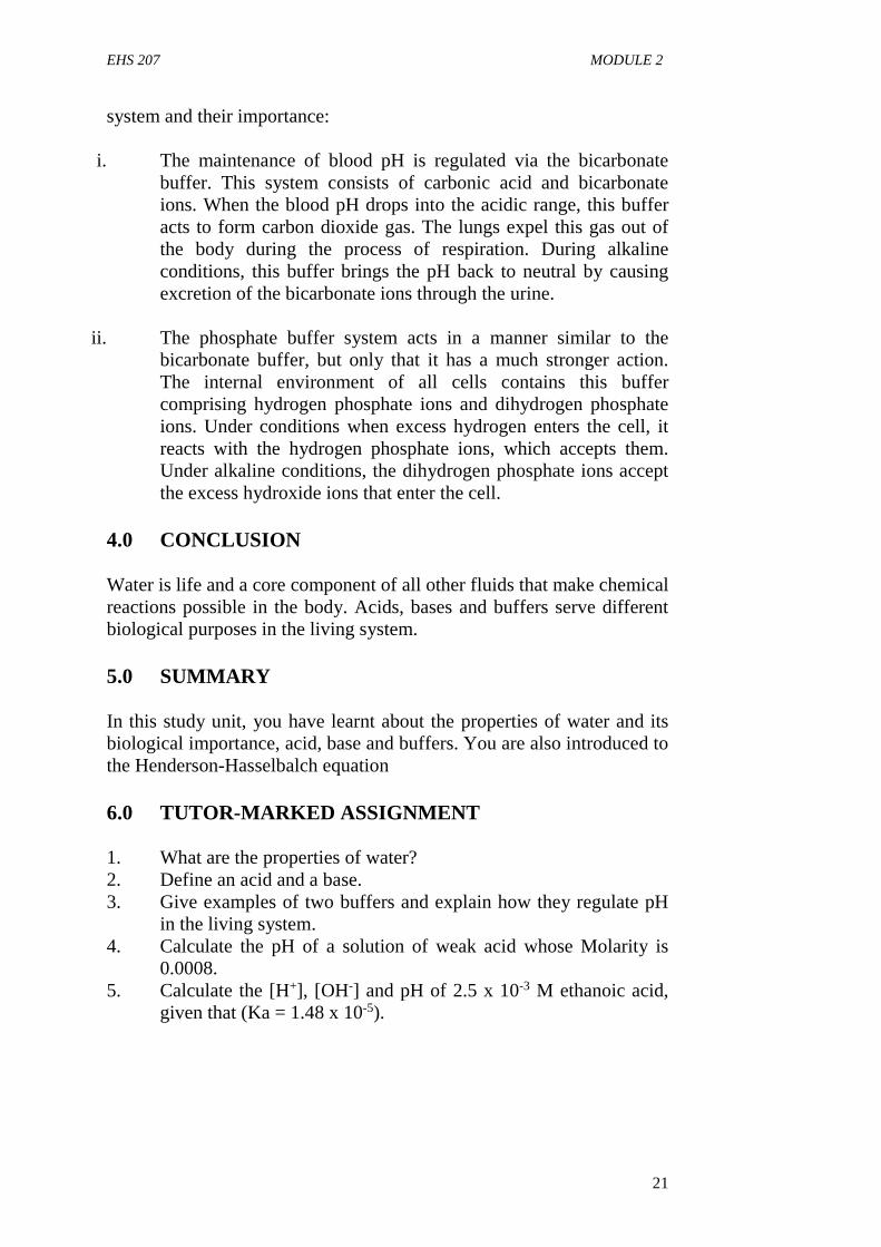
pentoses, hexoses or heptoses, depending on the number of carbon (C)atoms, and as aldoses and ketoses depending on whether they have analdehyde or ketone group. Examples of monosaccharaides hexoses areglucose and galactose.
Table 2.1: Classification of SugarsClassification based onnumber of Carbonatom
Aldoses Ketoses
Trioses (C3H6O3) Glyceraldehyde Dihydroxyacetone
Tetroses (C4H8O4) Erythrose Erythrulose
Pentoses (C5H10O5) Ribose Ribulose
Hexoses (C6H12O6) Glucose Fructose
Heptoses (C7H14O7) - Sedoheptulose
Figure 2.1:Sugars with different no of carbon atomsSource: Nelson and Cox (2010).
Disaccharides: These are condensation products of 2 monosaccharideunits e.g. lactose (galactose + glucose), maltose (2 glucose i.e. glucose +glucose) and sucrose (glucose + fructose).
Oligosaccharides: These are condensation products of 3-10monosaccharides. Most are not digested by human enzymes rather theyplay structural roles.
Polysaccharides: These are condensation products of 10 or moremonosaccharide units.. Food contains a wide variety of otherpolysaccharides, collectively known as non-starch polysaccharideswhich are not digestible by the human enzymes and are the majorcomponents of dietary fibre. Examples include cellulose (a glucose
EHS 207 MODULE 2
25
polymer from plant cell walls) and inulin (a fructose polymer which thestorage CHO in some plants. Examples of polysaccharides are glycogen,starch and dextrin which may be linear or branched polymers
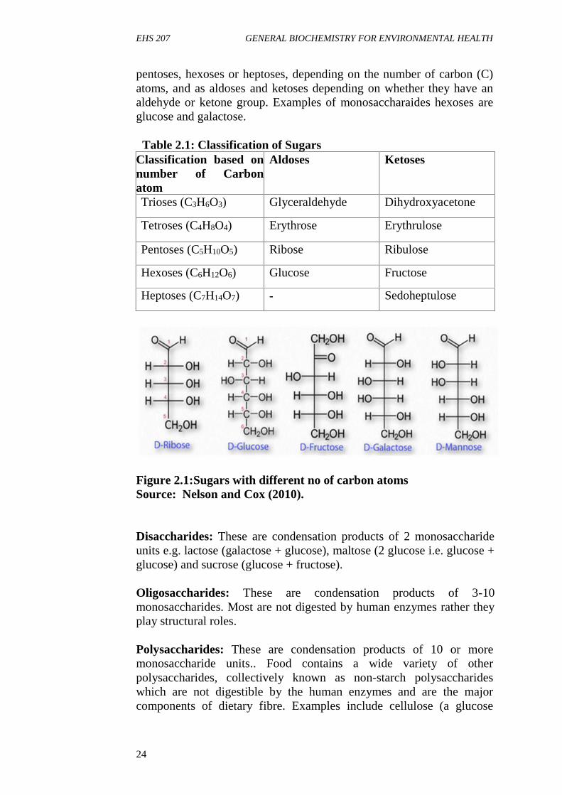
Glycogen: It is known as animal starch. It is made up of α-1, 4 linkagesin the linear and α-1, 6 linkages at the branching points. It is highlybranched. Glycogen is the storage form of energy (glucose) in each andevery cell of the body. Liver and muscle contains the highest amount ofglycogen. At least 5% of glycogen is present in each cell even undersevere fasting/starvation condition. It gives a red colour with iodine.
Starch: It is made up of α-D-glucose units, hence known as glucosan. Itis composed of amylose and amylopectin.
Figure 2.2: Structure of glycogen showing glycosidic linkageSource: Nelson and Cox (2010).
Structure of Glucose
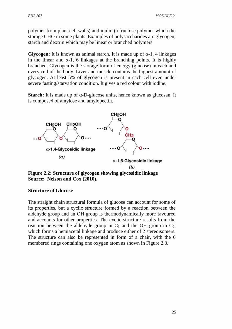
The straight chain structural formula of glucose can account for some ofits properties, but a cyclic structure formed by a reaction between thealdehyde group and an OH group is thermodynamically more favouredand accounts for other properties. The cyclic structure results from thereaction between the aldehyde group in C1 and the OH group in C5,which forms a hemiacetal linkage and produce either of 2 stereoisomers.The structure can also be represented in form of a chair, with the 6membered rings containing one oxygen atom as shown in Figure 2.3.
EHS 207 GENERAL BIOCHEMISTRY FOR ENVIRONMENTAL HEALTH
26
Figure 2.3: Structures of glucoseSource: Nelson and Cox (2010).
3.2 Isomers of Glucose
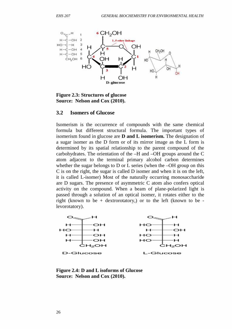
Isomerism is the occurrence of compounds with the same chemicalformula but different structural formula. The important types ofisomerism found in glucose are D and L isomerism. The designation ofa sugar isomer as the D form or of its mirror image as the L form isdetermined by its spatial relationship to the parent compound of thecarbohydrates. The orientation of the –H and –OH groups around the Catom adjacent to the terminal primary alcohol carbon determineswhether the sugar belongs to D or L series (when the –OH group on thisC is on the right, the sugar is called D isomer and when it is on the left,it is called L-isomer) Most of the naturally occurring monosaccharideare D sugars. The presence of asymmetric C atom also confers opticalactivity on the compound. When a beam of plane-polarized light ispassed through a solution of an optical isomer, it rotates either to theright (known to be + dextrorotatory,) or to the left (known to be -levorotatory).
Figure 2.4: D and L isoforms of GlucoseSource: Nelson and Cox (2010).

EHS 207 MODULE 2
27
Epimers: These are isomers differing as a result of variations inconfigurations of the –OH and –H on C atoms 2, 3 and 4 of glucose.The most important biological isomers of glucose are mannose (differsin configuration at C2) and galactose (differs in confriguration at C4).
Figure 2.5: Epimers of GlucoseSource: Nelson and Cox (2010).
3.3 Functions of carbohydrates
1. Provide instant energy to the body: This is the primaryfunction of carbohydrates in the body. Carbohydrates which weconsume as food in the form of starch get digested in the body to releaseglucose. This glucose after being absorbed into blood reaches all thebody tissues and cell. It gets metabolized to release energy in the formof ATP in the presence of oxygen inside the mitochondria.
2. Reserve food: Carbohydrate is also stored as the reserve food inthe body. This is a precautionary measure for the body to cope up intimes of hunger. The excess glucose which is obtained by food isconverted to glycogen in the body. This conversion of glucose toglycogen happens in the presence of the hormone insulin. This glycogenis stored in the liver and to a small extent in the skeletal muscles. Intimes of starvation, this glycogen converts back to glucose and providesenergy.
3. Detoxification of the body by metabolism: Many drugs andtoxic wastes in the body are metabolized for easy excretion in the body.Some of these are water-insoluble and hence they are difficult tobe expelled in urine. The body converts them into glucuronosylconjugates using the glucuronosyl moiety derived from carbohydrates. Acarbohydrate moiety like glucose combines with uronic acid to formglucuronate. These conjugates of insoluble substances with glucuronosylare more water-soluble and easily excreted from the body. Thusdetoxification of physiological importance is carried out to some extentwith carbohydrate derivatives.
EHS 207 GENERAL BIOCHEMISTRY FOR ENVIRONMENTAL HEALTH
28
4. Constitute genetic material: Carbohydrates form a part of DNAand RNA in the form of deoxyribose and ribose sugars. These arefive carbon sugars.
5. They are constituents of all the cellular organelles:Carbohydrates are also components of cell organelles like the cellmembrane, mitochondria, nucleus, endoplasmic reticulum, etc.They provide structural integrity, mechanical strength incombination with proteins and lipids. They help make up thebody mass by being included in all the parts of the cell andtissues. For example, in cell membranes, there are twoconstituents, i.e., glycolipid layer and glycoprotein layer. Herethe term “glyco” is a carbohydrate.
6. Transport of oxygen: Glucose is taken by red blood cells whichlack mitochondria and other cell organelles required forproducing energy. The energy in the form of ATP is produced bya non-oxidative pathway (anaerobic glycolysis). This energy thusproduced is necessary for hemoglobin to bind to oxygenmolecules which are transferred from lungs to the differenttissues.
4.0 CONCLUSION
Carbohydrates are one of the important biomolecules that are widelydistributed in plants and animals and they play important structural andmetabolic roles in the living cell.
5.0 SUMMARY
In this unit, you have learnt the various classifications of carbohydrateswith examples, the isomers of glucose and the biological roles ofcarbohydrate.
6.0 TUTOR-MARKED ASSIGNMENT
1. Explain the classification of carbohydrates and give twoexamples each
2. What is isomerism and list the isomers of glucose3. Enumerate the functions of carbohydrates

EHS 207 MODULE 2
29
7.0 REFERENCES/FURTHER READING
Amanullah, M. (2011). Medical Biochemistry and Biotechnology. 1st
edition.
Devlin T.M. (2010) Textbook of Biochemistry with Clinical Correlation7th edition. John Wiley & Sons Inc.
Murray, R.K., Bender, D.A., Botham, K., M., Kennelly, P.J., RodwellV.W. and Well, P.A., (2012). Harper’s IllustratedBiochemistry (29th Edition) McGraw-Hill Medical.
Nelson, D.L. and Cox M.M., (2009). Lehninger Principles ofBiochemistry 4th edition.
Sadler, T.W. (2004). Langman’s Medical Embryology 9th edition.
EHS 207 GENERAL BIOCHEMISTRY FOR ENVIRONMENTAL HEALTH
30
UNIT 3 CHEMISTRY OF AMINO ACIDS ANDPROTEINS
1.0 Introduction2.0 Objectives3.0 Main Content
3.1 Chemical Nature of Amino Acids3.2 The 20 Amino Acids found in Proteins3.3 Classification of Amino Acids3.4 Classification of Proteins3.5 Roles of Protein in Biological Process
4.0 Conclusion5.0 Summary6.0 Tutor-marked Assessment7.0 References/Further reading
1.0 INTRODUCTION
Amino acids are the basic structural units of proteins. Proteins in allspecies from bacteria to humans are made from the same set of twentyamino acids. Amino acids take part in many types of reactions, but themost important of these is the formation of a peptide bond. Thisinvolves the joining of the α- carboxyl group of one amino acid to the α-amino group of another amino acid, with the loss of a water molecule.Amino acids are grouped according to the nature of their side chains.Since amino acids are weak acids, their strength is expressed as pKa(negative log of ionization constant). The net charge on an amino aciddepends on the pKa of its functional groups and the pH of thesurrounding medium.
2.0 OBJECTIVES
At the end of this unit you will have an understanding of the chemicalnature of amino acids, different ways of classifying amino acids andproteins, formation of peptide bonds and the role of proteins inbiological process.
3.0 MAIN CONTENT
3.1 Chemical nature of amino acids
An amino acid consists of amino group, a carboxyl group (-COOH), ahydrogen atom (H) and a distinctive R group bonded to a carbon atom,called the α- carbon. The R group is specific to each amino acid.
EHS 207 MODULE 2
31
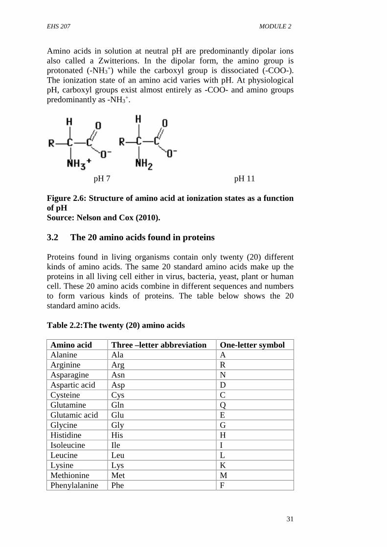
Amino acids in solution at neutral pH are predominantly dipolar ionsalso called a Zwitterions. In the dipolar form, the amino group isprotonated (-NH3
+) while the carboxyl group is dissociated (-COO-).The ionization state of an amino acid varies with pH. At physiologicalpH, carboxyl groups exist almost entirely as -COO- and amino groupspredominantly as -NH3
+.
pH 7 pH 11
Figure 2.6: Structure of amino acid at ionization states as a functionof pHSource: Nelson and Cox (2010).
3.2 The 20 amino acids found in proteins
Proteins found in living organisms contain only twenty (20) differentkinds of amino acids. The same 20 standard amino acids make up theproteins in all living cell either in virus, bacteria, yeast, plant or humancell. These 20 amino acids combine in different sequences and numbersto form various kinds of proteins. The table below shows the 20standard amino acids.
Table 2.2:The twenty (20) amino acids
Amino acid Three –letter abbreviation One-letter symbolAlanine Ala AArginine Arg RAsparagine Asn NAspartic acid Asp DCysteine Cys CGlutamine Gln QGlutamic acid Glu EGlycine Gly GHistidine His HIsoleucine Ile ILeucine Leu LLysine Lys KMethionine Met MPhenylalanine Phe F
EHS 207 GENERAL BIOCHEMISTRY FOR ENVIRONMENTAL HEALTH
32
Proline Pro PSerine Ser SThreonine Thr TTryptophan Trp WTyrosine Tyr YValine Val V
3.3 Classification of amino acids
Amino acids may be classified based on the following:
Classification based on nutritional requirement
1. Essential Amino AcidsThese are amino acids which are not synthesized in the body butmust be provided in the diet to meet the body’s metabolic needs.About ten of the amino acids are grouped under this categoryindicating that mammals require about half of the amino acids intheir diet for growth and maintenance of normal nitrogen balance.Examples are Arginine*, Histidine*, Isoleucine, Leucine, Lysine,Methionine, Phenylalanine, Threonine, Tryptophan, Valine.
Note that *Arginine and *Histidine are also grouped as semi essentialamino acids
2. Non- Essential Amino AcidsThese amino acids are amino acids that need not be providedthrough diet, because they can be biosynthesized in adequateamounts within the organism. Examples are Alanine, Asparagine,Aspartic acid (aspartate), Cysteine, Glutamic Acid (glutamate),Glutamine, Glycine, Proline, Serine, and Tyrosine.
3. Semi-essential Amino AcidsThese are amino acids that can be synthesized within theorganism but their synthesis is not in sufficient amounts, howeverthey should also be provided in the diet. Two amino acids aregrouped under semi-essential amino acids and they are Arginineand Histidine.
Classification based on the fate of each amino acid in mammals
Amino acids can be classified here as Glucogenic (i.e. potentially can beconverted to glucose), ketogenic (i.e. potentially be converted to ketonebodies).
1.
EHS 207 MODULE 2
33
Glucogenic Amino Acids
Glucogenic amino acids are those amino acids in which their carbonskeleton gets degraded to pyruvate, α ketoglutarate, succinyl CoA,fumarate and oxaloacetate and then converted to glucose and glycogen.These include Alanine, Cysteine, Glycine, Arginine, Glutamine,Isoleucine, and Tyrosine.
2. Ketogenic Amino Acids
Those amino acids in which their carbon skeleton is degraded toacetoacetyl CoA, or acetylCoA and then get converted to acetone and β-hydroxy butyrate which are referred to ketone bodies. These includePhenylalanine, Tyrosine, Tryptophan, Isoleucine, Leucine, and Lysine.
These amino acids have the ability to form ketone bodies which isparticularly evident in untreated diabetes mellitus in which largeamounts of ketone bodies are produced by the liver. Degradation ofLeucine which is an exclusively ketogenic amino acid makes asubstantial contribution to the formation of ketone bodies especiallyduring starvation.
Classification depending on the charge
1. Neutral amino acids: These are amino acids that do not containany charge on the ‘R’ group. They are further classified into
Aliphatic amino acids which contains a chain of carbon atomse.g. Glycine, Alanine, Seine, Threonine, Valine, Leucine,Isoleucine, Asparagine, Glutamine
Aromatic amino acid which have an aromatic shape or containsbenzene ring e.g. Phenylalanine, Tyrosine, Tryptophan.
Heterocyclic amino acids which have heterocyclic ring i.e. any ofthe ring structures which contain different atoms.
Sulphur containing which are amino acids which contain Sulphuratom e.g. Cysteine and Methionine.
2. Acidic amino acids: These are amino acids that contain anegative charge or an acidic group e.g. Aspartate, Glutamate
3. Basic amino acids: These contain a positive charge or a basicgroup e.g. Arginine, Lysine and Histidine.
Formation of peptide bondsThe most important reaction of amino acids is the formation of a peptidebond. This involves the joining of the α- carboxyl group of one aminoacid to the α- amino group of another amino acid, with resultant loss of a
EHS 207 GENERAL BIOCHEMISTRY FOR ENVIRONMENTAL HEALTH
34
water molecule. The biosynthesis of peptide bonds requires an input offree energy, whereas their hydrolysis is thermodynamically favorable.Many amino acids (usually > 100) are joined by peptide bonds to form apolypeptide chain.
3.4 Classification of proteins
Proteins are macromolecules with a backbone formed by polymerizationof amino acids. They are nitrogenous compounds of high molecularweight which play a vital or prime role in living organisms.
Proteins may be classified on the basis of their composition, solubility,shape and their biological functions.
Classification based on composition
A. Simple protein: These proteins yield only amino acids duringhydrolysis with no other major organic or inorganic hydrolysisproducts.
B. Conjugated ProteinsThese yields amino acids and other organic and inorganiccomponents e.g. Nucleoprotein (a proteincontaining Nucleic acids), Lipoprotein (a protein containinglipids), Phosphoprotein (a protein containing phosphorous),Metalloprotein (a protein containing metal ions of Fe2+),Glycoprotein (a protein containing carbohydrates)
Classification based on Solubility
a) Albumins: These proteins such as egg albumin and serumalbumin are readily soluble in water and coagulated by heat.
b) Globulins: These proteins are present in serum, muscle and othertissues and are soluble in dilute salt solution but sparingly inwater.
c) Histones: Histones are present in glandular tissues (thymus,pancreas etc.) and are soluble in water. They combine withnucleic acids in cells and on hydrolysis to yield basic aminoacids.
Classification based on Shape
A. Fibrous proteinsThese proteins are made up of several coiled cross-linkedpolypeptide chains. They are insoluble in water and highlyresistant to enzyme digestion. A few sub groups are listed below:
EHS 207 MODULE 2
35
1. Collagens: These are major proteins of the connective tissue.They are insoluble in water, acids or alkalis but are convertible towater-soluble gelatin and are easily digestible by enzymes.
2. Elastins: present in tendons, arteries and other elastic tissues, notconvertible to gelatin.
3. Keratins: these are proteins found in the hair and nails etc.
B. Globular proteins: These are globular or ovoid in shape, solublein water and constitute the enzymes, oxygen carrying proteins,hormones etc.
Classification based on biological functionsProteinsare sometimes described as the "workhorses" of the cell becausethey do so many things like:
act as enzymes e.g. kinases, transaminases etc. act as storage proteins e.g. myoglobin, ferritin act as regulatory proteins e.g. peptide hormones, DNA binding
proteins act as structural protein e.g. collagen, proteoglycan act as protective proteins e.g. blood clotting factors,
Immunoglobins, act as transport protein e.g. hemoglobin, plasma lipoproteins act as contractile or motile proteins e.g. actin, tubulin
3.5 Roles of proteins in biological processes
Proteins play crucial roles in virtually all biological processes. Some ofthese roles include:
Enzymatic catalysis: Nearly all chemical reactions in biologicalsystems are catalyzed by enzymes. Chemical transformations rarelyoccur at perceptible rates in vivo in the absence of enzymes. Mostenzymes are proteins. Thus, proteins play the unique role of determiningthe pattern of chemical transformations in biological systems.
Transport and storage: Many small molecules and ions are transportedby specific proteins e.g. Hb (hemoglobin) transports oxygen inerythrocytes while myoglobin transports oxygen in muscle. Transferrincarries iron in the plasma of blood to the liver where it is stored as acomplex with ferritin, another protein.
Co-ordinated motion: Proteins are the major components of muscle.Muscle contraction is accomplished by the sliding motion of two kindsof protein filaments. On the microscopic scale, coordinated motion suchas the movement of chromosomes in mitosis and the propulsion of
EHS 207 GENERAL BIOCHEMISTRY FOR ENVIRONMENTAL HEALTH
36
sperm by their flagella are also produced by contractile assembliesconsisting of proteins.
Mechanical support: The high tensile strength of skin and bone is dueto the presence of collage, a fibrous protein.
Immune protection: Antibodies are highly specific proteins thatrecognize and combine with foreign substances such as viruses, bacteriaand cells from other organisms.
Generation and transmission of nerve impulses: The response ofnerve cells to specific stimuli is mediated by receptor proteins e.g.rhodopsin is the photoreceptor protein in retinal rod cells.
Control of Growth and Differentiation: Controlled sequentialexpression of genetic information is essential for the orderly growth anddifferentiation of cells. Repressor proteins are important controlelements that silence specific segments of the DNA of a cell. Nervegrowth factor, a protein complex serves to guide the formation of neutralnetworks in higher organisms.
4.0 CONCLUSION
Proteins are nitrogenous organic compounds of high molecular weightwhich play a primary role in living organisms. They are polymers ofamino acids and are essential nutrients for tissue growth, repairs andreplacement.
5.0 SUMMARY
In this unit you have learnt about the chemical nature of amino acids,different classification of amino acids and proteins, formation of peptidebonds and the role of proteins in biological system.
6.0 TUTOR-MARKED ASSIGNMENT
1. Describe the chemical nature of amino acids.2. Identify the 20 amino acids which make up proteins.3. Explain the formation of peptide bond.4. State the biological role of protein in the living system.
EHS 207 MODULE 2
37
7.0 REFERENCES/FURTHER READING
Amanullah, M. (2011). Medical Biochemistry and Biotechnology 1st
edition.
Devlin T. M. (2010). Textbook of Biochemistry with ClinicalCorrelation 7th Edition. John Wiley & Sons Inc.
Murray, R. K., Bender, D. A., Botham, K., M., Kennelly, P. J., RodwellV.W. and Well, P. A. (2012). Harper’s IllustratedBiochemistry (29th Edition) McGraw-Hill Medical.
Nelson, D.L. and Cox M.M., (2009). Lehninger Principles ofBiochemistry 4th edition.
EHS 207 GENERAL BIOCHEMISTRY FOR ENVIRONMENTAL HEALTH
38
UNIT 4 CHEMISTRY OF LIPIDS
1.0 Introduction2.0 Objectives3.0 Main Content
3.1 Fatty Acids3.2 Classification of Lipids3.3 General Functions of Lipids
4.0 Conclusion5.0 Summary6.0 Tutor-marked Assignment7.0 Reference/further reading
1.0 INTRODUCTION
The word lipid was derived from the Greek word lipos meaning fats.Lipids are heterogeneous group of compounds related either directly orindirectly to fatty acids. They are insoluble in water but soluble in non-polar organic solvents such us benzene, chloroform, and ether. They arepresent in all living organisms.
2.0 OBJECTIVES
At the end of this unit, you will learn about fatty acids, classification oflipids and the biological functions of lipids.
3.0 MAIN CONTENT
3.1 Fatty Acids
Fatty acids (FA) are the monocarboxylic acids with a long hydrocarbonchain. The minimum number of carbon atoms required to be called asfatty acid is 4. They are two types of fatty acids namely saturated andfatty unsaturated.
1. Saturated fatty acids are the fatty acids without double bondsi.e. they contain only single bonds along the length of the carbonchain.
Name of fatty acids Number of carbon atomsButyric acid 4Lauric acid 12Myristic acid 14Palmitic acid 16Stearic acid 18

EHS 207 MODULE 2
39
Arachidic acid 20
All these fatty acids are solids at room temperature.
2. Unsaturated fatty acids are the FA that contains one or moredouble bonds along the length of the hydrocarbon chain. Most naturallyoccurring unsaturated FAs have a cis-configuration i.e. the hydrogenatoms are on the same side of the chain. To represent the position of thedouble bond in a fatty acid, it is represented as Delta n (∆), where nshows the position of double bond between the nth carbon atom and thecarbon atom next to it towards the omega (last) carbon atom. Unsaturatedfatty acids are all liquids at room temperature. They can be furtherclassified depending on the number of double bonds present per fattyacid into:
(a). Monounsaturated Fatty Acids (MUFA): They contain only onedouble bond per fatty acid. Oleicacid is the most abundantmonounsaturated FA in nature with C18 atoms and cis ∆9 (cis ∆9 meansposition of the double bond). Palmitoleic acid is another example ofMUFA, and it is present nearly in all fats. Palmitoleic has C16 atomsand cis ∆9 (cis ∆9 means position of the double bond)
Name of fatty acid Number of carbonatoms
Position of thedouble bond
Palmitoleic acid 16 cis ∆9
Oleic acid 18 cis ∆9
(b). Polyunsaturated fatty acids (PUFA)– These are the FAsobtained from plant seeds. They usually contain 2 or more double bonds.In PUFA, double bonds are usually separated by a methylene (CH2)group. PUFA are present in oils such as soya bean oil, groundnut oil,sunflower, benne-seed oil etc. Examples of PUFA are linoleic, linolenicand arachidonic acids; they are also called omega 6 and omega 3 fattyacid respectively. These two PUFA are also referred to as essentialPUFA because animals cannot synthesize them, therefore they must besupplied to the body in the diet.
Name of fattyacid
Number ofcarbon atoms
Number ofdouble bonds
Position of thedouble bond
Linoleic acid 18 2 cis ∆9,12
Linolenic acid 18 3 cis ∆9,12,15
Arachidonicacid
20 4 cis ∆5,8,11,14
EHS 207 GENERAL BIOCHEMISTRY FOR ENVIRONMENTAL HEALTH
40
3.2 Classification of Lipids
Lipids can be classified into the following:
1. Simple lipids: - These are esters of fatty acids with differentalcohols. They are further classified as:
Neutral Fats: - These are esters of fatty acids with glycerol. They areknown as triacylglycerols (TAG) or triglycerides.
Waxes: - Esters of fatty acids with high molecular weight monohydricalcohols
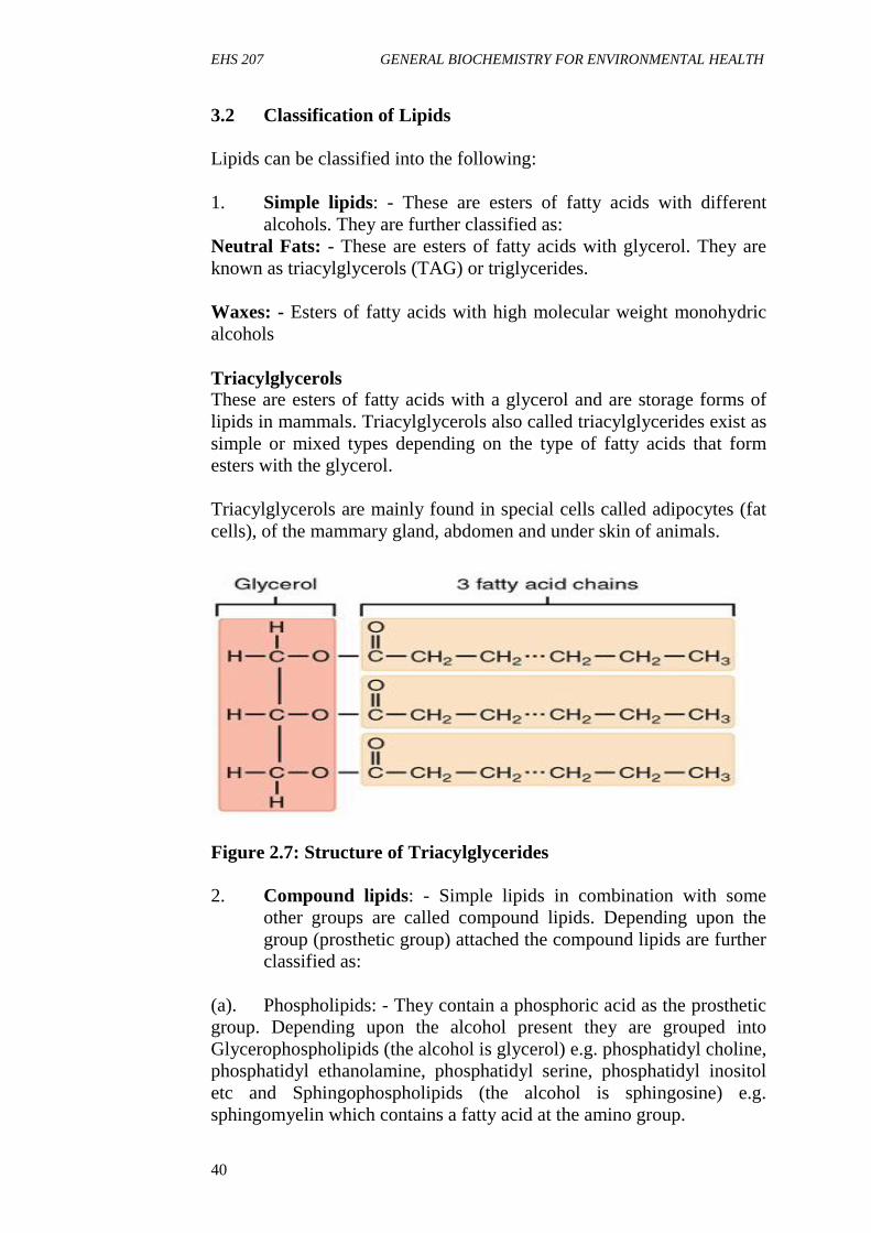
TriacylglycerolsThese are esters of fatty acids with a glycerol and are storage forms oflipids in mammals. Triacylglycerols also called triacylglycerides exist assimple or mixed types depending on the type of fatty acids that formesters with the glycerol.
Triacylglycerols are mainly found in special cells called adipocytes (fatcells), of the mammary gland, abdomen and under skin of animals.
Figure 2.7: Structure of Triacylglycerides
2. Compound lipids: - Simple lipids in combination with someother groups are called compound lipids. Depending upon thegroup (prosthetic group) attached the compound lipids are furtherclassified as:
(a). Phospholipids: - They contain a phosphoric acid as the prostheticgroup. Depending upon the alcohol present they are grouped intoGlycerophospholipids (the alcohol is glycerol) e.g. phosphatidyl choline,phosphatidyl ethanolamine, phosphatidyl serine, phosphatidyl inositoletc and Sphingophospholipids (the alcohol is sphingosine) e.g.sphingomyelin which contains a fatty acid at the amino group.
EHS 207 MODULE 2
41
(b). Glycolipids: - These lipids contain a fatty acid, sphingosine andcarbohydrate residues. They are also known as cerebroside.
3. Derived lipids: Derived lipids are the hydrolytic products of thesimple and compound lipids. They include fatty acids andsteroids.
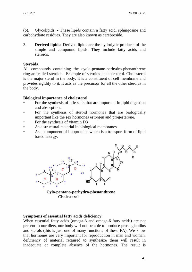
SteroidsAll compounds containing the cyclo-pentano-perhydro-phenanthrenering are called steroids. Example of steroids is cholesterol. Cholesterolis the major sterol in the body. It is a constituent of cell membrane andprovides rigidity to it. It acts as the precursor for all the other steroids inthe body.
Biological importance of cholesterol• For the synthesis of bile salts that are important in lipid digestion
and absorption.• For the synthesis of steroid hormones that are biologically
important like the sex hormones estrogen and progesterone.• For the synthesis of vitamin D3• As a structural material in biological membranes.• As a component of lipoproteins which is a transport form of lipid
based energy.
Cylo-pentano-perhydro-phenanthreneCholesterol
Symptoms of essential fatty acids deficiencyWhen essential fatty acids (omega-3 and omega-6 fatty acids) are notpresent in our diets, our body will not be able to produce prostaglandinsand sterols (this is just one of many functions of these FA). We knowthat hormones are very important for reproduction in man and woman,deficiency of material required to synthesize them will result ininadequate or complete absence of the hormones. The result is
EHS 207 GENERAL BIOCHEMISTRY FOR ENVIRONMENTAL HEALTH
42
infertility. So, foods that are rich in essential fatty acids are good for ourhealth, examples of such foods are fish, poultry products, fruits,vegetables, nuts (especially walnuts), legumes (especially soya beans),beef etc. The symptoms of essential fatty acids deficiency are:
i. Growth retardationii. Poor wound healingiii. Dermatitis and hair lossiv. Kidney and liver diseasesv. Infertilityvi. Depression
The most noticeable symptoms of essential fatty acids deficiency areskin disorders such as scaly dermatitis. It usually occur on the hands,shoulders, forearms and face however it can show up on other parts ofthe body. When essential fatty acids are included in the diets, thesesymptoms disappear within 7 days.
3.3 General functions of lipids
i. They serve as efficient energy sources: Lipids provide energyhigher amount of energy for the body than carbohydrates andprotein.
ii. They are structural components of the cell membrane: Cellmembranes are made up of lipids such as phospholipids andcholesterol that gives cell stability.
iii. They assist in digestion: Lipids dissolve fat soluble vitamins (Vit.A, D, E, K) and therefore help in their digestion.
iv. They occur as free fatty acids in the plasma where they act astransporter of various biological molecules, especially plasmaalbumin.
iv. They serve as thermal insulators.v. Lipids serve as precursors for hormones especially steroid
hormones.
4.0 CONCLUSION
Lipids are efficient energy sources for the body and also one of thestructural components of the plasma membrane. Deficiency of essentialfatty acids which are a class of lipid in our body give rise to symptomslike growth retardation, poor wound healing, dermatitis and hair loss etc.
5.0 SUMMARY
In this unit, you have learnt about fatty acids, classification of lipids andthe symptoms of fatty acid deficiency.

EHS 207 MODULE 2
43
6.0 TUTOR-MARKED ASSIGNMENT
1. List the classes of lipids and give 2 examples in each2. Enumerate the biological functions of lipids3. Differentiate between saturated and unsaturated fatty acids, give
two examples of each4. Enumerate the symptoms of fatty acids deficiency
7.0 REFERENCES/FURTHER READING
Amanullah, M. (2011). Medical Biochemistry and Biotechnology 1st
edition.
Devlin T. M. (2010). Textbook of Biochemistry with ClinicalCorrelation 7th Edition. John Wiley & Sons Inc.
Murray, R. K., Bender, D. A., Botham, K., M., Kennelly, P. J., RodwellV.W. and Well, P. A. (2012). Harper’s IllustratedBiochemistry (29th Edition) McGraw-Hill Medical.
Nelson, D.L. and Cox M.M., (2009). Lehninger Principles ofBiochemistry 4th edition.
EHS 207 GENERAL BIOCHEMISTRY FOR ENVIRONMENTAL HEALTH
44
UNIT 5 CHEMISTRY OF NUCLEIC ACID
1.0 Introduction2.0 Objectives3.0 Main Content
3.1 Nucleic acids4.0 Conclusion5.0 Summary6.0 Tutor-marked Assignment7.0 Reference/Further Reading
1.0 INTRODUCTION
Nucleic acids are acidic compounds first discovered in the cell nucleus.Later they were found in the cytoplasm. They are high molecular weightnitrogenous organic compounds which play an important role in storage,transmission and control of all the cellular activities. They are defined aspolynucleotides i.e. chain-like polymers of up to thousands of nucleotideunits. Each nucleotide is a molecular complex of nucleoside andphosphoric acid.
2.0 OBJECTIVES
On the completion of this unit you will get to understand the chemicalcomponents of nucleic acids, the structure of the DNA and RNA.
3.0 MAIN CONTENT
3.1 Nucleic acids
The term nucleic acid is the overall name for DNA i.e. deoxyribonucleicacid and RNA i.e. ribonucleic acid. They are composed of nucleotides,which are the monomers made of three components: a 5-carbon sugar, aphosphate group and a nitrogenous base.
Nucleic acids are the most important macromolecules in all living thingsbecause it is the genetic material responsible for the transfer of geneticinformation from one generation of organisms to another. The traits weshare with our parents are due to the genes inherited from them. This isnot limited to physical appearance, colour of skin, size of eye balls,intelligence etc.
Nucleic acids are present in nucleus, mitochondria and chloroplast inplant. They are found in two basic structural forms, deoxyribonucleicacid (DNA) and ribonucleic acid (RNA).

EHS 207 MODULE 2
45
The structure of DNADeoxyribonucleic acid (DNA) is the principal informationalmacromolecule of the cell which stores, translates and transfers thegenetic information. In prokaryotes, the DNA is found mostly in thenuclear zone. In eukaryotes it is found in the nucleus, mitochondria andchloroplast. The structure of the DNA is based on the discovery ofWatson and Creek in 1953.
Double helical structure of DNA as proposed by Watson and CrickModel
The three dimensional structure of DNA as proposed by Watson andCrick and the recent advances in it are summarized below:
1. DNA is made of two helical chains coiled around the same axis,to form a right-handed double helix.
2. The two chains in the helix are anti-parallel to each other i.e. the5’-end of the polynucleotide chain and the 3’-end of the otherpolynucleotide chain is on the same side and close together.
3. The distance between each turn is 3.6 nm4. There are 10 nucleotides per turn5. The spatial relationship between the two strands creates major
and minor grooves between the two strands. In these groovessome protein interact.
6. The hydrophilic backbones of alternating deoxyribose andnegatively charged phosphate groups are on the outside of thedouble bond helix.
7. The hydrophobic pyrimidine and purine bases are inside thedouble helix, which stabilizes the double helix of the DNA.
8. The double helix is also stabilized by inter-chain hydrogen bondformed between a purine and pyrimidine base.
9. A particular purine base, pairs by hydrogen bonds, only with aparticular pyrimidine base i.e. Adenine (A) pairs with Thymine(T) and Guanine (G) pairs with Cytosine only.
10. Two hydrogen bonds pairs Adenine and Thymine (A = T)whereas three hydrogen bonds pairs Guanine and Cytosine (G =C).
11. The base pairs A = T and (G = C) are known as complementarybase pairs.
12. Due to the presence of complementary base pairing, the twochains of the DNA double helix are complementary to each other.Hence the number of ‘A’ bases are equal to the number of ‘T’bases (or ‘G’ is equal to ‘C’) in a given double stranded DNA.
13. One of the strands in the double helix is known as sense strandi.e. which codes for
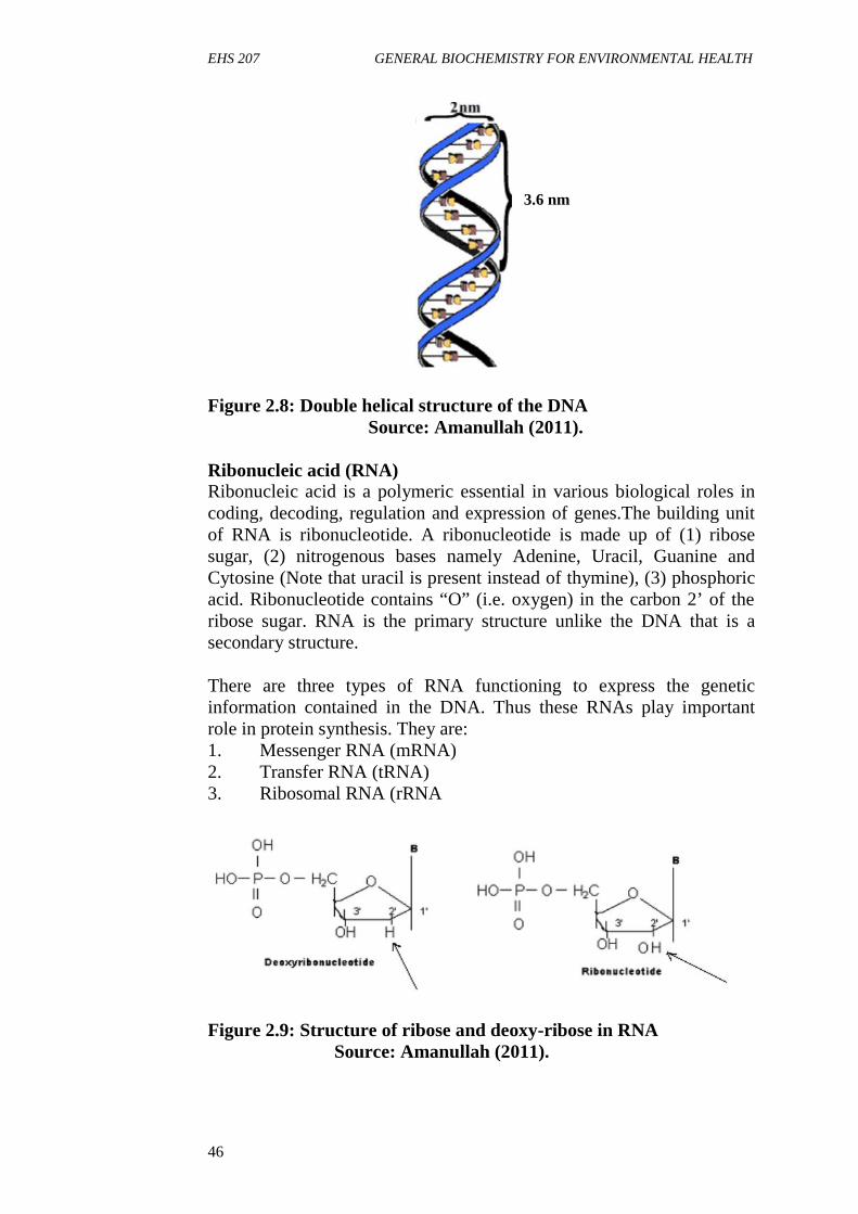
EHS 207 GENERAL BIOCHEMISTRY FOR ENVIRONMENTAL HEALTH
46
Figure 2.8: Double helical structure of the DNASource: Amanullah (2011).
Ribonucleic acid (RNA)Ribonucleic acid is a polymeric essential in various biological roles incoding, decoding, regulation and expression of genes.The building unitof RNA is ribonucleotide. A ribonucleotide is made up of (1) ribosesugar, (2) nitrogenous bases namely Adenine, Uracil, Guanine andCytosine (Note that uracil is present instead of thymine), (3) phosphoricacid. Ribonucleotide contains “O” (i.e. oxygen) in the carbon 2’ of theribose sugar. RNA is the primary structure unlike the DNA that is asecondary structure.
There are three types of RNA functioning to express the geneticinformation contained in the DNA. Thus these RNAs play importantrole in protein synthesis. They are:1. Messenger RNA (mRNA)2. Transfer RNA (tRNA)3. Ribosomal RNA (rRNA
Figure 2.9: Structure of ribose and deoxy-ribose in RNASource: Amanullah (2011).
3.6 nm
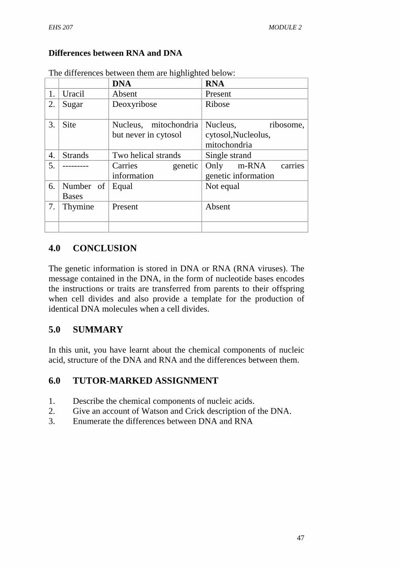
EHS 207 MODULE 2
47
Differences between RNA and DNA
The differences between them are highlighted below:DNA RNA
1. Uracil Absent Present2. Sugar Deoxyribose Ribose
3. Site Nucleus, mitochondriabut never in cytosol
Nucleus, ribosome,cytosol,Nucleolus,mitochondria
4. Strands Two helical strands Single strand5. --------- Carries genetic
informationOnly m-RNA carriesgenetic information
6. Number ofBases
Equal Not equal
7. Thymine Present Absent
4.0 CONCLUSION
The genetic information is stored in DNA or RNA (RNA viruses). Themessage contained in the DNA, in the form of nucleotide bases encodesthe instructions or traits are transferred from parents to their offspringwhen cell divides and also provide a template for the production ofidentical DNA molecules when a cell divides.
5.0 SUMMARY
In this unit, you have learnt about the chemical components of nucleicacid, structure of the DNA and RNA and the differences between them.
6.0 TUTOR-MARKED ASSIGNMENT
1. Describe the chemical components of nucleic acids.2. Give an account of Watson and Crick description of the DNA.3. Enumerate the differences between DNA and RNA
EHS 207 GENERAL BIOCHEMISTRY FOR ENVIRONMENTAL HEALTH
48
7.0 REFERENCES/FURTHER READING
Amanullah, M. (2011). Medical Biochemistry and Biotechnology 1st
edition.
Devlin T. M. (2010). Textbook of Biochemistry with ClinicalCorrelation 7th edition. John Wiley & Sons Inc.
Murray, R. K., Bender, D. A., Botham, K., M., Kennelly, P. J., RodwellV. W. and Well, P.A. (2012). Harper’s IllustratedBiochemistry (29th edition) McGraw-Hill Medical.
Nelson, D. L. and Cox M. M., (2009). Lehninger Principles ofBiochemistry 4th edition.
EHS 207 MODULE 3
49
MODULE 3 METABOLISM OF BIOMOLECULES
Unit 1 Metabolism of CarbohydratesUnit 2 Krebs Cycle and Oxidative PhosphorylationUnit 3 Metabolism of ProteinsUnit 4 Metabolism of Lipids
UNIT 1 METABOLISM OF CARBOHYDRATES
CONTENTS
1.0 Introduction2.0 Objectives3.0 Main Content
3.1 Digestion of Carbohydrates3.2 Absorption of Carbohydrates3.3 Glycolysis3.4 Clinical Conditions Associated with Impaired Glycolysis3.5 Fate of Pyruvate
4.0 Conclusion5.0 Summary6.0 Tutor-Marked Assignment7.0 Reference/Further Reading
1.0 INTRODUCTION
Carbohydrates comprises of the polysaccharides such as starch andglycogen, the disaccharides (lactose, maltose, sucrose) andmonosaccharaides (like glucose, fructose etc). The complexpolysaccharides and disaccharides are converted into simplemonosaccharaides which are absorbed by the body. This unit gives thedetails of the metabolism of carbohydrates.
2.0 OBJECTIVES
By the end of this unit, you will be able to:
3.0 MAIN CONTENT
3.1 Digestion of Carbohydrates
Liquid food materials like milk, soup, fruit juice escape digestion inmouth as they are swallowed, but solid foods are masticated thoroughlybefore they are swallowed to undergo digestion.
EHS 207 GENERAL BIOCHEMISTRY FOR ENVIRONMENTAL HEALTH
50
1. Digestion in MouthDigestion of carbohydrates begins in the mouth, where they come incontact with saliva during mastication. Saliva contains a carbohydratesplitting enzyme called salivary amylase (ptyalin).
Action of ptyalin (salivary amylase)
It is α – amylase that requires chloride (Cl-) ion for activation andoptimum pH 6-7. The enzyme hydrolyzes α- (1, 4) glycosidic linkagefrom molecules like starch, glycogen and dextrin, producing smallermolecules such as maltose and glucose. Ptyalin action stops in stomachwhen pH falls to 3.0. There is no digestion of carbohydrates in thestomach as the enzymes for carbohydrate digestion are absent in thegastric secretions.
2. Digestion in IntestineFood from the stomach reaches the duodenum where it meets thepancreatic juice. The pancreatic juice contains a carbohydrate-splittingenzyme called pancreatic amylase.
a. Pancreatic amylaseIt is also an α – amylase with an optimum pH 7.1. Like salivary ptyalinpancreatic amylase also requires Cl- for its activity. The enzymehydrolyzes α-(1, 4) glycosidic linkage situated in the polysaccharidemolecules thereby liberating free glucose molecules. The pancreaticamylase acts only on α –1, 4 glycosidic linkages, it cannot act on the α –1,6 glycosidic linkages which are present at the branching points ofstarch and glycogen. These undigested branching points are known asisomaltose which are digested by an enzyme called isomaltase or α–dextrinase.
The disaccharides are hydrolyzed by respective disaccharides which aresecreted by the intestinal mucosa. The disaccharide maltose is digestedby the enzyme maltase to yield two glucose units. Lactase splits thedisaccharide lactose into glucose and galactose. Sucrose is hydrolyzedby sucrase or invertase into glucose and fructose.
Cellulase cannot be digested by human beings as the enzyme is absent,yet cellulose is specifically included in the diet so as to increase the bulk(fiber) of the food and thus help in the mobility of the food through thegastrointestinal tract. The end products of carbohydrate digestion areglucose, fructose, mannose, ribose etc. some of the sugars are convertedinto glucose before absorption.
EHS 207 MODULE 3
51
3.2 Absorption of carbohydrates
The monosaccharides are absorbed in the small intestine by threemechanisms- (1) Simple diffusion (2) Active transport and (3)Facilitated transport.
1. Simple diffusion: As the digestion proceeds, the concentration ofglucose in the intestinal lumen increases more than the bloodglucose level. This results in an osmotic difference between thetwo, due to which glucose simply diffuses downhill from a regionof higher concentration of glucose (lumen) to the region of lowerconcentration of glucose (blood).
2. Active transport: Simple diffusion continues till theconcentration of glucose in the lumen equals to that of the blood,then glucose is transported by active transport. Here glucosebinds to a carrier protein situated in the outer membrane of theintestinal wall. This carrier protein also binds two sodium (Na+)ions. When both glucose and Na+ are bound to the carrier proteinit moves into the cell and releases glucose and Na+ into thecytoplasm of the cell, from where glucose simply diffuses intothe blood. To continue the active process Na+ must be expelledout of the cell, so as to maintain a low concentration of Na+insidethe cell, when compared to the lumen. Hence Na+ is expelled outof the cell into the blood plasma, in exchange of K+ through aNa+, K+ ATPase pump, which hydrolyses ATP for exchangingNa+ with K+. As this overall process requires energy it is knownas active transport. So, during active transport, glucose movesagainst concentration gradient. Galactose is also absorbed fromthe intestine in a similar manner as that of glucose activetransport.
3. Facilitated transport: Fructose is transported by a carrierprotein which does not require energy (ATP). Hence it is knownas facilitated transport.
3.3 Glycolysis
Glycolysis is the breakdown of glucose or glycogen to pyruvate orlactate. The glycolytic pathway is also called Embden Meyerhoffpathway. It occurs virtually in all tissues. Erythrocytes and nervoustissues derive their energy mainly form glycolysis. This pathway isunique in the sense that it can utilize O2 if available (‘aerobic’) and itcan function in absence of O2 also (‘anaerobic’). Enzymes involved inglycolysis are present in cytoplasm.
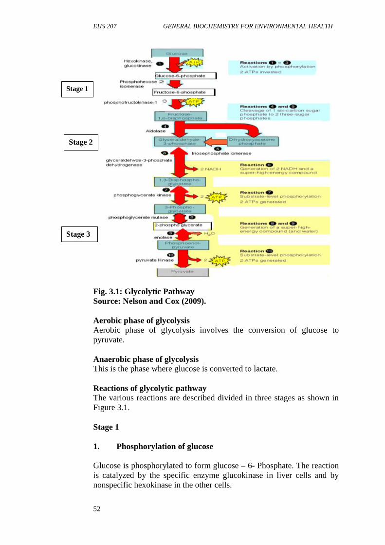
EHS 207 GENERAL BIOCHEMISTRY FOR ENVIRONMENTAL HEALTH
52
Fig. 3.1: Glycolytic PathwaySource: Nelson and Cox (2009).
Aerobic phase of glycolysisAerobic phase of glycolysis involves the conversion of glucose topyruvate.
Anaerobic phase of glycolysisThis is the phase where glucose is converted to lactate.
Reactions of glycolytic pathwayThe various reactions are described divided in three stages as shown inFigure 3.1.
Stage 1
1. Phosphorylation of glucose
Glucose is phosphorylated to form glucose – 6- Phosphate. The reactionis catalyzed by the specific enzyme glucokinase in liver cells and bynonspecific hexokinase in the other cells.
Stage 1
Stage 2
Stage 3

EHS 207 MODULE 3
53
ATP acts as the phosphate (PO4) donor in the presence of Mg. One highenergy PO4 bond is utilized and ADP is produced. The reaction isaccompanied by considerable loss of free energy as heat.
2. Conversion of G-6-phosphate to Fructose-6-phosphate
Glucose-6-phosphate after formation is converted to fructose-6-P byphospho-hexose isomerase.
3. Conversion of Fructose-6-phosphate to Fructose 1, 6-disphosphate
Fructose-6-P is phosphorylated with ATP to form fructose 1, 6-disphosphate catalyzed by the enzyme phosphofructokinase.
Note:Reaction one is irreversible.One ATP is utilized for phosphorylation of glucose at position 6.Phosphofructokinase is the key enzyme in glycolysis that regulates thepathway.
Stage 2This stage involves the cleavage of fructose 1,6-diphosphate into twothree-carbon fragments which are glyceraldehyde-3-phosphate and adihydroxyacetone phosphate catalyzed by aldolase. These resulting threecarbon units are readily interconvertible and are catalyzed byphosphotriose isomerase.
NoteThe reaction is reversible.There is neither expenditure of energy nor formation ATP.
Stage 3In this stage, ATP is harvested when the three carbon-fragments areoxidized to pyruvate. This is an energy-yielding reaction. It consists ofthe following reactions:
(1). Oxidation of Glyceraldehyde-3-phosphate to 1,3-disphosphoglycerate catalyzed by Glyceraldehyde-3-phosphatedehydrogenase.
(2). 1,3- diphosphoglycerate which is the end- product of the previousstage, still retains the PO4 group originally derived from ATP instage 1 and is converted to 3-phosphoglycerate by the enzyme
EHS 207 GENERAL BIOCHEMISTRY FOR ENVIRONMENTAL HEALTH
54
phosphoglycerate kinase which is activated by magnesium ion(Mg2+).
(3). 3-Phosphoglycerate formed in the above reaction is converted to2-phosphoglycerate, catalyzed by the enzyme phosphoglyceratemutase.
(4). 2-phosphoglycerate is converted to phospoenol pyruvate in areaction catalyzed by the enzyme enolase which requires thepresence of either Mg2+ or Mn2+.
(5). Phosphoenol pyruvate is converted to ‘Enol’ pyruvate. Thereaction is catalyzed by the enzyme pyruvate kinase. The highenergy phosphate (PO4) of phosphoenol pyruvate is directlytransferred to ADP producing ATP.
In the absence of oxygen (anaerobic condition), pyruvate is converted tolactic acid by the enzyme lactate dehydrogenase.
Significance of glycolysis1. It is the main route of glucose metabolism.2. It occurs in all cells of the body.3. The glycolytic pathway is meant for provision of energy4. Brain and red blood cells (RBCs) depends only on glucose for
oxidation and production of oxygen5. In the brain aerobic glycolysis occurs whereas in the RBC, there
is always anaerobic glycolysis due to the absence of themitochondria.
6. The initiation of glycolysis is regulated by the ATP concentrationin the cytoplasm.
7. In skeletal muscle, aerobic glycolysis occurs in normal conditionsbut during vigorous muscular contraction, anaerobic glycolysis isthe major pathway for energy production as glycolysis providesATP. In absence of oxygen, muscles can survive under anaerobiccondition.
3.4 Clinical Conditions Associated with Impaired Glycolysis
Lactic acidosisIt occurs as a result of overproduction of lactate, underutilization oflactate or inhibition of pyruvate dehydrogenase. It may also be as aresult of rare congenital disorders where the mitochondria do notfunction at full capacity or diabetic ketoacidosis as well as liver/kidneydisease. It is characterized by Lactate levels > 5 mm/L and serum pH <7.35.

EHS 207 MODULE 3
55
Symptoms: Nausea, Vomiting, Hyperventilation, Irregular heart rate.Pyruvate kinase deficiency
The deficiency of pyruvate kinase causes a rare genetic defect ofglycolysis which results in hemolytic anemia. In this conditionglycolytic intermediates accumulate, whereas pyruvate and lactateconcentrations decrease.
3.5 Fate of Pyruvate
Pyruvate is an important regulatory point for energy production. Theultimate fate of pyruvate depends on the energy state of the cell and thedegree of oxidative phosphorylation taking place. When the energy stateof the cell is low (low ADP, low ATP), pyruvate enters the TCA Cycle(Tricarboxylic cycle) as acetyl CoA via the pyruvate dehydrogenasecomplex and oxidized completely to carbon dioxide and water to yieldenergy. The pyruvate dehydrogenase complex is one of the mostcomplex proteins in the body and consists of more than 60 subunits.
Pyruvate dehydrogenase complex is a multi-enzyme complex made upof three enzymes which are (1). Pyruvate dehydrogenase (2).Dihydrolipoyl transacetylase and (3) Dihydrolipoyl dehydrogenase. Thisreaction requires five coenzymes which are (i) Thiamine pyrophosphate(ii) Lipoic acid (iii) Coenzyme A (iv) Flavin adenine dinucleotide(FAD) and (v) Nicotinamide adenine dinucleotide (NAD+)
4.0 CONCLUSION
Complex carbohydrates (starch) are broken down during digestion intomonosaccharaides unit such as glucose and fructose. Glucose is finallyoxidized into pyruvate in an aerobic condition or lactate in anaerobiccondition both producing different amount of energy in the form ofATP.
EHS 207 GENERAL BIOCHEMISTRY FOR ENVIRONMENTAL HEALTH
56
5.0 SUMMARY
In this unit you have learnt about the digestion and absorption ofcarbohydrates, reactions of glycolytic pathway and the significance ofthe pathway.
6.0 TUTOR-MARKED ASSIGNMENT
1. Explain the process of carbohydrate digestion2. Explain the concept of aerobic and anaerobic glycolysis3. Enumerate the different reactions which make up the pathway
and the enzymes which catalyze these reactions4. Enumerate the significance of the glycolytic pathway5. State the clinical conditions associated with impaired glycolysis
7.0 REFERENCES/FURTHER READING
Amanullah, M. (2011). Medical Biochemistry and Biotechnology. (1st
ed.).
Devlin T. M. (2010). Textbook of Biochemistry with ClinicalCorrelation. (7th ed.). JohnWiley & Sons Inc.
Murray, R. K., Bender, D. A., Botham, K., M., Kennelly, P. J., RodwellV. W. and Well, P.A. (2012). Harper’s IllustratedBiochemistry (29th edition) McGraw-Hill Medical.
Nelson, D. L. and Cox M. M., (2009). Lehninger Principles ofBiochemistry 4th edition.

EHS 207 MODULE 3
57
UNIT 2 KREBS CYCLE AND OXIDATIVEPHOSPHORYLATION
CONTENTS
1.0 Introduction2.0 Objectives3.0 Main Content
3.1 Description of TCA Cycle3.2 Amphibolic Nature of TCA Cycle3.3 Anaplerotic Nature of TCA Cycle3.4 Summary of Oxidative Phosphorylation
4.0 Conclusion5.0 Summary6.0 Tutor-Marked Assignment7.0 References/Further Reading
1.0 INTRODUCTION
The Krebs cycle also known as tricarboxylic cycle or the critic acidcycle is named after the scientist Sir Hans Krebs (1900-1981) whodiscovered it. He proposed the key elements of this pathway in 1937 andwas awarded the Nobel Prize in medicine for the discovery in 1953.Krebs cycle is a set of continuous reactions (8 steps) occurring in acyclic manner in the mitochondrial matrix in eukaryotes and within thecytoplasm in prokaryotes. Acetyl CoA, the fuel of TCA cycle, enters thecycle inside the mitochondrial matrix, and gets oxidized to carbon (IV)oxide (CO2) and H2O while at the same time reducing NAD to NADHand FAD to FADH2. The NADH and FADH2 can be used by theelectron transport chain to create ATP.
2.0 OBJECTIVES
By the end of this unit, you should understand the TCA cycle,amphibolic and anaplerotic nature of TCA cycle, oxidativephosphorylation and inhibitors of the electron transport chain.
3.0 MAIN CONTENT
3.1 Description of Krebs Cycle
In eukaryotes, Krebs cycle takes place in the mitochondria because allthe enzymes of the cycle are located inside the mitochondria matrix. TheTCA cycle is an important source of precursors or building blocks for
EHS 207 GENERAL BIOCHEMISTRY FOR ENVIRONMENTAL HEALTH
58
the synthesis of molecules such as amino acids, purine bases, cholesteroland porphyrins. The following describes the various steps in TCA cycle.
Step 1Condensation: In step 1, the two-carbon compound, acetyl CoAparticipates in a condensation reaction with the four-carbon compound,oxaloacetate to produce citrate, a six-carbon compound catalyzed bythe enzyme citrate synthase. This is the first stable TCA in the cycleand hence the name TCA cycle.
Step 2Isomerisation of citrate: Step 2 involves moving the hydroxyl group inthe citrate molecule so that it can later form α-keto acid. This processinvolves a sequential dehydration and hydration reaction, to form the D-isocitrate isomer (with the hydroxyl group now in the desired α-location), with cis-aconitase as the intermediate. A single enzyme,aconitase performs this two-step process.
Step 3Generation of CO2 and NAD+ linked enzyme: Oxidativedecarboxylation takes place in the next reaction. The reaction iscatalyzed by the enzyme isocitrate dehydrogenase. The reactioninvolves dehydrogenation to oxalosuccinate, an unstable intermediatewhich spontaneously decarboxylates to give α-ketoglutarate. Inaddition to decarboxylation, this step produces a reduced nicotinamideadenine dinucleotide phosphate (NADH) cofactor.
Step 4A second oxidative decarboxylation step: This step is performed by amulti-enzyme complex, the α-ketoglutarate dehydrogenationcomplex. The multi-step reaction performed by the α-ketoglutaratedehydrogenation complex is analogous to the pyruvate dehydrogenasecomplex, i.e. an α-keto acid undergoes oxidative decarboxylation withformation of an acetyl CoA i.e. succinyl CoA.
Step 5Substrate level phosphorylation: Succinyl CoA is a high potentialenergy molecule. The energy stored in this molecule is used to form ahigh energy phosphate bond in a guanine nucleotide diphosphate (GDP)molecule. Most of the GTP (Guanine triphosphate) formed is used in theformation of ATP, by the action of nucleoside diphosphate.
Step 6Flavin dependent dehydrogenation: The succinate produced bysuccinyl CoA-synthetase in the prior reaction needs to be converted to
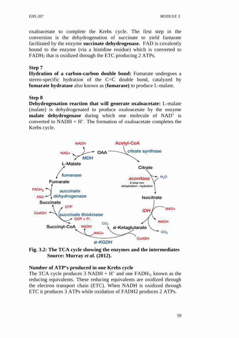
EHS 207 MODULE 3
59
oxaloacetate to complete the Krebs cycle. The first step in theconversion is the dehydrogenation of succinate to yield fumaratefacilitated by the enzyme succinate dehydrogenase. FAD is covalentlybound to the enzyme (via a histidine residue) which is converted toFADH2 that is oxidized through the ETC producing 2 ATPs.
Step 7Hydration of a carbon-carbon double bond: Fumarate undergoes astereo-specific hydration of the C=C double bond, catalyzed byfumarate hydratase also known as (fumarase) to produce L-malate.
Step 8Dehydrogenation reaction that will generate oxaloacetate: L-malate(malate) is dehydrogenated to produce oxaloacetate by the enzymemalate dehydrogenase during which one molecule of NAD+ isconverted to NADH + H+. The formation of oxaloacetate completes theKrebs cycle.
Fig. 3.2: The TCA cycle showing the enzymes and the intermediatesSource: Murray et al. (2012).
Number of ATP’s produced in one Krebs cycleThe TCA cycle produces 3 NADH + H+ and one FADH2, known as thereducing equivalents. These reducing equivalents are oxidized throughthe electron transport chain (ETC). When NADH is oxidized throughETC it produces 3 ATPs while oxidation of FADH2 produces 2 ATPs.
EHS 207 GENERAL BIOCHEMISTRY FOR ENVIRONMENTAL HEALTH
60
Calculation of ATPs produced in each round of citric acid cycle
Enzymeinvolved
Equivalentproduced
No. of ATPsproduced
The step inTCA
Isocitratedehydrogenase
NADH 3 Step 3
α-ketogluteratedehydrogenase
NADH 3 Step 4
Succinatethiokinase
GTP 1 Step 5
Succinatedehydrogenase
FADH2 2 Step 6
Malatedehydrogenase
NADH 3 Step 8
Total ATP produced 12 ATPs
3.2 Amphibolic role of TCA cycle
TCA cycle is the common pathway for the oxidation of carbohydrates,fats and proteins (catabolic role). The anabolic role is synthesis ofvarious carbohydrates, amino acids and fats. As its takes part both inanabolism (synthesis of large organic molecules from smaller organicmolecules which involves the utilization of energy) and catabolism(breakdown of larger organic molecules to smaller organic moleculesaccompanied with the release of energy), it is said to be amphibolicpathway of metabolism.
3.3 The Anaplerotic nature of the TCA Cycle
Anaplerosis is the replacement of the depleted intermediates of TCAcycle. As the TCA cycle takes part in the anabolic reactions, theintermediates of TCA cycle are utilized for the synthesis of variouscompounds. This results in the deficiency of one or more of the TCAcycle intermediates. In order to continue the TCA cycle, thoseintermediates which are deficient must be filled up by some otherprocess and this process is known as anaplerosis. For exampleoxaloacetate is utilized for the synthesis of the amino acid aspartic acidand oxaloacetate is replaced via anaplerosis by carboxylation ofpyruvate to oxaloacetate by the enzyme pyruvate carboxykinase asshown below.

EHS 207 MODULE 3
61
3.4 Summary of Oxidative Phosphorylation
The reduced coenzymes (NADH) and FADH2 from the TCA cycle arethemselves oxidized when they released their protons and electrons. Theelectrons are transferred to oxygen, which is the final electron acceptorthrough a complex chain of electron-carrying molecules known as theelectron transport chain. During the electron transferring process, largeamount of energy is released and it is conserved in the form of ATP.This process is called oxidative phosphorylation as shown below.
ADP + Pi ATP
Inhibitors of electron transport chainCompounds capable of inhibiting the electron transport chain includeAmytal (a barbiturate drug), rotenone (a plant product commonly usedas an insecticide), piericidin A, oligomycin etc.
Piericidin A and oligomycin (antibiotics) block the electron flowthrough the respiratory chain and thereby shut down energy productionin their respective targets.
4.0 CONCLUSION
The Krebs cycle is the cycle that takes place in the mitochondrial matrixof cells and it is responsible for the complete oxidation of acetyl CoA tocarbon (iv) oxide and water.
5.0 SUMMARY
In this unit, you have learnt about the TCA cycle reactions, amphibolicnature of the TCA cycle, anaplerotic nature of the TCA cycle, oxidativephosphorylation and inhibitors of the electron transport chain.
EHS 207 GENERAL BIOCHEMISTRY FOR ENVIRONMENTAL HEALTH
62
6.0 TUTOR-MARKED ASSIGNMENT
1. Describe the reactions of TCA cycle in detail2. Explain the amphibolic nature of the TCA cycle3. Explain the anaplerotic nature of the TCA cycle4. Give a summary account of oxidative phosphorylation5. List two examples of inhibitors of electron transport chain.
7.0 REFERENCES/FURTHER READING
Amanullah, M. (2011). Medical Biochemistry and Biotechnology 1st
edition.
Devlin T. M. (2010). Textbook of Biochemistry with ClinicalCorrelation 7th edition. JohnWiley & Sons Inc.
Murray, R. K., Bender, D. A., Botham, K., M., Kennelly, P. J., RodwellV. W. and Well, P.A. (2012). Harper’s IllustratedBiochemistry (29th edition) McGraw-Hill Medical.
Nelson, D. L. and Cox M. M., (2009). Lehninger Principles ofBiochemistry 4th edition.

EHS 207 MODULE 3
63
UNIT 3 AMINO ACIDS METABOLISM
CONTENTS
1.0 Introduction2.0 Objectives3.0 Main Content
3.1 Digestion and Absorption of Protein3.2 Catabolism of Amino Acids
4.0 Conclusion5.0 Summary6.0 Tutor-Marked Assignment7.0 References/Further Reading
1.0 INTRODUCTION
Amino acids are produced from the breakdown of proteins catalyzed byenzymes called proteolytic enzymes. Unlike the digestion ofcarbohydrates which begins in the month, the digestion of protein startsin the stomach this is because proteolytic enzymes that degrade proteinsare absent in the salivary secretions.
2.0 OBJECTIVES
On completion of this unit, you should have an understanding of thedigestion and absorption of proteins, catabolism of amino acids and thereactions of urea cycle.
3. 0 MAIN CONTENT
3.1 Digestion and Absorption of Protein
The entry of proteins into the stomach stimulates the gastric mucosa tosecrete a hormone gastrin which in turn stimulates the secretion of HClby the parietal cells of the gastric glands and pepsinogen by the chiefcells.
The HCl thus produced lower the pH of stomach to (pH 1.5 – 2.5) andacts as an antiseptic killing most of the bacteria and other foreign cellsingested along with food. The acid also denatures the protein by makingthem susceptible to hydrolysis by the action other proteolytic enzymescalled proteases.
Proteases are endopeptidases (a group of enzymes) that attack theinternal bonds of proteins and liberate large fragments of peptides. Thenpepsinogen, a zymogen (inactive form of an enzyme) is converted into
EHS 207 GENERAL BIOCHEMISTRY FOR ENVIRONMENTAL HEALTH
64
active pepsin which cleaves the ingested protein at their amino terminusof aromatic amino acids (Phe, Tyr, and Trp.) to produce large peptidefragments and some free amino acids.
Pancreatic Digestion
As the food passes from the stomach to small intestine the low pH of thefood triggers the secretion of the hormone ‘secretin’ into the blood. Itstimulates the pancreas to secrets bicarbonate HCO3 into the smallintestine in order to neutralize HCl. The secretion of HCO3 into theintestine abruptly raises the pH from 2.5 to 7.0 the entry of amino acidsinto the duodenum releases the hormone ‘cholecystokinin’ whichtriggers the release of pancreatic juice (that contains many pancreaticenzymes like trypsinogen, chymotrypsinogen, procarboxypeptidases) bythe exocrine cells of the pancreas. Most of these enzymes are producedas zymogens (inactive enzymes) by the pancreas in order to protect theexocrine cells from being digested.
Subsequent to the entry of trypsinogen into the small intestine it isactivated to trypsin by enterokinase secreted in the intestinal cells.Chymotrypsin is secreted in an inactive form called chemotrypsinogenwhich is activated by trypsin. Carboxypeptidase secreted asprocarboxypeptidase is activated again by trypsin. Also aminopeptidasesecreted as inactive proaminopeptidase is once again activated bytrypsin. It is an exopeptidase that cleaves the amino acids from the freeamino terminal end.Dipeptidase acts only on dipeptides and hydrolyzes it into 2 aminoacids.
3.2 Catabolism of Amino Acids
Amino acids are degradation in two steps: Removal of the α-aminogroup and conversion of the carbon chain to an intermediate of lipid andcarbohydrate metabolism.
Removal of the α-amino group (Nitrogen)Removal of the amino group from amino acids is accomplished byvariety of processes such as transamination, oxidative and non-oxidativedeamination.
TransaminationTransamination is the transfer of amino group from an amino acid to anα-keto acid or other amino acids.
EHS 207 MODULE 3
65
Alanine + α-Ketoglutarate Pyruvate + Glutamate
Oxaloacetate + Glutamate Aspartate + α-ketoglutarate (Urea cycle)
This reaction is catalyzed by aminotransferases that are dependent onthe cofactor pyridoxal phosphate, a derivative of vitamin B6. Theimportance of transamination reaction is to collect the amino groupsfrom different amino acid in the form of L-glutamate. The glutamatemolecules channel amino groups either into biosynthetic pathway or intosequence of reactions to form nitrogenous compounds. Therefore,transamination reactions are particularly important in the eventualremoval of the amino group from amino acids.
The oxidative deamination reaction is catalyzed by glutamatedehydrogenase. This is the major reaction in mammal cells throughwhich ammonia is synthesized. α-ketoglutarate acts as a sink for aminogroups by accepting amino groups from various amino acids, with theresultant formation of glutamate. In mammals, all α- amino acids canundergo transamination, except lysine and threonine. The glutamatedehydrogenase (GDH) in animal tissues occurs in the inner matrix ofmitochondria and will utilize NAD+ or NADP+. GDH system use NADor NADP but not both. The coupling of transamination and deaminationreactions, catalyzed by glutamic acid dehydrogenase account for most ofthe ammonia production in animals. The combined action of aminotransferase and glutamate dehydrogenase is referred to as transdeamination and provide a common route for removing and producingnitrogen.
Nitrogen excretion and the Urea cycle:
Excess amino Nitrogen from amino acids is removed as ammonia,which is toxic to the human body. Some ammonia is excreted in urine,but nearly 90% of it is utilized by the liver to form urea, which is highlysoluble and is passed in to circulation for being excreted by the kidneys.Daily excretion of urea amounts to about 30 g with a protein intake ofnearly 100 g in the food. It is less with lower protein intake. The urea-cycle starts in the mitochondrial matrix of hepatocytes and few of thesteps occur in the cytosol: the cycle spans two cellular compartments.The first amino group to enter the cycle is derived from ammonia insidethe mitochondria. Some ammonia also arrives at the liver via the portalvein from the intestine, when it is produced by bacterial oxidation ofamino acids.
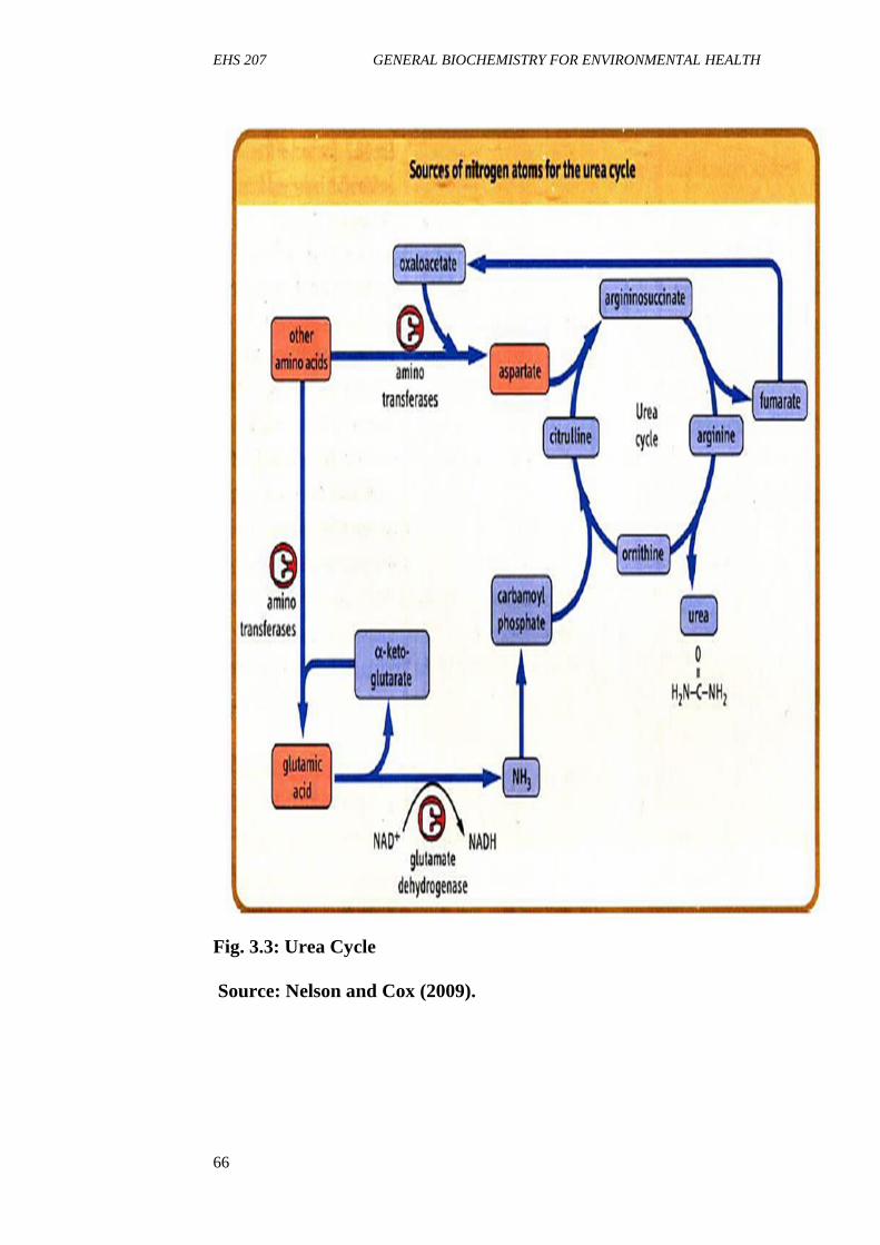
EHS 207 GENERAL BIOCHEMISTRY FOR ENVIRONMENTAL HEALTH
66
Fig. 3.3: Urea Cycle
Source: Nelson and Cox (2009).
EHS 207 MODULE 3
67
The reactions of urea cycle are described below:
Step 1 CO2 from bicarbonate and NH4 combine together in the livermitochondria to form carbamoyl phosphate in presence of ATP andMg2+ by the enzyme Carbamoyl phosphate synthetase I (CPSI).
Step 2 Carbamoyl phosphate reacts with ornithine transferring thecarbamoyl moiety to produce citrulline catalyzed by the enzymeornithine transcarbomylase
Step 3 Argininosuccinic acid is formed by the reaction of Aspartic acidand citrulline: the NH2 group of the argininosuccinic acid is linked to –CO group of the aspartic acid. The enzyme that catalyzed the reaction isargininosuccinic acid synthase.
Step 4 Argininosuccinic acid is cleaved to form Arginine and fumarateby the enzyme arginiosuccinate lyase. Fumarate goes to the pool ofTCA-cycle.
Step 5. Arginine gets cleared off to urea and ornithine by the cytosolicenzyme arginase. Ornithine is thus re-generated and can be transportedin to the mitochondrion to initiate another round of the urea - cycle.
Energetics of the urea cycle
The synthesis of one molecule of urea require four high energyphosphate groups
1. 2 ATPs used up to make up carbamoyl Phosphate2. 1 ATP and two High- energy bonds to make up Arginosuccinate.
4.0 CONCLUSION
Amino acids are degraded by transamination and deamination processes.Transamination of amino acid is catalyzed by transaminases whileoxidative deamination is catalyzed by glutamate dehydrogenase andproduces ammonia which is toxic to the human body. The toxicammonia are eliminated from the body as urea via the urea cycle whichoccurs in the liver and for each molecule of urea produced in the ureacycle, four ATP is required.
5.0 SUMMARY
In this unit you have learnt about the digestion and absorption ofproteins and the catabolism of amino acids and the urea cycle.
EHS 207 GENERAL BIOCHEMISTRY FOR ENVIRONMENTAL HEALTH
68
6.0 TUTOR-MARKED ASSIGNMENT
Discuss:
1. Digestion and absorption of proteins2. Catabolism of amino acids3. Reactions of the urea cycle
7.0 REFERENCES/FURTHER READING
Amanullah, M. (2011). Medical Biochemistry and Biotechnology (1st
ed.).
Devlin T. M. (2010). Textbook of Biochemistry with ClinicalCorrelation 7th edition. John Wiley & Sons Inc.
Murray, R. K., Bender, D. A., Botham, K., M., Kennelly, P. J., RodwellV. W. and Well, P.A. (2012). Harper’s IllustratedBiochemistry (29th edition) McGraw-Hill Medical.
Nelson, D. L. & Cox M. M., (2009). Lehninger Principles ofBiochemistry 4th edition.
EHS 207 MODULE 3
69
UNIT 4 LIPID METABOLISM
CONTENTS
1.0 Introduction2.0 Objectives3.0 Main Content
3.1 Digestion and Absorption of Lipids3.2 β-Oxidation of Fatty Acids
4.0 Conclusion5.0 Summary6.0 Tutor-Marked Assignment7.0 References/Further Reading
1.0 INTRODUCTION
Salivary juice produces a lipid hydrolyzing enzyme, especially in infantsand children, known as lingual lipase. Lipids are not digested in themouth, though lingual lipase is present because the high pH of themouth does not favour the action of lingual lipase and also because thefood remains here for a short period of time.
2.0 OBJECTIVE
By the end of this unit, you will be able to:
discuss digestion and absorption of lipids and β-oxidation of fattyacids.
3.0 MAIN CONTENT
3.1 Digestion and Absorption of Lipids
Digestion of lipids in the stomachAs soon as the lipids enter the stomach, they get liquefied due to heat ofthe stomach. The lipids get emulsified due to the peristaltic movementsof the stomach. The gastric juice contains gastric lipase, which isinactive at low pH of the stomach, whereas lingual lipase is active at hispH and it hydrolysis triacylglycerols of short chain fatty acids (whichare generally found in milk, so digestion in the stomach is seen only inchildren). The released fatty acids are absorbed via the stomach wall andenter the portal vein. The TAG with longer chain fatty acids dissolveand form fat droplets and finally enter the intestine.
EHS 207 GENERAL BIOCHEMISTRY FOR ENVIRONMENTAL HEALTH
70
Digestion of lipids in the intestine
In the intestine, the bile salts (sodium glycocholate and sodiumtaurocholate) and bile acids (cholic acid, chenodeoxycholic acid andcholesterol) help in emulsifying fats thereby making them susceptible tothe digestive enzymes. The presence secretions in the intestine containpancreatic lipase which along with the help of a protein called co-lipaseand lecithin acts on TAG at the water-oil interface.
Phospholipids are hydrolyzed by phospholipase A, B, C and cholesterolis hydrolyzed by cholesterol esterase. The end products of fat digestionare (1) monoacylglycerols (2) diacylglycerols (3) triacylglycerols (4)free fatty acids (5) glycerol.
Digestion of lipids takes place for a longer duration of time. Until andunless digestion of fats has not taken place, other food materials(carbohydrates and proteins) cannot be digested because of fats willcover the food and prevent enzymes reaching the food (hence take afatty meal while going on a long journey). The digested lipids enter theintestinal epithelium by diffusion or by a process called pinocytosis. Inthe intestinal wall the free fatty acids and glycerol re-aggregate to formTAG i.e. here re-synthesis of lipids). These lipids surround a littleamount of protein around them to form chylomicrons which enters intosystemic circulation. The chylomicrons of the circulation move, towardsthe adipose tissue, heart, kidney, liver and skeletal muscle. The capillarywalls of these tissues contain an enzyme called lipoprotein lipase whichhydrolyzes the lipids of the chylomicrons and help in their entry intorespective tissue. The lipids are stored as triacylglycerols mainly in theadipose tissue.
3.2 Beta Oxidation (β-oxidation) of Fatty Acids
Oxidation of the fatty acids at the β-carbon atom to a carboxylic group isknown as β-oxidation. It takes place in the matrix of mitochondria.The steps involved in the β-oxidation of fatty acids as presented inFigure 3.4 are summarized below:
1. Activation of fatty acids: The fatty acids are activated in thecytosol of the cell wherein the enzyme fatty acyl CoA synthasecondenses the fatty acids with coenzyme. A esterification whichfrequires two high energy bonds.
2. Formation of α-β unsaturated acyl-CoA (Enoyl-CoA): The fattyacyl-CoA undergoes dehydrogenation at the α and β-carbonatoms forming trans α-β unsaturated acyl-CoA.
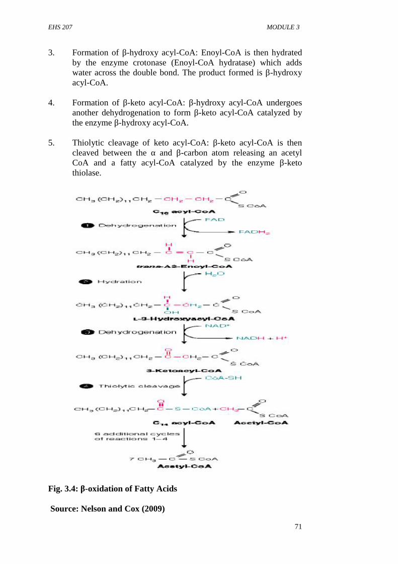
EHS 207 MODULE 3
71
3. Formation of β-hydroxy acyl-CoA: Enoyl-CoA is then hydratedby the enzyme crotonase (Enoyl-CoA hydratase) which addswater across the double bond. The product formed is β-hydroxyacyl-CoA.
4. Formation of β-keto acyl-CoA: β-hydroxy acyl-CoA undergoesanother dehydrogenation to form β-keto acyl-CoA catalyzed bythe enzyme β-hydroxy acyl-CoA.
5. Thiolytic cleavage of keto acyl-CoA: β-keto acyl-CoA is thencleaved between the α and β-carbon atom releasing an acetylCoA and a fatty acyl-CoA catalyzed by the enzyme β-ketothiolase.
Fig. 3.4: β-oxidation of Fatty Acids
Source: Nelson and Cox (2009)

EHS 207 GENERAL BIOCHEMISTRY FOR ENVIRONMENTAL HEALTH
72
4.0 CONCLUSION
Digestion of lipids begins in the stomach and is acted upon by group ofenzymes called lipase which hydrolyze the lipids into triacylglycerol,free fatty acids and glycerol.
5.0 SUMMARY
In this unit, you have learnt about been the digestion and absorption oflipids, metabolism of fatty acids and triacylglycerol and β-oxidation offatty acids.
6.0 TUTOR-MARKED ASSIGNMENT
1. Discuss the digestion and absorption of lipids2. Discuss β-oxidation of fatty acids
7.0 REFERENCES/FURTHER READING
Amanullah, M. (2011). Medical Biochemistry and Biotechnology. (1st
ed.).
Devlin T. M. (2010). Textbook of Biochemistry with ClinicalCorrelation 7th edition. John Wiley & Sons Inc.
Murray, R. K., Bender, D. A., Botham, K., M., Kennelly, P. J., RodwellV. W. and Well, P.A. (2012). Harper’s IllustratedBiochemistry (29th edition) McGraw-Hill Medical.
Nelson, D. L. & Cox M. M., (2009). Lehninger Principles ofBiochemistry. (4th ed.).
EHS 207 MODULE 4
73
MODULE 4 MICRONUTRIENTS (VITAMINS ANDMINERALS) AND DETOXIFICATION
Unit 1 Water Soluble VitaminsUnit 2 Trace ElementsUnit 3 Detoxification
UNIT 1 WATER SOLUBLE VITAMINS
CONTENTS
1.0 Introduction2.0 Objectives3.0 Main Content
3.1 Water Soluble Vitamins3.2 Fat Soluble Vitamins
4.0 Conclusion5.0 Summary6.0 Tutor-Marked Assignment7.0 References/Further Reading
1.0 INTRODUCTION
Vitamins organic compounds which cannot be synthesized in the humanbody and so must be provided in the diet. They are essential for thenormal processes of metabolism, including growth and maintenance ofhealth. It is known that the body is able to produce part or even all of itsrequirements for some of the vitamins. Examples of vitamins such areVitamin D from cholesterol and Niacin (Vit. B3) from tryptophan.
2.0 OBJECTIVES
By the end of this unit, you will be able to: discuss vitamins as micronutrients explain classes of vitamins and their functions state symptoms associated with deficiency of vitamins.
3.0 MAIN CONTENT
3.1 Water Soluble Vitamins
The water soluble vitamins are the Vitamin B complex and Vitamin C.Vitamin B and C share few common properties besides their solubility

EHS 207 GENERAL BIOCHEMISTRY FOR ENVIRONMENTAL HEALTH
74
characteristics. Since they are water soluble excess can be excretedthrough urine. Most of the water soluble vitamins act as coenzymes.
The B- Vitamins are essential and must be provided through diet andthey include Thiamine (Vit B1), Riboflavin (Vit B2), Vit B3 Niacin(Nicotinic acid (or Nicotinamide), Pantothenic acid (Vit B5), Vit B6
(Pyridoxine, pyridoxal, and Pyridoxamine), Vit B9 (Folic Acid), Vit B12
(Cobalamine).
Thiamine (vitamin B1)Thiamineis Vitamin B1. Addition of a pyrophosphate to thiamine (fromATP) converts it to thiamine pyrophosphate, a molecule that is thecoenzyme for all decarboxylation of keto acids.
Sources: The good sources of thamine are seeds, nuts, wheat,leguminous plants (rich source) and lean meat.
Required daily allowance (RDA): Minimum requirement is 1.0 mg foradults, 0.4-1.3 mg for infants and children. Requirement increases inconditions of anoxia-shock, hemorrhage, injury, illness, fever andhyperthyroidism.
Deficiency causes Beriberi and related deficiency syndromes. It ismainly caused by carbohydrate rich diets. In such individuals TPPdependent reactions are prevented, leading to accumulation of substrateslike pyruvate, pentose sugars etc.
Wernicke Korsakoff syndrome which is frequently found in Alcoholicsis associated with thiamin deficiency.
SymptomsSymptoms include Peripheral neuropathy, exhaustion and anorexia. Thesigns may progress to edema and cardiovascular disorders, neurologicaland muscular degeneration.
Riboflavin (Vitamin B2)Riboflavin, also known as vitamin B2, is a component of the flavincoenzymes, FAD (Flavin adenine dinucleotide and FMN (Flavinmonophosphate nucleotide) thereby acting as a co-factor.
Source: meats, nuts, legumes, milk, fish, egg etc.
Required daily allowance (RDA): 1.5-2.5 mg for adults, infants 0.6mg, children 1.0-1.8 mg
EHS 207 MODULE 4
75
Deficiency: Lack of riboflavin in the diet causes non-fatal syndrome ofinflammation of the corner of mouth (angular stomatitis), painfulglossitis of tongue (Purple) and scaly dermatitis.
Niacin (Vitamin B3)Niacin can be synthesized from tryptophan. However, conversion oftryptophan to niacin is relatively inefficient (60 mg of tryptophan isrequired to produce 1 mg of Niacin) and occurs only after all the bodyrequirements for tryptophan is met. Thus most people require dietarysources of both tryptophan and niacin.
Source: Milk, lean meat, unrefined grains, cereals and from metabolismof tryptophan.
Required daily allowance (RDA): Adults 17-21 mg, infants 6 mg. Therequirement increases with increased intake of calories, illness, severeinjury, infection, burns, high corn (maize) diet, pregnancy and lactation.
DeficiencyDeficiency leads to failure of growth, loss of weight and anemia.Pellagra, a disease involving gastrointestinal tract (GIT) and CNS(central nervous system) also develops.
SymptomsThe disease is characterized by intense irritation and inflammation of themucous membranes of the mouth and other parts of the GIT, leading togastro- intestinal hemorrhage, dermatitis, dementia and diarrhea. Skinlesion develops when exposed to sunlight, becomes red, thickened andscaly. The patient develops gingivitis and stomatitis (tongue getsswollen)
Vit B6 (Pyridoxine)This vitamin exists in three forms:Pyridoxine, Pyridoxal andpyridoxamine and their corresponding phosphates.
Sources: Wheat, corn, egg yolk, liver and muscle meat
RDA: 1.4-2.2 mg for adults, children 0.3-0.4 mg. Patients with anti-tubercular treatment needs more Vitamin B6.
DeficiencyThe deficiency is usually not common, but may result due to intake ofdrugs like contraceptives. Alcoholics also suffer from such deficiency.Oral contraceptives stimulate the synthesis of the enzyme which requiresthis vitamin, thus causing deficiency.
EHS 207 GENERAL BIOCHEMISTRY FOR ENVIRONMENTAL HEALTH
76
BiotinBiotin is a vitamin and a coenzyme commonly associated with enzymesperforming carboxylation reactions. Biotinis typically linked covalentlyto carboxylase enzymes through the α-amino nitrogen of lysine.Source: Intestinal bacteria, liver, egg, peanuts and milk.
RDA: 100-200 μg/day. Requirement increase in pregnancy andlactation. Patients on oral antibiotics for a long period of time requiremore of this vitamin.
DeficiencyThe deficiency is rare since it is found in almost all food stuffs. Butlarge consumption of raw egg white may lead to deficiency of Biotin.Avidin, a glycoprotein in egg white binds tightly to biotin and makes itunavailable for the necessary carboxylation reactions.
SymptomsDermatitis, glossitis, Muscle pain, depression, alopecia (loss of hair),loss of appetite and nausea.
Vit B12 (Cobalamin)The metal cobalt in vitamin B12is coordinated with a tetrapyrrole ringsystem, called a corrin ring similar to the porphyrin ring of hemecompounds. The cyanide attached to the cobalt in the structure is anartifact of the isolation and is replaced by water or a hyrdoxyl group incells. The presence of cobalt and amide nitrogens gives B12compoundsthe name cobamides or cobalamins.
Source: Synthesized by microorganisms
RDA: 3 mg/day.
DeficiencyDeficiency of vitamin B12 causes Anemia (a condition where there islow level of red blood cells in the body).
Folic Acid (Vitamin B9)The active form of folic acid is Tetra hydro folate (THF). Chemically,folic acid is formed from three distinct moieties: (1) a bicyclic,heterocyclic pteridine ring, (2) 6-methylpterin (p-aminobenzoic acid,PABA) (3) glutamic acid.
Source: The vitamin is abundant in leafy green vegetables such asspinach, so is named folic acid, from the same root as foliage, wholegrain cereals and Liver.

EHS 207 MODULE 4
77
RDA: 100 μg/day. (The RDA during lactation and pregnancy is 500 –800 μg/day)
DeficiencyThe causes of folate deficiency are inadequate intake, impairedabsorption, increased demand during pregnancy, lactation and impairedmetabolism that leads to megaloblastic anemia. In this conditionproduction of erythrocytes slows down, macrocytic erythrocytes withfragile membrane are formed. Inadequate folate levels during the earlystages of pregnancy increases the risk of neural tube defects (a type ofbirth defect) and spontaneous abortions.
Folate deficiency is common in alcoholics and in people who are ondrugs like anti convulsions and oral contraceptives.
Pantothenic Acid (Vitamin B5) Coenzyme APantothenic acid is a vitamin that forms an essential part of the acyl-carrier moiety known as coenzyme A. Coenzyme A (A for acyl)participates in the activation of all acyl groups in including the acetylgroup derived from pyruvate. The coenzyme is derived metabolicallyfrom ATP, the vitamin pantothenic acid, and mercaptoethylamine.
Sources: Eggs, liver, animal tissue, whole grain cereals, yeast andlegumes
RDA: 4-7 mg/day
DeficiencyThe deficiency is rare due to its wide distribution however the burningfoot syndrome in prisoners which is associated with reduced capacity foracetylation is ascribed to pantothenic acid deficiency.
Vitamin C (Ascorbic Acid)Vitamin Cis a water-soluble vitamin and can act as an antioxidant (i.e.fight/prevent against the oxidation of free radicals that causes cell.Tissue and DNA damage in the body).In general hydroxylation reactions require Vit C. Example:Hydroxylation of cholesterol.
FunctionsVitamin C plays a major role in collagen biosynthesis, degradation oftyrosine, absorption of iron, formation of steroids(Steroidogenesis),adrenaline synthesis, bile acid formation, bone mineralmetabolism and acts as potent antioxidant.
EHS 207 GENERAL BIOCHEMISTRY FOR ENVIRONMENTAL HEALTH
78
White blood cells (WBC) are rich in vit C and play an important role inimmunity.
Source: Citrus fruits, potato, tomato and green vegetablesRDA: 60 mg/day
DeficiencyDeficiency of vitamin C causes scurvy.
SymptomsA symptom of extreme vitamin Cdeficiency called scurvy is theweakening of collagen fibers caused by the failure tohydroxylate prolineand lysine.
Fat soluble VitaminsFat soluble vitamins are vitamins that are not readily absorbed into thebody from diet. The fats soluble vitamins are vitamins A, D, E and K.Ample reserves of fat soluble vitamins are stored in the tissues as theyare not readily absorbed from the food with the exception of Vit. K. Fatsoluble vitamins do not serve as coenzymes.
Vitamin AThe vitamin is present in the diet as retinol or as β-carotene some ofwhich is hydrolyzed in the intestine to form retinol. It is a generic termfor a collection of three forms of Vitamins, retinol, retinal and retinoicacid (Retinoids) all of which are derived from animal and plant sources.The vitamin is stored mainly in the liver.
Source: A rich source is liver, egg yolk, butter and milk but leafyvegetables and some fruits provide the largest amount of β-carotene,
Functionsβ-carotene plays as an antioxidant role and prevents the development ofdiseases like cancer and cardiovascular disease.
Deficiency
Effects on eyeVitamin A affects growth and differentiation of epithelial cells of theleading to defective epitheliazation, (a condition affecting the cornea ofthe eye). It causes formation of abnormal keratin (keratinization) in themucosal cells or cornea of the eye.
Effect on skinThe deficiency causes dryness and roughness of skin developingkeratosis of hair follicles with concomitant deficiency of Vit-B complex.
EHS 207 MODULE 4
79
Effect on bone and teethBone growth is markedly impaired. Osteoclastic activity is alsohampered, causing defective bone formation.Vitamin DVitamin D is the only vitamin that is usually not required in the diet, forthis reason it is rather assumed to function as a hormone.
SourcesSunlight, Fish oils, egg yolk are naturally rich sources of Vit D.
Functionsi. It promotes bone mineralization.ii. It promotes absorption of calcium, phosphates.iii. It enhances the reabsorption of calcium and phosphate by the
kidney.
DeficiencyDeficiency of vitamin D causes rickets (deformation of lower limbs).Rickets is characterized by the production of soft pliable bones due todefective mineralization which is secondary to calcium deficiency.
Usually deficiency of Vitamin D is due to insufficient exposure tosunlight, inadequate dietary intake, GIT disorder, obstructive jaundiceand Partial gastrectomy.
Vit D deficiency is also characterized by low concentration of calciumin blood in association with increased serum alkaline phosphatase.
Vitamin EVitamin E is required in the human diet but deficiency is rare, except inpregnancy and the new born, where it is associated with hemolyticanemia. It exists in the diet as a mixture of eight closely relatedcompounds called Tocopherols.
Source: The richest source is vegetable oils and nuts
FunctionsThe main function of Vitamin E is that it acts as an antioxidant.
DeficiencyVitamin E deficiency is a rare but found in complication of prolongedand severe steatorrhoea, and of prolonged parenteral nutrition.Deficiency of Vitamin E causes anemia. Generally deficiency isinvestigated by measuring plasma (Vitamin E).

EHS 207 GENERAL BIOCHEMISTRY FOR ENVIRONMENTAL HEALTH
80
Vitamin KIt refers to a group of related compounds, varying the number ofisoprenoid units in its side chain. There are three types, Menaquinone(K2) present in animals, Phylloquinone (K1) present in plants. Like vitE, the absorption of Vitamin k is dependent on appropriate fatabsorption.
FunctionsIt is the only one acting as co-enzyme from the group of Fat solublevitamins. This vitamin is also synthesized by intestinal bacteria. It isrequired for post translational modifications of several proteins requiredin the coagulation cascade
DeficiencyIt is widely distributed in nature and produced by the intestinal microflora. Virtually ensures that dietary deficiency does not occur in man.However, it is found in patients suffering from liver diseases(obstructive jaundice), in new born infants and in patients withmalabsorption. It is associated with bleeding disorders. The placenta isinefficient at passing maternal Vit K to the fetus and immediately afterbirth the circulation concentration drops, but recovers on absorption offoods. In addition the gut of the new born is sterile, so that the intestinalmicro flora does not provide a source of vit K for several days afterbirth. This is the reason why adults who are on prolonged antibiotictreatment require supplementation of Vit.E.
Specific inhibitors of vitamin D dependent Carboxylation reactions areused in the treatment of thrombosis related diseases. These drugs of thedicoumarin groups e.g. Warfarin, which inhibit the action of Vit K -probably via the mechanisms involved in the regeneration of the activehydroquinone.
Tests to asses Vitamin K status include the prothrombin time-animportant test in the investigation and management of jaundiced patientsand of those on anticoagulant treatment.
4.0 CONCLUSION
Vitamins are one of the organic molecules required in the body fromdiet for the proper functioning of the body. Some of them act as co-enzymes that enhances biochemical reactions in the living system whilesome act as antioxidants that help prevent the development of diseases.
EHS 207 MODULE 4
81
5.0 SUMMARY
In this unit, you have learnt about the water and fats soluble vitaminsand symptoms of their deficiencies
6.0 TUTOR-MARKED ASSIGNMENT
1. Define water soluble vitamin and list all.2. Enumerate the sources and deficiency of all vitamin B complex.3. State the functions of vitamin A, D and K.
7.0 REFERENCES/FURTHER READING
Amanullah, M. (2011). Medical Biochemistry and Biotechnology. (1st
ed.).
Devlin T. M. (2010). Textbook of Biochemistry with ClinicalCorrelation 7th edition. John Wiley & Sons Inc.
Murray, R. K., Bender, D. A., Botham, K., M., Kennelly, P. J., RodwellV. W. and Well, P.A. (2012). Harper’s IllustratedBiochemistry (29th edition) McGraw-Hill Medical.
Nelson, D. L. & Cox M. M., (2009). Lehninger Principles ofBiochemistry. (4th ed.).
EHS 207 GENERAL BIOCHEMISTRY FOR ENVIRONMENTAL HEALTH
82
UNIT 2 TRACE MINERALS
CONTENTS
1.0 Introduction2.0 Objectives3.0 Main Content
3.1 Macro and Micro Minerals3.2 Trace Elements
4.0 Conclusion5.0 Summary6.0 Tutor-Marked Assignment7.0 References /Further Reading
1.0 INTRODUCTION
Dietary minerals are the chemical elements required by livingorganisms, other than the four elements carbon, hydrogen, nitrogen, andoxygen present in common organic molecules.
For any nutrient, there is a range of intake between that which is clearlyinadequate, leading to clinical deficiency disease, and that which is somuch in excess of the body’s metabolic capacity that there may be signsof toxicity. Between the 2 extremes, there is a level of intake that isadequate for normal health and the maintenance of metabolic integrity.Individuals do not have the same requirement for nutrients, even whencalculated on the basis of body size or energy expenditure. Therefore, inorder to assess the adequacy of diets, it is necessary to set a referencelevel of intake high enough to ensure that no one either suffers fromdeficiency or is at risk of toxicity. Many of the essential minerals arewidely distributed in foods, and most people eating a mixed diet arelikely to receive adequate intakes, although supplements can be usedwhen some requirements are not adequately met by the diet, or whenchronic or acute deficiencies arise from pathology or injury, etc.
2.0 OBJECTIVES
By the end of this unit, you will be able to:
discuss macro and micro minerals explain functions, food sources, required daily allowance,
deficiency and toxicity symptoms of each trace mineral studied.
EHS 207 MODULE 4
83
3.0 MAIN CONTENT
3.1 Macro and Micro Minerals
Dietary Minerals include the macro and micro minerals. Macro mineralsare required in the diet in large amounts (>100 mg/day). They representabout 80% of body organic matter and include calcium, phosphorus,potassium, sulfur, sodium, chlorine, and magnesium.
Micro minerals or Trace elements are needed in doses < 100 mg per day.Important "trace" or minor minerals include iron, cobalt, copper, zinc,molybdenum, iodine, selenium and cobalt.
Some minerals are necessary for the body, but their exact functions areunknown. Such include Chromium, Nickel, Bromine, Lithium andBarium. Non-essential minerals found as contaminants in foodstuffsinclude rubidium, silver, gold and bismuth. Toxic minerals include Al,Pb, Cd and Hg.
3.2 Trace Elements
Zinc
Total zinc content of the body is about 2g, out of which 60% is inskeletal muscles and 30% in bones. The highest concentration of Zinc isseen in Hippocampus area of brain and prostate fluid. More than 300enzymes are zinc-dependent, including RNA and DNA polymerases,alkaline phosphatase and carbonic anhydrase. It also forms what isknown as zinc fingers (Zn2+) co-ordinated to four amino acid sidechains), which provide structural stability to many proteins and areimportant for protein-protein interactions. These are found in manysignal transduction proteins. Zn is also involved in DNA and proteinsynthesis as well as transport of vitamin A, taste perception, woundhealing, Zinc plays a vital role in fertility. In males, it protects theprostate gland from infection (prostates) and ultimately fromenlargement (prostatic hypertrophy). It also helps maintain sperm countand mobility and normal levels of serum testosterone. Zinc is importantduring pregnancy, for the growing foetus whose cells are rapidlydividing. Zinc also helps to avoid congenital abnormalities and pre-termdelivery. Zinc is vital in ensuring proper growth and development ininfants, children and teenagers.
Sources: Meat, shellfish, poultry, whole grains, vegetable, cheese
Required Daily Allowance (RDA): 11 mg for men, 8 mg for women
EHS 207 GENERAL BIOCHEMISTRY FOR ENVIRONMENTAL HEALTH
84
Deficiency: Zn deficiency in children is marked by poor growth andimpairment of sexual development. In both children and adults, it resultsin poor wound healing and dermatitis as well as impaired immunefunction. Toxicity effects include loss of appetite, impaired immunity,decreased HDL, iron and copper deficiencies. Toxicity is common inwelders due to inhalation of zinc oxide fumes.
IronTotal body content is 3-5 g, 75% of this is found in blood and the rest inliver, bone marrow and muscles. Iron is present in almost all cells.Blood contains 14.5g of Hb per 100 ml. About 75% of total iron is inhemoglobin, 5% is in myoglobin and 15% in ferritin. Iron carriesoxygen as part of haemoglobin in blood or myoglobin in muscles. It isrequired for cellular energy metabolism. Transferrin is the transportform while Ferritin is the storage form of iron. Transferrin is aglycoprotein, with a mol wt of 76,500 Daltons. Total iron bindingcapacity (TIBC) is a measure of the blood’s capacity to bind iron withtransferring. The ref range is about 400mg/100ml. One third of thiscapacity is saturated with iron. Transferrin has a half-life of 7-19 days,and is a useful index of nutritional status. One molecule of transferrincan bind two ferric ions. In blood, ceruloplasmin is the ferroxidase,which oxidizes ferrous to ferric state. Transferrin receptors are presenton most of the body cells, especially on cells which synthesize heme.The iron-transferrin complex is taken up by the body cells by thereceptor mechanism.
RDA: for men 8 mg, women (19-50 yrs) 18 mg, women > 50 yrs 8 mg.
Sources: Red meat, fish, poultry, eggs, dried fruits, leafy vegetables,pulses.
Deficiency: Includes anemia characterized by weakness, fatigue,headache and impaired mental and physical performance. It alsoimpaired immunity and pale skin.
Toxicity leads to GIT distress, infections, fatigue, joint pain, skinpigmentation and organ damage.
Copper
Total body copper is about 100 mg. It is found in muscles, liver, bonemarrow, brain, kidney, heart and hair. It is a required component ofmany redox enzymes. Copper containing enzymes are ceruloplasmin,cytochrome oxidase, tyrosinase, superoxide dismutase and others.Required for iron absorption and incorporation of iron into hemoglobin.
EHS 207 MODULE 4
85
Only about 10% dietary copper is absorbed. Excretion is mainly throughbile.
Sources: Legumes, nuts and seeds, whole grains, organ meats, drinkingwater.
RDA: 900 μg
Deficiency: Results in microcytic normochromic anemia, cardiovasculardiseases, defective cross-linking of connective tissue and hypopigmentation of hair. Toxicity is manifested as diarrohea and blue-greendiscoloration of saliva. Copper poisoning may result in hemolysis,hemoglobinuria, proteinuria and renal failure. Toxicity could occur fromeating acid foods cooked in uncoated copper cookware or exposure toexcess copper in drinking water or other environmental sources. Couldresult from Copper poisoning, or Wilson’s disease Results in vomiting,hematemesis, hypotension, coma, GIT distress, jaundice.
FluorideFluoride is known to prevent caries. In the pits and fissures of premolarand molar teeth, bacterial fermentation of residual food leads to acidproduction. The acid removes enamel and dentine to expose the pulp,leading to inflammation and toothache. Presence of fluoride will resultin a fluoroapatite layer on the enamel, which protects it from decay. Thesafe limit of F is about 1ppm in water (= 1 mg in 1000 ml of water)
RDA: 4 mg for men, 3 mg for women.
Toxicity leads to fluorosis (pitting and discoloration of teeth withalternate areas of osteoporosis, osteosclerosis and brittle bones),intestinal upset, loss of appetite and loss of weight.
SeleniumSelenium (Se) intake depends on the nature of soil in which food cropsare grown. In mammals, glutathione peroxidase is the most important secontaining enzyme. 5”-de-iodinase, which converts thyroxin to T3 alsocontains Se. Selenium concentration in testis is the highest in adulttissue. It is necessary for normal development of spermatozoa. Seleniumalso acts as a non specific intracellular anti oxidant, its action beingcomplementary to that of Vitamin E.
RDA: 50-100 μg/ day.
Deficiency: Causes Liver necrosis, cirrhosis, cardio myopathy andmuscular dystrophy.
EHS 207 GENERAL BIOCHEMISTRY FOR ENVIRONMENTAL HEALTH
86
Se Toxicity is called selenosis. Symptoms include hair loss, falling ofnails, diarrhea, weight loss and garlicky odour in breath.
IodineDietary iodine is efficiently absorbed and transported to the thyroidgland, where it is stored and used for the synthesis of triiodothyronineand thyroxine. These hormones function in regulating the basalmetabolic rate of adults and the growth and development of children.Also functions as antioxidant for extrathyroidal organs such asmammary and salivary glands and for gastric juice production.
Sources: seafood, dairy products, iodized salt RDA: 150 μg
Deficiency: Very common results in an enlargement of the thyroid gland(Goitre)
4.0 CONCLUSION
Minerals are organic molecules which are required in the body for bonemineralization and for proper functioning of the body tissues.
5.0 SUMMARY
In this unit, you have been exposed to Macro and Micro minerals.
6.0 TUTOR-MARKED ASSIGNMENT
1. Distinguish between Macro and Micro minerals (Trace elements)2. List important microminerals (Trace Elements) and enumerate
the functions, food sources, required daily allowance, deficiencyand toxicity symptoms of each trace mineral studied.
7.0 REFERENCES/FURTHER READING
Amanullah, M. (2011). Medical Biochemistry and Biotechnology. (1st
ed.).
Devlin T. M. (2010). Textbook of Biochemistry with ClinicalCorrelation 7th edition. John Wiley & Sons Inc.
Murray, R. K., Bender, D. A., Botham, K., M., Kennelly, P. J., RodwellV. W. and Well, P.A. (2012). Harper’s IllustratedBiochemistry (29th edition) McGraw-Hill Medical.
Nelson, D. L. & Cox M. M., (2009). Lehninger Principles ofBiochemistry. (4th ed.).
EHS 207 MODULE 4
87
UNIT 3 DETOXIFICATION
CONTENTS
1.0 Introduction2.0 Objectives3.0 Main Content
3.1 Mechanism of Detoxification3.2 Detoxification Reactions3.3 Excretion of Detoxified Xenobiotic
4.0 Conclusion5.0 Summary6.0 Tutor Marked Assignment7.0 References/Further Reading
1.0 INTRODUCTION
Humans are exposed to various foreign chemicals (called xenobiotic)from air, nutrition (food additives, preservatives), therapy for disease(drugs) etc. Hence an understanding of the ability of the body to copewith the chemical onslaught at the cellular level is an important aspectof metabolism. Detoxification is a series of biochemical reactions thatconvert converts toxic substances into less harmful products and theninto more easily excretable forms. Detoxification is often referred to asdrug metabolism. All the detoxification reactions majorly occur in theliver, however, the kidney and intestines are also involved to a lesserextent. Several unwanted and harmful substances e.g. drugs get entryinto the body by absorption from the gastrointestinal tract and so need tobe eliminated regularly to prevent accumulation and prolongedcumulative action.
Compounds that are detoxified are foreign compounds or xenobioticsuch as drugs, food additives, insecticides, pollutants etc, compoundsproduced in the body which are to be eliminated e.g. bilirubin, steroidsammonia etc and compounds produced in the intestine by bacterialputrefaction and fermentation e.g. indole and skatole (from tryptophan),histamine (from histidine), tyramine (from tyrosine) etc.
EHS 207 GENERAL BIOCHEMISTRY FOR ENVIRONMENTAL HEALTH
88
2.0 OBJECTIVE
By the end of this unit, you will be able to:
discuss detoxification, mechanism and reactions of detoxification,and the excretion of xenobiotic.
3.0 MAIN CONTENT
3.1 Mechanism of Detoxification
The overall mechanism of detoxification is to increase the watersolubility (polarity) of toxic products and thus facilitate their excretionfrom the body mostly in urine and also through bile and faeces. Toxicproducts are rendered water-soluble by adding polar functionalgroups/conjugates.
Toxic compounds / products (less polar)
Addition of polar functionalgroups/conjugates
Less toxic compounds(more polar, water soluble, easily excretable forms)
Excreted through urine / bile / faces
Fig. 4.1: Mechanism of Detoxification
3.2 Reactions of Detoxification
Foreign substances undergo some preliminary changes which are notdifferent from the metabolic changes occurring to physiologicalsubstances. Detoxification reactions are grouped into two phases namelyPhase-I and Phase-II detoxification reactions.

EHS 207 MODULE 4
89
Phase I
Phase I detoxification reaction involves oxidation (also known ashydroxylation), reduction or hydrolysis. Minor reactions likedeamination, desulfuration, dealkylation etc. are also induced under
Phase I
a. Detoxification by oxidation: In this reaction hydrocarbons areoxidized to their corresponding alcohols; alcohols to aldehydesand aldehydes to acids. Also aromatic hydrocarbons are reducedto phenols.Example is the oxidation of methanol to formic acid, oxidation ofbenzene to benzoic acid.Cytochrome P450 (hemeproteins) are enzymes involved inoxidation reaction and are located in the liver microsomes(endoplasmic reticulum). Fifty percent (50%) of drugs aredetoxified through this system.
Methanol (CH3-OH) Formic acid (H-COOH)
Benzoyl Benzoic acid
b. Detoxification by reduction: This reaction involves the additionof hydrogen atom to hydrocarbons. Examples are given below:
Nitrobenzene Amino benzene (Aniline)
Chloral Trichloroethanol(Chloral is a sedative)
c. Detoxification by hydrolysis: This reaction involves thehydrolysis of ester, amide, glycosidic bond, ether bond etc by theaddition of water. Also Drugs like procaine and acetyl salicylic(aspirin) acid and cardiac glycosides e.g. digitalin, atropineundergo hydrolysis.
H2O
Aspirin salicylic acid + acetic acid
H2O
Atropine tropic acid + tropine
EHS 207 GENERAL BIOCHEMISTRY FOR ENVIRONMENTAL HEALTH
90
Phase-II
Phase-II detoxification process consists of conjugation reaction. Thisreaction involves the coupling of the foreign substances after theprocesses of oxidation, reduction and hydrolysis with some polarsubstances in the body making the toxic compounds more water solubleand hence easily excretable. The following are some examples:
a. Glucuronic acids: Conjugation with glucuronic acid is the mostcommon conjugation reaction. UDP (uridine diphosphate)-glucuronic acid is the glucuronyl donor, UDP-glucuronyltransferases are the enzymes. Aromatic acids (e.g. benzoic acid)and phenols are conjugated with glucuronic acid. The glucuronicacid is derived from uridine diphosphate glucuronic acid. Thedrug chloramphenicol and the bile pigments are among theimportant substances conjugated with glucuronic acid.Derivatives of steroid hormones also are conjugated withglucuronic acid before excretion.
b. Active sulfate: It is used to conjugate phenolic compounds. Thederivatives are called ethereal sulfates. An increase in theiramount in urine excessive intestinal putrefaction or stasis.Adrenal cortical hormones are also excreted after conjugationwith sulfuric acid. Yeast and mammalian liver contain enzymesthat can activate inorganic sulfate by adding it to 3-phosphoadenosine-5-phosphate. Active sulfate is adenine-3-phosphoribose-5-phosphosulfate. Incorporation of sulfate intosulfated mucopolysaccharaides and conjugation of steroidhormones and others with sulfate are brought about afterpreliminary activation of sulfate.
c. Glycine: This is used to conjugate aromatic acids, cholic acid andalso nicotinic acid. The formation of bile acid (glycocholic acid)from cholic acid is also brought about by conjugation withglycine.
d. Cysteine: This is used in the conjugation of aromatic compoundslike benzene and halogenated ring compounds likebromobenzene.
e. Acetic acid: Acetic acid is used to conjugate with aromaticamino compounds like sulfanilamide.
f. Glutamine: This is conjugated with phenylacetic acid to formphenylacetylglutamine.
g. Methyl groups: Active methionine (s-adenosyl methionine) isused for the conjugation of certain pyridine and otherheterocyclic nitrogen containing compounds like nicotinamide.
h. Interaction of highly toxic cyanides: There may also beconjugation with thiosulfate to form relatively nontoxic

EHS 207 MODULE 4
91
thiocyanates. The enzyme rhodanase converts cyanide tothiocyanate.
3.3 Excretion of Detoxified Xenobiotic
i. The products of phase-I reactions are either excreted directly orundergo further metabolism by phase-II reactions and thenexcreted.
ii. In some cases, a xenobiotic may undergo only phase-II reactionwithout undergoing phase-I reaction.
iii. Occasionally, a xenobiotic may be excreted unchanged.
4.0 CONCLUSION
Detoxification is the breakdown of toxic substances to less harmfulsubstances. Xenobiotic are detoxified by the process of oxidation,reduction and conjugation.
5.0 SUMMARY
In this unit you have learnt about detoxification, mechanism ofdetoxification and reactions of detoxification.
6.0 TUTOR-MARKED ASSIGNMENT
1. Define detoxification.2. Explain the mechanism of detoxification.3. Write in details with examples reactions involved in oxidation,
reduction, hydrolysis and conjugation.
7.0 REFERENCES/FURTHER READING
Amanullah, M. (2011). Medical Biochemistry and Biotechnology. (1st
ed.).
Devlin T. M. (2010). Textbook of Biochemistry with ClinicalCorrelation 7th edition. John Wiley & Sons Inc.
Murray, R. K., Bender, D. A., Botham, K., M., Kennelly, P. J., RodwellV. W. and Well, P.A. (2012). Harper’s IllustratedBiochemistry (29th edition) McGraw-Hill Medical.
Nelson, D. L. & Cox M. M., (2009). Lehninger Principles ofBiochemistry. (4th ed.).