Embed Size (px)
Citation preview

COVER SHEET
This is the author version of article published as: Suzuki, Shuko and Grondahl, Lisbeth and Leavesley, David I. and Wentrup-Byrne, Edeline (2005) In vitro bioactivity of MOEP grafted ePTFE membranes for craniofacial applications. Biomaterials 26(26):pp. 5303-5312. Copyright 2005 Elsevier Accessed from http://eprints.qut.edu.au
In vitro bioactivity of MOEP grafted ePTFE membranes for craniofacial
applications
Shuko Suzuki1, Lisbeth Grøndahl2, David Leavesley1, Edeline Wentrup-Byrne*,1
1: Tissue BioRegeneration and Integration Program, Queensland University of Technology, 2 George Street, GPO Box 2434, Brisbane QLD 4001, Australia
2: Department of Chemistry, The School of Molecular and Microbial Sciences, The University of Queensland, Brisbane QLD 4072, Australia
*Corresponding Author Dr. Edeline Wentrup-Byrne Tissue BioRegeneration and Integration Program Queensland University of Technology 2 George Street, GPO Box 2434 Brisbane QLD 4001 Australia Tel: +61 7 3864 1226 Fax: +61 7 3864 1804 [email protected] Key Words Surface modification Simulated Body Fluid Protein adsorption Osteoblast SaOS-2 cell attachment FTIR spectroscopy

2
Abstract
The bioactivity of three methacryloyloxyethyl phosphate (MOEP) grafted expanded
polytetrafluoroethylene (ePTFE) membranes with varying surface coverage as well as
unmodified ePTFE was investigated through a series of in vitro tests: calcium phosphate
growth in simulated body fluid (SBF), serum protein adsorption, and a morphology and
attachment study of human osteoblast-like SaOS-2 cells. The graft copolymers were
prepared by means of gamma irradiation induced grafting and displayed various surface
morphologies and wettabilities depending on the grafting conditions used. Unmodified
ePTFE did not induce nucleation of calcium phosphate minerals, whereas all the grafted
membranes revealed the growth of calcium phosphate minerals after 7 days immersion in
SBF. The sample with lowest surface grafting yield (24% coverage), a smooth graft
morphology and relatively high hydrophobicity (θadv = 120º, θrec = 80º) showed
carbonated hydroxyapatite growth covering the surface. On the other hand, the samples
with high surface grafting yield (76 and 100%), a globular graft morphology and
hydrophilic surfaces (θadv = 60º and 80º, θrec = 25º and 15º, respectively) exhibited
irregular growth of non-apatitic calcium phosphate minerals. Irreversibly adsorbed
protein measured after a 1 hour immersion in serum solution was quantified by the
amount of nitrogen on the surface using XPS, as well as by weight increase. All grafted
membranes adsorbed 3-6 times more protein than the unmodified membrane. The sample
with the highest surface coverage adsorbed the most protein. Osteoblast-like SaOS-2 cells
cultured for 3 hours revealed significantly higher levels of cell attachment on all grafted
membranes compared to unmodified ePTFE. Although the morphology of the cells was

3
heterogeneous, in general, the higher grafted surfaces showed a much better cell
morphology than both the low surface-grafted and the control unmodified sample.
The suite of in vitro tests confirms that a judicious choice of grafted monomer such as the
phosphate-containing methacrylate monomer (MOEP) significantly improves the
bioactivity of ePTFE in vitro.

4
Introduction
Expanded polytetrafluoroethylene (ePTFE) is widely used in clinical medicine including
facial reconstructive surgery where it is used in facial augmentation. The general
biocompatibility of this material is good, and its microporous structure allows ingrowth
of bone and blood vessels after about six months [1]. However, since the material does
not have functional groups to interact with the cellular environment and is classified as
bioinert, lack of contact with the bone tissue means in vivo fixation of the implant is
delayed. ePTFE neither supports human osteoblast growth (in vitro cell studies) [2] or
induces calcium phosphate growth (simulated body fluid (SBF) for up to 4 weeks) [3].
Negatively charged functional groups on a material surface are capable of inducing
heterogeneous calcium phosphate (CaP) nucleation and growth in SBF [4, 5]. Among
these, phosphate groups are found to have the largest effect [6, 7]. The phosphate
containing monomer, methacryloyloxyethyl phosphate (MOEP) (Figure 1a) has been
grafted onto high density polyethylene (HDPE) and it was shown to increase carbonated
hydroxyapatite growth in vitro [8], as well as to significantly enhance bone-bonding
properties in vivo [8, 9]. In our previous study, the surface of an ePTFE membrane was
successfully grafted with monoacryloxyethyl phosphate (MAEP) (Figure 1b) by means of
radiation induced graft copolymerization. Subsequent investigation of CaP growth
revealed formation of a thick CaP material after immersion in SBF for 7 days when the
external surface grafting coverage was 44% or above [3, 10]. The initial CaP crystals had
a Ca/P ratio of 1.0 and the secondary growth a Ca/P ratio of 1.5 indicating that these CaP
phases are not hydroxyapatite [3]. In a recent study by Stancu et al. it was found that
5
MOEP containing co-polymers induced CaP nucleation in SBF. However, they also
found Ca/P ratios of 1.5-1.6, i.e. less than the theoretical value for hydroxyapatite (Ca/P =
1.67) [11].
Protein adsorption to biomaterial surfaces is affected by a number of surface properties
such as hydrophobicity / hydrophilicity, topography and surface chemistry. Kato et al.
investigated protein adsorption onto surfaces with ionic and non-ionic groups and showed
that protein adsorption onto the charged surfaces was governed by the electrostatic
interactions [12]. It has been shown that positively charged surfaces attract different
proteins [13] and show higher osteoblast cell attachment to that of negatively charged
surfaces[14]. The morphology of the cells also differs as observed by electron
microscopy. Cells were flattened and highly extended on positive surfaces but
maintained compact morphology and were anchored through discrete focal attachments
on negative ones [13]. The area between cells and the membrane is considered important
for osteoblast secretion of extracellular matrix directly onto the underlying substrate.
Interaction between osteoblasts and a MOEP grafted polyethylenetelephthalate (PET)
substrate has been investigated by transmission electron microscopy (TEM) and showed
similar cell morphology and deposition of extracellular matrix after 1 week of cultivation
[15]. A calcium phosphate layer, thought to be formed by both cell-dependent and cell-
independent mechanisms, was observed after 2 weeks.
Although several studies combine the in vitro tests used to assess the bioactivity of
biomaterials (SBF, protein adsorption or osteoblast cell culture studies) eg bioglasses [16,
6
17] and their composites [18-20] combine SBF and osteoblast assays, very few studies
make use of all these tests [21]. Sometimes SBF is better at differentiating between
similar materials than cell studies. Thus, in the performance of HDPE/Bioglass®
materials in SBF and in human primary osteoblast-like (HOB) cell viability studies both
showed improved outcomes compared to pure HDPE [20]. However, although the rate of
apatite growth in SBF increased with increasing Bioglass® content (0, 20, and 40%), cell
viability did not differ between the 20 and 40% composites. Similarly, HA growth on
poly(D,L-lactic acid) (PDLLA)/Bioglass® composites in SBF correlated with the
composition whereas osteoblast like MG-63 cell attachment showed no difference [18].
In contrast to uncoated PDLLA, Bioglass® coated materials showed enhanced HA
growth whereas HOB cell attachment was unchanged [19]. Although many studies have
been carried out on bioglasses and bioglass materials it is poorly understood how these in
vitro assays correlate when carried out on polymeric materials.
Graft copolymerization of MOEP (Figure 1) onto ePTFE by simultaneous irradiation
technique resulted in materials with various surface graft morphologies and wettabilities
depending on the grafting conditions used [22]. In this study, we investigate the
bioactivity of three types of MOEP grafted membranes with different surface graft
morphologies, surface grafting yields and wettabilities and compare them to that of
unmodified ePTFE. A series of in vitro tests; the growth of CaP in a simulated body
environment, irreversible protein adsorption, and human osteoblast-like SaOS-2 cell
attachment and morphology, was used to investigate the bioactivity of these materials.
7
Experimental
Materials
Sumitomo ePTFE Poreflon® 020-80 membranes were from Sumitomo Electric, Japan.
The preparation of the MOEP grafted ePTFE membranes in methanol and 2-butanone
(also known as methyl ethyl ketone, MEK) used in this study has been described
separately [22]. A third sample was prepared by simultaneous graft copolymerization of
MOEP in a mixed solvent system of methanol, water and dichloromethane (ratio of 2: 2:
1) using 60Co gamma radiation (220 Nordian Gamma-cell; Canada). All solvents were of
analytical grade and MilliQ water was used throughout. The conditions used for
polymeric grafting, the grafting yields (surface and overall) and the water contact angle
results are summarised in Table 1.
SBF Immersion
The simulated body fluid was prepared according to the method described by Kokubo et
al. [23]. Appropriate quantities of NaCl (99.9%), NaHCO3 (99.0%), Na2CO3 (99.9%),
KCl (99.0%), K2HPO4 (99.0%), MgCl2 · 6H2O (99.0%), CaCl2 · 2H2O (99.5%), and
NaSO4 (99.0%) were dissolved in water that had been boiled for one hour prior to
preparation and buffered with HEPES (2-[4-(2-Hydroxyethyl)-1-
piperazinyl]ethanesulfonic acid) (99.9%) and 1M NaOH at pH = 7.4 at 36.5 oC.
Approximately 25 mL of SBF solution was added to a 30 mL polystyrene container
containing a membrane sample either grafted or ungrafted. Samples U and A floated due
to their hydrophobic nature. They were held down in the SBF solution with an inert
plastic mesh. Each sample was processed in duplicate. The vials were immersed in a
8
water bath for a period of seven (7) days at 36.5 + 0.2 oC. The solution was changed at
three-day intervals and the pH was regularly checked. No significant pH change was
observed. After 7 days, the samples were removed from the SBF and washed thoroughly
with ultra-pure water and dried to constant weight.
Total deposition of CaP was measured by the weight increase of the membranes.
Scanning electron microscopy with energy dispersive X-ray
analysis (SEM/EDX) (FEI Quanta 200 Environmental SEM equipped with an Evarhart
Thomley secondary electron detector) was performed at 10 kV to examine the
morphology of the calcium phosphate deposit and to obtain the elemental composition of
the CaP crystals on the surfaces. Fourier transform infrared spectroscopy with attenuated
total reflectance (FTIR-ATR) (Nicolet Fourier Transform Infrared Spectroscopy
equipped with a diamond ATR) was used to analyse the mineral deposits (64 scans, 4 cm-
1 resolution, wave number range 4000 – 525 cm-1). This technique examines the sample
to a depth of approximately 1-2 μm.
Protein Adsorption
Foetal calf serum (Invitrogen, Auckland, NZ) was diluted with ultrapure water, which
had been pre-boiled for one hour, to a final concentration of 10% v/v FCS. Each
membrane was immersed in serum solution (5 mL) in a vial which had previously been
treated with Acrylease (Stratagene, Integrated Sciences, AU), and kept at 36.5 ± 0.2 oC
for one hour. Measurement on duplicate samples was performed. Following treatment,
the membranes were washed twice with ultrapure water and dried under low vacuum to
constant weight.
9
The proteins on the surfaces were analysed by X-ray photoelectron spectroscopy (XPS)
(PHI Model 560 XPS/SAM/SIMSI Multi-technique Surface Analysis System) with a
Model 225-270AR Cylindrical Mirror Analyser (CMA) and MgKα X-ray radiation
source (1253.6 eV). The N1s peaks from the multiplex scan were used as a measure of
the amount of protein adsorbed to the surface.
Cell Culture
The cell line used in this study was the human osteosarcoma-derived SaOS-2 line which
has previously been characterised as consisting of osteoblast-like cells [24]. SaOS-2 cells
were cultured in α-Minimum Essential Medium (α-MEM) (Invitrogen, Auckland, NZ),
supplemented with 10% foetal bovine serum (FBS) (Invitrogen, Auckland, NZ), 1%
penicillin/streptomycin (Invitrogen, Auckland, NZ), and 50 μL of gentamicin reagent
solution (Invitrogen, Auckland, NZ) in a humidified atmosphere of 5% CO2, 95% air at
37oC. The cells were subcultured at 70-80% confluency with 0.25% trypsin/EDTA
following two washes with Hank’s balanced salt solution (HBSS), 2-3 times a week and
the medium changed every second day.
Cell attachment assay
Cell attachment onto individual samples was quantified by detection of the radio-labelled
amino acid [4, 5-3H]-Leucine liberated from attached pre-labelled SaOS-2 cells. For cell
attachment analysis, three flasks of actively growing cells, passages 13-15, were
incubated in growth media containing 2 μCi/mL [4, 5-3H]-Leucine (Amersham, UK) for
18 hours before the assay.
10
The membranes were sterilized in 70% ethanol and air dried. Since the phosphate groups
on the grafted membranes are acidic, each sterilized grafted and control ungrafted
membrane was immersed in approximately 1 mL of α−MEM (serum free) for five
minutes to allow for ion exchange prior to the addition of cells. This procedure was
repeated three times to adjust the pH to 7. Three membranes of each grafting type were
then transferred to individual wells of a 24-well tissue culture polystyrene (TCPS) plate
(Nalge Nunc, Rochester, NY, USA) before drying. The hydrophobic and hydrophilic
membranes were treated differently: 1) Samples A and U (Table 1) floated in the cell
suspension due to their hydrophobic nature, therefore stainless steel holders with silicon
seals were applied. 2) Samples B and C (Table 1) were very hydrophilic and the holders
failed to seal the cell suspension. These membranes were placed in Poly(2-hydroxyethyl
methacrylate) (Sigma) coated wells without holders. TCPS both with and without holders
were used as controls.
Pre-labelled cells were harvested using 0.25% trypsin/EDTA and resuspended in culture
media to a concentration of 2 × 10⁵ cells/mL. 200 μL and 1mL of the cell suspension
were then added to the wells both with and without holders. The cells were incubated at
37oC in 5% CO2, 95% air for 3 hours.
Following incubation, the membranes were rinsed twice with medium to remove non-
adherent cells. Cellular proteins were precipitated using two washes of 5% w/vol
trichloroacetic acid (TCA) (BDH, Poole, England). The precipitated proteins were then
solubilised with 0.5 M NaOH / 0.1% Triton X-100 (Sigma) and triplicated sub-samples
transferred into individual scintillation vials containing 5 mL of “Readysafe” scintillation
cocktail (Beckman, Inc. Fullerton, USA). Radioactivity was determined in a Beckman
11
LS5000TA liquid scintillation counter (Beckman, Inc. Fullerton, USA). Cell-free samples
were also tested to evaluate background radioactivity. Since these results showed no
significant background, they are not included.
Cell morphological analysis
After 3 hours of incubation of SaOS-2 cells on the samples, morphological analysis of
attached cells was performed. A similar method to that used in the radio-assay was
employed except the cells were not pre-labelled. After three hours of attachment, non-
attached cells were removed by washing with growth media and the membranes were
immersed in 3% v/v glutaraldehyde in 0.1 M sodium cacodylate buffer solution (pH 7.3)
overnight to fix the attached cells. They were then dehydrated by progressive treatments
with duplicate ethanol solutions (50%, 70%, 90% and 100% ethanol). Incubation times
were 10 minutes between each treatment. Critical point drying was carried out in a
Denton Vacuum Critical Point Drying Apparatus (Denton Vacuum, Inc., Cherry Hill, NJ,
USA) using CO2. After mounting onto specimen stubs, the samples were gold coated in a
BioRad Gold Sputter Coater (BioRad, Swaziland). The cell morphology was visualized
by SEM at 15.0 kV using a FEI Quanta 200 scanning electron microscope (FEI Company,
Oregon, USA).
Statistical Analysis
Cell attachment data are expressed as the mean + the standard error of the mean for
triplicates. For comparison of the different samples, iner-STAT-a v1.3 software (Mario H

12
Vargas, Mexico) was used to perform Tukey’s test to determine significance between
samples.
13
Results
The in vitro bioactivity of three MOEP grafted ePTFE (sample A-C) and a control
unmodified membrane (sample U) were evaluated using simulated body fluid (SBF),
serum protein adsorption, and attachment of human osteoblast-like SaOS-2 cells. The
three grafted membranes had different grafting yields, graft morphology and wettability
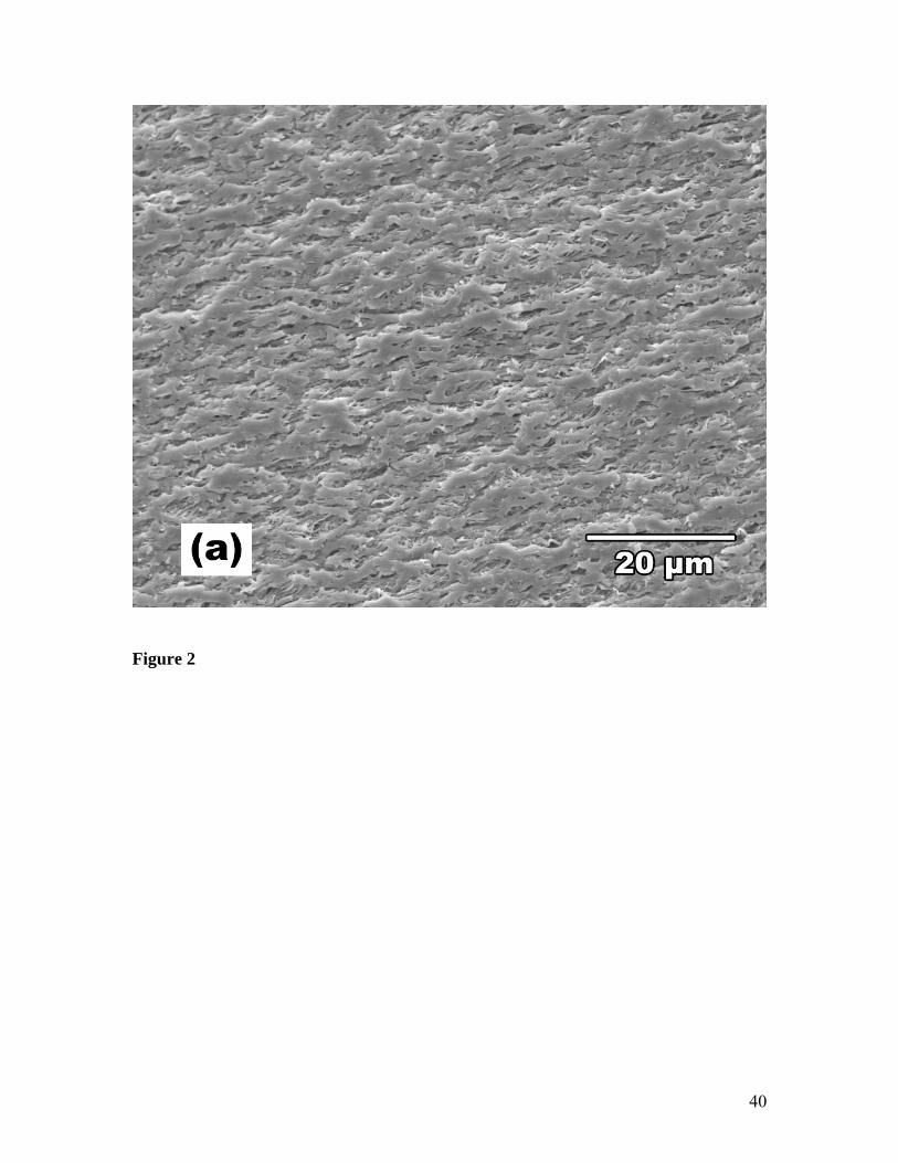
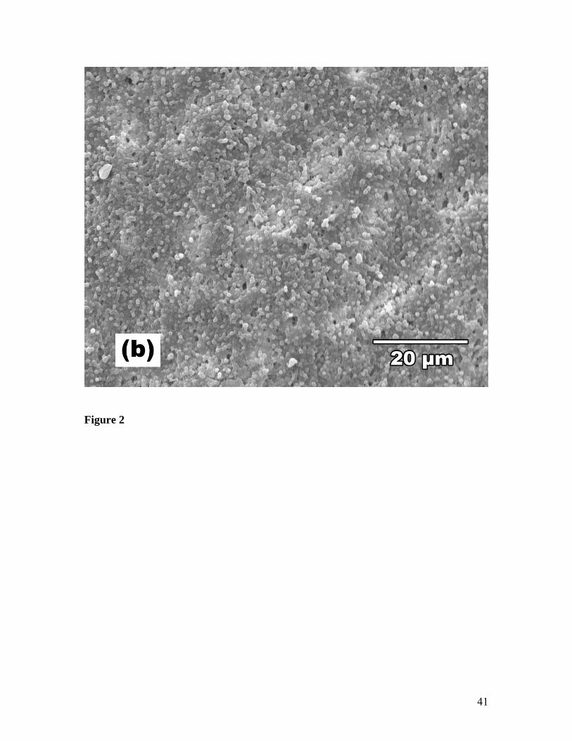
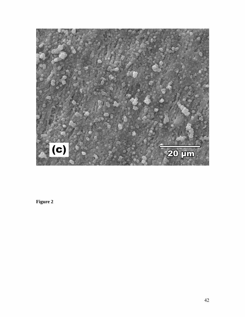
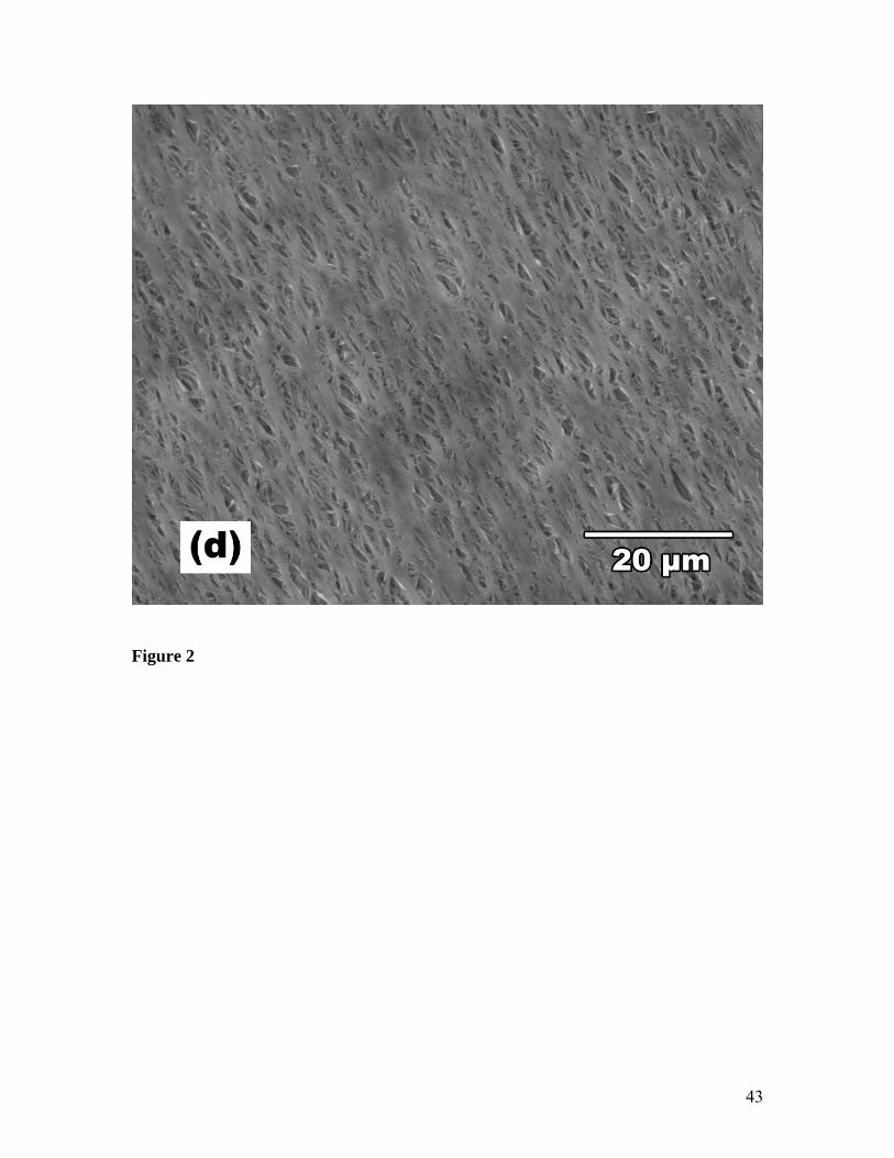
(Table 1 and Figure 2). Sample A grafted in methanol showed smooth but patchy
morphology of the graft co-polymer (Figure 2a). Whereas for the MEK (sample B), a
globular morphology of the graft co-polymer (ø = 1 μm) was obtained (Figure 2b). An
additional sample prepared for this study using a solvent mixture (methanol, water and
DCM) exhibited a higher yield of large globular (ø = 2 μm) graft co-polymers (sample C;
Figure 2c).
SBF Immersion
After 7 days immersion in SBF, all grafted membranes exhibited mineral formation
whereas no mineral was observed on untreated ePTFE. The weight increases of the
membranes after immersion are reported in Table 2. No significant difference between
the grafted samples was observed.
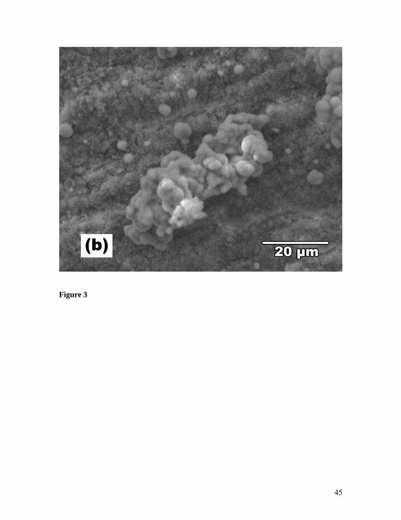
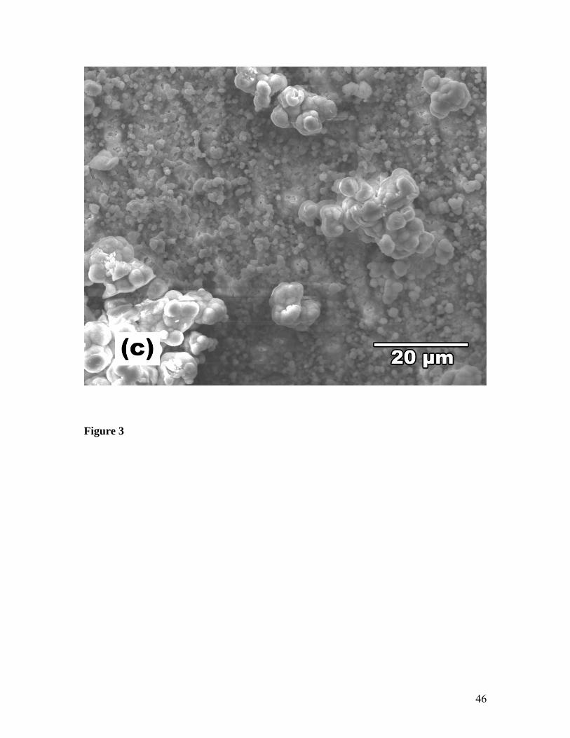
An SEM image of sample A (Figure 3a) shows the presence of round mineral nodules of
various sizes including some large clusters (sphere diameter of ~ 5μm). This spherical
morphology is often observed for apatite formed on materials in vitro [25, 26]. The
mineral formation on samples B and C was patchy with large areas where no mineral was
observed (Figure 3b and 3c). On areas with larger inorganic deposits, the spheres had
different sizes (ø ~ 5μm and ~ 1μm). Since the total amount of mineral growth on the

14
three grafted membranes was the same within experimental error, it is possible that
samples B and C have mineral growing within the pores of the material that is not visible
on the SEM images.
EDX analyses of the minerals revealed the presence of P, Ca, O, C, and minor amounts of
Mg. Since Mg is capable of substituting for Ca in the hydroxyapatite lattice, both the
Ca/P and (Ca + Mg)/P atomic ratios are shown in Table 2. The (Ca + Mg)/P ratio on
sample A was 1.63 which is close to the theoretical value for hydroxyapatite (1.67). On
samples B and C, the values were higher than the theoretical value of apatite (1.89 and
1.72, respectively).
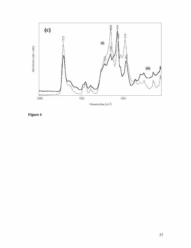
The ATR-FTIR spectrum of untreated ePTFE shows two C-F vibrations at 1201 and
1146 cm-1 (Figure 4ai). The PMOEP grafted samples show additional bands at 1721-
1727 (C=O stretching), ~1066 (P-O-(C) stretching) and ~977 cm-1 (P-O-(H) stretching)
(Figures 4aii, 4bi, and 4ci). Small bands in the region of 1490-1370 cm-1 correspond to
the C-H bending. The intensity of these PMOEP peaks correlate with the surface grafting
yields. Figure 4aiii shows the FTIR spectrum of sample A after SBF treatment. The C-F
peaks at 1204 and 1149 cm-1 are no longer visible in the spectrum indicating that a thick
CaP mineral phase was present on the grafted membrane. A large broad band at 1019
cm-1 was assigned to a phosphate vibration mode of HA. Bands at 1486, 1418 and 872
cm-1 corresponding to carbonate vibrations are also present. The small peak at 1596 cm-1
and the large broad band around 3600 - 2600 cm-1 were assigned to OH peaks from the
bound water. This result indicates that the calcium phosphate mineral formed on sample
A is carbonated HA correlating with the (Ca+Mg)/P ratio of 1.63 observed by EDX.
15
The spectrum of sample B (Figure 4b) after SBF treatment shows overlapping peaks from
the grafted membrane and the inorganic material. However, the C-F peaks at 1204 and
1149 cm-1 are suppressed and distinctive features of phosphate mineral can be observed
at 1013 cm-1. This large phosphate band is broader than that of sample A and a shoulder
at 1069 cm-1 can be observed. This indicates that other forms of CaP have formed in
combination with HA. The peaks at 1480, 1415 and 871 cm-1 are assigned to carbonate
vibrations. Clearly, a mixture of HA and other CaP phases has formed on sample B. This
is supported by the observed EDX (Ca+Mg)/P ratio of 1.89.
After SBF immersion, the spectrum of sample C (Figure 4c) showed large peaks from
both the grafted membrane and CaP mineral. The intensity of the MOEP carbonyl band at
1721 cm-1 is still significant. However, since the C-F peak at 1148 cm-1 decreased
dramatically, the band at 1066 cm-1 is assigned to a CaP mineral rather than PMOEP
peaks. The position of this band is significantly different from that of HA and does not
seem to correlate with other known CaP phases [27]. HA is not detected on sample C,
which could not have been predicted from the EDX (Ca+Mg)/P ratio of 1.72.
These results clearly show that FTIR is an excellent tool for examining the nature of CaP
mineral phases formed in SBF and serves as a complementary technique to EDX.
Although XRD is often the technique of choice for mineral characterization, it is limited
in that it requires crystalline samples. However, the mineral growth formed in SBF is
often highly amorphous [3] or nano-crystalline, in any case giving very poorly resolved
XRD spectra.
16
Protein Adsorption
Protein adsorption tests were conducted by immersing membranes in 10% foetal bovine
serum for 1 hour and washing the membranes to allow only irreversibly bound protein to
remain. Changes in weight of the dried samples are shown in Table 2. The N1s peak in
the XPS spectra was also used as a measure of adsorbed proteins, since grafted samples
without proteins showed no nitrogen in their XPS spectra. A good correlation between
the XPS result and weight increase measurements was found. All grafted membranes
exhibited greater protein adsorption compared to the untreated membrane (sample U).
Sample C with the highest surface grafting yield showed the highest protein adsorption
(Table 2) whereas there was no significant difference in the amount of protein adsorbed
on samples A and B.
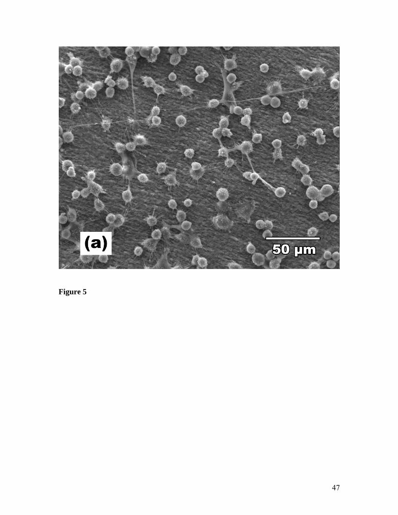
Cell Attachment Assay
Cell attachment relative to the control (TCPS) is shown in Table 3. The extent of short-
term cell attachment onto the three grafted membranes was significantly greater (p< 0.01)
to that on untreated ePTFE. However, there was no statistical difference between the
three grafted membranes.
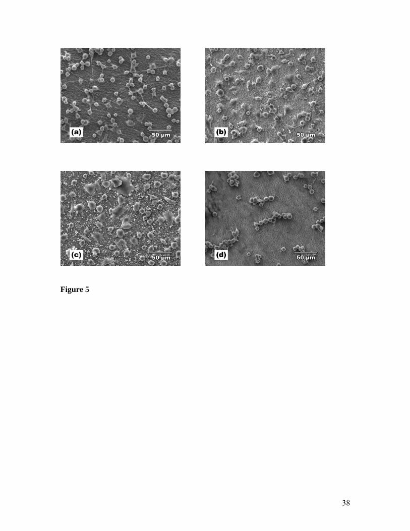
Cell morphology was evaluated using SEM. The same time point was used as in the cell
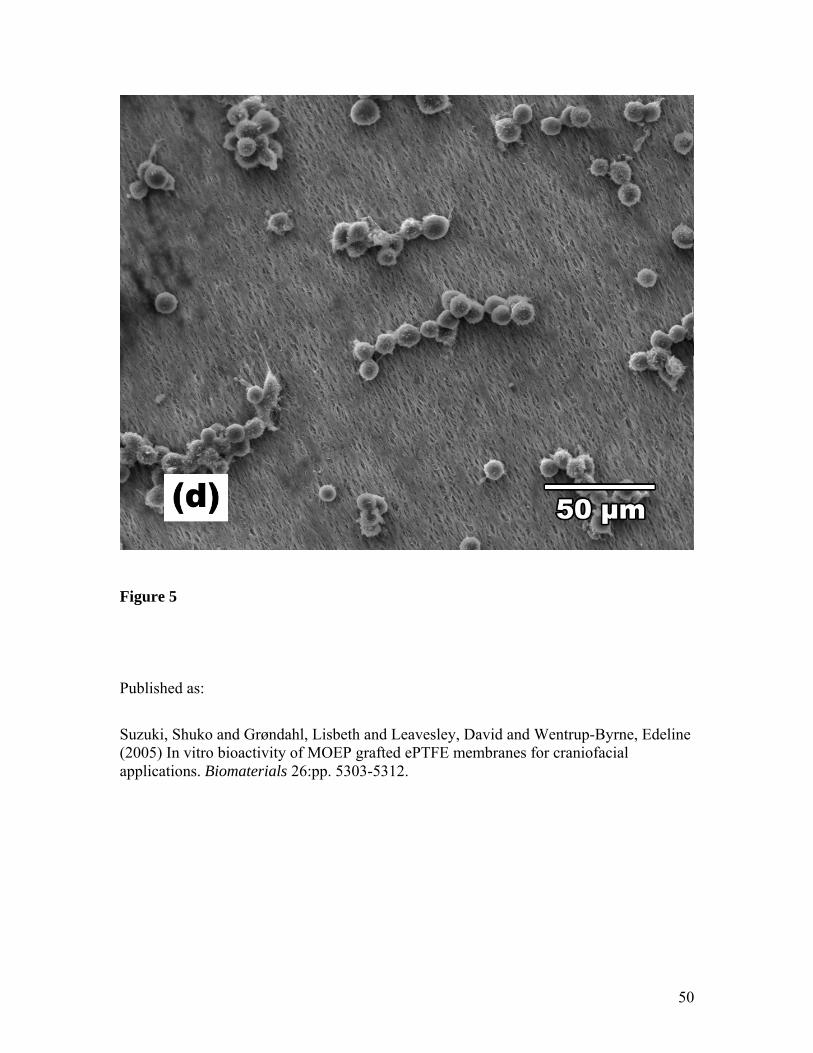
attachment assay. Few cells were found to attach and spread on untreated membranes
(Figure 5d). The attached cells clustered together and on top of each other rather than on
the material.
The majority of the cells on sample A (Figure 5a) failed to spread although they appear to
attach and form cellular processors both to the substrate surface and between cells.

17
Typically, these cells expressed blebs and micro-processes on their surfaces. In addition,
some attached cells possessed long processes to more distant cells.
Samples B and C (Figures 5b and 5c) demonstrate a high proportion of well attached
cells with a well-spread morphology. They had numerous cellular processes to the
membrane surface and the cell bodies were covered by micro-processes.
The number of spread and round cells were counted from the SEM images of four
randomly selected areas (260 μm × 225 μm) from each membrane and the average cell
numbers are shown in Table 3. The combined measured area by SEM was 1/330 of the
total material area. Significantly more cell spreading were observed on samples B and C
(56 and 53% of total cells, respectively) compared to A (35%), and much fewer spread
cells were observed on sample U (5%).
18
Discussion
Radiation-induced graft copolymerization of MOEP onto ePTFE showed interesting
morphological differences of grafted pMOEP depending on the grafting conditions used
[22]. Three samples with different graft morphologies, surface coverages, grafting yields,
and wettabilities were selected for in vitro analysis to investigate how these differences
might translate into different bioactivity performance. In addition, three different in vitro
assessments were carried out in order to investigate the degree of correlation between
them.
Simulated Body Fluid
Apatite formation in SBF is a well defined test for giving some indication of bone
bonding capability in vivo, thus, the formation of an apatite layer on a material in SBF
correlates with good bone bonding ability in vivo [8, 9, 26, 28]. In a recent study, a co-
polymer acting as an ion exchanger for calcium failed to induce CaP nucleation in SBF.
This co-polymer was subsequently found to perform poorly in in vivo tests [11, 29], thus
confirming the correlation between apatite formation in SBF with in vivo behaviour. In
the present study, the same amount of CaP nucleation was found on samples A-C and no
CaP nucleation was observed on the untreated sample in agreement with previous results
[3]. A detailed investigation of the samples after immersion in SBF revealed that sample
A with the lowest surface grafting yield (24% coverage) and a smooth but patchy graft
morphology showed formation of carbonated HA (similar to biological HA) covering the
surface after 7 days immersion in SBF. On the other hand, samples B and C with higher
surface grafting yields (76 and 100%, respectively), lower contact angles and a globular
19
polymer morphology showed 3-dimensional mineral growth rather than covering the
surface (2-D). This would seem to indicate that, in this case, growth is not strongly
template dependent [30]. Moreover, on samples B and C, although the Ca/P ratios from
EDX were only slightly higher than the theoretical value for HA, the FTIR analysis
revealed a mixture of CaP phases on sample B, and the CaP formed on sample C was not
HA. Although it is not known if the formation of HA (rather than other CaP phases) in
SBF is necessary for good bone bonding in vivo, it is generally assumed that HA
formation is the positive outcome in SBF tests. This would indicate that material A of this
study is the better candidate.
Previous studies have shown that the higher the surface grafting coverage, generally the
more CaP formed. MAEP grafted ePTFE membranes showed no nucleation of CaP on
samples with less than 44% surface grafting coverage, whereas highly grafted samples
had large mineral depositions with (Ca+Mg)/P ratios of 1.1 and a secondary growth with
a ratio of 1.5 [3]. MOEP grafted HDPE also showed more HA growth in SBF on samples
with higher surface grafting yields [8]. In these studies, grafting only occurred on the
outer most surface and no morphological differences were observed by SEM before and
after grafting. By contrast, in the present study, there was no correlation between surface
coverage and the amount of CaP growth in SBF. The samples A - C had different
wettabilities, overall grafting yields and displayed different surface morphologies. The
synergistic effects between hydrophobicity, surface chemistry and topography make it
difficult to investigate the effect of any one of them in isolation and it is therefore not
20
possible to conclude which parameter(s) are affecting the CaP growth on samples A – C
in SBF.
Protein Adsorption
Protein adsorption is the first event that occurs when a material is implanted. The amount
and types of proteins as well as the conformation of the adsorbed proteins are important
factors for subsequent cell adhesion and proliferation. In this study, a simple
determination of irreversibly adsorbed protein was performed. This technique is used
extensively in the literature with the assumption that larger amounts of protein will lead
to enhanced osteoblast cell attachment, a correlation that has been found [31-33].
Only small amounts of protein adsorbed on the unmodified membrane. Here protein-
surface interactions are most likely governed by hydrophobic interactions since there are
no functional groups present. On the other hand, all the grafted membranes displayed
enhanced protein adsorption with the highest amount of protein adsorption found for
sample C; the sample with the highest surface grafting yield. It can be concluded,
therefore, that the protein-surface interactions in this system are dominated by
electrostatic rather than hydrophobic interactions.
Cell Attachment
Cellular mediated biomineralisation is an important contributor to osseointegration.
Ideally a material surface should provide support for osteoblast adhesion, proliferation
and differentiation. It has been suggested that attachment of bone cells is essential for
their proliferation and differentiation stages [34]. The quantitative measurements in this
21
study revealed that MOEP grafted membranes significantly improve cell attachment
compared to that of untreated ePTFE.
Distinct morphological differences were found between SaOS-2 cells attached to the
various grafted and non-grafted membranes. Cells on the unmodified controls appeared
rounded with most cells attached to each other and very few spread cells. A previous
morphological study of osteoblasts on the outer surfaces of ePTFE vascular grafts (Gore-
Tex) showed similar results in that cells were rounded without any spreading after three
hours of attachment and the material did not support their growth [2].
Cells on sample A were rounded in appearance but had many projections and some
filopodia. Samples B and C showed many more spread, well attached cells. Therefore it
can be concluded that, after 3 hours of culture, samples with higher surface grafting
coverage and higher wettability exhibited an enhanced cell response compared to samples
with lower surface coverage and higher hydrophobicity. Although no significant
differences in cell numbers were observed (Table 3), the differences in cell morphology
(Figure 5) clearly indicate that the cell response to the different grafted materials is
indeed different. It is thus possible that cell response at a later time point will show
significant differences between the grafted samples. This hypothesis is supported by a
study by Bosetti et al. who compared two types of bioglass coatings with uncoated
zirconia [21]. They found that although the early cell attachment and spreading of cells
showed no difference between coated and uncoated zirconia, the results of a 5-day cell
growth assay indicated better performance of the coated materials.
22
Comparison of in vitro tests
In the present study, a larger amount of protein adsorption was found on the modified
materials compared to unmodified membrane. This correlates with improved osteoblast
attachment to the modified materials. This is similar to findings in other studies where a
correlation between the total amount of irreversibly adsorbed protein and degree of cell
attachment has been found [31-33]. The SBF result of this study also showed much better
performance for modified materials than for the unmodified substrate, and thus, all in
vitro assays indicate that modification of ePTFE with the phosphate containing monomer
MOEP enhances the bioactivity of the material.
Comparing the performance of samples A-C in the protein adsorption assay suggests
sample C is the better material while samples A and B perform equally well. The number
of cells attaching does not differ whereas the cell morphology studies suggest samples B
and C perform differently to sample A. Thus, these two in vitro assays do not correlate
between the grafted samples. In order to assess the reason for this, a much more detailed
study of specific protein adsorption to the various surfaces is necessary. For example, it
has been found that there is a direct correlation between the amount of specific proteins
(fibronectin and vitronectin) on a material surface (adsorbed from foetal bovine serum)
and the binding of osteoblast precursor cells [32].
Sample A gives rise to extensive carbonated HA growth in SBF, whereas a mixture of
CaP minerals are formed on sample B and a non-apatite CaP mineral is formed on sample
C. Cell morphology indicates that materials B and C are more cell friendly than material
23
A. This apparent disagreement between cell attachment and SBF in in vitro test results is
similar to the results of a study by Itälä et al. where the attachment of MG-63 cells were
compared to SBF results for a number of bioglasses [17]. They found that the thickness
of HA formed in SBF and the cell assay results did not identify the same material as the
best candidate.
Conclusion
A set of in vitro studies show that the grafting of MOEP increases the bioactivity of
ePTFE membranes compared to the untreated control. Results from the suite of tests
highlight the caution which should be applied when using any one relatively simple in
vitro assay to predict the in vivo bioactivity of a biomaterial, since based on the choice of
test different optimum modified materials are can be identified. Since the samples all had
different wettabilities, overall grafting yields and displayed different surface
morphologies, the synergistic effects between hydrophobicity, surface chemistry and
topography make it very difficult to investigate the effect of any one factor in isolation
and it is impossible to conclude which parameter(s) are affecting the overall bioactivity
as measured by the in vitro tests . Also it is important to note that the test times were all
different; protein adsorption was a one hour assay, cell attachment a 3 hour assay, and
SBF a 7 day experiment. Further cell and in vivo studies will be required in order to
confirm whether improved cell interaction observed on the more highly grafted,
hydrophilic surfaces will prove more supportive of cell proliferation and hence a more
suitable surface for craniofacial applications.

24
Acknowledgement
The authors would like to thank Dr. Thor Bostrom, Analytical Electron Microscopy
Facility, QUT, for his technical support with SEM and EDX characterisations, Dr. Barry
Wood, Brisbane Surface Analysis Facility, UQ, for his expert assistance in the XPS, Mr.
Tony Parker, QUT, for his help and guidance in cell culture, Dr. Robert A. Johnson, QUT,
for supplying the ePTFE membranes, and Ms. Fabienne Dragin, visiting student,
Polytech Montpellier, for her contribution. Special thanks also go to Dr. Richard
Lewandowski, plastic surgeon, Brisbane, for sharing his vital knowledge in facial
reconstruction.
25
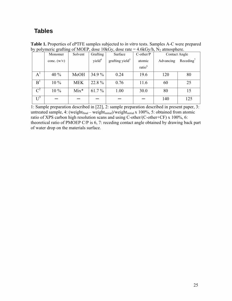
Tables
Table 1. Properties of ePTFE samples subjected to in vitro tests. Samples A-C were prepared by polymeric grafting of MOEP, dose 10kGy, dose rate = 4.6kGy/h, N2 atmosphere.
Monomer
conc. (w/v)
Solvent Grafting
yield4
Surface
grafting yield5
C-other/P
atomic
ratio6
Contact Angle
Advancing Receding7
A1 40 % MeOH 34.9 % 0.24 19.6 120 80
B1 10 % MEK 22.8 % 0.76 11.6 60 25
C2 10 % Mix* 61.7 % 1.00 30.0 80 15
U3 ─ ─ ─ ─ ─ 140 125
1: Sample preparation described in [22], 2: sample preparation described in present paper, 3: untreated sample, 4: (weightfinal – weightinitial)/weightinitial x 100%, 5: obtained from atomic ratio of XPS carbon high resolution scans and using C-other/(C-other+CF) x 100%, 6: theoretical ratio of PMOEP C/P is 6, 7: receding contact angle obtained by drawing back part of water drop on the materials surface.

26
Table 2: SBF and protein adsorption study results
SBF Protein Adsorption Sample
Weight
increase (%)*
Ca/P (Ca + Mg)/P Weight
increase (%)*
N % from
XPS
A 37 1.52 1.63 5 9
B 35 1.74 1.89 3 9
C 39 1.62 1.72 6 12
D ― ― ― 0.6 2
*: (weightfinal – weightinitial)/weightinitial x 100%
Table 3: SaOS-2 Cell Attachment1
From SEM’s 3 Sample Number of attached cells
(%)2 Spread Cells Round Cells % Spread Cells
A 68 + 8 44 + 5 80 + 15 35
B 72 + 0.5 58 + 5 45 + 4 56
C 82 + 4 52 + 3 46 + 6 53
U 12 + 0.4 4 + 0.3 81 + 9 5
1: Error is the standard error of the mean, 2: % relative to TCPS, 3: Average number of spread and round cells in the area of 260 μm × 225 μm.
27
References
[1] Piattelli A, Scarano A, and Paolantonio M. Bone formation inside the material
interstices of ePTFE membranes: a light microscopical and histochemical study in man.
Biomaterials 1996;17:1725-1731.
[2] Walsh WR, Olmedo M, Kim HD, Zou L, Weiss A-PC. Human osteoblast
response to PTFE surfaces. Clinical Materials 1994;16:201-210.
[3] Grøndahl L, Bostrom T, Cardona F, Chiem K, Wentrup-Byrne E. Calcium
phosphate nucleation on surface-modified PTFE membranes. J.Mater Sci Mater Med
2003;14:503-510.
[4] Filmon R, Grizon F, Basle MF, Chappard D. Effects of negatively charged groups
(carboxymethyl) on the calcification of poly(2-hydroxyethyl methacrylate). Biomaterials
2002;23:3053-3059.
[5] Zhu P, Masuda Y, Koumoto K. The effect of surface charge on hydroxyapatite
nucleation. Biomaterials 2004;25:3915-3921.
[6] Tanahashi M, Matsuda T. Surface functional group dependence on apatite
formation on self-assembled monolayers in a simulated body fluid. J Biomed Mater Res
1997;34:305-315.
[7] Liu Q, Ding J, Mante FK, Wunder SL, Baran JR. The role of surface functional
groups in calcium phosphate nucleation on titanium foil: a self-assembled monolayer
technique. Biomaterials 2002;23:3103-3111.
[8] Tretinnikov ON, Kato K, Ikada Y. In vitro hydroxyapatite deposition onto a film
surface-grafted with organophosphate polymer. J Biomed Mater Res 1994;28:1365-1373.
28
[9] Kamei S, Tomita N, Tamai S, Kato K, Ikada Y. Histologic and mechanical
evaluation for bone bonding of polymer surfaces grafted with a phosphate-containing
polymer. J Biomed Mater Res 1997;37:384-393.
[10] Grøndahl L, Cardona F, Chiem K, Wentrup-Byrne E. Preparation and
characterisation of the copolymers obtained by grafting of Monoacryloxyethyl Phosphate
onto Polytetrafluoroethylene membranes and Poly(tetrafluoroethylene-co-
hexafluoropropylene) films. J Appl Poly Sci 2002;86:2550-2556.
[11] Stancu IC, Filmon R, Cincu C, Zaharia C, Tourmen Y, Baslé MF, Chappard D.
Synthesis of methacryloyloxyethyl phosphate copolymers and in vitro calcification
capacity. Biomaterials 2004;25:205-213.
[12] Kato K, Sano S, Ikada Y. Protein adsorption onto ionic surfaces. Colloids and
Surfaces B: Biointerfaces 1995;4:221-230.
[13] Shelton RM, Rasmussen AC, Davies JE. Protein adsorption at the interface
between charged polymer substrata and migrating osteoblasts. Biomaterials 1988;9:24-29.
[14] Lee JH, Lee JW, Khang G, Lee HB. Interaction of cells on chargeable functional
group gradient surfaces. Biomaterials 1996;18:351-358.
[15] Yamamoto M, Kato K, Ikada Y. Ultrastructure of the interface between cultured
osteoblasts and surface-modified polymer substrates. J Biomed Mater Res 1997;37:29-36.
[16] Olmo N, Martin AI, Salinas AJ, Turnay J, Vallet-Regi M, Lizarbe MA. Bioactive
sol-gel glasses with and without a hydroxycarbonate apatite layer as substrates for
osteoblast cell adhesion and proliferation. Biomaterials 2003;24:3383-3393.
29
[17] Itälä A, Ylänen HO, Yrjans J, Heino T, Hentunen T, Hupa M, Aro HT.
Characterization of microrough bioactive glass surface: surface reactions and osteoblast
responses in vitro. J Biomed Mater Res 2002;62:404-411.
[18] Blaker JJ, Gough JE, Maquet V, Notingher I, Boccaccini AR. In vitro evaluation
of novel bioactive composites based on bioglass® -filled polylactide foams for bone tissue
engineering scaffolds. J Biomed Mater Res 2003;67A:1401-1411.
[19] Roether JA, Gough JE, Boccaccini AR, Hench LL, Maquet V, Jerome R. Novel
bioresorbable and bioactive composites based on bioactive glass and polylactide foams
for bone tissue engineering. J Mater Sci Mater Med 2002;13:1207-1214.
[20] Huang J, Di Silvio L, Wang M, Rehman I, Ohtsuki C, Bonfield W. Evaluation of
in vitro bioactivity and biocompatibility of bioglass® - reinforced polyethylene composite.
J Mater Sci Mater Med 1997;8:809-813.
[21] Bosetti M, Verne E, Ferraris M, Ravaglioli A, Cannas M. In vitro characterisation
of zirconia coated by bioactive glass. Biomaterials 2001;22:987-994.
[22] Wentrup-Byrne E, Grøndahl L, Suzuki S. Radiation induced graft
copolymerization of methacryloyloxyethyl phosphate onto polytetrafluoroethylene
membranes. in preparation.
[23] Kim HM, Miyazaki T, Kokubo T, Nakata T. Revised simulated body fluid.
Bioceramics 2000;13:47-50.
[24] Rodan SB, Imai Y, Thiede MA, Wesolowski G, Thompson D, Bar-Shavit Z, Shull
S, Mann K, Rodan GA. Characterization of a human osteosarcoma cell line (Saos-2) with
osteoblastic properties. Cancer Res 1987;47:4961-4966.
30
[25] Uchida M, Kin HM, Kokubo T. Bonelike apatite formation induced on zirconia
gel in a simulated body fluid and its modified solutions. J Am Ceram Soc 2001;84:2041-
2044.
[26] Kokubo T. Bioactive glass ceramics: prooperties and applications. Biomaterials
1991;12:155-163.
[27] Fowler BO, Moreno EC, Brown WE. Infra-red spectra of hydroxyapatite,
octacalcium phosphate and pyrolysed octacalcium phosphate. Arch Oral Biol
1966;11:477-492.
[28] Hench LL. Bioceramics. J Am Ceram Soc 1998;81:1705-1728.
[29] Stancu IC, Filmon R, Grizon F, Zaharia C, Cincu C, Baslé MF, Chappard D. The
in vitro calcification capacity of a copolymer, based on methacryloyloxyethyl phosphate,
does not favor osteoconduction. J Biomed Mater Res 2004;69A:584-589.
[30] Song J, Saiz E, Bertozzi CR. A new approach to mineralization of biocompatible
hydrogel scaffolds: An efficient process toward 3-dimensional bonelike composites. J
Am Chem Soc 2003;125:1236-1243.
[31] Scotchford CA, Ball M, Winkelmann M, Voros J, Csucs C, Brunette DM,
Danuser G, Textor M. Chemically patterned, metal-oxide-based surfaces produced by
photolithographic techniques for studying protein- and cell-interactions. II: protein
adsorption and early cell interactions. Biomaterials 2003;24:1147-1158.
[32] Kilpadi KL, Chang PL, Bellis SL. Hydroxyapatite binds more serum proteins,
purified integrins, and osteoblast precursor cells than titanium or steel. J Biomed Mater
Res 2001;57:258-267.
31
[33] Steele JG, Dalton BA, Underwoos PA. Polystyrene chemistry affects vitronectin
activity: An explanation for cell attachment to tissue culture polystyrene but not to
unmodified polystyrene. J Biomed Mater Res 1993;27:927-940.
[34] Ingber DE. Engineering cell shape and function through control of substrate
adhesion. In: Mittal KL, Lee K-W, editors. Polymer surfaces and interfaces:
Characterization, modification and application. Utrecht, The Netherlands: VSP; 1997. p.
413-424.

32
Figures
Figure 1 Chemical structures of the monomers methacryloyloxyethyl phosphate
(MOEP) and monoacryloxyethyl phosphate (MAEP)
Figure 2 SEM images of (a) sample A, (b) sample B, (c) sample C and (d) untreated
ePTFE membranes
Figure 3 SEM images of mineral formations on (a) sample A, (b) sample B, and (c)
sample C
Figure 4 ATR-FTIR spectra of grafted membranes before and after SBF immersion
(a) sample A: (i) ePTFE, (ii) grafted, (iii) after SBF, (b) sample B: (i)
grafted, (ii) after SBF, and (c) sample C: (i) grafted, (ii) after SBF
Figure 5 SEM images of attached SaOS-2 cells on (a) sample A, (b) sample B, (c),
sample C and (d) untreated ePTFE
33
Figure 1
R = H; Monoacryloxyethyl phosphate (MAEP) R = CH3; Methacryloyloxyethyl phosphate (MOEP)
CH2
OO
PR
O
O
OHOH
34
Figure 2

35
Figure 3
36
(i)
(ii) (iii)
(ii)
(i)
(a)
(b)
37
Figure 4
(i)
(ii)
(c)
38
Figure 5

39
APPENDIX
40
Figure 2
41
Figure 2
42
Figure 2
43
Figure 2

44
Figure 3
45
Figure 3
46
Figure 3
47
Figure 5

48
Figure 5

49
Figure 5
50
Figure 5
Published as:
Suzuki, Shuko and Grøndahl, Lisbeth and Leavesley, David and Wentrup-Byrne, Edeline (2005) In vitro bioactivity of MOEP grafted ePTFE membranes for craniofacial applications. Biomaterials 26:pp. 5303-5312.