Embed Size (px)
Citation preview

Alma Mater Studiorum - Università di Bologna
Dottorato di ricerca
Biologia e Fisiologia Cellulare
- XX Ciclo -
Settore Scientifico / Disciplinare di afferenza: BIO-09
RNA Interference and cyclooxygenase-2
(COX-2) regulation in colon cancer cells
Presentata da: Dr. Antonio Strillacci
Coordinatore Dottorato Relatore
Prof.ssa Michela Rugolo Prof. Vittorio Tomasi
Esame Finale Anno 2008

Summary
Despite new methods and combined strategies, conventional cancer chemotherapy still lacks
specificity and induces drug resistance. Gene therapy can offer the potential to obtain the success in
the clinical treatment of cancer and this can be achieved by replacing mutated tumour suppressor
genes, inhibiting gene transcription, introducing new genes encoding for therapeutic products, or
specifically silencing any given target gene. Concerning gene silencing, attention has recently
shifted onto the RNA interference (RNAi) phenomenon. Gene silencing mediated by RNAi
machinery is based on short RNA molecules, small interfering RNAs (siRNAs) and microRNAs
(miRNAs), that are fully o partially homologous to the mRNA of the genes being silenced,
respectively. On one hand, synthetic siRNAs appear as an important research tool to understand the
function of a gene and the prospect of using siRNAs as potent and specific inhibitors of any target
gene provides a new therapeutical approach for many untreatable diseases, particularly cancer. On
the other hand, the discovery of the gene regulatory pathways mediated by miRNAs, offered to the
research community new important perspectives for the comprehension of the physiological and,
above all, the pathological mechanisms underlying the gene regulation. Indeed, changes in miRNAs
expression have been identified in several types of neoplasia and it has also been proposed that the
overexpression of genes in cancer cells may be due to the disruption of a control network in which
relevant miRNAs are implicated. For these reasons, I focused my research on a possible link
between RNAi and the enzyme cyclooxygenase-2 (COX-2) in the field of colorectal cancer (CRC),
since it has been established that the transition adenoma-adenocarcinoma and the progression of
CRC depend on aberrant constitutive expression of COX-2 gene. In fact, overexpressed COX-2 is
involved in the block of apoptosis, the stimulation of tumor-angiogenesis and promotes cell
invasion, tumour growth and metastatization.
On the basis of data reported in the literature, the first aim of my research was to develop an
innovative and effective tool, based on the RNAi mechanism, able to silence strongly and
specifically COX-2 expression in human colorectal cancer cell lines. In this study, I firstly show
that an siRNA sequence directed against COX-2 mRNA (siCOX-2), potently downregulated COX-2
gene expression in human umbilical vein endothelial cells (HUVEC) and inhibited PMA-induced
angiogenesis in vitro in a specific, non-toxic manner. Moreover, I found that the insertion of a
specific cassette carrying anti-COX-2 shRNA sequence (shCOX-2, the precursor of siCOX-2

previously tested) into a viral vector (pSUPER.retro) greatly increased silencing potency in a colon
cancer cell line (HT-29) without activating any interferon response. Phenotypically, COX-2
deficient HT-29 cells showed a significant impairment of their in vitro malignant behaviour. Thus,
results reported here indicate an easy-to-use, powerful and high selective virus-based method to
knockdown COX-2 gene in a stable and long-lasting manner, in colon cancer cells. Furthermore,
they open up the possibility of an in vivo application of this anti-COX-2 retroviral vector, as
therapeutic agent for human cancers overexpressing COX-2.
In order to improve the tumor selectivity, pSUPER.retro vector was modified for the shCOX-2
expression cassette. The aim was to obtain a strong, specific transcription of shCOX-2 followed by
COX-2 silencing mediated by siCOX-2 only in cancer cells. For this reason, H1 promoter in basic
pSUPER.retro vector [pS(H1)] was substituted with the human Cox-2 promoter [pS(COX2)] and
with a promoter containing repeated copies of the TCF binding element (TBE) [pS(TBE)]. These
promoters were chosen because they are particularly activated in colon cancer cells. COX-2 was
effectively silenced in HT-29 and HCA-7 colon cancer cells by using enhanced pS(COX2) and
pS(TBE) vectors. In particular, an higher siCOX-2 production followed by a stronger inhibition of
Cox-2 gene were achieved by using pS(TBE) vector, that represents not only the most effective, but
also the most specific system to downregulate COX-2 in colon cancer cells.
Because of the many limits that a retroviral therapy could have in a possible in vivo treatment of
CRC, the next goal was to render the enhanced RNAi-mediate COX-2 silencing more suitable for
this kind of application. Xiang and et al. (2006) demonstrated that it is possible to induce RNAi in
mammalian cells after infection with engineered E. Coli strains expressing Inv and HlyA genes,
which encode for two bacterial factors needed for successful transfer of shRNA in mammalian
cells. This system, called “trans-kingdom” RNAi (tkRNAi) could represent an optimal approach for
the treatment of colorectal cancer, since E. Coli in normally resident in human intestinal flora and
could easily vehicled to the tumor tissue. For this reason, I tested the improved COX-2 silencing
mediated by pS(COX2) and pS(TBE) vectors in the tkRNAi system. Results obtained in HT-29 and
HCA-7 cell lines were in high agreement with data previously collected after the transfection of
pS(COX2) and pS(TBE) vectors in the same cell lines. These findings suggest that tkRNAi system
for COX-2 silencing, in particular mediated by pS(TBE) vector, could represent a promising tool
for the treatment of colorectal cancer.

Flanking the studies addressed to the setting-up of a RNAi-mediated therapeutical strategy, I
proposed to get ahead with the comprehension of new molecular basis of human colorectal cancer.
In particular, it is known that components of the miRNA/RNAi pathway may be altered during the
progressive development of colorectal cancer (CRC), and it has been already demonstrated that
some miRNAs work as tumor suppressors or oncomiRs in colon cancer. Thus, my hypothesis was
that overexpressed COX-2 protein in colon cancer could be the result of decreased levels of one or
more tumor suppressor miRNAs.
In this thesis, I clearly show an inverse correlation between COX-2 expression and the human miR-
101(1) levels in colon cancer cell lines, tissues and metastases. I also demonstrate that the in vitro
modulating of miR-101(1) expression in colon cancer cell lines leads to significant variations in
COX-2 expression, and this phenomenon is based on a direct interaction between miR-101(1) and
COX-2 mRNA. Moreover, I started to investigate miR-101(1) regulation in the hypoxic
environment since adaptation to hypoxia is critical for tumor cell growth and survival and it is
known that COX-2 can be induced directly by hypoxia-inducible factor 1 (HIF-1). Surprisingly, I
observed that COX-2 overexpression induced by hypoxia is always coupled to a significant
decrease of miR-101(1) levels in colon cancer cell lines, suggesting that miR-101(1) regulation
could be involved in the adaption of cancer cells to the hypoxic environment that strongly
characterize CRC tissues.

Index
Chapter I: Cyclooxygenases (Prostaglandin Endoperoxide H Synthases)……………………1
1. Cyclooxygenase Isozymes…………………………………………………………………………………1
1.1 Prostaglandins and Cyclooxygenase………………………………………………………2
1.2 Early Evidence for Multiple Cyclooxygenases……………………………………………3
1.3 Studies of Cell Division and the Discovery of Cyclooxygenase-2………………………..4
1.4 Structure of Cyclooxygenase-1 and Cyclooxygenase-2…………………………………..5
1.5 Variants of Cyclooxygenase Isoenzymes.......................................................................... 12
1.6 Cyclooxygenase Inhibitors—Nonsteroidal Anti-Inflammatory Drugs (NSAIDs)………15
2. Pharmacological Actions of Cyclooxygenase Isozyme-Generated Prostanoids………………….19
2.1 Prostaglandin Receptors…………………………………………………………………19
2.2 Inflammation…………………………………………………………………………….19
2.3 Pain ………………………………………………………………………………………20
2.4 Fever……………………………………………………………………………………...21
2.5 Immune System…………………………………………………………………………..21
2.6 Gastrointestinal Tract…………………………………………………………………….22
2.7 Cardiovascular System…………………………………………………………………...23
2.8 Kidney……………………………………………………………………………………27
2.9 Lungs……………………………………………………………………………………..28
2.10 Reproduction……………………………………………………………………………30
2.11 Brain and Spinal Cord…………………………………………………………………..31
3. Cyclooxygenase Isozymes in Human Disease…………………………………………………………33
3.1 Treatment of Inflammatory Diseases…………………………………………………….33
3.2 Neoplastic Disease ……………………………………………………………………….33
3.3 Alzheimer's Disease……………………………………………………………………...38
4. COX-2 Regulation………………………………………………………………………………………...41
4.1 Molecular aspects of COX-2 regulation…………………………………………………41
4.2 Dynamic Regulation of COX-2 Transcriptional Activation by C/EBPß ………………..42
4.3 Other signaling pathways………………………………………………………………...44

4.4 Post-transcriptional regulation…………………………………………………………...46
5) COX-2 and Colorectal Cancer………………………………………………………………………….49
5.1 COX-2 expression in colorectal cancer…………………………………………………..50
5.2 Role of COX-2 in tumorigenesis…………………………………………………………52
5.3 Anti-tumor effects of nonsteroidal anti-inflammatory drugs (NSAIDs)…………………55
5.4 COX-2 and colo-rectal cancer: regulation by Wnt pathway……………………………...57
5.5 COX-2 and colo-rectal cancer: regulation by the hypoxic pathway……………………...59
Chapter II: The Mechanism of RNA Interference (RNAi)…………………………………...61
1. Origins of RNA Interference…………………………………………………………………………….61
2. Mechanism of action of RNAi…………………………………………………………………………...63
2.1 Small interfering RNAs (siRNAs)………………………………………………………..63
2.2 MicroRNAs (miRNAs)…………………………………………………………………...64
3. Applications of RNAi in mammalian systems…………………………………………………………68
3.1 RNAi effectors used for biological analysis in mammalian cells………………………..68
3.2 Off-target effects…………………………………………………………………………70
3.3 Application of RNAi-based technologies………………………………………………...73
3.4 “Trans-kingdom” RNAi…………………………………………………………………..76
3.5 Prediction of microRNA targets………………………………………………………….76
4. MicroRNAs and Cancer………………………………………………………………………………….84
4.1 Disruption of miRNA-directed regulation………………………………………………..84
4.2 MiRNAs as tumor suppressors and oncogenes............... .................................................... 85
4.3 Genetics and epigenetics changes associated with altered miRNA expression.................. 88
4.4 MiRNAs as new targets for therapy.................................................................. .................. 91
4.5 Altered miRNAs expression in colorectal cancer………………………………………...93
4.6 COX-2 and miRNAs........................................................................................................... 96

Chapter III: The Research Project…………………………………………………….............97
1. Preliminary Remarks……….…………………………………………………………………………….97
2. Objectives……………………..…………………………………………………………………………...98
2.1 COX-2 silencing in colon cancer cells……………………………………………………98
2.2 COX-2 regulation analysis……………..………………………………………………....99
Chapter IV: COX-2 silencing RNAi-mediated……………………………………………....101
1. Results Part 1……………………………………….…………………………………………………...101
2. Figures Part 1…………………………………………………………………………………………...105
3. Discussion Part 1………………………………………………………………………………………..125
Chapter V: mir-101 regulates COX-2 in cancer cells………………………………………..131
1. Results Part 2……………………………………….…………………………………………………..131
2. Figures Part 2…………………………………………………………………………………………..133
3. Discussion Part 2……………………………………………………………………………………….145
Chapter VI……………………………………………….……………………………………..149
1. Materials and methods………………………….……………………………………………………...149
2. Supplementary Materials……………………………………………………………………………….157
3. Publications………………………………………………………………………………………..........163
4. Publications………………………………………………………………………………………..........165
5. References………………………………………………………………………………………………..167

1
Chapter I
Cyclooxygenases (Prostaglandin Endoperoxide H Synthases)
1. Cyclooxygenase Isozymes
Prostaglandin endoperoxide H synthases (PGHSs) catalyze the conversion of arachidonic acid and
O2 to PGH2, the committed step in prostanoid biosynthesis. Before 1991, only one PGHS had been
described, the isozyme now called PGHS-1, COX-1 (for cyclooxygenase-1) or the constitutive
enzyme. At that time Simmons and Herschman and their colleagues discovered mRNAs whose
expression was induced in chicken and mouse fibroblasts in response to src and tumor-promoting
phorbol esters, respectively, and which encoded proteins having 60% amino acid sequence identity
with PGHS-1. Subsequent work has shown that the new protein, called PGHS-2, COX-2 or the
inducible isoform, is very similar to PGHS-1 in structure but differs substantially from PGHS-1
with respect to its pattern of expression and its biology. The reason for the existence of the two
PGHS isozymes is unknown. However, PGHS-1 and -2 are often coexpressed in the same cell and
may act as parts of separate prostanoid biosynthetic systems that function somewhat independently
to channel prostanoids to the extracellular milieu and the nucleus, respectively. Nonsteroidal anti-
inflammatory drugs (NSAIDs) represent one of the most highly utilized classes of pharmaceutical
agents in medicine. All NSAIDs act through inhibiting prostaglandin synthesis, a catalytic activity
possessed by two distinct COX isozymes encoded by separate genes. The discovery of COX-2
launched a new era in NSAID pharmacology, resulting in the synthesis, marketing, and widespread
use of COX-2 selective drugs. These pharmaceutical agents have quickly become established as
important therapeutic medications with potentially fewer side effects than traditional NSAIDs.
Additionally, characterization of the two COX isozymes is allowing the discrimination of the roles
each play in physiological processes such as homeostatic maintenance of the gastrointestinal tract,
renal function, blood clotting, embryonic implantation, parturition, pain, and fever. Of particular
importance has been the investigation of COX-1 and -2 isozymic functions in cancer, dysregulation
of inflammation, and Alzheimer's disease. More recently, additional heterogeneity in COX-related
proteins has been described, with the finding of variants of COX-1 and COX-2 enzymes. These
variants may function in tissue-specific physiological and pathophysiological processes and may
represent important new targets for drug therapy.

2
1.1 Prostaglandins and Cyclooxygenase
Prostaglandins, potent bioactive lipid messengers derived from arachidonic acid (AA), were first
extracted from semen, prostate, and seminal vesicles by Goldblatt and von Euler in the 1930s and
shown to lower blood pressure and cause smooth muscle contraction. Bergström and colleagues
purified the first prostaglandin isomers during the 1950s and 60s, and in 1964, van Dorp et al. and
Bergstrom et al. independently identified AA, a 20-carbon tetraenoic fatty acid (C20:4Ω6) as the
precursor to prostaglandins. The cyclooxygenase reaction through which AA is enzymatically
cyclized and is oxygenated to yield endoperoxide-containing prostaglandin G2 (PGG2) was later
identified by Samuelsson and colleagues (Hamberg and Samuelsson, 1973; Hamberg et al., 1974)
(Fig.1). The enzyme, cyclooxygenase (COX) that catalyzes this cyclooxygenation reaction also
reduces a hydroperoxyl in PGG2 to a hydroxyl to form PGH2 via a separate peroxidase active site on
the enzyme. Isomerases and oxidoreductases produce various bioactive prostaglandin isomers using
PGH2 as substrate as shown in Fig.1.
Figure 1. The arachidonic acid cascade.
3
Until 1976 (Hemler and Lands, 1976; Miyamoto et al., 1976), when purified COX preparations
were first described, tissue homogenates were used as a source of COX enzyme activity, which was
frequently referred to at that time as a prostaglandin synthetase. Because the COX enzyme reaction
does not require ATP, the nomenclature was later changed to synthase. The COX enzyme, also
known as prostaglandin H synthase (PGHS) or prostaglandin endoperoxide synthase (E.C.
1.14.99.1), was identified as the major enzyme in the oxidative conversion of AA to PGG2 and
PGH2 (Smith and Lands, 1972; Hamberg et al., 1974), with seminal vesicles of sheep, bovines being
a rich enzyme source (Smith and Lands, 1972). Thus, purification of PGHS enzyme to homogeneity
was first achieved from the sheep (Hemler and Lands, 1976; van der Ouderaa et al., 1977) and
bovine (Miyamoto et al., 1976; Ogino et al., 1978) seminal vesicles. This purified enzyme migrated
in the region of approximately 67 kDa in SDS-polyacrylamide gel electrophoresis and contained
cyclooxygenase and peroxidase activities, which were later found to be at separate sites (Marshall
and Kulmacz, 1988). Since detergents such as Tween 20 were needed to solubilize the enzyme, it
was classified as an integral microsomal membrane protein. In 1971, John Vane used a cell-free
homogenate of guinea pig lung to demonstrate that aspirin, indomethacin, and salicylate, popular
nonsteroidal anti-inflammatory drugs (NSAIDs), were inhibitors of this enzyme—thus defining the
mechanism of action of this important class of drugs.
1.2 Early Evidence for Multiple Cyclooxygenases
Researchers, beginning in the early 1970s, speculated on whether there was more than one COX
enzyme. Flower and Vane (1972) postulated the existence of an acetaminophen-inhibitable COX
activity that was in dog brain but not in rabbit spleen. The same year, two catalytically distinct
prostaglandin synthase activities were reported to be present in acetone powder extracts of sheep
vesicular glands (Smith and Lands, 1972). Studies of autoinactivation rates of COX, inhibition by
NSAIDs, and time course profiles of PGE2 and PGF2 synthesis led Lysz and colleagues (1982,
1988) to propose that rabbit and mouse, but not rat brain, contained two forms of COX. It was,
however, through the study of prostaglandin induction by mitogens and proinflammatory agents, as
well as prostaglandin down-regulation by glucocorticoids, that the most provocative data regarding
the potential of more than one COX were obtained. The phenomenon that was observed by many
laboratories was that prostaglandin synthesis and release in some situations, such as in activated
platelets, occurs within a few minutes after stimulation. In other cases, such as in mitogen-
stimulated fibroblasts, prostaglandin synthesis may take hours to occur. In 1985, Habenicht and
colleagues (1985) reported that platelet-derived growth factor treatment of Swiss 3T3 cells resulted
in an early (10 min) and a late (2–4 h) peak in induction in prostaglandin synthesis. Only the late

4
peak was blocked by cycloheximide, leading to the conclusion that platelet-derived growth factor-
stimulated PG synthesis occurred by "direct effects on the PG-synthesizing enzyme system, one
involving a protein synthesis-independent mechanism and another that requires rapid translation of
cyclooxygenase". The activities revealed in Habenicht's early study were indicative of an
endogenous COX enzyme (COX-1) and an inducible enzyme (COX-2). Many other laboratories at
this time did similar studies on induction of prostaglandin synthesis, but with only nucleic acid and
antibody probes to the seminal vesicle form of COX to work with, many investigators observed the
paradoxical phenomenon that, in many instances, prostaglandin induction occurred without an
increase in the seminal vesicle COX—an enzyme which was found to be present in most cells and
tissues investigated (DeWitt and Smith, 1988). Frequently only marginal increase in seminal vesicle
COX was observed despite robust increase in PG synthesis. Similar anomalies in which PG
synthesis and seminal vesicle COX did not coincide were observed with regard to the action of
glucocorticoids, which strongly decreased PG synthesis but typically had little to no effect on
seminal vesicle COX levels. Various postulates were proposed that were consistent with these
observed phenomena, the most common of which was that changes in substrate delivery were
responsible for these fluctuations in PG synthesis. In 1989, Rosen et al. used low-stringency
Northern blot hybridization with an ovine seminal vesicle COX cDNA as probe to detect a 4.0-kb
RNA, in addition to the known 2.8-kb mRNA encoding seminal vesicle COX. This 4.0-kb mRNA
was inducible and paralleled induction of enzyme activity. These investigators concluded that "the
larger mRNA may encode for a cyclooxygenase" encoded by a distinct gene. In 1990, Needleman
and colleagues (Fu et al., 1990) studying lipopolysaccharide (LPS) stimulation of monocytes
concluded that these "cells may contain two pools of COX, each with a differential sensitivity to
LPS or DEX (dexamethasone)." During this time, Young, Macara, and colleagues (Han et al., 1990)
identified, by using giant two-dimensional protein gel electrophoresis, proteins immunoreactive
with COX-1 antibodies that were induced in v-src-transformed cells. The evidence in these and
other early studies was consistent with distinct inducible and constitutive COX isozymes encoded
by separate genes but was also compatible with other explanations.
1.3 Studies of Cell Division and the Discovery of Cyclooxygenase-2
The answer to the mechanism of how COX enzyme activity rapidly increases PG induction in
inflammation and in other physiological contexts came from studies of cell division. In the late
1980s, Simmons et al. (1989) and Herschman (Varnum et al., 1989) and colleagues independently
identified immediate-early genes in fibroblast-like cells activated by mitogens. Genes found by
Simmons in chicken (Simmons et al., 1989; Xie et al., 1991) and mouse (Simmons et al., 1991)

5
were activated by the v-src oncogene, phorbol esters and serum. Herschman used Swiss 3T3 cells to
identify tetradecanoyl-13-phorbol acetate-inducible sequences (or TIS genes), which were also
induced by other mitogens (Varnum et al., 1989; Kujubu et al., 1991). In 1991, each laboratory
independently reported that one of their sequences encoded a new inducible COX enzyme. Also
contributing to the identification of COX-2 in 1991, Young and colleagues (O'Banion et al., 1991)
reported a partial predicted sequence of COX-2 from a murine cDNA. The inducible enzyme cloned
in these studies is now most frequently referred to as COX-2 and the seminal vesicle form of the
enzyme as COX-1. Herschman and colleagues expressed the mouse TIS10 cDNA in heterologous
cells and showed that increased prostaglandin E2 synthesis was induced by this cDNA (Fletcher et
al., 1992 ). Ectopic overexpression was also done by Young's laboratory (O'Banion et al., 1992) and
Meade et al. (1993), who also demonstrated the importance of the 3'-untranslated region of the
COX-2 mRNA in governing overexpression of the enzyme. Using mouse COX-2 sequences as
probe, Hla and Neilson (1992) identified and published the sequence of the human homolog of
TIS10/CEF147, which they named COX-2. Overexpression of human COX-2 cDNA in Cos cells
also induced COX enzymatic activity, similar to that of TIS10, and this activity was inhibited by
nonsteroidal anti-inflammatory drugs (Hla and Neilson, 1992). The human COX-2 cDNA was
widely expressed as an inducible gene in nonimmortalized vascular and inflammatory cells.
1.4 Structure of Cyclooxygenase-1 and Cyclooxygenase-2
Pure preparations of the COX-1 enzyme obtained from seminal vesicles were instrumental in the
elucidation of the primary structure of this enzyme by molecular cloning. Both the N terminus and
the internal sequences following limited trypsin digestion of the sheep seminal enzyme were
reported (Chen et al., 1987). Roth and colleagues (1975), using sheep and bovine seminal vesicle
enzyme preparations, showed that aspirin acetylated the COX enzyme. The region of the active site
residues and the determination of the serine acetylated by aspirin were elucidated by sequencing 3H-
aspirin-labeled peptides of the sheep seminal vesicle enzyme (Roth et al., 1980; DeWitt et al.,
1990); however, molecular cloning by three different laboratories ultimately elucidated the
complete primary structure of the COX-1 enzyme (DeWitt and Smith, 1988; Merlie et al., 1988;
Yokoyama et al., 1988). Sequence analysis of COX-1 cDNAs indicated that they contained an open
reading frame of ~1.8 kb, which contained all the polypeptide sequences from protein
microsequencing efforts (Roth et al., 1980, 1983; DeWitt and Smith, 1988), including the aspirin
acetylation site (DeWitt et al., 1990). These data strongly suggested that the isolated cDNA clone
encoded the sheep seminal vesicle COX enzyme. In addition to ovine COX-1, the gene for the
human homolog of this enzyme was also cloned (Yokoyama and Tanabe, 1989). These cloning

6
efforts were followed by the demonstration that the cDNA for sheep seminal vesicle COX exhibited
both cyclooxygenase and peroxidase activities upon ectopic overexpression in mammalian and
insect cells (DeWitt et al., 1990; Funk et al., 1991; Meade et al., 1993). Mutagenesis experiments
were conducted to identify critical residues for catalysis, such as the heme coordination sites, aspirin
acetylation sites, etc. (Shimokawa et al., 1990; Shimokawa and Smith, 1992). Northern blot analysis
with cDNA probes of COX-1 identified a major mRNA species of 2.8 kb and a minor species of 5.2
kb in human endothelial cells (Hla, 1996). Further sequence analysis of a human endothelial cell-
derived cDNA, which encoded the 3'-end of the 5.2-kb mRNA, indicated that the 5.2-kb cDNA and
the 2.8-kb cDNAs represent alternatively polyadenylated mRNA species with differing lengths of
the 3'-UTR (Hla, 1996). These alternative polyadenylation states were also confirmed in cDNAs
encoding the 3' of the 5.2-kb mRNA from a human megakaryoblastic cell line (Plant and
Laneuville, 1999). The predicted amino acid sequences of COX-2 cloned in chicken and mammals
showed it to possess approximately 60% amino acid identity with COX-1 (Simmons et al., 1991).
COX-1 and COX-2 were found to be approximately 600 amino acids in size in all species. The
COX-1 and/or -2 cDNA sequences from many organisms, including bony and cartilaginous fish,
birds, and mammals have been characterized. Furthermore COX genes appear to be expressed in
invertebrates, such as coral and sea squirts, where two COXs have been identified in two different
species of each phylum (Valmsen et al., 2001, Jarving et al., 2004). These data suggest that the
cyclooxygenase pathway was present in early invertebrate speciation in the animal kingdom. From
an evolutionary standpoint COX-1 and COX-2 appear to have resulted from a gene duplication
event that occurred early in or before vertebrate speciation. Cyclooxygenases in unicellular
organisms, insects, or the plant kingdom have not been identified; however, COX enzymes have
recently been determined to be members of a larger fatty acid oxygenase family that includes
pathogen-inducible oxygenases (PIOXs). These latter enzymes have been identified in
monocotyledon and dicotyledon plants, Caenorhabditis elegans, and bacteria (Pseudomonas). Like
COXs, PIOXs oxygenate polyunsaturated fatty acids using molecular oxygen (Sanz et al., 1998;
Hamberg et al., 1999; Koeduka et al., 2000). They also introduce a hydroperoxyl moiety into the
fatty acid, which is introduced at the α carbon by PIOXs to form 2R-hydroperoxy fatty acids.
Generation of α-peroxyl-fatty acids by PIOXs has been proposed to be a signaling response in these
organisms to activate genes needed for antipathogen defense (Sanz et al., 1998). COXs and PIOXs
share approximately 30% sequence identity, and PIOXs contain conserved critical residues needed
for fatty acid oxygenation seen in COX. Sequence similarity to COXs in the region of the Tyr385
has also been found in the enzyme linoleate diol synthase (LDS) from fungus (Oliw et al., 1998; Su
et al., 1998; Hörnsten et al., 1999). This enzyme is a homotetrameric ferric hemeprotein that

7
catalyzes the dioxygenation of linoleic acid to (8R)-hydroperoxylinoleate and isomerization of this
latter compound to (7S,8S)-dihydroxylinoleate. Like COXs, the enzyme is known to form ferryl
intermediates and a tyrosyl radical. PIOXs and LDSs are clearly related to peroxidases; however,
there is no evidence that PIOXs or LDS possess peroxidase activity. These findings lead to the
conclusion that PIOXs, LDS, and COXs each represent distinct subfamilies of fatty acid oxygenases
that are descended from ancient peroxidases. If they descend from a common peroxidase progenitor,
LDS, PIOXs and COXs have additionally gained hydrophobic pockets for binding and oxygenation
of fatty acid substrates; however, the PIOX and LDS branches of this fatty acid oxygenase family
have a degenerate peroxidase active site, that likely functions solely to activate the enzyme. PIOXs
and LDSs, therefore, are predicted to perform primarily the dioxygenation reaction typical of the
COX active site. Since PIOXs and LDS are found in plants, bacteria, fungus, and lower animals, the
fatty acid oxygenase progenitor of COXs and PIOXs is predicted to exist very early in evolution,
underscoring the concept that generation of oxygenated fatty acids by these enzymes represents an
evolutionarily ancient mechanism of cell signaling. Recently a cyclooxygenase enzyme from the
protozoan Entamoeba histolytica has been identified that lacks structural similarity with other
COXs, PIOXs, or LDS enzymes, but makes PGE2 from arachidonic acid (Dey et al., 2003).
Therefore, prostaglandin-synthesizing enzymes distinct from the COX lineage characterized in
vertebrates, coral, and sea squirts appear to have arisen during speciation of some organisms.
Landmark studies by Garavito and colleagues (Picot et al., 1994) elucidated the tertiary and
quaternary structure of COX-1. Early studies in the 1970s showed that COX-1 was likely a dimer
and was tightly bound to microsomal membranes; however, the topology of the enzyme in
microsomal membranes was unknown. At crystallographic resolution, Garavito's studies described
COX-1's distinct domains for dimerization, membrane binding, and catalysis. A fourth domain, the
N-terminal signal peptide, which is clearly evident in the primary structure of COX-1, was not
observed because this sequence is cotranslationally cleaved from the nascent polypeptide by
microsomal signal peptidase. Crystallographic structures of COX-2 have been obtained by Luong et
al. (1996), Bayly et al. (1999), and Kurumbail et al. (1996) and show striking similarity with COX-
1. In fact, all known COX enzymes share the same functional domains. Outstanding reviews of
COX structure and enzyme kinetics have recently been written (Garavito and DeWitt, 1999;
Marnett, 2000; Smith et al., 2000) and thus only the essential aspects of these topics needed to
understand the pharmacology of NSAIDs are discussed here. The structures of COX-1 and COX-2
predict that both enzymes are located in the lumen of the nuclear envelope and endoplasmic
reticulum. Structural aspects of each of the four domains (Figs. 2 and 3) of COX-1 and COX-2 lead
to this conclusion.

8
Figure 2. Crystallographic structures of ovine COX-1 (left) and murine COX-2 (right) homodimers.
Figure 3. Structure of a single subunit of COX.
1. Amino-Terminal Signal Peptide. Nascent COX-1 and COX-2 polypeptides are directed into the
lumen of the endoplasmic reticulum by amino-terminal signal peptides. Although cleaved from the
nascent polypeptide, these hydrophobic peptides show a size difference between COX-1 and COX-2
that, until recently, has been of unknown biological significance. The signal peptide for COX-1 is
always 22 to 26 amino acids in length with a large hydrophobic core comprised of four or more
leucines or isoleucines. COX-2's signal peptide is 17 amino acids long in all species and appears to
be less hydrophobic. In vitro translation experiments demonstrate that COX-1 is rapidly translocated
into the lumen of canine pancreatic microsomes, whereas COX-2 is inefficiently translocated (Xie et
al., 1991). Immediately following the signal peptide in COX-1 are eight amino acids that are not
found in COX-2. The function of this sequence is unknown. Recently, as described below, variants

9
of COX-1 have been identified in which retention of all or part of intron-1 results in a retained
signal peptide in COX-1, altering the biological properties of the enzyme (Chandrasekharan et al.,
2002). Also, one coral isozyme has seven amino acids inserted in the same location; however, in this
case, the insertion appears to change the location of the cleavage site in COS-7 cells rather than to
affect retention of the signal peptide (Jarving et al., 2004).
2. Dimerization Domain. COX-1 and COX-2 dimers are held together via hydrophobic interactions,
hydrogen bonding, and salt bridges between the dimerization domains of each monomer.
Heterodimerization of COX-1 and COX-2 subunits does not occur. The dimerization domain is
encoded by approximately 50 amino acids near the amino terminus of the proteolytically processed
protein. Three disulfide bonds hold this domain together in a structure reminiscent of epidermal
growth factor. A fourth disulfide bond links the dimerization domain with the globular catalytic
domain. The presence of disulfide bonds, which require an oxidizing environment, is consistent with
the location of COXs inside the lumen of the nuclear envelope, ER, or Golgi, which have redox
states that are significantly more oxidized than cytosol.
3. Membrane Binding Domain. COX isozymes associate with the intraluminal surface of
microsomal membranes in an unusual fashion. Rather than employing transmembrane spanning
sequences or covalently bound lipids for attachment, COX isozymes contain a tandem series of four
amphipathic helices, which creates a hydrophobic surface that penetrates into the upper portion of
the luminal side of the hydrophobic core of the lipid bilayer. These helices are encoded by
approximately 50 amino acids found immediately carboxy-terminal to the dimerization domain. The
helices allow COX dimers to float on the surface of the lumen of the ER/nuclear envelope, with the
majority of the protein protruding into the luminal space of these compartments. The membrane
binding domain also forms the mouth of a narrow hydrophobic channel that is the cyclooxygenase
active site.
4. Catalytic Domain. Carboxy-terminal to the membrane binding domain in COX primary structures
is the catalytic domain, which comprises 80% (approximately 480 amino acids) of the protein and
contains two distinct enzymatic active sites.
a. Peroxidase Active Site. The catalytic domain is globular with two distinct intertwining lobes.
The interface of these lobes creates a shallow cleft on the upper surface of the enzyme (i.e., the
surface furthest from the membrane) where the peroxidase active site is located and where heme
is bound. Coordination of the heme is via an iron-histidine bond involving His388 in sheep COX-
1. (All numbering hereafter uses sheep COX-1 as reference.). Other important interactions

10
between the protoporphyrin also occur, and specific amino acids that may function in
coordinating PGG2 have been identified (Malkowski et al., 2000; Thuresson et al., 2001). The
geometry of heme binding leaves a large portion of one side of the heme exposed in the open
cleft of the peroxidase active site for interaction with PGG2 and other lipid peroxides.
b. Cyclooxygenase Active Site. The cyclooxgyenase active site is a long, narrow, dead-end
channel of largely hydrophobic character whose entrance is framed by the four amphipathic
helices of the membrane binding domain. The channel extends approximately 25 Å into the
globular catalytic domain and is on average about 8 Å wide (Picot et al., 1994). However,
significant narrowing of the channel is observed where arginine 120, one of only two ionic
residues found in the COX active site, protrudes into the channel and forms a hydrogen bonded
network with glutamate 524 (the other ionic residue in the channel) and tyrosine 355. Arginine
120 is essential for binding substrates and carboxylate-containing NSAIDs in COX-1. In
contrast, this residue is unessential in binding substrate in COX-2 (Rieke et al., 1999), where it
also appears to be nonessential in coordinating carboxylate-containing NSAIDs (Greig et al.,
1997).
The upper portion of the channel, or catalytic pocket, contains tyrosine 385 that forms a tyrosyl
radical, abstracts hydrogen from the pro-S side of carbon 13 of AA, and creates an activated
arachidonyl radical that undergoes the cyclization/oxygenation reaction shown in Fig. 4.
a b
Figure 4. Priming of COX enzymes by formation of a tyrosyl radical (a). Catalytic steps in
cyclooxygenation of arachidonic acid (b).

11
Also in the hydrophobic pocket is Ser530, which is transacetylated by aspirin. The hydroxyl of
serine 530, together with valine 349, appears to be essential in governing the stereochemistry of
oxygen attack at carbon 15 in the production of PGG2 (Schneider et al., 2002); however, its
acetylation prevents abstraction of hydrogen from AA in COX-1 by sterically preventing AA from
binding productively in the active site (Rowlinson et al., 2000). In contrast, abstraction of hydrogen
does occur in acetylated COX-2, but cyclization of the arachidonyl radical and formation of the
endoperoxide does not occur, yielding 15-R-hydroxyeicosotetraenoic acid (15R-HETE) rather than
PGH2 (Holtzman et al., 1992). A crucial structural difference between the active sites of COX-1 and
COX-2 is a substitution of isoleucine 523 in COX-1 for a valine in COX-2. This single difference
opens a hydrophobic outpocketing in COX-2 that can be accessed by some COX-2-selective drugs
(Kurumbail et al., 1996). There are other changes in residues that are near but do not line the COX
active site, so-called second shell residues, that result in subtle changes and a slightly enlarged
COX-2 active site relative to COX-1 (Kurumbail et al., 1996; Luong et al., 1996). The evolutionary
conservation of an enlarged cyclooxygenase active site in COX-2 relative to COX-1 may be
essential to the recognition of bulkier substrates by COX-2. Anandamide (arachidonylethanolamide)
and 2-arachidonylglycerol are endocannabinoids that are efficiently oxidized by COX-2 to
endocannabinoid-derived prostanoids (Kozak et al., 2002). COX-2 utilizes these bulkier substrates
as efficiently as arachidonic acid, and the resulting endoperoxide can be utilized by downstream
isomerases (Kozak et al., 2002). The function of these prostanoid-like oxidized endocannabinoids is
unknown but may represent new biological roles of COX-2. The endocannabinoid analog
methandamide upregulates COX-2 expression, further linking this enzyme with metabolism of
endocannabinoids (Gardner et al., 2003; Ramer et al., 2003). Elegant studies done collaboratively by
the laboratories of Garavito and Smith (Malkowski et al., 2000; Thuresson et al., 2001) have
succeeded in defining the productive structure of COX-1 with its substrate AA as well as with
eicosapentaenoic and linoleic acids. COX-1 binds AA in an extended L shape, its carboxylate
forming both a salt bridge with the guanidinium group of arginine 120 and also a hydrogen bond
with tyrosine 355. The remainder of the fatty acid makes more than 50 mostly hydrophobic
interactions with 19 amino acid residues, which position substrate for hydrogen abstraction and
facilitate conversion to PGG2 rather than to HETEs (Thuresson et al., 2001). Two molecules of
oxygen for the bisoxygenation reaction and hydroperoxidation reaction that yield the endoperoxide
and hydroperoxide moieties, respectively, have been postulated to diffuse into the COX active site
from the direction of the membrane, thus resulting in the observed fact that attack of oxygen at
carbon 11, to eventually result in the PGG2 endoperoxide, occurs from the opposite or anterofacial
orientation from that of hydrogen abstraction at carbon 13. At the carboxy terminus of the catalytic

12
domain of COX-1 and COX-2 are modified versions of the KDEL sequence that act as a signal for
retention of proteins in the endoplasmic reticulum (Song and Smith, 1996). Additionally, COX-2
has an 18-amino acid sequence located next to this retention signal. This structure, which is not
found in COX-1, is not fixed in crystallographic studies, and its function is unknown. The above
structural features are consistent with localization of COX isozymes inside the lumen of the ER, a
fact that is further supported by numerous studies using fluorescence and immuno-electron
microscopy (Song and Smith, 1996; Liou et al., 2000); however COX-1 has been found by Weller
and colleagues to be localized to lipid bodies in leukocytes and other cells (Bozza et al., 1996).
Lipid bodies in these cells are rapidly formed following treatment with platelet-activating factor
(PAF), nonesterified fatty arachidonate, or other fatty acids (Bozza and Weller, 2001) and are
induced in endothelial cells by hypoxia (Scarfo et al., 2001). Unlike the ER, which contains a
lumen, the structure of lipid bodies is less defined and may contain a central core of neutral lipids
surrounded by a monolayer of phospholipid, which is thought to be derived from the cytosolic side
of the ER bilayer (Murphy and Vance, 1999). In addition to containing COX-1, lipid bodies have
also been shown to be rich in other lipid-metabolizing enzymes (Bandeira-Melo et al., 2001). In
addition to lipid bodies, COX-1 has been localized to unusual filamentous structures in endothelial
ECV304 cells (Liou et al., 2000), and COX-2 was localized to caveolin-1-containing vesicles in
bovine arterial endothelial cells treated with phorbol ester (Liou et al., 2000) or human fibroblasts
treated with either phorbol ester or IL-1. COX-1 and COX-2 have been identified by a number of
laboratories to traffic within the nucleus following a variety of stimuli (Coffey et al., 1997; Neeraja
et al., 2003). How extraluminal COX isozymes might structurally and enzymatically differ from
their intraluminal counterparts or result in differential targeting of prostaglandins (e.g., to the
nucleus) is currently unknown.
1.5 Variants of Cyclooxygenase Isoenzymes
Recently it has become clear that the transcriptome and proteome is significantly larger than the
genome. Much of the discrepancy is due to alternative splicing. The first COX-1 splice variant was
identified by Diaz in 1992 from a cDNA clone that contained the complete coding region for human
lung COX-1; however, the cDNA contained an in-frame removal, due to alternative splicing, of the
last 111 base pairs encoded by exon 9. This deletion eliminated the N-glycosylation site at residue
409, which had previously been shown by others to be essential for proper folding of the enzyme
and for enzyme activity. Differential expression of this variant relative to COX-1 was observed
following treatment of human lung fibroblasts with transforming growth factor-β, IL-1β, TNFβ,
serum, and phorbol esters. Human myometrium was found to express this transcript at low levels

13
that do not change during parturition (Moore et al., 1999). A second COX-1 variant, which lacks
exon 1 and instead contains part of intron 2, was identified in a rat tracheal cell line (EGV-6). This
transcript was expressed at low levels; however, more than 90% of the COX-1 transcripts in this cell
line are in this variant form. Primary rat tracheal epithelial cells and fibroblasts were also found to
contain the variant transcript, but at only 1% of the level of COX-1 mRNA. Because this transcript
lacks exon 1, which contains the initiating codon for translation, it has been considered to encode a
nonsense COX protein (Kitzler et al., 1995). Interestingly, however, studies of the rat
gastrointestinal tract show differential expression of this variant relative to COX-1 in aging stomach
(Vogiagis et al., 2000). Moreover, expression of this variant was elevated in colorectal tumors, and
its expression was reduced following treatment with NSAIDs. One of these, termed by the authors
COX-3, consists of the COX-1 mRNA that retains intron-1. Intron-1 is small in all mammalian
COX-1 genes thus far characterized. In dogs, it is 90 nucleotides in length and represents an in-
frame insertion into the portion of the COX-1 open reading frame encoding the N-terminal
hydrophobic signal peptide. The COX-3 variant produces protein containing the encoded intron-1
sequence when expressed in insect cells. The protein possesses reduced prostaglandin synthesis
activity relative to COX-1, but analgesic/antipyretic drugs such as acetaminophen and dipyrone
preferentially inhibit this activity. Evolutionary comparisons show that intron-1 is of similar size in
all species but is not always in frame as in canines. For example, it is out of frame in humans and
rodents and would require additional mechanisms such as the use of alternative splice sites,
ribosomal frameshifting, or RNA editing to make a functional protein (Chandrasekharan et al.,).
Other COX-1 splice variants recently identified encode PCOX-1 (partial COX-1) proteins
(Chandrasekharan et al., 2002). PCOX-1 variants exhibit in-frame deletion of exons 5 through 8.
This deletion results in the removal of 219 amino acids from the catalytic domain corresponding to
amino acids 119–337 in COX-1. Two forms of PCOX-1 are known, PCOX-1a and PCOX-1b.
PCOX-1a contains intron-1 whereas this sequence is removed by splicing in PCOX-1b. The deleted
portion of PCOX-1 proteins contains structural helices HE, H1, H2, H3, H5, and part of H6, which
constitute part of the cyclooxygenase and peroxidase catalytic sites. Consequently, PCOX-1 proteins
do not make prostaglandins (Chandrasekharan et al., 2002); however, the critical proximal ligand to
heme is not deleted and, therefore, PCOX-1, like their distant relatives PIOX and linoleate diol
synthase, may be fatty acid oxidases or isomerases. It is important to note that the intron/exon
placements in mammalian COX-1 and COX-2 genes are strictly conserved except for intron-1 in
COX-1. COX-2 genes lack this intron. Therefore, it is possible that a PCOX-2 protein exists that
would be analogous to PCOX-1b; however, a PCOX-2a could not exist because COX-2 lacks the
equivalent of intron-1 in COX-1. Exons 2 through 5 and 7 in COX-1 and exons 2 through 4 and 6 in

14
COX-2 genes all have the potential of producing in-frame deletions if excised during pre-mRNA
splicing. Simultaneously skipping exons 6 and 8 in COX-1 or exons 5 and 7 in COX-2 transcripts
also produces in-frame deletions. Thus many different splice variants of COX-1 and COX-2 can be
generated by exon skipping that produce proteins that potentially contain a heme binding site. In
addition to the above splice variants that affect the coding region of COX-1, a number of
alternatively polyadenylated transcripts are known. COX-1 in some human cells and tissues (e.g.,
endothelial cells) is expressed as three transcripts of 2.8, 4.5, and 5.2 kb (Hla, 1996). The 2.8-kb
transcript encodes COX-1 and is the most abundant of these mRNAs. The 4.5-kb transcript has been
poorly characterized. The 5.2-kb transcript arises by read-through of the consensus polyadenylation
site and termination at another consensus termination site that is approximately 2.7 kb downstream
(Plant and Laneuville, 1999). The 5.2-kb transcript was expressed at highest levels in human
bladder and colon where its level exceeded that of the 2.8-kb transcript. A 5.2-kb COX-1 mRNA in
cerebral cortex, other regions of the forebrain, heart, and muscle can contain all or part of intron-1
and is the human analog of the COX-3 mRNA in dog (Chandrasekharan et al., 2002). In
megakaryocytes, all three transcripts can be induced to different extents by mitogens such as
phorbol esters (Plant and Laneuville, 1999). COX-1 in NIH3T3 cells is expressed as two transcripts
of 2.8 and >7.0 kb in size (Evett et al., 1993). The 2.8-kb transcript encodes COX-1 and is greater
than 10 times the abundance of the >7.0-kb transcript, which has been poorly characterized. At least
some of the >7.0-kb transcript contains intron-1 and is analogous to the 5.2-kb intron-1-containing
transcript in humans. COX-2 is expressed in many organisms as three alternatively polyadenylated
transcripts of 4.2, 3.8, and 2.2 kb in size. The 3.8- and 2.2-kb transcripts arise from polyadenylation
at cryptic nonconsensus sites containing the sequence AUUAAA (Evanson, 2002). Noncoordinated
expression of these transcripts has been observed (Evanson, 2002). For example, rat spermatogonial
cells contain primarily a 2.8-kb COX-2 transcript, and COX-2 in these cells was found to localize
primarily within the nucleus (Neeraja et al., 2003). Thus alternative 3'-untranslated regions may
serve to direct subcellular locations of COX isoenzymes. In addition to variant COX mRNAs,
which potentially produce COX or PCOX proteins with altered or expanded biological function, is
the issue of mutations and epigenetic (e.g., CpG methylation; Deng et al., 2002) changes in COX
genes or regulatory regions that may be involved in disease states. Numerous COX-1 and COX-2
single nucleotide polymorphisms (SNPs) have been identified, and a more complete discussion of
them has been done by Cipollone and Patrono (2002). Silent and nonsilent SNPs have been
identified in COX coding regions, and SNPs of unknown function have also been identified in COX
introns, untranslated regions, and upstream regulatory regions (Cipollone and Patrono, 2002).
Because of the central role that COX-1 and COX-2 play in physiological and pathophysiological

15
processes such as inflammation and cancer, it is anticipated that SNPs in COX genes may result in
altered susceptibility to diseases. Although the genetic/epidemiological data are at present limited,
early studies suggest this to be the case. Lin et al. (2002 ) associated a Val511Ala polymorphism
found in some African Americans with a potential decreased susceptibility to colon cancer (odds
ratios 0.56 and 0.67 in two separate study populations). Other SNPs found in the COX-2 promoter
region and in intron-6 have been associated with a higher prevalence of type 2 diabetes mellitus in
Pima Indians (Konheim and Wolford, 2003). Patients heterozygous for two single nucleotide
changes in the COX-1 gene (A842G/C50T) demonstrated greater inhibition of platelet COX activity
by aspirin. Finally, a SNP (-756 G>C) in the COX-2 gene promoter has been associated with lower
promoter activity. Patients carrying this allele had lower C-reactive protein levels 1 to 4 days after
coronary artery bypass graft surgery (Papafili et al., 2002). Thus, future studies of COX variants and
mutants are likely to yield new and exciting insights into the roles of COX gene products.
1.6 Synthetic Cyclooxygenase Inhibitors—Nonsteroidal Anti-Inflammatory Drugs (NSAIDs)
NSAIDs have been prominent analgesic/anti-inflammatory/antipyretic medications since 1898 when
aspirin was first marketed. COX-2-selective drugs were introduced in 1999. All NSAIDs act as
inhibitors of the cyclooxygenase active site of COX isozymes. Important mechanistic differences in
the actions of individual NSAIDs with the COX active site are complex.
1. Aspirin. Of the NSAIDs in medical use only aspirin is a covalent modifier of COX-1 and COX-2.
The crystallographic studies of Garavito and colleagues (Loll et al., 1995) demonstrated why this
drug so efficiently acetylates serine 530 of COX-1. Like other NSAIDs, aspirin diffuses into the
COX active site through the mouth of the channel and traverses up the channel to the constriction
point formed by Arg120, Tyr355, and Glu524. At this point in the channel, the carboxyl of aspirin
forms a weak ionic bond with the side chain of Arg 120, positioning aspirin only 5 Å below Ser530
and in the correct orientation for transacetylation (Loll et al., 1995). Because the catalytic pocket of
the channel is somewhat larger in COX-2 than in COX-1, orientation of aspirin for attack on Ser530
is not as good, and transacetylation efficiency in COX-2 is reduced. This accounts for the 10- to
100-fold lowered sensitivity to aspirin of COX-2 in comparison to COX-1.
2. Competitively Acting Nonsteroidal Anti-Inflammatory Drugs. Other NSAIDs besides aspirin
inhibit COX-1 and COX-2 by competing with AA for binding in the COX active site. However,
NSAIDs significantly differ from each other in whether they bind the COX active site in a time-
dependent or independent fashion.

16
a. Time Dependence. NSAIDs differ dramatically with regard to how quickly they productively
bind in the COX active site and how quickly they come out of the COX channel (Marnett and
Kalgutkar, 1998). Some NSAIDs have very rapid on and off rates, such as ibuprofen (Selinsky et
al., 2001). Such drugs do not show time dependence. They inhibit COX activity essentially
instantaneously after addition of the NSAID, and they readily wash out of the COX active site
when the NSAID is removed from the environment of the enzyme. In contrast, many NSAIDs
such as indomethacin and diclofenac are time-dependent. They require typically seconds to
minutes to bind the COX active site. Once bound, however, these drugs typically have low off-
rates that may require hours for the NSAID to wash out of the active site. Time-dependent
NSAIDs compete very poorly with AA in instantaneous assays of COX activity. Time-dependent
NSAIDs bind the COX active site first in a loose interaction and then in a productive tight
complex. The rate-limiting step in drug binding is the formation of the tight binding
conformation of the NSAID within the COX channel. Of particular importance to this second
step in NSAID binding is the constriction point created by the hydrogen bonding network of
Arg120, Tyr355, and Glu524 and the proposed difficulty for some NSAIDs to traverse it. A
plausible scenario is that time-dependent NSAIDs likely require conformational heterogeneity in
the constriction site caused by molecular breathing of the polypeptide to enter into the upper
portion of the catalytic channel. One open state of the COX-2 enzyme has been identified
crystallographically (Luong et al., 1996). An open state of the COX-1 enzyme that allows
NSAIDs to pass the constriction point is likely to be transient since crystallographic studies show
no difference in COX-1 conformation bound to time-dependent or nondependent NSAIDs
(Selinsky et al., 2001). Once having passed through the constriction site into the catalytic pocket,
carboxyl-containing NSAIDs form a salt bridge between the carboxylate of the NSAID and the
guanidinium moiety of Arg120 in COX-1 (Loll et al., 1995). The ionic bond formed, however, is
stronger for competitively acting NSAIDS than for aspirin. Hydrophobic interactions between
the aromatic ring(s) of NSAIDs and the hydrophobic amino acids lining the channel further
stabilize binding. The sum of these interactions results in tight binding of many NSAIDs at the
constriction point of the channel, where they totally block entry of AA. Cocrystallization studies
have been performed for flurbiprofen and COX-1 and COX-2 as well as indomethacin and COX-
1, which define the precise binding interactions of carboxyl-containing NSAIDs in the COX
binding site (Picot et al., 1994).
b. Selective Cyclooxygenase Inhibitors. Celecoxib (Celebrex) and rofecoxib (Vioxx) were
marketed in 1999 as the first NSAIDs developed as selective COX-2 inhibitors. Other NSAIDs
including meloxicam (Mobic), nimesulide, and etodolac (Lodine), which were marketed earlier

17
in Europe or the United States as safer NSAIDs, were found after the discovery of COX-2 to be
preferential inhibitors of this enzyme (Fig. 5). Currently, second generation COX-2 inhibitors,
such as valdecoxib (Bextra; Smith and Baird, 2003) and etoricoxib (Hunt et al., 2003) are in use
or are coming to market as are other COX-2-selective agents such as lumiracoxib (Ding and
Jones, 2002). NS398 is a particularly important COX-2 inhibitor that is not in clinical use but is
commercially available and, therefore, is widely used in pharmacology studies. Celecoxib and
rofecoxib are diaryl compounds containing a sulfonamide and methylsulfone, respectively, rather
than a carboxyl group. Each of these compounds is a weak time-independent inhibitor of COX-1,
but a potent time-dependent inhibitor of COX-2. Like time-dependent carboxyl-containing
NSAIDs, time dependence for celecoxib and rofecoxib requires these compounds to enter and be
stabilized in the catalytic pocket (Gierse et al., 1999). However, because these drugs lack a
carboxyl group, stabilization of binding for both of these drugs does not require Arg120. Instead,
a sum of hydrophobic and hydrogen bonding interactions stabilizes binding. Of particular
importance is penetration of the sulfur-containing phenyl ring into the hydrophobic outpocketing
in the COX-2 catalytic pocket shown in (Kurumbail et al., 1996). The structural basis for NS398
selectivity toward COX-2 is unclear, since its sulfonamide moiety is coordinated in the COX
active site by ion pairing, just like carboxyl moieties in nonselective NSAIDs (Marnett and
Kalgutkar, 1998).
3. Analgesic/Antipyretic Drugs. Acetaminophen (paracetamol in the United Kingdom) and dipyrone
(Fig. 5) are important pain and fever relievers that lack anti-inflammatory activity. Acetaminophen
is used primarily in North America and Western Europe whereas dipyrone is used extensively in
Mexico, South America, Eastern Europe, and Africa. Although older than aspirin and used
extensively for decades, acetaminophen has no certain mechanism of action. Flower and Vane
(1972) proposed a central action for acetaminophen of inhibiting COX activity in brain. Indeed,
neither acetaminophen nor dipyrone is acidic and both agents cross the blood-brain barrier well, but
acetaminophen is a poor inhibitor of purified COX enzymes (Ouellet and Percival, 2001). Marginal
inhibition of COX-1 can be achieved by performing inhibition studies at low arachidonate levels in
the presence of low oxidant tone (Ouellet and Percival, 2001). Even under these conditions COX-2
was not inhibited at physiological concentrations. In whole cells, COX inhibition by acetaminophen
has been observed in microglia (Greco et al., 2003), platelets, and leukocytes (Sciulli et al., 2003).
Oates et al. (Boutaud et al., 2002) showed that in human umbilical vein endothelial cells in culture,
acetaminophen inhibits COX-2 with an IC50 of 66 µM, well within the therapeutic range in humans.
It is unclear what factors may make COX susceptible to inhibition by acetaminophen in these whole
cells, although changes in oxidant tone have been proposed (Boutaud et al., 2002). Recently,

18
Chandrasekharan et al. (2002) identified a COX-1 variant, COX-3, that was sensitive to inhibition
by acetaminophen and dipyrone in whole insect cells expressing the protein. The variant was
identified in dog brain and may represent a central target of analgesic/antipyretic drugs. Salicylate
has analgesic, antipyretic, and anti-inflammatory activity, but unlike aspirin, is a poor inhibitor of
COXs in vitro. In this regard, it resembles acetaminophen. Mitchell et al. (1997) found that
salicylate does inhibit COX activity when substrate concentrations are maintained at low levels,
similar to the findings of Ouellet and Percival (2001) for acetaminophen. Recently Oates and
Marnett have proposed that acetaminophen and salicylate both inhibit COX by redox mechanisms
with sodium acetaminophen acting as a peroxidase cosubstrate and sodium salicylate acting at the
cyclooxygenase active site (Aronoff et al., 2003).
Figure 5. Structural comparison of selected commercially available COX-2 selective inhibitors.
19
2. Pharmacological Actions of Cyclooxygenase Isozyme-Generated Prostanoids
2.1 Prostaglandin Receptors
Prostaglandin receptors are designated by the letter "P" and a prefix of "D", "E", "F", "I", or "T" to
signify preference for prostaglandins D, E, F, I, or thromboxane, respectively. To date, four
subtypes of EP receptors have been identified, EP1–EP4. In addition to classical prostanoids that act
via plasma membrane-derived G-protein-coupled receptors, several COX products such as PGJ2, 15-
deoxy- 12,14-PGJ2 (15d-PGJ2) and PGA2 can activate nuclear receptors of the PPAR class. Although
it is not clear whether these classes of compounds are generated under physiological conditions and
thus act as physiologically relevant inducers of PPAR receptors, they are stimulators of this
nuclear receptor pathway (Forman et al., 1995). Recent studies show that 15d-PGJ2 is produced
from the COX-2 pathway. 15d-PGJ2 is found in chronic inflammatory exudates of animal models
during the late resolution phase. In this study, the authors showed that treatment with COX-2
inhibitors inhibited the appearance of 15d-PGJ2, suggesting that it is produced from the COX-2
pathway (Gilroy et al., 1999). Recent studies show that in addition to stimulating the PPAR
receptors, these nuclear-acting prostanoid ligands inhibit the I B kinase activity and thereby block
the NF B transcription factor pathway (Rossi et al., 2000). Indeed, treatment of vascular endothelial
cells and ECV304 bladder cancer cells resulted in cellular apoptosis that requires the PPAR
activity, suggesting that nuclear-acting prostanoids may act to down-regulate angiogenesis. Indeed,
PPAR -activating prostanoids, such as 15d-PGJ2 induce synoviocyte apoptosis and inhibit the
development of adjuvant-induced arthritis in animal models (Kawahito et al., 2000). These data
raise the possibility that the COX pathway may induce anti-angiogenic effects by nuclear-acting
prostanoids.
2.2 Inflammation
Both PGE2 and PGI2 have been found in the synovial fluid from knee joints of arthritic patients
(Bombardieri et al., 1981). In the rat model of carrageenan-induced paw edema, PGE2 is the major
PG involved in inflammation and pain, since antibodies to PGE2 inhibit both symptoms in this rat
model (Portanova et al., 1996). Carrageenan-induced hyperalgesia in the rat paw was also reversed
by administration of SC58635 (now known as celecoxib), a selective COX-2 inhibitor,
demonstrating that PGE2 synthesis by the COX-2 enzyme is responsible for inflammatory
symptoms in this animal model (Zhang et al., 1997). PGI2 has also been detected in inflammatory
lesions, and there may well be species differences, because inflammation is completely suppressed

20
in mice in which the IP receptor for PGI2 has been deleted. It is likely, therefore, that both PGE2 and
PGI2 contribute to the development of inflammatory erythema and pain (Higgs et al., 1978).
Unexpectedly, in COX-2 gene-deleted mice the inflammatory response was not affected. However,
only an acute inflammatory response was tested, which almost certainly involved COX-1 rather
than COX-2. The cyclopentenone PGs may be antiinflammatory since 15-deoxy-PGJ2 seems to
resolve carrageenan-induced pleural inflammation in rats (Rossi et al., 2000). This anti-
inflammatory activity may be mediated, at least in part, by inhibition of I B kinase (Straus et al.,
2000).
2.3 Pain
Prostaglandin E2 does not cause pain when applied to an unprotected blister base on a human
forearm, but greatly potentiates the pain induced by pain-producing mediators such as bradykinin or
histamine (Ferreira, 1972). Therefore, Ferreira concluded that the pain-producing action of
inflammatory mediators such as bradykinin or histamine was increased when PGs sensitized
chemical receptors on primary afferent nerve terminals. PGs are therefore hyperalgesic. To produce
its hyperalgesic action, PGE2 released during the inflammatory response or by other trauma, lowers
the activation threshold of tetrodotoxin-resistant sodium channels on sensory neurons. PGI2 rather
than PGE2 may be involved in short-lasting hyperalgesia since it was more potent than PGE2 in
producing hyperalgesia in the rat and dog models (Ferreira et al., 1978). PGI2 is mainly responsible
for the stretching response to an intraperitoneal (i.p.) injection of zymosan in mice, and IP receptor-
deficient mice showed greatly reduced nociceptive responses to i.p. administration of dilute acetic
acid (Murata et al., 1997). The stretching response to acetic acid is mediated mainly by COX-1,
since it is abolished in COX-1-/- mice (Ballou et al., 2000). Although the major PG involved in the
stretching response is PGI2, stretching responses to acetic acid or phenylbenzoquinone are reduced
by 50% in mice with a deleted EP1 receptor. This provides evidence that PGE2 as well as PGI2
mediates nociceptive responses to these hyperalgesic agents (Stock et al., 2001). Thus, both PGE2
and PGI2 can sensitize nociceptors on sensory nerve terminals to painful stimulation. Several
studies, however, suggest that agonists for the IP receptor can activate sensory neurons in the
absence of any other nociceptive stimuli. For example, the stable prostacyclin analogs,
carbaprostacyclin and iloprost, produce stretching responses when injected i.p. into mice (Akarsu et
al., 1989). In addition, PGI2 and cicaprost increased spontaneous activity and mechanically evoked
discharges of articular mechanonociceptors in the rat ankle joint arthritis model. Infusions of
iloprost or cicaprost into patients suffering from vascular occlusive disease cause pain at the
infusion site and headaches are a frequent side effect of this treatment (Shindo et al., 1991). A high

21
density of IP receptors have been found on sensory neurons. Binding sites for [3H]iloprost were
observed in the rat dorsal root ganglion and the dorsal horn of the spinal cord (Pierce et al., 1995).
In the mouse dorsal root ganglion, almost 40% of neurons demonstrated binding for IP receptor
mRNA (Oida et al., 1995). IP receptors in sensory neurons are linked to the activation of adenyl
cyclase and phospholipase C and can thus modulate the activity of ion channels and
neurotransmitter release through activation of protein kinases A and C (Smith et al., 1998).
2.4 Fever
Fever is caused by PGE2 released by inflammatory mediators from endothelial cells lining the blood
vessels of the hypothalamus. Bacterial LPS from infecting organisms, or circulating IL-1, stimulate
the expression of COX-2 and of PGE synthase in endothelial cells that constitute the blood-brain
barrier (Samad et al., 2001). PGE2 generated by PGE synthase diffuses out of the endothelial cells
into the organum vasculosum lamina terminalis (OVLT) region of the hypothalamus, which is
responsible for controlling fever. The pyretic action of PGE2 is mediated by the EP3 receptor, since
mutant mice lacking this receptor do not develop fever after administration of PGE2, IL-1, or LPS
(Ushikubi et al., 1998). Pyrexia-producing PGE2 is formed by COX-2 for selective COX-2
inhibitors, such as rofecoxib, abolish fever in several species, including humans and LPS fails to
raise the core temperature of COX-2-/- mice (Li et al., 1999). Although COX-2 is primarily involved
in the fever response to LPS, source components of this response are dependent on COX-1 (Zhang
et al., 2003)
2.5 Immune System
Mouse macrophages stimulated with inflammatory mediators to induce COX-2 release PGE2 and
PGI2, whereas stimulated human monocytes and macrophages secrete large amounts of PGE2
together with TXA2 (Fels et al., 1986). Neutrophils make moderate amounts of PGE2, whereas mast
cells produce almost exclusively PGD2. No prostanoids appear to be made by lymphocytes,
although both COX-1 and COX-2 have been detected in these cells (Pablos et al., 1999). Release of
PGE2 by macrophages may act as a negative feedback control mechanism, reducing further
activation through increase of cAMP thus resulting in inhibition of immune function. PGE2 also
inhibits IL-2 and interferon (IFN ) production from T lymphocytes (Betz and Fox, 1991) and IL-1
and TNF release from macrophages; however, immature cells of the immune system are stimulated
by PGE2. For example, PGE2 induces immature thymocytes and B lymphocytes to differentiate and
acquire the functional characteristics of mature cells (Parker, 1986). It has been suggested that PGE2
produced by tumor cells accounts for the depression of the immune system associated with cancer.

22
Large amounts of PGs are produced by certain tumor cells, which induce a generalized state of
immunodeficiency (Plescia et al., 1975). This immunosuppression was prevented in tumor-bearing
mice by inhibitors of PG synthesis such as indomethacin (Pollard and Luckert, 1981). Treatment of
rheumatoid arthritis with aspirin-like drugs leads to inhibition of PG formation and thus to removal
of the immunosuppressant effect of these eicosanoids. Removal of immunosuppression may be one
of the factors responsible for the cancer-inhibiting action of the NSAIDs. Another consequence of
removing the suppression of immune processes by PGs may be the enhancement of cartilage
breakdown seen with NSAIDs in vitro and in vivo.
2.6 Gastrointestinal Tract
PG synthesis can be demonstrated to occur in every part of the gastrointestinal tract. In rat tissues,
using vortex generation, the rank order of PG synthesis, as determined by bioassay techniques, was
greatest in gastric muscle and forestomach, followed by gastric mucosa, colon, rectum, ileum,
cecum, duodenum, jejunum, and esophagus (Whittle and Salmon, 1983). PGE2 contracts
gastrointestinal smooth muscle through stimulation of smooth muscle EP1 receptors. Prostanoids
are "cytoprotective" in the gastrointestinal tract, as strikingly demonstrated by the finding in rat that
gastric damage induced by topical application of strong acids, hypertonic solutions or ethanol, could
be reduced by coadministration of various PGs (Miller, 1983). The mechanism of the cytoprotective
action is complex and depends on a combination of several mechanisms.
1. Both PGE2 (acting on the EP3 receptor) and PGI2 (acting on the IP receptor) reduce secretion of
gastric acid, even histamine-stimulated acid secretion, by the parietal cells of the stomach. This
action is species-dependent since PGI2 is more active than PGE2 in anesthetized rat, conscious
dog, and monkey, whereas PGE2 is a more potent inhibitor of acid secretion in the stomach of the
anesthetized dog (Shea-Donohue et al., 1982).
2. Intravenous infusions of PGE2 or PGI2 exert a direct vasodilator action on the gastric mucosa.
Increase in gastric mucosal blood flow is obviously beneficial in maintaining the functional
integrity of the gastric tissue (Whittle et al., 1978).
3. PGE2 is synthesized by epithelial and smooth muscle cells in the stomach, and intragastric
administration of PGE2 to humans stimulates the release of viscous mucus, which could play a
defensive role against mucosal injury (Allen and Garner, 1980) by gastric acid. Other than
providing a physical barrier, mucus may act to create an unstirred layer of secreted bicarbonate
on the epithelium (Bahari et al., 1982) and hence help to neutralize hydrogen ions diffusing back

23
from the lumen into the mucosa. PGE2 stimulates bicarbonate secretion via the EP3 receptor, thus
application of acid induces more severe damage to the stomach mucosa in EP3-/- mice than in
wild-type animals (Takeuchi et al., 1999).
Most surprising has been the finding that animals without the COX-1 gene did not spontaneously
develop stomach ulcers (Langenbach et al., 1995). This has been explained by postulation of an
adaptation process whereby increased production of nitric oxide or calcitonin gene-related peptide
may have taken over the cytoprotective role of the absent PGs. An alternative explanation is that
both COX-1 and COX-2 may be required for gastrointestinal mucosal defense. COX-1-/- or COX-2-/-
mice were more susceptible to colonic injury with dextran sodium sulfate than wild-type mice, but
the administration of a selective COX-2 inhibitor exacerbated the mucosal injury with dextran
sodium sulfate in COX-1-/- mice (Morteau et al., 2000). Similarly, neither the selective COX-1
inhibitor, SC-560, nor the selective COX-2 inhibitor, celecoxib, administered to rats produced
gastric damage, even though SC-560 reduced both gastric PGE2 synthesis and gastric blood flow;
however, the combination of SC-560 with celecoxib resulted in gastric erosions in all rats.
Celecoxib, but not SC-560, increased leukocyte adherence to the vascular endothelium of the
gastrointestinal microcirculation. Thus, it appears that inhibition of the activity of both COX-1 and
COX-2 is required to produce gastric damage (Wallace et al., 2000). This work was confirmed by
Gretzer et al. (2001) who showed that cotreatment of rats with SC-560 and the COX-2-selective
inhibitor, rofecoxib, induced severe gastric lesions.
2.7 Cardiovascular System
Various prostanoids are secreted by vascular cells, including PGI2, PGE2, and PGF2 , among others.
In addition, cells in the vascular wall respond to various prostanoids. The major prostanoid secreted
by endothelial cells is PGI2, as the prostacyclin synthase enzyme is enriched in this cell type. This
prostanoid binds to the IP receptors on vascular smooth muscle cells and inhibits vascular
contraction (FitzGerald et al., 1983). The IP receptor couples to the Gs protein and increases
intracellular cAMP concentrations, thus antagonizing the contractile agonists and inhibiting the
mitogen-activated protein kinase pathway (FitzGerald and Patrono, 2001). In platelets, the IP
receptor signaling antagonizes the aggregation response and thus inhibits thrombosis. PGI2
synthesis by the COX pathway is important in normal control of vascular homeostasis and
thrombosis. Interestingly, an unexpected role of PGI2 in the control of the inflammatory process was
elucidated by the deletion of the IP receptor in knockout mice (Murata et al., 1997). This is probably
related to the ability of PGI2 to induce vascular relaxation, which is important in the increased blood

24
flow that occurs during inflammation. PGE2 and PGF2 , in contrast to PGI2 can induce either
vasoconstriction or vasorelaxation, depending on the vascular bed (FitzGerald et al., 1983 ). These
effects are mediated by specific expression of the respective receptor subtypes on the vascular
smooth muscle cells. These findings indicate that the products of the COX pathway mediate
complex and critical homeostatic interactions in the vessel wall. PGE2 can also potently relax
vascular smooth muscle contributing to the characteristic vasodilatation (via the EP2 receptor)
leading to the erythema seen in acute inflammation. This increases blood flow through inflamed
tissues and thus augments the extravasation of fluid, facilitating edema formation (Williams and
Peck, 1977). EP2 receptors generally mediate arterial dilatation and are also involved in salt-
sensitive hypertension. An infusion of PGE2, normally hypotensive, raised blood pressure in EP2-/-
mice, whereas EP2-/- mice fed a high salt diet became hypertensive. There was no change in systolic
blood pressure of control animals on a high salt diet (Kennedy et al., 1999). Blood platelets contain
only COX-1, which converts AA to the potent pro-aggregatory and vasoconstrictor eicosanoid
TXA2, the major COX product formed by platelets. TXA2 has a half-life at body pH and
temperature of 30 s, degrading to inactive TXB2. It was proposed in 1976 that PGI2 and TXA2
represent the opposite poles of a homeostatic mechanism for regulation of hemostasis in vivo
(Moncada et al., 1976). Stimulation of the TP receptor on platelets leads to their aggregation and TP-
/- mice have greatly prolonged bleeding times, demonstrating the importance of TXA2 in hemostasis
(Murata et al., 1997). TP receptors are coupled through regulatory G proteins to increased
intracellular phosphoinositol hydrolysis. Antagonists for the TP receptor on platelets are of interest
to inhibit platelet aggregation and prevent further thrombosis after myocardial infarction. TXA2 also
causes contraction of all vascular and airway smooth muscle by stimulating the TP receptor. As
expected, blood platelets of mice with a nonfunctional COX-1 gene did not aggregate to AA
(Langenbach et al., 1995). Normal endothelial cells and vascular smooth muscle cells express COX-
1; however, COX-2 was identified as a shear stress-inducible gene in vascular endothelial cell
cultures (Topper et al., 1996). These data suggest that vascular endothelial cells express COX-2 in
response to normal blood flow. Indeed, recent studies in human volunteers after administration of
COX-2 inhibitors suggest that total body prostacyclin synthesis (as measured by the quantitation of
urinary metabolites) is contributed significantly by the COX-2 isoenzyme (McAdam et al., 1999).
Immunocytochemical studies, however, suggest that COX-2 expression in normal vessels (both
large and small) is negligible to undetectable, whereas that of COX-1 is readily detectable (Crofford
et al., 1994). These observations suggest that COX-1 is highly expressed whereas COX-2 is
expressed at a much lower level in the normal vascular tissues. In contrast, high levels of COX-2 are
detected in activated and proliferating vascular tissues, for example angiogenic microvessels,

25
atherosclerotic lesions, and inflamed tissues. Normal production of prostacyclin is critical for vessel
tone control and inhibition of thrombosis (FitzGerald et al., 1983). This is because PGI2/TXA2
balance is critical. After ingestion of aspirin, platelet thromboxane synthesis and vascular
prostacyclin synthesis are inhibited rapidly; however, nucleated vascular cells recover their ability to
synthesize prostacyclin rapidly, estimated to be 6 h. In contrast, thromboxane synthesis is inhibited
for a much longer period, since platelets lack the ability to resynthesize COX via de novo protein
synthesis (Jaffe and Weksler, 1979). Thus, aspirin is a first line of defense against thrombotic and
vaso-occlusive vascular diseases. Whether COX-2 inhibition presents a risk for thrombotic events
requires further study. Atherosclerotic lesions occur in large vessels like the carotid and the
coronary arteries. Plaques are classified as stable and unstable plaques, depending on the degree of
lipid accumulation and inflammation in the pathologic tissue. Unstable plaques have high levels of
metalloproteinase production, contain more activated foam cells, and have lipid accumulation in the
lesion. They are highly prothrombotic and lead to rupture, culminating in the occlusion of the vessel
(Lusis, 2000). COX-2 expression was found to be elevated in human atherosclerotic plaques.
Monocytic foam cells and vascular endothelial cells, express high levels of COX-2 in these lesions
(Schonbeck et al., 1999). It is likely that oxidized low-density lipoprotein components, such as
lysophosphatidyl choline, as well as cytokines and growth factors found in the lesions induce the
COX-2 gene expression. The role of COX-2 in these lesions in not well understood. Studies with
COX-2 null mice and COX-2 inhibitors are required to better define the causal role (if any) of
COX-2 in atherosclerosis and other vascular pathologies. Recently there is renewed interest in the
role of COX products in the formation of new vessels, a process commonly referred to as
angiogenesis (Hla et al., 1993). Early studies by Gullino showed that PGE2 is a potent inducer of
angiogenesis in the corneal models of angiogenesis (Ziche et al., 1982); however, the mechanisms
involved are not well understood since PGE2 does not potently stimulate endothelial cell migration,
proliferation, and morphogenesis. In addition, various in vivo studies indicate that NSAIDs inhibit
angiogenesis in various in vivo models. Such studies were inconclusive since the doses of drugs
used to inhibit angiogenesis are much higher than the doses required to inhibit COX-1 or -2 activity.
It is known that high concentration of NSAIDs have effects independent on COX enzyme activity.
Various studies reported that PGE2 is a potent inducer of vascular endothelial cell growth factor
(VEGF) expression in rheumatoid synovial fibroblasts and osteoblasts. These data suggest that
PGE2 can induce angiogenesis indirectly by up-regulating VEGF expression of stromal cells. The
receptor subtype involved in VEGF expression is not known, but cAMP increases may be involved,
suggesting that EP2 and/or EP4 subtypes may be critical (Ben-Av et al., 1995). The induction of
angiogenesis by the COX-derived PGE2 may be potentially involved in colon cancer . Tsujii et al.

26
(1998) reported that enhanced COX-2 expression in colon cancer cells modulates the angiogenic
behavior of endothelial cells in the coculture system. The authors showed that secretion of
angiogenic factors, such as VEGF and fibroblast growth factor, is modulated by COX-2
overexpression. The mechanistic details of how COX-2 regulates angiogenic growth factor
expression secretions are not well understood. Furthermore, the DuBois laboratory has recently
extended these findings and showed that tumor angiogenesis and growth of explanted tumors are
reduced in the COX-2 null mice; suggesting that host COX-2 induction by the tumor cells
contributes to tumor angiogenesis and ultimately the growth of the tumors. These observations,
coupled with the findings that COX-2 is overexpressed in the angiogenic lesions, suggest that COX-
2 inhibitors may possess an antiangiogenic effect in various pathologic conditions such as
rheumatoid arthritis and solid tumors; however, effective inhibition of angiogenesis should lead to
disease modification in rheumatoid arthritis, an effect that is not observed in chronic clinical use of
NSAIDs and COX-2 inhibitors (Crofford, 2000). Thus, the role of COX-2 and its products in
angiogenesis in various pathological contexts is unclear at present. The effects of COX-1 and -2 on
angiogenesis are assumed to be due to the actions of secreted prostanoids that act in an autocrine
and/or paracrine manner; however, the ability of the peroxidase activity to utilize various reducing
equivalents may also contribute to those effects (Ohki et al., 1979). Some in vitro and in vivo studies
have raised the possibility that NSAID-treated COX enzymes as well as active site mutants of the
COX enzymes that fail to synthesize prostanoids induce effects on transfected cells (Trifan et al.,
1999). Although these studies are suggestive, definitive involvement of the peroxidase activity of
the COX enzyme in various physiological phenomena dependent on COX-1 or -2 expression is not
yet demonstrated. Although the mechanistic studies have yielded a multitude of possibilities of the
COX pathway to regulate angiogenesis, unequivocal evidence that COX enzymes regulate
angiogenesis is lacking at present. Clearly, angiogenesis during embryo development does not
require the COX pathway since the vascular system develops normally in both COX-1- and COX-2-
deficient mice. However, embryo implantation defects seen in COX-2-/- female mice may be related
to the effect of prostanoid induction of angiogenesis in the uterine implantation site. In this system,
the PPAR nuclear receptor is essential and induction can be restored by carbaprostacyclin, a
pharmacological agonist of the PPAR receptor; however, various NSAIDs inhibit angiogenesis in
models of angiogenesis, such as the sponge and corneal model (Majima et al., 1997). Further studies
using both selective antagonists, enzyme inhibitors, as well as receptor gene null mice are required
to establish the role of the COX enzymes in angiogenesis in vivo. In contrast, prostanoids are
required for maintenance of an open ductus and for its closure in the postnatal period (Loftin et al.,
2001). The transition from maternal to fetal circulation is followed by the closure of the ductus,

27
which separates the arterial and venous systems and thereby allows for efficient pulmonary blood
flow (Loftin et al., 2001). Premature closure of the ductus leads to abnormal pulmonary pressure and
lung dysfunction. Clinical studies have shown that indomethacin treatment produces closure of the
ductus, suggesting that prostanoids mediate this process. Indeed, COX-1 and -2 double null
homozygous mice develop neonatal circulatory failure due to the failure to close the ductus
arteriosus. This phenotype was also seen in EP4 null mice, suggesting that PGE2 signaling via the
EP4 receptor is involved; however, the role of other prostanoids such as thromboxane A2 is also
implicated (Loftin et al., 2001). These data indicate the essential nonredundant function of
prostanoids in vascular development and remodeling in the neonatal period (Ushikubi et al., 2000).
Whether similar mechanisms operate in pathological vascular remodeling is not known.
2.8 Kidney
The cortex of normal kidneys produces mainly PGE2 and PGI2 with very small amounts of TXA2.
The renal medulla produces mostly PGE2 for which it has a synthetic capacity approximately 20
times that of the cortex (Zusman and Keiser, 1977). Urinary PGE2 levels are generally regarded as
reflecting production of PGE2 by the kidneys. PGE2 and PGI2 have vasodilator actions in the kidney,
and intrarenal infusions of these PGs increase renal blood flow. PGs are also natriuretic, inhibiting
tubular sodium reabsorption, and in the thick ascending limb of the loop of Henle, they reduce
chloride transport. Glomerular epithelial and mesangial cells have the synthetic capacity to form
both PGI2 and PGE2. These prostanoids are therefore uniquely situated to influence renal blood
flow, glomerular filtration rate, and the release of renin. PGI2 and PGE2 synthesized in the renal
cortex are important stimulators of renin release (Osborn et al., 1984). PGI2 formed by COX-2 in
mesangial cells may directly stimulate renin secretion since upregulation of COX-2 has been
observed in the macula densa following salt deprivation (Harris et al., 1994). Different nephron
segments synthesize a distinctive spectrum of AA metabolites that behave as either modulators or
mediators of the actions of hormones on tubular function. Studies on rabbit medullary cells in the
thick ascending limb of Henle's loop (mTALH) revealed that the principal pathway of AA
metabolism in this segment of the nephron is via cytochrome P450 (P450) and not COX-1
(Schwartzman et al., 1985). Thus, the major P450-derived AA products synthesized by the rabbit
mTALH are 19- and 20-hydroxyeicosatetraenoates (HETEs) and 20-COOH HETE, a metabolite of
20-HETE; however, some COX-2 protein is also expressed constitutively in unstimulated mTALH
cells and COX-2 expression increases after treatment with TNF or phorbol 12-myristate 13-acetate
(Ferreri et al., 1999). In addition, the products of P450 interact with TNF formed by mTALH cells
and with angiotensin II to regulate ion transport in cells of the mTALH. Maintenance of kidney

28
function in animal models of disease states and in patients with congestive heart failure, liver
cirrhosis, or renal insufficiency is dependent on vasodilator PGs. These patients are, therefore, at
risk of renal ischemia when PG synthesis is reduced by NSAIDs. Synthesis of PGE2 is mainly by
COX-1, although as mentioned above, there are discrete cells in the macula densa that contain
constitutive COX-2. Prostacyclin, made by constitutive COX-2 may drive the renin-angiotensin
system (Harris, 1996). FitzGerald's group compared the renal effects of the nonselective COX
inhibitor indomethacin with those of the selective COX-2 inhibitor rofecoxib and with placebo in
healthy older adults over 2 weeks of treatment. Both active regimes were associated with a transient
but significant decline in urinary sodium excretion during the first 72 h. The glomerular filtration
rate (GFR) was decreased by indomethacin but not changed significantly by rofecoxib. Thus, acute
sodium retention by NSAIDs in healthy adults is mediated by inhibition of COX-2, whereas
depression of GFR is due to inhibition of COX-1. The urinary excretion of the PGI2 metabolite, 2,3-
dinor-6-keto-PGF1 was decreased by both rofecoxib and indomethacin, but not by placebo
(Catella-Lawson et al., 1999). The implication of this is that prostacyclin is synthesized in
endothelial cell by COX-2 rather than COX-1. COX-2 is possibly continuously induced by the shear
stress on the arterial wall, rather than being present constitutively (Topper et al., 1996). Young male
and female COX-2 gene-deficient mice showed arrested development of the kidneys. Rodent
kidneys develop fully only after birth, and COX-2 appears to be important in this process. Failure to
develop mature kidneys shortened the life span of the COX-2 gene null mice to approximately 8
weeks. This retardation of renal cortical development could be mimicked in mice and rats by
chronic administration of a selective COX-2 inhibitor to the mother during pregnancy and to the
pups until weaning (Kömhoff et al., 2000). Failure to develop mature kidneys shortens the life span
of COX-2 null mice to approximately 2 weeks in some genetic backgrounds; however, it may be
possible to overcome this developmental defect by cross-breeding the original COX-2-/- C57BL/6
mice with a DBA/1 strain (Ballou et al., 2000). The animals of this mixed strain live a full life span,
and their kidneys appear to develop normally. This suggests the potent effects of modifier genes on
COX-2 regulation of kidney development.
2.9 Lungs
PGs have potent actions on bronchiolar tone and on the diameter of the pulmonary blood vessels.
The airways of most species, including humans, contract to PGF2 , TXA2, and PGD2, whereas PGE2
and PGI2 are weak bronchodilators. PGD2 and PGF2 potently constrict the airways in asthmatic
patients and potentiate the constrictor responses to other spasmogens (Fuller et al., 1986). The
concentrations of PGD2 and PGF2 in bronchoalveolar lavage fluid of asthmatic subjects were 10-

29
fold higher than in control nonasthmatic and atopic individuals. Excretion of the stable metabolite of
TXA2 increases after allergen challenge (Sladek et al., 1990). Thus, raised levels of
bronchoconstrictor PGs in the lungs may contribute to allergic bronchospasm during asthmatic
attacks. Pulmonary blood vessels are constricted by PGF2 and TXA2, but in some species they
dilate to PGE2. Prostacyclin is a potent vasodilator of the pulmonary circulation in humans and
other species. Blood levels of prostacyclin increase 15- to 20-fold in anesthetized patients with
artificial ventilation. This endothelium-derived prostacyclin is well placed to function as a local
vasodilator and to prevent the formation of microthrombi. PGI2 may be important in regulating
pulmonary vascular tone during chronic hypoxia. Overexpression of prostacyclin synthase (PGIS) in
lung epithelium of transgenic mice prevented development of pulmonary hypertension after
exposure to hypobaric hypoxia, whereas lungs of patients with severe pulmonary hypertension
expressed lower levels of PGIS than those of control subjects (Tuder et al., 1999). Mediators of
inflammation such as bradykinin, histamine, and 5-hydroxytryptamine release PGs from lung tissue.
Histamine releases PGF2 from human lung fragments by stimulating H1 receptors. Lungs of
asthmatics produce more histamine than normal lungs, which correlates with the greater number of
mast cells found in asthmatic lungs (Holgate, 1986). Pro-inflammatory cytokines such as IL-1 and
TNF are present in the inflamed airways of asthmatic patients and induce COX-2 expression in
lung epithelial cells, airway smooth muscle, pulmonary endothelial cells, and alveolar macrophages
(Mitchell et al., 1995). In the carrageenan-induced pleurisy model of inflammation, levels of COX-2
in the cell pellets of pleural exudate increased maximally 2 h after the injection of carrageenan. This
was accounted for by induction of COX-2 in 100% of mast cells, in 65% of resident mononuclear
leukocytes, and in 8% of extravasated neutrophils present in the exudates (Hatanaka et al., 1996).
Inflammatory stimuli cause differential release of PGs from various regions of the lungs. Human
cultured pulmonary epithelial cells stimulated with LPS, IL-1 , TNF , or a mixture of cytokines
synthesize mainly PGE2 together with smaller amounts of PGF2 , PGI2, and TXA2. This PG
production can be suppressed by dexamethasone (Mitchell et al., 1994). Thus, PGE2 is the main
product of COX-2 induced in lung epithelial cells and in vitro studies in animals suggest that
epithelial PGE2 may protect against bronchoconstriction induced by bradykinin, tachykinins, and
endothelin (Devillier et al., 1991). Moreover, evidence in COX-2 null mice suggests that COX-2-
produced PGE2 prevents chemically induced pulmonary fibrogenesis. Endogenous PGE2 may
therefore be bronchoprotective in asthma and other pulmonary conditions and may act as an
endogenous anti-inflammatory factor (Pavord and Tattersfield, 1995). Aspirin-induced asthma may
be triggered by increased release of leukotrienes from inflammatory cells caused by removal of the
inhibitory influence of PGE2, a major product of COX-2 in airways. The true role of PGs in asthma

30
is unclear. The nonselective NSAIDs have very little effect on airway function in most patients with
asthma except to make the disease worse in aspirin-sensitive asthmatics. Perhaps the actions of the
bronchoconstrictor PGs are counterbalanced by the protective dilator action of PGE2. It remains to
be seen whether selective COX-2 inhibitors will be beneficial in allergic asthma; however, mast
cells, by producing PGD2 in response to an allergic challenge, may have a pathological role in
allergic asthma. Disruption of the gene encoding the DP receptor in ovalbumin-sensitized mice
prevented the infiltration of cytokine-responsive cells into the lungs. This suggests that PGD2
released from mast cells stimulates production of cytokines and chemokines by an action on DP
receptors, which leads to recruitment of inflammatory cells into the lungs. A physiological role has
been proposed for constitutive COX-2 in the lung, for it was found in noninflamed rat lungs. COX -2
is present in vascular smooth muscle cells of normal rat lungs as well as in lung macrophages and
mast cells. Arachidonic acid perfused through isolated rat lungs forms TXA2 and causes
vasoconstriction, which is blocked dose dependently with selective COX-2 inhibitors, suggesting a
physiological role for COX-2 in the regulation of pulmonary blood flow (Ermert et al., 1998).
2.10 Reproduction
Human seminal fluid contains high concentrations of several PGs including PGE2, PGE1, PGE3, and
PGF2 (Samuelsson, 1963), which perhaps function to relax corporeal smooth muscle. These PGs
may also facilitate conception by stimulating contractions of the cervix, fallopian tubes, and uterus.
Prostaglandin E1 given by injection into the corpus cavernosum has been used as a treatment for
impotence. The EP2 receptor is important for in vivo fertilization, as EP2-/- mice ovulated normally
but the ova failed to become fertilized (Tilley et al., 1999). Because of these problems with
ovulation and fertilization, EP2-/- female mice give birth to unusually small litters, emphasizing the
importance of PGE2 in reproductive processes. PGE2 is involved in ripening of the cervix prior to
labor and can itself induce labor at any stage of pregnancy. It is made by COX-1 and COX-2, as is
PGF2 , in the pregnant uterus, fetal membranes, and umbilical cord. COX-2 mRNA in the amnion
and placenta increases considerably immediately before and after the start of labor (Gibb and Sun,
1996). PGF2 is involved in reproductive processes such as ovulation, luteolysis and parturition. In
most species, except primates and humans, PGF2 activity made by the induction of COX-2 is
required for commencement of parturition, since mutant mice lacking the gene for the FP receptor,
are unable to give birth. The action of PGF2 on the FP receptor of the corpus luteum induces
luteolysis, which terminates progesterone production, and hence triggers parturition, for, in the
absence of progesterone, the uterus becomes more sensitive to oxytocin (Sugimoto et al., 1997).
Analogs of PGF2 are, in fact, used to synchronize estrus and to produce luteolysis in farm animals.

31
In humans, the induction of COX-2 in the amniotic membranes and uterine wall at parturition leads
to the synthesis of PGF2 and PGE2, which contract the smooth muscle to expel the fetus (Allport
and Bennett, 2001). Normally, PGs acting on the EP4 receptor maintain the patency of the ductus
arteriosus before birth, yet most EP4-/- mouse neonates die within 72 h after birth. Histological
examination of these animals showed that the ductus arteriosus remained open. Presumably, in the
absence of the EP4 receptor, PGs are no longer involved in maintaining a patent ductus arteriosus.
Other mechanisms have taken over the role of PGs, but no means exist for the termination of their
action at birth. Thus, normal function of the EP4 receptor is essential to mediate neonatal adaptation
of the cardiovascular system (Segi et al., 1998). Moreover, some fetuses of COX-1 null mice and all
neonates born to homozygous COX-1/COX-2 null animals did not survive, most likely because
their ductus arteriosus remained patent after birth (Langenbach et al., 1995). Deletion of the COX-2
gene in female mice resulted in infertility because they did not ovulate. Thus, COX-2 appears to be
essential for ovulation in mice. Ovulation was restored in these animals by treatment with PGE2 or
IL-1 demonstrating the role of PGE2 in ovulation. The role of IL-1 in restoring ovulation of COX-
2-/- mice requires further clarification.
2.11 Brain and Spinal Cord
COX-1 is found in neurons throughout the brain but it is most abundant in the forebrain (Breder et
al., 1995), where PGs may be involved in complex integrative functions such as control of the
autonomic nervous system and in sensory processing. COX-2 mRNA is induced in brain tissue and
cultured glial cells by pyrogenic substances such as LPS, IL-1, or TNF. Low levels of COX-2
protein and COX-2 mRNA have been detected in neurons of the forebrain without previous
stimulation by pro-inflammatory stimuli (Breder et al., 1995). These "basal" levels of COX-2 are
particularly high in neonates and are probably induced by nervous activity. Intense nerve
stimulation, leading to seizures, induces COX-2 mRNA in discrete neurons of the hippocampus,
whereas acute stress raises levels in the cerebral cortex (Yamagata et al., 1993). COX-2 mRNA is
also constitutively expressed in the spinal cord of normal rats and may be involved with processing
of nociceptive stimuli by releasing PGE2 (Yaksh and Svensson, 2001). The antihyperalgesic action
of NSAIDs is mediated by inhibition of constitutive spinal COX-2 but not COX-1 (Yaksh and
Svensson, 2001). Endogenous fever-producing PGE2 is thought to originate from COX-2 induced in
endothelial cells lining the blood vessels of the hypothalamus by circulating LPS or IL-1. PGE2 is
synthesized in the human brain as well as PGD2, which has a more limited distribution. Large
amounts of PGD2 are found in the brains of mammals and in mast cells but practically nowhere else.
In addition to PGD2 itself, PGD synthase and 15-hydroxy-PGD2 dehydrogenase (which metabolizes

32
PGD2) have been identified in mammalian brains. In young rodents, PGD synthase is localized in
neurons, whereas in adult animals it is mainly restricted to oligodendrocytes (Urade et al., 1987).
The reason for this selective distribution and the significance of PGD2 in the brain is unknown. DP
receptors have been found in the brain as well as in some vascular smooth muscle and blood
platelets. They are coupled to adenylate cyclase through a Gs protein and stimulation results in
formation of cyclic AMP. PGD2 and PGE2 have opposing actions in sleep and temperature
regulation. Microinjections of PGD2 into the preoptic area of the rat brain induces normal sleep,
whereas PGE2 infused into the region of the posterior hypothalamus causes wakefulness (Hayaishi,
1991). Similarly, administration of PGD2 lowers body temperature and PGE2 has a pyretic action. It
is interesting that patients with systemic mastocytosis fall deeply asleep after periods of production
of large amounts of PGD2 by their mast cells (Roberts et al., 1980).

33
3. Cyclooxygenase Isozymes in Human Disease
3.1 Treatment of Inflammatory Diseases
NSAIDs are currently used as first-line therapeutics in the treatment of osteoarthritis (OA),
rheumatoid arthritis (RA), systemic lupus erythematosis, and other inflammatory syndromes. In
each case, NSAID treatment is palliative rather than disease modifying. NSAIDs reduce
inflammation and pain in these syndromes. In the short period of 8 years after the discovery of
COX-2, selective inhibitors of this enzyme were developed for use in RA, OA, and for pain relief.
Even before the discovery of COX-2, pharmaceutical companies were searching for anti-
inflammatory drugs that would have less damaging effects on the stomach than existing therapies. In
the 1980s, these experiments resulted in the development of three drugs with anti-inflammatory
activity but with very little inhibitory effect on PG production by the stomach. Nimesulide, etodolac,
and meloxicam emerged from preclinical studies as anti-inflammatory compounds with less
damaging effects on the stomach than established NSAIDs. After the discovery of COX-2 in 1991
(Xie et al., 1991), these drugs were shown to have a selective inhibitory action on COX-2 compared
with COX-1; however, after the cloning of COX-2, inhibitors were designed with an even greater
selectivity for COX-2. Selectivity for the inducible isoform was established by comparing inhibitory
potency against COX-1 measured as the IC50 with inhibition of COX-2 in isolated enzymes,
cultured cells, or in the whole blood assay. Slightly different measurements of selectivity were
obtained in each system, but the relative values between drugs and their order of potency generally
remained the same. The most reproducible estimates of selectivity have been obtained by comparing
inhibitory potency on recombinant human enzymes or by measuring the selectivity with the human
whole blood assay (Patrignani et al., 1994). The latter is considered to resemble most closely the
clinical situation in patients taking NSAIDs. Blood proteins are present so that drug binding to
protein is accounted for and endogenous human enzymes are used. The inhibitory potency against
COX-1 is measured on platelets in clotting blood, whereas potency for inhibition of COX-2 is
estimated in blood monocytes previously incubated with endotoxin to induce COX-2. The modified
William Harvey whole blood assay (Warner et al., 1999) uses cultured A549 cells, instead of blood
monocytes, incubated with IL-1 and added to human blood for estimation of activity against COX-2.
3.2 Neoplastic Disease
Prostanoids may be involved in the pathogenesis of cancers. Early studies have recognized that
growth factors, tumor promoters, and oncogenes induce prostanoid synthesis. It is now recognized

34
that such effects are due to the induction of COX-2 in various cell types (Dubois et al., 1998). Early
studies also pointed out that metabolism of AA via the COX pathway is enhanced in various human
tumors, compared with the nontumorigenic counterparts. For example, mammary tumors secrete
high levels of PGE2 compared with the normal adjacent mammary tissue. The functional role of
such a finding has not been clear; however, several theories have been proposed on the role of
tumor-derived prostanoids. For example, induction of angiogenesis, induction of tumor cell
proliferation, suppression of immune response, and inhibition of cell death. Early work on animal
models of tumorigenesis, indicated that NSAIDs profoundly inhibit colon and breast tumors induced
by carcinogens in rodents (Reddy et al., 1987). These data, although correlative, strongly suggested
that products of the COX pathway may participate in carcinogen-induced tumorigenesis. These
evidences were further supported by epidemiological studies of humans who use aspirin and other
NSAIDs chronically; it was found in various studies that incidence of various cancers, including,
colon, intestinal, gastric, breast, and bladder cancers, were reduced up to 40 to 50%. These large-
scale epidemiologic studies strongly suggested that the COX pathway is involved in the cancer
chemopreventive activity of NSAIDs in the GI tract (Baron, 2003). Aspirin and sulindac are the best
studied NSAIDs with regard to chemoprevention and induction of tumor regression in the
colon/rectum, respectively; however, all NSAIDs may share these properties. Epidemiologic
evidence implicates both COX-1 and COX-2 in the chemopreventive roles of NSAIDs. Aspirin,
which is a preferential COX-1 inhibitor, has recently been shown in three randomized placebo-
controlled trials to be "moderately effective" in preventing the appearance of sporadic colorectal
adenomas in patients with a history of these tumors (Sandler et al., 2003). Significantly, doses of
only 80 to 325 mg/per day, which would be expected to inhibit primarily COX-1, were sufficient to
prevent adenoma incidence by 45%, although there was some variation in the degree of effect
between studies. COX-2 selective drugs celecoxib and rofecoxib have also been shown to reduce
adenoma incidence and to evoke tumor regression in patients with familial polyposis. Duodenal
adenomas, which are otherwise untreatable, show some reduction by treatment with celecoxib
(Phillips et al., 2002). Many studies have examined the expression of COX-1 and -2 in tumor tissues
from various cancers. Early studies by Eberhart et al. (1994) and Sano et al. (1995) showed that
COX-2 is overexpressed in 80% of colorectal cancer tissues. Interestingly, epithelial cells as well
as inflammatory cells and stromal cells express this enzyme. In contrast, COX-2 expression is
detectable but lower in adjacent normal tissues. COX-1 isoenzyme is expressed in both normal and
tumor tissue. This finding has been repeatedly confirmed in other tumors such as pancreas, skin,
gastric, bladder, lung, head and neck, among others. These studies suggested that COX-2 may play a
role in tumor formation and/or maintenance. After the development of the COX-2 inhibitors, they

35
were employed in animal models of carcinogenesis. They were also shown to be effective in
reducing the incidence of carcinogen-induced tumors; however, nonselective inhibitors of COX and
derivatives of some NSAIDs that do not inhibit COX enzyme activity (for example, sulindac
sulfone) were also effective, raising questions about the requirement of COX enzymes in
tumorigenesis (Marx, 2001). Various other lines of evidence also cast a doubt on the causal role of
COX enzymes in tumorigenesis. Aspirin and salicylates, albeit at high doses, were shown to inhibit
the I B kinase pathway, suggesting that additional targets exist for the NSAIDs. In addition,
sulindac sulfone was shown to inhibit the PPAR pathway, which was induced by APC gene
deletion; however, recent studies indicate that PPAR is not required for sulindac-induced epithelial
cell apoptosis, which occur at very high nonphysiological levels of this drug (Park et al., 2001). In
addition, NSAIDs at high doses inhibited oncogene-induced transformation of mouse embryonic
fibroblasts derived from COX-1 and -2 double null embryos (Zhang et al., 1999). Furthermore,
neither COX-1 nor COX-2 acted as classical oncogenes in cellular models of transformation.
Indeed, overexpression of the COX enzymes was associated with cellular growth arrest in many cell
types. Together, these studies have suggested that overexpression of COX enzymes in tumors may
not be simply acting as oncogenes in tumor development. Oshima et al. (1996) provided definitive
evidence that COX-2 is required for intestinal tumorigenesis in the Apc 715 deletion mouse model
neoplasia. This mutation results in the truncation of the APC tumor suppressor gene, which
regulates the level and activity of the -catenin protein. Enhanced levels of -catenin results in
transcriptional activation of various growth regulatory genes, for example, c-myc, cyclin D1 via the
TCF/LEF family of transcriptional regulators. Thus, Apc 715 deletion mice develop intestinal
polyps with a very high penetrance. Deletion of the COX-2 gene in these mice resulted in gene
dose-dependent reduction in polyps. In addition, COX-2 inhibitor also reduced polyps in these mice.
These data strongly suggested that COX-2 expression is required for intestinal tumorigenesis
(Oshima et al., 1996); however, the expression of COX-2 was detected in the stromal tissues of the
small intestine and not in the epithelial compartment, suggesting that it may function in a paracrine
manner to regulate epithelial cell transformation. Therefore, an endocrine role for COX-2-derived
prostanoids cannot be ruled out from this study. Thus, these critical experiments provided strong
evidence that COX-2 is required for intestinal tumorigenesis. Langenbach's laboratory showed that
deletion of the COX-1 gene also attenuated polyp formation. These data further suggest that both
COX-1 and -2 may be important in tumorigenesis. In addition, in carcinogen-induced tumor
initiation and promotion model in skin papilloma formation also exhibited similar requirement for
both COX-1 and -2 gene expression. Although papilloma numbers were decreased in both COX-1
and -2 deleted mice, the phenotype of the skin polyps was distinct. For example, papillomas formed

36
in COX-2-/- mice showed increased cellular apoptosis, increased differentiation of keratinocytes, and
formed small elongated polyps. In contrast, COX-1-deficient animals formed larger dometopped
polyps and did not show differences in keratinocyte apoptosis and differentiation (Langenbach et al.,
1999). Takeda and colleagues (2003) have recently advanced the postulate that COX-1 expressed in
intestinal stromal cells provides basal expression of PGE2 sufficient for polyps to grow to 1 mm,
whereafter COX-2 and microsomal PGE2 synthase are induced to support further polyp growth and
development of tumor vasculature. These studies indicate that both COX-1 and -2 enzymes play
complex roles in tissue homeostasis and participate in multiple nodes of the tumorigenesis process.
The question of whether COX-2 overexpression is sufficient to induce tumorigenesis was recently
addressed (Liu et al., 2001). Liu et al. overexpressed the human COX-2 gene in the mammary
glands of transgenic mice using the mouse mammary tumor virus promoter. This promoter is highly
selectively expressed in the mammary epithelium and is hormonally induced. Thus, expression of
mouse mammary tumor virus-linked transgenes is induced in the mammary glands during
pregnancy and lactation. Transgenic mice expressing the human COX-2 gene exhibited high levels
of COX-2 mRNA, protein, and enzymatic activity in the mammary glands, particularly during
pregnancy and lactation. The expression of exogenous COX-2 gene did not influence the expression
of COX-1 gene in the mammary glands of transgenic mice. The COX-2 transgenic mice showed
precocious mammary gland differentiation, which was characterized by premature expression of the
-casein gene and premature lobuloalveolar development in the virgin animals. These effects were
reversed by the administration of the COX inhibitor indomethacin, suggesting that they are
mediated by secreted prostanoids. The COX-2 transgenic mice underwent normal pregnancy and
lactation in the first cycle. Mammary gland involution was delayed, which was associated with
decreased apoptosis of the mammary epithelial cells. These findings were in concert with decreased
apoptosis observed in COX-2 overexpressing epithelial cells. After repeated cycles of pregnancy
and lactation, however, the COX-2 transgenic mice developed tumors in the mammary glands (Liu
et al., 2001). The histology of the mammary tumors indicated that invasive, metastatic tumors of
both alveolar and ductal histotypes were observed. In addition, the tumors were focal in nature,
suggesting that COX-2 overexpression as well as other mutagenic events were required to fully
transform the mammary epithelium. COX-2-induced tumors continued to express COX-2 and
contained lower levels of the apoptotic regulatory proteins Bax and Bcl-XL as well as containing
higher levels of the anti-apoptotic protein Bcl-2. These data suggest that ectopic overexpression of
COX-2 gene is sufficient to transform the mammary gland after repeated cycles of pregnancy and
lactation (Liu et al., 2001). These data support the notion that unregulated expression of COX-2,
perhaps induced by carcinogenic stimuli or other tumor promoters is an important contributor of

37
tumorigenesis. Recent transgenic overexpression studies in which COX-2 was targeted to the skin
of transgenic mice also support this concept. In this study, hyperplasia of the epidermis and
abnormal sebaceous gland differentiation was observed, although spontaneous skin tumors did not
develop. Recent work from the same group showed that carcinogen-induced skin papilloma
formation is enhanced in COX-2TG mice, suggesting that COX-2 acts as a potent tumor progression
factor in the skin (Marks et al., 2003). Even though the effect of COX-2 to induce tissue changes
that ultimately lead to tumorigenesis is beginning to be appreciated, the mechanisms involved are
not well understood. Many in vitro studies, however, support the notion that COX-2 overexpression
inhibits apoptosis and that tumor angiogenesis is induced as well (Dubois et al., 1998). Tsujii and
DuBois showed that overexpression of COX-2 in intestinal epithelial cells resulted in enhanced E-
cadherin expression and decreased apoptosis, when cells are induced with butyrate (Tsujii and
DuBois, 1995). Later studies from the same laboratory indicated that PGE2 stimulation of these cells
results in cell survival due to the phosphatidylinositol 3-kinase/Akt pathway. In addition, epithelial
cell motility and invasive behavior were also induced (Sheng et al., 2001). The ability of COX-2
overexpression to inhibit apoptosis was observed in other epithelial cells and neuronal PC-12 cells
but not in ECV-304 bladder carcinoma cells. Coupled with the finding that apoptosis of mammary
epithelial cells are altered in COX-2 transgenic mice (Liu et al., 2001), regulation of apoptosis may
be an important event in COX-2-induced tissue changes that lead to tumorigenesis. In addition,
induction of angiogenesis by the COX-2 (and the COX-1 pathway) may contribute to the
development of tumors (Tsujii et al., 1998). Angiogenesis is regulated by a plethora of factors, the
balance of which is thought to be critical. As discussed above, COX-2 expression and secretion of
prostanoids such as PGE2 may induce angiogenesis during cancer development; however, the reason
why normal tissue that produces PGE2 abundantly via the COX-1 pathway fails to induce
angiogenesis is not understood. The enhanced production of COX-2, together with other changes
that occur during cancer development, may contribute to the overall balance of the angiogenic
response during tumor development. Further studies are needed to better define the molecular
mechanisms involved in COX-2-induced tumorigenesis. Evidence that NSAIDs reduce cancer
incidence and evoke tumor regression in the GI tract has been extensively reviewed elsewhere, as
has overexpression of COX-2 in epithelial cancers in humans (Dixon, 2003). The antineoplastic
activity of NSAIDs is likely multifactorial, but the induction of apoptosis is essential to the ability
of these drugs to cause tumor regression and may also be integral to their ability to prevent tumor
growth. Induction in experimentally induced tumors in rodents of tumor regression has been
observed in several studies (Oshima et al., 1996). Giardello has also reported an increase in
apoptosis in the carcinomas of the colon/rectum following 3 months of treatment with sulindac
38
(Pasricha et al., 1995). Currently the mechanism(s) by which NSAIDs induce apoptosis in tumors of
the GI tract, and perhaps elsewhere, is unknown. Cyclooxygenase and noncyclooxygenase pathways
that have been proposed to govern NSAID-induced apoptosis have been reviewed elsewhere
(Simmons and Wilson, 2001).
3.3 Alzheimer's Disease
Epidemiologic evidence indicates that NSAID use is associated with a lower incidence or risk of
AD. An inverse relationship is seen between NSAID use (particularly aspirin) and AD incidence in
case-controlled studies of patients who have osteoarthritis, rheumatoid arthritis, or who use NSAIDs
for other purposes. A similar inverse correlation between NSAID use and AD was seen in a co-twin
control study of 50 elderly twins with AD onset separated by 3 years or more (Breitner et al., 1995).
Both decreased risk of AD among NSAID users as well as a decreased risk of AD with increased
duration of NSAID use was found in the prospective Baltimore Longitudinal Study of Aging. In this
study, 1686 participants were followed for 15 years, and participants 55 and older were assessed for
AD. A decrease in cognitive decline was also associated with NSAID use in the 1-year longitudinal
Rotterdam Study (Andersen et al., 1995); however, another longitudinal study, The Medical
Research Council Treatment Trial of Hypertension in Older Adults, found that increased beneficial
cognitive effects among NSAID users compared with controls was not evident in AD patients over
74. A fourth longitudinal study also found no beneficial cognitive effect of aspirin or other NSAIDs
in AD patients with a mean age of 80 (Henderson et al., 1997). Together these studies suggest that
the window of efficacy for NSAID use may precede the age of 75; however, in a recent case-control
study of specific dementias including AD, vascular, and other dementias in patients 75 and older, a
strong inverse correlation between NSAID usage and the presence of AD, but not other dementias,
was observed. This study suggests that a lack of effect observed in older patients may be due to
confounding neurodegenerative conditions in elderly people that are not affected by NSAIDs. A
number of excellent reviews have recently explored the complex and, as yet, unclear roles that COX
isozymes play in AD (O'Banion, 1999). This disease exhibits a strong inflammatory component
initiated and/or exacerbated by fibrillar -sheet -amyloid deposits (Halliday et al., 2000).
Proinflammatory cytokines, acute phase proteins, prostaglandins, and other mediators of
inflammation are elevated in and around the senile plaques present in AD brains. COX-2 has been
reported to be increased in the cortex of AD brains; however, it is important to note that COX-2 is
also normally expressed in neurons of the neocortex and hippocampus and appears to be
preferentially expressed in glutamatergic pyramidal neurons (Ho et al., 2001). Within these neurons
it shows a perinuclear subcellular localization typical of other cells but also may be found

39
accumulated in dendrites and particularly in dendritic. Pasinetti and colleagues (Ho et al., 2001)
have recently correlated increased COX-2 expression in the CA2 and CA3 subdivision of the
hippocampal pyramidal layer with increasing dementia through mild to severe stages. Increased
COX-2 in the CA1 subdivision was evident only in severe stage AD. The normal function of COX-2
in brain neurons is unknown, and it is unclear whether long-term use of COX-2 inhibitors will have
a physiological effect through inhibition of this function. COX-2 can be induced in neurons,
microglia, and astrocytes by a variety of neurotoxic stimuli including hypoxia and excitotoxins, such
as kainic acid. The current debate is whether COX-2 induction after neuronal insult serves to protect
against cell death or promote apoptosis in the expressing neuron itself or in neighboring neurons.
Data using animal models and in vitro systems support both protective and pro-apoptotic roles
(Iadecola et al., 2001). Increased neuronal PG synthesis resulting from increased COX-2 may evoke
increased levels of other proinflammatory agents produced by astroglial cells and cause neuronal
cell death. This is consistent with the finding that NSAIDs result in a decrease in activated
microglial cells in AD patients and in the Tg2576 mouse, which is predisposed to an AD-like
syndrome. Ibuprofen treatment in these mice decreases concentrations of inflammatory mediators
such as IL-1 , slows -amyloid plaque deposition, and decreases the number of dystrophic neurites
(Lim et al., 2000). These findings suggest that COX isozymes influence the rate of -amyloid
secretion or deposition as well as function in any inflammatory process that results from plaque
formation; however, NSAIDs used in both human and animal studies described above were
nonselective toward COX enzymes or COX-1 preferential and suggests that COX-1 and COX-2 are
important in the pathogenesis of AD. Aspirin is a strongly COX-1 preferential NSAID and a small
(44 patients), double blind, placebo-controlled clinical trial of the nonselective drug indomethacin
showed reduction in the rate of cognitive decline in AD patients. Diclofenac administered with
misoprostol showed no statistically significant effects in an even smaller study although a trend to
prevention was seen (Scharf et al., 1999). Several lines of evidence suggest that COX-2 is important
in AD. COX-2 is clearly induced following various neurotoxic stimuli as mentioned above.
Furthermore, elevated COX-2 is also found in other neurodegenerative diseases such as in the spinal
cord of patients with sporadic amyotrophic lateral sclerosis and in the spinal cords of transgenic
mice that exhibit an amyotrophic lateral sclerosis-like syndrome (Almer et al., 2001). Genetic
deletion of COX-2 in laboratory animals decreases susceptibility to ischemic brain injury and N-
methyl-D-aspartate-mediated neurotoxicity (Iadecola et al., 2001), and COX-2 overexpression in
transgenic mice increases susceptibility to -amyloid-induced neurotoxicity. A COX-2 selective
NSAID reduces focal ischemic brain injury in a rat model, suggesting a role for COX-2 in stroke
and a possible role of COX-2-selective inhibitors in stroke treatment; however, pretreatment of mice

40
with NS398 leads to markedly increased neuronal cell death in the hippocampus and increased
mortality following kainate treatment. This latter finding suggests that inhibition of COX-2 induced
by excitotoxins may be neuroprotective, but that inhibition of constitutive COX-2 expression may
be deleterious in the event of a seizure. In addition to determining the specific roles that COX-1 and
COX-2 play in AD, other important issues remain to be addressed with regard to the action of these
drugs in this disease. First, dose levels that maximally evoke a protective effect in AD need to be
determined for NSAIDs. Limited data suggest that low dose NSAID treatment may be as effective
as high doses (Broe et al., 2000). If prevention of AD requires inhibition of both COX-1 and COX-2
as present data suggests, appropriate dose levels may be difficult to establish due to NSAID-induced
gastric toxicity; however, if low doses are required, the establishment of regimens with acceptable
toxicity levels for most people should be feasible. Second, it will be important to determine whether
aspirin or competitively acting NSAIDs more effectively reduce risk of AD. Most studies have
detected a preventive effect of aspirin in AD, but some studies comparing aspirin with competitively
acting NSAIDs have shown the latter to be better at reducing risk of AD (Stewart et al., 1997).
Third, the cells directly involved in the NSAID-induced protection mechanism need to be
determined. NSAIDs may inhibit the inflammatory component of AD through inhibiting COX
isozymes in neurons or glial cells. Alternatively, if low doses of NSAIDs maximally reduce AD, the
important target for anti-AD therapy may be cells of the vascular system such as platelets or
endothelial cells.

41
4. COX-2 Regulation
4.1 Molecular aspects of COX-2 regulation
COX-2 was discovered in 1991 as a primary response gene (Kujubu et al., 1991; Hla and Neilson,
1992). COX-2 expression is regulated by both transcriptional and post-transcriptional mechanisms.
The human COX-2 gene maps to chromosome 1 q25.2–q25.38 (Inoue et al., 1995). It is
approximately 8.3 kb long and comprises 10 exons (Fig. 6a). Three mRNA transcript variants have
been reported, they are respectively 2.8, 4,0, and 4.6 kb in length. The largest exon in the COX-2
gene encodes the entire 3′-untranslated region (3′-UTR). The latter contains several copies of the
“ATTTA” RNA instability element. Sequence analysis of the 5′-flanking region reveals several
potential transcription regulator elements (Fig. 6b). However, in all studied species, only a limited
number of elements, namely the cAMP response element (CRE), the C/EBP-NF-IL6
(“CAAT/enhancer binding protein”), NFAT (“nuclear factor of activated T cells”) and NFkB
(“nuclear factor kappa B cells”) sites, and the E-box are known to be involved in the regulation of
COX-2 gene expression.
Figure 6. Structure of the human Cox-2 gene (a). Transcription factor binding sites in the 5 ′-
flanking sequences (promoter) of the human Cox-2 gene.

42
4.2 Dynamic Regulation of COX-2 Transcriptional Activation by C/EBPß
The core COX-2 promoter region harbors a canonical TATA element and several functionally
important enhancer elements, which are well-conserved between humans and mice (Fletcher et al.,
1992; Tazawa et al., 1994) The enhancer elements are localized within a 500-basepair 5'-
untranslated region of human COX-2 promoter. Functional analysis of promoter activation in
several laboratories including ours has shed light on several salient features regarding COX-2
transcriptional regulation. First, the cAMP response element (CRE) located at –59 to –53 from the
transcription start site of human COX-2 is indispensable for basal and induced COX-2
transcriptional activation in human fibroblasts and endothelial cells, and mutation of the CRE
element results in a complete collapse of promoter activity. A large number of transactivators
including CRE binding protein (CREB), ATF, C/EBP, C-Jun, C-Fos, and USF bind to an
overlapped CRE and AP-1 region located at –60 to –40 from the transcription start site (Schroer et
al., 2002). Binding of this cluster of transactivators to a small stretch of DNA sequence close to the
TATA box is essential for basal and induced COX-2 transcriptional activation. Second,
transactivation of COX-2 by proinflammatory mediators requires a concerted upregulation of
binding of distinct transactivators to their respective enhancer elements. This point is illustrated by
the regulation of transactivator binding in response to stimulation by phorbol 12-myristate 13-
acetate (PMA), and tumor necrosis factor-alpha (TNFα). PMA increases binding of CREB-2, C-Jun,
and C-Fos to the CRE/AP-1 region and C/EBPß to the C/EBP element at –124 to –132 of human
COX-2 (Schroer et al., 2002; Saunders et al., 2001) Mutation of CRE site or C/EBP element
completely abolishes the increase in COX-2 expression stimulated by PMA. These results indicate
that COX-2 transcriptional activity by PMA depends on upregulation of CREB-2, C-Fos/C-Jun
(AP-1), and C/EBPß binding to CRE region and C/EBP site, respectively. In contrast, TNFα induces
activation and binding of NF-κB to 2 separate κB enhancer elements on the core promoter region
and mutation of either enhancer element greatly reduces the TNFα transcriptional activity (Deng et
al., 2003). Thus, COX-2 transcriptional activation by TNFα depends largely on NF-κB activation
and binding. Third, DNA-bound transactivators induced by proinflammatory mediators recruit
predominantly p300 coactivators to the complex, which interact with the transcription machinery.
Furthermore, p300 histone acetyltransferase acetylates histones, thereby opening up chromatin to
allow more access to transactivators and acetylates transactivators such as p50 NF-κB subunit to
augment NF-κB binding (Deng et al., 2004). CREB binding protein, which is highly homologous to
p300, plays a relatively unimportant role because it is expressed in very low abundance in primary
cultured human cells such as human umbilical vein endothelial cells and fibroblasts. Lastly, COX-2

43
transcriptional activation by proinflammatory mediators is regulated by a time-dependent alteration
in transactivator levels and switch in transactivator binding. One example is the switch of C/EBP
isoforms during PMA-induced COX-2 transactivation (Zhu et al., 2002). C/EBPß has emerged as a
key transactivator for COX-2 expression induced by proinflammatory mediators. It has been shown
to regulate COX-2 transcriptional activation in murine and human cells induced by diverse
proinflammatory mediators (Wadleigh et al., 2000). The C/EBP family proteins comprise 6
members of basic leucine zipper transcription factors (Akira et al., 1997). They are divided into 2
subgroups based on sequence homology: one group comprises C/EBPα, ß, and δ, and the other
comprises C/EBPγ, ε , and ζ. Several studies have shown that the ß and δ isoforms are involved in
COX-2 transcriptional activation by proinflammatory mediators in murine and human cells (Zhu et
al., 2002; Wadleigh et al., 2000). C/EBPß and C/EBPδ bind to C/EBP enhancer elements at –132/–
124 and CRE at –59/–53 of human COX-2 promoter in resting fibroblasts or endothelial cells. After
treatment with PMA, C/EBPß protein levels are unaltered, whereas C/EBPδ declines in a time-
dependent manner. Binding of the ß isoform to C/EBP site is time-dependently increased.
Overexpression of C/EBPδ in human fibroblasts by transient transfection results in a marked
increase in basal but not PMA-induced COX-2 promoter activity, whereas overexpression of
C/EBPß stimulates neither basal nor PMA-induced COX-2 promoter activity. These results suggest
that C/EBPδ binds constitutively to CRE and C/EBP enhancer elements and is involved in
regulating the basal COX-2 promoter activity. By contrast, C/EBPß is dormant and does not bind to
C/EBP site at resting state. Its binding activity is increased by signaling from PMA, and its
increased binding to the C/EBP site plays an important role in COX-2 transcriptional activation. It is
interesting that C/EBPδ proteins undergo degradation after PMA treatment. It is unclear how
C/EBPδ is degraded. Nevertheless, the results suggest that C/EBPδ degradation may be important
for making the C/EBP enhancer element unoccupied and accessible to activated C/EBPß. COX-2
transcriptional activation is further regulated by C/EBPß variants. Three variants of C/EBPß are
detected in human fibroblasts and endothelial cells: 46-kDa full-length (FL), 41-kDa liver-enriched
transcription activating protein (LAP), and 16-kDa liver-enriched transcription inhibitory protein
(LIP). LAP and LIP are truncated forms of FL C/EBPß with deletion of the amino-terminal regions
of C/EBPß. They are translated from C/EBPß mRNA by using alternative translation start sites
because of a leaky ribosomal scanning mechanism. LAP, like FL C/EBPß, activates transcription,
whereas LIP suppresses gene transcription (Descombes and Schibler, 1991; Liao et al., 1999). We
observed LIP but not LAP binding to C/EBP site of COX-2 promoter in resting cells, and PMA
enhanced LIP and induced LAP binding, without altering the cellular LIP or LAP protein levels.
LIP is a dominant-negative mutant of LAP, and its overexpression in human cells abrogates PMA-

44
induced LAP binding and COX-2 promoter activity (Zhu et al., 2002). LIP controls basal and PMA-
induced COX-2 transcriptional activation and may play an important role in limiting the extent and
duration of COX-2 expression. C/EBPß (FL and LAP) in resting cells does not bind to C/EBP
enhancer element because C/EBPß harbors an intramolecular bipartite inhibitory element located
between the N-terminal transactivating domain and C-terminal DNA-binding and leucine zipper
(dimerization) region. This intramolecular inhibitory element is abolished by phosphorylation of
serine or threonine residues adjacent to this element. It has been shown that phosphorylation of
C/EBPß at Thr-235 by p42/p44 mitogen-activated protein kinase (ERK1/2), Thr-266 by p90
ribosomal S6 kinase (RSK), Ser-288 by protein kinase A, and Ser-325 by calmodulin-dependent
kinase IV enhance C/EBPß binding activity (Wegner et al., 1992; Buck et al., 1999). The signaling
pathway via which proinflammatory mediators activate C/EBPß has not been reported.
Identification of kinases that activate C/EBPß should be valuable for providing specific target for
drug discovery. C/EBPß plays an important role in mediating vascular diseases. It is involved in
mediating transcription of COX-2 as well as inducible nitric oxide synthase and cytokines
(Matsusaka et al., 1993). The results of a recent study have shown that administration of a C/EBP
decoy oligonucleotide into a balloon-injured carotid artery of a rabbit atherosclerosis model reduced
intimal hyperplasia and attenuated vascular inflammation accompanied by a complete inhibition of
C/EBP protein binding to a consensus C/EBP sequence (Kelkenberg et al., 2002). Taken together,
these findings underscore the importance of C/EBPß and C/EBPδ in regulating COX-2 promoter
activity induced by proinflammatory stimuli. The extent and duration of COX-2 expression are
regulated by several inter-related molecular events, including C/EBPδ degradation, C/EBPß
activation and binding, and competitive inhibition of LIP. These events are likely to influence the
extent of vascular lesions and are potential targets for developing new therapeutic strategies.
4.3 Other signaling pathways
1. Lipopolysaccharide (LPS). Historically, gram (−) bacterial LPS was the first inducer of COX-2
expression to be identified in macrophages (Lee et al., 1992). It is now known that most pro-
inflammatory mediators induce the expression of COX-2. More specifically, LPS and other TLR
ligands bind to MyD88-associated receptors and via MEK/ERK induce the transcription factor
activator protein 1 (AP1). LPS also activates the TRAF6/NIK/Tpl2/IKK/NFkB pathway, which also
leads to induction of COX-2 transcription. Tpl2 signals also lead to ERK1/2 activation, which in
turn activates p90RSK and MSK1, which phosphorylate CREB, a central regulator of COX-2
transcription (Eliopoulos et al., 2002). LPS activates C/EBPβ and C/EBPδ via p38MAPK and
ERK1/2. It should be noted that C/EBPβ and CREB play a major role during the initial stage of

45
COX-2 transcriptional activation while C/EBPδ maintains an already induced transcription (Chen et
al., 2005). The transcription complex at the COX-2 promoter requires the transcriptional co-
activator p300. It now appears that p300 binds on CREB, AP1, C/EBP and NFkB, controlling the
initiation of transcription (Deng et al., 2004).
2. Nitric oxide (NO). NO affects COX-2 directly by increasing its catalytic activity and indirectly by
triggering several signalling cascades that affect transcription. Thus, NO reacts acutely with
superoxide anions to form peroxynitrite anions (ONOO−), which enhance COX-2 catalytic activity
in a direct manner. It should be noted here that NO may affect carcinogenesis in two parallel ways:
first, via activation of COX-2 catalytic activity and secondly in synergy to COX-2 by inducing
VEGF production (Surh et al., 2001). NO and reactive oxygen species (ROs) induce COX-2 mRNA
expression via the beta catenin/TCF pathway leading to activation of the polyoma enhancer
activator 3 (PEA3) transcription factor. Moreover, NO utilizes cAMP/PKA/CREB and
JNK/Jun/ATF2 signalling cascades affecting COX-2 transcription (Liu et al., 2004 and Park et al.,
2005).
3. Cytokine. Several pro-inflammatory cytokines such as IL-1 or IFN-γ induce COX-2 expression.
The major signalling pathway involved is a cAMP- and PKA-dependent activation of CREB. It is
of interest that among the MAPK pathways, p38MAPK appears to be involved while ERK1/2
appears not to participate (Wu et al., 2005). On the contrary, TNF-α-induced COX-2 expression
depends heavily on activation of ERK1/2 and NFkB.
4. Growth factor and growth factor receptor. COX-2 expression is induced by several growth
factors including IGF, TGFα and EGF. Growth factor-induced COX-2 expression takes place in
both normal as well as cancer cells. The IGF-induced signals are mediated by PI3Kinase and
Src/ERK but not by p38MAPK while the effect of TGF and EGF is mediated by p38MAPK,
ERK1/2 and PI3K (Chun and Surh, 2004).
5. UVB irradiation. UVB induces COX-2 mRNA expression in cancer cells. UVB activates the
transcription factors CREB and ATF1 via a cAMP-dependent pathway. Induction of p38MAPK and
PI3K but not NFkB appears to be essential for UVB-induced COX-2 expression (Bachelor et al.,
2005).
6. Neuropeptide. Fine-tuning of COX-2 expression levels can occur by extracellular signals such as
G-protein coupled receptor (GPCR) binding proteins. The stress neuropeptides corticotropin

46
releasing factor (CRF) and urocortins augment LPS-induced COX-2 expression via
cAMP/PKA/CREB activation (McEvoy et al., 2004).
7. Ceramide. Accumulation in the cytoplasm of this sphingosine-based lipid-signalling molecule
appears to regulate COX-2 expression resulting in inflammation and tumorigenesis. It has been also
implicated in the aging process. More specifically, it has been shown that ceramide is the mediator
of TNF-α-induced COX-2 expression in fibroblasts and of cannabinoid-induced COX-2 expression
in neuroglioma cells (Pettus et al., 2003). It should be noted that the induction of COX-2 expression
by ceramide involves NFkB but not AP1 or CREB (Wu et al., 2003).
Figure 7. COX-2 transcriptional regulation.
4.4 Post-transcriptional regulation
The mRNA stability has been reported to be crucially involved in the regulation of COX-2
expression (Dixon et al., 2000). Exon 10 of the cyclooxygenase-2 gene encodes the entire 3′-UTR,
and contains several copies of the pentameric ATTTA sequence. The AUUUA motif (also called
AU-rich element — ARE) is found in the 3′-UTR of many unstable cytokine- and proto-oncogene-
encoding mRNAs, and these motifs regulate mRNA stability and translation (Barreau et al., 2005).

47
Involvement of these ARE in the post-transcriptional regulation of COX-2 expression has been
reported (Cok and Morrison, 2001). Interestingly a small fragment of the human COX -2 3′-UTR
(CR1; + 1 to + 123 of the 3′-UTR) containing 6 AREs was sufficient to destabilize a reporter
mRNA in transfection assays. These AREs have been shown to regulate gene expression by
interaction with different RNA-binding proteins. Important RNA-binding proteins shown to bind to
COX-2 AREs and to regulate COX-2 expression (mRNA stability or translatability) are AUF1,
HuR, TIA-1, β-catenin, CUGBP2, TIAR and TTP (Phillips et al., 2004; Lee and Jeong, 2006).
Interestingly, the overexpression of COX-2 in most colon carcinoma cells seems to be promoted by
a parallel enhanced expression of the ARE-mRNA stabilizing protein HuR (Dixon et al., 2001).
ARES aside, other sequence elements of the human COX-2 mRNA have been shown to be involved
in the post-transcriptional regulation of COX-2 protein expression. For instance, transfection
experiments using the 3′-UTR of the human COX-2 mRNA with or without the AREs (ΔARE)
demonstrated that the AREs play a role in the IGF-1-mediated enhancement of COX-2 mRNA
stability. Depending on the cell type analyzed, different signalling pathways (MAPK, AMPK, PKC,
PI3K etc., alone or in combination) are involved in regulating COX-2 expression at the post-
transcriptional level (Eberhardt et al., 2007). These signalling pathways directly or indirectly
regulate the activity, localization or expression of several of the RNA-binding proteins discussed
above. Several pro-inflammatory stimuli, which induce COX-2 gene expression, also stimulate
MAPK. In particular, the activation of p38MAPK has been shown to be critically involved in the
post-transcriptional regulation of several pro-inflammatory genes. Furthermore, p38MAPK has
been shown to regulate the localization and activity of several RNA-binding proteins involved in
post-transcriptional control of COX-2 expression. Accordingly, specific inhibitors of p38 block the
accumulation of COX-2 mRNA in a variety of cells subjected to pro-inflammatory signals. In Hela
cells stimulated with IL-1α and β, and in primary human monocytes stimulated with LPS, inhibition
of p38 results in a rapid and specific destabilization of COX-2 mRNA but has little effect on COX-
2 transcription. Similar findings of p38 activation on mRNA stability were reported in human
monocytes as well as myofibroblasts (Mifflin et al., 2004). In addition to p38MAPK, ERK1/2 and
JNK have also been shown to be involved in the post-transcriptional regulation of COX-2
expression in some cell types. The post-transcriptional regulation of COX-2 expression by bile acid
and ceramide in rat intestinal epithelial cells and IGF in human ovarian cancer cells has been shown
to depend on ERK1/2. However, the exact mechanisms (e.g., RNA-binding proteins involved) have
not been determined. In murine J774 macrophages, specific inhibition of JNK by SP600125
resulted in reduction of LPS-induced COX-2 expression by destabilization of the COX-2 mRNA
(Nieminen et al., 2006). JNK activity has been shown to modulate Tristetraprolin (TTP) expression

48
and TTP destabilizes the COX-2 mRNA and (Phillips et al., 2004). However, whether the
SP600125-mediated modulation of TTP expression is involved in the SP600125-dependent
inhibition of LPS-induced COX-2 expression in J774 macrophages is not known. In addition to
MAPK, other signalling pathways seem to be involved in the post-transcriptional regulation of
COX-2 expression in different cell types. In human ovarian cancer cells IGF-1-induced stabilization
of the COX-2 mRNA depends on the activation of PI3K and PKC in addition to the activation of
ERK1/2 and p38MAPK (Dean et al., 1999). Furthermore, in human mesangial cells the ATP-
induced stabilization of the COX-2 mRNA is inhibited by PKC inhibitors or specific
downregulation of PKC-α by siRNAs. Interestingly, in this cell type ATP incubation induces
PKCα-dependent HuR phosphorylation, which results in enhanced nuclear to cytoplasmic shuttling
of HuR and in turn in stabilization of COX-2 mRNA (Doller et al., 2007). Activation of the AMP-
activated kinase (AMPK) has been shown to reduce COX-2 expression. AMPK has been shown to
regulate the nucleo-cytoplasmic shuttling of HuR. Activation of AMPK reduces cytoplasmic HuR
levels (Wang et al., 2002). However, whether AMPK-activation reduces COX-2 mRNA stability
has not been determined.

49
5. COX-2 and Colorectal Cancer
With an estimated 147,500 new cases and 57,100 deaths in 2003, colorectal cancer (CRC) is the
third most common cancer in the United States and is the third leading cause of cancer-related
mortality (Jemal et al. ,2003). Given the relatively high incidence of colorectal cancer and its
refractoriness to available anti-cancer therapies, prevention and treatment of this disease is of major
public health importance. Interest in cyclooxygenase (COX) as a therapeutic target for CRC derives
from epidemiologic studies that have consistently shown a 40–50% reduction in CRC incidence
among chronic users of nonsteroidal anti-inflammatory drugs (NSAIDs) (Giovannucci et al., 1995).
COX is the best defined molecular target of NSAIDs and regulates the synthesis of prostaglandins
(PGs) and related eicosanoids from arachidonic acid. Isoforms of COX include the constitutively
expressed COX-1 and the inducible COX-2 enzymes. COX-2 (also known as prostaglandin H
synthase-1 or prostaglandin endoperoxidase synthase 2) is up-regulated in response to inflammatory
cytokines, growth factors and tumor promoters. COX-2, but not COX-1, has also been shown to be
overexpressed in an estimated 40% of human colorectal adenomas and 80% of adenocarcinomas
relative to normal epithelial cells. In animal models of colon cancer, COX-2 expression has been
detected in intestinal adenomas in azoxymethane (AOM)-treated rodents and in the ApcMin mouse
which carries a fully penetrant, dominant mutation in the murine APC gene. In another murine
model of FAP, deletion of the murine COX-2 gene in APC716 mice was found to dramatically
reduce the number of intestinal polyps in double knockout mice relative to COX-2 wild-type
animals (Oshima et al., 1996). These data provide the best evidence of the association of COX-2
and intestinal tumorigenesis. Potential mechanisms by which COX-2 may contribute to
tumorigenesis include stimulation of angiogenesis, inhibition of apoptosis, and increased cell
invasiveness. In genetic and in carcinogen-induced colon cancer model systems, treatment with
traditional NSAIDs or selective COX-2 inhibitors has been shown to reduce tumor size and
multiplicity (Mahmoud et al., 1996). The relevance of these findings to human neoplasia was shown
in patients with germline mutations in the APC gene and the syndrome of familial adenomatous
polyposis (FAP), where treatment with the NSAID sulindac or the selective COX-2 inhibitor
celecoxib has been shown to effectively regress existing colorectal adenomas relative to placebo.
Selective COX-2 inhibitors, known as coxibs, were developed recognizing that COX-2 is induced at
sites of inflammation and that inhibition of COX-1 is believed to be largely responsible for the
adverse effects associated with NSAID treatment. Studies indicate that coxibs are associated with
significantly fewer gastrointestinal toxic events relative to traditional NSAIDs (Bombardier et al. ,

50
2000). While the specific anti-tumor mechanism(s) of NSAIDs and coxibs remain unknown, studies
have identified potential COX-dependent and -independent targets.
Figure 8. A genetic model for colorectal tumorigenesis
5.1 COX-2 expression in colorectal cancer
COX-2 proteins are overexpressed in approximately 80% of human colorectal cancers and in 40%
of colorectal adenomas relative to normal mucosa. By immunohistochemistry, COX-2 localizes
predominantly to the cytoplasm of neoplastic epithelial cells although some stromal staining is
observed (Fig. 9). Normal epithelium, however, is negative for COX-2. Results have been
consistent regarding the frequency of COX-2 protein expression in human colorectal cancers
(Sinicrope et al., 1999). COX-2 messenger RNA (mRNA) levels have also been shown to be
increased in colorectal cancers relative to normal mucosa. Levels of COX-1, however, are
equivalent between the normal mucosa and colorectal neoplams consistent with its constitutive
expression. Stromal cell expression of COX-2 in macrophages, fibroblasts, and endothelial cells has
been detected in these tumors but to a lesser extent than in epithelial cells (Masferrer et al., 2000).
COX-2 overexpression in other epithelial cancers including breast, pancreas, esophagus, stomach,
and lung is also found primarily in the malignant epithelial cells. Furthermore, intestinal adenomas
from APCMin mice (Williams et al., 1996) and colon carcinomas from AOM-treated rats [6,41]
show a predominant localization of COX-2 to neoplastic epithelial cells. Some expression of COX-

51
2 has been seen in stromal cells, specifically in macrophages or fibroblasts within polyps from
ApcMin mice [42–44]. In Apc716 mice, COX-2 appears to be limited to stromal cells in intestinal
polyps, shown to be fibroblasts and endothelial cells (Sonoshita et al., 2002).
Figure 9. COX-2 expression in tumor colon cells (Immunohistochemistry, x200)
Apc716 mice carry a truncated APC gene and serve as a model for FAP. The explanation for these
differences in COX-2 localization remain unknown, but suggest that COX-2-mediated synthesis of
PGs may promote tumor growth by acting on neoplastic epithelial cells, stromal cells, or both.
Studies by Karnes et al. (1998) have shown that human colon cancers with defective DNA
mismatch repair (MMR), as manifested by a high frequency of microsatellite instability (MSI)
and/or germline mutations in MMR genes, have reduced rates of COX-2 expression relative to
microsatellite stable (MSS) cases. The lack of COX-2 expression in some of these cases raised the
possibility of a specific silencing mechanism for the COX-2 gene. Since aberrant hypermethylation
of 50 CpG islands has been implicated in the transcriptional silencing of genes including the
hMLH1 gene promoter in sporadic colon cancers with MSI, we examined the methylation status of
the COX-2 promoter in a series of colorectal tumors (Toyota et al. , 2000). Aberrant methylation of
COX-2 was detected in 12 of 92 (13%) sporadic CRCs and in 7 of 50 (14%) colorectal adenomas.
No association was observed between COX-2 methylation and MSI status. COX-2 methylation was
significantly associated with the presence of the CpG island methylator phenotype (CIMP) and
inversely related to p53 mutation. Although the functional consequences of aberrant methylation of
COX-2 in colorectal tumorigenesis remain to be determined, these findings suggest that
overproduction of PGs may not be necessary for tumor progression in these tumors. The

52
chemopreventive efficacy of a COX-2 inhibitor in hMSH2-deficient APCMin mice was studied by
Lal et al. (2001). An equivalent decrease in small intestinal polyp number was found in hMSH2-
deficient APCMin mice vs. MMR-proficient APCMin mice treated with the COX-2 inhibitor. COX-2
expression in small and large intestinal polyps did not correlate with treatment response and no
decrease in colonic polyps was found. These data suggest that coxibs may be effective
chemopreventive agents in patients with defective DNA MMR due to germline defects or
methylation abnormalities. Finally, COX-2 overexpression may confer prognostic information in
human colorectal cancers and in selected other epithelial cancers. In a study of 76 colorectal cancer
patients, COX-2 overexpression in primary tumors was associated with more advanced tumor stage,
increased tumor size, and positive lymph node status. Moreover, a high level of dichotomized
COX-2 expression was associated with reduced patient survival rates (Sheehan et al. , 1999).
5.2 Role of COX-2 in tumorigenesis
Multiple studies have shown that NSAIDs can prevent experimental colon cancer. Specifically,
studies in carcinogen-induced colon cancer in rodents demonstrate that NSAIDs and coxibs can
prevent aberrant crypt foci, adenomas and carcinomas of the colon (Reddy et al., 2000). In the
APCMin mouse model wherein adenomas develop predominantly in the small intestine, suppression
of intestinal tumorigenesis has been observed with NSAIDs and coxibs (Jacoby et al. 2000).
Administration of coxibs has also been shown to suppress intestinal polyposis in the APC716
mouse. A critical link between COX-2 and colorectal tumorigenesis was shown in Apc716 mutant
mice mated to COX-2 knockouts, where a dramatic reduction in intestinal polyp number was found
in doubly null progeny compared to COX-2 wild-type animals (Oshima et al., 1996). The
observation that these animals had incomplete suppression of all polyps suggests that while COX-2
is important, it is not essential for intestinal polyp development. Taken together, these data
represent the best evidence of the importance of COX-2 in intestinal tumorigenesis. Data also
indicate that COX-2 can induce cellular transformation in vivo. In transgenic mice that overexpress
the human COX-2 gene in mammary glands using a mammary tumor virus (MMTV) promoter, Liu
et al. (2001) found that multiparous but not virgin mice showed significant increases in mammary
gland carcinomas compared with age-matched controls. Similarly, constitutive COX-2 expression
in basal keratinocytes of transgenic mice resulted in epidermal hyperplasia and dysplasia. Thus, the
relationship between COX-2 and tumorigenesis has been clearly demonstrated. While the specific
mechanism underlying this relationship remains unknown, studies indicate that COX-2 may

53
potentially mediate tumor development and progression by effects on apoptosis, angiogenesis, and
tumor invasiveness.
1. Apoptosis resistance. Epithelial tissue homeostasis is maintained by a balance between cell
proliferation and cell death. It is believed that disruption of this homeostatic balance may give rise
to tumors. Bedi et al. (1995) found a progressive decrease in apoptotic rates during colorectal
tumorigenesis. Furthermore, studies by Moss et al. (1996) have shown that adenomas are
characterized by an inverted apoptotic gradient relative to normal mucosa. In vitro studies have
addressed the consequences of forced COX-2 overexpression. Transfection of rat intestinal
epithelial cells with the COX-2 cDNA resulted in increased adherence to extracellular matrix,
resistance to butyrate-induced apoptosis, and up-regulation of Bcl-2 expression; these alterations
were reversed by sulindac sulfide. Forced COX-2 expression in HCT-15 human colon cancer cells
could inhibit apoptosis induction by anti-neoplastic drugs. Specifically, COX-2 overexpression was
associated with inhibition of the mitochondrial apoptotic pathway characterized by reduced
cytochrome c release, attenuated caspase-9 and -3 activation, and up-regulation of Bcl-2 in HCT-15
cells (Sun et al., 2002). Additionally, ectopic COX-2 expression in HCT-15 cells also suppressed
the membrane death receptor (DR) apoptotic pathway via transcriptional repression of DR5
expression. Treatment of stable COX-2 transfected cells with the DR5 ligand, recombinant human
TNF-related apoptosis-inducing ligand (TRAIL also known as Apo2L), resulted in decreased
apoptosis compared to vector alone transfectants. Interestingly, the NSAID sulindac sulfide was
shown to up-regulate DR5 expression in HCT-15 cells and in other colon cancer cells. Furthermore,
the combination of TRAIL and sulindac sulfide reduced cell survival to a greater extent than did
either drug alone (Tang et al., 2002). COX-2 overexpression is also associated with apoptotic
resistance in vivo. In mammary glands, decreased apoptotic indices were found in mammary
epithelial cells from transgenic mice that overexpress COX-2 (Liu et al., 2001). Furthermore,
increased anti-apoptotic Bcl-2 and Bcl-XL proteins and reduced pro-apoptotic BAX expression were
found in these cells. Collectively, these data suggest that COX-2 can reduce apoptotic susceptibility
which may contribute to tumor development and/or progression.
2. Increased invasiveness and angiogenesis. COX-2 expression has been shown to increase cellular
adhesion and invasiveness which may influence tumor metastasis. COX-2 and the matrix
metalloproteinase (MMP) matrilysin, were shown to be co-expressed in 80% of human colorectal
cancers. Matrilysin was strongly expressed at the invading tumor edge whereas COX-2 was
expressed throughout the neoplastic epithelium (Shattuck-Brandt et al., 1999) . In the Caco-2 colon

54
cancer cell line, forced COX-2 expression resulted in increased invasiveness, activation of matrix
metalloproteinase-2 (MMP-2), and increased RNA levels for the membrane-type metalloproteinase
(Tsujii et al., 1997). Interestingly, these effects were reversed by sulindac sulfide. Similarly, forced
COX-2 expression in a transitional cell carcinoma cell line was associated with increased MMP-2
activation and angiogenesis. Levels of MMP-2 and MMP-9 were reduced by NS398, a selective
COX-2 inhibitor, in the human prostate tumor cell line DU-145 (Attiga et al., 2000). In the 1980s, a
link between PGs and angiogenesis was shown wherein NSAIDs inhibited vascularization in tumor
xenografts. In an in vitro angiogenesis model, Jones et al. (1999) found that nonselective and COX-
2 selective NSAIDs inhibit angiogenesis through direct effects on endothelial cells and have PG-
dependent and PG-independent components. Ectopic expression of COX-2 in Caco-2 and HCA-7
cells was associated with production of high levels of vascular endothelial growth factor (VEGF),
basic fibroblast growth factor (bFGF), transforming growth factor -1 (TGFβ-1), platelet-derived
growth factor and endothelin-1 which stimulate endothelial migration and endothelial tube
formation (Tsujii et al., 1998) (Fig. 10).
Figure 10. COX-2 activation pathways in tumor angiogenesis.
COX-2 expression enhanced bFGF-induced angiogenesis by PG-mediated expression of VEGF in
rat sponge implants. A significant correlation between COX-2 and VEGF expression was found in
human colorectal cancers and both were correlated with increased microvessel density. When Lewis
lung carcinoma cell xenografts were implanted into COX-2 null mice, a decrease in vascular
density was observed relative to those in wild-type mice. More recent data have also shown that
selective COX-2 inhibitors can reduce angiogenesis in vitro and in vivo (Masferrer et al., 2000).

55
Aspirin and NS398 were shown to inhibit angiogenesis and the production of angiogenic factors in
colon cancer cells with COX-2 overexpression. In a rat model of angiogenesis, celecoxib blocked
corneal blood vessel formation while SC-560, a specific COX-1 inhibitor, had no effect. In this
same assay, Daniel et al. (1999) reported that endothelial migration and angiogenesis can be
inhibited by a TxA2 receptor antagonist, suggesting that COX-2 derived TxA2 may be an important
activator of angiogenesis.
5.3 Anti-tumor effects of nonsteroidal anti-inflammatory drugs (NSAIDs)
Numerous epidemiological studies indicate that regular and prolonged intake of NSAIDs,
particularly aspirin, is associated with a 40–50% reduction in CRC incidence [2–4]. Anti-tumor
effects of NSAIDs have been shown in the ApcMin mouse, the AOM-treated rat, and in tumor
xenografts. Colorectal adenoma regression has also been observed for the NSAID sulindac and
celecoxib compared to placebo in patients with FAP (Steinbach et al., 2000). To date, the exact
mechanism(s) by which NSAIDs exert their anti-tumor effects are incompletely understood. While
COX is the best defined molecular target of NSAIDs, evidence indicates that there are both COX-
dependent and COX-independent effects of NSAIDs. Importantly, at lower doses of NSAIDs,
COX-dependent mechanisms appear to the most relevant to the human disease. The
chemopreventive properties of NSAIDs have traditionally been attributed to COX inhibition and a
reduction in prostaglandin levels. Inhibition of the COX-2 enzyme by genetic or pharmacologic
approaches has been shown to be sufficient to inhibit tumorigenesis. Studies in animal models show
equivalent or greater efficacy for the prevention of intestinal neoplasms with coxibs compared to
traditional NSAIDs. Treatment of APC716 mutant mice or COX-2 wild-type mice with a novel
COX-2 inhibitor or sulindac was shown to reduce polyp number (Oshima et al., 1996). In a recent
study using this mouse model, the selective COX-2 inhibitor rofecoxib was shown to significantly
reduce the number and size of small intestinal and colon polyps at clinically achievable
concentrations. Studies have also attempted to address the role of COX-1. Chulada et al. (2000)
showed an equivalent reduction in tumor multiplicity in COX-1 and COX-2 knockout ApcMin mice
when compared to wild-type controls. In contrast, using Lewis lung carcinoma cell xenografts in
C57BL/6 mice, tumor growth was markedly attenuated in COX-2 null, but not COX-1 null or wild-
type ApcMin mice. The explanation for these disparate results is unknown and the contribution of
COX-1 inhibition to the anti-tumor effects of NSAIDs awaits further study. Inhibition of COX and
PG synthesis by NSAIDs cannot entirely explain many of the experimental results obtained. Both
nonselective COX and selective COX-2 inhibitors have been shown to inhibit cell proliferation and

56
to induce apoptosis of colon and several cultured tumor cell lines (Piazza et al., 1995). However, the
doses of NSAIDs found to exert these effects in vitro have generally been well in excess of doses
that can be achievable in vivo, thereby casting suspicion as to the clinical relevance of these
findings. Yet, in intestinal tissues from animals and humans treated with NSAIDs, modulation of
apoptotic rates has been found in association with tumor inhibition and regression (Samaha et al.,
1997). Specifically, NSAID treatment is associated with increased rates of apoptosis in the
intestinal epithelium of animal models of colon cancer as well as in FAP patients (Sinicrope et al.,
2003). To date, the molecular and biochemical pathways responsible for the pro-apoptotic effects of
NSAIDs remain poorly understood. Sulindac sulfide triggers the caspase-dependent mitochondrial
apoptotic pathway in colon cancer cells. Furthermore, this drug can also modulate the levels of
membrane death receptor DR5 which regulates the apoptotic response to its ligand TRAIL (He et
al., 2002). A role for the pro-apoptotic BAX gene in NSAID-induced apoptosis in colon cancer cell
lines has been demonstrated (Zhang et al., 2000). In this study, NSAIDs decreased levels of the
antiapoptotic gene BCL-XL, thereby increasing the cellular ratio of BAX: BCL-XL; BAX(-/-) cells
were resistant to NSAID-induced apoptosis (Fig. 11). Additional evidence for COX-independent
effects include the observation that (1) NSAIDs can inhibit the growth of cancer lines devoid of
COX-2 expression as they do for those producing COX-2 (Hanif et al., 1996); (2) murine
embryonic fibroblasts with homologous knockout of COX-1 and COX-2 alleles remain sensitive to
the antiproliferative and pro-apoptotic effects of NSAIDs (Zhang et al., 1999); (3) suppression of
growth factor-stimulated mitogenesis in HCA-7 colon cancer cell lines by the COX-2 inhibitors
NS983 and SC-58125 did not correlate with a reduction in PG levels, and exogenous PGs (PGE2
and PGF2) failed to restore mitogenesis (Coffey et al., 1997). The addition of PGE2 to NSAID-
treated colon cancer cells has been shown to reverse their growth inhibitory and pro-apoptotic
effects in some (Tsujii et al., 1995) but not other (Elder et al., 2000) reports. (4) The NSAID
sulindac is a pro-drug that is metabolized to its sulfide and sulfone derivatives (Piazza et al., 1995).
While the sulfide derivative is a potent inhibitor of COX, the sulfone metabolite lacks COX-
inhibitory activity. However, sulindac sulfone has been shown to exert anti-proliferative and pro-
apoptotic effects in vitro and is an effective chemopreventive agent in an animal model of colon
cancer, offering yet further evidence of COX-independent anti-tumor effects. Other biochemical
targets may contribute to the COX-independent effects of NSAIDs. High doses of aspirin were
found to inhibit signaling by the transcription factor NF-kB (Kopp et al., 1994). This effect appears
to be mediated by inhibition of the IkB kinase enzyme (Yamamoto et al., 1999) which is
responsible for activation of the NF-kB pathway by phosphorylating the inhibitory subunit of NF-
kB and targeting it for destruction (Fig. 11). Aspirin and sulindac can inhibit the activity of IkB

57
kinase in vitro. Peroxisome proliferator-activated receptors or PPARs have also been proposed as
important targets for NSAIDs (He et al., 1999). PPARs are a family of ligand-activated
transcription factors that are members of the nuclear-hormone receptor superfamily. In a study by
He et al. (1999), PPARexpression was repressed by the APC tumor suppressor gene in CRC cells.
This repression was mediated by beta-catenin/Tcf-4 responsive elements within the PPAR
promoter. Furthermore, the NSAID sulindac was shown to bind to and inhibit the DNA-binding
activity of PPARFig. 11). Overexpression of PPARwas also found to suppress the induction of
apoptosis by sulindac. In a subsequent study, however, PPARnull cell lines grown as xenografts
in nude mice displayed similar sensitivity to sulindac-induced apoptosis when compared to PPAR
± and wild-type controls (Park et al., 2001). Taken together, multiple lines of evidence suggest that
cellular targets of NSAIDs other than inhibition of PG biosynthesis may be important for their
antitumor effects.
Fig. 11. Anti-tumor effects of NSAIDs
5.4 COX-2 and colorectal cancer: regulation by Wnt pathway
Direct genetic evidence that COX-2 plays a key role in colorectal tumorigenesis was provided by
Oshima et al. (1996), who showed that knocking out the COX-2 gene caused a marked reduction in
the number and size of intestinal polyps in Apc knockout mice, a murine model of familial
adenomatous polyposis. APC gene inactivation plays a critical role at an early stage in the
development of both inherited and sporadic forms of colorectal cancer (Kinzler and Vogelstein,
1996), also, mutant APC DNA has been detected in feces from patients with this cancer. APC is a
member of the Wnt signal transduction pathway (Bienz and Clevers, 2000) and interacts with a

58
variety of cytoplasmic proteins including glycogen synthase kinase-3β and axin family proteins, and
β-catenin. Wild-type APC can bind to β-catenin and direct its intracellular degradation. However,
mutated APC does not bind to β-catenin, resulting in its nuclear translocation (Behrens et al., 1996).
Moreover, upon the activation of EP2 receptors by PGE2, the α subunit of Gs binds the RGS domain
of Axin, thereby promoting the release of GSK-3ß from its complex with Axin. Concomitantly, free
ßγ subunits stimulate the PI3K-PDK1-AKT signaling route, which causes the phosphorylation and
inactivation of GSK-3ß. β-catenin is a major component of adherence junctions linking the actin
cytoskeleton to members of the cadherin family of transmembrane cell-cell adhesion receptors.
PGE2 can also activate EGFR indirectly through the stimulation of c-Src and the activation of
matrix metalloproteinases (MMP) that convert latent EGFR ligands (such as heparin-binding EGF-
like growth factor and TGFα) into their active forms, thereby stimulating the EGFR-initiated
signaling network. β-Catenin translocates into the nucleus, where it complexes with Tcf-4
transcription factors and regulates the expression of specific genes, e.g., c-myc, cyclin D1, and
PPARδ (Tetsu and McCormick, 1999). Wnt family members are critical in developmental processes
and have been shown to promote carcinogenesis. The Wnt-signaling pathway inactivates GSK-3β,
which results in subsequent stabilization of β-catenin and stimulates Tcf-4-mediated gene
transcription. Although the APC status affects COX-2 expression, and COX-2 has been proposed to
be a downstream target of the Wnt-signaling pathway (Howe et al., 1999), this hypothesis has not
been fully investigated.
Figure 12. The Wnt signalling pathway and COX-2 regulation.

59
5.5 COX-2 and colo-rectal cancer: regulation by the hypoxic pathway
Adaptation to hypoxia is critical for tumor cell growth and survival and is achieved largely by
transcriptional activation of genes that facilitate short- and long-term adaptive responses (Semenza,
2002). Hypoxia induces COX-2 in colorectal tumor cells and this up-regulation is mediated directly
by hypoxia-inducible factor 1 (HIF-1). In addition, COX-2 up-regulation by hypoxia represents a
critical adaptive mechanism that promotes colorectal tumor cell survival and angiogenesis under
hypoxic conditions. It is of interest to note that, in response to hypoxia, COX-2 expression is
enhanced in both colorectal adenoma and carcinoma cells, suggesting that hypoxia may contribute
to COX-2 overexpression at early stages of colorectal tumorigenesis. Additionally, hypoxia up-
regulates COX-2 expression in colorectal tumor cells with different basal levels of COX-2,
suggesting that hypoxia may act synergistically with other pathways implicated in COX-2 up-
regulation. COX-2 up-regulation by hypoxia has been described previously in human umbilical
vascular endothelial (Schmedtje et al., 1996) and corneal epithelial cells (Bonazzi et al., 2000) to be
mediated by NF- B and peroxisome proliferator-activated receptors, respectively. In addition, Csiki
et al. reported that COX-2 is up-regulated in hypoxic lung cancer cells in an HIF-1-dependent
manner (Csiki et al., 2006). Kaidi et al. (2006) showed that HIF-1 directly binds a specific HRE
located at –506 on the COX-2 promoter, enhancing COX-2 protein expression and PGE2 production.
This could explain, at least in part, how colorectal tumor cells maintain their growth and survival
under hypoxic conditions. Until now, great emphasis has been placed on the role of COX-2/PGE2 in
tumor cell growth and survival under normoxic conditions, mediated by activating
phosphatidylinositol 3-kinase/MAPK pathways (Sheng et al., 2001) as well as up-regulating the
prosurvival protein Bcl-2. The increase in COX-2 and PGE2 levels in hypoxic colorectal tumor cells
represents a short-term adaptive response that allows cell survival during hypoxia, which could have
important implications for colorectal tumorigenesis. Although the mechanisms by which PGE2
promotes cell survival in hypoxia are not completely elucidated here, data suggest that it is likely to
be occurring through the activation of MAPK (Wang et al., 2005; Kaidi et al. 2006). In addition to
mediating short-term survival and metabolic responses in tumors, hypoxia also induces long-term
responses, mediated chiefly by the secretion of VEGF. For colorectal tumors, in particular, COX-2
plays a critical role in VEGF induction and stimulation of angiogenesis (Tsujii et al., 1998). Infact,
PGE2 has important role in increasing VEGF levels in colorectal cancer cells in normoxia and also
during hypoxia. This potentiation is achieved by the effect of PGE2 in enhancing the transcriptional
activity of HIF-1 through the activation of MAPK pathway, consistent with previous reports that
described the involvement of MAPK pathways in the modulation of HIF-1 transcriptional activity
(Sang et al., 2003). The ability of PGE2 to potentiate HIF-1 transcriptional activity is particularly

60
interesting because HIF-1 is involved in the regulation of several other pathways implicated in
tumorigenesis (Semenza, 2003). Therefore, PGE2 up-regulation in colorectal tumor cells during
hypoxia may modulate the expression of several other HIF-1-target genes, which could have
implications for tumor cell survival, angiogenesis, invasion and metastasis, and subsequently tumor
progression. The link between hypoxia and COX-2 suggests possible overlapping functions that
collectively drive the progression of colorectal and potentially other solid tumors where hypoxia is
commonly observed. Recently, PGE2 has been shown to amplify the expression of COX-2 during
colorectal tumorigenesis through a positive feedback loop involving the constitutive activation of
Ras-MAPK. Increased levels of PGE2 during hypoxia, resulting from COX-2 up-regulation,
potentiate HIF-1 transcriptional activity, which results in further up-regulation of COX-2 because
COX-2 is a HIF-1 target gene. This positive feedback loop may be important in maintaining COX-2
overexpression in hypoxic colorectal tumor cells (Fig. 13).
Figure 13. The hypoxic survival pathway in colo-rectal cancer.

61
Chapter II
The Mechanism of RNA Interferenc e (RNAi)
1. Origins of RNA Interference
RNA Interference (RNAi) has revolutionized studies to determine the role of a gene. The advent of
massive genome sequencing projects has highlighted the marked need for a means of elucidating
gene function. Loss of function studies using antisense and homologous recombination are
cumbersome and variably successful. RNAi now provides a rapid means of depleting mRNAs by
introducing double-stranded RNA homologous to a particular message leading to its sequence-
specific degradation. As with many great discoveries, the history of RNAi is a tale of scientists able
to interpret unexpected results in a novel and imaginative way. Napoli and Jorgensen were the first
to report an RNAi type of phenomenon in 1990 (Napoli et al., 1990). The goal of their studies was
to determine whether chalcone synthase (CHS), a key enzyme in flavonoid biosynthesis, was the
rate-limiting enzyme in anthocyanin biosynthesis. The anthocyanin biosynthesis pathway is
responsible for the deep violet coloration in petunias. In an attempt to generate violet petunias,
Napoli and Jorgensen overexpressed chalcone synthase in petunias, which unexpectedly resulted in
white petunias. The levels of endogenous as well as introduced CHS were 50-fold lower than in
wild-type petunias, which led them to hypothesize that the introduced transgene was
"cosuppressing" the endogenous CHS gene. In 1992, Romano and Macino reported a similar
phenomenon in Neurospora crassa (Romano and Mancino, 1992), noting that introduction of
homologous RNA sequences caused "quelling" of the endogenous gene. RNA silencing was first
documented in animals by Guo and Kemphues, who observed that the introduction of sense or
antisense RNA to par-1 mRNA resulted in degradation of the par-1 message in Caenorhabditis
elegans (Guo and Kemphues, 1995). At that time, antisense was one of the most attractive means of
eliminating gene expression. Antisense was thought to function by hybridization with endogenous
mRNAs resulting in double-stranded RNA (dsRNA), which either inhibited translation or was
targeted for destruction by cellular ribonucleases. Surprisingly, when Guo and colleagues performed
control experiments using only the sense par-1 RNA, which would not hybridize with the
endogenous par-1 transcript, the par-1 message was still targeted for degradation. This finding
caused investigators to rethink the current dogma. In 1998, Fire and Mello published a seminal
paper that provided an explanation for the previously reported silencing of endogenous genes by
"cosuppression”, quelling and sense mRNA" (Fire et al., 1998). Working with C. elegans, they

62
tested the hypothesis that the trigger for gene silencing was not single-stranded RNA (ssRNA) but
double-stranded RNA (dsRNA). They reasoned that the seemingly paradoxical finding of Guo and
Kemphues showing that introduction of sense RNA leads to gene silencing could have been due to
the contamination of preparations of ssRNA by dsRNA resulting from the activity of bacteriophage
RNA polymerases. To address this possibility, Fire and Mello extensively purified the sense and
antisense ssRNA preparations, then directly compared their effects to dsRNA on the unc-22 gene.
The purified ssRNAs (sense or antisense) were consistently found to be 10- to 100-fold less
effective than dsRNA targeting the same mRNA. Indeed, ssRNA was found to be effective only if
the sense strand was injected into the animals, followed by the antisense strand or vice versa,
suggesting that hybridization of the ssRNA to form dsRNA occurred in vivo. Thus, Fire and Mello
provided the first explanation for previous observations, implicating integrated transgenes in the
production of dsRNA in plants and fungi, and contamination of sense RNA by dsRNA in worms.
While this work established an entirely new conceptual framework for the effects of RNA on gene
silencing by highlighting a role for dsRNA, a plethora of questions remained regarding the
mechanism by which dsRNA could cause the degradation of endogenous mRNA. When dsRNA
was injected into one region of a worm or plant, it caused systemic silencing, which led to the
hypothesis that the RNAi effect was mediated by a stable silencing intermediate. This hypothesis
was further supported by the observation that gene silencing could be passed from parent to progeny
in C. elegans (Voinnet and Baulcombe, 1997; Grishok et al., 2000). The existence of stable
intermediates was first demonstrated by plant virologists Hamilton and Baulcombe (1999).
Although it was generally thought that the dsRNA had to unwind in order for the antisense strand to
bind to the mRNA, the full-length antisense strand was never detected. This led Hamilton and
Baulcombe to search for shorter forms of the antisense RNA derived from the dsRNA. They
hypothesized that antisense RNA could serve as a guide, binding to the mRNA and causing its
degradation. When Hamilton and Baulcombe detected antisense RNA that had an estimated length
of 25 nucleotides (nt), they suggested that this length was necessary for RNAi specificity. The
following year, two independent teams of biochemists used extracts from Drosophila cells to
identify the silencing intermediate (Hammond et al., 2000; Zamore et al., 2000) . Upon
fractionation, both groups found that 21–23 nt RNA always copurified with RNAi, suggesting that
dsRNA was converted to shorter intermediates, small interfering RNAs (siRNAs) capable of
binding to their homologous target mRNAs, leading to cleavage of the transcript. To determine
definitively that the 21–23 nt dsRNAs are the effector molecules of the RNAi pathway, Tuschl and
colleagues incubated Drosophila cell extracts with chemically synthesized 21–22 nt dsRNAs
targeting a firefly luciferase transcript (Elbashir et al., 2001b). The siRNAs were able to act as

63
guides to mediate cleavage of the target mRNA. siRNAs with 2–3 nt overhangs on their 3' ends
were more efficient in reducing the amount of target mRNA than siRNAs with blunt ends. The
target mRNA was found to be cleaved near the center of the region encompassed by the 21–22 nt
RNAs 11 or 12 nt downstream of the first base pair between the siRNA and target mRNA (Elbashir
et al., 2001b). These short chemically synthesized 21–22 nt siRNAs were capable of silencing not
only heterologous but also endogenous genes in mammalian cells (Elbashir et al., 2001a). Up to this
point, the use of RNAi was limited to flies, worms, and plants, as the introduction of long dsRNA
into mammalian cells elicits an interferon response that causes a general inhibition of translation
abrogating the specificity of RNAi. The finding that short dsRNA could silence genes heralded the
use of RNAi in mammalian cells.
2. Mechanism of action of RNAi
2.1 Small interfering RNAs (siRNAs)
The initial descriptions of RNAi focused on the post-transcriptional suppression of target genes
mediated by the introduction of homologous dsRNA [over ≈100 nucleotides (nts)] into model
organisms. Subsequently, these dsRNAs were found to be processed into smaller 21–23 nt dsRNAs,
termed small interfering RNAs (siRNAs), with 3' dinucleotide overhangs generated by the RNase
III endoribonuclease Dicer (Bernstein et al., 2001; Elbashir et al., 2001b). siRNAs were found to be
active independent of processing from larger dsRNAs. As the immune response precludes the use of
long dsRNAs in mammalian cells, it was not until this discovery that RNAi could be identified in
these systems (Elbashir et al., 2001a). siRNAs direct the cleavage of targeted mRNAs. Cleavage is
mediated by a single strand of the siRNA duplex, termed the guide strand, after incorporation into a
ribonucleoprotein complex known as the RNA-induced silencing complex (RISC). RISC contains
Argonaute proteins. This family of proteins is highly diverse, but all members are characterized by
the presence of two domains, the Piwi-Argonaute-Zwille (PAZ) and PIWI domains (Parker and
Barford, 2006). The PAZ domain specifically recognizes the characteristic 3' termini of processed
effectors and the PIWI domain adopts an RNase H-like structure that can catalyze the enzymatic
cleavage of RNA. There are eight known Argonaute proteins in humans, but of these only
Argonaute 2 (Ago2) has been found to generate cleavage-competent RISC (Liu et al., 2004a). In
addition to target cleavage, Ago2 is also responsible for guide strand selection. This occurs through
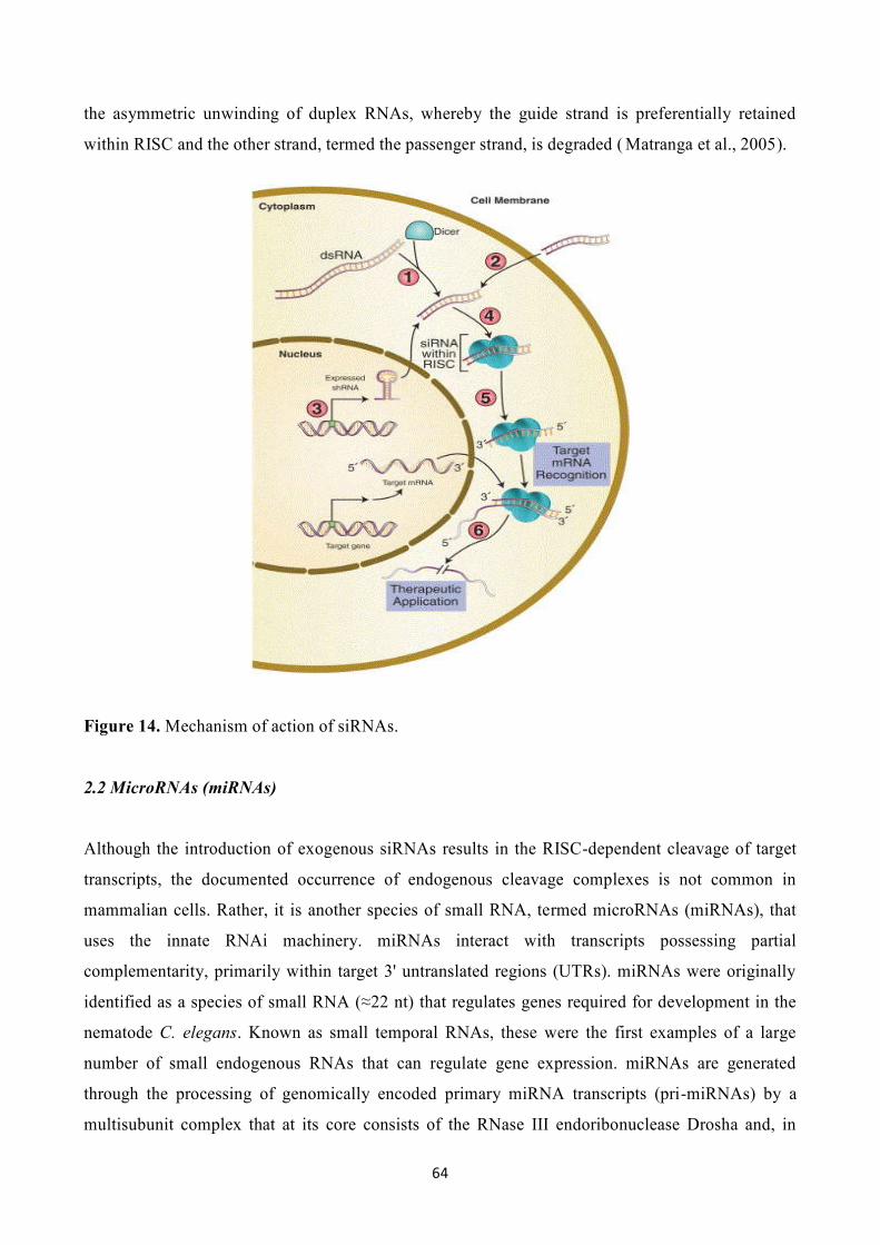
64
the asymmetric unwinding of duplex RNAs, whereby the guide strand is preferentially retained
within RISC and the other strand, termed the passenger strand, is degraded ( Matranga et al., 2005).
Figure 14. Mechanism of action of siRNAs.
2.2 MicroRNAs (miRNAs)
Although the introduction of exogenous siRNAs results in the RISC-dependent cleavage of target
transcripts, the documented occurrence of endogenous cleavage complexes is not common in
mammalian cells. Rather, it is another species of small RNA, termed microRNAs (miRNAs), that
uses the innate RNAi machinery. miRNAs interact with transcripts possessing partial
complementarity, primarily within target 3' untranslated regions (UTRs). miRNAs were originally
identified as a species of small RNA (≈22 nt) that regulates genes required for development in the
nematode C. elegans. Known as small temporal RNAs, these were the first examples of a large
number of small endogenous RNAs that can regulate gene expression. miRNAs are generated
through the processing of genomically encoded primary miRNA transcripts (pri-miRNAs) by a
multisubunit complex that at its core consists of the RNase III endoribonuclease Drosha and, in

65
mammalian cells, the DGCR8 protein. The processing of primary miRNA transcripts yields hairpin
structures known as precursor miRNAs (pre-miRNAs). Following export to the cytoplasm via
Exportin5 (Exp5), pre-miRNAs are processed by Dicer to produce mature miRNAs that incorporate
into miRNA ribonucleoprotein complexes (miRNPs). These complexes are similar, if not identical,
to RISC. The precise mechanism by which individual miRNAs recognize their target sites on the
mRNAs has not yet been completely unraveled but some general patterns have been determined
(Figs. 15 and 16). The sequence motif bound by the miRNA is situated in the 3'-untranslated region
(3'-UTR) of the transcript, i.e. between the protein-coding region of the mRNA and its poly(A) tail
(Stark et al., 2005). By sequence comparison of miRNAs and their cognate mRNA target sequences
it has been found that nucleotides 2 to 8 of the miRNA constitute a "seed region" that in most cases
binds to a perfectly complementary recognition sequence on the mRNA. The central part of the
miRNA usually lacks complementarity to the mRNA (typically nucleotides 10 and 11), whereas the
3'-region of the miRNA binds more or less specifically to the mRNA and contributes partly to the
specificity and affinity of the miRNA:mRNA complex (Brennecke et al., 2005). In a few instances,
the seed region does not show complete complementarity to the target sequence and in these cases a
strong binding of the miRNA 3'-region to the mRNA is required to stabilize the RNA duplex (as
seen in the interaction of miR-10 with the sex combs reduced (scr) transcript of Drosophila
melanogaster (Enright et al., 2003). miRNAs that rely mainly on their seed sequence for binding
may exert a function on the mRNA by themselves, whereas those that bind less strongly due to a
weaker seed sequence often have to act in concert with other miRNAs binding to the same mRNA
to cause an effect. Based on these binding requirements, computational calculations indicate that
each miRNA on average recognizes about 100 different mRNA targets (Brennecke et al., 2005).
Depending on the mode of base pairing between the miRNA and mRNA, one of two regulatory
pathways is employed—the short interfering RNA (siRNA) or the miRNA pathway. If perfect base
complementarity exists between the miRNA and mRNA, the mRNA will be processed through the
siRNA pathway and cleaved in a miRNA-directed manner. An endonucleolytic cleavage of the
mRNA is catalyzed by the ribonuclease in RISC (Ago2 in humans) in the center of the region
binding the miRNA and as a result the mRNA is degraded (Bagga et al., 2005). This mode of gene
silencing is common in plants, whereas only a few examples of mRNA silencing in this manner
have been reported for animals (one example is the miR-196-directed cleavage of the HOXB8
transcript during mouse embryogenesis (Yekta et al., 2004). However, this mechanism has proven
to be invaluable as an experimental tool for specific inactivation of target mRNAs in cells through
RNA interference (RNAi), where binding of a synthetic miRNA with perfect match to the target
mRNA causes degradation of the latter through the siRNA pathway.

66
Despite the existence of the siRNA pathway in human cells, most human miRNAs regulate mRNA
function through the miRNA pathway. Three modes of action have been unraveled, which result in
translational repression and partial mRNA decay: Repression of the initiation step of translation,
repression of the elongation phase of translation, and a general destabilization of the transcript as a
result of poly(A)-tail shortening. miRNA-directed inhibition of translational initiation was
supported by the finding of an effect of let-7 on the translation of reporter mRNAs in HeLa cells
(Pillai et al., 2005). Here, the distribution of polysomes of let-7-repressed mRNAs was shifted
towards the lighter fractions of a sucrose gradient in a manner similar to that observed when using
known inhibitors of translational initiation. Analogously, the cationic amino acid transporter 1
(CAT-1) mRNA, which is repressed by miR-122 in hepatocarcinoma cells under regular growth
conditions, was found in the light polysomal fraction. If, however, the cells were starved,
translational derepression of CAT-1 mRNA occurred and was accompanied by a shift in polysomal
distribution towards the heavier fractions (Bhattacharyya et al., 2006). The mechanism of miRNA-
directed repression requires a mRNA with a 5'-terminal m7G cap structure—as demonstrated by the
lack of inhibition by let-7 and CXCR4 miRNA of mRNAs, which are made to use a different
mechanism of translational initiation, namely internal ribosomal entry sites (IRES). Protein
production may also be inhibited by miRNAs during the process of translation. In Caenorhabditis
elegans, repression of the lin-14 mRNA by the miRNA lin-4 does not involve a change in polysome
distribution, indicating that repression occurs after initiation of translation. A number of reports
demonstrate an association between miRNAs and actively translating polyribosomes also in human
cells. The exact mechanism of repression has not been clarified yet but is probably due to either a
decrease in the elongation rate and/or degradation of the nascent protein (Nottrott et al., 2006).
Besides translational inhibition, an accelerated decay of the transcripts may also be observed
following their interaction with miRNAs by a mechanism distinct from the siRNA-type mRNA
cleavage. This mechanism of degradation relies on the recruitment of deadenylating and decapping
enzymes by the miRNAs with a subsequent degradation of the cognate transcript as result.
Mounting data indicate that mRNAs silenced by miRNA accumulate in cytoplasmic compartments
known as processing bodies (P-bodies) (Sen et al., 2005). The mRNAs found in these locations are
devoid of ribosomes and other translation factors. The P-bodies are rich in enzymes involved in
mRNA deadenylation, decapping, and degradation, and are believed to cause decay of the miRNA-
inhibited mRNAs (Sheth and Parker, 2003). In some instances, however, the mRNAs instead appear
to be stored in an inactive form in the P-body with the potential to re-enter the cytoplasm and re-
engage in translation (Brengues et al., 2005). One example of this is the miR-122-directed

67
repression of CAT-1 in hepatocarcinoma cells during normal growth, which is relieved by
starvation and results in re-translation of the CAT-1 mRNA (Bhattacharyya et al., 2006).
Figure 15. Mechanism of miRNAs formation.
Figure 16. Possible mechanisms of miRNA-mediated repression of target mRNAs in animals.

68
3. Applications of RNAi in mammalian systems
In most cases, the aim of RNAi-based experiments is the sequence-dependent cleavage and
reduction of protein-encoding mRNAs. Although most studies have focused on the RNAi analysis
of these targets, any RNA species can be targeted (for example, noncoding RNA transcripts or viral
RNAs). Only a limited number of mammalian cell types can tolerate RNAi induced by large,
exogenous dsRNAs (e.g., embryonic stem cells). Thus, it is usually necessary to use one of two
broad categories of RNAi effector molecules in mammalian systems. These include siRNA
duplexes, formed through the annealing of two independent RNA strands, or single-stranded RNA
molecules that contain a dsRNA domain, termed short-hairpin RNAs (shRNAs). In both cases,
RNAi effectors are designed to possess full complementarity with target transcripts, thereby
resulting in their cleavage.
3.1 RNAi effectors used for biological analysis in mammalian cells
siRNAs can be generated through the annealing of synthetic oligonucleotides. Most synthetic
siRNAs consist of 19 perfectly matched complementary ribonucleotides and 3' dinucleotide
overhangs that, for ease of synthesis, often consist of deoxyribonucleotides. Synthetic siRNAs are
available from a number of commercial vendors. More rarely, siRNAs are generated by a number of
other methods including in vitro transcription, plasmid-based tandem or convergent expression
cassettes, polymerase chain reaction (PCR) or the endonuclease digestion of large dsRNAs that
produce pools of siRNAs. The introduction of synthetic siRNAs into cultured mammalian cells
usually uses standard physico-chemical transfection methods, such as those based on cationic lipids,
cationic polymers, or electroporation. Empirical testing is required to determine the most
efficacious transfection conditions for any given cell system. Once well-optimized, transfected
siRNAs can yield a substantial decrease in the steady-state levels of target mRNAs for 24–120 h.
As opposed to direct transfection, shRNAs are usually expressed from plasmids or viral-based
expression vectors. shRNAs are designed to mimic miRNA precursors. Consequently, they are
processed by the endogenous RNAi machinery and loaded into RISC. A number of different
shRNA expression systems have been described. Variations include differences in promoter-
terminator combinations, linker sequences, flanking sequences, duplex length, and regulatory
elements that can be used for spatial and/or temporal-specific expression. Additionally, selection
markers, used to generate stable cell lines, and unique sequence elements, used to identify active
shRNAs among larger populations (discussed below), have been employed. As opposed to siRNAs,
the stable expression of shRNAs allows for a nontransient reduction of targeted mRNAs. Thus, the

69
choice of RNAi effector (siRNA or shRNA) depends on the question under investigation. No matter
the choice, it is always important to include negative control effector molecules in any RNAi-based
experiment. These controls, which are commercially available, incorporate sequences with minimal
complementarity to any endogenous transcript. In addition to improving conditions for their cellular
introduction and/or expression and subsequent processing, many studies have been directed toward
maximizing activity as a function of effector sequence (Schwarz et al., 2003). For example, an
understanding of any bias related to guide strand selection has obvious implications for design.
Analysis has revealed that the strand most easily unwound from its 5' end is preferentially
incorporated into RISC (Reynolds et al., 2004). Thus, effector design incorporates such bias to
encourage selection of intended guide strands. Studies have indicated other positional biases. For
example, high thermodynamic stability is preferred between nucleotides 5–10 of the guide strand
(Reynolds et al., 2004). Furthermore, empirical comparisons between large sets of effective and
ineffective siRNAs have led to the development of algorithms that assist in the generation of active
siRNAs. These types of design tools are incorporated into the production of commercial siRNAs
and are also publicly available. Sequences generated by these tools merely have an increased
probability of mediating RNAi. Only experimentation will establish the activity of any given RNAi
effector. Of note, an increasing number of validated sequences are available from commercial
sources and are being characterized and collated by the scientific community. Considerations of the
target are also important for maximizing RNAi. As RNAi effectors are designed according to
reported reference sequences, any discrepancies between those and the actual target sequences
within systems under study, for example as a consequence of single nucleotide polymorphisms
(SNPs), may prevent efficient RNAi (Martin and Caplen, 2006). However, the influence of
sequence discrepancies may be less than predicted owing to the fact that RISC can sometimes
tolerate mismatches within targets, especially those distal from the cleavage site (Martin et al.,
2007). Despite possibly interfering with RNAi, sequence aberrations can potentially be used to
selectively target mutated transcripts associated with disease. This approach has been applied in a
number of contexts including the targeting of cancer-specific mutations, the targeting of a single-
base mutation associated with the dominant genetic disorder spinocerebellar ataxia, and, most
recently, for the silencing of mutant β-globin as an approach toward treating sickle cell anemia
(Miller et al., 2003). In addition to potential sequence discrepancies, one should also ensure that
RNAi effectors target all known transcript variants of genes under study. Inevitably, even with an
increased understanding of RNAi, effectors invariably exhibit a spectrum of activity. Thus, it may
be prudent to obtain more than one effector against targets under investigation.
70
3.2 Off-target effects
The ability of RNAi effectors to elicit specific downregulation of intended targets while minimizing
or controlling for unintended effects, termed off-target effects, is critical for the meaningful
application of RNAi. Off-target effects are known to arise from a variety of mechanisms, which
include both sequence-independent and sequence-dependent processes. Sequence-independent
effects, or nonspecific effects, generally involve those relating to transfection conditions (e.g., lipid
transfection reagents), inhibition of endogenous miRNA activity, or stimulation of pathways
associated with the immune response. Sequence-dependent effects primarily concern the
unintentional silencing of targets sharing partial complementarity with RNAi effector molecules
through miRNA-like interactions, but also include receptor-mediated immune stimulation through
the recognition of certain nucleotide motifs. As discussed below, there are a number of approaches
toward controlling for both types of off-target effects.
1. Nonspecific Effects. Nonspecific effects resulting from the inhibition of endogenous miRNA
activity appear to depend on saturation of Exp5. For example, the shRNA-mediated inhibition of
miRNA activity is mitigated by the overexpression of Exp5. Also consistent with Exp5 as a
saturatable component of RNAi, its overexpression, but not the overexpression of other RNAi
components, enhances the activity of both miRNAs and shRNAs (Yi et al., 2005). As siRNAs do
not require export from the nucleus, their activity would not be expected to depend on Exp5.
However, some studies have found that siRNA-mediated RNAi is dependent on Exp5, where Exp5
prevents entry, and subsequent dilution, of siRNAs into non-nucleolar areas of the nucleus (Ohrt et
al., 2006). This is still controversial, as other studies have found no relationship between Exp5 and
siRNA activity (Yi et al., 2005). At the very least, it is clear that the activity of endogenous
miRNAs can be disrupted by the overexpression of shRNAs. The consequences of this not only
manifest in cell culture, but also in vivo, as Grimm and colleagues have shown that a high
percentage of shRNAs can cause lethality in mice regardless of shRNA target, or even the presence
of a target. Moreover, this toxicity correlated with high shRNA expression (Grimm et al., 2006).
Findings that RNAi effectors can saturate the endogenous machinery emphasize the importance of
using RNAi effectors at the lowest possible effective concentrations. Additionally, the use of
negative control siRNAs or shRNAs is paramount for the proper interpretation of res ults. In the case
of shRNAs, it does not seem adequate to simply use an empty vector control, as this does not
control for shRNA-mediated inhibition of the endogenous miRNA machinery.

71
Despite early perceptions that siRNAs of less than 30 nts would avoid the immuno-stimulatory
activity exhibited by larger RNA molecules (Elbashir et al., 2001), it has since been shown that
siRNAs can activate the immune response in a sequence-independent, concentration-dependent
manner. For example, 21-nt siRNAs have been shown to induce an interferon response in human
glioblastoma T98G cells through a process dependent on the activation of the dsRNA-dependent
protein kinase (PKR), and at least partially dependent on siRNA concentration (Sledz et al., 2003).
Similarly, both externally delivered siRNAs and shRNAs were found to induce an interferon
response in HEK293 and HaCaT keratinocyte cell lines (Kariko et al., 2004). Additional studies in
HEK293 cells found this response to be primarily dependent on Toll -like receptor 3 (TLR3) (Kariko
et al., 2004). Importantly, the induction of an interferon response is cell-line dependent, with long
siRNAs of 27 nt unable to activate a response in certain cell lines, including HeLa cells (Reynolds
et al., 2006). The expression of shRNAs can also induce an interferon response. As with siRNAs,
shRNA-mediated activation also appears to be concentration dependent (Bridge et al., 2003). Thus,
similar to saturation of the endogenous RNAi machinery, the use of lowest effective concentrations
and negative control RNAi effectors are necessary to control for stimulation of interferon-type
responses. Additionally, chemical modifications that help prevent the activation of PKR have been
described.
2. Sequence-Dependent Effects. siRNAs can also induce an immune response through sequence-
dependent effects, particularly when it is part of a lipid or polycation complex in vivo. More
specifically, certain nucleotide motifs, especially GU-rich sequences, can induce interferon-α (IFN-
α), interleukin-6 (IL-6), and tumor necrosis factor-α (TNF-α), probably through activation of TLRs.
For example, a subset of liposome-encapsulated siRNAs was found to induce a substantial, dose-
dependent IFN-α response in mice. These siRNAs also stimulated an immune response in human
peripheral blood mononuclear cells and isolated plasmacytoid dendritic cells. The stimulatory
siRNAs were found to share UGUGU motifs that were presumably recognized by endosomal TLR7
and/or TLR8 (Judge et al., 2005). Similarly, siRNAs were found to stimulate IFN-α production in
human plasmacytoid dendritic cells through a GUCCUUCAA motif. In this case, experiments
confirmed that stimulation was dependent on recognition by TLR7. Because nonimmune cells do
not express detectable TLR7 or TLR8, sequence-dependent immune stimulation is not thought to
influence experiments conducted in commonly used cell lines (Marques et al., 2006), but sequence-
dependent stimulation is clearly an important issue regarding in vivo applications. Although specific
siRNA nucleotide motifs can activate an immune response, the primary source of sequence-
dependent off-target effects originates from partial complementarity between RNAi effectors and
off-target transcripts. Such interactions are similar to those exhibited by endogenous miRNAs,
72
which usually share complementarity between nucleotides within their 5' ends and regions within
target 3' UTRs (Lewis et al., 2005). In fact, much like miRNA targets, off-targeted transcripts are
enriched in those containing complementarity between their 3' UTRs and hexamer (nts 2–7) and
heptamer (nts 2–8) sequences within 5' ends of RNAi effectors (Jackson et al., 2006). Some studies
have found these effects to be nontitratable, with dose responses mirroring that of on-target
transcripts (Jackson et al., 2003). Others have found these effects to be concentration-dependent,
whereby the use of low siRNA concentrations can significantly mitigate off-target interactions
(Semizarov et al., 2003). Importantly, most detailed studies of off-target effects are conducted using
gene expression analysis. However, since miRNAs can impede translation in a manner
disproportionate with alterations in target mRNA levels, the magnitude of off-target effects may be
underestimated. Sequence-dependent off-target effects can have functional consequences. For
example, different siRNAs targeting the same gene can exhibit varying effects on the mRNA and
protein levels of key cellular genes, independent of on-target silencing (Scacheri et al., 2004).
Accordingly, a high percentage of siRNAs can induce a toxic phenotype. For example, 51 of 176
randomly selected siRNAs directed against either firefly luciferase or human DBI reduced the
viability of HeLa cells by more than 25%, a trend that was reproducible in different cell lines. From
a practical perspective, off-target effects can have a profound effect on experimental results. For
example, Lin and colleagues determined that the top three “hits” from a siRNA-based screen for
targets affecting the hypoxia-related HIF-1 pathway resulted from off-target effects (Lin et al.,
2005). For two of these three “hits,” activity could be traced to interactions within the 3' UTR of
HIF-1A itself. Of note, off-target effects not only affect experiments conducted in mammalian
systems, but can also influence studies in Drosophila (Ma et al., 2006). There are a number of ways
to control for, and help minimize, sequence-dependent off-target effects. Many of these relate to
siRNA design features. For example, the use of asymmetric design, which helps to minimize the
loading of passenger strands into RISC, thereby reducing associated off-target effects, and the use
of siRNAs designed to avoid homology with untargeted transcripts. Both of these considerations are
typically incorporated into the design of commercially available siRNAs. Increased stringency may
be gained through the development of new algorithms that include emphasis on avoiding
complementarity between siRNAs and untargeted 3' UTRs. Chemical modifications that reduce
sequence-dependent off-target effects have also been described. For example, the incorporation of
2'-O-methyl groups within the first two 5' nucleotides of siRNA passenger strands reduces
passenger strand-mediated activity. Similarly, a 2'-O-methyl ribosyl substitution at position 2 of the
guide strand can significantly reduce sequence-dependent off-target effects (Jackson et al. , 2006).
Modified siRNAs exhibiting reduced off-target effects are commercially available. As with

73
sequence-independent effects, experimental conditions should be optimized to use the lowest
effective dose of the RNAi effector. Despite all of these considerations, the occurrence of sequence-
dependent off-target effects may be unavoidable. Consequently, efforts should be made to help
validate hits. All RNAi-derived phenotypes should be confirmed with additional RNAi effectors
against the same target. Moreover, the downregulation of target mRNA and protein levels should be
characterized and correlated with the observed effects. For example, the inactivity of a follow-up
siRNA does not necessarily imply that the activity of the first resulted from off-target effects,
especially if the second siRNA is unable to downregulate target levels. Conversely, a phenotype
induced by only a fraction of siRNAs directed against the same target, despite equivalent silencing
by all siRNAs, would be suspicious. Overall, it is difficult to prescribe the number of independent
RNAi effectors necessary for target validation, but it would certainly require at least two. No matter
how many independent RNAi effectors are tested, it could be possible that observed phenotypes
result from cooperative effects between target-specific downregulation and nonspecific effects.
Even a rescue experiment using a target construct resistant to RNAi could not control for such
scenarios. Thus, confirmation of phenotypes under different experimental conditions (e.g., the use
of a different lipid reagent or the use of an siRNA to confirm an shRNA-derived phenotype) may
help to eliminate some of these possibilities. Additionally, RNAi-independent methods, such as the
chemical inhibition of identified targets, should be used to corroborate phenotypes where possible.
3.3 Application of RNAi-based technologies
RNAi has enormous potential for the treatment of many genetic and acquired diseases. For
example, RNAi could potentially be used to reduce the levels of toxic gain-of-function proteins,
trigger cytotoxicity within tumors, or block viral replication. The use of RNAi-based therapeutics is
especially appealing as RNAi can be used to modulate the expression of proteins not normally
accessible by more traditional pharmaceutical approaches. For example, nondruggable targets
lacking ligand-binding domains or proteins sharing high degrees of structural homology that are
difficult to target as individuals are all accessible by RNAi. The in vivo application of RNAi was
described within a year of the first cell culture experiments, with reports describing the transient
inhibition of transgenes within the livers of mice. This was accomplished through high-pressure tail
vein injection of both siRNAs and shRNAs (McCaffrey et al., 2002). Subsequent in vivo studies
have focused on the improved delivery and efficacy of RNAi effectors. These efforts have used the
experience gained through two decades of developing ribozyme and antisense-based therapeutics
and the gene therapy field as a whole. Currently, most in vivo studies using synthetic siRNAs use

74
lipid-based carriers with or without modification of the siRNA itself, whereas most shRNA-based
studies employ the standard viral vector expression systems used in traditional gene therapy.
1. In Vivo Application of Synthetic siRNAs. The in vivo delivery of synthetic siRNAs must account
for the need to ensure resistance to exonuclease digestion, the maintenance of duplex stability, good
pharmacokinetics, and the minimization of nonspecific immunological responses. Accordingly, a
number of siRNA chemical modifications that address these issues have been examined. Many of
these modifications are analogous to those incorporated in RNase H-dependent antisense
oligonucleotides. A common modification to improve stability is the use of a partial
phosphorothioate backbone, particularly within the 3' overhangs of both siRNA strands.
Furthermore, the inclusion of 2'-O-methyl dinucleotides at the 3' end of the antisense strand has also
been shown to improve stability. As mentioned, avoiding immune stimulation is also critical. The
selection of sequences that avoid GU-rich sequences and/or modification with 2'-O-Me nucleotides
or locked nucleic acids (LNAs) have all been shown to inhibit stimulation of the immune system
without concomitant loss of efficacy (Judge et al., 2006). Chemical modifications have also been
engineered to improve cellular uptake. For example, cholesterol-conjugated siRNAs, corresponding
to the ApoB gene, have been delivered into the livers of mice as a potential strategy for the
treatment of familial cholesterolemia and, possibly, for the broader treatment of atherosclerosis
(Soutschek et al., 2004). These conjugates were found to induce a significant decrease in both liver
ApoB mRNA and plasma ApoB protein levels, as well as downstream lipoprotein and cholesterol
levels. These effects were much greater than those observed with nonconjugated analogs.
Unfortunately, the quantity of material necessary for efficient silencing was incompatible with
scale-up to larger preclinical models, thus follow-up studies in nonhuman primates used a different
delivery strategy (Zimmerman et al., 2006). In addition to directly modifying siRNAs for improved
characteristics, carrier molecules also have the potential to protect siRNAs from the extracellular
environment and improve intracellular delivery. A wide variety of polymer- or lipid-based delivery
systems have been described. For example, cationic polyethylenimines have been used for siRNA
transfection in vivo, including delivery to lung and xenografts following subcutaneous,
intraperitoneal, and intrathecal administration (Thomas et al., 2005). A large number of different
liposome-based carriers have also been developed for the in vivo delivery of siRNAs. One such
system that has been relatively well characterized uses lipid-polyethylene glycol (PEG) mixtures to
encapsulate siRNAs. This delivery system has been used for the systemic delivery of APOB-
targeted siRNAs into the livers of nonhuman primates, causing a significant reduction in both
APOB mRNA and protein levels. Furthermore, a relatively sustained (11-day) reduction in low-
density lipoprotein was observed in animals receiving the highest dose of siRNA lipid (2.5 mg/kg).

75
Similarly, this lipid-encapsulated siRNA system has been used in studies directed toward inhibiting
viral infections (Mousses et al., 2003).
2. In Vivo Application of shRNAs. The first studies applying shRNAs in vivo used plasmid DNA
(Lewis et al., 2002); however, most subsequent studies have focused on the use of viral vectors. The
choice of viral delivery system usually depends on the cell type under investigation and on the need
for short- or long-term shRNA expression. For example, adenoviral (AV) and herpes simplex viral
vector systems have been primarily used for short-term expression, while adeno-associated viral
(AAV) vectors and the integrating viral vector systems based on retroviruses (RVs) and lentiviruses
(LVs) have usually been used for long-term expression or for applications in nondividing cells. An
important adaptation of RNAi has come from the ability to stably express shRNAs in blastocytes or
embryonic stem cells, from which transgenic animals can be generated. Initial “proof of concept”
experiments used shRNAs to target overexpressed marker genes (e.g., green fluorescence protein)
in transgenic animals. These studies used either direct injection or lentiviral transduction of early
embryos (Hasuwa et al., 2002). Subsequent studies have demonstrated the feasibility of targeting
endogenous genes within embryonic stem (ES) cells (Rubinson et al., 2003). These models broadly
mimic the phenotype of traditional knockout mice. Consequently, the constitutive expression of an
shRNA for the generation of an RNAi-based transgenic is only compatible with genes that do not
compromise animal viability. To circumvent embryonic lethality, shRNA-based conditional
expression systems have been developed. These include Cre-Lox-based systems whereby the
shRNA is flanked by LoxP sites that prevent shRNA expression. Tissue-specific or temporal-
specific shRNA expression can then be achieved by crossing shRNA transgenic mice with Cre
recombinase expressing mice (Ventura et al., 2004). Although Cre-lox RNAi-based systems are
irreversible, reversible expression, predominantly using doxycycline-based control systems, has
been described. RNAi transgenics have also been used in animals not normally amenable to
traditional homologous recombination techniques, including rat and goat. In the case of goat, a
RNAi transgenic was generated through somatic cell nuclear transfer from a LV-transduced goat
fibroblast stably expressing an shRNA corresponding to the prion protein (Golding et al., 2006).
While the development of RNAi transgenics was initially hailed with great excitement, its broader
use has not been adapted as quickly as may have been anticipated. This may be due to difficulties in
obtaining efficient lentiviral transfection of embryos or ES cells, difficulties in generating ES clones
that stably express shRNAs or problems associated with variations in knockdown efficiency. The
recent adaptation of the more conventional pronuclear injection procedure may enable wider use of
RNAi in the development of transgenics. shRNAs have also been used in xenograft tumor models,
particularly in mice. One of the first examples of this was targeting an activated mutant of K-RAS

76
found in the pancreatic carcinoma cell line CAPAN-1 using a mutant-specific shRNA expressed
from a retroviral vector (Brummelkamp et al., 2002). In contrast to control cells, the KRAS-targeted
cells failed to form tumors in athymic mice, demonstrating the ability of RNAi-mediated silencing
to suppress tumor formation in vivo. Another interesting example involves targeting the tumor
suppressor TP53 in mouse Eμ-Myc hematopoietic stem cells. By using shRNAs that mediated
different degrees of TP53 message reduction, the percentage of mice developing lymphoma could
be varied as a function of TP53 protein levels. More recent variants of this method include the use
of an inducible expression system that can be activated upon xenograft tumor formation, potentially
generating a better clinical model for the identification and validation of anticancer molecular
targets (Li et al., 2005).
3.4 “Trans-kingdom” RNAi
Several years ago, it was demonstrated that systemic gene silencing could be attained in the
nematode Caenorhabditis elegans when it ingested E. coli engineered to produce interfering RNAs,
suggesting that RNAi-mediated information transfer between species or kingdoms might be
possible (Timmons and Fire, 1998; May and Plasterk, 2005). Bacteria engineered to produce a short
hairpin RNA (shRNA) can induce trans-kingdom RNAi in vitro and in vivo also in mammalian
systems. Nonpathogenic Escherichia coli can be engineered to transcribe shRNAs from a plasmid
containing the invasin gene Inv and the listeriolysin O gene HlyA, which encode two bacterial
factors needed for successful transfer of the shRNAs into mammalian cells. Upon oral or
intravenous administration, E. coli encoding shRNA induce significant gene silencing in the
intestinal epithelium and in human colon cancer xenografts in mice (Xiang et al., 2006). These
results provide an example of trans-kingdom RNAi in higher organisms and suggest the potential of
bacteria-mediated RNAi for functional genomics, therapeutic target validation and development of
clinically compatible RNAi-based therapies.
3.5 Prediction of microRNA targets (Mazière and Enright, 2007. review)
Currently, there are 474 confirmed microRNAs (miRNAs) in humans, although there might be
many more. miRNAs are expected to have multiple targets; however, few have been confirmed
experimentally (only 66 of potentially thousands so far). In the absence of high-throughput
experimental techniques to determine the targets of miRNAs, it is vital that computational
techniques are developed to unravel their regulatory effects and implications for diseases and

77
diagnostics. Indeed, recent studies have already implicated miRNAs in numerous human diseases,
such as colorectal cancer, chronic lymphocytic leukaemia and fragile X syndrome. Hence, both the
miRNA itself and its regulatory targets are potentially druggable. The prediction of miRNA targets
has been ongoing since the 3′ untranslated regions (3′UTRs) of transcripts were determined to
contain binding sites for them. The efficacy of computational approaches to locate and rank
potential genomic binding sites is supported by the relatively high degree of miRNA
complementarity to experimentally determined binding sites. Despite the later identification of
hundreds of miRNAs in a variety of species, through large-scale and sequencing projects, only a
handful of targets had been identified experimentally, for an even smaller number of miRNAs.
Given the laborious nature of experimental validation of targets, and despite the limited data
available, it was imperative that computational approaches be developed that could produce reliable
and testable predictions.
1. miRNA size. The apparent complementarity between miRNA and target could have been seen as
an advantage for computational analysis. However, other features of miRNA–UTR associations
make matters more complicated. Conventional sequence alignment algorithms assume longer
sequences than the 20–23 nucleotides of miRNAs. This short length makes ranking and scoring of
targets difficult because statistical techniques for sequence matching (such as Karlin–Altschul
statistics) require longer sequences. Binding sites actually consist of regions of complementarity,
bulges and mismatches. Because standard sequence analysis tools were designed for sequences with
longer stretches of matches and fewer gaps, they are much less useful for miRNA target prediction.
Recently, position 2–7 of miRNAs, the so-called ‘seed’ region, has been described as a key
specificity determinant of binding, and requires perfect complementarity and. If one ignores GC
content and performs an order of magnitude calculation, then a perfect match for a six-nucleotide
seed region of a miRNA should occur approximately once in every 1.3 kb in a genome – in other
words, on average, almost once in every human 3′UTR. However, it would not seem realistic for a
single miRNA to regulate more than a few hundred targets. Effective regulation of transcript
translation requires that miRNAs and their targets are located in the same cellular compartments.
Hence, most of these theoretical targets correspond to false positives.
2. Identification of 3′UTRs. To identify miRNA targets in a given species, knowledge of the set of
3′UTRs for this species is a vital step. Despite the accumulation of genome sequences for many
species, the location, extent or splice variation of 3′UTRs is still poorly characterized for many
mammals. Some species-specific projects, such as the Berkeley Drosophila Genome Project
(BDGP), produce high-quality transcript information that makes possible the accurate determination

78
of a 3′UTR, from stop codon to polyadenylation site. For other species, such as Homo sapiens,
some transcripts are well defined, whereas others remain poor in their description. The Ensembl
database uses alignment of cDNAs and expressed sequence tags to genomic sequences to extract
3′UTR regions, and so far, evidence is available for human, mouse and zebrafish genomes.
However, 30% of human genes lack definitive 3′UTR boundaries. These regions can be estimated
by selecting a downstream flanking sequence of the stop codon, corresponding to the length of an
average human 3′UTR (e.g. 1 kb). Experimental techniques, such as tiling arrays, and ditag or cage
tagging, seem to be promising approaches for the generation of high-quality 3′UTR datasets.
Attaining reliably annotated and verified 3′UTR datasets will potentially benefit target prediction
more than making small improvements to existing prediction methods. In the context of drug
discovery, both 3′UTRs and miRNA genes represent drug target candidates through either the
generation of synthetic miRNAs or the repression or overexpression of existing miRNAs.
3. Conservation analysis. Solutions to reduce the number of false positives in target predictions
include filtering out those binding sites that do not seem to be conserved across species. The use of
predicted binding sites conserved across orthologous 3′UTRs in multiple species are considered
more likely to reduce the number of false positives. However, recently evolved miRNAs, such as
miR-430 in zebrafish, might not have conserved targets in the scope of the currently available set of
fish genomes. One caveat of conservation analysis concerns the set of species that are compared:
looking for conserved targets between humans and chimpanzees will not be helpful, given that at
least 99% of the entire transcript will be conserved. Other species might seem more relevant for
comparing with human transcripts (e.g. mouse, rat, or dog), but the fact is that genomes are not
sequenced according to their evolutionary distances. As a result, the number of false positives can
effectively be greatly reduced but this is at the expense of increased false negatives.
4. Computational target-prediction approaches. Different methods have been developed for
computational target prediction (Tab. 1). These might or might not be made available as functional
packages but the results are always available, at least as a precomputed set of transcripts, through
online resources.
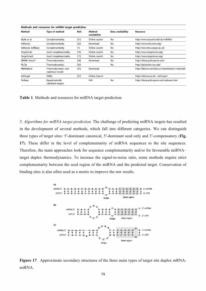
79
Table 1. Methods and resources for miRNA target prediction.
5. Algorithms for miRNA target prediction. The challenge of predicting miRNA targets has resulted
in the development of several methods, which fall into different categories. We can distinguish
three types of target sites: 5′-dominant canonical, 5′-dominant seed only and 3′-compensatory (Fig.
17). These differ in the level of complementarity of miRNA sequences to the site sequences.
Therefore, the main approaches look for sequence complementarity and/or for favourable miRNA–
target duplex thermodynamics. To increase the signal-to-noise ratio, some methods require strict
complementarity between the seed region of the miRNA and the predicted target. Conservation of
binding sites is also often used as a metric to improve the raw results.
Figure 17. Approximate secondary structures of the three main types of target site duplex mRNA-
miRNA.

80
6. Complementarity searching. The first algorithms did not develop statistical background models
to evaluate the significance of each detected hit; rather, they were oriented towards recovery of
known targets and the detection of further targets for experimental validation, so that our
knowledge of miRNA binding dynamics might be improved. In most cases, they used
complementarity initially to identify potential targets, followed by iterative rounds of filtering based
on thermodynamics, binding site structure and conservation. After these filtering steps, a score is
typically applied to each detected target; this score can be useful for target ranking. Initial attempts
at false-positive rate estimation usually relied on comparing detection methods for real miRNAs
and shuffled control miRNAs.
7. The method of Stark and co-workers. Large-scale prediction of miRNA targets was first
successfully published for Drosophila melanogaster, for which well-annotated and accurate 3′UTRs
could be obtained from the BDGP. The sequence search tool HMMer was used to identify the
reverse complement miRNA sequences. Similar profiles were built to enable G:U wobble matches.
Following the prediction algorithm, the resulting 3′UTRs were filtered for conservation in
Drosophila pseudoobscura and Anopheles gambiae. The detected target sites were scored and used
as an input for the MFold algorithm, to evaluate the thermodynamic stability of the miRNA–target
association. Despite a statistical model based on the normal distribution rather than the extreme
value distribution, the method predicted previously validated D. melanogaster miRNA binding
sites. Many previously unknown binding sites were also predicted, six of which were
experimentally validated.
8. miRanda. The miRanda algorithm was the second method to be published. As with the method
by Stark et al., miRanda identifies potential binding sites by looking for high-complementarity
regions on the 3′UTRs. The scoring matrix used by the algorithm is built so that complementary
bases at the 5′ end of the miRNA are rewarded more than those at the 3′ end. Hence, the binding
sites exhibiting a perfect or almost-perfect match at the seed region of miRNAs display a better
score. The resulting binding sites are then evaluated thermodynamically, using the Vienna RNA
folding package. This first version of miRanda successfully predicted many known targets in D.
melanogaster. The BDGP 3′UTRs dataset was used, and the results were filtered, as described
above, to limit predictions to targets conserved in D. pseudoobscura. When classified according to
gene ontology terms, the miRanda-predicted targets were shown to display specific functional
patterns for each miRNA. Expression data analysis confirmed this property by suggesting that many
individual miRNAs have highly specific roles in particular tissues, processes and pathways. When
basic parameter settings are used, the approximated false-positive rate was between 24% and 39%.

81
These values are significantly decreased when multiple sites are considered. Newer miRanda
versions implement a strict model for the binding sites that requires almost-perfect complementarity
in the seed region with only a single wobble pairing. Other variations of these algorithms are
currently under development for a version 3.0 of miRanda. These incorporate a statistical model
equivalent to that used by RNAHybrid, thereby they efficiently reduce the rate of false-positive
predictions. Despite their similar methods and identical input datasets, the scoring and ranking
strategies devised by Stark et al. and miRanda are different: only 40% of the respective top-ten
miRNA targets predicted by both methods overlap. It seems that even small differences in the
criteria used for ranking and scoring lead to large differences in the set of predicted targets. The
multiplicity of miRNA binding sites on the same 3′UTRs drastically improves their statistical
significance in both methods. This is confirmed by experimental evidence showing that multiple
sites enhance the silencing effect of miRNAs. However, many miRNAs still seem to operate at a
single site on their targets. One given explanation implies that a miRNA exhibiting high
complementarity to its single-site target could have the same regulatory effect as a miRNA with a
lower level of complementarity but a multiple-site target.
9. TargetScan and TargetScanS. Although the previously mentioned methods attempt to find all
potentially complementary sites and then filter them according to different criteria, TargetScan uses
a different approach. This method requires perfect complementarity to the seed region of a miRNA
and then extends these regions to unravel complementarity outside the region. This aims at filtering
many false positives from the beginning of the prediction process. For the same purpose, the
conservation criteria, based on the presence of the seed region in an island of conservation, are
introduced early in the process by using groups of orthologous 3′UTRs as input data. The following
step is common to the other methods: the predicted binding sites are tested for their thermodynamic
stability, in this case with RNAFold from the Vienna Package. TargetScan was the first method to
be applied for human miRNA target prediction, using mouse, rat and fish genomes for conservation
analysis. Shuffled sequences, with maintained dinucleotide compositions that mimic real 3′UTRs,
were used to determine the significance of binding sites. The estimated false-positive rate varies
between 22% and 31%. The method was shown to predict not only known miRNA binding sites but
also novel sites. Luciferase reporter constructs validated 11 of the 15 tested sites. TargetScanS
simplified the TargetScan method and improved the target prediction fidelity. The miRNA
complementarity is now limited to a six-nucleotide seed, followed by an additional 3′ match of an
adenosine anchor at position 1. No other criteria are required once the previous conditions are met;
contrary to previous algorithms, single-site 3′UTRs are sufficient for a reliable prediction.
TargetScan and TargetScanS feature an efficient reduction in the false-positive rate but, because of

82
the required strict complementarity in the seed region, loosely conserved targets and those
containing wobble pairings are more likely to be missed, including 3 ′-compensatory sites.
10. PicTar. The PicTar algorithm uses a group of orthologous 3′UTRs from multiple species as the
input dataset. The algorithm scans the alignments of 3′UTRs for those displaying seed matches to
miRNAs. The retained alignments are then filtered according to their thermodynamic stability. Each
predicted target is scored by using a Hidden Markov Model (HMM) maximum-likelihood fit
approach. PicTar is the first method that uses the criteria of co-expression in space and time of
miRNAs and their targets. The experimental validation of seven out of 13 tested predicted targets,
as well as the confirmation of eight of nine previously known targets, demonstrates the efficiency of
the algorithm.
11. DIANA-microT. The DIANA-microT method uses a 38-nucleotide window that is progressively
moved across a 3′UTR sequence. Using dynamic programming, the free energy (ΔG kcal/mol) of
the potential binding site is calculated at each step and compared with the results obtained from
shuffled sequences with the same dinucleotide composition as real 3′UTRs. Contrary to sequence
complementarity-based methods, DIANA-microT demands 3′ complementarity to the miRNA and
does not bother with site multiplicity. Using this technique, all currently known C. elegans miRNA
binding sites were predicted successfully, with false-positive rates similar to those found in
previously described methods.
12. RNAHybrid. The lack of strong statistical models is one of the main criticisms that can be
levelled at the methods previously described here. RNAHybrid was the first method to address this
issue by developing a model as robust as those used for large-scale sequence comparison. Contrary
to tools such as MFold and Vienna, which are designed for single-sequence folding and therefore
require an artificial linker between the miRNA and its potential binding site, RNAHybrid identifies
regions in the 3′UTRs that have the potential to form a thermodynamically favourable duplex with a
specific miRNA. The maximum free energy of a miRNA is calculated for every 3′UTRs of a set of
shuffled 3′UTR sequences with maintained dinucleotide frequencies. Normalisation for both 3′UTR
and miRNA length using Snorm = log(S/mn) is applied to these energies. Random energies derived in
this manner should exhibit an extreme value distribution (EVD). Subsequently, the parameters of
the EVD that best describe the data for a given miRNA are empirically calculated using the derived
distribution from shuffled sequences. Each hit to any 3′UTR for this miRNA is then assigned a P
value calculated directly from these parameters. Hence, at the scanning stage, miRNAs are scanned
against a database of real 3′UTRs, and each hit is compared with the expected distribution and
assigned a P value. Moreover, the statistical model implemented in RNAHybrid takes into account

83
multiple sites and conserved sites, by respectively combining individual P values using Poisson
statistics and calculating conservative P values for conserved sites. A statistical fitting approach
corrects for highly conserved 3′UTRs by evaluating the overall conservation in the group of
sequences compared with the conservation at the site. The resulting statistics cover individual site
quality, quantity of sites, whether they are conserved and how significant this conservation is, given
the input sequences. The method was successfully tested to predict known targets in D.
melanogaster, with a low false-positive rate. The association of P values with predicted targets is an
appreciable asset for directly comparing predicted binding sites.

84
4. MicroRNAs and Cancer
4.1 Disruption of miRNA-directed regulation
As each miRNA is expected to regulate the translation of up to 100 mRNAs (Lim et al., 2005) it is
clear that disturbances of the miRNA expression level, processing of the miRNA precursors, or
mutations in the sequence of the miRNA, its precursor, or its target mRNA, may have detrimental
effects on cell physiology. A number of such aberrations have been associated with cancer, as
described in detail below, and can in short be categorized as the following types of lesions:
a) Alterations of the miRNA expression level. This may occur by gross genomic alterations
such as deletions, insertions, inversions, and translocations (Calin et al., 2004), by
epigenetic changes of the miRNA gene (Saito et al., 2006), by insertion of a viral element
near the miRNA gene that disrupts normal transcriptional regulation (Wang et al., 2006), or
minor genetic changes such as a point mutation in a promoter element of the miRNA gene
or in the coding region for a transcription factor that is crucial for miRNA transcription (Xi
et al., 2006).
b) Alterations affecting miRNA processing. Changes in the expression level of a large number
of miRNAs may be a consequence of a disruption of the miRNA-processing apparatus
(Sugito et al. ,2006). Changes affecting translation of only a few mRNAs can be caused by
an alteration in the primary sequence of the pri-miRNA, which affects its downstream
processing efficiency (Diederichs et al., 2006).
c) Mutations in the miRNA:mRNA interacting sequences. Alterations in the translation
efficiency of a mRNA may also be a result of a base change in the mature miRNA or in the
target sequence of the mRNA, which weakens the interaction between the miRNA and the
mRNA (Iwai and Naraba, 2005). This interaction is especially sensitive to mutations in the
seed region.
There is now ample evidence that the expression of miRNAs is altered in cancer, and that certain
changes may be directly implicated in the carcinogenic process. A number of miRNAs have been
shown to promote cell proliferation and survival, while others diminish cell proliferation and
survival. These two classes of miRNAs may play a central role in cancer development as novel
oncogenes and tumor suppressors, respectively. In general, the majority of miRNAs are
downregulated in cancer specimens (Lu et al., 2005). In normal tissues, some of these miRNAs
have been documented to inhibit the translation of proto-oncogenes by targeting the 3'ends of their

85
mRNAs. Such miRNAs are therefore considered as "tumor suppressor miRNAs" (TS-miRs) since
their normal function is to control the expression of an oncogene. Conversely, certain miRNAs
seem to be upregulated in cancer and may act as "oncomiRs" since they can enable the
downregulation of a tumor suppressor. Since miRNAs have several potential targets that may be the
mRNAs of both oncogenes and tumor suppressors, the actual function of a particular miRNA as
either TS-miR or onco-miR may depend on the cellular context. The mechanisms by which miRNA
expression is altered in cancer are multifaceted. The function of miRNAs in cancer seems to be
disrupted by the same mechanisms as those that affect the expression of protein-encoding genes, i.e.
amplifications, translocations, deletions, and point mutations of the pri-miRNA-encoding DNA
sequence, and by epigenetic disruption of miRNA transcription (Fig. 18).
4.2 MiRNAs as tumor suppressors and oncogenes
1. Tumor suppressor miRNAs. One of the first indications of a direct involvement of miRNAs in
cancer was the linking of the miR-15- and miR-16-encoding sequences to a critical region of
deletion of only 30 kb at 13q14, which is lost in more than half the cases of chronic lymphocytic
leukemia (CLL) (Calin et al., 2002). Previous comprehensive analyses for candidate tumor
suppressor genes within this region had turned out to be unsuccessful. However, the targeting of
miR-15 and miR-16 to the minimal region of LOH (loss of heterozygosity) in CLL combined with
the recent findings of germline mutations of pri-miRs-15 and -16 in a case of familial CLL indicated
that they might represent the long sought CLL-associated tumor suppressor located in this region
(Calin et al., 2005). Furthermore, expression analyses have shown that as many as 68% of all CLLs
show downregulation of miRs-15 and -16. Both miRNAs were shown to act as tumor suppressors
by targeting translation of the anti-apoptotic BCL-2 mRNA (Calin et al., 2002), an oncogene that
frequently is found to be overexpressed in CLL. Downregulation of miR-15 and -16 has been
shown to correlate with overexpression of the BCL-2 protein, and transfection with either of the two
miRNAs completely abolished protein expression and re-established apoptosis in a leukemia model.
Another early and well-documented finding was the downregulation of oncogenic Ras by the let-7
family members of miRNAs in lung cancer (Johnson et al., 2005). It was observed that low Let-7
expression correlated with a shortened post-operative survival in lung cancer patients who had
undergone potentially curative operative procedures. Since then, a large number of miRNAs have
been shown to be downregulated in various cancers, including the downregulation of let-7 (Akao et
al., 2006a), miR-143, and miR-145 in colorectal cancer (Akao et al., 2006b), miR-145 in breast
cancers (Iorio et al., 2005), and miR-29b in CLL (Pekarsky et al., 2006) and AML (Garzon et al.,
2007) (Tab. 2).

86
Figure 18. Mechanism of action of tumor suppressor and oncogenic miRNAs
2. Oncogenic miRNAs. In contrast to TS-miRs, oncomiRs are frequently upregulated in cancers and
show proliferative and/or anti-apoptotic activity. One of the first oncomiRs to be identified was
miR-155, which is co-expressed with the non-protein-coding gene BIC. The exact target mRNAs of
miR-155 remain to be established, but early observations showed that high expression of this
miRNA led to an increase in leukemia and lymphoma formation in chicken. More recent studies
showed overexpression of miR-155 in diffuse large B-cell lymphoma (DLBCL), Hodgkin's disease,
and primary mediastinal DLBCL (Eis et al., 2005). Initially, high expression was also reported in
pediatric Burkitt's lymphoma; however, recent observations show disruption of miR-155 processing
in Burkitt lymphoma cell lines (Kluiver et al., 2006). In DLBCL and lung adenocarcinomas, high
expression of miR-155 has been associated with aggressive variants of tumors and poor survival
(Yanaihara et al., 2006). The members of the miR-17-92 cluster represent another intensely studied
group of potential oncomiRs that are frequently upregulated in lymphomas. This cluster consists of
seven individual miRs: miR-17-5p, 17-3p, -18, -19a,-19b1, -20, and 92, which are all encoded from
a frequently amplified locus at 13q31.3 (Ota et al., 2004). It was shown in Eμ-Myc transgenic mice
that the miR-17-92 cluster, but not the individual miRNAs, could enhance tumorigenesis by
inhibiting apoptosis in the c-Myc-overexpressing tumor. Further studies in human cell lines showed
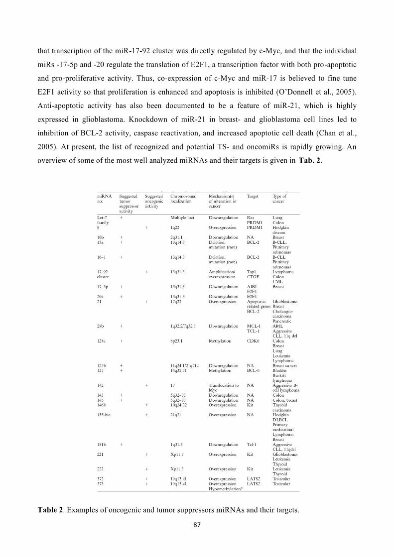
87
that transcription of the miR-17-92 cluster was directly regulated by c-Myc, and that the individual
miRs -17-5p and -20 regulate the translation of E2F1, a transcription factor with both pro-apoptotic
and pro-proliferative activity. Thus, co-expression of c-Myc and miR-17 is believed to fine tune
E2F1 activity so that proliferation is enhanced and apoptosis is inhibited (O’Donnell et al., 2005).
Anti-apoptotic activity has also been documented to be a feature of miR-21, which is highly
expressed in glioblastoma. Knockdown of miR-21 in breast- and glioblastoma cell lines led to
inhibition of BCL-2 activity, caspase reactivation, and increased apoptotic cell death (Chan et al.,
2005). At present, the list of recognized and potential TS- and oncomiRs is rapidly growing. An
overview of some of the most well analyzed miRNAs and their targets is given in Tab. 2.
Table 2. Examples of oncogenic and tumor suppressors miRNAs and their targets.

88
4.3 Genetics and epigenetics changes associated with altered miRNA expression
Early reports stated that more than half of all miRNA-encoding genes are located at chromosomal
regions showing frequent genomic instability in cancer, i.e. at sites of minimal deletions/LOH and
amplifications, and in relation to common chromosomal breakpoint regions (Calin et al., 2004).
This led to the assumption that the miRNAs may play an important role in cancer, and the possible
connection between miRNA genes within such regions and cancer has been—and is being—
intensively investigated.
1. Mutations in miRNAs. As for miR-15 and -16 in CLL, many miRNAs are encoded from
chromosomal regions showing LOH in cancer, and some miRNA genes may be the tumor
suppressors targeted in these regions. However, as with other tumor suppressor genes, the most
compelling evidence of a miRNA's contribution to cancer is a targeted inactivation of the particular
miRNA by an acquired mutation in the cancer cells. The initial observations demonstrated that the
frequency of mutations in the pri-miRNA-encoding DNA sequences is high and may have an
important influence on mature miRNA formation (Calin et al., 2005). A germ-line mutation was
observed in the miR-16-1-miR-15a pri-miRNA, and the presence of this mutation correlated with
low miRNA expression and deletion of the second allele. The exact mechanism whereby this is
mediated is unclear; however, it was suggested that the mutations affect the miRNA hairpin
formation. This mutation was found in 2 of 75 CLL patients, and, in total, germline or somatic
sequence variations were found in 5 of 42 miRNAs in 11 of the 75 CLL patients, but in none of 160
controls. Many (73%) of the patients with mutant miRNAs had a family history of CLL or other
cancers. From this, it was predicted that miRNA gene mutations might play a major role in cancer.
However, recent analyses of a large panel of cancer cell lines revealed that although such mutations
are predicted to dramatically affect the folding and cleavage of pri-miRNAs, functional studies
documented that the processing of pre-miRNA and mature miRNA formation was unaffected
(Diederichs et al., 2006). Thus, at present, the role of targeted miRNA mutations in cancer is
uncertain and for the majority of TS-miRNAs their association with cancer relies solely on the
location of the miRNA gene in a region of minimal chromosomal deletion or their downregulation
in cancer. Accordingly, the tumor suppressor properties of these miRNAs need to be further
confirmed in functional studies and mouse models.
2. Epigenetic regulation of miRNA expression. It is now well documented that tumor suppressor
genes can be silenced by methylation changes of the promoter cytosines and histones.
Approximately 40% of miRNAs are encoded within the introns of known genes and are

89
coordinately expressed with the host gene (Rodriguez et al., 2004). However, other miRNAs are
transcribed in the direction opposite to their host gene or located separately from the protein-
encoding regions of the DNA. Such miRNAs may be transcribed from their own promoter and can
be expected to be regulated similarly to protein-encoding genes, e.g. by epigenetic mechanisms.
Therefore, another way to inactivate a TS-miRNA may be by hypermethylation and histone
deacetylation of the miRNA promoter region. Recent studies now indicate that this mechanism
works to regulate miRNA expression in at least some types of cancer. One of the first studies to
show epigenetic regulation of miRNAs demonstrated that treatment of the breast cancer cell line
SKBr3 with the histone deacetylase (HDAC) I/II inhibitor LAQ824 resulted in massive changes in
miRNA expression. Five miRNAs showed significant upregulation, while most miRNAs, including
miR-27, were downregulated. Several potential target mRNAs of miR-27 were upregulated,
including the pro-apoptotic protein RYBP and the Sp1 repressor ZBTB10, and it was demonstrated
that antisense treatment of miR-27 resulted in upregulation of the same mRNAs (Scott et al., 2006).
Another study systematically documented the involvement of epigenetic regulation of miRNAs in
cancer. It was shown that a small but significant proportion of miRNA becomes upregulated upon
treatment by a combination of an HDAC inhibitor (HDACi), phenyl butyric acid, and the
demethylating agent 5-aza-2-deoxycytidine (Saito et al., 2006). In particular, miR-127 was
epigenetically downregulated in various cancer cell lines derived from bladder-, breast-, cervix-,
pancreas-, lung-, and colon cancer, and Burkitts' lymphoma. In normal human fibroblasts, the miR-
127 is transcribed as part of a cluster, whose expression was downregulated in the cancer cell lines.
However, by treatment with a DNA methyltransferase inhibitor (DNMTi) and an HDACi the
transcription of miR-127 could be upregulated from its own promoter, which is methylated in both
normal and cancer cells. The miRNA was next demonstrated to be a specific inhibitor of translation
of the proto-oncogene BCL-6, which is upregulated in a large proportion of B-cell lymphomas,
where it acts as a transcriptional suppressor of TP53 and downstream effectors of TP53. By
reactivation of miRNA-127 by epigenetic therapy it was possible to downregulate BCL-6 at the
protein level while leaving the mRNA unaffected, and transfection of miR-127 into non-expressing
cells confirmed its ability to downregulate BCL-6 (Saito et al., 2006). A recent study shows that a
number of miRNAs are upregulated in the DNMT1/DNMT3b double knockout (DKO) cells, when
compared to the wild-type colon cancer cell line HCT-116 (Lujambio et al., 2007). The miR-124a
was selected from this panel, and further studies showed that it was inactivated by promoter
methylation in a variety of tumors, including colon-, breast-, lung-, and hematopoietic cancers. One
oncogenic target of this miRNA was the cell cycle regulator cyclin-dependent kinase 6 (CDK6),
which accelerates cell cycle progression through the G1/S checkpoint by phosphorylating Rb.

90
Indeed, hypermethylation of miR-124a correlated with CDK6 activation and Rb phosphorylation in
this study (Lujambio et al., 2007). Although the above studies strongly indicated that epigenetic
therapy may at least in part exert its anti-cancer activity by reactivating epigenetically silenced
miRNAs, a recent study showed no effect of DNMTi and HDACi on miRNA expression in lung
cancer. Since many miRNAs are downregulated in this malignancy this may be mediated via
different regulatory mechanisms (Diederichs et al., 2006). Indeed, further studies are required to
fully demonstrate the importance and the extent to which epigenetic regulation of miRNAs
contributes to cancer. Since DNMTi and HDACi are now used for treating cancer patients it will be
interesting to see whether treatment with these drugs will influence the miRNA expression and the
translational regulation of oncogenes in vivo.
3. miRNAs at translocation breakpoints. Several observations document the presence of miRNAs at
chromosomal breakpoints, suggesting their role as translocation partners. The classical example is
the translocation of the miR-142 to the MYC oncogene in the t(8;17). In analogy to the
translocations of MYC to the immunoglobulin gene locus, this translocation brings MYC under the
control of the miR-142 gene promoter, which leads to its upregulation in aggressive B-cell
lymphoma (Lagos-Quintana et al., 2002). Other indications of a role for miRNAs at translocations
include the observation that the pri-miR-122a gene is located at chromosome 18 near the MALT1
gene, which is involved in translocations of the majority of mucosa-associated lymphoid tissue type
lymphoma (Calin et al., 2004). Furthermore, insertion of the miR-125b, which is a homologue of
lin-4, into the rearranged immunoglobulin heavy (IgH) chain gene locus, has been demonstrated in
a case of precursor B-cell acute lymphoblastic leukemia. Since most translocations in B-cell tumors
involve the IgH-locus, this observation may further support a role for miRNAs at translocation
breakpoints (Sonoki et al., 2005).
4. Changes in the miRNA processing apparatus. A number of recent studies have focused on
changes in the expression levels of the miRNA processing RNase III enzymes. Pri-miRNAs are
cleaved in the nucleus by RNASEN (Drosha) and DGCB8 (Pasha) and the resulting pre-miRNAs
are subsequently cleaved by Dicer in the cytoplasm. In esophagous cancer it was shown that high
expression of RNASEN correlated with a significantly shortened post-operative survival, and it was
suggested that RNASEN might be involved in tumor invasion since particularly strong expression
of this enzyme was noticed at the periphery of tumors. The underlying mechanism for RNASEN
upregulation was not investigated; however, the gene localizes to 5q13.3, a region that is often
amplified in this type of cancer (Sugito et al., 2006). In another study it was shown that low
expression of Dicer was associated with a shorter post-operative survival in lung cancer patients.

91
The potential mechanisms of Dicer downregulation were investigated in this case but although the
DICER1 promoter contains a CpG-island, no methylation was seen in the cases with low expression
levels. Studies of allelic loss were not performed; however, in lung cancer, deletions of 14q, which
contains the Dicer-encoding gene, are common, and it was suggested that haploinsufficiency may
play a role in its downregulation (Karube et al., 2005). Although these studies show a potential role
for the expression levels of the miRNA-regulating enzymes in the prognostication of cancer, more
studies are needed to establish whether these alterations are causative, i.e. related to specific
enzyme-encoding gene disruption, or merely reflect changes in enzyme expression that are
secondary to other carcinogenic events.
5. Sequence variations in the miRNA-mRNA binding sites. Inappropriate base pairing due to
variations in the 3'-UTR sequence of the target mRNAs or in the mature miRNA sequence may be
another mechanism of translational disruption in cancer. Tumors may evade growth inhibition by
TS-miRs if they do not bind properly to their target oncogenic mRNA sequence, or alternatively,
tumor growth may be enhanced by sequence variations that promote the binding properties of
oncomiRs to tumor suppressor mRNAs. Loss of the KIT protein in thyroid cancers has been
associated with high expression of the miRs -221, -222, and -146b, and polymorphic changes in 3'-
UTR of the KIT-mRNA were demonstrated in half of these cases (He at al., 2005). Owing to the
high incidence of familial thyroid cancer, it was speculated whether these polymorphisms might
predispose to this disease. Polymorphic changes in the mature miRNA sequences have also been
observed. For example, a sequence variation that may alter the target selection has been identified
in the mature miR-30c-2, which is overexpressed in many solid tumors (Iwai et al. ,2005).
4.4 miRNAs as new targets for therapy
1. Epigenetic therapy. As described above, miRNAs are downregulated by epigenetic mechanisms
in at least some types of cancer, and re-activation of miRNAs by epigenetic therapy has been
demonstrated in cancer cell lines (Fig. 16). In what to our knowledge is the first in vivo study of the
effect of epigenetic therapy on miRNA transcription, it was shown that the malignant cells from
CLL patients had a significantly altered miRNA expression profile after treatment with the DNMTi
5-azacytidine (Yu et al., 2006). This study holds promise that epigenetic therapy may also have an
effect on miRNA re-activation in vivo. Whether there is also a correlation with miRNA re-
activation and treatment response remains to be established.

92
2. Anta- and AgomiRs. In mouse models it has been possible to effectively silence endogenously
expressed miRNAs. It was demonstrated that miR-122, which is abundant in liver cells, could be
specifically silenced by an antagomiR. It was subsequently shown that cholesterol levels were
downregulated by this treatment. This study has raised hopes that similar treatment strategies can be
used against oncomiRs, which in this way may be silenced before they reach their target mRNAs
(Fig. 19). Conversely, therapy may also be directed against oncogenic mRNAs. In a study of
pancreatic cell lines it was demonstrated that a synthetically designed miRNA (a so-called
AgomiR), which targets the oncogenic Gli-1 mRNAs' 3'- UTR, could inhibit cell proliferation
(Tsuda et al., 2006). Many research institutions and biotechnological companies are currently
working on therapies that directly target miRNAs. However, the in vivo function of miRNAs and
the possibilities of manipulating their expression levels in patients are still largely unknown and
will present a major challenge for future translational research.
Figure 19. miRNAs as tools for therapy

93
4.5 Altered miRNAs expression in colorectal cancer
Components of the miRNA/RNAi pathway may be altered during the progressive development
of neoplasia. Michael et al., (2003) firstly demonstrated a reduced accumulation of miR-143 and
miR-145 in colorectal neoplasia. They supposed that altered accumulation of these mature
miRNAs may reflects early changes in the cellular composition of tumors, compared with normal
mucosae. The identification of miRNAs that consistently display reduced steady-state levels in
tumors raises the possibility that they, or their targets, may be directly involved in the processes
that lead to neoplasia. The public miRNA database contains 321 human miRNA sequences, 234
of which have been experimentally verified. To explore the possibility that additional miRNAs
are present in the human genome, Cummings and collaborators (Cummings et al., 2006) have
developed an experimental approach called miRNA serial analysis of gene expression
(miRAGE) and used it to perform the largest experimental analysis of human miRNAs to date.
Sequence analysis of 273,966 small RNA tags from human colorectal cells allowed us to id entify
200 known mature miRNAs, 133 novel miRNA candidates, and 112 previously uncharacterized
miRNA forms. To aid in the evaluation of candidate miRNAs, they disrupted the Dicer locus in
three human colorectal cancer cell lines and examined known and novel miRNAs in these cells.
These studies suggest that the human genome contains many more miRNAs than currently
identified and provide an approach for the large-scale experimental cloning of novel human
miRNAs in human tissues. Xi et al., (2006) explored the potential relationship between the
transcription factor function of p53 and miRNA expression in a colon cancer–related context, as
p53 is one of the most frequently altered tumor suppressor genes in colon cancer due to
mutations and deletions. The human HCT-116 (wt-p53) and HCT-116 (null-p53) colon cancer
cell lines were chosen as model systems to investigate the role of p53 on the expression of
miRNAs. Since the functional miRNAs are localized in the actively translated polyribosome
complexes, they have investigated the effect of wt-p53 on miRNAs and their translationally
regulated mRNA targets by isolating both actively translated mRNA transcripts and miRNAs
from polyribosome complexes from these two colon cell lines. The effect of p53 on miRNA
expression and on the expression levels of both steady-state and actively translated mRNA
transcripts were analyzed. Their study indicated that the expression levels of a number of
miRNAs were affected by wt-p53. Down-regulation of wt-p53 via siRNA abolished the effect of
wt-p53 in regulating miRNAs in HCT-116 (wt-p53) cells. Global sequence analysis revealed that
>46% of the 326 miRNA putative promoters contain potential p53-binding sites, suggesting that
some of these miRNAs were potentially regulated directly by wt-p53. Nearly 200 cellular mRNA
transcripts were regulated at the posttranscriptional level, and sequence analysis revealed that

94
some of these mRNAs may be potential targets of miRNAs. Nakajima et al. (2006) profiled 5
mature miRNAs (hsa-let-7g, hsa-miR-143, hsa-miR-145, hsa-miR-181b and hsa-miR-200c) on
46 formalin-fixed paraffin-embedded (FFPE) colorectal cancer patient samples that were treated
with S-1, fourth-generation of 5-Fluorouracil (5-FU)-based drugs that has been one of the main
anti-neoplastic drugs for treating various solid tumors for nearly a half century. The expression
levels of hsa-miR-143 and hsa-miR-145 were not significantly different in tumor samples
compared to their corresponding normal samples. This was a bit surprising since it had been
reported by Michael et al. that the expression of hsa-miR-143 and hsa-miR-145 in human
colorectal cancers decreased compared to normal samples (Michael et al., 2003). The expression
level of hsa-miR-200c and hsa-let-7g was significantly over-expressed in the colorectal tumor
samples compared to the corresponding normal. Based on the bioinformatics analysis, hsa-let-7g
can potentially interact with more than 200 mRNA targets. This includes several critical cell
cycle control genes such as RAS, cyclin D, c-myc and E2F family members. It’s known that E2F
family proteins are key transcription factors for regulating the expression of enzymes involved in
DNA synthesis, such as TS and TK. This study firstly demonstrated that hsa-let-7g is associated
with chemosensitivity to S-1 based chemotherapy. The expression of hsa-miR-181b was also
strongly associated with patient response to S-1 and many genes, such as cytochrome C, ECIP-1,
MAPPKKK1, TEM6, E2F5, GATA6, PP2B and eIF5A, are predicted to be regulated by hsa-miR-
181b. Lanza et al. (2007) investigated colon cancer samples (23 characterized by microsatellite
stability, MSS, and 16 by high microsatellite instability, MSI-H) for genome-wide expression of
microRNA (miRNA) and mRNA. Based on combined miRNA and mRNA gene expression, a
molecular signature consisting of twenty seven differentially expressed genes, inclusive of 8
miRNAs, could correctly distinguish MSI-H versus MSS colon cancer samples. Among the
differentially expressed miRNAs, various members of the oncogenic miR-17-92 family were
significantly up-regulated in MSS cancers. The majority of protein coding genes were also up-
regulated in MSS cancers. Their functional classification revealed that they were most frequently
associated with cell cycle, DNA replication, recombination, repair, gastrointestinal disease and
immune response. miR-34a was found to be highly up-regulated in a human colon cancer cell
line, HCT 116, treated with a DNA-damaging agent, adriamycin (Tazawa et al., 2007). Transient
introduction of miR-34a into two human colon cancer cell lines, HCT 116 and RKO, caused
complete suppression of cell proliferation and induced senescence-like phenotypes. Moreover,
miR-34a also suppressed in vivo growth of HCT 116 and RKO cells in tumors in mice when
complexed and administered with atelocollagen for drug delivery. Gene-expression microarray
and immunoblot analyses revealed down-regulation of the E2F pathway by miR-34a

95
introduction. Up-regulation of the p53 pathway was also observed. Furthermore, 9 of 25 human
colon cancers (36%) showed decreased expression of miR-34a compared with counterpart
normal tissues. These results provide evidence that miR-34a functions as a potent suppressor of
cell proliferation through modulation of the E2F signaling pathway. Abrogation of miR-34a
function could contribute to aberrant cell proliferation, leading to colon cancer development.
Schetter et al. (2008) performed a largest study to date analyzing microRNA profiles in colon
cancer tissues using 2 independent cohorts. 5 microRNAs showed a differentiate expression
between tumor and nontumorous tissue suggesting that predictable and systematic changes of
microRNA expression patterns may occur during tumorigenesis and may be representative of
sporadic colon adenocarcinomas. miR-20a, miR-21, miR-106a, miR-181b, and miR-203 were all
found to be expressed at higher levels in colon tumors, although it is uncertain whether these
changes in microRNA expression patterns are merely associated with colon cancer or causal to
the histological progression to cancer. Our data are consistent with published studies that provide
evidence for changes in microRNA expression promoting tumor formation, especially for miR-
20a and miR-21. miR-20a is part of the miR-17-92 polycistronic microRNA cluster (Tanzer and
Stadler, 2004). Overexpression of this cluster enhances cell proliferation in vitro and accelerates
tumor formation in animal models. Enforced expression of the miR-17-92 cluster causes
increased tumor size and tumor vascularization in mice by negatively regulating the anti-
angiogenic thrombospondin 1 (Tsp1) protein (Dews et al., 2006) Experimental evidence also
suggests that increased miR-21 expression promotes tumor development. miR-21 is expressed at
high levels in most solid tumors (Iorio et al., 2005). Overexpression of miR-21 acts as an
antiapoptotic factor in human glioblastoma cells (Chan et al., 2005). Inhibition of miR-21 inhibits
cell growth in vitro and inhibits tumor growth in xenograft mouse models through an indirect
down-regulation of the antiapoptotic factor, B-cell lymphoma 2 (Bcl-2) (Si et al., 2007). Studies
in human cell lines have shown miR-21 can also target the tumor suppressor genes, phosphatase
and tensin homolog (PTEN) and tropomyosin 1 (TPM1) (Zhu et al., 2007). These data, taken
together, support a causal role for altered microRNA expression during tumorigenesis. Adenomas
represent a precursor stage of adenocarcinoma. Adenomas express high levels of miR-21. If
increased miR-21 expression promotes colon tumor progression, increased expression in
adenomas may be an early cellular event in the progression to cancer. Inhibiting miR-21 activity
may help prevent tumor promotion in populations at high risk of colon cancer, such as
individuals with familial adenomatous polyposis. Finally, a recent study reports that expression
of hsa-miR-342, a microRNA encoded in an intron of the gene EVL, is commonly suppressed in
human colorectal cancer (Grady et al., 2008). The expression of hsa-miR-342 is coordinated with

96
that of EVL and these results indicate that the mechanism of silencing is CpG island methylation
upstream of EVL. The methylation at the EVL/hsa-miR-342 locus in 86% of colorectal
adenocarcinomas and in 67% of adenomas, indicating that it is an early event in colorectal
carcinogenesis. In addition, there is a higher frequency of methylation (56%) in histologically
normal colorectal mucosa from individuals with concurrent cancer compared to mucosa from
individuals without colorectal cancer (12%), suggesting the existence of a 'field defect' involving
methylated EVL/hsa-miR-342. Furthermore, reconstitution of hsa-miR-342 in the colorectal
cancer cell line HT-29 induces apoptosis, suggesting that this microRNA could function as a
proapoptotic tumor suppressor.
4.6 COX-2 and miRNAs
The implantation process is complex, requiring reciprocal interactions between implantation-
competent blastocysts and the receptive uterus. Because microRNAs (miRNAs) have major roles in
regulating gene expression, Chakrabarty et al. (2007) speculated that they participate in directing
the highly regulated spatiotemporally expressed genetic network during implantation. They showed
that two miRNAs, mmu-miR-101a and mmu-miR-199a*, are spatiotemporally expressed in the
mouse uterus during implantation coincident with expression of cyclooxygenase-2, critical for
implantation. More interestingly, the in vitro gain- and loss-of-function experiments show that
cyclooxygenase-2 expression is posttranscriptionally regulated by these two miRNAs.

97
Chapter III
The Research Project
1. Preliminary remarks
Despite new methods and combined strategies, conventional cancer chemotherapy still lacks
specificity and induces drug resistance. Thus, gene therapy offers the potential to obtain the success
in the clinical treatment of cancer. This can be achieved by replacing mutated tumour suppressor
genes, inhibiting gene transcription, introducing new genes encoding for therapeutic products, or
specifically silencing any given target gene. Concerning gene silencing, the antisense approach
presented many practical constraints that have limited its application to cancer therapy.
Nevertheless, attention has now shifted onto a more recent discovery in gene silencing, the RNA
interference (RNAi). RNAi is a physiological, post-transcriptional mechanism that can effect gene
silencing through chromatin remodelling, blocking protein synthesis and, in particular, cleaving
specifically targeted mRNA. The effectors of RNAi are short RNA molecules, small interfering
RNAs (siRNAs) and microRNAs (miRNAs), that are fully o partially homologous to the mRNA of
the genes being suppressed, respectively. On one hand, synthetic siRNAs appear as an important
research tool to understand the function of a gene and the prospect of using siRNAs as potent and
specific inhibitors of any target gene provides a new therapeutical approach for many untreatable
diseases, particularly cancer. On the other hand, the discovery of the gene regulatory pathways
mediated by miRNAs, offered to the research community new important perspectives for the
comprehension of the physiological and, above all, the pathological mechanisms underlying the
gene regulation. Indeed, changes in miRNAs expression have been identified in several types of
neoplasia and it has also been proposed that the overexpression of genes in cancer cells may be due
to the disruption of a control network in which relevant miRNA are implicated. For these reasons, I
focused my research on a possible link between RNAi and the enzyme cyclooxygenase-2 (COX-2)
in the field of colorectal cancer (CRC), since it has been established that the transition adenoma-
adenocarcinoma and the progression of CRC depend on aberrant constitutive expression of COX-2
gene. Overexpressed COX-2 is involved in the block of apoptosis, the stimulation of tumor-
angiogenesis and promotes cell invasion, tumour growth and metastatization.

98
2. Objectives
2.1 COX-2 silencing in colon cancer cells
On the basis of data reported in the literature, the first aim of my research was to develop an
innovative and effective tool, based on the RNAi mechanism, able to silence strongly and
specifically COX-2 expression in human colorectal cancer cell lines, in order to: 1) better
comprehend the role of COX-2 overexpression in CRC; 2) provide a new technology to suppress
the malignancy of tumor cells, COX-2 mediated, in the hope of a possible in vivo application in
therapy. The steps of this research line were:
1) Characterization of synthetic siRNA sequences capable to downregulate COX-2 expression
effectively and specifically, in a model based on human umbilical vein endothelial cells
(HUVEC).
2) Constitutive silencing of COX-2 gene in HT-29 colon cancer cell line, through the use of a
retroviral vector system (pSUPER.retro) able to permanently transduct in tumor cells an
expression cassette for a short hairpin RNA (shRNA) anti -COX-2.
3) Modification of the pSUPER.retro system, in order to improve COX-2 silencing mediated
by shRNA and gain its tumor-specificity in colon cancer cell lines. In particular, the aim was
to put the expression of shRNA and then siRNA anti-COX-2 (shCOX2 and siCOX2) under
control of specific molecular pathway that result particularly activated in tumor cells (e.g.
Wnt/-catenin signalling pathway).
4) Development of a better RNAi-mediated silencing system, more suitable for a possible in
vivo application. The starting point was the discovery of “trans-kingdom RNAi” (tkRNAi)
by Xiang and collaborators (2006). They found that recombinant E. Coli strains (expressing
Inv and HlyA genes) are able to transfer active siRNA in human colon cancer cells either in
vitro or in vivo. My objective was to link the phenomenon of tkRNAi with the improved
pSUPER.retro technology, to potently and specifically silence COX-2 enzyme in colon
cancer cells.

99
2.2 COX-2 regulation analysis
Flanking the studies addressed to the setting-up of a RNAi-mediated therapeutical strategy, I
proposed to get ahead with the comprehension of new molecular basis of human colorectal cancer.
In particular, my second research line was based on two hypothesis regarding the causes that let
colon cancer be and develop: 1) overexpression of COX-2 in tumor cells could be due to a
misregulation of some microRNAs (miRNAs) important on controlling COX-2 signalling
pathways; 2) hypoxia-related pathway could represent a driving force to develop an aggressive
cancer phenotype in CRC. The steps of this second objective were:
1) Selection of miRNAs able to regulate COX-2 mRNA, based on computational analyses and
data available from literature.
2) Analysis of the expression of selected miRNAs in different colon cancer cell lines, tumor
tissues and metastases derived from CRC patients, in order to find a correlation between
COX-2 and miRNA expression.
3) Validation of the miRNA-mediated COX-2 regulation by performing in vitro assays (e.g.
miRNA transfections, luciferase assay).
4) Elucidation of possible molecular pathways that underlie the miRNA-mediated COX-2
regulation.
5) Analysis of hypoxia-induced survival in colon cancer cells, in order to find out a rational
molecular model in which COX-2, hypoxia and tumor growth are connected.

100

101
Chapter IV
Results Part 1: COX-2 silencing RNAi-mediated
1. COX-2 specific knockdown by siRNAs in HUVE cells and evaluation of 6-keto-PGF1
production. Considering the relevance of endothelial COX-2 in the angiogenic process, I used an in
vitro angiogenesis experimental model, based on primary human endothelial cells (HUVEC), to
detect whether siRNA molecules were capable of downregulating COX-2 expression and inhibiting
COX-2-dependent angiogenesis. Four different siRNAs, directed against COX-2 mRNA (Figure
1A), were transfected at 200 pM concentration, by using the Oligofectamine reagent, in HUVEC
treated with PMA to enhance COX-2 expression. As shown in Figure 1 (B and C), only two
siRNAs (sequences B and C) were capable of reducing COX-2 protein levels by more than 50%,
whereas a scrambled siRNA, used as a negative control, was found to be completely devoid of
effects. Moreover, I demonstrated that the transient knockdown mediated by siRNAs in HUVEC
was highly specific since COX-1 expression resulted unaffected (Figure 1B). In samples in which
COX-2 was downregulated, also PGI2 production, evaluated by ELISA assay, significantly
decreased up to more than 40% (Figure 1C).
2. siRNA-B inhibition of PMA-induced angiogenesis on 3-D collagen gel. Thus, I chose siRNA
sequence-B to perform an in vitro angiogenesis test (Figure 2). HUVE cells were able to organize
into capillary-like tubular structures when seeded on 3-D collagen gel and stimulated with PMA
(compare PMA-stimulated cells in panel B to control cells in panel A). I observed that transfection
of siRNA-B in HUVEC strongly affected their ability to organize in tubular structures (panel C),
with a significant reduction of vessels number after PMA stimulation (as shown in panel E). Cells
transfected with scrambled siRNA (panel D) were still able to differentiate in tubular structures with
the same efficiency of PMA-stimulated control cells (as shown in panel E), allowing to exclude
toxicity and non-specific effects of siRNA-B on angiogenesis. These results demonstrate that
siRNAs are capable to affect the in vitro angiogenic process by downregulating COX-2 expression
in a strong specific manner.
3. siRNA-B activates the interferon-signalling cascade in HUVEC only at high concentration. I
also evaluated whether the transfection of synthetic siRNAs in HUVE cells may activate the
interferon-mediated Jak-STAT pathway, as previously reported for other siRNAs molecules.
Western Blot analysis of phospho-STAT-1[Tyr701] (active form) levels, normalized against

102
p85/p91 STAT-1 total protein levels, showed that only an high concentration (200 nM) of
transfected siRNA-B is able to trigger the interferon system response, while a lower but effective
dose of siRNA (200 pM) does not have any effect on STAT-1 phosphorylation (Figure 3A). PMA-
treated samples were used as positive controls. Data from immunofluorescence analysis were in
high agreement with these findings (Figure 4B). STAT-1 phosphorylation, followed by nuclear
translocation, was strongly increased in samples transfected with siRNA 200 nM, while no relevant
differences were detected between samples transfected with siRNA 200 pM and controls.
4. Stable knockdown of COX-2 gene by RNAi in HT-29 cells. A specific sequence for anti-COX-2
short hairpin-RNA, corresponding to siRNA-B, was cloned into pSUPER.retro vector in order to
achieve a stable down-regulation of COX-2 in cancer cells. The transcription of this sequence is
regulated by H1 promoter for RNA pol-III. Green Fluorescent Protein (GFP) gene expression
provides a rapid test for infection efficiency and the gene for puromycin resistance is necessary to
select clones expressing shRNAs against COX-2 mRNA. All cassettes are included into retroviral
5’-3’ LTRs to allow provirus integration in host cells genome. 3’ LTR is inactivated by deletion to
avoid virus replication inside infected cells (Figure 4A). The recombinant vector was transfected
into Phoenix packaging cells to produce retroviral ecotropic supernatant, used to infect HT29 cells.
Infected cells were selected by using standard puromycin treatment (1 g/ml) for 48 h. Selected
HT29 cells [HT29 pSUPER(+)] were analyzed by Western Blot for COX-2 expression. As shown
in Figure 4B, COX-2 levels were found to be significantly decreased (more than 70%) in HT29
pSUPER(+) when compared to control cells. The inhibition was still effective when the COX-2
gene expression was stimulated by PMA treatment. COX-2 mRNA levels were analyzed in HT29
pSUPER(+) by Real-Time PCR. Results were in strict accordance with data obtained by Western
Blot, confirming the specific COX-2 mRNA degradation by RNAi. In fact, I obtained an 80%
reduction of COX-2 mRNA levels either in the absence or in the presence of PMA stimulation
(Figure 4C). As a further demonstration of the efficiency of the COX-2 knockdown mediated by
RNA Interference, I also found a significant decrease of PGE2 production in HT29 pSUPER(+)
cells (Figure 4D).
5. Effect of anti-COX-2 shRNA expression on the interferon-signalling cascade in HT29
pSUPER(+) cells. Since I found, as mentioned above, that the transfection of exogenous synthetic
siRNAs is capable to activate the interferon system at high concentrations in HUVE cells, the
following aim was to demonstrate whether an endogenous and constitutive production of shRNA in
the HT29 pSUPER(+) model had a different effect. Surprisingly, I found that shRNAs, that strongly
downregulate COX-2 expression in HT29 pSUPER(+) cells, did not trigger the interferon system

103
response in the absence of PMA treatment, compared to the control. Both Western Blot (Figure 5A)
and immunofluorescence (Figure 5B) analysis of phospho-STAT-1[Tyr701] levels and localization
confirmed this evidence.
6-7. Effect of pSUPER.retro infection system on viability, cell cycle distribution, migration and
soft-agar colony formation of HT29 cells. Moreover, in order to obtain more data on the effects of
the constitutive COX-2 downregulation in HT29 pSUPER(+) cells, I performed four different
assays to evaluate the proliferation profile and the invasiveness of these clones, compared to two
different controls: HT29 wild type and HT29 pSUPER(-) cells. HT29 pSUPER(-) cells were
selected with puromycin after infection with the retroviral vector, devoid of the anti-COX-2 shRNA
expression cassette. Although the MTT proliferation assay (Figure 6A) and the cell cycle
distribution analysis (Figure 6B) didn’t show significant differences between controls and HT29
pSUPER(+), data from migration assay performed with Boyden chambers (Figure 7A and 7B) and
soft-agar colony formation assay (Figure 7C) suggest that the stable knockdown of COX-2 gene by
RNAi promotes a significant reduction of the migratory ability as well as a strong inhibition of
colony formation in soft-agar in infected pSUPER(+) colon cancer cells. Interestingly, the loss of
the malignant behaviour in vitro of pSUPER(+) HT29 cells did not seem to depend on an
impairment of cell growth, since constitutive expression of anti-COX-2 shRNA in HT29 cells only
slightly modified their proliferation profile and their cell cycle distribution, but it derived from a
reduction of the ability to invade the extracellular matrix and to grow in anchorage-independent
manner, which are indexes of an invasive and aggressive behaviour.
8. Efficiency of pSUPER.retro infection system. I tested the efficiency of the pSUPER.retro
infection system on HUVEC and other cancer cell lines. The infection efficiency on HUVE cells
was very low (less than 5%), even if repeated attempts were performed. In contrast, HT29 and
HCA7 colon cancer cell lines, compared to HeLa cells (used as positive control), were easily
infected showing higher efficiency levels (Figure 9). The infection efficiency for both HT29 and
HCA7 was around 45%, whereas it was around 35% for HeLa cells.
9. COX-2 silencing mediated by enhanced pSUPER.retro system. In order to improve the
efficiency and selectivity of COX-2 silencing in colon cancer cells, two different modifications
were applied to the pSUPER.retro vector. In the basic form of the vector [pS(H1), described above],
the transcription of the anti-COX-2 shRNA (shCOX-2) is driven by H1 human promoter for RNA
pol III (Figure 9A.1) and the transcription STOP signal is represented by a repeat of five adenines.
The H1 promoter allows the expression of shRNA, and then mature siRNA, in almost every human
cell. In the light of a possible in vivo application, this could represent a limit for COX-2 silencing

104
only in tumoral tissues. For this reason, pS(H1) vector was modified and H1 promoter was
substituted with the human Cox-2 promoter [pS(COX2), Figure 9A.1] and with a promoter
containing two sets (with the second set in the reverse orientation) of three copies of the TCF
binding element (TBE) upstream of the Thymidine Kinase (TK) minimal promoter, derived from
TOPFlash plasmid distributed by Upstate, USA [pS(TBE), Figure 9A.3]. In these two vectors,
shCOX-2 is transcribed by the RNA pol II and then the transcription STOP signal was also
substituted with the SV40 polyA signal. Cox-2 promoter was chosen because it is highly activated
in colon cancer cells that overexpress COX-2 protein. TBE promoter was chosen because it is
bound by TCF-4/LEF-1 transcription factors and should promote the transcription of shCOX-2 only
in cells in which the Wnt/-catenin signalling pathway is strongly activated (e.g. colon cancer
cells). As shown in Figure 9 (panels B and C), the efficiency of COX-2 silencing mediated by
pS(COX-2) and pS(TBE) was evaluated in HT-29 and HCA-7 colon cancer cell lines and compared
with the negative control [pS(-) empty vector, not expressing shCOX-2] and with the efficiency of
pS(H1) vector. Levels of COX-2 protein, mRNA and siCOX-2 were analyzed in both cell lines.
Data clearly show that enhanced pS(COX2) and, most of all, pS(TBE) vectors are more effective in
silencing COX-2, both in HT-29 and HCA-7 cells, compared to pS(H1). This observation was
confirmed by the increased levels of siCOX-2.
10. COX-2 silencing mediated by trans-kingdom RNA Interference (tkRNAi). Xiang and et al.
(2006) demonstrated that it is possible to induce RNAi in mammalian cells after infection with
engineered E. Coli strains expressing Inv and HlyA genes, which encode for two bacterial factors
needed for successful transfer of shRNA in mammalian cells (Figure 10A). Four different E. Coli
strains were produced after a co-transformation with pGB2Ωinv-hly plasmid and pS(-), pS(H1),
pS(COX2) and pS(TBE) vectors, respectively. After the bacterial infection, the efficiency of the
enhanced pSUPER.retro vectors was evaluated also in this system and the levels of COX-2 protein,
mRNA and siCOX-2 were analyzed in HT-29 and HCA-7 cells (Figure 10, B and C). Data obtained
were in great agreement with the data previously obtained after Lipofectamine transfection of HT-
29 and HCA-7 cells with the same vectors. In HT-29 cells, the efficiency of pS(COX2) and
pS(TBE) in silencing COX-2 protein was greatly enhanced compared to pS(H1) vector. The highest
expression of siCOX-2, resulting in the highest COX-2 silencing, was obtained after infection with
E. Coli transformed with pS(TBE) vector. In HCA-7 cells, while siCOX-2 expression didn’t show
great differences for the three vectors, only cells infected with E. Coli transformed with pS(TBE)
resulted in a strong COX-2 inhibition.

105
FIGURE 1
A
B
C
Figure 1. COX-2 specific knockdown by siRNAs in HUVE cells and evaluation of 6-keto-
PGF1 production. HUVE cells were transiently transfected with siRNAs directed against COX-2
mRNA (sequences A, B, C, D; final concentration 200 pM; see panel A): COX-2 levels (dark bars)
and 6-keto-PGF1 production (bright bars) were analysed by Western Blot and ELISA assay
respectively (B and C). Panel B also shows COX-1 expression after siRNAs treatment. After the
evaluation of the bands intensity by Image Master VDS software, both COX-2 and COX-1 levels
were normalized against -actin expression. All samples (lanes 2-7) except control in lane 1 were
treated with PMA 40 nM. Lanes 3-6: samples treated with siRNA A, B, C and D, respectively. Lane
7: HUVE cells transfected with siRNA-Scr (scrambled), representing a negative control. Data are
expressed as % of PMA-stimulated control value (lane 2) and represent the mean ± SEM of three
independent experiments. * (P<0.01); # (P<0.05).

106

107
FIGURE 2
E
Figure 2. siRNA-B inhibition of PMA-induced angiogenesis on 3-D collagen gel. HUVE cells,
seeded on 3-D collagen gels, were transfected with siRNA-B and siRNA-scrambled (C and D
respectively; final concentration 200 pM) and treated for 48 h with PMA 40 nM in order to
stimulate the early formation of capillary-like tubular structures. Results were compared with
negative control (no PMA treatment, A) and PMA-stimulated positive control (B). The graph in
panel E shows the number of capillary structures formed for each sample after treatments. All
procedures are described under Material and Methods and results are expressed as mean ± SEM of
three different experiments. # (P<0.05). Bar: 20 m.

108
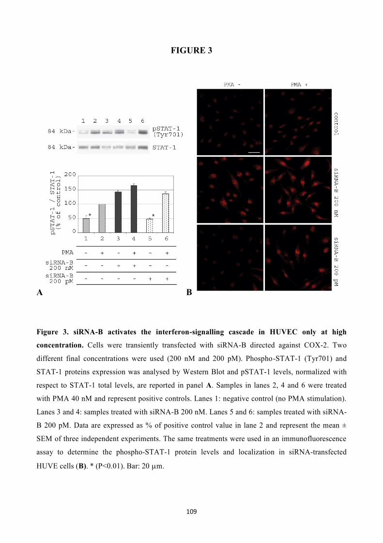
109
FIGURE 3
A B
Figure 3. siRNA-B activates the interferon-signalling cascade in HUVEC only at high
concentration. Cells were transiently transfected with siRNA-B directed against COX-2. Two
different final concentrations were used (200 nM and 200 pM). Phospho-STAT-1 (Tyr701) and
STAT-1 proteins expression was analysed by Western Blot and pSTAT-1 levels, normalized with
respect to STAT-1 total levels, are reported in panel A. Samples in lanes 2, 4 and 6 were treated
with PMA 40 nM and represent positive controls. Lanes 1: negative control (no PMA stimulation).
Lanes 3 and 4: samples treated with siRNA-B 200 nM. Lanes 5 and 6: samples treated with siRNA-
B 200 pM. Data are expressed as % of positive control value in lane 2 and represent the mean ±
SEM of three independent experiments. The same treatments were used in an immunofluorescence
assay to determine the phospho-STAT-1 protein levels and localization in siRNA-transfected
HUVE cells (B). * (P<0.01). Bar: 20 m.

110

111
FIGURE 4
Figure 4. Stable knockdown of COX-2 gene by RNAi in HT-29 cells. (A) Scheme of
pSUPER.retro vector (see details under Results Part 1). HT-29 cells were infected using
pSUPER.retro system, selected and analysed for COX-2 protein and COX-2 mRNA levels by
Western Blot and Real-Time PCR (B and C, respectively). PGE2 production (D) was evaluated by
using an ELISA assay. COX-2 expression in infected cells (lanes 2 and 4) was compared with that
of control cells (lanes 1 and 3), in the absence (lanes 1-2) or in the presence (lanes 3-4) of 40 nM
PMA-stimulation. Data from Western Blot are expressed as % of PMA-stimulated control in lane 1.
All results are expressed as mean ± SEM of three different experiments. * (P<0.01).

112

113
FIGURE 5
A B
Figure 5. Effect of anti-COX-2 shRNA expression on the interferon-signalling cascade in
HT29 pSUPER(+) cells. An analysis of STAT-1 phosphorylation in HT29 wild type cells and
HT29 cells infected using pSUPER.retro system was performed. Phospho-STAT-1 (Tyr701) and
STAT-1 proteins expression was analysed by Western Blot and pSTAT-1 levels, normalized with
respect to STAT-1 total levels, are reported in panel A. Lanes 1 and 2: HT29 wild type. Lanes 3 and
4: HT29 pSUPER(+). Samples in lanes 2 and 4 were treated with PMA 40 nM. Data are expressed
as % of positive control value in lane 2 and represent the mean ± SEM of three independent
experiments. The same samples were analysed in an immunofluorescence assay to determine the
phospho-STAT-1 protein level and localization (results are shown in panel B). * (P<0.01). Bar: 20
m.

114

115
FIGURE 6
A
B
Figure 6. Effect of pSUPER.retro infection system on the viability and the cell cycle
distribution of HT29 cells. Proliferation curves were determined by the MTT assay (A) and the
cell cycle distribution analysis was carried out on 1x10⁶ cells/samples after 60 min of incubation
with BrdU (B). Control: HT29 wild type cells ( and lane 1); pSUPER(-): HT29 cells infected with
vector non expressing anti-COX2 shRNA ( and lane 2); pSUPER(+): infected HT29 cells
expressing shRNA against COX-2 mRNA ( and lane 3). All data represent the mean of three
independent experiments.

116

117
FIGURE 7
Figure 7. Effect of pSUPER.retro infection system on migration and soft-agar colony
formation in HT29 cells. The migration assay was performed by using Boyden chambers and 8-
m polycarbonate membranes coated with Matrigel (40-fold dilution). After 24 h of incubation,
cells that migrated through the Matrigel coated membranes were fixed, stained, photographed (A)
and counted under light microscopy (B). Regarding the soft-agar colony formation assay, the
number of colonies was evaluated 7 days after the seeding in soft-agar (C). Control: HT29 wild
type cells; pSUPER(-): HT29 cells infected with vector non expressing anti-COX2 shRNA;
pSUPER(+): infected HT29 cells expressing shRNA against COX-2 mRNA. In the migration assay
samples were tested in the absence and presence of PMA 40 nM. Data in panels B and C represent
the mean ± SEM of three independent experiments. * (P<0.01); # (P<0.05). Bar: 20 m.

118

119
FIGURE 8
Figure 8. Efficiency of pSUPER.retro infection system. The images represent four different cell
types that underwent the infection by pSUPER.retro system (A: HUVEC; B: HT29; C: HCA7; D:
HeLa). Cells were fixed 24 h after infection and observed by using confocal microscopy. Nuclei
were stained with propidium iodide and the infected cells appear GFP-positive since the
pSUPER.retro vector contains an expressing cassette for GFP gene. Bar 20 m.

120
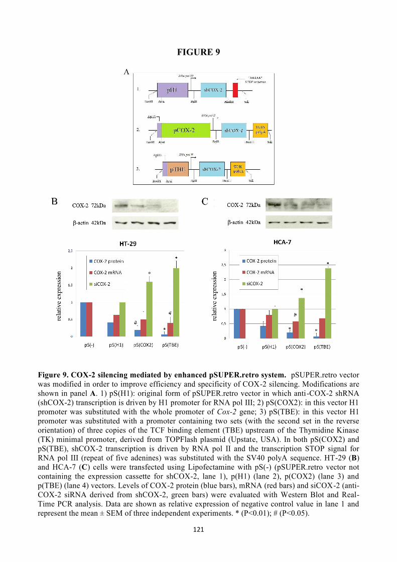
121
FIGURE 9
Figure 9. COX-2 silencing mediated by enhanced pSUPER.retro system. pSUPER.retro vector was modified in order to improve efficiency and specificity of COX-2 silencing. Modifications are shown in panel A. 1) pS(H1): original form of pSUPER.retro vector in which anti-COX-2 shRNA (shCOX-2) transcription is driven by H1 promoter for RNA pol III; 2) pS(COX2): in this vector H1 promoter was substituted with the whole promoter of Cox-2 gene; 3) pS(TBE): in this vector H1 promoter was substituted with a promoter containing two sets (with the second set in the reverse orientation) of three copies of the TCF binding element (TBE) upstream of the Thymidine Kinase (TK) minimal promoter, derived from TOPFlash plasmid (Upstate, USA). In both pS(COX2) and pS(TBE), shCOX-2 transcription is driven by RNA pol II and the transcription STOP signal for RNA pol III (repeat of five adenines) was substituted with the SV40 polyA sequence. HT-29 (B) and HCA-7 (C) cells were transfected using Lipofectamine with pS(-) (pSUPER.retro vector not containing the expression cassette for shCOX-2, lane 1), p(H1) (lane 2), p(COX2) (lane 3) and p(TBE) (lane 4) vectors. Levels of COX-2 protein (blue bars), mRNA (red bars) and siCOX-2 (anti-COX-2 siRNA derived from shCOX-2, green bars) were evaluated with Western Blot and Real-Time PCR analysis. Data are shown as relative expression of negative control value in lane 1 and represent the mean ± SEM of three independent experiments. * (P<0.01); # (P<0.05).

122

123
FIGURE 10
Figure 10. COX-2 silencing mediated by trans-kingdom RNA Interference (tkRNAi). (A)
Schematic representation of tkRNAi. E. Coli strains were co-transformed with both pGB2Ωinv-hly
and pSUPER.retro plasmids. Four different E. Coli strains were created, each containing one of the
four different forms of pSUPER.retro vector previously described. HT-29 (B) and HCA-7 (C) were
infected with the four recombinant E. Coli strains and levels of COX-2 protein (blue bars), mRNA
(red bars) and siCOX-2 (anti-COX-2 siRNA derived from shCOX-2, green bars) were evaluated
with Western Blot and Real-Time PCR analysis. Lanes 1-4: cells infected with E. Coli co-
transformed with pS(-) (lane 1), pS(H1) (lane 2), pS(COX2) (lane 3) and pS(TBE) (lane 4). Data
are shown as relative expression of negative control value in lane 1 and represent the mean ± SEM
of three independent experiments. * (P<0.01).

124

125
Discussion part 1: COX-2 silencing RNAi-mediated
RNA Interference (RNAi) represents a brand new approach in the field of reverse genetics, since it
is a potent tool capable of silencing genes in a high, long lasting and selective manner. The
understanding of RNAi mechanism of action has soon disclosed a wide spectrum of possible
applications, either in vitro or in vivo. In the past decade, many studies have shown that small
dsRNA molecules are capable of downregulating genes with sequence homology and that the
knockdown mediated by RNAi machinery is an ubiquitous phenomenon in eukaryotic cells. Much
of the knowledge regarding RNAi comes from studies on C. elegans. In this organism, Mello and
co-workers in 1998 demonstrated that microRNAs (miRNAs) physiologically regulate the
expression of genes involved in worm development (Fire et al, 1998; Elbashir et al, 2001). Today,
siRNAs and shRNAs are widely used by researchers to silence the expression of many target genes,
because of their high specificity and their apparent non-toxicity. Moreover, systems based on DNA
plasmids or retroviral vectors have provided new solutions to achieve a stable knockdown mediated
by shRNAs.
In this work, I used a RNAi-based approach to obtain an efficient knockdown of COX-2 mRNA.
The overexpression of COX-2 seems to play a critical role in many pathological processes. In
particular, the concept that high levels of PGI2 and PGE2, the main products of arachidonate
metabolism in vascular tissues and cancer cells respectively, stimulate both tumor-induced
angiogenesis and tumor progression is widely accepted (Leahy et al, 2002; Iñiguez et al, 2003),
whereas the overexpression of the inducible form of COX enzyme in colorectal cancer cells seems
to promote tumor invasion, tumor growth and tumor metastasis (Gupta and Dubois, 2001;
Yamauchi et al, 2002; Tuynman et al, 2004). Moreover, the possibility that the overexpression of
genes implicated in human cancers may be due to a down-regulation or a general imbalance of
specific miRNAs has been recently supported (Micheal et al, 2003; Calin et al, 2004; He et al,
2005; Lu et al, 2005) and it could be investigated in the case of colorectal cancer.
Data collected in this thesis show the effect of the transfection of four different sequences of anti-
COX-2 siRNAs on HUVE cells, stimulated with PMA in order to overexpress COX-2 mRNA. It
has been previously shown that PMA stimulates COX-2 expression in HUVEC, strongly increasing
both mRNA and protein levels (Hirai et al, 1999). Under these conditions, the release of
prostacyclin (PGI2) was also found to be highly augmented. It is well known that PGI2 stimulates
angiogenesis, probably by acting on a nuclear receptor belonging to peroxisome proliferator-

126
activated receptors (PPARs) family. Two of the synthetic siRNAs tested (sequences B and C)
resulted to be effective in downregulating COX-2 levels and enzymatic activity in HUVE cells in a
specific manner, having no effect on COX-1 expression. Moreover, COX-2 silencing by siRNAs
was observed even at very low concentrations (200 pM). Has to be mentioned that one of these
active siRNA sequences (sequence B) overlaps the same sequence used by Denkert and co-workers
(2003) to efficiently downregulate COX-2 expression in ovarian carcinoma cells. As a consequence
of COX-2 downregulation and PGI2 reduction, endothelial cells transfected with siRNA-B failed to
organize capillary-like tubular structures in 3-D collagen gel when stimulated with PMA, whereas
control cells (non-transfected or transfected with a scrambled siRNA) rapidly formed several
lengthened structures sprouting inside the gel.
It has been reported that synthetic siRNAs (Sledz et al, 2003; Judge et al, 2005), as well as short
single-stranded RNAs (ssRNAs) (Kim et al, 2004) and some DNA vectors expressing shRNAs
(Bridge et al, 2003), are able to trigger an interferon response in vitro. On the contrary, other
findings demonstrate that is even possible to administer naked, synthetic siRNA to mice without
inducing any interferon response (Heidel et al, 2004). In my experimental model I analyzed the
phosphorylation status (on Tyr701) of STAT-1 protein as a marker of the interferon system
activation (Brierley et al, 2005). As STAT-1 activation can be triggered by PMA treatment (Cohen
et al, 2005), I compared the levels of STAT-1 phosphorylated isoform in HUVEC transfected with
siRNA-B at two different doses (200 nM and 200 pM) with respect to PMA-stimulated control
cells. While the treatment with a 200 nM dose of siRNA-B significantly increased STAT-1
phosphorylation levels, both in the absence or in the presence of PMA, the treatment with the same
siRNA molecule at a low dose (200 pM) did not show any significant effect on STAT-1
phosphorylation when compared with controls. Similar results were ob tained by analyzing phospho-
STAT-1 expression and its nuclear translocation by immunofluorescence assay, indicating that low
but effective doses of anti-COX-2 siRNA are devoid of effects on the interferon system.
Following the detection of a siRNA sequence able to efficiently interfere with COX-2 expression in
HUVE cells, I focused my attention on a new RNAi strategy based on the use of a shRNA-
expressing vector. The aim was to obtain a stable and efficient knockdown of COX-2 gene in HT-
29 cells, a cell line derived from a human colorectal cancer and known to overexpress this enzyme.
The approach I chose was based on pSUPER.retro technology (Brummelkamp et al, 2002). Many
advantages came from this approach, first of all the possibility to achieve, by using inexpensive
tools, a potent silencing of target genes that is also highly specific and long-lasting. Therefore,
interesting results were achieved, since data from Western Blot, Real-Time PCR analysis and

127
ELISA assay clearly showed a strong and selective reduction of COX-2 protein and mRNA levels,
with consequent inhibition of PGE2 production, in HT-29 cells infected with a pSUPER.retro
vector expressing anti-COX-2 shRNA (shCOX-2) and then anti-COX-2 siRNA-B (siCOX-2) [HT-
29 pSUPER(+)], even in the presence of PMA-induced COX-2 overexpression. These data support
the evidence of a strong and long-lasting COX-2 mRNA degradation driven by siRNA molecules
processed from shRNA precursors.
As previously tested in HUVEC treated with anti-COX-2 siRNA-B, I analyzed the effect of the
permanent expression of shCOX-2 molecules on STAT-1 activation in HT-29 pSUPER(+) cells,
detecting no significant increase neither in STAT-1 phosphorylation levels nor in phospho-STAT-1
nuclear accumulation. These data suggest that a vector-based stable expression of shRNA
molecules induces a weaker interferon system response with respect to the transfection of synthetic
siRNAs.
Performing experiments aimed to analyze the phenotype of COX-2-deficient HT-29 pSUPER(+)
cells, no significant effects of COX-2 down-regulation on cell proliferation and cell cycle
distribution were detected, observing only a slight decrease of the proliferation rate and a slight
accumulation of cells in the G0/G1 phase. These results are in line with other data recen tly collected
in a model of ovarian carcinoma cells (Denkert et al., 2003) and in a model of human hepatocellular
carcinoma cells (Park et al, 2005) based on siRNA-mediated COX-2 down-regulation, suggesting
that COX-2 itself is not decisively involved in the proliferation of human cancer cells as well.
However, interesting results were obtained by testing the in vitro invasive behavior of HT-29
pSUPER(+) cells and their anchorage-independent growth ability. PMA-stimulated HT-29 cells,
expressing high levels of COX-2, resulted to be able to degrade and migrate through ECM
components of Matrigel-coated membranes, which indicates their malignant behavior in vitro,
associated with a well described invasive and metastatic ability in vivo (Kakiuchi et al, 2002).
Moreover, HT-29 wild type cells easily formed colonies in soft-agar, which is an accepted criterion
for transformation and an experimental condition that better represents tumor cells growth and
invasiveness in vivo. The knockdown of COX-2 enzyme in HT-29 cells abrogated their ability to
invade Matrigel-coated membranes in a Boyden chamber assay, either in the absence or in the
presence of PMA-stimulation, and strongly impaired their anchorage-independent growth in soft
agar basal conditions. These results support the involvement of COX-2 in the malignant behavior of
human colon carcinoma cells and underline the relevance of a stable virus-based COX-2
knockdown mediated by RNA interference in order to impair the invasive and metastatic ability of
CRC.

128
My study confirms the predominant role that RNA Interference is assuming in the field of gene
silencing due to its high efficacy, specificity and non-toxicity. One of the novel targets in cancer
therapies is to obtain a selective down-regulation of those genes overexpressed in tumor tissues.
COX-2 is certainly one of them. Results reported here indicate an easy-to-use, powerful and high
selective virus-based method to knockdown COX-2 gene in a stable and long-lasting manner, in
colon cancer cells. Furthermore, they open up the possibility of an in vivo application of this anti-
COX-2 retroviral vector, as therapeutic agent for human cancers overexpressing COX-2. In fact, a
significant resistance of HUVE cells to the pSUPER.retro viral infection was observed in
comparison with different types of human cancer cells, either CRC cells (HT-29 and HCA-7) and
cervix carcinoma cells (HeLa). This observation indicates that endothelial cells are refractory to
retroviral infection and suggests that this kind of virus-based approach might not affect the
physiological prostaglandins production in vascular tissues, avoiding some of the well known side
effects coming from therapies based on selective COX-2 inhibitors (coxibs) (Mamdani et al, 2004;
Mitchell and Warner, 2005).
In order to improve the tumor selectivity, pSUPER.retro vector was modified for the shCOX-2
expression cassette. The aim was to gain a strong, specific transcription of shCOX-2 followed by
COX-2 silencing only in cancer cells. It is well known that intracellular activation of different
molecular pathways are responsible of COX-2 overexpression in colon cancer. This highlights that
COX-2 promoter is overstimulated in tumor cells, compared with other normal tissues in which
COX-2 gene is finely regulated. Moreover, there are some pathways that are typically activated in
colon cancer cells: one of those is Wnt/-catenin signalling pathway. -catenin protein is frequently
mutated in colon adenomas and adenocarcinomas and this alteration causes COX-2 overexpression
mediated by transcriptional factors like TCF-4/LEF-1 that bind and activate COX-2 promoter. For
this reason, my purpose was to put the shCOX-2 expression cassette under the control of TCF/LEF
consensus sequences (TBEs, TCF Binding Elements) which can modulate the expression of the
shRNA in those colon cancer cells presenting an high nuclear -catenin. In pSUPER.retro vector
previously used, the expression cassette of shCOX-2 is under control of RNA pol-III human
promoter H1 [pS(H1) vector] that brings, after gene trasduction, a constitutive non-regulated
transcription of siCOX-2 processed from the shCOX-2 precursor. This means that the COX-2
silencing system allows siCOX-2 production in every mammalian cell type where viral trasduction
is effective. Thus, H1 promoter was substituted with the human Cox-2 promoter [pS(COX2)] and
with a promoter containing repeated copies of the TCF binding element (TBE) [pS(TBE)]. The
transcription stop signal was also substituted with SV40 polyA signal. Following these
modification, shCOX-2 transcription became strong and specific only in cells in which COX-2 and

129
Wnt/-catenin signalling pathway are activated. These evidences were largely supported by data
collected using HT-29 and HCA-7 colon cancer cells, in which the COX-2 silencing mediated by
enhanced pS(COX2) and pS(TBE) vectors was evaluated. In particular, a stronger inhibition of
Cox-2 gene (at both protein and mRNA level) and a higher siCOX-2 production were achieved by
using pS(TBE) vector, that represents not only the most effective, but also the most specific system
to downregulate COX-2 in colon cancer cells.
Because of the many limits that a retroviral therapy could have in a possible in vivo treatment of
CRC, the next goal was to render the enhanced RNAi-mediate COX-2 silencing more suitable for
this kind of application. Xiang and et al. (2006) demonstrated that it is possible to induce RNAi in
mammalian cells after infection with engineered E. Coli strains expressing Inv and HlyA genes,
which encode for two bacterial factors needed for successful transfer of shRNA in mammalian
cells. This system, called “trans-kingdom” RNAi (tkRNAi) could represent an optimal approach for
the treatment of colorectal cancer, since E. Coli in normally resident in human intestinal flora and
could easily vehicled to the tumor tissue. For this reason, I tested the improved COX-2 silencing
mediated by pS(COX2) and pS(TBE) by using tkRNAi system. Results obtained in HT-29 and
HCA-7 cell lines were in high agreement with data collected after the Lipofectamine transfection of
pS(COX2) and pS(TBE) vectors in the same cell lines, previously discussed. These findings suggest
that tkRNAi system for COX-2 silencing, in particular mediated by pS(TBE) vector, could
represents a promising tool for the treatment of colorectal cancer.

130
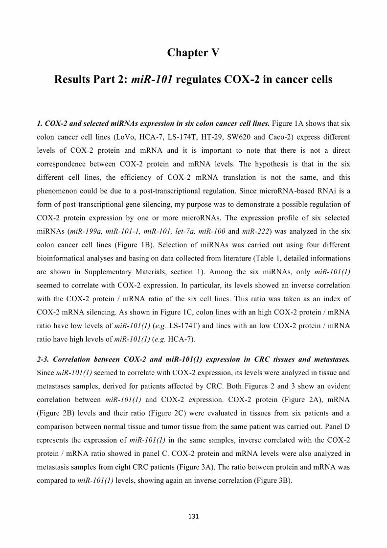
131
Chapter V
Results Part 2: miR-101 regulates COX-2 in cancer cells
1. COX-2 and selected miRNAs expression in six colon cancer cell lines. Figure 1A shows that six
colon cancer cell lines (LoVo, HCA-7, LS-174T, HT-29, SW620 and Caco-2) express different
levels of COX-2 protein and mRNA and it is important to note that there is not a direct
correspondence between COX-2 protein and mRNA levels. The hypothesis is that in the six
different cell lines, the efficiency of COX-2 mRNA translation is not the same, and this
phenomenon could be due to a post-transcriptional regulation. Since microRNA-based RNAi is a
form of post-transcriptional gene silencing, my purpose was to demonstrate a possible regulation of
COX-2 protein expression by one or more microRNAs. The expression profile of six selected
miRNAs (miR-199a, miR-101-1, miR-101, let-7a, miR-100 and miR-222) was analyzed in the six
colon cancer cell lines (Figure 1B). Selection of miRNAs was carried out using four different
bioinformatical analyses and basing on data collected from literature (Table 1, detailed informations
are shown in Supplementary Materials, section 1). Among the six miRNAs, only miR-101(1)
seemed to correlate with COX-2 expression. In particular, its levels showed an inverse correlation
with the COX-2 protein / mRNA ratio of the six cell lines. This ratio was taken as an index of
COX-2 mRNA silencing. As shown in Figure 1C, colon lines with an high COX-2 protein / mRNA
ratio have low levels of miR-101(1) (e.g. LS-174T) and lines with an low COX-2 protein / mRNA
ratio have high levels of miR-101(1) (e.g. HCA-7).
2-3. Correlation between COX-2 and miR-101(1) expression in CRC tissues and metastases.
Since miR-101(1) seemed to correlate with COX-2 expression, its levels were analyzed in tissue and
metastases samples, derived for patients affected by CRC. Both Figures 2 and 3 show an evident
correlation between miR-101(1) and COX-2 expression. COX-2 protein (Figure 2A), mRNA
(Figure 2B) levels and their ratio (Figure 2C) were evaluated in tissues from six patients and a
comparison between normal tissue and tumor tissue from the same patient was carried out. Panel D
represents the expression of miR-101(1) in the same samples, inverse correlated with the COX-2
protein / mRNA ratio showed in panel C. COX-2 protein and mRNA levels were also analyzed in
metastasis samples from eight CRC patients (Figure 3A). The ratio between protein and mRNA was
compared to miR-101(1) levels, showing again an inverse correlation (Figure 3B).

132
4. miR-101(1) regulates COX-2 expression in colon cancer cell lines. Supported by the data
obtained in colon cancer cell lines and tissues, a series of in vitro experiments was carried out to
demonstrate a direct regulation of COX-2 expression mediated by miR-101(1). Three colon cancer
cell lines (LS-174T, HT-29 and HCA-7) were transfected with synthetic miR-101(1) using
Lipofectamine and the silencing of COX-2 protein was evaluated. The effect of miR-101(1)
transfection was compared to two different negative controls (cells treated only with Lipofectamine
and cells transfected with cel-miR-67, a C. Elegans miRNA,) and a positive control (siCOX-2,
previously tested in my experiments). The effect of the inhibitors of both miR-101(1) and cel-miR-
67 was also evaluated. All the RNA molecules were transfected at 200 nM final concentration. As
expected, transfection of miR-101(1) led to a COX-2 silencing in all the three cell lines (Figure 4,
A-B and C), compared to negative controls. Moreover, the transfection of miR-101(1) inhibitor (that
blocks the action of miR-101(1) by pairing its molecule), induced an overexpression of COX-2
protein, slight in LS-174T cell and stronger in HT-29 and HCA-7 cells. Finally, Figure 4E shows
the putative binding site of miR-101(1) in COX-2 3’-UTR region as detected by MiRanda software.
5. miR-101(1) downregulation is involved in COX-2 overexpression induced by hypoxia. In order
to investigate a possible molecular pathway by which miR-101(1) regulates COX-2 expression, I
started to analyze the involvement of miR-101(1) in COX-2 overexpression induced by hypoxia.
The hypoxic context was chosen because it strongly enhance COX-2 expression in colon cancer
cells. An hypoxic environment can be induced in vitro treating the cells with the iron chelator
desferrioxamine (DFX). Figure 5 shows that COX-2 is overexpressed after 24 and 48 h of DFX 100
M treatment in LS-174T (5A), HT-29 (5B) and HCA-7 (5C). The strongest induction of COX-2
was observed in HT-29 after 24 h and HCA-7 after 48 h while it was only slight in LS-174T.
Surprisingly, in all the three cell lines COX-2 overexpression wasn’t a consequence of increased
mRNA levels. On the contrary, DFX treatment produced a significant decrease of COX-2 mRNA
either after 24 h or 48 h. Moreover, a miR-101(1) transfection was performed on DFX treated cells.
The transfected miR-101(1) strongly inhibited the COX-2 overexpression in LS-174T after 24 and
48 h, and in HT-29 and HCA-7 after 48 h. Under these conditions, the COX-2 protein / mRNA ratio
was compared to miR-101(1) levels in all samples. For the three cell lines, the increased ratio was
always coupled to a miR-101(1) decreased expression. After 48 h treatment with DFX and miR-
101(1) transfection instead, the ratio values were comparable to the negative control samples, not
treated with DFX while in LS-174T the ratio returned to normal levels already after 24 h (Figure 5,
D-E and F).

133
FIGURE 1
Figure 1. COX-2 and selected miRNAs expression in six colon cancer cell lines. (A) COX-2
protein (dark bars) and mRNA (light bars) levels were analyzed in six colon cancer cell by Western
Blot and Real-Time PCR. COX-2 protein expression was normalized against -actin protein and
COX-2 mRNA expression was normalized against GUSB mRNA. (B) selected miRNAs expression
was evaluated in the same cell lines samples by using Real-Time PCR and normalized against U6
RNA levels. (C) COX-2 protein / mRNA ratio (light bars) was taken as an index of COX-2 mRNA
silencing and it was compared to miR-101(1) expression (dark bars) for the six colon cell lines. In
all panels, data show the relative expression of each sample compared to the reference sample
(LoVo cell line) and represent the mean ± SEM of three independent experiments.

134

135
TABLE 1
Table 1. Selection of six anti-COX-2 microRNAs candidates.
Six human microRNAs were selected as possible silencers of COX-2 mRNA translation. Selection
was carried out using four different bioinformatical analyses and basing on data collected from
literature. Detailed informations are shown in Supplementary Materials, section 1.
miRNA1 PicTar Score2
(kcal/mol)
MiRanda Score3
(kcal/mol)
Target Scan Score4
miRNA Target (hits)5
HCT-116 / Caco-2
(miR expr.)6
HCT-116 / HT-29 (Ct)7
hsa-miR-199a 7.6 (-21.9) 114 (-17.3) - 0 0 / 0 -16.6 /
-16.8
hsa-miR-101 2.2 (-23.8) 111
(-14.4) 0.14 1 229 / 24 -22.2 / -21.7
hsa-miR-103 0 123 (-18) - 0 285 / 238 -13.1 / -12.9
hsa-let-7a 0 121
(-17.1) - 0 92 / 3 -12.4 / -22.4
hsa-miR-100 0 103
(-16.1) - 0 20 / 0 -11.7 /
-21
hsa-miR-222 0 116
(-24.2) - 0 242 / 22 -13.3 / -17.5

136

137
FIGURE 2
Figure 2. Correlation between COX-2 and miR-101(1) expression in CRC tissues. COX-2
protein and mRNA levels were analyzed in selected colon tissue samples from CRC patients by
Western Blot and Real-Time PCR respectively. Densitometric analysis of COX-2 bands is available
in Supplementary Materials, section 2. Data were normalized against -actin protein and mRNA
expression. For each patient, COX-2 protein (A) and mRNA (B) were evaluated in both normal
(light bars) and tumor (dark bars) colon tissue. COX-2 protein / mRNA ratio (C) was analyzed and
compared to miR-101(1) expression (D).

138
139
FIGURE 3
Figure 3. Correlation between COX-2 and miR-101(1) expression in CRC metastases. COX-2
protein and mRNA levels were analyzed in selected metastasis samples from CRC patients by
Western Blot and Real-Time PCR respectively (A). Densitometric analysis of COX-2 bands is
available in Supplementary Materials, section 2. Data were normalized against -actin protein and
mRNA expression. For each patient, COX-2 protein / mRNA ratio was analyzed and compared to
miR-101(1) expression (B).

140

141
FIGURE 4
Figure 4. miR-101(1) regulates COX-2 expression in colon cancer cell lines. COX-2 regulation
mediated by miR-101(1) was evaluated in three different colon cancer cell lines: LS-174T (A), HT-
29 (B) and HCA-7 (C). Cell were transfected with: siRNA anti-COX2 (siCOX-2 positive control,
lane 2); miR-101(1) (lane 3); miR-101(1) inhibitor (lane 4); miR-negative control (lane 5); miR-
negative control inhibitor (lane 6). All treatments are described in D. COX-2 protein (dark bars) and
mRNA (light bars) levels and miR-101(1) (medium bars) expression were analyzed by Western Blot
and Real-Time PCR 48h after transfection and referred to the negative control sample in lane 1.
Data represent the mean ± SEM of three independent experiments. E: putative binding site of miR-
101(1) in COX-2 3’-UTR region as detected by MiRanda software.

142

143
FIGURE 5
Figure 5. miR-101(1) downregulation is involved in COX-2 overexpression induced by
hypoxia. miR-101(1) expression regulated by hypoxia was investigated in three different colon
cancer cell lines: LS-174T (A, D), HT-29 (B, E) and HCA-7 (C, F). Cell were treated with
desferrioxamine 100 M (DFX , lanes 2, 3, 5 and 6) and transfected with miR-101(1) (lanes 3 and
6). A, B and C: COX-2 protein (dark bars) and mRNA (light bars) levels were analyzed by Western
Blot and Real-Time PCR respectively 24 h and 48 h after transfection and referred to samples in
lane 1 and 4. D, E and F: COX-2 protein / mRNA ratio (dark bars) was compared to miR-101(1)
levels (light bars) analyzed with Real-Time PCR. Expression data refer to samples in lane 1 and 4.
All treatments are described in G. Data represent the mean ± SEM of three independent
experiments.

144

145
Discussion part 2: miR-101 regulates COX-2 in cancer cells
As each microRNA (miRNA) is expected to regulate the translation of up to 100 mRNAs (Lim et
al., 2005) it is clear that disturbances of the miRNA expression level (Calin et al., 2004), processing
of the miRNA precursors (Sugito et al. ,2006), or mutations in the sequence of the miRNA, its
precursor, or its target mRNA (Iwai and Naraba, 2005), may have detrimental effects on cell
physiology. There is now ample evidence that the expression of miRNAs is altered in cancer, and
that certain changes may be directly implicated in the carcinogenic process. A number of miRNAs
have been shown to promote cell proliferation and survival, while others diminish cell proliferation
and survival. These two classes of miRNAs may play a central role in cancer development as novel
oncogenes and tumor suppressors, respectively. In general, the majority of miRNAs are
downregulated in cancer specimens (Lu et al., 2005). In normal tissues, some of these miRNAs
have been documented to inhibit the translation of proto-oncogenes by targeting the 3'ends of their
mRNAs. Such miRNAs are therefore considered as "tumor suppressor miRNAs" (TS-miRs) since
their normal function is to control the expression of an oncogene (Calin et al., 2002). Conversely,
certain miRNAs seem to be upregulated in cancer and may act as "oncomiRs" since they can enable
the downregulation of a tumor suppressor (O’Donnell et al., 2005; Chan et al., 2005).
Components of the miRNA/RNAi pathway may be altered during the progressive development of
colorectal cancer (CRC) as well. Michael et al., (2003) firstly demonstrated a reduced accumulation
of miR-143 and miR-145 in colorectal neoplasia. They supposed that altered accumulation of these
mature miRNAs may reflects early changes in the cellular composition of tumors, compared with
normal mucosae. Moreover, hsa-miR-342 showed an anti-apoptotic effect in the colorectal cancer
cell line HT-29, suggesting that this microRNA could function as a proapoptotic tumor suppressor
(Grady et al., 2008). On the contrary, many other studies were carried out to find out an
upregulation in miRNA expression patterns in CRC. In particular, hsa-miR-200c, hsa-let-7g
(Nakajima et al., 2006), various members of the oncogenic miR-17-92 family (Dews et al., 2006;
Lanza et al., 2007), miR-34a (Tazawa et al., 2007), miR-21 (Tanzer and Stadler, 2004; Chan et al.,
2005; Iorio et al., 2005; Si et al., 2007) and other miRNAs as miR-20a miR-106a, miR-181b, miR-
203 (Schetter et al., 2008) were find to be significantly overexpressed in the colorectal tumor
samples compared to the corresponding normal, acting as oncomiRs.
Basing on the data from literature, the first aim of this second part of my research was to elucidate a
possible relation between cyclooxygenase-2 (COX-2) overexpression in CRC and altered miRNA

146
expression patterns. Since Cox-2 gene expression, as for other human genes, could be modulated by
the machinery of RNAi, the hypothesis was that overexpressed COX-2 protein in colon cancer cells
could be the result of decreased levels of one or more tumor suppressor miRNAs. In this thesis, I
clearly show an inverse correlation between COX-2 expression and the human miR-101(1) levels in
colon cancer cell lines, tissues and metastases. Moreover, I demonstrate that the in vitro modulating
of miR-101(1) expression in colon cancer cell lines leads to significant variations in COX-2
expression. In these experiments, an important parameter was considered as an index of COX-2
silencing mediated by miRNA: the COX-2 protein / mRNA ratio was compared to the expression
profile of miR-101(1) in each sample analyzed. Since the most common mechanism of gene
silencing mediated by miRNA is based on the translational repression of cellular mRNAs (Pillai et
al., 2005; Nottrott et al., 2006), the assumption was that high levels of miRNA, miR-101(1) in my
case, should correspond to low values of COX-2 protein / mRNA ratio and lower levels of miR-
101(1) should correspond to higher levels of the same ratio. miRNAs can also induce a rapid
degradation of the mRNAs target, as in the small interfering RNA (siRNAs) pathway, if a perfect
base complementarity exists between miRNA and mRNA (Bagga et al., 2005). Nevertheless, it can
be excluded that miR-101(1) silences COX-2 messenger by this latter mechanism. In fact, analyzing
the putative binding site of miR-101(1) in COX-2 3’-UTR region as detected by predictive
software, there is not a complete pairing of miR-101(1) on COX-2 mRNA (Figure 4E).
The starting point of my research was the selection of possible miRNA candidates for the COX-2
silencing. After a computational analysis of all the known human miRNAs and the COX-2 mRNA
3’UTR region, the expression profile of six selected miRNAs (miR-199a, miR-101-1, miR-101, let-
7a, miR-100 and miR-222) was analyzed in six colon cancer cell lines (LoVo, HCA-7, LS-174T,
HT-29, SW620 and Caco-2). Surprisingly, miR-101(1) levels showed a perfect inverse correlation
with the COX-2 protein / mRNA ratio of these cells. In order to demonstrate a direct regulation of
COX-2 protein expression mediated by miR-101(1), further in vitro tests were carried out in LS-
174T, HT-29 and HCA-7 cells. In all the three cases, the transfection of synthetic exogenous miR-
101(1) or its inhibitor, induced a significant silencing or upregulation of COX-2, respectively. It
must be notice that the highest level of COX-2 silencing (induced by transfected miR-101-1) and
the lowest level of COX-2 upregulation (induced by transfected miR-101-1 inhibitor) were recorded
in LS-174T cells that normally express the lowest level of endogenous miR-101(1), compared to the
other cell lines. On the contrary, the lowest level of COX-2 silencing and the highest level of COX-
2 upregulation were recorded in HCA-7 cells that normally express the highest level of miR-101(1).
All these findings demonstrate that miR-101(1) regulates COX-2 expression in human cells.
However, it is not to be excluded that miR-101(1) could regulate COX-2 expression not directly, but

147
through a silencing mechanism acting on other cellular factors involved in COX-2 regulation. To
discard this last hypothesis, I performed a Luciferase assay in which miR-101(1) “seed” region of
COX-2 3’UTR mRNA was cloned downstream the luciferase gene in pGL3.basic reporter plasmid.
Transfection of both plasmid and miR-101(1) in LS-174T determined a decreased amount of
luciferase protein production (compared to positive and negative controls), showing the evidence of
a direct interaction between miR-101(1) and COX-2 mRNA (preliminary data, not shown). These
data are supported by the work of Chakrabarty et al. (2007) that recently demonstrated that two
miRNAs, mmu-miR-101a and mmu-miR-199a*, are spatiotemporally expressed in the mouse uterus
during implantation coincident with expression of COX-2, critical for implantation. The in vitro
gain- and loss-of-function experiments showed that COX-2 expression is posttranscriptionally
regulated by these two miRNAs.
It is well known that COX-2 is overexpressed in colorectal cancer (Sinicrope et al., 1999) and this
contributes to an increased apoptosis resistance (Moss et al., 1996) and invasiveness (Tsujii et al.,
1997) of the tumor cells and an increased tumor angiogenesis (Tsujii et al., 1998) in colon tissues.
Since miR-101(1) was found to regulate COX-2, I started to investigate the role of miR-101(1) in
colon tumorigenesis. The analysis of COX-2 protein-mRNA and miR-101(1) levels in different
colon tissues (normal vs. tumor) and metastases from patients affected by CRC, confirmed the
inverse correlation between COX-2 and miR-101(1), previously described in the in vitro
experiments. Combining together data collected in vitro and ex vivo, I can show evidences that, not
only miR-101(1) regulates COX-2 in colon cancer cell lines but, more interestingly, alterations of its
expression profile could be involved in colorectal cancer growth and development.
The main question now is: if COX-2 overexpression depends on a miR-101(1) expression
impairment, who controls miR-101(1) repression in the CRC pathology? I will try to answer to this
question carrying on my personal research on RNAi, COX-2 and colon cancer and some data were
already collected. In particular, I started to investigate miR-101(1) regulation in the hypoxic
environment.
Adaptation to hypoxia is critical for tumor cell growth and survival and is achieved largely by
transcriptional activation of genes that facilitate short- and long-term adaptive responses (Semenza,
2002). Hypoxia induces COX-2 in colorectal tumor cells and this upregulation is mediated directly
by hypoxia-inducible factor 1 (HIF-1) (Kaidi et al. (2006). An hypoxic environment can be induced
in vitro treating the cells with the iron chelator desferrioxamine (DFX) (Woo et al. 2006). For this
reason I decided to analyze the effect of hypoxia on miR-101(1) expression and a great enthusiasm
derived from this sequence of experiments. In fact, treating LS-174T, HT-29 and HCA-7 colon

148
cancer cell lines with DFX, I observed a strong decrease in miR-101(1) levels, and this phenomenon
was always coupled to a COX-2 overexpression. Moreover, COX-2 overexpression DFX-induced
was completely abrogated when DFX treated cells were also transfected with synthetic miR-101(1).
Even if further investigations are needed, I can conclude that hypoxia-induced COX-2
overexpression, fundamental for cancer cells survival in vivo, could be also connected with the
RNAi mechanism mediated by miR-101(1).
In conclusion, in this second part of my thesis I demonstrated that COX-2 regulation can be directed
by the mechanism of RNA Interference, through the action of human miR-101(1). miR-101(1)
silences COX-2 in vitro, and this evidence was found in LS-174T, HT-29 and HCA-7 colon cancer
cell lines. Moreover, a reduced accumulation of miR-101(1) was found both in tissues and
metastases samples derived from patients affected by colorectal cancer. As a consequence, miR-
101(1) deregulation in cancer tissues could represent one of the main causes underlying COX-2
enzyme overexpression in CRC. Finally, preliminary studies pointed out that miR-101(1)
impairment could be a consequence of the adaptation of cancer cells to the hypoxic environment
that strongly characterize CRC tissues.

149
Chapter VI
Materials and Methods
Cell Lines. HUVE cells were isolated from freshly collected umbilical cords. Cells were grown in
M199 medium supplemented with 20% foetal calf serum, 100 mg/ml ECGS, 100 mg/ml heparin, 2
mM L-glutamine and antibiotics (penicillin 100 U/ml and streptomycin 100 mg/ml. Cells were
maintained at 37° C in 5% CO2 and used for experiments between 3rd and 5th passage. HT-29, SW-
620 and LoVo (S, doxorubicin-sensitive) human colon cancer cells were obtained from American
Type Culture Collection (Manassas, VA, USA). HCA-7, Caco-2 and LS-174T cells were obtained
from European Collection of Cell Cultures (ECACC; Salisbury, Wiltshire, UK). All the cell lines
were cultured at 37°C in 5% CO2 in Dulbecco’s modified Eagle’s medium (DMEM) supplemented
with 10% heat-inactivated Foetal Calf Serum (FCS), 2 mM L-glutamine, penicillin 100 U/ml and
streptomycin 100 mg/ml. FCS and DMEM were purchased from Cambrex Biowittaker, USA. All
the other reagents were purchased from Sigma (Sigma Chemical, St. Louis, USA).
Synthetic siRNAs Design. Four different siRNAs against COX-2 mRNA were designed as
suggested by Elbashir and collaborators (2002) and chemically synthesized (PROLIGO Primers and
Probes, USA). The identified target sequences on COX-2 mRNA (NM000963) were respectively:
bases 290-310 (siRNA-A, 5’aaactgctcaacaccggaatt3’), bases 291-311 (siRNA-B,
5’aactgctcaacaccggaattt3’), bases 1020-1040 (siRNA-C, 5’aacagagtatgcgatgtgctt3’) and bases 1429-
1449 (siRNA-D, 3’aagtatcacaggcttccattg5’). We also used a scrambled siRNA (Ambion, USA) as a
negative control, with no significant homology to any known gene sequences in human, mouse and
rat genome. All siRNA sequences were controlled for their specificity by using BLAST database
and did not show any homology to other human gene.
siRNAs Transfection. HUVE cells were seeded in 25 cm2 flasks (150.000 cells/flask) and grown
up to 50% confluence. After 24 h, cells were transfected with siRNAs by using Oligofectamine
reagent (Invitrogen, USA) according to the manufacturer’s instructions. Briefly, each siRNA (400
fmoles) was diluted in 350 l of serum-free medium. For each flask, 10 l of Oligofectamine

150
reagent was incubated with 40 l of serum-free medium for 10 minutes at room temperature. 50 l
of Oligofectamine solution were added to diluted siRNAs. After incubation for 20 minutes at room
temperature, siRNAs-Oligofectamine complexes were added to flasks containing 1.6 ml of serum-
free medium, resulting in 2 ml of total transfection volume and in a siRNA final concentration of
200 pM. After 4 h of incubation at 37°C, 3 ml of complete medium containing FCS and phorbol 12-
myristate 13-acetate (PMA) was added (final concentration: 10% serum and 40 nM PMA).
Western Blot. Cells were scraped and lysed in lysis buffer (50 mM Tris-HCl, pH 7.5, 2 mM EDTA,
100 mM NaCl, 1% Triton X-100 and protease inhibitors mixture), additionated with 5 mM NaF, 1
mM Na3VO4, 10 mM -glycerolphosphate, for the phospho-STAT-1 immunoblotting experiments.
Cell lysates were incubated 1 h on ice and centrifuged at 12000g to collect supernatants. Protein
concentration in supernatants was evaluated by using the Lowry method. After addition of SDS-
PAGE sample buffer and boiling, 40 g of denatured proteins were separated in 12% SDS-PAGE
and then transferred to nitrocellulose papers. After the blotting, nitrocellulose papers were incubated
with specific antibodies. The primary antibodies used were: polyclonal anti-COX-2 (Cayman
Chemicals, USA), anti-phospho-STAT-1[Tyr701] (Cell Signaling, USA) and anti--actin (Sigma,
USA); monoclonal anti-COX-1 (Cayman Chemicals, USA) and anti-STAT-1 p84/p91 (Santa Cruz
Biotechnology, USA). Secondary antibodies (HRP-conjugated) were purchased from Santa Cruz
Biotechnology, USA. Immunolabelling was visualised by using the ECL procedure (Amersham
Biosciences, USA). Bands were quantified by using a densitometric image analysis software (Image
Master VDS, Pharmacia Biotech, Uppsala, Sweden). Normalization was made against β-actin
expression.
Determination of COX-2 Activity. Conditioned media from HUVE cells were collected 48 h after
transfection with siRNAs and stored at -80°C. COX-2 enzymatic activity was evaluated by
measuring the 6-keto-PGF1 release in conditioned media by using an ELISA assay (Assay
Designs, USA). 6-keto-PGF1 production was then related to the protein concentration in cell
lysates. PGE2 levels in HT29 culture media were evaluated by using an ELISA assay (Cayman
Chemicals, USA) and related to the protein concentration in cell lysates.

151
In Vitro Angiogenesis Test. In vitro angiogenesis was evaluated by seeding HUVE cells on a 3-D
collagen gel in a 24-well plate and transfecting them with siRNAs. Briefly, collagen gel was
prepared by adding 8 volumes of collagen solution (3 mg/ml, Roche Applied Science, USA) to 2
volumes of a mixture containing M199 5X, HEPES 0.02 M and NaHCO3 7.5 mg/ml. After pH
adjusting to 7.2-7.4, the mixture was quickly dispensed to the wells and gelification was achieved at
37°C. After gelification, wells were washed twice with M199. HUVE cells (5x10⁴/well) were
seeded onto the gels and then transfected with siRNAs (final concentration 200 pM) by using
Oligofectamine reagent (Invitrogen, USA) according to the manufacturer’s instructions. In vitro
capillary-like formation was stimulated with 40 nM PMA, examined 48 h after transfection by
using a phase contrast microscope and the number of tubular capillary-like structures per well was
counted.
Cloning anti-COX-2 short hairpin RNA (shCOX-2) into pSUPER.retro Vector. Constructs,
coding for anti-COX-2 shRNA, were prepared as described by Brummelkamp et al (2002a).
pSUPER.retro vector (Oligoengine, Seattle, WA, USA), based on the murine s tem cell virus
(MSCV) genome, was a kind gift of Dr. P. Chieco (C.R.B.A. lab, Bologna, Italy). Forward and
reverse sequences for sh(COX-2) construct were 5’-
gatccccaactgctcaacaccggaatttcaagagaattccggtgttgagcagtttttttggaa-3’ and 5’-
agcttttccaaaaaaactgctcaacaccggaattctcttgaaattccggtgttgagcagttggg-3’, respectively, as shown in
Figure 1. 64 nt-containing oligos were synthesized and purchased from PROLIGO (USA).
Sequence design started from the most effective anti-COX-2 synthetic siRNA (siCOX-2, sequence
B). Steps for cloning oligonucleotides into pSUPER.retro vector were made accordingly to
pSUPER RNAi system protocol (www.oligoengine.com). Forward and reverse oligonucleotides
were annealed to form a duplex. The annealed oligos were then ligated into the BglII–HindIII
cleavage site within the pSUPER.retro vector pre-linearized with the same restriction enzymes.
Recombinant vector containing inserts was transformed into competent E. coli cells. After selection
in ampicillin-containing medium, colonies were recovered and checked for the presence of
recombinant pSUPER.retro vector.
Virus Production and Cell Infections. Anti-COX-2 pSUPER.retro vector was transfected into Phi-
NX (“Phoenix”) packaging cell line kindly provided by Dr. P. Chieco (C.R.B.A lab, Bologna, Italy)
to produce ecotropic retroviral supernatants. Phoenix cells were cultured in Dulbecco’s modified

152
Eagle’s medium (DMEM) supplemented with 10% foetal calf serum and pre-treated with
Chloroquine at final concentration of 25 M. The day before transfection, Phoenix cells were
seeded in 10 cm dishes (3x10⁶ cells/dish) in order to reach 60% confluence at the time of
transfection. Cells were transfected with 10 g of viral vector DNA by using calcium-phosphate
precipitation method. 48 h after transfection, culture medium was filtered through a 0.45 µm filter
and the viral supernatant was used for HT29 cells infection after addition of 8 µg/ml of polybrene
(Sigma, USA). After infection, HT29 cells were incubated at 32°C in 5% CO2 for 6 h. Then
medium was changed with fresh medium and HT29 cells were allowed to recover for 48 h at 37°C
in 5% CO2. Infection efficiency was examined under a fluorescence microscope to check the GFP
expression. Infected cells were selected by adding puromycin (1 µg/ml) for 48 h to the culture
medium. COX-2 expression in HT29 wild type cells and in HT29 pSUPER(+) clones (producing
anti-COX-2 shRNA) was analyzed by Western Blot and by Real-Time Polymerase Chain Reaction,
as described in this section. The same procedure was used to infect HUVE, HCA7 and HeLa cells
and to obtain HT29 pSUPER(-) clones infected with empty vector, not expressing anti-COX-2
shRNA. Infection efficiencies were evaluated by using confocal microscopy analysis.
RNA Extraction and Real-Time PCR. Total RNA was extracted using Eurozol reagent (CELBIO,
Milan, Italy) according to the manufacturer’s instructions. RNA was quantified by
spectrophotometry and analysed by electrophoresis on 1% agarose/formaldehyde denaturing gel to
exclude the presence of RNA degradation. Extracted RNA samples were then treated with DNase I,
to remove any genomic DNA contamination, by using DNA-free kit (Ambion, USA) and reverse-
transcripted by using RevertAid™ First Strand cDNA Synthesis Kits (Fermentas, Canada).
miRNAs were reverse-transcribed with stem-loop RT-PCR technique (Chen et al. 2005). COX-2
mRNA and miRNAs levels were analyzed by Real-Time PCR by using SYBR supermix kit and a
Bio-Rad iCycler system (Bio-Rad, USA) according to the manufacturer’s instructions. The melting
curve data were collected to check PCR specificity. Each cDNA sample was analysed as triplicate
and corresponding samples with no cDNAs were included as negative controls. COX-2 mRNA
levels were normalized against GUSB mRNA while miRNAs expression was normalized against
RNA U6 levels. All relative expressions were calculated by using Ct values and all primers and
parameters for RT-PCR and Real-Time PCR are summarized in Supplementary Materials, section
3.

153
MTT Assay. HT29 cells [wild type, pSUPER(-) and pSUPER(+)] were seeded in 24-well plates
(1x10⁴ cells/well) and the MTT assay was performed in triplicate at days 2, 5, 8, 12, 15 and 20.
Briefly, medium was replaced with fresh complete medium (450 l). 50 l of PBS containing 5
mg/ml MTT (Sigma, USA) was added to each well. In the absence of light, samples were incubated
for 2 hours and precipitates were resuspended by adding 100 µl of 10% SDS solution to each well.
Absorbance was measured spectrophotometrically on a plate reader (Bio-Rad, USA) at 570 nm.
BrdU Labelling Index. The cell cycle distribution of HT29 cells [wild type, pSUPER(-) and
pSUPER(+)] was evaluated by using a cytofluorimeter. Briefly, 1x10⁶ cells were seeded in
complete medium. After 24 h from seeding, cell cultures were incubated with 10 µM BrdU (Sigma,
USA) for 1 h in a CO2 atmosphere at 37°C. Harvested cells were fixed in 70% ethanol for 30 min.
After DNA denaturation with 2N HCl for 30 min at room temperature, cells were washed with 0.1M
Na2B4O7 (pH 8.5). Cells were then processed for indirect immunofluorescence staining, using -
BrdU (Becton Dickinson, Milan, Italy) diluted 1:4 as a primary MAb, and stained with 20 g/ml
Propidium Iodide before flow cytometry analysis (FACSCalibur, Becton Dickinson, Milan, Italy).
Cell Migration Assay. Migration assay was performed by using Boyden chambers (New
Technologies Group, Italy) with 8-m pore polycarbonate membranes (New Technologies Group,
Italy). Membranes were coated with Matrigel (Sigma, USA) at 40-fold dilution. Assay was
performed using fresh DMEM supplemented with 10% heat-inactivated FCS as chemoattractant
agent. HT29 cells [wild type, pSUPER(-) and pSUPER(+)] were added into the upper chamber at
high density (500x103 cells) either in the absence or presence of PMA 40 nM stimulation and then
incubated for 24 hours at 37°C. Following incubation, membranes were disassembled and
nonmigratory cells on the upper surface of the membrane were wiped with a cotton swab. Cell
invasion was determined by counting under light microscopy the number per optical fields (200X
magnification) of the cells that migrated to the lower side of each membrane, after fixing and
staining membranes with 2% Toluidine Blue.
Soft-Agar Colony Formation Assay. Anchorage-independent growth was determined in 0.33%
agarose (SeaPlaque, FMC BioProducts Rockland, ME, USA). HT29 cell suspensions [wild type,
pSUPER(-) and pSUPER(+); 1000 cells/sample] were plated in a semisolid medium (DMEM

154
supplemented with 10% FCS and 0.5% agar). Dishes were incubated at 37°C in a humidified
atmosphere containing 5% CO2 and colonies were counted (under light microscopy) after 7 days.
Confocal Microscopy and Immunofluorescence Analysis. To visualize the localization of
phospho-STAT-1 (Tyr701) protein, HUVE and HT29 cells were grown on cover slides, washed in
TBS buffer, fixed with 4% paraformaldehyde, permeabilized with PBS-Triton X-100 0.1% and
quenched with cold 0.1% sodium borohydride in TBS. Cells were treated with blocking buffer
(PBS containing 10% horse serum and 1% BSA) and then incubated with phospho-STAT-1
(Tyr701) primary antibody diluted 1:100 in TBS-BSA 1%. After washings, cells were incubated
with secondary anti-rabbit TRITC conjugated antibody (Dako, Denmark) diluted 1:50 in TBS-BSA
1%. Finally, slides were washed and mounted in glycerol-PBS medium containing 50 mg/ml
DABCO. To evaluate pSUPER.retro infection system efficiency, cells (HUVE, HT29, HCA7 and
HeLa) were seeded on cover slides 24 h after infection, fixed with 4% paraformaldehyde and
permeabilized with PBS-Triton X-100 0.1%. Nuclei were stained with propidium iodide 0.05 g/ml
and slides were mounted, after washings, in glycerol-PBS medium containing 50 mg/ml DABCO.
The imaging was performed on a confocal microscope (Leica, Germany) equipped with an
argon/krypton laser. Optical sections were obtained at increments of 0.5 m in the Z-axis and were
digitized with a scanning mode format of 512 x 512 pixels. The image processing and the volume
rendering were performed using the Leica TCS software.
Production of pS(COX2) and pS(TBE) Vectors. Basic pSUPER.retro vector, not expressing
shCOX-2 [pS(-)] was modified as follow: after digestion of pS(-) with HindIII and SalI restriction
enzymes, SV40 polyA signal sequence derived from pSG5-HER plasmid was introduced. To obtain
pS(COX2) vector, entire human Cox-2 promoter sequence (derived from HCA-7 genomic DNA)
was firstly subcloned with shCOX-2 sequence in pcDNA-3.1(-) plasmid (Invitrogen, USA) between
ApaI and BamHI restriction sites and then the whole sequence was cloned in pS(-)+SV40polyA
after digestion with ApaI and BglII restriction enzymes. To obtain pS(TBE) vector, a promoter
containing two sets (with the second set in the reverse orientation) of three copies of the TCF
binding element (TBE, derived from TOPFlash plasmid, Upstate, USA) was subcloned with
shCOX-2 sequence in pcDNA-3.1(-) plasmid between ApaI and BamHI restriction sites and then
the whole sequence was cloned in pS(-)+SV40polyA after digestion with ApaI and BglII restriction

155
enzymes. All primers used for SV40 polyA signal, Cox-2 promoter and TBE promoter
amplification and cloning are shown in Supplementary Materials, section 4.
pSUPER.retro Vectors Transfection. HT-29 and HCA-7 cells were seeded in a 6-wells plate
(8x10⁵ and 4x10⁵ cells/well) at 60% confluence. After 24 h, cells were transfected with by using
Lipofectamine 2000 transfection reagent (Invitrogen, USA) according to the manufacturer’s
instructions. Briefly, 2 g of each pSUPER.reto vector [pS(-), pS(H1), pS(COX2) and pS(TBE)]
was diluted in 400 l of serum-free medium. For each well, 9 l of Lipofectamine was incubated
with 400 l of serum-free medium for 5 minutes at room temperature. Lipofectamine solution was
added to diluted vectors. After 20 minutes of incubation at room temperature, DNA-Lipofectamine
complexes were added to each well. After 6 h of incubation at 37°C, transfection medium was
replaced with 1 ml of complete medium containing 10% FCS and 2 g/ml puromycin. After 48 h
transfected cells were lysated for Western Blot and Real-Time PCR analyses.
tkRNAi Infections. E. Coli cells were co-transformed with both pGB2Ωinv-hly plasmid (kindly
gift of prof. Courvalin, Pasteur Institute, Paris) and pSUPER.retro vectors. After selection with
ampicillin 100 g/ml and streptomycin 25 g/ml, four different E. Coli strains were created, each
containing pGB2Ωinv-hly plasmid and one of the four different forms of pSUPER.retro vector
previously described. HT-29 and HCA-7 cells were seeded in a 6-wells plate (8x10⁵ and 4x10⁵
cells/well) at 50% confluence. After 24 h, cells were infected with engineered E. Coli, previously
grown o.n. in LB medium at 37°C, diluted in cells medium 1:1250. After 3 h incubation at 37°C,
5% CO2 cells were washed and fresh complete medium containing antibiotics was replaced. After
18 h of incubation at 37°C, puromycin was added to the medium, 2 g/ml final concentration. After
24 h infected cells were lysated for Western Blot and Real-Time PCR analyses.
Patients and CRC Tissues. Formalin-fixed, paraffin-embedded samples from 14 CRC patients were
used in this investigation. Tissue samples frozen in liquid nitrogen, immediately after surgical
procedure, were used for Western Blot and Real-Time PCR analyses.

156
miRNAs Transfections. miR-101(1) (5’-uacaguacugugauaacugaa-3’), miR-negative control (cel-
miR-67: 5’-ucacaaccuccuagaaagaguaga-3’) and their inhibitors used for transfection were
purchased from Dharmacon (Lafayette, CO, USA). Anti-COX-2 siRNA (siCOX-2, 5’-
aactgctcaacaccggaattt-3’) was purchased from PROLIGO Primers and Probes, USA. LS-174T, HT-
29 and HCA-7 cells were seeded in a 12-wells plate (4x10⁵ and 2x10⁵ cells/well) at 60%
confluence. After 24 h, cells were transfected with by using Lipofectamine 2000 transfection
reagent (Invitrogen, USA) according to the manufacturer’s instructions. Briefly, miRNAs and
siCOX-2 (80 pmoles) were diluted in 200 l of serum-free medium. For each well, 4.5 l of
Lipofectamine was incubated with 200 l of serum-free medium for 5 minutes at room temperature.
Lipofectamine solution was added to diluted RNAs. After 20 minutes of incubation at room
temperature, RNAs-Lipofectamine complexes were added to each well, with a RNA final
concentration of 200 nM. After 6 h of incubation at 37°C, transfection medium was replaced with 1
ml of complete medium containing 10% FCS. After 48 h transfected cells were lysated for Western
Blot and Real-Time PCR analyses.
Statistical analysis. Data were expressed as mean ± SEM. Differences were analysed by Student’s t
test and considered statistically significant at P<0.05 and P<0.01.
157
Supplementary Materials
Section 1.
(1) hsa-miRNAs sequences (http://microrna.sanger.ac.uk/sequences)
hsa-mir-199a: CCCAGUGUUCAGACUACCUGUUC
hsa-mir-101(1): UACAGUACUGUGAUAACUGAAG
hsa-mir-103: AGCAGCAUUGUACAGGGCUAUGA
hsa-let-7a: UGAGGUAGUAGGUUGUAUAGUU
hsa-mir-100: AACCCGUAGAUCCGAACUUGUG
hsa-mir-222: AGCUACAUCUGGCUACUGGGUCUC
(2) PicTar (http://pictar.bio.nyu.edu)
“Seed” sequence on: Homo sapiens prostaglandin-endoperoxide synthase 2 (prostaglandin G/H
synthase and cyclooxygenase) (PTGS2), 3’UTR (pos. 1950-4465 from NM_000963)
- miR-199a: 1722
- miR-101(1): 1892
(3) MiRanda (http://www.microrna.org/miranda_new.html)
Alignments on: Homo sapiens prostaglandin-endoperoxide synthase 2 (prostaglandin G/H
synthase and cyclooxygenase) (PTGS2), 3’UTR (pos. 1950-4465 from NM_000963)
- miR-199a: 1201-1232
- miR-101(1): 1718-1743
- miR-103: 1222-1247
- let-7a: 1215-1240
- miR-100: 2061-2087
- miR-222: 997-1021
General setting: score threshold = 80; energy threshold = -14 kcal/mol; scaling factor = 2

158
(4) Estimated false discovery rate (http://genes.mit.edu/tscan/ucsc.html)
(5) http://cbio.mskcc.org/cgi-bin/mirnaviewer/mirnaviewer.pl
(6) Expression level of the miRNAs detected by miRNA serial analysis of gene expression
(miRAGE). Cummins et al. 2005 Table 2
(7) Data are presented as ΔCT [CT miRNA - CT U6 RNA]. Jiang et al. 2005.Supplemental Table 5.
Section 2.
Densitometric analysis of COX-2 and -actin bands from Western Blot performed on CRC tissue
and metastasis samples.
Tissue samples (N, normal; T, tumor):
Metastasis Samples:
Sample 1 2 7 8 13 18 20 22
COX-2 1,92 4,44 2,32 2,15 3,51 4,64 2,11 0,89
-actin 6,68 11,16 16,56 13,71 8,07 6,58 6,89 6,48
Sample 19N 19T 83N 83T 77N 77T 92N 92T 72N 72T 69N 69T
COX-2 1,00 0,89 0,74 2,29 1,14 5,59 1,06 2,25 4,71 4,70 2,54 3,34
-actin 1,00 0,52 1,76 2,03 4,23 6,21 0,79 2,25 5,01 3,22 1,45 1,37

159
Section 3.
Primers and parameters for RT-PCR
mRNA, miRNA sequence
COX-2 5’-ctgatgcgtgaagtgctg-3’
GUSB 5’-gtatctctctcgcaaaaggaac-3’
U6 5’-aaaatatggaacgcttcacg-3’
hsa-miR-199a 5’-gtcgtatccagtgcagggtccgaggtattcgcactggatacgacgaacagg-3’
hsa-miR-101(1) 5’-gtcgtatccagtgcagggtccgaggtattcgcactggatacgaccttcagt-3’
hsa-miR-103 5’-gtcgtatccagtgcagggtccgaggtattcgcactggatacgactcatagc-3’
hsa-let-7a 5’-gtcgtatccagtgcagggtccgaggtattcgcactggatacgacaactata-3’
hsa-miR-100 5’-gtcgtatccagtgcagggtccgaggtattcgcactggatacgaccacaagt-3’
hsa-miR-222 5’-gtcgtatccagtgcagggtccgaggtattcgcactggatacgacgagaccc-3’
Step 1 20 min 15°C
Step 2 10 min 25°C
Step 3 40 min 42°C
Step 4 10 min 75°C

160
Primers and parameters for real-time PCR
cDNAs Forward Primer Reverse Primer
COX-2 5’-cctgtgcctgatgattgc-3’ 5’-ctgatgcgtgaagtgctg-3’
GUSB 5’-tggtataagaagtatcagaagcc-3’ 5’-gtatctctctcgcaaaaggaac-3’
U6 5’-cttcggcagcacatatact-3’ 5’-aaaatatggaacgcttcacg-3’
hsa-miR-199a 5’-cccagtgttcagactacct-3’ 5’-tgcagggtccgaggtat-3’
hsa-miR-101(1) 5’-gggtactgtgataactgaagg-3’ 5’-tgcagggtccgaggtat-3’
hsa-miR-103 5’-cgagcagcattgtacagg-3’ 5’-tgcagggtccgaggtat-3’
hsa-let-7a 5’-ggtgaggtagtaggttgtatag-3’ 5’-tgcagggtccgaggtat-3’
hsa-miR-100 5’-ggaacccgtagatccgaa-3’ 5’-tgcagggtccgaggtat-3’
hsa-miR-222 5’-agctacatctggctactgg-3’ 5’-tgcagggtccgaggtat-3’
HOLD 95°C 2 min
Cycle 1 (x5) 95°C 30 sec
46°C 20 sec
72°C 30 sec
Cycle 2 (x35) 95°C 30 sec
51°C 20 sec
72°C 30 sec 30 sec
HOLD 48°C 60 sec
Cycle (x104) +0,5°C/cycle 10 sec/cycle
U6 and miRNAs cDNAs were diluted 1000 fold before real -time PCR amplification.

161
Section 4.
Primers Sequences
SV40 polyA FW 5’-CCCAAGCTTAAATAAAGCAATAGCATCAC-3’
SV40 polyA REV 5’-TAGAGTCGACCAGACATGATAAGAT-3’
COX-2 promoter FW1 5’-CCTGAGCACTACCCATGATA-3’
COX-2 promoter REV1 5’-CCGAGAGAACCTTCCTTTTTA-3’
COX-2 promoter FW2 5’-CGGGCCCTGAGCACTACCCATGATA-3’
COX-2 promoter REV2 5’-CGGGATCCACCGAGAGAACCTTCC-3’
TBE promoter FW 5’-CGGGCCCAAGCTATCAAAGGG-3’
TBE promoter REV 5’-CGGGATCCGGCGCCTCAGCTGGC-3’

162
163
Collaborations
1. Selective inhibition of prostacyclin synthase activity by rofecoxib (2007)
Griffoni C, Spisni E, Strillacci A, Toni M, Bachschmid MM, Tomasi V.
Department of Experimental Biology, University of Bologna, Via Selmi 3, 40126 Bologna, Italy.
The development of cyclooxygenase-2 (COX-2) selective inhibitors prompted studies aimed at
treating chronic inflammatory diseases and cancer by using this new generation of drugs. Yet,
several recent reports pointed out that long-term treatment of patients with COX-2 selective
inhibitors (especially rofecoxib) caused severe cardiovascular complicances. The aim of this study
was to ascertain whether, in addition to inhibiting COX-2, rofecoxib may also affect prostacyclin
(PGI2) level by inhibiting PGI2 forming enzyme (prostacyclin synthase, PGIS). In order to evaluate
if selective (celecoxib, rofecoxib) and non-selective (aspirin, naproxen) anti-inflammatory
compounds could decrease PGI2 production in endothelial cells by inhibiting PGIS, we analyzed
the effect of anti-inflammatory compounds on the enzyme activity by ELISA assay after addition of
exogenous substrate, on PGIS protein levels by Western blotting and on its subcellular distribution
by confocal microscopy. We also analyzed the effect of rofecoxib on PGIS activity in bovine aortic
microsomal fractions enriched in PGIS. This study demonstrates an inhibitory effect of rofecoxib on
PGIS activity in human umbilical vein endothelial (HUVE) cells and in PGIS-enriched bovine
aortic microsomal fractions, which is not observed by using other anti-inflammatory compounds.
The inhibitory effect of rofecoxib is associated neither to a decrease of PGIS protein levels nor to an
impairment of the enzyme intracellular localization. The results of this study may explain the
absence of a clear relationship between COX-2 selectivity and cardiovascular side effects.
Moreover, in the light of these results we propose that novel selective COX-2 inhibitors should be
tested on PGI2 synthase activity inhibition.

164
2. Cyclooxygenase-2/Carbonic anhydrase-IX up-regulation promotes invasive
potential and hypoxia survival in colorectal cancer cells (2008)
Sansone P 1,2, Piazzi G 1,5#, Paterini P 1,4,5#, Strillacci A 4, Ceccarelli C 5, Minni F 6, Biasco G 5,
Chieco P 1, and Bonafè M 1, 3.
1Center for Applied Biomedical Research (CRBA), St. Orsola-Malpighi University Hospital,
University of Bologna, Bologna, Italy; 2Department of Pharmacology, University of Bologna, Italy; 3Department of Experimental Pathology, University of Bologna, Italy; 4Department of Experimental
Evolutionary Biology, University of Bologna, Italy; 5Institute of Hematology and Medical
Oncology, St. Orsola-Malpighi University Hospital, University of Bologna, Bologna, Italy, 6
Department of Surgical and Anesthesiological Sciences, University of Bologna, Bolo gna, Italy.
Inflammation promotes colorectal carcinogenesis. Tumor growth often generates an hypoxic
environment in the inner tumor mass. We here show that, in colon cancer cells, the expression of
the pro-inflammatory enzyme Cyclooxygenase-2 (COX-2) associates with that of the hypoxia
survival gene Carbonic Anhydrase-IX (CA-IX). The modulation of COX-2 gene expression by the
stable infection of a specific short hairpin RNA (shCOX-2) in colorectal cancer cells reveals that
CA-IX gene expression relies upon the capacity of COX-2 gene and of COX-2 products PGE2 to
activate ERK1/2 pathway. In normoxic environment, shCOX-2 infected/CA-IX siRNA transfected
colorectal cancer cells show a reduced level of active Metalloproteinase-2 (MMP-2) that associates
with a decreased extracellular matrix invasion capacity. In presence of hypoxia that increases COX-
2 gene expression and PGE2 production, shCOX-2 infected/CA-IX siRNA transfected colorectal
cancer cells disclose a blunted survival capability. At a high cell density, a culture condition
associated with mild pericellular hypoxia, that promotes the expression of COX-2/CA-IX genes and
enhances the invasive potential of colon cancer cells, shCOX-2/CA-IX siRNA transfected cells
show an impaired invasive potential. Finally, in human colon cancer tissues, COX-2/CA-IX protein
expression level, assessed by western blot and immunohistochemistry, correlate each other and both
increase with tumor stage. In conclusion, these data indicate that COX-2/CA-IX interplay promotes
the aggressive behaviour of colorectal cancer cells.

165
Publications
1. Strillacci A, Griffoni C, Spisni E, Manara MC, Tomasi V (2006). RNA interference as a key
to knockdown overexpressed cyclooxygenase-2 gene in tumour cells. Br J Cancer.
94(9):1300-10.
2. Spisni E, Toni M, Strillacci A, Galleri G, Santi S, Griffoni C, Tomasi V (2006). Caveolae
and caveolae constituents in mechanosensing: effect of modeled microgravity on cultured
human endothelial cells. Cell Biochem Biophys. 46(2): 155-164
3. Tomasi V, Griffoni C, Santi S, Lenaz P, Iorio RA, Strillacci A, Spisni E (2006). Assays for
membrane and intracellular signalling events. Angiogenesis Assays. A critical appraisal of
current techniques. (CA Stanton, C Lewis and R Bicknell). John Wiley & Sons, Ltd. pp:
139-157.
4. Griffoni C, Spisni E, Strillacci A, Toni M, Bachschmid MM, Tomasi V (2007). Selective
inhibition of prostacyclin synthase activity by rofecoxib. J Cell Mol Med. 11(2):327-38.
5. Sansone P, Piazzi G, Paterini P, Strillacci A, Ceccarelli C, Minni F, Biasco G, Chieco P,
Bonafè M (2008). Cyclooxygenase-2/Carbonic anhydrase-IX interplay promotes malignant
features in colorectal cancer cells. J Pathol (submitted).
6. Strillacci A, Griffoni C, Sansone P, Spisni E, Tomasi V (2008). miR-101 is involved in
cyclooxygenase-2 regulation in human colon cancer cells. (in preparation).
7. Strillacci A, Griffoni C, Sansone P, Spisni E, Tomasi V (2008). COX-2 silencing mediate by
“trans-kingdom” RNA Interference in colon cancer cells. (in preparation).

166

167
References
Akao Y, Nakagawa Y, Naoe T (2006a). let-7 microRNA functions as a potential growth suppressor
in human colon cancer cells. Biol Pharm Bull 29: 903– 6.
Akao Y, Nakagawa Y, Naoe T (2006b). MicroRNAs 143 and 145 are possible common onco-
microRNAs in human cancers. Oncol Rep; 16: 845– 50.
Akarsu ES, Palaoglu O, and Ayhan IH (1989) Iloprost-induced writhing in mice and its suppression
by morphine. Methods Find Exp Clin Pharmacol 11: 273-275.
Akira S, Kishimoto (1997). TNF. IL6 and NF-kappa B in cytokine gene regulation. Adv Immunol.;
65: 1–46.
Allen A and Garner A (1980) Mucus and bicarbonate secretion in the stomach and their possible
role in mucosal protection. Gut 21: 249-262.
Allport V and Bennett P (2001) Cyclooxygenase enzymes and human labour, in Therapeutic Roles
of Selective COX-2 Inhibitors (Vane JR and Botting RM eds) pp 252-273, William Harvey Press,
London.
Almer G, Guegan C, Teismann P, Naini A, Rosoklija G, Hays AP, Chen C, and Przedborski S
(2001) Increased expression of the pro-inflammatory enzyme cyclooxygenase-2 in amyotrophic
lateral sclerosis. Ann Neurol 49: 176-185.
Andersen K, Launer LJ, Ott A, Hoes AW, Breteler MMB, and Hofman A (1995) Do nonsteroidal
anti-inflammatory drugs decrease the risk for Alzheimer's disease? The Rotterdam Study.
Neurology 45: 1441-1445.
Aronoff DM, Boutaud O, Marnett LJ, and Oates JA (2003) Inhibition of prostaglandin H2 synthases
by salicylate is dependent on the oxidative state of the enzymes. J Pharmacol Exp Ther 304: 589-
595.
Bachelor MA, Cooper SJ, Sikorski ET and Bowden GT (2005) Inhibition of p38 mitogen-activated
protein kinase and phosphatidylinositol 3-kinase decreases UVB-induced activator protein-1 and
cyclooxygenase-2 in a SKH-1 hairless mouse model, Mol. Cancer Res. 3:90–99.

168
Bagga S, Bracht J, Hunter S, Massirer K, Holtz J, Eachus R, et al. (2005). Regulation by let-7 and
lin-4 miRNAs results in target mRNA degradation. Cell; 122: 553– 63.
Bahari HM, Ross IN, and Turnberg LA (1982) Demonstration of a pH gradient across the mucus
layer on the surface of human gastric mucosa in vitro. Gut 23: 513-516.
Ballou LR, Botting RM, Goorha S, Zhang J, and Vane JR (2000) Nociception in cyclooxygenase
isozyme-deficient mice. Proc Natl Acad Sci USA 97: 10272-10276.
Bandeira-Melo C, Phoofolo M, and Weller PF (2001) Extranuclear lipid bodies, elicited by CCR3-
mediated signaling pathways, are the sites of chemokineenhance leukotriene C4 production in
eosinophils and basophils. J Biol Chem 276: 22779-22787.
Baron JA (2003) Epidemiology of non-steroidal anti-inflammatory drugs and cancer. Prog Exp
Tumor Res 37: 1-24.
Barreau C, Paillard L and Osborne HB (2005). AU-rich elements and associated factors: are there
unifying principles?. Nucleic Acids Res. 33:7138–7150.
Bayly CI, Black WC, Leger S, Ouimet N, Ouellet M, and Percival MD (1999) Structure-based
design of COX-2 selectivity into flurbiprofen. Bioorg Med Chem Lett 9: 307-312.
Behrens J, von Kries J P, Kuhl M, Bruhn L, Wedlich D, Grosschedl R, and Birchmeier W (1996).
Functional interaction of β-catenin with the transcription factor LEF-1. Nature (Lond.), 382: 638–
642.
Ben-Av P, Crofford LJ, Wilder RL, and Hla T (1995) Induction of vascular endothelial growth
factor expression in synovial fibroblasts by prostaglandin E and interleukin-1: a potential
mechanism for inflammatory angiogenesis. FEBS Lett 372: 83-87.
Bernstein E, Caudy AA, Hammond SM, Hannon GJ. 2001. Role for a bidentate ribonuclease in the
initiation step of RNA interference. Nature 409:363–66
Betz M and Fox BS (1991) Prostaglandin E2 inhibits the production of Th1 lymphokines but not
Th2 lymphokines. J Immunol 146: 108-113.
Bhattacharyya SN, Habermacher R, Martine U, Closs EI, Filipowicz W (2006). Relief of
microRNA-mediated translational repression in human cells subjected to stress. Cell; 125: 1111–
24.

169
Bienz, M, and Clevers (2000). H. Linking colorectal cancer to Wnt signaling. Cell, 103:311–320.
Bombardier C, Laine L, Reicin A, Shapiro D, Burgos–Vargas R, Davis B, Day R, Ferraz MB,
Hawkey CJ, Hochberg MC, Kvien TK, Schnitzer TJ (2000). Comparison of upper gastrointestinal
toxicity of rofecoxib and naproxen in patients with rheumatoid arthritis. VIGOR Study Group. N
Engl J Med 343(21): 1520–1528.
Bombardieri S, Cattani P, Ciabattoni G, Di Munno O, Pasero G, Patrono C, Pinca E, and Pugliese F
(1981) The synovial prostaglandin system in chronic inflammatory arthritis: differential effects of
steroidal and nonsteroidal anti-inflammatory drugs. Br J Pharmacol 73: 893-901.
Bonazzi A, Mastyugin V, Mieyal PA, Dunn MW, Laniado-Schwartzman M (2000). Regulation of
cyclooxygenase-2 by hypoxia and peroxisome proliferators in the corneal epithelium. J Biol Chem.
275:2837–44.
Boutaud O, Aronoff DM, Richardson JH, Marnett LJ, and Oates JA (2002) Determinants of the
cellular specificity of acetaminophen as an inhibitor of prostaglandin H(2) synthases. Proc Natl
Acad Sci USA 99: 7130-7135.
Bozza PT, Payne JL, Morham SG, Langenbach R, Smithies O, and Weller PF (1996) Leukocyte
lipid body formation and eicosanoid generation: cyclooxygenaseindependent inhibition by aspirin.
Proc Natl Acad Sci USA 93: 11091-11096.
Bozza PT and Weller PF (2001) Arachidonyl triflouromethyl ketone induces lipid body formation
in leukocytes. Prostaglandins Leukot Essent Fatty Acids 64: 227-230.
Breder CD, Dewitt D, and Kraig RP (1995) Characterization of inducible cyclooxygenase in rat
brain. J Comp Neurol 355: 296-315.
Breitner JC, Welsh KA, Helms MJ, Gaskell PC, Gau B, Roses AD, Pericak-Vance MA, and
Saunders AM (1995) Delayed onset of Alzheimer's disease with nonsteroidal anti-inflammatory and
histamine H2 blocking drugs. Neurobiol Aging 16: 523-530.
Brengues M, Teixeira D, Parker R (2005). Movement of eukaryotic mRNAs between polysomes
and cytoplasmic processing bodies. Science. 310: 486– 9.
Brennecke J, Stark A, Russell RB, Cohen SM (2005). Principles of microRNA-target recognition.
PLoS Biol; 3: e85.

170
Bridge AJ, Pebernard S, Ducraux A, Nicoulaz AL, Iggo R (2003). Induction of an interferon
response by RNAi vectors in mammalian cells. Nat. Genet. 34:263–64.
Brierley MM, Fish EN (2005) Stats: multifaceted regulators of transcription. J Interferon Cytokine
Res 25: 733 – 744.
Broe GA, Grayson DA, Creasey HM, Waite LM, Casey BJ, Bennett HP, Brooks WS, and Halliday
GM (2000) Anti-inflammatory drugs protect against Alzheimer disease at low doses. Arch Neurol
57: 1586-1591.
Buck M, Poli V, van der Geer P, Chojkier M, Hunter T (1999). Phosphorylation of rat serine 105 or
mouse threonine 217 in C/EBP beta is required for hepatocyte proliferation induced by TGF alpha.
Mol Cell. 4: 1087–1092.
Brummelkamp TR, Bernards R, Agami R (2002). Stable suppression of tumorigenicity by virus-
mediated RNA interference. Cancer Cell 2:243–47.
Calin GA, Dumitru CD, Shimizu M, Bichi R, Zupo S, Noch E, et al. (2002). Frequent deletions and
down-regulation of micro- RNA genes miR15 and miR16 at 13q14 in chronic lymphocytic
leukemia. Proc Natl Acad Sci U S A. 99: 15524– 9.
Calin GA, Ferracin M, Cimmino A, Di Leva , Shimizu M, Wojcik SE, et al. (2005). A MicroRNA
signature associated with prognosis and progression in chronic lymphocytic leukemia. N Engl J
Med. 353: 1793– 801.
Calin GA, Sevignani C, Dumitru CD, Hyslop T, Noch E, Yendamuri S, et al. (2004). Human
microRNA genes are frequently located at fragile sites and genomic regions involved in cancers.
Proc Natl Acad Sci U S A. 101: 2999– 3004.
Catella-Lawson F, McAdam B, Morrison B, Kapoor S, Kujubu D, Antes L, Lasseter KC, Quan H,
Gertz BJ, and FitzGerald GA (1999). Effects of specific inhibition of cyclooxygenase-2 on sodium
balance, hemodynamics, and vasoactive eicosanoids. J Pharmacol Exp Ther 289: 735-741.
Chakrabarty A, Tranguch S, Daikoku T, Jensen K, Furneaux H, Dey SK (2007). MicroRNA
regulation of cyclooxygenase-2 during embryo implantation. Proc Natl Acad Sci U S A.
104(38):15144-9.
Chan JA, Krichevsky AM, Kosik KS (2005). Micro RNA-21 is an antiapoptotic factor in human
glioblastoma cells. Cancer Res. 65: 6029– 33.

171
Chandrasekharan NV, Dai H, Roos KL, Evanson NK, Tomsik J, Elton TS, and Simmons DL
(2002). COX-3, a cyclooxygenase-1 variant inhibitied by acetaminophen and other
analgesic/antipyretic drugs: cloning, structure, and expression. Proc Natl Acad Sci USA 99: 13926-
13931.
Chen C, Ridzon DA, Broomer AJ, Zhou Z, Lee DH, Nguyen JT, Barbisin M, Xu NL, Mahuvakar
VR, Andersen MR, Lao KQ, Livak KJ, Guegler KJ (2005). Real-time quantification of microRNAs
by stem-loop RT-PCR. Nucleic Acids Res. 33(20):e179.
Chen JJ, Huang WC and Chen CC (2005). Transcriptional regulation of cyclooxygenase-2 in
response to proteasome inhibitors involves reactive oxygen species-mediated signalling pathway
and recruitment of CCAAT/enhancer-binding protein delta and CREB-binding protein, Mol. Biol.
Cell. 16: 5579–5591.
Chen YN, Bienkowski MJ, and Marnett LJ (1987) Controlled tryptic digestion of prostaglandin H
synthase. Characterization of protein fragments and enhanced rate of proteolysis of oxidatively
inactivated enzyme. J Biol Chem 262: 16892-16899.
Cohen S, Dovrat S, Sarid R, Huberman E, Salzberg S (2005) JAK-STAT signaling involved in
phorbol 12-myristate 13-acetate- and dimethylsulfoxide-induced 2'-5' oligoadenylate synthetase
expression in human HL-60 leukemia cells. Leuk Res 29: 923 – 931
Chun KS and Surh YJ (2004). Signal transduction pathways regulating cyclooxygenase-2
expression: Potential molecular targets for chemoprevention, Biochem. Pharmacol. 68: 1089–1100.
Cipollone F and Patrono C (2002) Cyclooxygenase-2 polymorphism: putting a brake on the
inflammatory response to vascular injury? Arterioscler Thromb Vasc Biol 22: 1516-1518.
Coffey RJ, Hawkey CJ, Damstrup L, Graves-Deal R, Daniel VC, Dempsey PJ, Chinery R, Kirkland
SC, DuBois RN, Jetton TL, et al. (1997) Epidermal growth factor receptor activation induces
nuclear targeting of cyclooxygenase-2, basolateral release of prostaglandins and mitogenesis in
polarizing colon cancer cells. Proc Natl Acad Sci USA 94: 657-662.
Cok SJ and Morrison AR (2001). The 3′-untranslated region of murine cyclooxygenase-2 contains
multiple regulatory elements that alter message stability and translational efficiency, J. Biol. Chem.
276: 23179–23185.
Crofford LJ (2000) Clinical experience with specific COX-2 inhibitors in arthritis. Curr Pharm Des
6: 1725-1736.

172
Crofford LJ, Wilder RL, Ristimaki AP, Sano H, Remmers EF, Epps HR, and Hla T (1994)
Cyclooxygenase-1 and –2 expression in rheumatoid synovial tissues. Effects of interleukin-1 beta,
phorbol ester, and corticosteroids. J Clin Investig 93: 1095-1101.
Csiki I, Yanagisawa K, Haruki N, et al. (2006). Thioredoxin-1 modulates transcription of
cyclooxygenase-2 via hypoxia-inducible factor-1α in non-small cell lung cancer. Cancer Res.
66:143–50.
Cummins JM, He Y, Leary RJ, Pagliarini R, Diaz LA Jr, Sjoblom T, Barad O, Bentwich Z,
Szafranska AE, Labourier E, Raymond CK, Roberts BS, Juhl H, Kinzler KW, Vogelstein B,
Velculescu VE (2006). The colorectal microRNAome. Proc Natl Acad Sci U S A. 103(10):3687-92.
Dean JL, Brook M, Clark AR and Saklatvala J (1999). p38 mitogen-activated protein kinase
regulates cyclooxygenase-2 mRNA stability and transcription in lipopolysaccharide-treated human
monocytes, J. Biol. Chem. 274: 264–269.
Deng D, Deng G, Smith MF, Zhou J, Xin H, Powell SM, and Lu Y (2002) Simultaneous detection
of CpG methylation and single nucleotide polymorphism by denaturing high performance liquid
chromatography. Nucleic Acids Res 30: E13.
Deng WG, Zhu Y, Wu KK (2004). Role of p300 and PCAF in regulating cyclooxygenase-2
promoter activation by inflammatory mediators. Blood. 103: 2135–2142.
Deng WG, Zhu Y, Wu KK (2003). Up-regulation of p300 binding and p50 acetylation in tumor
necrosis factor-alpha-induced cyclooxygenase-2 promoter activation. J Biol Chem. 278: 4770–4777.
Denkert C, Furstenberg A, Daniel PT, Koch I, Kobel M, Weichert W, Siegert A, Hauptmann S
(2003) Induction of G0/G1 cell cycle arrest in ovarian carcinoma cells by the anti-inflammatory
drug NS-398, but not by COX-2-specific RNA interference. Oncogene 22: 8653 – 8661
Descombes P, Schibler UA (1991). liver-enriched transcriptional activator protein, LAP, and a
transcriptional inhibitory protein, LIP, are translated from the same mRNA. Cell. 67: 569–579.
Devillier P, Acker GM, Advenier C, Regoli D, and Frossard N (1991) Respiratory epithelium
releases relaxant prostaglandin E2 through activation of substance P (NK1) receptors. Am Rev Respir
Dis 139: A351.
DeWitt DL, El-Harith EA, Kraemer SA, Andrews MJ, Yao EF, Armstrong RL, and Smith WL
(1990) The aspirin and heme-binding sites of ovine and murine prostaglandin endoperoxide
synthases. J Biol Chem 265: 5192-5198.

173
DeWitt DL and Smith WL (1988) Primary structure of prostaglandin G/H synthase from sheep
vesicular gland determined from the complementary DNA sequence. Proc Natl Acad Sci USA 85:
1412-1416.
Dews M, Homayouni A, Yu D; et al. (2006). Augmentation of tumor angiogenesis by a Myc-
activated microRNA cluster. Nat Genet. 38(9):1060-1065.
Dey I, Keller K, Belley A, and Chadee K (2003) Identification and characterization of a
cyclooxygenase-like enzyme from Entamoeba histolytica. Proc Natl Acad Sci USA 100: 13561-
13566.
Diederichs S, Haber DA (2006). Sequence variations of microRNAs in human cancer: alterations in
predicted secondary structure do not affect processing. Cancer Res. 66: 6097– 104.
Ding C and Jones G (2002) Lumiracoxib (novartis). Drugs 5: 1168-1172.
Dixon DA (2003) Regulation of COX-2 expression in human cancers. Prog Exp Tumor Res 37: 52-
71.
Dixon DA, Kaplan CD, McIntyre TM, Zimmerman GA and Prescott SM (2000). Post-
transcriptional control of cyclooxygenase-2 gene expression. The role of the 3′-untranslated region,
J. Biol. Chem. 275: 11750–11757.
Dixon DA, Tolley ND, King PH, Nabors LB, McIntyre TM, Zimmerman GA and Prescott SM
(2001). Altered expression of the mRNA stability factor HuR promotes cyclooxygenase-2
expression in colon cancer cells, J. Clin. Invest. 108: 1657–1665.
Doller A, Huwiler A, Muller R, Radeke HH, Pfeilschifter H and Eberhardt W (2007). Protein kinase
C alpha-dependent phosphorylation of the mRNA-stabilizing factor HuR, Mol. Biol. Cell 18: 2137–
2148.
Dubois RN, Abramson SB, Crofford L, Gupta RA, Simon LS, Van De Putte LB, and Lipsky PE
(1998) Cyclooxygenase in biology and disease. FASEB J 12: 1063-1073.
Eberhart CE, Coffey RJ, Radhika A, Giardiello FM, Ferrenbach S, and DuBois RN (1994) Up-
regulation of cyclooxygenase 2 gene expression in human colorectal adenomas and
adenocarcinomas. Gastroenterology 107: 1183-1188.
Eberhardt W, Doller A, Akool S and Pfeilschifter J (2007). Modulation of mRNA stability as a
novel therapeutic approach, Pharmacol. Ther. 114: 56–73.

174
Eis PS, Tam W, Sun L, Chadburn A, Li Z, Gomez MF, et al. (2005). Accumulation of miR-155 and
BIC RNA in human B cell lymphomas. Proc Natl Acad Sci U S A. 102: 3627– 32.
Elbashir, S. M., Harborth, J., Lendeckel, W., Yalcin, A., Weber, K., Tuschl, T. (2001a) Duplexes of
21-nucleotide RNAs mediate RNA interference in cultured mammalian cells. Nature 411,494-498.
Elbashir, S. M., Lendeckel, W., Tuschl, T. (2001b) RNA interference is mediated by 21- and 22-
nucleotide RNAs. Genes Dev. 15,188-200.
Eliopoulos A.G., Dumitru C.D., Wang C.C., Cho J. and Tsichlis P.N.(2002). Induction of COX -2
by LPS in macrophages is regulated by Tpl2-dependent CREB activation signals, EMBO J. 21:
4831–4840.
Enright AJ, John B, Gaul U, Tuschl T, Sander C, Marks DS (2003). MicroRNA targets in
Drosophila. Genome Biol. 5: R1.
Evanson NK (2002) Post-transcriptional regulation of chicken and mammalian cyclooxygenase 2.
Honors Thesis, Brigham Young University, Provo, UT.
Evett GE, Xie W, Chipman JG, Robertson DL, and Simmons DL (1993) Prostaglandin G/H
synthase isoenzyme 2 epxression in fibroblasts: regulation by dexamethasone, mitogens, and
oncogenes. Arch Biochem Biophys 306: 169-177.
Fels OAS, Pawlowski NA, Abraham EL, and Cohn ZA (1986) Compartmentalized regulation of
macrophage arachidonic acid metabolism. J Exp Med 163: 752-757.
Ferreira SH (1972) Prostaglandins, aspirin-like drugs and analgesia. Nature (Lond) 240: 200-203.
Ferreira SH, Nakamura M, and Abreu Castro MS (1978) The hyperalgesic effects of prostacyclin
and PGE2. Prostaglandins 16: 31-37.
Ferreri NR, An SJ, and McGiff JC (1999) Cyclooxygenase-2 expression and function in the
medullary thick ascending limb. Am J Physiol 277: F360-F368.
Fire, A., Xu, S., Montgomery, M. K., Kostas, S. A., Driver, S. E., Mello, C. C. (1998) Potent and
specific genetic interference by double-stranded RNA in Caenorhabditis elegans. Nature 391,806-
811
FitzGerald GA and Patrono C (2001) The coxibs, selective inhibitors of cyclooxygenas e-2. N Engl J
Med 345: 433-442.

175
FitzGerald GA, Pedersen AK, and Patrono C (1983) Analysis of prostacyclin and thromboxane
biosynthesis in cardiovascular disease. Circulation 67: 1174-1177.
Fletcher BS, Kujubu DA, Perrin DM, Herschman HR (1992). Structure of the mitogen-inducible
TIS10 gene and demonstration that the TIS10-encoded protein is a functional prostaglandin G/H
synthase. J Biol Chem. 267: 4338–4344.
Flower RJ and Vane JR (1972) Inhibition of prostaglandin synthetase in brain explains the anti-
pyretic activity of paracetamol (4-acetamidophenol). Nature (Lond) 240: 410-411.
Forman BM, Tontonoz P, Chen J, Brun RP, Spiegelman BM, and Evans RM (1995) 15-Deoxy-
delta 12, 14-prostaglandin J2 is a ligand for the adipocyte determination factor PPAR gamma. Cell
83: 803-812.
Fu JY, Masfarrer JL, Seibert K, Raz A, and Needleman P (1990) The induction and suppression of
prostaglandin H2 synthase (cyclooxygenase) in human monocytes. J Biol Chem 265: 16737-16740.
Fuller RW, Dixon CMS, Dollery CT, and Barnes PJ (1986) Prostaglandin D2 potentiates airway
responses to histamine and methacholine. Am Rev Respir Dis 133: 252-254.
Funk CD, Funk LB, Kennedy ME, Pong AE, and Fitzgerald GA (1991) Human
platelet/erythroleukemia cell prostaglandin G/H synthase: cDNA cloning, expression and gene
chromosomal assignment. FASEB J 5: 2304-2312.
Garavito RM and DeWitt DL (1999) The cyclooxygenase isoforms: structural insights into the
conversion of arachidonic acid to prostaglandins. Biochim Biophys Acta 1441: 278-287.
Gardner B, Zhu LX, Sharma S, Tashkin DP, and Dubinett SM (2003) Methanandamide increases
COX-2 expression and tumor growth in murine lung cancer. FASEB J 17: 2157-2159.
Garzon R, Pichiorri F, Palumbo T, Visentini M, Aqeilan R, Cimmino A, et al. MicroRNA gene
expression during retinoic acid-induced differentiation of human acute promyelocytic leukemia.
Oncogene 2007; 26: 4148– 57.
Gibb W and Sun M (1996) Localization of prostaglandin H synthase type 2 protein and mRNA in
term human fetal membranes and decidua. J Endocrinol 150: 497-503.
Gierse JK, Koboldt CM, Walker MC, Seibert K, and Isakson PC (1999) Kinetic basis for selective
inhibition of cyclo-oxygenases. Biochem J 339: 607-614.

176
Gilroy DW, Colville-Nash PR, Willis D, Chivers J, Paul-Clarke MJ, and Willoughby DA (1999)
Inducible cyclooxygenase may have anti-inflammatory properties. Nat Med 5: 698-701.
Giovannucci E, Egan KM, Hunter DJ, Stampfer MJ, Colditz GA, Willett WC, Speizer FE (1995).
Aspirin and the risk of colorectal cancer in women. N Engl J Med. 333(10): 609–614.
Golding MC, Long CR, Carmell MA, Hannon GJ, Westhusin ME (2006). Suppression of prion
protein in livestock by RNA interference. Proc. Natl. Acad. Sci. USA 103:5285–90
Grady WM, Parkin RK, Mitchell PS, Lee JH, Kim YH, Tsuchiya KD, Washington MK, Paraskeva
C, Willson JK, Kaz AM, Kroh EM, Allen A, Fritz BR, Markowitz SD, Tewari M (2008).
Epigenetic silencing of the intronic microRNA hsa-miR-342 and its host gene EVL in colorectal
cancer. Oncogene. In press.
Greco A, Ajmone-Cat MA, Nicolini A, Sciulli MG, and Minghetti L (2003) Paracetamol effectively
reduces prostaglandin E2 synthesis in brain macrophages by inhibiting enzymatic activity of
cyclooxygenase but not phospholipase and prostaglandin E synthase. J Neurosci Res 71: 844-852.
Greig GM, Francis DA, Falgueyret JP, Ouellet M, Percival MD, Roy P, Bayly C, Mancini JA, and
O'Neill GP (1997) The interaction of arginine 106 of human prostaglandin G/H synthase-2 with
inhibitors is not a universal component of inhibition mediated by nonsteroidal anti-inflammatory
drugs. Mol Pharmacol 52: 829-838.
Gretzer B, Maricic N, Respondek M, Schuligoi R, and Peskar BM (2001) Effects of specific
inhibition of cyclo-oxygenase-1 and cyclo-oxygenase-2 in the rat stomach with normal mucosa and
after acid challenge. Br J Pharmacol 132: 1565-1573.
Grimm D, Streetz KL, Jopling CL, Storm TA, Pandey K, et al. (2006). Fatality in mice due to
oversaturation of cellular microRNA/short hairpin RNA pathways. Nature 441:537–41
Grishok, A., Tabara, H., Mello, C. C. (2000) Genetic requirements for inheritance of RNAi in C.
elegans. Science 287,2494-2497
Guo, S., Kemphues, K. J. (1995) par-1, a gene required for establishing polarity in C. elegans
embryos, encodes a putative Ser/Thr kinase that is asymmetrically distributed. Cell 81,611-620
Gupta RA and Dubois RN (2001). Colorectal cancer prevention and treatment by inhibition of Cox-
2. Nature Rev Cancer 1: 11 – 21.

177
Habenicht AJ, Goerig M, Grulich J, Rothe D, Gronwald R, Loth U, Schettler G, Kommerell B, and
Ross R (1985) Human platelet-derived growth factor stimulates prostaglandin synthesis by
activation and by rapid de novo synthesis of cyclooxygenase. J Clin Investig 75: 1381-1387.
Halliday G, Robinson SR, Shepherd C, and Kril J (2000) Alzheimer's disease and inflammation: a
review of cellular and therapeutic mechanisms.Clin Exp Pharmacol Physiol 27: 1-4.
Hamberg M and Samuelsson B (1973) Detection and isolation of an endoperoxide intermediate in
prostaglandin biosynthesis. Proc Natl Acad Sci USA 70: 899-903.
Hamberg M, Sanz A, and Castresana C (1999) α-Oxidation of fatty acids in higher plants. J Biol
Chem 274: 24503-24513.
Hamberg M, Svensson J, and Samuelsson B (1974) Prostaglandin endoperoxides. A new concept
concerning the mode of action of prostaglandins. Proc Natl Acad Sci USA 71: 3824-3828.
Hamilton, A. J., Baulcombe, D. C. (1999). A species of small antisense RNA in posttranscriptional
gene silencing in plants. Science 286: 950-952.
Hammond, S. M., Bernstein, E., Beach, D., Hannon, G. J. (2000). An RNA -directed nuclease
mediates post-transcriptional gene silencing in Drosophila cells. Nature 404: 293-296
Han JW, Sadowski H, Young DA, and Macara IG (1990) Persistent induction of cyclooxygenase in
p60v-src transformed 3T3 fibroblasts. Proc Natl Acad Sci USA 87: 3373-3377.
Harris RC (1996) The macula densa: recent developments. J Hypertens 14: 815-822.
Harris RC, McKanna JA, Akai Y, Jacobson HR, Dubois RN, and Breyer MD (1994)
Cyclooxygenase-2 is associated with the macula densa of rat kidney and increases with salt
restriction. J Clin Investig 94: 2504-2510.
Hasuwa H, Kaseda K, Einarsdottir T, Okabe M. 2002. Small interfering RNA and gene silencing in
transgenic mice and rats. FEBS Lett. 532:227–30
Hatanaka K, Harada Y, Kawamura M, Ogino M, Saito M, and Katori M (1996) Cell types
expressing COX-2 in rat carrageenin-induced pleurisy. Jpn J Pharmacol 71 (Suppl I): 304P.
Hayaishi O (1991) Molecular mechanisms of sleep-wake regulation: roles of prostaglandins D2 and
E2. FASEB J 5: 2575-2581.
178
He H, Jazdzewski K, Li W, Liyanarachchi S, Nagy R, Volinia S, et al. The role of microRNA genes
in papillary thyroid carcinoma. Proc Natl Acad Sci U S A 2005; 102: 19075– 80.
Heidel JD, Hu S, Liu XF, Triche TJ and Davis ME (2004). Lack of interferon response in animals
to naked siRNAs. Nature Biotechnol 22: 1579 – 1582.
Hemler M and Lands WE (1976). Purification of the cyclooxygenase that forms prostaglandins:
demonstration of two forms of iron in the holoenzyme. J Biol Chem 251: 5575-5579.
Henderson AS, Jorm AF, Christensen H, Jacomb PA, and Korten AE (1997) Aspirin, anti-
inflammatory drugs and risk of dementia. Int J Geriatr Psychiatry 12: 926-930.
Higgs EA, Moncada S, and Vane JR (1978) Inflammatory effects of prostacyclin (PGI2) and 6-oxo-
PGF1a in the rat paw. Prostaglandins 16: 153-162.
Hla T (1996) Molecular characterization of the 5.2 KB isoform of the human cyclooxygenase-1
transcript. Prostaglandins 51: 81-85.
Hla T and Neilson K (1992) Human cyclooxygenase-2 cDNA. Proc Natl Acad Sci USA 89: 7384-
7388.
Hla T, Ristimaki A, Appleby S, and Barriocanal JG (1993) Cyclooxygenase gene expression in
inflammation and angiogenesis. Ann N Y Acad Sci 696: 197-204.
Ho L, Purohit D, Haroutunian V, Luterman JD, Willis F, Naslund J, Buxbaum JD, Mohs RC, Aisen
PS, and Pasinetti GM (2001) Neuronal cyclooxygenase 2 expression in the hippocampal formation
as a function of the clinical progression of Alzheimer Diseases. Arch Neurol 58: 487-492.
Holgate ST (1986) The pathophysiology of bronchial asthma and targets for its drug treatment.
Agents Actions 18: 281-287.
Holtzman MJ, Turk J, and Shornick LP (1992) Identification of a pharmacologically distinct
prostaglandin H synthase in cultured epithelial cells. J Biol Chem 267: 21438-21445.
Howe LR, Subbaramaian K, Chung WJ, Dannenberg AJ and Brown MCA (1999). Transcriptional
activation of cyclooxygenase-2 in Wnt-1-transformed mouse mammary epithelial cells. Cancer Res.
59: 1572–1577.

179
Hörnsten L, Su C, Osbourn AE, Garosi P, Hellman U, Wernstedt C, and Oliw EH (1999) Cloning
of linoleate diol synthase reveals homology with prostaglandin H synthases. J Biol Chem 274:
28219-28224.
Hunt RH, Harper S, Watson DJ, Yu C, Quan H, Lee M, Evans JK, and Oxenius B (2003) The
gastrointestinal safety of the COX-2 selective inhibitor etoricoxib assessed by both endoscopy and
analysis of upper gastrointestinal events. Am J Gastroenterol 98: 1725-1733.
Iadecola C, Niwa K, Nogawa S, Zhao X, Nagayama M, Araki E, Morham S, and Ross ME (2001)
Reduced susceptibility to ischemic brain injury and N-methyl-D-aspartate-mediated neurotoxicity
in cyclooxygenase-2-deficient mice. Proc Natl Acad Sci USA 98: 1294-1299.
Inoue H, Kosaka T, Miyata A, Hara S, Yokoyama C, Nanayama T and Tanabe T (1995). Structure
and expression of the human prostaglandin endoperoxide synthase 2 gene, Adv. Prostaglandin
Thromboxane Leukotriene Res. 23: 109–111.
Iñiguez MA, Rodriguez A, Volpert OV, Fresno M, Redondo JM (2003) Cyclooxygenase-2: a
therapeutic target in angiogenesis. TRENDS Mol Med 9: 73 – 78.
Iorio MV, Ferracin M, Liu CG, Veronese A, Spizzo R, Sabbioni S, et al. MicroRNA gene
expression deregulation in human breast cancer. Cancer Res 2005; 65: 7065– 70.
Hirai K, Ezumi Y, Nishida E, Uchiyama T, Takayama H (1999) Comparative study of vanadate -and
phorbol ester-induced cyclo-oxygenase-3 expression in human endothelial cells. Thromb Haemost
82: 1545 – 1552.
Iwai N, Naraba H (2005). Polymorphisms in human pre-miRNAs. Biochem Biophys Res Commun.
331: 1439– 44.
Jackson AL, Bartz SR, Schelter J, Kobayashi SV, Burchard J, et al. (2003). Expression profiling
reveals off-target gene regulation by RNAi. Nat. Biotechnol. 21:635–37.
Jackson AL, Burchard J, Leake D, Reynolds A, Schelter J, et al. (2006). Position-specific chemical
modification of siRNAs reduces “off-target” transcript silencing. RNA 12:1197–205
Jaffe EA and Weksler BB (1979) Recovery of endothelial cell prostacyclin production after
inhibition by low doses of aspirin. J Clin Investig 63: 532-535.
Jarving R, Jarving I, Kurg R, Brash AR, and Samel N (2004) On the evolutionary origin of
cyclooxygenase (COX) isozymes: characterization of marine invertebrate COX genes points to

180
independent duplication events in vertebrate and invertebrate lineages. J Biol Chem 279: 13624-
13633.
Johnson SM, Grosshans H, Shingara J, Byrom M, Jarvis R, Cheng A, et al. (2005) RAS is regulated
by the let-7 microRNA family. Cell; 120: 635– 47.
Judge AD, Bola G, Lee AC, MacLachlan I.(2006). Design of noninflammatory synthetic siRNA
mediating potent gene silencing in vivo. Mol. Ther. 13:494–505.
Judge AD, Sood V, Shaw JR, Fang D, McClintock K, MacLachlan I. 2005. Sequence-dependent
stimulation of the mammalian innate immune response by synthetic siRNA. Nat. Biotechnol.
23:457–62.
Kaidi A, Qualtrough D, Williams AC and Paraskeva C (2006) Direct Transcriptional Up-regulation
of Cyclooxygenase-2 by Hypoxia-Inducible Factor (HIF)-1 Promotes Colorectal Tumor Cell
Survival and Enhances HIF-1 Transcriptional Activity during Hypoxia. Cancer Res. 66: 6683-6691.
Kakiuchi Y, Tsuji S, Tsujii M, Murata H, Kawai N, Yasumaru M, Kimura A, Komori M, Irie T,
Miyoshi E, Sasaki Y, Hayashi N, Kawano S, Hori M (2002) Cyclooxygenase-2 activity altered the
cell-surface carbohydrate antigens on colon cancer cells and enhanced liver metastasis. Cancer Res
62: 1567 – 1572.
Kariko K, Bhuyan P, Capodici J, Weissman D. 2004. Small interfering RNAs mediate sequence-
independent gene suppression and induce immune activation by signaling through toll-like receptor
3. J. Immunol. 172:6545–49
Kawahito Y, Kondo M, Tsubouchi Y, Hashiramoto A, Bishop-Bailey D, Inoue K, Kohno M,
Yamada R, Hla T, and Sano H (2000) 15-deoxy-delta(12,14)-PGJ(2) induces synoviocyte apoptosis
and suppresses adjuvant-induced arthritis in rats. J Clin Investig 106: 189-197.
Kelkenberg U, Wagner AH, Sarhaddar J, Hecker M, von der Leyen, H.E. (2002). CCAAT.
/enhancer-binding protein decoy oligodeoxynucleotide inhibition of macrophage-rich vascular
lesion formation in hypercholesterolemic rabbits. Arterioscler Thromb Vasc Biol.; 22: 949–954.
Kennedy CR, Zhang Y, Brandon S, Guan Y, Coffee K, Funk CD, Magnuson MA, Oates JA, Breyer
MD, and Breyer RM (1999) Salt-sensitive hypertension and reduced fertility in mice lacking the
prostaglandin EP2 receptor. Nat Med 5: 217-220.

181
Kinzler KW, and Vogelstein B (1996). Lessons from hereditary colorectal cancer. Cell, 87: 159–
170.
Kitzler J, Hill E, Hardman R, Reddy N, Philpot R, and Eling TE (1995) Analysis and quantitation
of splicing variants of the TPA-inducible PGHS-1 mRNA in rat tracheal epithelial cells. Arch
Biochem Biophys 316: 856-863.
Kluiver J, Haralambieva E, de Jong D, Blokzijl T, Jacobs S, Kroesen BJ, et al. (2006). Lack of BIC
and microRNA miR-155 expression in primary cases of Burkitt lymphoma. Genes Chromosomes
Cancer; 45: 147– 53.
Koeduka T, Matsui K, Akakabe Y, and Kajiwara T (2000) Molecular characterization of fatty acid
-hydroperoxide-forming enzyme ( -oxygenase) in rice plants. Biochem Soc Trans 28: 765-768.
Konheim YL and Wolford JK (2003) Association of a promoter variant in the inducible
cyclooxygenase-2 gene (PTGS2) with type 2 diabetes mellitus in Pima Indians. Hum Genet 113:
377-381.
Kozak KR, Crews BC, Morrow JD, Wang LH, Ma YH, Weinander R, Jakobsson PJ, and Marnett
LJ (2002) Metabolism of the endocannabinoids, 2-arachidonylglycerol and anandamide, into
prostaglandin, thromboxane, and prostacyclin glycerol esters and ethanolamides. J Biol Chem 277:
44877-44885.
Kujubu DA, Fletcher BS, Varnum BC, Lim RW, and Herschman HR (1991) TIS 10, a phorbol ester
tumor promoter-inducible mRNA from Swiss 3T3 cells, encodes a novel prostaglandin
synthase/cyclooxygenase homologue. J Biol Chem 266: 12866-12872.
Kurumbail RG, Stevens AM, Gierse JK, McDonald JJ, Stegeman RA, Pak JY, Gildehaus D,
Miyashiro JM, Penning TD, Seibert K, et al. (1996) Structural basis for selective inhibition of
cyclooxygenase-2 by anti-inflammatory agents. Nature (Lond) 384: 644-648.
Lagos-Quintana M, Rauhut R, Yalcin A, Meyer J, Lendeckel W, Tuschl T. Identification of tissue-
specific microRNAs from mouse. Curr Biol 2002; 12: 735– 9.
Langenbach R, Loftin CD, Lee C, and Tiano H (1999) Cyclooxygenase-deficient mice. A summary
of their characteristics and susceptibilities to inflammation and carcinogenesis. Ann N Y Acad Sci
889: 52-61.

182
Langenbach R, Morham SG, Tiano HF, Loftin CD, Ghanayem BI, Chulada PC, Mahler JF, Lee CA,
Goulding EH, Kluckman KD, et al. (1995) Prostaglandin synthase 1 gene disruption in mice
reduces arachidonic acid-induced inflammation and indomethacin-induced gastric ulceration. Cell
83: 483-492.
Lanza G, Ferracin M, Gafà R, Veronese A, Spizzo R, Pichiorri F, Liu CG, Calin GA, Croce CM,
Negrini M (2007). mRNA/microRNA gene expression profile in microsatellite unstable colorectal
cancer. Mol Cancer. 6:54.
Leahy KM, Ornberg RL, Wang Y, Zweifel BS, Koki AT, Masferrer JL (2002) Cyclooxygenase-2
inhibition by celecoxib reduces proliferation and induces apoptosis in angiogenic endothelial cells
in vivo. Cancer Res 62: 625 – 631.
Lee H.K. and Jeong S. (2006). Beta-Catenin stabilizes cyclooxygenase-2 mRNA by interacting with
AU-rich elements of 3′-UTR, Nucleic Acids Res. 34: 5705–5714.
Lee S.H., Soyoola E., Chanmugam P., Hart S., Sun W., Zhong H., Liou S., Simmons D. and Hwang
D. (1992). Selective expression of mitogen-inducible cyclooxygenase in macrophages stimulated
with lipopolysaccharide, J. Biol. Chem. 267: 25934–25938.
Lewis BP, Burge CB, Bartel DP. 2005. Conserved seed pairing, often flanked by adenosines,
indicates that thousands of human genes are microRNA targets. Cell 120:15–20.
Lewis DL, Hagstrom JE, Loomis AG, Wolff JA, Herweijer H. 2002. Efficient delivery of siRNA
for inhibition of gene expression in postnatal mice. Nat. Genet. 32:107–8
Li L, Lin X, Staver M, Shoemaker A, Semizarov D, et al. 2005. Evaluating hypoxia-inducible
factor-1alpha as a cancer therapeutic target via inducible RNA interference in vivo. Cancer Res.
65:7249–58.
Li S, Wang Y, Matsumura K, Ballou LR, Moreham SG, and Blatteis CM (1999) The febrile
response to lipopolysaccharide is blocked in cyclooxygenase-2-/-mice. Brain Res 825: 86-94.
Liao J, Piwien-Pilipuk G, Ross SE, Hodge CL, Sealy L, MacDougald OA, Schwartz J. (1999)
CCAAT/enhancer-binding protein beta (C/EBPbeta) and C/EBPdelta contribute to growth
hormone-regulated transcription of c-fos. J Biol Chem. 274: 31597–31604.
Lim GP, Yang F, Chu T, Chen P, Beech W, Teter B, Tran T, Ubeda O, Ashe KH, Frautschy SA, et
al. (2000) Ibuprofen suppresses plaque pathology and inflammation in a mouse model for
Alzheimer's disease. J Neurosci 20: 5709-5714.

183
Lim LP, Lau NC, Garrett-Engele P, Grimson A, Schelter JM, Castle J, et al. (2005). Microarray
analysis shows that some microRNAs downregulate large numbers of target mRNAs. Nature; 433:
769– 73.
Lin X, Ruan X, Anderson MG, McDowell JA, Kroeger PE, et al. (2005). siRNA-mediated off-
target gene silencing triggered by a 7 nt complementation. Nucleic Acids Res. 33:4527–35
Liou JY, Shyue SK, Tsai MJ, Chung CL, Chu KY, and Wu KK (2000) Colocalization of
prostacyclin synthase with prostaglandin H synthase-1 (PGHS-1) but not phorbol ester-induced
PGHS-2 in cultured endothelial cells. J Biol Chem 275: 15314-15320.
Liu CH, Chang SH, Narko K, Trifan OC, Wu MT, Smith E, Haudenschild C, Lane TF, and Hla T
(2001) Overexpression of cyclooxygenase-2 is sufficient to induce tumorigenesis in transgenic
mice. J Biol Chem 276: 18563-18569.
Liu Y., Borchert G.L. and Phang J.M. (2004). Polyoma enhancer activator 3, an ets transcription
factor, mediates the induction of cyclooxygenase-2 by nitric oxide in colorectal cancer cells, J. Biol.
Chem. 279: 18694–18700.
Liu J, Carmell MA, Rivas FV, Marsden CG, Thomson JM, et al. (2004a). Argonaute2 is the
catalytic engine of mammalian RNAi. Science 305:1437–41.
Loftin CD, Trivedi DB, Tiano HF, Clark JA, Lee CA, Epstein JA, Morham SG, Breyer MD,
Nguyen M, Hawkins BM, et al. (2001) Failure of ductus arteriosus closure and remodeling in
neonatal mice deficient in cyclooxygenase-1 and cyclooxygenase-2. Proc Natl Acad Sci USA 98:
1059-1064.
Loll PJ, Picot D, and Garavito RM (1995) The structural basis of aspirin activity inferred from the
crystal structure of inactivated prostaglandin H2 synthase. Nat Struct Biol 2: 637-643.
Lu J, Getz G, Miska EA, varez-Saavedra E, Lamb J, Peck D, et al. (2005). MicroRNA expression
profiles classify human cancers. Nature; 435: 834– 8.
Lujambio A, Ropero S, Ballestar E, Fraga MF, Cerrato C, Setien F, et al. (2007). Genetic
unmasking of an epigenetically silenced microRNA in human cancer cells. Cancer Res; 67: 1424–
9.
Luong C, Miller A, Barnett J, Chow J, Ramesha C, and Browner MF (1996) Flexibility of the
NSAID binding site in the structure of human cyclooxygenase-2. Nat Struct Biol 3: 927-933.

184
Lusis AJ (2000) Atherosclerosis. Nature (Lond) 407: 233-241.
Lysz TW and Needleman P (1982) Evidence for two distinct forms of fatty acid cyclooxygenase in
brain. J Neurochem 38: 1111-1117.
Lysz TW, Zweig A, and Keeting PE (1988) Examination of mouse and rat tissues for evidence of
dual forms of the fatty acid cyclooxygenase. Biochem Pharmacol 37: 921-927.
Ma Y, Creanga A, Lum L, Beachy PA. 2006. Prevalence of off-target effects in Drosophila RNA
interference screens. Nature 443:359–63.
Mahmoud NN, Boolbol SK, Dannenberg AJ, Mestre JR, Bilinski RT, Martucci C, Newmark HL,
Chadburn A, Bertagnolli MM (1998). The sulfide metabolite of sulindac prevents tumors and
restores enterocyte apoptosis in a murine model of familial adenomatous polyposis. Carcinogenesis
19(1): 87–91.
Majima M, Isono M, Ikeda Y, Hayashi I, Hatanaka K, Harada Y, Katsumata O, Yamashina S,
Katori M, and Yamamoto S (1997) Significant roles of inducible cyclooxygenase (COX)-2 in
angiogenesis in rat sponge implants. Jpn J Pharmacol 75: 105-114.
Malkowski MG, Ginell SL, Smith WL, and Garavito RM (2000) The productive conformation of
arachidonic acid bound to prostaglandin synthase. Science (Wash DC) 289: 1933-1937.
Mamdani M, Juurlink DN, Lee DS, Rochon PA, Koop A, Naglie G, Austin PC, Laupacis A and
Stukel TA (2004) Cyclo-oxygenase-2 inhibitors versus non-selective non-steroidal anti-
inflammatory drugs and congestive heart failure outcomes in elderly patients: a population-based
cohort study. Lancet 363: 1751 – 1756.
Marks F, Furstenberger G, Neufang G, and Muller-Decker K (2003) Mouse skin as a model for
cancer chemoprevention by nonsteroidal anti-inflammatory drugs. Cancer Res 163: 46-47.
Marnett LJ (2000) Cyclooxygenase mechanisms. Curr Opin Chem Biol 4: 545-552.
Marnett LJ and Kalgutkar AS (1998) Design of selective inhibitors of cyclooxygenase-2 as
nonulcerogenic anti-inflammatory agents. Curr Opin Chem Biol 2: 482-490.
Marques JT, Devosse T, Wang D, Zamanian-Daryoush M, Serbinowski P, et al. 2006. A structural
basis for discriminating between self and nonself double-stranded RNAs in mammalian cells. Nat.
Biotechnol 24:559–65.

185
Marshall PJ and Kulmacz RJ (1988) Prostaglandin H synthase: distinct binding sites for
cyclooxygenase and peroxidase substrates. Arch Biochem Biophys 266: 162-170.
Martin SE, Caplen NJ. 2006. Mismatched siRNAs downregulate mRNAs as a function of target site
location. FEBS Lett. 580:3694–98
Martin SE, Jones TL, Thomas CL, Lorenzi PL, Nguyen DA, et al. 2007. Multiplexing siRNAs to
compress RNAi-based screen size in human cells. Nucleic Acids Res. doi:10.1093/nar/gkm141.
Marx J (2001) Cancer research. Anti-inflammatories inhibit cancer growth – but how? Science
(Wash DC) 291: 581-582.[
Matranga C, Tomari Y, Shin C, Bartel DP, Zamore PD. 2005. Passenger-strand cleavage facilitates
assembly of siRNA into Ago2-containing RNAi enzyme complexes. Cell 123:607–20
Matsusaka T, Fujikawa K, Nishio Y, Mukaida N, Matsushima K, Kishimoto T, Akira S. (1993).
Transcription factors NF-IL6 and NF-kappa B synergistically activate transcription of the
inflammatory cytokines, interleukin 6 and interleukin 8. Proc Natl Acad Sci U S A. 90: 10193–
10197.
May, R.C. and Plasterk, R.H. (2005). RNA interference spreading in C. elegans. Methods Enzymol.
392, 308–315.
Mazière P and Enright AJ (2007). Prediction of microRNA targets. Drug Discov Today.12(11-
12):452-8.
McAdam BF, Catella-Lawson F, Mardini IA, Kapoor S, Lawson JA, and FitzGerald GA (1999)
Systemic biosynthesis of prostacyclin by cyclooxygenase (COX)-2: the human pharmacology of a
selective inhibitor of COX-2. Proc Natl Acad Sci USA 96: 272-277.
McCaffrey AP, Meuse L, Pham TT, Conklin DS, Hannon GJ, Kay MA. 2002. RNA interference in
adult mice. Nature 418:38–39.
McEvoy A.N., Bresnihan B., FitzGerald O. and Murphy E.P. (2004). Cyclooxygenase 2-derived
prostaglandin E2 production by corticotropin-releasing hormone contributes to the activated cAMP
response element binding protein content in rheumatoid arthritis synovial tissue, Arthritis Rheum.
50: 1132–1145.
Meade EA, Smith WL, and DeWitt DL (1993) Expression of the murine prostaglandin (PGH)
synthase-1 and PGH synthase-2 isozymes in Cos-1 cells. J Lipid Mediat 6: 119-129.

186
Merlie JP, Fagan D, Mudd J, and Needleman P (1988) Isolation and characterization of the
complementary DNA for sheep seminal vesicle prostaglandin endoperoxide synthase
(cyclooxygenase). J Biol Chem 263: 3550-3553.
Michael MZ, O' Connor SM, van Holst Pellekaan NG, Young GP, James RJ (2003). Reduced
accumulation of specific microRNAs in colorectal neoplasia. Mol Cancer Res. 1(12):882-91.
Mifflin R.C., Saada J.I., Di Mari J.F., Valentich J.D., Adegboyega P.A. and Powell D.W. (2004).
Aspirin-mediated COX-2 transcript stabilization via sustained p38 activation in human intestinal
myofibroblasts, Mol. Pharmacol. 65: 470–478.
Miller TA (1983) Protective effects of prostaglandins against gastric mucosal damage: current
knowledge and proposed mechanisms. Am J Physiol 245: G601-G623.
Miller VM, Xia H, Marrs GL, Gouvion CM, Lee G, et al. 2003. Allele-specific silencing of
dominant disease genes. Proc. Natl. Acad. Sci. USA 100:7195–200.
Mitchell JA, Belvisi MG, Akarasereenont P, Robbins RA, Kwon O-J, Croxtall J, Barnes PJ, and
Vane JR (1994) Induction of cyclo-oxygenase-2 by cytokines in human pulmonary epithelial cells:
regulation by dexamethasone. Br J Pharmacol 113: 1008-1014.
Mitchell JA, Larkin S, and Williams TJ (1995) Cyclooxygenase-2: regulation and relevance in
inflammation. Biochem Pharmacol 50: 1535-1542.
Mitchell JA, Saunders M, Barnes PJ, Newton R, and Belvisi MG (1997) Sodium salicylate inhibits
cyclo-oxygenase-2 activity independently of transcription factor (nuclear factor kappaB) activation:
role of arachidonic acid. Mol Pharmacol 51: 907-912.
Mitchell JA and Warner TD (2005) Discontinuation of Vioox. Lancet 365: 27 – 28.
Miyamoto T, Ogino N, Yamamoto S, and Hayaishi O (1976) Purification of prostaglandin
endoperoxide synthetase from bovine vesicular gland microsomes. J Biol Chem 251: 2629-2636.
Moncada S, Needleman P, Bunting S, and Vane JR (1976) Prostaglandin endoperoxide and
thromboxane generating systems and their selective inhibition. Prostaglandins 12: 323-329.
Moore SD, Brodt-Eppley J, Cornelison LM, Burk SE, Slater DM, and Myatt L (1999) Expression
of prostaglandin H synthase isoforms in human myometrium at parturition. Am J Obstet Gynecol
180: 103-109.

187
Morteau O, Morham SG, Sellon R, Dieleman LA, Langenbach R, Smithies O, and Sartor RB
(2000) Impaired mucosal defense to acute colonic injury in mice lacking cyclooxygenase-1 or
cyclooxygenase-2. J Clin Investig 105: 469-478.
Mousses S, Caplen NJ, Cornelison R, Weaver D, Basik M, et al. 2003. RNAi microarray analysis in
cultured mammalian cells. Genome Res. 13:2341–47.
Murata T, Ushikubi F, Matsuoka T, Hirata M, Yamasaki A, Sugimoto Y, Ichikawa A, Aze Y,
Tanaka T, Yoshida N, et al. (1997) Altered pain perception and inflammatory response in mice
lacking prostacyclin receptor. Nature (Lond) 388: 678-682.
Murphy DJ and Vance J (1999) Mechanisms of lipid-body formation. Trends Biochem Sci 24: 109-
115.
Nakajima G, Hayashi K, Xi Y, Kudo K, Uchida K, Takasaki K, Yamamoto M, Ju J (2006). Non-
coding MicroRNAs hsa-let-7g and hsa-miR-181b are Associated with Chemoresponse to S-1 in
Colon Cancer. Cancer Genomics Proteomics.3(5):317-324.
Napoli, C., Lemieux, C., Jorgensen, R. (1990) Introduction of a chimeric chalcone synthase gene
into petunia results in reversible co-suppression of homologous genes in trans. Plant Cell 2,279-289
Neeraja S, Sreenath AS, Reddy PR, and Reddanna P (2003) Expression of cyclooxygenase-2 in the
testis. Reprod Biomed Online 6: 302-309.
Nieminen R., Lahti A., Jalonen U., Kankaanranta H. and Moilanen E. (2006). JNK inhibitor
SP600125 reduces COX-2 expression by attenuating mRNA in activated murine J774 macrophages,
Int. Immunopharmacol. 6: 987–996.
Nottrott S, Simard MJ, Richter JD (2006). Human let-7a miRNA blocks protein production on
actively translating polyribosomes. Nat Struct Mol Biol; 13: 1108– 14.
O'Banion MK (1999) COX-2 and Alzheimer's disease: potential roles in inflammation and
neurodegeneration. Expert Opin Investig Drugs 8: 1521-1536.
O'Banion MK, Sadowski HB, Winn V, and Young DA (1991) A serumand glucocorticoid-regulated
4-kilobase mRNA encodes a cyclooxygenase-related protein. J Biol Chem 266: 23261-23267.
O'Banion MK, Winn V, and Young DA (1992) cDNA cloning and functional activity of a
glucocorticoid-regulated inflammatory cyclooxygenase. Proc Natl Acad Sci USA 89: 4888-4892.

188
O'Donnell KA, Wentzel EA, Zeller KI, Dang CV, Mendell JT (2005). c-Myc-regulated microRNAs
modulate E2F1 expression. Nature; 435: 839– 43.
Ogino N, Ohki S, Yamamoto S, and Hayaishi O (1978) Prostaglandin endoperoxide synthetase
from bovine vesicular gland microsomes: inactivation and activation by heme and other
metalloporphyrins. J Biol Chem 253: 5061-5068.
Ohki S, Ogino N, Yamamoto S, and Hayaishi O (1979) Prostaglandin hydroperoxidase, an integral
part of prostaglandin endoperoxide synthetase from bovine vesicular gland microsomes. J Biol
Chem 254: 829-836.
Ohrt T, Merkle D, Birkenfeld K, Echeverri CJ, Schwille P. 2006. In situ fluorescence analysis
demonstrates active siRNA exclusion from the nucleus by Exportin 5. Nucleic Acids Res. 34:1369–
80
Oida H, Namba T, Sugimoto Y, Ushikubi F, Ohishi H, Ichikawa A, and Narumiya S (1995) In situ
hybridization studies of prostacyclin receptor mRNA expression in various mouse organs. Br J
Pharmacol 116: 2828-2837.
Oliw EH, Su C, Skogstrom T, and Benthin G (1998) Analysis of novel hydroperoxides and other
metabolites of oleic, linoleic, and linolenic acids by liquid chromatography-mass spectormetry and
ion trap MSn. Lipids 33: 843-852.
Osborn JL, Kopp UC, Thames MD, and DiBona GF (1984) Interactions among renal nerves,
prostaglandins, and renal arterial pressure in the regulation of renin release. Am J Physiol 247:
F706-F713.
Oshima M, Dinchuk JE, Kargman SL, Oshima H, Hancock B, Kwong E, Trzaskos JM, Evans JF,
and Taketo MM (1996) Suppression of intestinal polyposis in Apc delta716 knockout mice by
inhibition of cyclooxygenase 2 (COX-2). Cell 87: 803-809.
Ota A, Tagawa H, Karnan S, Tsuzuki S, Karpas A, Kira S, et al. Identification and characterization
of a novel gene, C13orf25, as a target for 13q31-q32 amplification in malignant lymphoma. Cancer
Res 2004; 64: 3087– 95. Ouellet M and Percival MD (2001) Mechanism of acetaminophen
inhibition of cyclooxygenase isoforms. Arch Biochem Biophys 387: 273-280.
Pablos JL, Santiago B, Carreira PE, Galindo M, and Gomez-Reino JJ (1999) Cyclooxygenase-1 and
-2 are expressed by human T cells. Clin Exp Immunol 115: 86-90.

189
Papafili A, Hill MR, Brull DJ, McAnulty RJ, Marshall RP, Humphries SE, and Laurent GJ (2002)
Common promoter variant in cyclooxygenase-2 represses gene expression: evidence of role in
acute-phase inflammatory response. Arterioscler Thromb Vasc Biol 22: 1641-1646.
Park BH, Vogelstein B, and Kinzler KW (2001) Genetic disruption of PPARdelta decreases the
tumorigenicity of human colon cancer cells. Proc Natl Acad Sci USA 98: 2598-2603.
Park S.W., Sung M.W., Heo D.S., Inoue H., Shim S.H. and Kim K.H. (2005). Nitric oxide
upregulates the cyclooxygenase-2 expression through the cAMP-response element in its promoter
in several cancer cell lines, Oncogene 24: 6689–6698.
Parker CW (1986) Leukotrienes and prostaglandins in the immune system. Adv Prostaglandin
Thromboxane Leukot Res 16: 113-134.
Parker JS, Barford D. 2006. Argonaute: a scaffold for the function of short regulatory RNAs.
Trends Biochem. Sci. 31:622–30.
Pasricha PJ, Bedi A, O'Connor K, Rashid A, Akhatar AJ, Zahurak ML, Piantadosi S, Hamilton SR,
and Giardiello FM (1995) The effects of sulindac on colorectal proliferation and apoptosis in
familial adenomatous polyposis. Gastroenterology 109: 994-998.
Patrignani P, Panara MR, Greco A, Fusco O, Natoli C, Iacobelli S, Cipollone F, Ganci A, Creminon
C, Maclouf J, et al. (1994) Biochemical and pharmacological characterization of the
cyclooxygenase activity of human blood prostaglandin endoperoxide synthases. J Pharmacol Exp
Ther 271: 1705-1712.
Pavord ID and Tattersfield AE (1995) Bronchoprotective role for endogenous prostaglandin E2.
Lancet 344: 436-438.
Pekarsky Y, Santanam U, Cimmino A, Palamarchuk A, Efanov A, Maximov V, et al. (2006). Tcl1
expression in chronic lymphocytic leukemia is regulated by miR-29 and miR-181. Cancer Res; 66:
11590– 3.
Pettus B.J., Bielawski J., Porcelli A.M., Reames D.L., Johnson K.R., Morrow J., Chalfant C.E.,
Obeid L.M. and Hannun Y.A. (2003). The sphingosine kinase 1/sphingosine-1-phosphate pathway
mediates COX-2 induction and PGE2 production in response to TNF-alpha, FASEB J. 17: 1411–
1421.

190
Phillips K., Kedersha N., Shen L., Blackshear P.J. and Anderson P. (2004) . Arthritis suppressor
genes TIA-1 and TTP dampen the expression of tumor necrosis factor alpha, cyclooxygenase 2, and
inflammatory arthritis, Proc. Natl. Acad. Sci. U. S. A. 101: 2011–2016.
Phillips RK, Wallace MH, Lynch PM, Hawk E, Gordon GB, Saunders BP, Wakabayashi N, Shen
Y, Zimmerman S, Godio L, et al.; FAP Study Group (2002) A randomized, double blind, placebo
controlled study of celecoxib, a selective cyclooxygenase 2 inhibitor, on duodenal polyposis in
familial adenomatous polyposis. Gut 50: 857-860.
Picot D, Loll P, and Garavito M (1994) The X-ray crystal structure of the membrane protein
prostaglanind H2 synthase-1. Nature (Lond) 367: 243-249.
Pierce KL, Gil DW, Woodward DF, and Regan JW (1995) Cloning of human prostanoid receptors.
Trends Pharmacol Sci 16: 253-256.
Pillai RS, Bhattacharyya SN, Artus CG, Zoller T, Cougot N, Basyuk E, et al. (2005). Inhibition of
translational initiation by Let-7 MicroRNA in human cells. Science; 309: 1573– 6.
Plant MH and Laneuville O (1999) Characterization of a novel transcript of prostaglandin
endoperoxide H synthase 1 with a tissue-specific profile of expression. Biochem J 344: 677-685.
Plescia OJ, Smith A, and Grinwich K (1975) Subversion of the immune system by tumor cells and
the role of prostaglandins. Proc Natl Acad Sci USA 72: 1848-1852.
Pollard M and Luckert PH (1981) Treatment of chemically induced intestinal cancers with
indomethacin. Proc Soc Exp Biol Med 167: 161-164.
Portanova JP, Zhang Y, Anderson GD, Hauser SD, Masferrer JL, Seibert K, Gregory SA, and
Isakson PC (1996) Selective neutralization of prostaglandin E2 blocks inflammation, hyperalgesia,
and interleukin 6 production in vivo. J Exp Med 184: 883-891.
Ramer R, Weinzierl U, Schwind B, Brune K, and Hinz B (2003) Ceramide is involved in r(+)-
methanandamide-induced cyclooxygenase-2 expression in human neuroglioma cells. Mol
Pharmacol 64: 1189-1198.
Reddy BS, Maruyama H, and Kelloff G (1987) Dose-related inhibition of colon carcinogenesis by
dietary piroxicam, a nonsteroidal anti-inflammatory drug, during different stages of rat colon tumor
development. Cancer Res 47: 5340-5346.

191
Reynolds A, Anderson EM, Vermeulen A, Fedorov Y, Robinson K, et al. 2006. Induction of the
interferon response by siRNA is cell type- and duplex length-dependent. RNA 12:988–93
Reynolds A, Leake D, Boese Q, Scaringe S, Marshall WS, Khvorova A. 2004. Rational siRNA
design for RNA interference. Nat. Biotechnol. 22:326–30.
Rieke CJ, Mulichak AM, Garavito RM, and Smith WL (1999) The role of arginine 120 of human
prostaglandin endoperoxide H synthase-2 in the interaction with fatty acid substrates and inhibitors.
J Biol Chem 274: 17109-17114.
Roberts LJ 2nd, Sweetman BJ, Lewis RA, Austen KF, and Oates JA (1980) Increased production of
prostaglandin D2 in patients with systemic mastocytosis. N Engl J Med 303: 1400-1404.
Rodriguez A, Griffiths-Jones S, Ashurst JL, Bradley A (2004). Identification of mammalian
microRNA host genes and transcription units. Genome Res; 14: 1902– 10.
Romano, N., Macino, G. (1992) Quelling: transient inactivation of gene expression in Neurospora
crassa by transformation with homologous sequences. Mol. Microbiol. 6,3343-3353
Rossi A, Kapahi P, Natoli G, Takahashi T, Chen Y, Karin M, and Santoro MG (2000) Anti-
inflammatory cyclopentenone prostaglandins are direct inhibitors of I B kinase. Nature (Lond) 403:
103-108.
Roth GJ, Machuga ET, and Ozols J (1983) Isolation and covalent structure of the aspirin-modified,
active site region of prostaglandin synthetase. Biochemistry 22: 4672-4675.
Roth GJ, Siok CJ, and Ozols J (1980) Structural characteristics of prostaglandin synthetase from
sheep vesicular glands. J Biol Chem 255: 1301-1304.
Roth GJ, Stanford N, and Majerus PW (1975) Acetylation of prostaglandin synthase by aspirin.
Proc Natl Acad Sci USA 72: 3073-3076.
Rowlinson SW, Crews BC, Goodwin DC, Schneider C, Gierse JK, and Marnett LJ (2000) Spatial
requirements for 15-(R)-hydroxy-5Z,8Z,11Z,13E-eicosatetraenoic acid synthesis within the
cyclooxygenase active site of murine COX-2. Why acetylated COX-1 does not synthesize 15-(R)-
hete. J Biol Chem 275: 6586-6591.
Rubinson DA, Dillon CP, Kwiatkowski AV, Sievers C, Yang L, et al. 2003. A lentivirus-based
system to functionally silence genes in primary mammalian cells, stem cells and transgenic mice by
RNA interference. Nat. Genet. 33:401–6

192
Saito Y, Liang G, Egger G, Friedman JM, Chuang JC, Coetzee GA, et al. (2006). Specific
activation of microRNA-127 with downregulation of the proto-oncogene BCL6 by chromatin-
modifying drugs in human cancer cells. Cancer Cell; 9: 435– 43.
Samad TA, Moore KA, Sapirstein A, Billet S, Allchorne A, Poole S, Bonventre JV, and Woolf CJ
(2001) Interleukin-1 -mediated induction of COX-2 in the CNS contributes to inflammatory pain
hypersensitivity. Nature (Lond) 410: 471-475.
Samuelsson B (1963) Isolation and identification of prostaglandins from human seminal plasma.
18. Prostaglandins and related factors. J Biol Chem 238: 3229-3234.
Sandler RS, Halabi S, Baron JA, Budinger S, Paskett E, Keresztes R, Petrelli N, Pipas JM, Karp
DD, Loprinzi CL, et al. (2003) A randomized trial of aspirin to prevent colorectal adenomas in
patients with previous colorectal cancer. N Engl J Med 348: 883-890.
Sang N, Stiehl DP, Bohensky J, Leshchinsky I, Srinivas V, Caro J. (2003). MAPK signaling up-
regulates the activity of hypoxia-inducible factors by its effects on p300. J Biol Chem. 278:14013–
9.
Sano H, Hla T, Maier JA, Crofford LJ, Case JP, Maciag T, and Wilder RL (1992) In vivo
cyclooxygenase expression in synovial tissues of patients with rheumatoid arthritis and
osteoarthritis and rats with adjuvant and streptococcal cell wall arthriti s. J Clin Investig 89: 97-108.
Sanz A, Moreno JI, and Castresana C (1998) PIOX, a new pathogen-induced oxygenase with
homology to animal cyclooxygenase. Plant Cell 10: 1523-1537.
Saunders MA, Sansores-Garcia L, Gilroy DW, Wu KK (2001). Selective suppression of
CCAAT/enhancer-binding protein beta binding and cyclooxygenase-2 promoter activity by sodium
salicylate in quiescent human fibroblasts. J Biol Chem.; 276: 18897–18904.
Scacheri PC, Rozenblatt-Rosen O, Caplen NJ, Wolfsberg TG, Umayam L, et al. 2004. Short
interfering RNAs can induce unexpected and divergent changes in the levels of untargeted proteins
in mammalian cells. Proc. Natl. Acad. Sci. USA 101:1892–97.
Scarfo LM, Weller PF, and Farber HW (2001) Induction of endothelial cell cytoplasmic lipid bod ies
during hypoxia. Am J Physiol Heart Circ Physiol 280: H294-H301.
Scharf S, Mander A, Ugoni A, Vajda F, and Christophidis N (1999) A double blind, placebo-
controlled trial of diclofenac/misoprostol in Alzheimer's disease. Neurology 53: 197-201.

193
Schetter AJ, Leung SY, Sohn JJ, Zanetti KA, Bowman ED, Yanaihara N, Yuen ST, Chan TL,
Kwong DL, Au GK, Liu CG, Calin GA, Croce CM, Harris CC (2008). MicroRNA expression
profiles associated with prognosis and therapeutic outcome in colon adenocarcinoma. JAMA.
299(4):425-36.
Schmedtje JF, Ji YS, Liu WL, DuBois RN, Runge MS (1996). Hypoxia induces cyclooxygenase-2
via the NF- B p65 transcription factor in human vascular endothelial cells. Circulation. 94:2392.
Schneider C, Boeglin WE, Prusakiewicz JJ, Rowlinson SW, Marnett LJ, Samel N, and Brash AR
(2002) Control of prostaglandin stereochemistry at the 15-carbon by cyclooxygenase-1 and -2. A
critical role for serine 530 and valine 349. J Biol Chem 277: 478-485.
Schonbeck U, Sukhova GK, Graber P, Coulter S, and Libby P (1999) Augmented expression of
cyclooxygenase-2 in human atherosclerotic lesions. Am J Pathol 155: 1281-1291.
Schroer K, Zhu Y, Saunders MA, Deng WG, Xu XM, Meyer-Kirchrath J, Wu KK (2002).
Obligatory role of cyclic adenosine monophosphate response element in cyclooxygenase-2
promoter induction and feedback regulation by inflammatory mediators. Circulation.; 105: 2760–
2765.
Schwartzman M, Ferreri NR, Carroll MA, Songu-Mize E, and McGiff JC (1985) Renal cytochrome
P450-related arachidonate metabolite inhibits (Na+-K+)ATPase. Nature (Lond) 314: 620-
622.Sciulli MG, Seta F, Tacconelli S, Capone ML, Ricciotti E, Pistritto G, and Patrignani P (2003)
Effects of acetaminophen on constitutive and inducible prostanoid biosynthesis in human blood
cells. Br J Pharmacol 138: 634-641.[
Schwarz DS, Hutvagner G, Du T, Xu Z, Aronin N, Zamore PD. 2003. Asymmetry in the assembly
of the RNAi enzyme complex. Cell 115:199–208.
Scott GK, Mattie MD, Berger CE, Benz SC, Benz CC (2006). Rapid alteration of microRNA levels
by histone deacetylase inhibition. Cancer Res; 66: 1277– 81.
Segi E, Sugimoto Y, Yamasaki A, Aze Y, Oida H, Nishimura T, Murata T, Matsuoka T, Ushikubi
F, Hirose M, et al. (1998) Patent ductus arteriosus and neonatal death in prostaglandin receptor
EP4-deficient mice. Biochem Biophys Res Commun 246: 7-12.
Selinsky BS, Gupta K, Sharkey CT, and Loll PJ (2001) Structural analysis of NSAID binding by
prostaglandin H2 synthase: time-dependent and time-independent inhibitors elicit identical enzyme
carformation. Biochemistry 40: 5172-5180.

194
Semenza GL (2002). HIF-1 and tumor progression: pathophysiology and therapeutics. Trends Mol
Med. 8:S62–7.
Semenza GL (2003). Targeting HIF-1 for cancer therapy. Nat Rev Cancer. 3:721–32.
Semizarov D, Frost L, Sarthy A, Kroeger P, Halbert DN, Fesik SW. 2003. Specificity of short
interfering RNA determined through gene expression signatures. Proc. Natl. Acad. Sci. USA
100:6347–52
Sen GL, Blau HM (2005). Argonaute 2/RISC resides in sites of mammalian mRNA decay known as
cytoplasmic bodies. Nat Cell Biol; 7: 633– 6.
Shea-Donohue PT, Nompleggi D, Myers L, and Dubois A (1982) A comparison of effects of
prostacyclin and the 15(S),15-methyl analogs of PGE2 and PGF2 on gastric parietal and non-
parietal secretion. Dig Dis Sci 27: 17-22.
Sheth U, Parker R (2003). Decapping and decay of messenger RNA occur in cytoplasmic
processing bodies. Science; 300: 805– 8.
Sheng H, Shao J, Washington MK, and DuBois RN (2001) Prostaglandin E2 increases growth and
motility of colorectal carcinoma cells. J Biol Chem 276: 18075-18081.
Shimokawa T, Kulmacz RJ, DeWitt DL, and Smith WL (1990) Tyrosine 385 of prostaglandin
endoperoxide synthase is required for cyclooxygenase catalysis. J Biol Chem 265: 20073-20076.
Shimokawa T and Smith WL (1992) Prostaglandin endoperoxide synthase: the aspirin acetylation
region. J Biol Chem 267: 12387-12392.
Shindo H, Tawata M, Aida K, and Onaya T (1991) Clinical efficacy of a stable prostacyclin analog,
iloprost, in diabetic neuropathy. Prostaglandins 41: 85-96.
Si ML, Zhu S, Wu H, Lu Z, Wu F, Mo YY (2007). miR-21-mediated tumor growth. Oncogene.
26(19):2799-2803.
Simmons DL, Levy DB, Yannoni Y, and Erikson RL (1989) Identification of a phorbol ester-
repressible v-src-inducible gene. Proc Natl Acad Sci USA 86: 1178-1182.
Simmons DL and Wilson JE (2001) Apoptosis induced with non-steroid antiinflammatory drugs, in
Therapeutic Roles of Selective COX-2 inhibitors (Vane JR and Botting R eds) pp 221-251, William
Harvey Press, London.

195
Simmons DL, Xie W, Chipman JG, and Evett GE (1991) Multiple cyclooxygenases: Cloning of an
inducible form, in Prostaglandins, Leukotrienes, Lipoxins and PAF (Bailey JM ed) pp 67-78,
Plenum Press, New York.
Sinicrope FA, Lemoine M, Xi L, Lynch PM, Cleary KR, Shen Y, Frazier ML (1999). Reduced
expression of cyclooxygenase 2 proteins in hereditary nonpolyposis colorectal cancers relative to
sporadic cancers. Gastroenterology. 117(2): 350–358.
Sladek K, Dworski R, FitzGerald GA, Buitkus KL, Block FJ, Marney SR Jr, and Sheller JR (1990)
Allergen-stimulated release of thromboxane A2 and leukotriene E4 in humans. Effect of
indomethacin. Am Rev Respir Dis 141: 1441-1445.
Sledz CA, Holko M, de Veer MJ, Silverman RH, Williams BR. (2003). Activation of the interferon
system by short-interfering RNAs. Nat. Cell Biol. 5:834–39.
Smith HS and Baird W (2003) Meloxicam and selective COX-2 inhibitors in the management of
pain in the palliative care population. Am J Hosp Palliat Care 20: 297-306.
Smith JA, Amagasu SM, Eglen RM, Hunter JC, and Bley KR (1998b) Characterisation of
prostanoid receptor-evoked responses in rat sensory neurones. Br J Pharmacol 124: 513-523.
Smith WL, DeWitt DL, and Garavito RM (2000) Cyclooxygenases: structural, cellular and
molecular biology. Annu Rev Biochem 69: 145-182.
Smith WL and Lands WE (1972) Oxygenation of polyunsaturated fatty acids during prostaglandin
biosynthesis by sheep vesicular gland. Biochemistry 11: 3276-3285.
Song I and Smith WL (1996) C-terminal Ser/Pro-Thr-Glu-Leu tetrapeptides of prostaglandin
endoperoxide H synthase-1 and -2 target the enzymes to the endoplasmic reticulum. Arch Biochem
Biophys 334: 67-72.
Sonoki T, Iwanaga E, Mitsuya H, Asou N. Insertion of microRNA-125b-1, a human homologue of
lin-4, into a rearranged immunoglobulin heavy chain gene locus in a patient with precursor B-cell
acute lymphoblastic leukemia. Leukemia 2005; 19: 2009– 10.
Soutschek J, Akinc A, Bramlage B, Charisse K, Constien R, et al. 2004. Therapeutic silencing of an
endogenous gene by systemic administration of modified siRNAs. Nature 432:173–78
Stark A, Brennecke J, Bushati N, Russell RB, Cohen SM. Animal MicroRNAs confer robustness to
gene expression and have a significant impact on 3' UTR evolution. Cell 2005; 123: 1133– 46.

196
Stewart WF, Kawas C, Corrada M, and Metter EJ (1997) Risk of Alzheimer's disease and duration
of NSAID use. Neurology 48: 626-632.
Stock JL, Shinjo K, Burkhardt J, Roach M, Taniguchi K, Ishikawa T, Kim HS, Flannery PJ,
Coffman TM, McNeish JD, et al. (2001) The prostaglandin E2 EP1 receptor mediates pain
perception and regulates blood pressure. J Clin Investig 107: 325-331.
Straus DS, Pascual G, Li M, Welch JS, Ricote M, Hsiang CH, Sengchanthalangsy LL, Ghosh G,
and Glass CK (2000) 15-Deoxy- 12,14-prostaglandin J2 inhibits multiple steps in the NF- B
signaling pathway. Proc Natl Acad Sci USA 97: 4844-4849.
Su C, Sahlin M, and Oliw EH (1998) A protein radical and ferryl intermediates are generated by
linoleate diol synthase, a ferric hemeprotein with dioxygenase and hydroperoxide isomerase
activities. J Biol Chem 273: 20744-20751.
Sugimoto Y, Yamasaki A, Segi E, Tsuboi K, Aze Y, Nishimura T, Oida H, Yoshida N, Tanaka T,
Katsuyama M, et al. (1997) Failure of parturition in mice lacking the prostaglandin F receptor.
Science (Wash DC) 277: 681-683.
Sugito N, Ishiguro H, Kuwabara Y, Kimura M, Mitsui A, Kurehara H, et al. (2006). RNASEN
regulates cell proliferation and affects survival in esophageal cancer patients. Clin Cancer Res; 12:
7322– 8.
Surh Y.J., Chun K.S., Cha H.H., Han S.S., Keum Y.S., Park K.K. and Lee S.S. (2001). Molecular
mechanisms underlying chemopreventive activities of anti-inflammatory phytochemicals: Down-
regulation of COX-2 and iNOS through suppression of NF-kappa B activation, Mutat. Res.
480/481: 243–268.
Takeda H, Sonoshita M, Oshima H, Sugihara K, Chulada PC, Langenbach R, Oshima M, and
Taketo MM (2003) Cooperation of cyclooxygenase 1 and cyclooxygenase 2 in intestinal polyposis.
Cancer Res 63: 4872-4877.
Takeuchi K, Ukawa H, Kato S, Furukawa O, Araki H, Sugimoto Y, Ichikawa A, Ushikubi F, and
Narumiya S (1999) Impaired duodenal bicarbonate secretion and mucosal integrity in mice lacking
prostaglandin E-receptor subtype EP3. Gastroenterology 117: 1128-1135.
Tanzer A, Stadler PF (2004). Molecular evolution of a microRNA cluster. J Mol Biol. 339(2):327-
335.

197
Tazawa H, Tsuchiya N, Izumiya M, Nakagama H (2007). Tumor-suppressive miR-34a induces
senescence-like growth arrest through modulation of the E2F pathway in human colon cancer cells.
Proc Natl Acad Sci U S A. 104(39):15472-7.
Tazawa R, Xu XM, Wu KK, Wang LH (1994). Characterization of the genomic structure,
chromosomal location and promoter of human prostaglandin H synthase-2 gene. Biochem Biophys
Res Commun. 203: 190–199.
Tetsu, O., and McCormick, F (1999). β-catenin regulates expression of cyclin D1 in colon
carcinoma cells. Nature (Lond.), 398: 422–426.
Thomas M, Lu JJ, Ge Q, Zhang C, Chen J, Klibanov AM. 2005. Full deacylation of
polyethylenimine dramatically boosts its gene delivery efficiency and specificity to mouse lung.
Proc. Natl. Acad. Sci. USA 102:5679–84
Thuresson ED, Lakkides KM, Rieke CJ, Sun Y, Wingerd BA, Micielli R, Mulichak AM,
Malkowski MG, Garavito RM, and Smith WL (2001) Prostaglandin endoperoxide H synthase-1:
the function of cyclooxygenase active site residues in the binding, positioning, and oxygenation of
arachidonic acid. J Biol Chem 276: 10347-10358.
Tilley SL, Audoly LP, Hicks EH, Kim HS, Flannery PJ, Coffman TM, and Koller BH (1999)
Reproductive failure and reduced blood pressure in mice lacking the EP2 prostaglandin E2 receptor.
J Clin Investig 103: 1539-1545.
Timmons L, and Fire A (1998). Specific interference by ingested dsRNA. Nature 395: 854.
Topper JN, Cai J, Falb D, and Gimbrone MA Jr (1996) Identification of vascular endothelial genes
differentially responsive to fluid mechanical stimuli: cyclooxygenase-2, manganese superoxide
dismutase, and endothelial cell nitric oxide synthase are selectively up-regulated by steady laminar
shear stress. Proc Natl Acad Sci USA 93: 10417-10422.
Trifan OC, Smith RM, Thompson BD, and Hla T (1999) Overexpression of cyclooxygenase-2
induces cell cycle arrest. Evidence for a prostaglandin-independent mechanism. J Biol Chem 274:
34141-34147.
Tsuda N, Ishiyama S, Li Y, Ioannides CG, Abbruzzese JL, Chang DZ (2006). Synthetic microRNA
designed to target glioma-associated antigen 1 transcription factor inhibits division and induces late
apoptosis in pancreatic tumor cells. Clin Cancer Res; 12: 6557– 64.

198
Tsujii M and DuBois RN (1995) Alterations in cellular adhesion and apoptosis in epithelial cells
overexpressing prostaglandin endoperoxide synthase 2. Cell 83: 493-501.
Tsujii M, Kawano S, Tsuji S, Sawaoka H, Hori M, and DuBois RN (1998) Cyclooxygenase
regulates angiogenesis induced by colon cancer cells. Cell 93: 705-716.
Tuder RM, Cool CD, Geraci MW, Wang J, Abman SH, Wright L, Badesch D, and Voelkel NF
(1999) Prostacyclin synthase expression is decreased in lungs from patients with severe pulmonary
hypertension. Am J Respir Crit Care Med 159: 1925-1932.
Tuynman JB, Peppelenbosch MP and Richel DJ (2004) Cox-2 inhibition as a tool to treat and
prevent colorectal cancer. Crit Rev Oncol Hematol 52: 81 – 101.
Urade Y, Fujimoto N, Kaneko T, Konishi A, Mizuno N, and Hayaishi O (1987) Postnatal changes
in the localisation of prostaglandin D synthetase from neurons to oligodendrocytes in the rat brain. J
Biol Chem 262: 15132-15136.
Ushikubi F, Segi E, Sugimoto Y, Murata T, Matsuoka T, Kobayashi T, Hizaki H, Tuboi K,
Katsuyama M, Ichikawa A, et al. (1998) Impaired febrile response in mice lacking the
prostaglandin E receptor subtype EP3. Nature (Lond) 395: 281-284.
Ushikubi F, Sugimoto Y, Ichikawa A, and Narumiya S (2000) Roles of prostanoids revealed from
studies using mice lacking specific prostanoid receptors. Jpn J Pharmacol 83: 279-285.
van der Ouderaa FJ, Buytenhek M, Nugteren DH, and Van Dorp DA (1977) Purification and
characterisation of prostaglandin endoperoxide synthetase from sheep vesicular glands. Biochim
Biophys Acta 487: 315-333.
Varnum BC, Lim RW, Sukhatme VP, and Herschman HR (1989) Nucleotide sequence of a cDNA
encoding TIS11, a message induced in Swiss 3T3 cells by the tumor promoter tetradecanoyl
phorbol acetate. Oncogene 4: 119-120.
Ventura A, Meissner A, Dillon CP, McManus M, Sharp PA, et al. 2004. Cre-lox-regulated
conditional RNA interference from transgenes. Proc. Natl. Acad. Sci. USA 101:10380–85
Vogiagis D, Glare EM, Misajon A, Brown W, and O'Brien PE (2000) Cyclooxygenase-1 and an
alternatively spliced mRNA in the rat stomach: effects of aging and ulcers. Am J Physiol
Gastrointest Liver Physiol 278: G820-G827.
Voinnet, O., Baulcombe, D. C. (1997) Systemic signalling in gene silencing. Nature 389,553

199
Wadleigh DJ, Reddy ST, Kopp E, Ghosh S, Herschman HR (2000). Transcriptional activation of
the cyclooxygenase-2 gene in endotoxin-treated RAW 264.7 macrophages. J Biol Chem.; 275:
6259–6266.
Wallace JL, McKnight W, Reuter BK, and Vergnolle N (2000) NSAID-induced gastric damage in
rats: Requirements for inhibition of both cyclooxygenase 1 and 2. Gastroenterology 119: 706-714.
Wang CL, Wang BB, Bartha G, Li L, Channa N, Klinger M, et al. Activation of an oncogenic
microRNA cistron by provirus integration. Proc Natl Acad Sci U S A 2006; 103: 18680– 4.
Wang D, Buchanan FG, Wang H, Dey SK, DuBois RN (2005). Prostaglandin E2 enhances
intestinal adenoma growth via activation of the Ras-mitogen-activated protein kinase cascade.
Cancer Res. 65:1822–9.
Wang W., Fan J., Yang X., Furer-Galban S., Lopez de Silanes I., von Kobbe C., Guo J., Georas
S.N., Foufelle F., Hardie D.G., Carling D. and Gorospe M. (2002). AMP -activated kinase regulates
cytoplasmic HuR, Mol. Cell. Biol. 22: 3425–3436.
Warner TD, Giuliano F, Vojnovic I, Bukasa A, Mitchell JA, and Vane JR (1999) Nonsteroidal drug
selectivities for cyclooxygenase-1 rather than cyclooxygenase-2 are associated with human
gastrointestinal toxicity: a full in vitro analysis. Proc Natl Acad Sci USA 96: 7563-7568.
Wegner M, Cao Z, Rosenfeld MG (1992). Calcium-regulated phosphorylation within the leucine
zipper of C/EBP beta. Science. 256: 370–373.
Whittle BJ, Boughton-Smith NK, Moncada S, and Vane JR (1978) Actions of prostacyclin (PGI2)
and its product 6-oxo-PGF1 on the rat gastric mucosa in vivo and in vitro. Prostaglandins 15: 955-
968.
Whittle BJR and Salmon JA (1983) in Intestinal Secretion (Turnberg LA ed) pp 69-73, Smith Kline
and French Publications, Welwyn Garden City, Hertfordshire, England, UK.
Williams TJ and Peck MJ (1977) Role of prostaglandin-mediated vasodilatation in inflammation.
Nature (Lond) 270: 530-532.
Woo KJ, Lee TJ, Park JW, Kwon TK (2006). Desferrioxamine, an iron chelator, enhances HIF-
1alpha accumulation via cyclooxygenase-2 signaling pathway. Biochem Biophys Res Commun.
343(1):8-14.

200
Wu D., Marko M., Claycombe K., Paulson K.E. and Meydani S.N. (2003). Ceramide-induced and
age-associated increase in macrophage COX-2 expression is mediated through up-regulation of NF-
kappa B activity, J. Biol. Chem. 278: 10983–10992.
Wu M.H., Wang C.A., Lin C.C., Chen L.C., Chang W.C. and Tsai S.J. (2005). Distinct regulation
of cyclooxygenase-2 by interleukin-1beta in normal and endometriotic stromal cells, J. Clin.
Endocrinol. Metab. 90: 286–295.
Xi Y, Formentini A, Chien M, Weir DB, Russo JJ, Ju J, et al. (2006). Prognostic values of
microRNAs in colorectal cancer. Biomarker Insights ; 2: 113– 121.
Xi Y, Shalgi R, Fodstad O, Pilpel Y, Ju J. (2006) Differentially regulated micro-RNAs and actively
translated messenger RNA transcripts by tumor suppressor p53 in colon cancer. Clin Cancer Res.
Apr 1;12(7 Pt 1):2014-24.
Xiang S, Fruehauf J, and Li CJ (2006) Short hairpin RNA–expressing bacteria elicit RNA
interference in mammals. Nature Biotechnology 24, 697 – 702.
Xie W, Chipman JG, Robertson DL, Erikson RL, and Simmons DL (1991) Expression of a
mitogen-responsive gene encoding prostaglandin synthase is regulated by mRNA splicing. Proc
Natl Acad Sci USA 88: 2692-2696.
Yaksh TL and Svensson C (2001) Role of spinal cyclooxygenases in nociceptive processing, in
Therapeutic Roles of Selective COX-2 Inhibitors (Vane JR and Botting RM eds) pp 168-190,
William Harvey Press, London.
Yamagata K, Andreasson KI, Kaufman WE, Barnes CA, and Worley PF (1993) Expression of a
mitogen-inducible cyclooxygenase in brain neurons: regulation by synaptic activity and
glucocorticoids. Neuron 11: 371-386.
Yamauchi T, Watanabe M, Kubota T, Hasegawa H, Ishii Y, Endo T, Kabeshima Y, Yorozuya K,
Yamamoto K, Mukai M, Kitajima M (2002) Cyclooxygenase-2 expression as a new marker for
patients with colorectal cancer. Dis Colon Rectum 45: 98 – 103.
Yanaihara N, Caplen N, Bowman E, Seike M, Kumamoto K, Yi M, et al. Unique microRNA
molecular profiles in lung cancer diagnosis and prognosis. Cancer Cell 2006; 9: 189– 98.
Yekta S, Shih IH, Bartel DP. MicroRNA-directed cleavage of HOXB8 mRNA. Science 2004; 304:
594– 6.

201
Yi R, Doehle BP, Qin Y, Macara IG, Cullen BR. 2005. Overexpression of exportin 5 enhances
RNA interference mediated by short hairpin RNAs and microRNAs. RNA 11:220–26
Yokoyama C and Tanabe T (1989) Cloning of human gene encoding prostaglandin endoperoxide
synthase and primary structure of the enzyme. Biochem Biophys Res Commun 165: 888-894.
Yu MK, Mendell JT, Glenn MG, Chen Z, Phillips JD (2006). DNA methylation inhibitors
upregulate miRNA expression in patients with chronic lymphocytic leukemia. Blood; 108: Abstract
2257.
Zamore, P. D., Tuschl, T., Sharp, P. A., Bartel, D. P. (2000) RNAi: double-stranded RNA directs
the ATP-dependent cleavage of mRNA at 21 to 23 nucleotide intervals. Cell 101,25-33
Zhang X, Morham SG, Langenbach R, and Young DA (1999) Malignant transformation and
antineoplastic actions of nonsteroidal anti-inflammatory drugs (NSAIDs) on cyclooxygenase-null
embryo fibroblasts. J Exp Med 190: 451-459.
Zhang Y, Shaffer A, Portanova J, Seibert K, and Isakson PC (1997) Inhibition of cyclooxygenase-2
rapidly reverses inflammatory hyperalgesia and prostaglandin E2 production. J Pharmacol Exp Ther
283: 1069-1075.
Zhang YH, Lu J, Elmquist JK, and Saper CB (2003) Specific notes of cyclooxygenase-1 and
cyclooxygenase-2 in liopolysaccharide induced fever and Fos expression in rat brain. J Comp
Neurol 463: 3-12.
Ziche M, Jones J, and Gullino PM (1982) Role of prostaglandin E1 and copper in angiogenesis. J
Natl Cancer Inst 69: 475-482.
Zhu S, Si ML, Wu H, Mo YY (2007). MicroRNA-21 targets the tumor suppressor gene
tropomyosin 1 (TPM1). J Biol Chem. 282(19):14328-14336.
Zhu Y, Saunders MA, Yeh H, Deng WG, Wu KK (2002). Dynamic regulation of cyclooxygenase-2
promoter activity by isoforms of CCAAT/enhancer-binding proteins. J Biol Chem. 277: 6923–6928.
Zimmermann TS, Lee AC, Akinc A, Bramlage B, Bumcrot D, et al. 2006. RNAi-mediated gene
silencing in nonhuman primates. Nature 441:111–14.
Zusman RM and Keiser HR (1977) Prostaglandin biosynthesis by rabbit renomedullary interstitial
cells in tissue culture. Stimulation by angiotensin II, bradykinin, and arginine vasopressin. J Clin
Investig 60: 215-22.

202


![Colon, Rectal, and Anal Cancers¬¬13...(NSAIDs) such as sulindac (Clinoril, nonspecific COX-1 [cyclo-oxygenase-1] and COX-2 inhibitor) and celecoxib (Celebrex, COX-2 inhibitor) has](https://img.pdfslide.net/doc/110x75/5e8a4462b031bf73bf0f6eae/colon-rectal-and-anal-13-nsaids-such-as-sulindac-clinoril-nonspecific.jpg)


![The anticancer effect of Bioconverted Danggui Liuhuang ... · also associated with AKT signaling in colon cancer cells [9,10]. Inhibition of COX-2 expression with selective COX-2](https://img.pdfslide.net/doc/110x75/600b08f94f5688543944ab8e/the-anticancer-effect-of-bioconverted-danggui-liuhuang-also-associated-with.jpg)


![Thymoquinoneloadedinnanostructured ... · SH-SY5Y human neuroblastoma cells [10], SW 626 human colon cancer cells [11], ES-2 human ovarian cancer cells [12], HeLa human cervical carcinoma](https://img.pdfslide.net/doc/110x75/604dba5a6c0cbc4067664fea/thymoquinoneloadedinnanostructured-sh-sy5y-human-neuroblastoma-cells-10-sw.jpg)