Embed Size (px)
DESCRIPTION
Cpc 9403 / paediatrics
Citation preview
七院聯合病例討論會七院聯合病例討論會
三軍總醫院三軍總醫院R2 R2 李宗翰 李宗翰 / / Vs. Vs. 喩永生主任喩永生主任
HistoryHistory
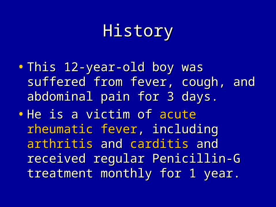
• This 12-year-old boy was suffered This 12-year-old boy was suffered from fever, cough, and abdominal from fever, cough, and abdominal pain for 3 days.pain for 3 days.
• He is a victim of He is a victim of acute rheumatic acute rheumatic feverfever, including , including arthritisarthritis and and carditiscarditis and received regular Penicillin-G and received regular Penicillin-G treatment monthly for 1 year.treatment monthly for 1 year.
HistoryHistory
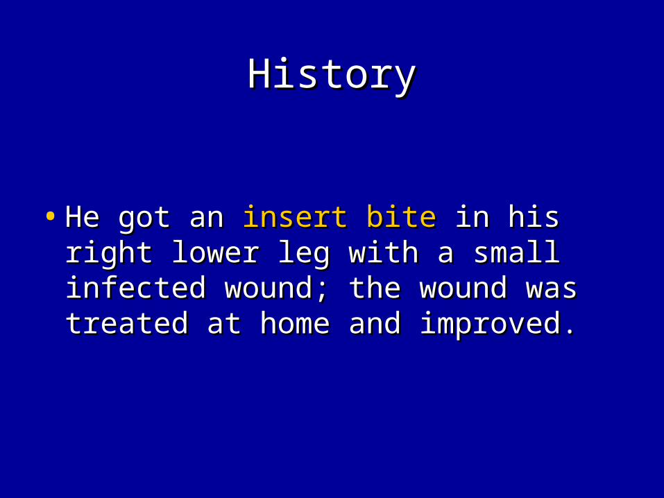
• He got an He got an insert biteinsert bite in his right in his right lower leg with a small infected lower leg with a small infected wound; the wound was treated at wound; the wound was treated at home and improved.home and improved.

HistoryHistory
• However, about 1 week later, the However, about 1 week later, the symptoms of symptoms of low-grade feverlow-grade fever, , coughcough and and abdominal painabdominal pain with fullness with fullness sensation were noted.sensation were noted.
• 3 days later, 3 days later, progressive dyspneaprogressive dyspnea during exertion was noted and then during exertion was noted and then he was sent to our hospital for help.he was sent to our hospital for help.

Physical examinationPhysical examination
• The physical examination showed The physical examination showed respiratory distressrespiratory distress, bilateral coarse , bilateral coarse breathing sounds, grade breathing sounds, grade Ⅰ-Ⅱ/Ⅳ Ⅰ-Ⅱ/Ⅳ systolic systolic murmurs over LSBmurmurs over LSB, distended abdomen, , distended abdomen, dilated jugular and superficial veinsdilated jugular and superficial veins, , hepatosplenomegalyhepatosplenomegaly and and pitting edema of pitting edema of bilateral lower legsbilateral lower legs..
• A small healed A small healed insert bite woundinsert bite wound over right over right lower leg.lower leg.

ImageImage
• The CXR showed minimal The CXR showed minimal right side right side pleural effusionpleural effusion with normal heart with normal heart size.size.
Laboratory dataLaboratory data
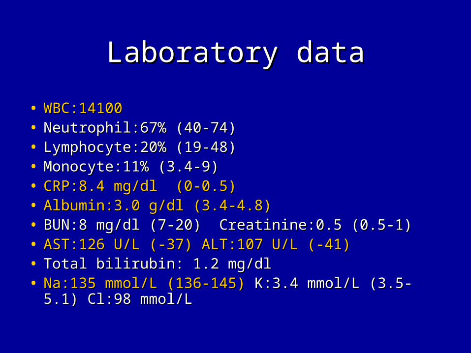
• WBC:14100 WBC:14100 • Neutrophil:67% (40-74)Neutrophil:67% (40-74)• Lymphocyte:20% (19-48)Lymphocyte:20% (19-48)• Monocyte:11% (3.4-9)Monocyte:11% (3.4-9)• CRP:8.4 mg/dl (0-0.5)CRP:8.4 mg/dl (0-0.5)• Albumin:3.0 g/dl (3.4-4.8)Albumin:3.0 g/dl (3.4-4.8)• BUN:8 mg/dl (7-20) Creatinine:0.5 (0.5-1)BUN:8 mg/dl (7-20) Creatinine:0.5 (0.5-1)• AST:126 U/L (-37) ALT:107 U/L (-41)AST:126 U/L (-37) ALT:107 U/L (-41)• Total bilirubin: 1.2 mg/dlTotal bilirubin: 1.2 mg/dl• Na:135 mmol/L (136-145)Na:135 mmol/L (136-145) K:3.4 mmol/L (3.5-5.1) K:3.4 mmol/L (3.5-5.1)
Cl:98 mmol/LCl:98 mmol/L

Admission courseAdmission course
• Echocardiography:Echocardiography:
• Tricuspid valve regurgitationTricuspid valve regurgitation
• Mitral valve regurgitationMitral valve regurgitation
• Pulmonary valve regurgitationPulmonary valve regurgitation

Admission courseAdmission course
• Abdominal sonographyAbdominal sonography
• HepatosplenomegalyHepatosplenomegaly
• Patent hepatic artery and portal veinPatent hepatic artery and portal vein
• Mild to moderate ascitesMild to moderate ascites

Subjective findingsSubjective findings
• FeverFever
• CoughCough
• Abdominal pain with fullnessAbdominal pain with fullness
• Exertional dyspneaExertional dyspnea
• Insert biteInsert bite

Objective findingsObjective findings
• Coarse breathing soundsCoarse breathing sounds
• Systolic heart murmurs over LLSBSystolic heart murmurs over LLSB
• Pitting edema over bilateral lower Pitting edema over bilateral lower legs.legs.
• HepatosplenomegalyHepatosplenomegaly
• Wound of insert biteWound of insert bite

Objective findingsObjective findings
• Liver function impairmentLiver function impairment
• HypoalbuminemiaHypoalbuminemia
• HyponatremiaHyponatremia
• Elevated CRP levelElevated CRP level
• Elevated WBC countElevated WBC count
• Pleural effusionPleural effusion
• MR, TR, PR.MR, TR, PR.
ProblemsProblems
• R/O Heart failureR/O Heart failure
• Fever Fever
• Insert biteInsert bite
• HepatosplenomegalyHepatosplenomegaly
• Impaired liver functionsImpaired liver functions
• Pleural effusionPleural effusion
• AscitesAscites

QuestionQuestion
• Patient traveling historyPatient traveling history• EKG finding (arrhythmia, ST or T wave change)EKG finding (arrhythmia, ST or T wave change)• Echocardiography findings (compared with previous)Echocardiography findings (compared with previous)• Arterial blood gas (hypoxia, hypercapnea)Arterial blood gas (hypoxia, hypercapnea)• Bacterial culture (blood,pleural effusion)Bacterial culture (blood,pleural effusion)• Virus isolation and identificationVirus isolation and identification• Pleural effusion study (transudate or exudate, AFB stain)Pleural effusion study (transudate or exudate, AFB stain)• Heart rate, blood pressure (arrhythmia, hypertension)Heart rate, blood pressure (arrhythmia, hypertension)• Anemia or thrombocytopenia (hematologic abnormalities)Anemia or thrombocytopenia (hematologic abnormalities)• Urine routine (Proteinuria ,hematuria)Urine routine (Proteinuria ,hematuria)• Indirect fluorescent antibody assay serologyIndirect fluorescent antibody assay serology

Causes of splenomegalyCauses of splenomegaly
• InfectionInfection– Bacterial: Typhoid fever, endocarditis, septicemia, abscessBacterial: Typhoid fever, endocarditis, septicemia, abscess– Viral:E-B virus, CMV, and othersViral:E-B virus, CMV, and others– Protozoal: Malaria, toxoplasmosisProtozoal: Malaria, toxoplasmosis
• Hematologic processesHematologic processes– Hemolytic anemia: Congenital, acquiredHemolytic anemia: Congenital, acquired– Extramedullary hematopoiesis: thalassemia, osteopetrosis, myelofibrosisExtramedullary hematopoiesis: thalassemia, osteopetrosis, myelofibrosis
• NeoplasmsNeoplasms– Malignant: Leukemia, lymphoma, histiocytoses, metastatic tumorsMalignant: Leukemia, lymphoma, histiocytoses, metastatic tumors– Benign: Hemagioma, hamartomaBenign: Hemagioma, hamartoma
• Metabolic diseasesMetabolic diseases– Lipidosis: Niemann-Pick, Gaucher diseaseLipidosis: Niemann-Pick, Gaucher disease– Mucopolysaccharidosis infiltration: HistiocytosisMucopolysaccharidosis infiltration: Histiocytosis
• CongestionCongestion• CirrhosisCirrhosis• CystsCysts• MiscellaneousMiscellaneous
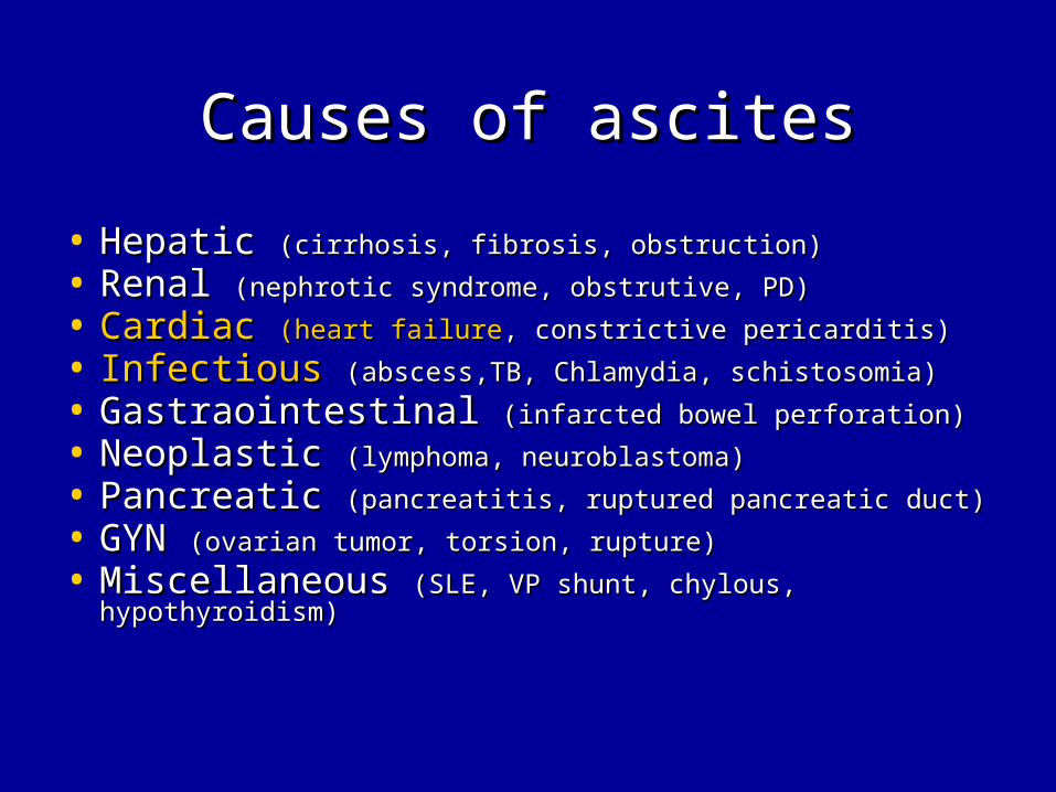
Causes of ascitesCauses of ascites
• Hepatic Hepatic (cirrhosis, fibrosis, obstruction)(cirrhosis, fibrosis, obstruction)
• Renal Renal (nephrotic syndrome, obstrutive, PD)(nephrotic syndrome, obstrutive, PD)
• CardiacCardiac (heart failure(heart failure, constrictive pericarditis), constrictive pericarditis)
• Infectious Infectious (abscess,TB, Chlamydia, schistosomia)(abscess,TB, Chlamydia, schistosomia)
• Gastraointestinal Gastraointestinal (infarcted bowel perforation)(infarcted bowel perforation)
• Neoplastic Neoplastic (lymphoma, neuroblastoma)(lymphoma, neuroblastoma)
• Pancreatic Pancreatic (pancreatitis, ruptured pancreatic duct)(pancreatitis, ruptured pancreatic duct)
• GYN GYN (ovarian tumor, torsion, rupture)(ovarian tumor, torsion, rupture)
• Miscellaneous Miscellaneous (SLE, VP shunt, chylous, (SLE, VP shunt, chylous, hypothyroidism)hypothyroidism)

Heart failureHeart failure
• SymptomsSymptoms– Fatigue, Fatigue, effort intoleranceeffort intolerance, anorexia, , anorexia,
abdominalabdominal painpain, , dyspneadyspnea,, coughcough
• SignSign– Engorged jugular veinEngorged jugular vein, , hepatomegalyhepatomegaly, , basilar basilar
ralesrales, , edema,edema, cardiomegaly, gallop rhythm, cardiomegaly, gallop rhythm, holosystolic murmur of mitral or tricuspid valve holosystolic murmur of mitral or tricuspid valve regurgitationregurgitation may be heard when ventricular may be heard when ventricular dilatation is advanced.dilatation is advanced.
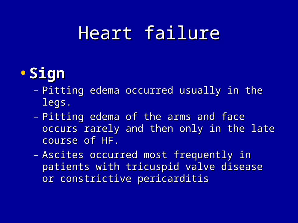
Heart failureHeart failure
•SignSign– Pitting edema occurred usually in the legs.Pitting edema occurred usually in the legs.– Pitting edema of the arms and face occurs Pitting edema of the arms and face occurs
rarely and then only in the late course of HF.rarely and then only in the late course of HF.– Ascites occurred most frequently in patients Ascites occurred most frequently in patients
with tricuspid valve disease or constrictive with tricuspid valve disease or constrictive pericarditispericarditis

Heart failureHeart failure
• Chest X rayChest X ray• Exaggeration of the pulmonary arterial Exaggeration of the pulmonary arterial
vessels to the periphery of the lung fields. vessels to the periphery of the lung fields. (Patients with (Patients with cardiomyopathycardiomyopathy may have a may have a relatively normal pulmonary vascular bed relatively normal pulmonary vascular bed early in the course of disease)early in the course of disease)
• CardiomegalyCardiomegaly is invariable noted. is invariable noted. • Pleural effusionPleural effusion was more frequent was more frequent in the in the
right side than in the left side. right side than in the left side.

Heart failureHeart failure
• EKGEKG• Left or right ventricular ischemic changes Left or right ventricular ischemic changes
may correlate well with clinical and other may correlate well with clinical and other non invasive parameters of ventricular non invasive parameters of ventricular function.function.
• Low voltage QRS morphologic characteristic Low voltage QRS morphologic characteristic with ST-T wave abnormaluties may also with ST-T wave abnormaluties may also suggest suggest myocardial inflammatory diseasemyocardial inflammatory disease but can be seen with but can be seen with pericarditispericarditis
• Rhythm disease is also a potential cause of Rhythm disease is also a potential cause of heart failure.heart failure.

Diagnosis of congestive heart Diagnosis of congestive heart failurefailure
(Framingham criteria)(Framingham criteria)• Major criteria (at least 1)Major criteria (at least 1)
– Paroxysmal nocturnal dyspneaParoxysmal nocturnal dyspnea– Neck vein distentionNeck vein distention– RalesRales– CardiomegalyCardiomegaly– Acute pulmonary edemaAcute pulmonary edema– S3 gallopS3 gallop– Increased venous pressure (>16 cm H2O)Increased venous pressure (>16 cm H2O)– Positive hepatojugular refluxPositive hepatojugular reflux
• Minor criteria (at least 2)Minor criteria (at least 2)– Extremity edemaExtremity edema– Night coughNight cough– Dyspnea on exertionDyspnea on exertion– HepatomegalyHepatomegaly– Pleural effusionPleural effusion– Vital capacity reduced by 1/3 from normalVital capacity reduced by 1/3 from normal– Tachycardia (>120 bpm)Tachycardia (>120 bpm)

Acute versus ChronicAcute versus Chronic
• AcuteAcute: : Sudden developmentSudden development of of myocardial infectionmyocardial infection or rupture of a or rupture of a cardiac valvecardiac valve, no cardiomegaly, no , no cardiomegaly, no peripheral edema.peripheral edema.
• Chronic: Dilated cardiomyopathy, Chronic: Dilated cardiomyopathy, multiple valvular heart disease, multiple valvular heart disease, cardiomegaly, peripheral edema.cardiomegaly, peripheral edema.
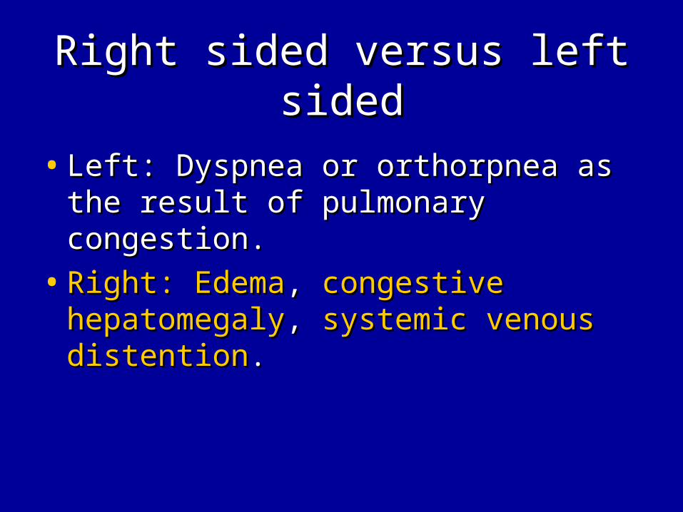
Right sided versus left sidedRight sided versus left sided
• Left: Dyspnea or orthorpnea as the Left: Dyspnea or orthorpnea as the result of pulmonary congestion.result of pulmonary congestion.
• Right:Right: EdemaEdema, , congestive congestive hepatomegalyhepatomegaly, , systemic venous systemic venous distentiondistention..
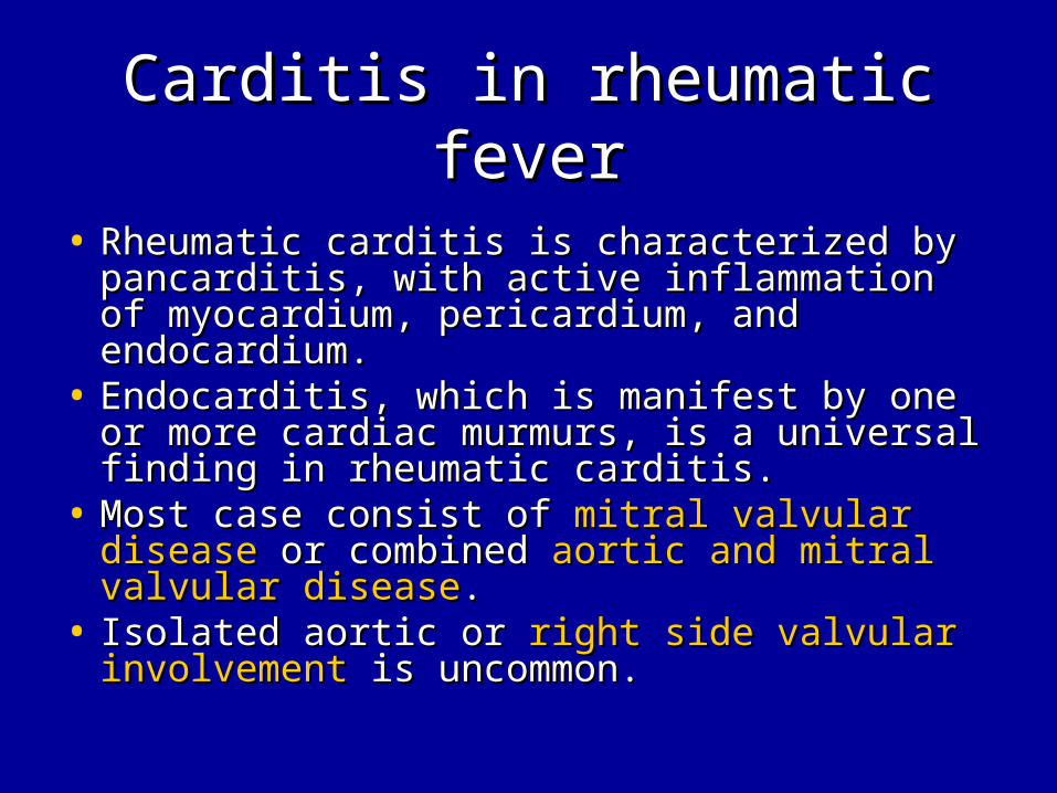
Carditis in rheumatic feverCarditis in rheumatic fever
• Rheumatic carditis is characterized by Rheumatic carditis is characterized by pancarditis, with active inflammation of pancarditis, with active inflammation of myocardium, pericardium, and endocardium.myocardium, pericardium, and endocardium.
• Endocarditis, which is manifest by one or more Endocarditis, which is manifest by one or more cardiac murmurs, is a universal finding in cardiac murmurs, is a universal finding in rheumatic carditis.rheumatic carditis.
• Most case consist of Most case consist of mitral valvular diseasemitral valvular disease or or combined combined aortic and mitral valvular diseaseaortic and mitral valvular disease..
• Isolated aortic or Isolated aortic or right side valvular involvementright side valvular involvement is uncommon.is uncommon.
Rheumatic heart diseaseRheumatic heart disease
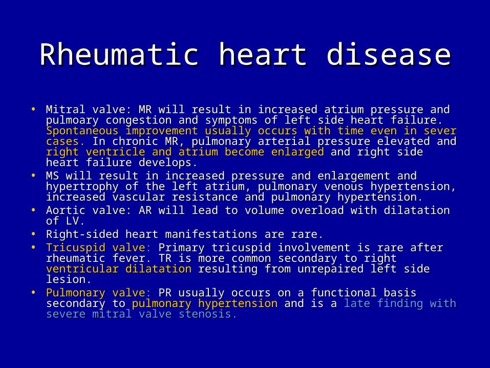
• Mitral valve: MR will result in increased atrium pressure and Mitral valve: MR will result in increased atrium pressure and pulmoary congestion and symptoms of left side heart failure. pulmoary congestion and symptoms of left side heart failure. Spontaneous improvement usually occurs with time even in sever Spontaneous improvement usually occurs with time even in sever cases.cases. In chronic MR, pulmonary arterial pressure elevated and In chronic MR, pulmonary arterial pressure elevated and right ventricle and atrium become enlargedright ventricle and atrium become enlarged and right side heart and right side heart failure develops. failure develops.
• MS will result in increased pressure and enlargement and MS will result in increased pressure and enlargement and hypertrophy of the left atrium, pulmonary venous hypertension, hypertrophy of the left atrium, pulmonary venous hypertension, increased vascular resistance and pulmonary hypertension.increased vascular resistance and pulmonary hypertension.
• Aortic valve: AR will lead to volume overload with dilatation of LV. Aortic valve: AR will lead to volume overload with dilatation of LV. • Right-sided heart manifestations are rare.Right-sided heart manifestations are rare.• Tricuspid valveTricuspid valve:: Primary tricuspid involvement is rare after Primary tricuspid involvement is rare after
rheumatic fever. TR is more common secondary to right rheumatic fever. TR is more common secondary to right ventricular dilatationventricular dilatation resulting from unrepaired left side lesion. resulting from unrepaired left side lesion.
• Pulmonary valvePulmonary valve:: PR usually occurs on a functional basis PR usually occurs on a functional basis secondary to secondary to pulmonary hypertensionpulmonary hypertension and is a and is a late finding with late finding with severe mitral valve stenosis.severe mitral valve stenosis.
Pulmonary regurgitationPulmonary regurgitation
• Isolated congenital pulmonary Isolated congenital pulmonary regurgitation is rare.regurgitation is rare.
• Pulmoary valvular insufficiency most Pulmoary valvular insufficiency most often accompanies other often accompanies other cardiovascular diseasecardiovascular disease or may be or may be secondary to severe secondary to severe pulmonary pulmonary hypertension.hypertension.

Tricuspid regurgitationTricuspid regurgitation
• Tricuspid regurgitation usually Tricuspid regurgitation usually functional.functional.
• Secondary to marked Secondary to marked RV dilatationRV dilatation of any of any cause and often associated with cause and often associated with pulmonary hypertension.pulmonary hypertension.
• When volume overload or intrinsic When volume overload or intrinsic myocardial disease -> dilated right myocardial disease -> dilated right ventricle. ventricle.
• Tricuspid regurgitation often accompanies Tricuspid regurgitation often accompanies with with right ventricular dysfunction.right ventricular dysfunction.

Pulmonary regurgitation & Pulmonary regurgitation & Tricuspid regurgitationTricuspid regurgitation
• Pulmonary hypertension Pulmonary hypertension
• Cardiovascular disease -> RV Cardiovascular disease -> RV dysfunctiondysfunction
Causes of pulmonary hypertensionCauses of pulmonary hypertension
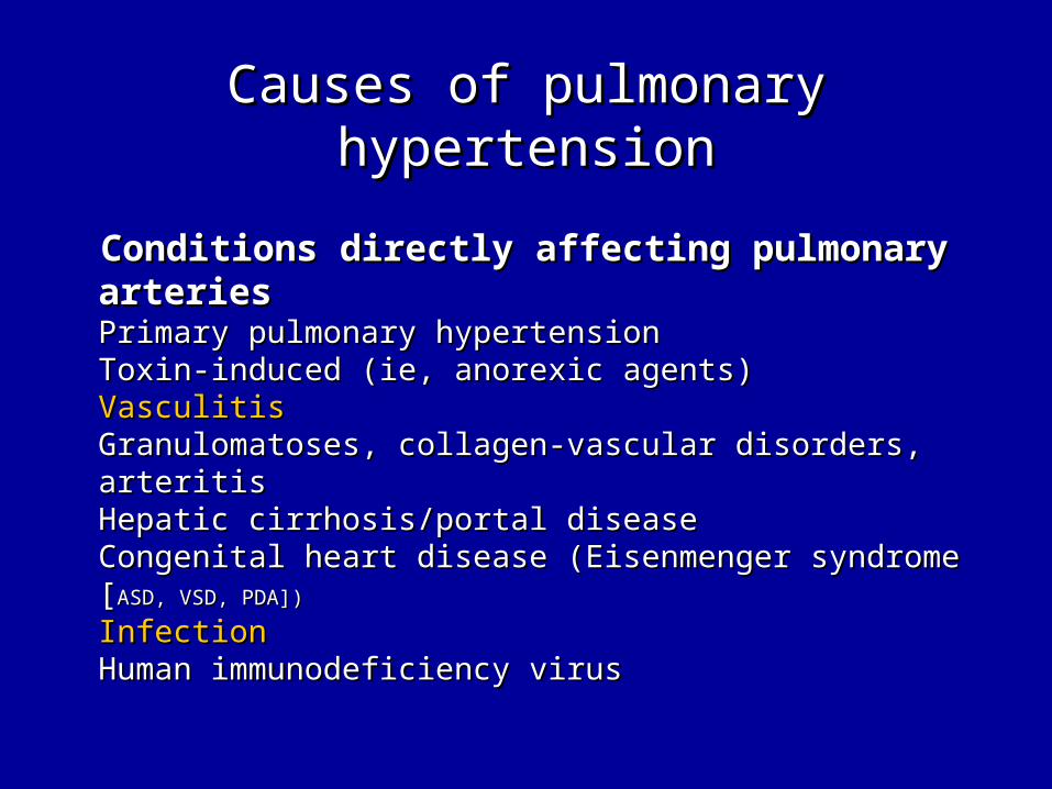
Conditions directly affecting pulmonary arteriesConditions directly affecting pulmonary arteriesPrimary pulmonary hypertensionPrimary pulmonary hypertensionToxin-induced (ie, anorexic agents)Toxin-induced (ie, anorexic agents)VasculitisVasculitisGranulomatoses, collagen-vascular disorders, arteritisGranulomatoses, collagen-vascular disorders, arteritisHepatic cirrhosis/portal diseaseHepatic cirrhosis/portal diseaseCongenital heart disease (Eisenmenger syndrome [Congenital heart disease (Eisenmenger syndrome [ASD, VSD, ASD, VSD, PDA])PDA])
InfectionInfectionHuman immunodeficiency virusHuman immunodeficiency virus

Causes of pulmonary hypertensionCauses of pulmonary hypertension
Conditions affecting pulmonary parenchymaConditions affecting pulmonary parenchymaChronic obstructive lung diseaseChronic obstructive lung diseaseInfiltrative/granulomatous diseasesInfiltrative/granulomatous diseasesSarcoidosis, pneumoconiosis, radiation, fibrosis, neoplasm, Sarcoidosis, pneumoconiosis, radiation, fibrosis, neoplasm, pneumonia, collagen-vascular diseasespneumonia, collagen-vascular diseasesCystic fibrosisCystic fibrosisUpper airway obstructionUpper airway obstructionHigh-altitude diseaseHigh-altitude diseaseArteriovenous fistulas within the lungArteriovenous fistulas within the lungRestrictive lung diseasesRestrictive lung diseases
Causes of pulmonary hypertensionCauses of pulmonary hypertension
Conditions affecting the thoracic cage and Conditions affecting the thoracic cage and neuromuscular systemneuromuscular systemObesity-hypoventilation/sleep apneaObesity-hypoventilation/sleep apneaPharyngeal-tracheal obstructionPharyngeal-tracheal obstructionKyphoscoliosisKyphoscoliosisPleural fibrosisPleural fibrosisNeuromuscular disordersNeuromuscular disordersMyasthenia gravis, poliomyelitis, central respiratory disordersMyasthenia gravis, poliomyelitis, central respiratory disorders
Causes of pulmonary hypertensionCauses of pulmonary hypertension
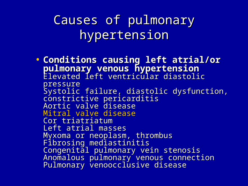
• Conditions causing left atrial/or pulmonary Conditions causing left atrial/or pulmonary venous hypertensionvenous hypertensionElevated left ventricular diastolic pressureElevated left ventricular diastolic pressureSystolic failure, diastolic dysfunction, constrictive Systolic failure, diastolic dysfunction, constrictive pericarditispericarditisAortic valve diseaseAortic valve diseaseMitral valve diseaseMitral valve diseaseCor triatriatumCor triatriatumLeft atrial massesLeft atrial massesMyxoma or neoplasm, thrombusMyxoma or neoplasm, thrombusFibrosing mediastinitisFibrosing mediastinitisCongenital pulmonary vein stenosisCongenital pulmonary vein stenosisAnomalous pulmonary venous connectionAnomalous pulmonary venous connectionPulmonary venoocclusive diseasePulmonary venoocclusive disease
Causes of pulmonary hypertensionCauses of pulmonary hypertension
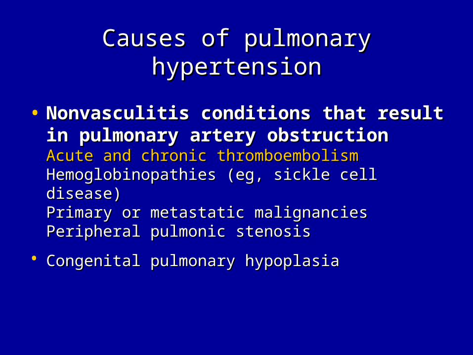
• Nonvasculitis conditions that result in pulmonary Nonvasculitis conditions that result in pulmonary artery obstructionartery obstructionAcute and chronic thromboembolismAcute and chronic thromboembolismHemoglobinopathies (eg, sickle cell disease)Hemoglobinopathies (eg, sickle cell disease)Primary or metastatic malignanciesPrimary or metastatic malignanciesPeripheral pulmonic stenosisPeripheral pulmonic stenosis
• Congenital pulmonary hypoplasiaCongenital pulmonary hypoplasia
Vasculitis syndromeVasculitis syndrome
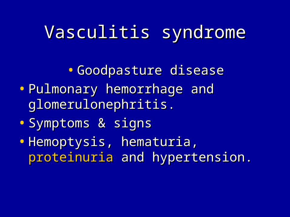
• Goodpasture diseaseGoodpasture disease
• Pulmonary hemorrhage and Pulmonary hemorrhage and glomerulonephritis.glomerulonephritis.
• Symptoms & signsSymptoms & signs
• Hemoptysis, hematuria, Hemoptysis, hematuria, proteinuriaproteinuria and hypertension.and hypertension.

Vasculitis syndromeVasculitis syndrome
• Wegener GranulomatosisWegener Granulomatosis
• Fever, myalgia, cough, nasal discharge, Fever, myalgia, cough, nasal discharge, hematuria proteinuria.hematuria proteinuria.
• Hemoptysis and dyspnea from lung Hemoptysis and dyspnea from lung lesion.lesion.
• Uveitis, conjuctivitis.Uveitis, conjuctivitis.
• Peripheral granuloma.Peripheral granuloma.
• Palpalbe purpuric nodules and ulcersPalpalbe purpuric nodules and ulcers

Vasculitis syndromeVasculitis syndrome
• Polyarteritis NodosaPolyarteritis Nodosa• Associated with infection of Associated with infection of group A group A
streptococcusstreptococcus, hepatitis B, CMV, , hepatitis B, CMV, parvovirus , TB, E-B virus.parvovirus , TB, E-B virus.
• Symptoms & signs.Symptoms & signs.• Fever Fever • Abdominal pain (mesenteric arterial inflammation)Abdominal pain (mesenteric arterial inflammation)• Hypertension, hematuria, Hypertension, hematuria, proteinuria (renal vessels)proteinuria (renal vessels)• Purpura, edema, painful nodule (skin involvement)Purpura, edema, painful nodule (skin involvement)• Heart failure (myocarditis, myocardial ischemia)Heart failure (myocarditis, myocardial ischemia)• Elevated liver function (hepatitis B infection more common in Elevated liver function (hepatitis B infection more common in
adults)adults)• Elevated ESR, anemia and leukocytosisElevated ESR, anemia and leukocytosis
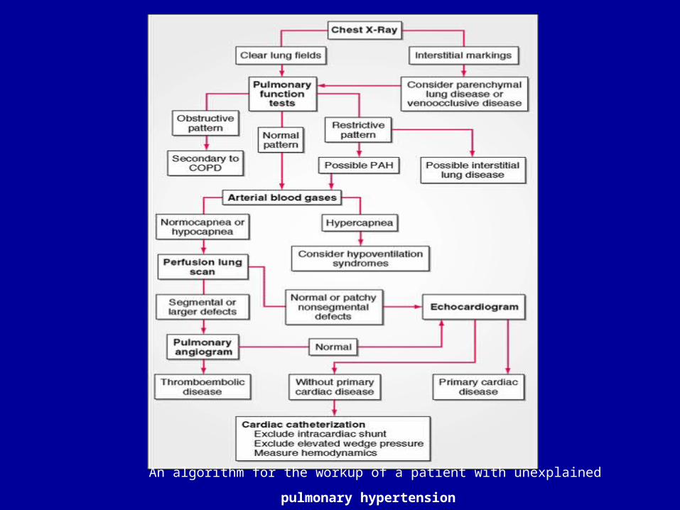
An algorithm for the workup of a patient with unexplained pulmonary
hypertension

Pulmonary embolismPulmonary embolism
• Precipitating factorPrecipitating factor• CVP insertion, immobility, CVP insertion, immobility, heart diseaseheart disease, oral , oral
contraceptives, ventriculoatrial shunt, trauma, contraceptives, ventriculoatrial shunt, trauma, infectioninfection, , dehydration, collagen vascular disease, shock , obesity, dehydration, collagen vascular disease, shock , obesity, hematologic disorder.hematologic disorder.
• SymptomsSymptoms• Chest pain, Chest pain, dyspneadyspnea, , coughcough, , feverfever, , hemoptysishemoptysis..
• SignsSigns• Rales, Rales, heart murmursheart murmurs, tachycardia, , tachycardia, feverfever, diaphoresis, , diaphoresis,
phlebitis, wheezing.phlebitis, wheezing.

Pulmonary embolismPulmonary embolism
• LabLab• ABG:ABG: Respiratory alkalosis, arterial hypoxemia Respiratory alkalosis, arterial hypoxemia• Elevated WBC countElevated WBC count, D-dimer, fibrinogen, protein C, protein , D-dimer, fibrinogen, protein C, protein
S.S.• Chest:Chest: Often normal in patient with PE.Often normal in patient with PE.• EKG:EKG: Nonspecific ST segment change and signs of cor Nonspecific ST segment change and signs of cor
pulmonalepulmonale• Ventilation-perfusion image:Ventilation-perfusion image: Regional blood flow Regional blood flow
and ventilation defects .and ventilation defects . But normal V-P scan cant exclude a But normal V-P scan cant exclude a PE and are interpreted as high probability, intermediate PE and are interpreted as high probability, intermediate probability, low probability, very low probability and normal. probability, low probability, very low probability and normal. Lung disease can limit the utility of this study. Lung disease can limit the utility of this study.
• Pulmonary angiographyPulmonary angiography is the gold standard is the gold standard diagnostic test for pulmonary embolism.diagnostic test for pulmonary embolism.

Cause of heart failure in Cause of heart failure in childchild
• Rheumatic feverRheumatic fever• Acute hypertensionAcute hypertension• ThyrotoxicosisThyrotoxicosis• Hemochromatosis-hemosiderosisHemochromatosis-hemosiderosis• Cancer therapy (radiation, doxorubincin)Cancer therapy (radiation, doxorubincin)• Sickle cell anemiaSickle cell anemia• EndocarditisEndocarditis• Cardiomyopathy Cardiomyopathy ((Viral myocarditisViral myocarditis, , nonviralnonviral))• MyocarditisMyocarditis, , hypertrophic, dilated.)hypertrophic, dilated.)• Cor pulmoale (cystic fibrosis)Cor pulmoale (cystic fibrosis)

Pulmonary regurgitation & Pulmonary regurgitation & Tricuspid regurgitationTricuspid regurgitation
• Pulmonary hypertension Pulmonary hypertension • InfectionInfection• Pulmonary embolismPulmonary embolism
• Cardiovascular disease -> RV Cardiovascular disease -> RV dysfunctiondysfunction
• Rheumatic feverRheumatic fever• EndocarditisEndocarditis• Cardiomyopathy (carditis, dilated, Cardiomyopathy (carditis, dilated,
hypertrophic )hypertrophic )

Acute rheumatic fever Acute rheumatic fever
• Major: (least 2 major or 1+2 minor)Major: (least 2 major or 1+2 minor)• CarditisCarditis, polyarthritis, erythema marginatum, , polyarthritis, erythema marginatum,
subcutaneous nodulessubcutaneous nodules
• Minor:Minor: • FeverFever, arthralgia, , arthralgia, elevated ESR elevated ESR oror CRP, CRP, prolonged PR prolonged PR
intervalinterval
• Supported evidence of GAS infection:Supported evidence of GAS infection:• Positive throat culture or streptococcal antigen testPositive throat culture or streptococcal antigen test• Elevated or increased streptococcal antibody titerElevated or increased streptococcal antibody titer

Infectious endocarditisInfectious endocarditis
• Etiology: Viridans-type streptococci and staphylococcus aureus Etiology: Viridans-type streptococci and staphylococcus aureus are the leading causative agents responsible for endocarditis in are the leading causative agents responsible for endocarditis in pediatric patients.pediatric patients.
• Infective endocarditis is often a complication of congenital or Infective endocarditis is often a complication of congenital or rheumatic heart diseaserheumatic heart disease
• Prolonged fever without other manifestation.Prolonged fever without other manifestation.• Low grade feverLow grade fever, fatigue, myalgia, arthralgia, headache, and at , fatigue, myalgia, arthralgia, headache, and at
times, chills, nausea, and vomiting.times, chills, nausea, and vomiting.• SplenomegalySplenomegaly and petechiae are relative common. and petechiae are relative common.• Osler nodes (tender, pea-sized intradermal nodules in the pads of Osler nodes (tender, pea-sized intradermal nodules in the pads of
the fingers and toes), Janeway lesion (painless small the fingers and toes), Janeway lesion (painless small erythematous or hemorrhagic lesion on the palms and soles), erythematous or hemorrhagic lesion on the palms and soles), splinter hemorrhages (linear lesions beneath the nails)splinter hemorrhages (linear lesions beneath the nails)
• Embolic strokes, cerebral abscess, mycotic aneurysm, and Embolic strokes, cerebral abscess, mycotic aneurysm, and hemorrhage in staphylococcus disease.hemorrhage in staphylococcus disease.

Infectious endocarditisInfectious endocarditis
• HistoryHistory• Congenital or rheumatic heart diseaseCongenital or rheumatic heart disease
• Preceding dental, urinary tract or intestinal procedurePreceding dental, urinary tract or intestinal procedure
• Intravenous drugs abuseIntravenous drugs abuse
• Central venous catheterCentral venous catheter
• Prosthetic heart valveProsthetic heart valve
• SymptomsSymptoms• FeverFever, chills, chest and , chills, chest and abdominal painabdominal pain, arthralgia, myalgia, , arthralgia, myalgia,
dyspneadyspnea, malaise, night sweats, weight loss, CNS , malaise, night sweats, weight loss, CNS manifestationsmanifestations

Infectious endocarditisInfectious endocarditis
• SignsSigns• Elevated temperatureElevated temperature, tachycardia, embolic phenomena (Roth , tachycardia, embolic phenomena (Roth
spots, petechia, splinter nail bed hemorrahge, Osler nodes, CNS or spots, petechia, splinter nail bed hemorrahge, Osler nodes, CNS or ocular lesions), Janeway lesions, ocular lesions), Janeway lesions, New or changing murmurNew or changing murmur,, splenomegalysplenomegaly, arthritis, , arthritis, heart failureheart failure, arrythmias, Metastatic , arrythmias, Metastatic infection (arthritis, meningitis, mycotic arterial aneurysm, infection (arthritis, meningitis, mycotic arterial aneurysm, pericarditis, abscesses, septic pulmonary emboli), clubbingpericarditis, abscesses, septic pulmonary emboli), clubbing
• LabsLabs• Blood culture (+), ESR ↑, Blood culture (+), ESR ↑, CRP↑,CRP↑, anemia, anemia, leukocytosisleukocytosis, immune , immune
complexes, hypergammaglobulinemia, hypocomplementemia, complexes, hypergammaglobulinemia, hypocomplementemia, rheumatoid factor, hematuria, renal failurerheumatoid factor, hematuria, renal failure
• Chest X ray: bilateral infiltration, nodules, Chest X ray: bilateral infiltration, nodules, pleural effusionspleural effusions• Echocardiography: valve vegetations, prosthetic valve dysfunction Echocardiography: valve vegetations, prosthetic valve dysfunction
or leak, myocardial abscess, new-onset or leak, myocardial abscess, new-onset valve insufficiencyvalve insufficiency..

Infectious endocarditisInfectious endocarditis
• Duke criteriaDuke criteria• Major: 1.Blood culture (+) 2.Evidence of endocarditis on Major: 1.Blood culture (+) 2.Evidence of endocarditis on
echocardiography (intracardiac mass on a valve or other echocardiography (intracardiac mass on a valve or other site, regurgitation flow near a prosthesis, abscess, partial site, regurgitation flow near a prosthesis, abscess, partial dehiscence of prosthetic valves or dehiscence of prosthetic valves or new valve regurgitation new valve regurgitation flowflow))
• Minor:Minor:feverfever, embolic-vascular signs, immune complex , embolic-vascular signs, immune complex phenomena (GN, arthritis, rheumatoid factor, Osler nodes, phenomena (GN, arthritis, rheumatoid factor, Osler nodes, Roth spots), Roth spots),
• 2 major + 1 minor2 major + 1 minor
• 1 major + 3 minors1 major + 3 minors
• 5 minors5 minors
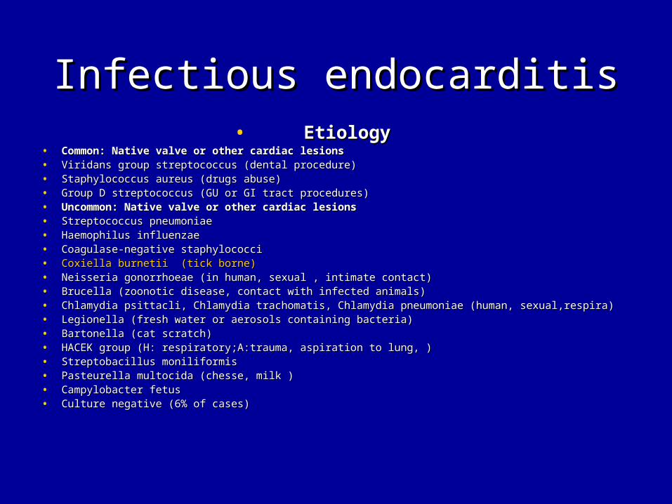
Infectious endocarditisInfectious endocarditis• EtiologyEtiology
• Common: Native valve or other cardiac lesionsCommon: Native valve or other cardiac lesions• Viridans group streptococcus (dental procedure)Viridans group streptococcus (dental procedure)• Staphylococcus aureus (drugs abuse)Staphylococcus aureus (drugs abuse)• Group D streptococcus (GU or GI tract procedures)Group D streptococcus (GU or GI tract procedures)• Uncommon: Native valve or other cardiac lesionsUncommon: Native valve or other cardiac lesions• Streptococcus pneumoniaeStreptococcus pneumoniae• Haemophilus influenzaeHaemophilus influenzae• Coagulase-negative staphylococciCoagulase-negative staphylococci• Coxiella burnetii (tick borne)Coxiella burnetii (tick borne)• Neisseria gonorrhoeae (in human, sexual , intimate contact)Neisseria gonorrhoeae (in human, sexual , intimate contact)• Brucella (zoonotic disease, contact with infected animals)Brucella (zoonotic disease, contact with infected animals)• Chlamydia psittacli, Chlamydia trachomatis, Chlamydia pneumoniae (human, sexual,respira)Chlamydia psittacli, Chlamydia trachomatis, Chlamydia pneumoniae (human, sexual,respira)• Legionella (fresh water or aerosols containing bacteria)Legionella (fresh water or aerosols containing bacteria)• Bartonella (cat scratch)Bartonella (cat scratch)• HACEK group (H: respiratory;A:trauma, aspiration to lung, )HACEK group (H: respiratory;A:trauma, aspiration to lung, )• Streptobacillus moniliformisStreptobacillus moniliformis• Pasteurella multocida (chesse, milk )Pasteurella multocida (chesse, milk )• Campylobacter fetusCampylobacter fetus• Culture negative (6% of cases)Culture negative (6% of cases)

MyocarditisMyocarditis
• Symptoms:Symptoms:• FeverFever, , severe heart failuresevere heart failure, , respiratory distressrespiratory distress..
• Incubation time:1-7 days of the onset of Incubation time:1-7 days of the onset of symptomssymptoms..
• Signs:Signs:• Cyanosis, distant heart sounds, weak pulses, Cyanosis, distant heart sounds, weak pulses,
tachycardia, Gallop rhythm, acidosis, and shock.tachycardia, Gallop rhythm, acidosis, and shock.
• Evidence of Evidence of hepatitishepatitis, aseptic meningitis, , aseptic meningitis, associated rash may be present.associated rash may be present.

MyocarditisMyocarditis
• Lab:Lab:• Elevated CK, LDHElevated CK, LDH
• EKG: Sinus tachycardia, reduced QRS voltage, ST-EKG: Sinus tachycardia, reduced QRS voltage, ST-segment and T-wave abnormalities, arrhythmia.segment and T-wave abnormalities, arrhythmia.
• Echo: poor ventricular function, pericardial Echo: poor ventricular function, pericardial effusion, effusion, mitral valve regurgitationmitral valve regurgitation, absence of , absence of congenital heart lesion or coronary arterycongenital heart lesion or coronary artery

Etiology of myocardial Etiology of myocardial diseasedisease• Familial-Hereditary Familial-Hereditary (myopathy, cardiomyopathy)(myopathy, cardiomyopathy)
• Infection Infection (Virus, Richettsiae, (Virus, Richettsiae, Bacteria,parasite,fungus)Bacteria,parasite,fungus)
• Metabolic, nutritional, endocarineMetabolic, nutritional, endocarine• Connective tissue-Granulomatous disease-Connective tissue-Granulomatous disease-
infiltrative infiltrative (SLE, (SLE, vasculitisvasculitis, Scleroderma, sarcoidosis, , Scleroderma, sarcoidosis, dermatomyositis, leukemia)dermatomyositis, leukemia)
• Drugs-Toxins Drugs-Toxins (chemo, alcohol,irradiation.)(chemo, alcohol,irradiation.)
• Coronary arteries Coronary arteries (Kawasaki disease, meidal necrosis, (Kawasaki disease, meidal necrosis, anomalous left coronary artery)anomalous left coronary artery)
• Other Other (anemia, ischemia)(anemia, ischemia)

Etiology of myocardial Etiology of myocardial diseasedisease• Infection:Infection:
• Virus:Virus: • Coxsackievirus A and BCoxsackievirus A and B• AdenovirusAdenovirus• HIV HIV • Echovirus Echovirus • Rubella Rubella • Varicella Varicella • Influenza Influenza • Mumps Mumps • Epstein-Barr Epstein-Barr • Measles Measles • Poliomyelitis. Poliomyelitis.

Etiology of myocardial Etiology of myocardial diseasedisease
• Infection:Infection:• Richettsiae: Richettsiae: • PsittacosisPsittacosis• Q fever, Coxiella burnetii(Ticks)Q fever, Coxiella burnetii(Ticks)• Rocky Moutain spotted fever. (Tick bites)Rocky Moutain spotted fever. (Tick bites)• BacterialBacterial• Diphtheria (respiratory, cutaneous)Diphtheria (respiratory, cutaneous)• Mycoplasma (airborne, human to human)Mycoplasma (airborne, human to human)• Meningococcus (respiratory)Meningococcus (respiratory)• Leptospirosis (water or soil contaminated with rat urine)Leptospirosis (water or soil contaminated with rat urine)• Lyme diseaseLyme disease (tick bites) (tick bites)• Typhoid fever (foods or water contaminate with human feces) Typhoid fever (foods or water contaminate with human feces) • Tuberculosis (airborne, human to human)Tuberculosis (airborne, human to human)• Streptococcus (respiratory, dental procedure)Streptococcus (respiratory, dental procedure)• Listeriosis (food(cheese, milk) contaminate with feces of animals)Listeriosis (food(cheese, milk) contaminate with feces of animals)

Etiology of myocardial Etiology of myocardial diseasedisease
• Infection:Infection:• Parasites:Parasites: • Chagas disease (kissing bugs)Chagas disease (kissing bugs)• Toxoplasmosis (food or water comtaminated by cat feces)Toxoplasmosis (food or water comtaminated by cat feces)• Loa loa (biting flies)Loa loa (biting flies)• Toxocara canis (dog feces)Toxocara canis (dog feces)• Schistosomiasis (contact water contaminated with cercaria)Schistosomiasis (contact water contaminated with cercaria)• CysticercosisCysticercosis• EchinococcusEchinococcus• Trichinosis. (meat with larvae of trichinella)Trichinosis. (meat with larvae of trichinella)• Fungi:Fungi: • Histoplasmosis.Histoplasmosis.• Coccidiomyocosis. Coccidiomyocosis. • Actiomycosis.Actiomycosis.

Differential problemsDifferential problems
• R/O Pulmonary hypertensionR/O Pulmonary hypertension
• R/O Pulmonary embolismR/O Pulmonary embolism
• R/O CardiomyopathyR/O Cardiomyopathy
• HepatosplenomegalyHepatosplenomegaly
• Insert bite transmitted diseaseInsert bite transmitted disease

DiseaseDisease TransmissionTransmission HeartHeart Pul. hypertentsionPul. hypertentsion
Pul. embolismPul. embolism
HepatomegalHepatomegalyy
SplenomegalSplenomegalyy
MalariaMalaria MosquitoesMosquitoes (-)(-) (-)(-) (+)(+)Yellow Yellow FeverFever
MosquitoesMosquitoes (-)(-) (-)(-) (-)(-)Dengue Dengue FeverFever
MosquitoesMosquitoes (-)(-) (-)(-) (-)(-)J. J. encephalitiencephalitiss
MosquitoesMosquitoes (-)(-) (-)(-) (-)(-)
FilariasisFilariasis MosquitoesMosquitoes (-)(-) (-)(-) (-)(-)Colorado Colorado tick fevertick fever
TickTick (-)(-) (-)(-) (-)(-)Tick bornTick born
EncephalitiEncephalitiss
TickTick (-)(-) (-)(-) (-)(-)
DiseaseDisease TransmissionTransmission HeartHeart Pul. hypertentsionPul. hypertentsion
Pul. embolismPul. embolism
HepatomegalHepatomegalyy
SplenomegalSplenomegalyy
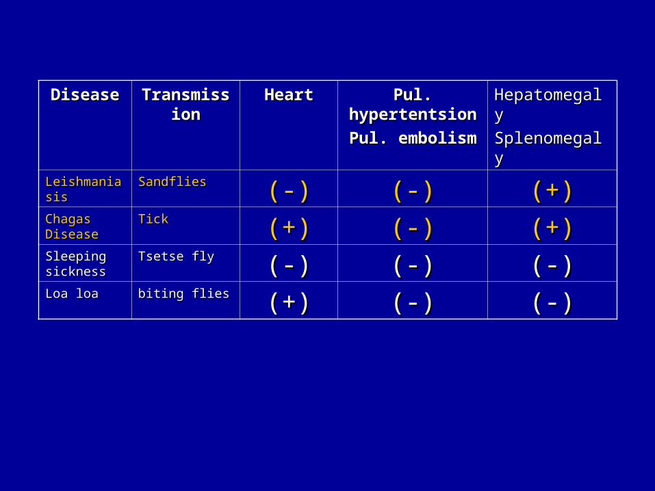
LeishmaniasLeishmaniasisis
SandfliesSandflies (-)(-) (-)(-) (+)(+)Chagas Chagas DiseaseDisease
TickTick (+)(+) (-)(-) (+)(+)Sleeping Sleeping sicknesssickness
Tsetse flyTsetse fly (-)(-) (-)(-) (-)(-)Loa loaLoa loa biting fliesbiting flies (+)(+) (-)(-) (-)(-)

DiseaseDisease TransmissionTransmission Heart Heart HepatomegalyHepatomegaly
SplenomegalySplenomegaly
Pul. E.Pul. E.
Pul. H.Pul. H.
Relapsing feverRelapsing fever Tick,louseTick,louse MyocarditisMyocarditis (+)(+) (-)(-)Lyme diseaseLyme disease Tick biteTick bite MyocarditisMyocarditis (-)(-) (-)(-)RMSFRMSF Tick biteTick bite MyocarditisMyocarditis (+) 33%(+) 33% ((+) +) P.E.P.E.
MSFMSF Tick biteTick bite (-)(-) (-)(-) (-)(-)Scrub typhusScrub typhus Chigger biteChigger bite MyocarditisMyocarditis (-)(-) (-)(-)Murine typhusMurine typhus Flea fecesFlea feces (-)(-) (-)(-) (-)(-)Epidemic typhusEpidemic typhus Louse fecesLouse feces (-)(-) (-)(-) (-)(-)HME, HEHME, HE Tick biteTick bite (-)(-) (+)(+) (-)(-)Q feverQ fever Tick borneTick borne MyocarditisMyocarditis
EndocariditisEndocariditis(+)(+) ((+)+)P.E.P.E.
Chagas diseaseChagas disease Kissing bugKissing bug MyocarditisMyocarditis (+)(+) (-)(-)Loa LoaLoa Loa Biting fliesBiting flies CardiomyopathyCardiomyopathy (-)(-) (-)(-)

Richettsial infectionRichettsial infection
• Spotted fever group Spotted fever group
• Typhus Typhus
• Scrub typhus Scrub typhus
• Ehrlichioses and AnaplasmosisEhrlichioses and Anaplasmosis
• Q feverQ fever

Richettsial infectionRichettsial infection
• Spotted fever groupSpotted fever group
• Rocky Mountain spotted feverrRocky Mountain spotted feverr
• Mediterranean Spotted feverMediterranean Spotted fever
• African tick-bite feverAfrican tick-bite fever
• RickettsialpoxRickettsialpox
• Murine typhus-likeMurine typhus-like

Spotted fever groupSpotted fever group• Rocky mountain spotted feverRocky mountain spotted fever
• Tick biteTick bite transmission disease (Host:Dogs, rodents) transmission disease (Host:Dogs, rodents)• United states, southwestern, Canada, Mexico, Central America, South AmericaUnited states, southwestern, Canada, Mexico, Central America, South America• The incubation period: 2-14 days (median 7 days)The incubation period: 2-14 days (median 7 days)• Headache,Headache, fever fever, anorexia, myalgia, restlessness., anorexia, myalgia, restlessness.• GI symptoms(39-63%): Nausea, vomiting, diarrhea, GI symptoms(39-63%): Nausea, vomiting, diarrhea, abdominal painabdominal pain..• Skin rash: Maculopapules onset after the 3Skin rash: Maculopapules onset after the 3rdrd day of illness -> petechia or purpura after day of illness -> petechia or purpura after
several days. (49~74%) several days. (49~74%) (1st day: 14%, 3rd day:49%)(1st day: 14%, 3rd day:49%)• Hepatosplenomegaly (33%)Hepatosplenomegaly (33%)• Clinical triad: Headache,Clinical triad: Headache, feverfever, rash, rash• Lab data: Low leukocyte count with left shift; low platelet count; Lab data: Low leukocyte count with left shift; low platelet count; low serum sodiumlow serum sodium are are
clues for RMSF, clues for RMSF, impaired liver function (38%)impaired liver function (38%)• Severe case may present: Severe case may present: myocarditismyocarditis, facial edema, DIC, non cardiogenic , facial edema, DIC, non cardiogenic pulmonary pulmonary
edemaedema, respiratory distress., respiratory distress.• Diagnosis: Immunohistologic demonstration of specific rickettsial antigen (IFA, DFA, IH; Diagnosis: Immunohistologic demonstration of specific rickettsial antigen (IFA, DFA, IH;
sensitivity 70%) (single IFA tilter>1:64 or 4 fold increased between acute and convalescent sensitivity 70%) (single IFA tilter>1:64 or 4 fold increased between acute and convalescent sera (2-4 wk apart))sera (2-4 wk apart))
• Treatment: Doxycycline (2.2mg/Kg/dose q12h->2.2mg/Kg/D divided q12h) Treatment: Doxycycline (2.2mg/Kg/dose q12h->2.2mg/Kg/D divided q12h) • Tetracycline (25-50mg/Kg/D q6h po); Chloramphenicol (50-100mg/Kg/D q6h iv) at least 5 Tetracycline (25-50mg/Kg/D q6h po); Chloramphenicol (50-100mg/Kg/D q6h iv) at least 5
days until afebrile 2-4 days.days until afebrile 2-4 days.

Spotted fever groupSpotted fever group
• Mediterranean Spotted feverMediterranean Spotted fever• Tick biteTick bite transmission disease (Host: Dogs, rodents) transmission disease (Host: Dogs, rodents)
• Pathogen: R. conoriiPathogen: R. conorii
• India, Pakistan, Russia, Ukraine, Georgia, Israel, Ethiopia, Kenya, South India, Pakistan, Russia, Ukraine, Georgia, Israel, Ethiopia, Kenya, South Africa, Morocco, and Southern Europe.Africa, Morocco, and Southern Europe.
• FeverFever, headache, myalgias, maculopapular rash which appears 3-5 days , headache, myalgias, maculopapular rash which appears 3-5 days after onset of symptoms.after onset of symptoms.
• 70% of patient have eschar, tache noire, regional lymphadenopathy.70% of patient have eschar, tache noire, regional lymphadenopathy.
• 6% of cases: Purpuric skin lesion, neurologic signs,6% of cases: Purpuric skin lesion, neurologic signs,myocarditismyocarditis, , respiratory respiratory distressdistress, acute renal failure, thrombocytopenia., acute renal failure, thrombocytopenia.
• Diagnosis: Immunohistologic demonstration of specific rickettsial antigenDiagnosis: Immunohistologic demonstration of specific rickettsial antigen
• Treatment: tetracycline, doxycycline, chloramphenicholTreatment: tetracycline, doxycycline, chloramphenichol
• Azithromycin (10mg/Kg/D once daily po) and clarithromycin (15mg/Kg/D Azithromycin (10mg/Kg/D once daily po) and clarithromycin (15mg/Kg/D bid po) bid po)
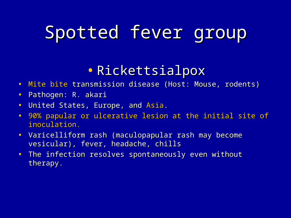
Spotted fever groupSpotted fever group
• RickettsialpoxRickettsialpox• Mite biteMite bite transmission disease (Host: Mouse, rodents) transmission disease (Host: Mouse, rodents)
• Pathogen: R. akariPathogen: R. akari
• United States, Europe, and United States, Europe, and AsiaAsia..
• 90% papular or ulcerative lesion at the initial site of inoculation.90% papular or ulcerative lesion at the initial site of inoculation.
• Varicelliform rash (maculopapular rash may become vesicular), Varicelliform rash (maculopapular rash may become vesicular), fever, headache, chillsfever, headache, chills
• The infection resolves spontaneously even without therapy.The infection resolves spontaneously even without therapy.

Scrub typhusScrub typhus
• Scrub typhusScrub typhus• Chigger biteChigger bite transmission disease (Host: Rats, ) transmission disease (Host: Rats, )• Pathogen: Orientia tsutsugamushiPathogen: Orientia tsutsugamushi• Southern Asia, Japan, Indonesia, Australia, Korea, Asiatic Russia, India, Southern Asia, Japan, Indonesia, Australia, Korea, Asiatic Russia, India,
ChinaChina• Incubation period:6-21 days.Incubation period:6-21 days.• <50% of cases necrotic eschar with an erythematous rim<50% of cases necrotic eschar with an erythematous rim• FeverFever, headache, myalgia, , headache, myalgia, cough cough and and GI symptomsGI symptoms..• Regional or generalized lymphadenopathy is common.Regional or generalized lymphadenopathy is common.• <50% of patients presents maculopapular rash.<50% of patients presents maculopapular rash.• Complications: Meningoencephalitis, Complications: Meningoencephalitis, myocarditismyocarditis, interstitial pneumonitis., interstitial pneumonitis.• Diagnosis:indirect fluorescent antibody assay or immunoperoxidase Diagnosis:indirect fluorescent antibody assay or immunoperoxidase
serologic tests of O. tsutsugamushi antigenserologic tests of O. tsutsugamushi antigen• Treatment: Doxycycline ; tetracycline; chloramphenicol at least 5 days Treatment: Doxycycline ; tetracycline; chloramphenicol at least 5 days
until afebrile 2-4 days.until afebrile 2-4 days.
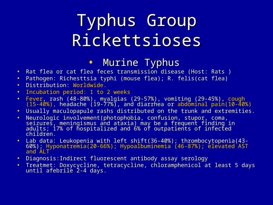
Typhus Group RickettsiosesTyphus Group Rickettsioses
• Murine TyphusMurine Typhus• Rat flea or cat flea feces transmission disease (Host: Rats )Rat flea or cat flea feces transmission disease (Host: Rats )• Pathogen: Richesttsia typhi (mouse flea); R. felis(cat flea)Pathogen: Richesttsia typhi (mouse flea); R. felis(cat flea)• Distribution: Distribution: Worldwide.Worldwide.• Incubation period: 1 to 2 weeksIncubation period: 1 to 2 weeks• FeverFever, rash (48-80%), myalgias (29-57%), vomiting (29-45%), , rash (48-80%), myalgias (29-57%), vomiting (29-45%), cough (15-cough (15-
40%),40%), headache (19-77%), and diarrhea or headache (19-77%), and diarrhea or abdominal pain(10-40%)abdominal pain(10-40%)• Usually maculopapule rashs distributed on the trunk and extremities.Usually maculopapule rashs distributed on the trunk and extremities.• Neurologic involvement(photophobia, confusion, stupor, coma, seizures, Neurologic involvement(photophobia, confusion, stupor, coma, seizures,
meningismus and ataxia) may be a frequent finding in adults; 17% of meningismus and ataxia) may be a frequent finding in adults; 17% of hospitalized and 6% of outpatients of infected children.hospitalized and 6% of outpatients of infected children.
• Lab data: Leukopenia with left shift(36-40%); thrombocytopenia(43-60%); Lab data: Leukopenia with left shift(36-40%); thrombocytopenia(43-60%); Hyponatremia(20-66%);Hyponatremia(20-66%); Hypoalbuminemia (46-87%);Hypoalbuminemia (46-87%); elevated AST and elevated AST and ALTALT
• Diagnosis:Indirect fluorescent antibody assay serologyDiagnosis:Indirect fluorescent antibody assay serology• Treatmet: Doxycycline, tetracycline, chloramphenicol at least 5 days until Treatmet: Doxycycline, tetracycline, chloramphenicol at least 5 days until
afebrile 2-4 days.afebrile 2-4 days.

Typhus Group RickettsiosesTyphus Group Rickettsioses
• Epidemic typhusEpidemic typhus• Louse feces (via wound or conjunctiva) disease (Host: Human )Louse feces (via wound or conjunctiva) disease (Host: Human )
• Pathogen: Richesttsia prowazekiiPathogen: Richesttsia prowazekii
• Distribution: Africa, South Amercia. Central America. Mexico, Distribution: Africa, South Amercia. Central America. Mexico, Asia.Asia.
• Incubation period:<14 days.Incubation period:<14 days.
• FeverFever, severe headache, , severe headache, abdominal painabdominal pain, rash in most people, rash in most people
• Chills(82%); myalgias(70%); arthralgia(70%); anorexia (48%), Chills(82%); myalgias(70%); arthralgia(70%); anorexia (48%), nonproductive cough (38%),nonproductive cough (38%), dizziness (35%), photophobia (33%), dizziness (35%), photophobia (33%), nausea (32%), nausea (32%), abdominal pain (30%),abdominal pain (30%), tinnitus (23%); tinnitus (23%); meningismus (17%), visual disturbances (15%), vomiting (10%)meningismus (17%), visual disturbances (15%), vomiting (10%)
• Treatment: Doxycycline; tetracycline; Chloraphenicol at least 5 Treatment: Doxycycline; tetracycline; Chloraphenicol at least 5 days until afebrile 2-4 days.days until afebrile 2-4 days.

Ehrlichioses and Ehrlichioses and AnaplasmosisAnaplasmosis
• Ehrlichiosis and anaplasmosisEhrlichiosis and anaplasmosis• Tick bite diseaseTick bite disease (Host: Deer, dogs(HME) ; deer rodents, ruminants (HGE)) (Host: Deer, dogs(HME) ; deer rodents, ruminants (HGE))• Pathogen: E. chaffeensis (Human monocytic ehrlichiosis (HME)); Pathogen: E. chaffeensis (Human monocytic ehrlichiosis (HME)); • Anaplasma phagocytophilum (Hauman anaplasmosis; Human granulocytic Anaplasma phagocytophilum (Hauman anaplasmosis; Human granulocytic
ehrlichiosis (HGE))ehrlichiosis (HGE))• Distribution: United states, Europe, AfricaDistribution: United states, Europe, Africa• Incubation period:2 days to 3 weeks.Incubation period:2 days to 3 weeks.• Fever(97%)Fever(97%), headache (81%), myalgias(68%), malaise (84%) anorexia, and nausea or , headache (81%), myalgias(68%), malaise (84%) anorexia, and nausea or
vomiting, maculopapular rash(2/3 of children with HME), conjunctivitis, pharyngitis vomiting, maculopapular rash(2/3 of children with HME), conjunctivitis, pharyngitis and lymphadenopathy (minority), and lymphadenopathy (minority),
• Hepatomegaly and splenomegaly (frequent), systolic ejection murmur (often)Hepatomegaly and splenomegaly (frequent), systolic ejection murmur (often)• Lab data: leuopenia (58-72%); lymphopenia (75-78%); thrombocytopenia(80-92%); Lab data: leuopenia (58-72%); lymphopenia (75-78%); thrombocytopenia(80-92%);
Granulomas and granulomatous inflammation in 75% in bone marrow examinations Granulomas and granulomatous inflammation in 75% in bone marrow examinations of HME; of HME; Elevated transaminase levelsElevated transaminase levels; ; HyponatremiaHyponatremia; prolonged PT/PTT; Morulae in ; prolonged PT/PTT; Morulae in peripheral blood monocyte or neutrophil.peripheral blood monocyte or neutrophil.
• Diagnosis: The demonstration of morulae, single high tilter (>=1:128) of A. Diagnosis: The demonstration of morulae, single high tilter (>=1:128) of A. phargocytophilum antibodies or seroconversion (>=1:64)phargocytophilum antibodies or seroconversion (>=1:64)
• Treatment: Doxycycline, Tetracycline at least 5 days until afebrile 2-4 days.Treatment: Doxycycline, Tetracycline at least 5 days until afebrile 2-4 days.

Q feverQ fever
• Acute Q feverAcute Q fever• Inhalation of infectious aerosols, ingestion of contaminated dairy products, Inhalation of infectious aerosols, ingestion of contaminated dairy products, ticks ticks
diseasedisease (Host: Cattle, sheep, goats, cats, rabbits ) (Host: Cattle, sheep, goats, cats, rabbits )• Pathogen: Coxiella burnetiiPathogen: Coxiella burnetii• Distribution: WorldwideDistribution: Worldwide• Incubation day: 3-30 days.Incubation day: 3-30 days.• FeverFever, sever headache, arthralgia, myalgia,, sever headache, arthralgia, myalgia, cough cough, fatigue, vomiting, , fatigue, vomiting, abdominal abdominal
painpain..• Hepatomegaly and splenomegalyHepatomegaly and splenomegaly may be detected in some patients. may be detected in some patients.• Fever (91%),Fever (91%), respiratory involvement (34%);respiratory involvement (34%); rash (11%), neurologic findings rash (11%), neurologic findings
(4%)(4%)• Lab data: Leukopenia with left shift (50%), thrombocytopenia (9-48%), reative Lab data: Leukopenia with left shift (50%), thrombocytopenia (9-48%), reative
thrombocytosis in recovery state results in DVT, thrombocytosis in recovery state results in DVT, Elevated transaminase levels Elevated transaminase levels (62%),(62%), elevated ESR (50%) elevated ESR (50%)
• Hepatitis (40%), Hepatitis (40%), Myocarditis, pericarditis.Myocarditis, pericarditis.• Right side endocarditis results in pulmonary embolism is reported.Right side endocarditis results in pulmonary embolism is reported.• Self-limited illness that last for 2-36 weeks.Self-limited illness that last for 2-36 weeks.

Q feverQ fever
• Chronic Q feverChronic Q fever• Chronic Q fever occurred months to years after acute Q fever or even in the Chronic Q fever occurred months to years after acute Q fever or even in the
absence of any history of acute Q fever.absence of any history of acute Q fever.• Presents with Presents with endocarditisendocarditis, , hepatitishepatitis, , myocarditismyocarditis, , FUOFUO, pneumonia, , pneumonia,
osteomyelitis.osteomyelitis.• Q fever endocarditisQ fever endocarditis: : FeverFever may be absent in up to 15% of cases. More than may be absent in up to 15% of cases. More than
75% of all identified have 75% of all identified have congestive heart failure.congestive heart failure.• Data: ESR > 20 mm/hr (80%), hypergammaglobulinemia (54%), Data: ESR > 20 mm/hr (80%), hypergammaglobulinemia (54%),
hyperfibrinogenemia (67%). Rheumatoid factor (>50%) , antiplatelet hyperfibrinogenemia (67%). Rheumatoid factor (>50%) , antiplatelet antibodies, anti-smooth muscle antibodies, antimitochondrial antibodies, antibodies, anti-smooth muscle antibodies, antimitochondrial antibodies, positive direct Coombs test.positive direct Coombs test.
• Diagnosis: (children with Diagnosis: (children with fever of unknown originfever of unknown origin, , atypical pneumoniaatypical pneumonia, , culture negative endocarditisculture negative endocarditis) 4-fold increase in indirect fluorescent ) 4-fold increase in indirect fluorescent antibody tilters to phase 1 and phase 2 antigens in acute and convalescent antibody tilters to phase 1 and phase 2 antigens in acute and convalescent (2-4 weeks) sera(2-4 weeks) sera
• Treatment: Acute:Treated within 3 days of onset of symptoms with Treatment: Acute:Treated within 3 days of onset of symptoms with tetracycline or doxycycline.tetracycline or doxycycline.
• Chronic:Tetracycline or doxycycline + rifampin, ofloxacin or pefloxacin. Chronic:Tetracycline or doxycycline + rifampin, ofloxacin or pefloxacin. (18months until phase 1 tilters of <1:200 for IgG and negative IgA tilters)(18months until phase 1 tilters of <1:200 for IgG and negative IgA tilters)
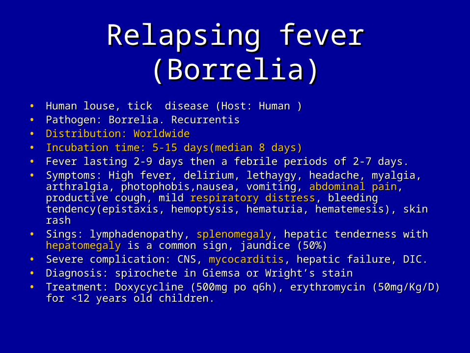
Relapsing fever (Borrelia)Relapsing fever (Borrelia)
• Human louse, tick disease (Host: Human )Human louse, tick disease (Host: Human )• Pathogen: Borrelia. RecurrentisPathogen: Borrelia. Recurrentis• Distribution: WorldwideDistribution: Worldwide• Incubation time: 5-15 days(median 8 days)Incubation time: 5-15 days(median 8 days)• Fever lasting 2-9 days then a febrile periods of 2-7 days.Fever lasting 2-9 days then a febrile periods of 2-7 days.• Symptoms: Symptoms: High feverHigh fever, delirium, lethaygy, headache, myalgia, arthralgia, , delirium, lethaygy, headache, myalgia, arthralgia,
photophobis,nausea, vomiting, photophobis,nausea, vomiting, abdominal painabdominal pain, , productive coughproductive cough, mild , mild respiratory distressrespiratory distress, bleeding tendency(epistaxis, hemoptysis, hematuria, , bleeding tendency(epistaxis, hemoptysis, hematuria, hematemesis), skin rashhematemesis), skin rash
• Sings: lymphadenopathy, Sings: lymphadenopathy, splenomegalysplenomegaly, hepatic tenderness with , hepatic tenderness with hepatomegalyhepatomegaly is a common sign, jaundice (50%) is a common sign, jaundice (50%)
• Severe complication: CNS, Severe complication: CNS, mycocarditismycocarditis, hepatic failure, DIC., hepatic failure, DIC.• Diagnosis: spirochete in Giemsa or Wright’s stainDiagnosis: spirochete in Giemsa or Wright’s stain• Treatment: Doxycycline (500mg po q6h), erythromycin (50mg/Kg/D) for Treatment: Doxycycline (500mg po q6h), erythromycin (50mg/Kg/D) for
<12 years old children.<12 years old children.
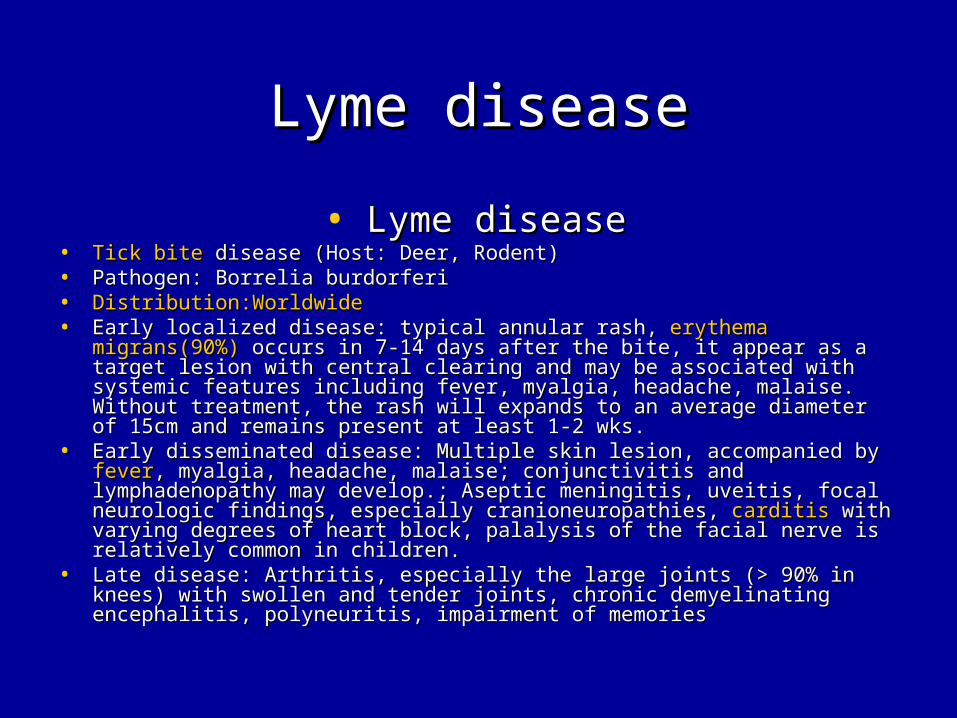
Lyme diseaseLyme disease
• Lyme diseaseLyme disease• Tick biteTick bite disease (Host: Deer, Rodent) disease (Host: Deer, Rodent)• Pathogen: Borrelia burdorferiPathogen: Borrelia burdorferi• Distribution:Worldwide Distribution:Worldwide • Early localized disease: typical annular rash, Early localized disease: typical annular rash, erythema migrans(90%)erythema migrans(90%)
occurs in 7-14 days after the bite, it appear as a target lesion with central occurs in 7-14 days after the bite, it appear as a target lesion with central clearing and may be associated with systemic features including fever, clearing and may be associated with systemic features including fever, myalgia, headache, malaise. Without treatment, the rash will expands to myalgia, headache, malaise. Without treatment, the rash will expands to an average diameter of 15cm and remains present at least 1-2 wks.an average diameter of 15cm and remains present at least 1-2 wks.
• Early disseminated disease: Multiple skin lesion, accompanied byEarly disseminated disease: Multiple skin lesion, accompanied by feverfever, , myalgia, headache, malaise; conjunctivitis and lymphadenopathy may myalgia, headache, malaise; conjunctivitis and lymphadenopathy may develop.; Aseptic meningitis, uveitis, focal neurologic findings, especially develop.; Aseptic meningitis, uveitis, focal neurologic findings, especially cranioneuropathies, cranioneuropathies, carditiscarditis with varying degrees of heart block, palalysis with varying degrees of heart block, palalysis of the facial nerve is relatively common in children.of the facial nerve is relatively common in children.
• Late disease: Arthritis, especially the large joints (> 90% in knees) with Late disease: Arthritis, especially the large joints (> 90% in knees) with swollen and tender joints, chronic demyelinating encephalitis, polyneuritis, swollen and tender joints, chronic demyelinating encephalitis, polyneuritis, impairment of memoriesimpairment of memories

Lyme diseaseLyme disease• Diagnosis: The presence of EM rashes > 5cm in diameter or serologic Diagnosis: The presence of EM rashes > 5cm in diameter or serologic
confirmation of infection with evidence of at least one manifestation of musculo confirmation of infection with evidence of at least one manifestation of musculo skeletal, neurological or cardiovascular disease . skeletal, neurological or cardiovascular disease .
• The culture of B. burgdorferi in Barbour-Stoenner-Kelly medium from the The culture of B. burgdorferi in Barbour-Stoenner-Kelly medium from the specimen of plasma, biopsy samples of erythema migrans lesions, occasionally specimen of plasma, biopsy samples of erythema migrans lesions, occasionally from cerebrospinal fluid samples in patients with meningitis can permit a from cerebrospinal fluid samples in patients with meningitis can permit a definitive diagnosis. definitive diagnosis.
• Serologic tests was used as diagnostic method in public health surveillance and Serologic tests was used as diagnostic method in public health surveillance and in clinical diagnosis. This Western Blot detects the presence or absence of in clinical diagnosis. This Western Blot detects the presence or absence of antibodies and is not quantitative. Antibody can still be detected with Western antibodies and is not quantitative. Antibody can still be detected with Western blot even after a successful. It was used to confirm equivocal and positive blot even after a successful. It was used to confirm equivocal and positive serology results obtained by ELISA or IFA.serology results obtained by ELISA or IFA.
• Treatment:doxycycline (100mg bid for 14-21 days) in children older than 8 yr of Treatment:doxycycline (100mg bid for 14-21 days) in children older than 8 yr of age; Amoxicillin (50 mg /Kg/D tid po 14-21 days); cefuroxime (30mg/kg/D bid po age; Amoxicillin (50 mg /Kg/D tid po 14-21 days); cefuroxime (30mg/kg/D bid po for 14-21 days ); erythromycin (30-50mg/Kg/D qid for 14-21 days.)for 14-21 days ); erythromycin (30-50mg/Kg/D qid for 14-21 days.)
• Meningitis: Ceftriaxone 50-80 mg/Kg/D iv qd for 14-28 days.Meningitis: Ceftriaxone 50-80 mg/Kg/D iv qd for 14-28 days.• Carditis: Mild to moderate as EM; Severe as meningitisCarditis: Mild to moderate as EM; Severe as meningitis• Late disease: the same as for EM except for 28 days->recurrence choose second Late disease: the same as for EM except for 28 days->recurrence choose second
dose or meningitis.dose or meningitis.

Chagas diseaseChagas disease
• Acute Chagas diseaseAcute Chagas disease• Kissing bugs biteKissing bugs bite transmission disease transmission disease
• Pathogen: Trypanosoma cruziPathogen: Trypanosoma cruzi
• Distribution: Western hemisphere (Mixeco, south America, Brazil, Distribution: Western hemisphere (Mixeco, south America, Brazil, Argentina)Argentina)
• Acute chagas disease: mild fever, malaise, facial edema, Acute chagas disease: mild fever, malaise, facial edema, lymphadenopathy, local sign of inflammation at the site entry(chagomas) .lymphadenopathy, local sign of inflammation at the site entry(chagomas) .
• 50% with Romana sign (Unilateral, painless eye swelling), conjunctivitis, 50% with Romana sign (Unilateral, painless eye swelling), conjunctivitis, preauricular lymphadenitispreauricular lymphadenitis
• Lymphadenopathy,Lymphadenopathy, hepatosplenomegalyhepatosplenomegaly, meningoencephalitis can occur , meningoencephalitis can occur in children younger than 2 years old.in children younger than 2 years old.
• The heart is the primarily target organ, 4 chamber dilatation, The heart is the primarily target organ, 4 chamber dilatation, diffuse diffuse myocarditismyocarditis and inflammation of the conduction system lead to the and inflammation of the conduction system lead to the development of fibrosis.development of fibrosis.

Chagas diseaseChagas disease
• Chronic Chagas diseaseChronic Chagas disease• Chronic Chagas disease: Cardiomyopathy included Chronic Chagas disease: Cardiomyopathy included congestive heart congestive heart
failurefailure, arrhythmia and thrombocytopenia events. EKG will showed , arrhythmia and thrombocytopenia events. EKG will showed complete A-V block, RBBB, LBBB, left ventricular apical aneurysm.complete A-V block, RBBB, LBBB, left ventricular apical aneurysm.
• Diagnosis: Giemsa-stain smear will demonstrate motile Diagnosis: Giemsa-stain smear will demonstrate motile trypanosomes.trypanosomes.
• These are only seen in the peripheral blood in the first 6-12 week of These are only seen in the peripheral blood in the first 6-12 week of the illness.the illness.
• Specific IgM antibodies in ELISA or IFA in acute Chagas disease.Specific IgM antibodies in ELISA or IFA in acute Chagas disease.• Complement fixation is considered the most reliable Complement fixation is considered the most reliable
immunodiagnostic method for chronic Chagas disease.immunodiagnostic method for chronic Chagas disease.• Treatment: Nifurtimox (15-20mg/kg/D qid po(1-10 y/o); Treatment: Nifurtimox (15-20mg/kg/D qid po(1-10 y/o);
12.5-15mg/KG/D (11-16 y/o), 8-10mg/Kg/D(>16 y/o)) for 90 days and 12.5-15mg/KG/D (11-16 y/o), 8-10mg/Kg/D(>16 y/o)) for 90 days and benznidazole (10mg/Kg/D bid po(<12y/o); 5-7 mg/kg/D(>12y/o)) for benznidazole (10mg/Kg/D bid po(<12y/o); 5-7 mg/kg/D(>12y/o)) for 60 days60 days

DiseaseDisease TransmissionTransmission Heart Heart HepatomegalyHepatomegaly
SplenomegalySplenomegaly
Pul. E.Pul. E.
Pul. H.Pul. H.
Relapsing feverRelapsing fever Tick,louseTick,louse MyocarditisMyocarditis (+)(+) (-)(-)Lyme diseaseLyme disease Tick biteTick bite MyocarditisMyocarditis (-)(-) (-)(-)RMSFRMSF Tick biteTick bite MyocarditisMyocarditis (+) 33%(+) 33% ((+) +) P.E.P.E.
MSFMSF Tick biteTick bite (-)(-) (-)(-) (-)(-)Scrub typhusScrub typhus Chigger biteChigger bite MyocarditisMyocarditis (-)(-) (-)(-)Murine typhusMurine typhus Flea fecesFlea feces (-)(-) (-)(-) (-)(-)Epidemic typhusEpidemic typhus Louse fecesLouse feces (-)(-) (-)(-) (-)(-)HME, HEHME, HE Tick biteTick bite (-)(-) (+)(+) (-)(-)Q feverQ fever Tick borneTick borne MyocarditisMyocarditis
EndocariditisEndocariditis(+)(+) ((+)+)P.E.P.E.
Chagas diseaseChagas disease Kissing bugKissing bug MyocarditisMyocarditis (+)(+) (-)(-)Loa LoaLoa Loa Biting fliesBiting flies CardiomyopathyCardiomyopathy (-)(-) (-)(-)

ImpressionImpression
• Q feverQ fever
• Rocky mountain spotted feverRocky mountain spotted fever
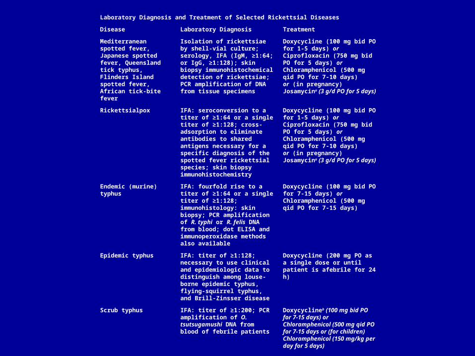
Laboratory Diagnosis and Treatment of Selected Rickettsial Diseases
Disease Laboratory Diagnosis Treatment
Mediterranean spotted fever, Japanese spotted fever, Queensland tick typhus, Flinders Island spotted fever, African tick-bite fever
Isolation of rickettsiae by shell-vial culture; serology, IFA (IgM, ≥1:64; or IgG, ≥1:128); skin biopsy immunohistochemical detection of rickettsiae; PCR amplification of DNA from tissue specimens
Doxycycline (100 mg bid PO for 1-5 days) orCiprofloxacin (750 mg bid PO for 5 days) orChloramphenicol (500 mg qid PO for 7-10 days)or (in pregnancy)Josamycina (3 g/d PO for 5 days)
Rickettsialpox IFA: seroconversion to a titer of ≥1:64 or a single titer of ≥1:128; cross-adsorption to eliminate antibodies to shared antigens necessary for a specific diagnosis of the spotted fever rickettsial species; skin biopsy immunohistochemistry
Doxycycline (100 mg bid PO for 1-5 days) orCiprofloxacin (750 mg bid PO for 5 days) orChloramphenicol (500 mg qid PO for 7-10 days)or (in pregnancy)Josamycina (3 g/d PO for 5 days)
Endemic (murine) typhus
IFA: fourfold rise to a titer of ≥1:64 or a single titer of ≥1:128; immunohistology: skin biopsy; PCR amplification of R. typhi or R. felis DNA from blood; dot ELISA and immunoperoxidase methods also available
Doxycycline (100 mg bid PO for 7-15 days) orChloramphenicol (500 mg qid PO for 7-15 days)
Epidemic typhus IFA: titer of ≥1:128; necessary to use clinical and epidemiologic data to distinguish among louse-borne epidemic typhus, flying-squirrel typhus, and Brill-Zinsser disease
Doxycycline (200 mg PO as a single dose or until patient is afebrile for 24 h)
Scrub typhus IFA: titer of ≥1:200; PCR amplification of O. tsutsugamushi DNA from blood of febrile patients
Doxycyclineb (100 mg bid PO for 7-15 days) orChloramphenicol (500 mg qid PO for 7-15 days or (for children)Chloramphenicol (150 mg/kg per day for 5 days)
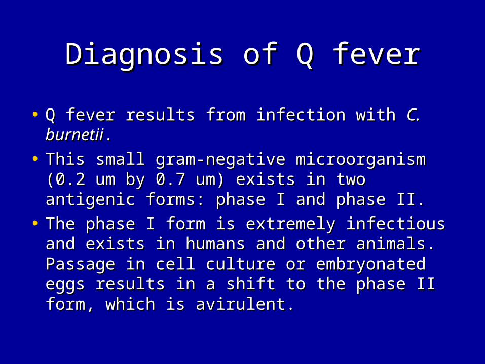
Diagnosis of Q feverDiagnosis of Q fever
• Q fever results from infection with Q fever results from infection with C. burnetiiC. burnetii. .
• This small gram-negative microorganism (0.2 This small gram-negative microorganism (0.2 um by 0.7 um) exists in two antigenic forms: um by 0.7 um) exists in two antigenic forms: phase I and phase II.phase I and phase II.
• The phase I form is extremely infectious and The phase I form is extremely infectious and exists in humans and other animals. Passage exists in humans and other animals. Passage in cell culture or embryonated eggs results in in cell culture or embryonated eggs results in a shift to the phase II form, which is avirulent. a shift to the phase II form, which is avirulent.

Diagnosis of Q feverDiagnosis of Q fever• C. burnetiiC. burnetii can be isolated from buffy-coat blood samples or can be isolated from buffy-coat blood samples or
tissue specimens by a shell-vial technique; tissue specimens by a shell-vial technique; C. burnetiiC. burnetii since it since it is considered highly infectious. is considered highly infectious.
• PCRPCR can be used to amplify can be used to amplify C. burnetiiC. burnetii DNA DNA from tissue or from tissue or biopsy specimens. This technique can also be used on biopsy specimens. This technique can also be used on paraffin-embedded tissues. paraffin-embedded tissues.
• Serology Serology is the most commonly used diagnostic tool. Three is the most commonly used diagnostic tool. Three techniques are available: techniques are available: complement fixationcomplement fixation, , indirect indirect immunofluorescenceimmunofluorescence, and , and enzyme-linked immunosorbent enzyme-linked immunosorbent assay.assay.
• Indirect immunofluorescence is sensitive and specific and is Indirect immunofluorescence is sensitive and specific and is the method of choice. the method of choice.
• Rheumatoid factor should be adsorbed from the specimen Rheumatoid factor should be adsorbed from the specimen before testing.before testing.

Diagnosis of Q feverDiagnosis of Q fever
• 4-fold increased in IFA titers to phase 4-fold increased in IFA titers to phase I and phase II antigens I and phase II antigens between acute and convalescent sera(2-4wks)between acute and convalescent sera(2-4wks)
• An An IgG titer of ≥1:800 to phase I antigenIgG titer of ≥1:800 to phase I antigen is suggestive of is suggestive of chronic Q fever. chronic Q fever.
• In almost all instances of chronic Q fever, In almost all instances of chronic Q fever, the antibody titer to the antibody titer to phase I antigen is much higher than that to phase II antigen.phase I antigen is much higher than that to phase II antigen.
• The reverse is true in acute Q feverThe reverse is true in acute Q fever. In addition, in acute Q . In addition, in acute Q fever, it is usually possible to demonstrate a fourfold rise in titer fever, it is usually possible to demonstrate a fourfold rise in titer between acute- and convalescent-phase serum samples.between acute- and convalescent-phase serum samples.
• Elevated phase I IgA antibodyElevated phase I IgA antibody are diagnostic for Q fever are diagnostic for Q fever endocariditis.endocariditis.
Diagnosis of RMSFDiagnosis of RMSF
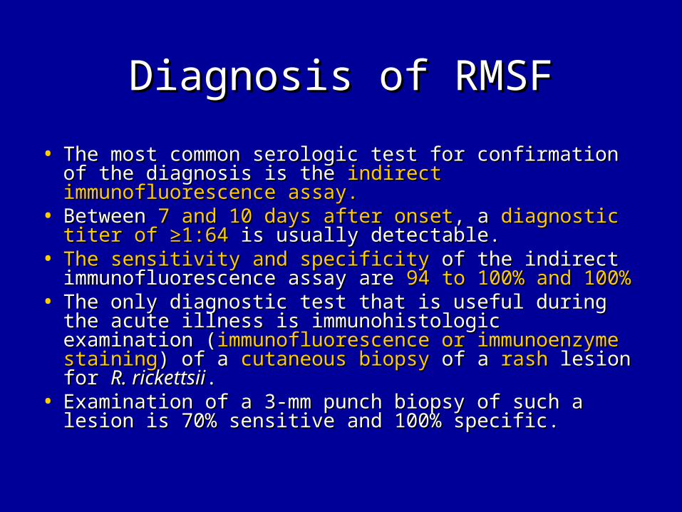
• The most common serologic test for confirmation of the The most common serologic test for confirmation of the diagnosis is the diagnosis is the indirect immunofluorescence assay.indirect immunofluorescence assay.
• Between Between 7 and 10 days after onset7 and 10 days after onset, a , a diagnostic titer of ≥1:64diagnostic titer of ≥1:64 is usually detectable. is usually detectable.
• The sensitivity and specificityThe sensitivity and specificity of the indirect of the indirect immunofluorescence assay are immunofluorescence assay are 94 to 100% and 100%94 to 100% and 100%
• The only diagnostic test that is useful during the acute illness The only diagnostic test that is useful during the acute illness is immunohistologic examination (is immunohistologic examination (immunofluorescence or immunofluorescence or immunoenzyme stainingimmunoenzyme staining) of a ) of a cutaneous biopsycutaneous biopsy of a of a rashrash lesion for lesion for R. rickettsiiR. rickettsii. .
• Examination of a 3-mm punch biopsy of such a lesion is 70% Examination of a 3-mm punch biopsy of such a lesion is 70% sensitive and 100% specific. sensitive and 100% specific.

![9403 - xycom.co.kr · 9403 Industrial Monitor P/N 99592-001 ... PKIM [/r /t]runs the full PKIM utility Where: /r = reduced functionality. Some keyboard controllers will not allow](https://img.pdfslide.net/doc/110x75/5ec3d7bfc3c16e594a76dd4b/9403-xycomcokr-9403-industrial-monitor-pn-99592-001-pkim-r-truns-the.jpg)