Embed Size (px)
Citation preview
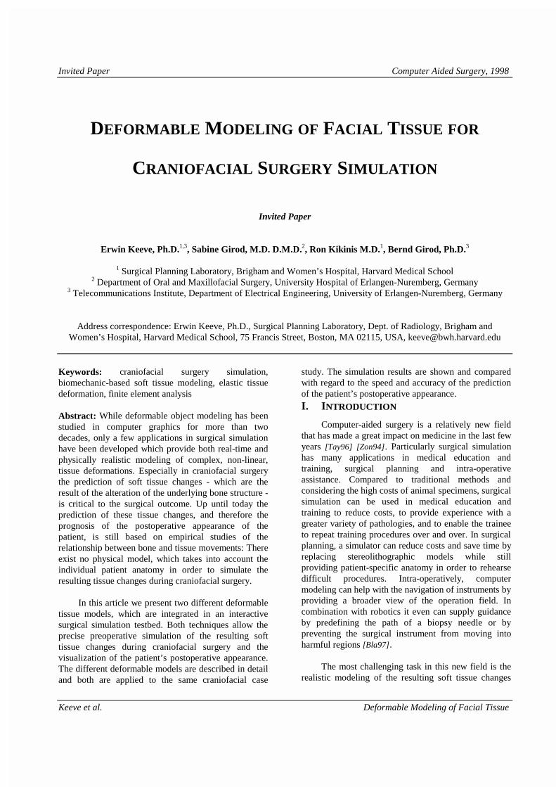
Invited Paper Computer Aided Surgery, 1998
Keeve et al. Deformable Modeling of Facial Tissue
DEFORMABLE MODELING OF FACIAL TISSUE FOR
CRANIOFACIAL SURGERY SIMULATION
Invited Paper
Erwin Keeve, Ph.D.1,3, Sabine Girod, M.D. D.M.D.2, Ron Kikinis M.D. 1, Bernd Girod, Ph.D.3
1 Surgical Planning Laboratory, Brigham and Women’s Hospital, Harvard Medical School2 Department of Oral and Maxillofacial Surgery, University Hospital of Erlangen-Nuremberg, Germany
3 Telecommunications Institute, Department of Electrical Engineering, University of Erlangen-Nuremberg, Germany
Address correspondence: Erwin Keeve, Ph.D., Surgical Planning Laboratory, Dept. of Radiology, Brigham andWomen’s Hospital, Harvard Medical School, 75 Francis Street, Boston, MA 02115, USA, [email protected]
Keywords: craniofacial surgery simulation,biomechanic-based soft tissue modeling, elastic tissuedeformation, finite element analysis
Abstract: While deformable object modeling has beenstudied in computer graphics for more than twodecades, only a few applications in surgical simulationhave been developed which provide both real-time andphysically realistic modeling of complex, non-linear,tissue deformations. Especially in craniofacial surgerythe prediction of soft tissue changes - which are theresult of the alteration of the underlying bone structure -is critical to the surgical outcome. Up until today theprediction of these tissue changes, and therefore theprognosis of the postoperative appearance of thepatient, is still based on empirical studies of therelationship between bone and tissue movements: Thereexist no physical model, which takes into account theindividual patient anatomy in order to simulate theresulting tissue changes during craniofacial surgery.
In this article we present two different deformabletissue models, which are integrated in an interactivesurgical simulation testbed. Both techniques allow theprecise preoperative simulation of the resulting softtissue changes during craniofacial surgery and thevisualization of the patient’s postoperative appearance.The different deformable models are described in detailand both are applied to the same craniofacial case
study. The simulation results are shown and comparedwith regard to the speed and accuracy of the predictionof the patient’s postoperative appearance.I. I NTRODUCTION
Computer-aided surgery is a relatively new fieldthat has made a great impact on medicine in the last fewyears [Tay96] [Zon94]. Particularly surgical simulationhas many applications in medical education andtraining, surgical planning and intra-operativeassistance. Compared to traditional methods andconsidering the high costs of animal specimens, surgicalsimulation can be used in medical education andtraining to reduce costs, to provide experience with agreater variety of pathologies, and to enable the traineeto repeat training procedures over and over. In surgicalplanning, a simulator can reduce costs and save time byreplacing stereolithographic models while stillproviding patient-specific anatomy in order to rehearsedifficult procedures. Intra-operatively, computermodeling can help with the navigation of instruments byproviding a broader view of the operation field. Incombination with robotics it even can supply guidanceby predefining the path of a biopsy needle or bypreventing the surgical instrument from moving intoharmful regions [Bla97].
The most challenging task in this new field is therealistic modeling of the resulting soft tissue changes
Invited Paper Computer Aided Surgery, 1998
Keeve et al. Deformable Modeling of Facial Tissue
- 2 -
during surgery. Although deformable object modelinghas been studied in computer graphics for more thantwo decades, only few applications in surgicalsimulation have been developed which provide bothreal-time and physically realistic modeling of complex,non-linear, tissue deformations [Gib98]. In particular, itis often difficult to obtain accurate tissue properties,which describe the elasticity of living tissue, and it iseven harder to conduct those kinds of experimentsrequired to verify the precision of the computersimulations. Therefore, the computer-based simulationof the biomechanical behaviour of human soft tissue hasbeen addressed only in the last few years by specializedresearch sites [Bro96a] [Cot96] [Cov93] [Del94] [Gib97][Koc96] [Pat96] [Pie95] [Tan95]. Although this approachrequires both high-performance computation as well ashigh-end graphics capabilities, the great advantage ofthe ability to simulate the elasticity of soft tissue and thefunctionality of human organs has led to establish a newresearch field in the area of computer aided surgery:deformable modeling in surgical simulation [Sze98].
Among other surgical areas in which deformablemodeling of soft tissue can lead to a betterunderstanding of the patient’s anatomy and mayimprove the outcome of surgical procedures,craniofacial surgery stands out: Craniofacial surgery haslong been an extremely productive application area forthe development of computer aided surgical methods -much of the initial work in 3D reconstruction andvisualization of patient anatomy has come from thisfield [Alt93] [Bil95] [Den88] [Euf94] [Has95] [Hem91][Lo94] [Van83] [Zei95]. In craniofacial surgery the goalis not only to improve the functionality, but also torestore an aesthetically pleasing face. Therefore therealistic prediction of the ensuing soft tissue changes issubstantial. With traditional methods only a limitedforecast of these soft tissue changes can be achieved[Ste94]. The prediction is still based on empiricalstudies of the relationship between bone and tissuemovements [Ath95] [Krü85] [See92]. Although thisrelationship can be found for forward and backwardshifts of the lower jaw, in operations involving theupper jaw or other displacements of the lower jaw,there is presently no satisfactory method of predictingthe resulting soft tissue changes.
To address this challenge we integrated twodifferent deformable tissue models into our surgicalsimulation system. One interactive more approximatingtechnique – described in section III.4 – that gives thesurgeon the ability to realize nearly real-timesimulations of the resulting soft tissue changes and toimprove his planning process. And one more precise
technique – described in section III.5 - which considersthe exact physical properties of the facial soft tissue andcan be used off-line to verify the chosen surgicalprocedure. Both techniques are described in detail andare applied to the same case study in order to validatethe accuracy of the simulation results.
II. R ELATED WORK
Although deformable soft tissue models are widelyused in computer graphics - for example in facialanimation - these applications have a different emphasisthan those applied in surgical simulation: Theirintention is to realistically animate facial expressions,not to simulate the exact physical behaviour of humansoft tissue [Che92] [Che95] [Gou89] [Lee95] [Par96][Ter90] [Ter91] [Wat92] [Wat95]. There is noconsideration of the individual anatomical structureneither are the actual elastic properties of living tissuetaken into account. Therefore these techniques can onlybe used as a brief guideline in order to develop newdeformable tissue models suitable for surgicalsimulation. However, in the last few years variousdeformable models have been evolved in order topredict intra- and postoperative modifications - forinstance in minimal invasive and orthopedic surgery[Cov93] [Fis96] [Gib97] . Beside these surgical areasmost of todays leading techniques in deformable tissuemodeling have been projected in plastic- andcraniofacial surgery.
• A Craniofacial Surgery Simulation Testbed wasdeveloped by Delingette et al. in 1994, usingdeformable surfaces and volumes called SimplexMeshes in order to simulate surface changes suchas those exerted on the facial skin by muscle action[Del94]. But this approach does not consider theexact anatomical structure of the soft tissue; insteadthe user has to specify virtual Muscles which thendefine the connection between the facial skin andthe underlying bone.
• To simulate facial palsy and cleft lip surgery,Tanaka et al. developed a deformable tissue modelbased on a facial muscle model [Tan95]. Thismuscle model is limited to only the mouth and doesnot take into account the underlying bone structure.It can not be used to determine soft tissue changesresulting from bone displacements as they occur incraniofacial surgery.
• CAPS, a computer-aided plastic surgery systemdeveloped by Pieper et al., can be used to predict
Invited Paper Computer Aided Surgery, 1998
Keeve et al. Deformable Modeling of Facial Tissue
- 3 -
soft tissue changes on individual skin data [Pie91][Pie92] [Pie95]. But this approach does also nottake into account the underlying bone structure ofthe patient and can not be used to predict soft tissuechanges affected by surgical bone realignment.
• Koch et al. presented an approach to model softtissue changes shaped by the realignment of theunderlying bone structure [Koc96]. In this approachmass-spring and finite-element methods are used inorder to model the elasticity of the facial tissue. Buttheir methods are not integrated into a surgicalsimulation system; instead commercial availablesoftware packages like AVS and Alias are used tobuild the necessarily three-dimensional surfacemodels [Adv98] [Ali98] . Because of these timeconsuming steps this approach has only be testedon the Visible Human Project dataset and has notbeen applied to actual patient data [Nat98].
Although the discussed methods are veryinteresting and auspicious, each of them only allows thesimulation of soft tissue changes in a very specificmanner and none of them is yet integrated into asurgical simulation system which empowers both real-time and physically realistic modeling of complex, non-linear, tissue deformations: Therefore none of themhave yet been applied to actual clinical cases.
III. M ATERIALS AND METHODS
The goal of the presented craniofacial surgerysimulation system is to enable precise and interactivemanipulations of the three-dimensional reconstructionof the patient’s bone and to predict the resulting softtissue changes in order to visualize the patient’spostoperative appearance. As stated, the focus of thispaper is the deformable modeling of facial tissue.Therefore, the next two sections elucidate only brieflythe general setup of our system, the data acquisition andvisualization and the simulation of the bone realignment(for a detailed description the reader is referred to[Kee96a]). Section III.4 describes in detail the mass-spring and section III.5 the finite-element tissue model.Both models are integrated into the surgery simulationsystem which is implemented in C++ on SiliconGraphics workstations, using the object-orientedgraphics library Open Inventor [Wer94]. Each algorithmhas its own Motif dialogue window, which gives theuser the flexibility to easily tailor parameters to suit theneeds of their application.
III.1 Data Acquisition and Visualization
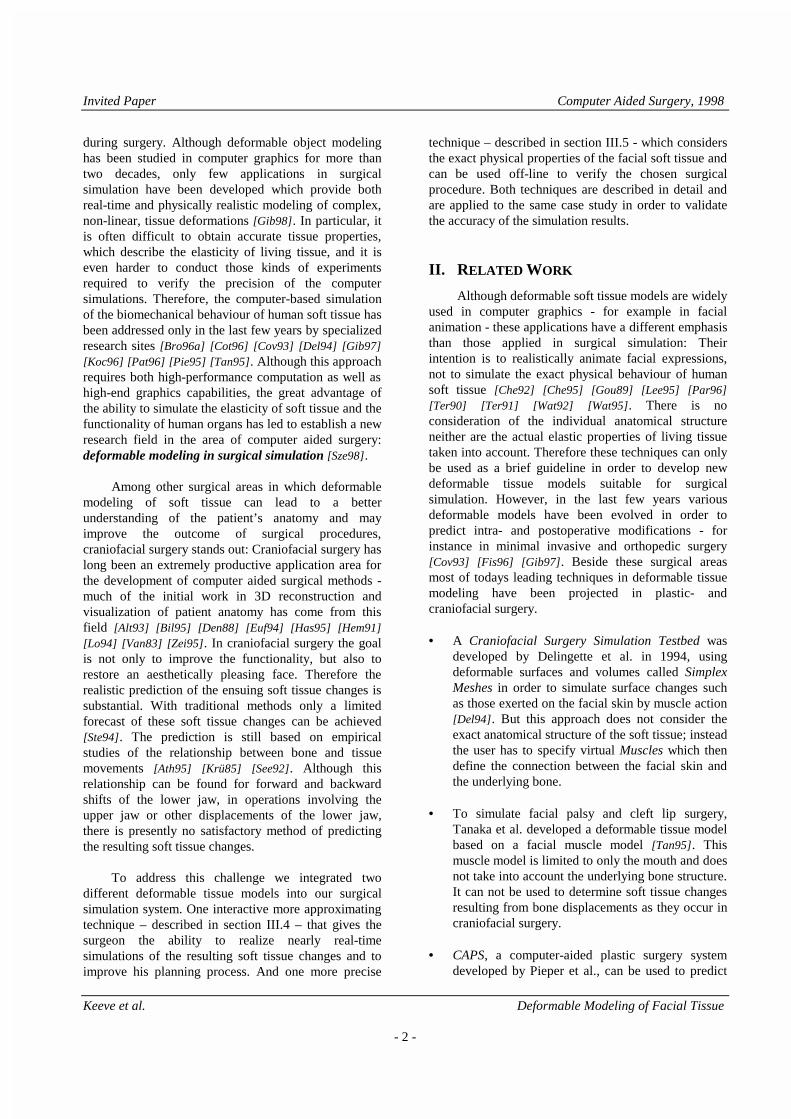
The Marching Cubes method is used toreconstruct the skull from computer tomography data[Lor87] . This three-dimensional skull reconstructionprovides the basis for the interactive surgical simulationof the bone realignment. A Cyberware scanner obtainsthe geometry of the patient’s skin surface [Cyb98]. Afterdata acquisition, and 3D surface reconstruction anadaptive reduction technique is used to decrease thesize of both datasets [Kee97]. This compressionalgorithm reduces the amount of triangles used torepresent complex anatomical structures up to 80 %without sacrificing the visible details of the objects. Asemi-automatic method registers the skin and skull data,which are obtained in different coordinate systems. Theuser provides corresponding anatomical points in bothdatasets. A matrix is calculated by least squares fitting,which transforms the data from one coordinate systemto the other. Figure 1a shows the Marching Cubesreconstruction of the patient's skull. As shown in Figure1b, a 3D Cephalometry tool allows the physician toeasily measure the exact anatomical structure of thepatient's skull and skin. Figures 1c and 1d illustrate theViewing Plane tool that can be used to generate asection at an arbitrary angle and visualize particularstructures of interest - a volume-rendered image is usedas a reference for a more detailed structure localization.The system also supports visualization of the scene instereo. As shown in Figure 1e, the Implant Carving toolcan be used to model a custom prosthesis with a desiredshape. To verify the tissue model, a Verification tool asshown in Figure 1f, is used to visualize the shapedifferences in the simulation with respect to the actualpostoperative finding.
a.) Marching cubes recononstr. b.) 3D cephalometry
c.) Viewing plane d.) Volume rendering
Invited Paper Computer Aided Surgery, 1998
Keeve et al. Deformable Modeling of Facial Tissue
- 4 -
e.) Implant carving f.) Verification
Figure 1: Data acquisition and visualizationIII.2 Simulation of Bone Realignment
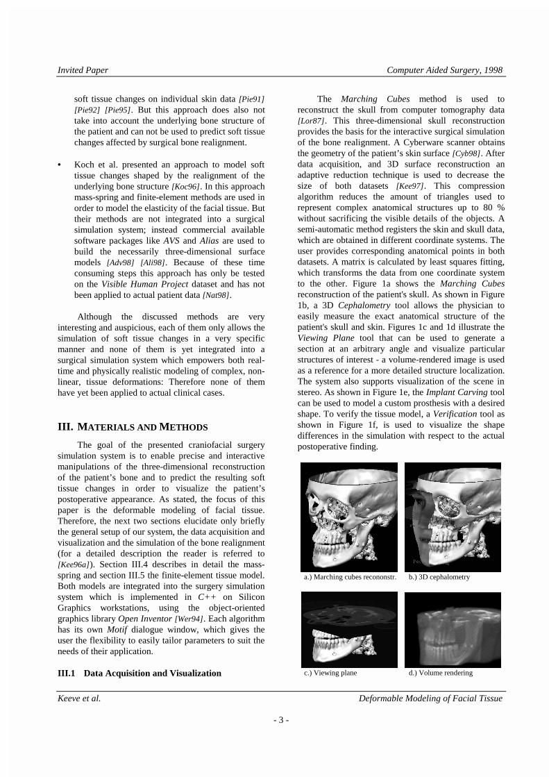
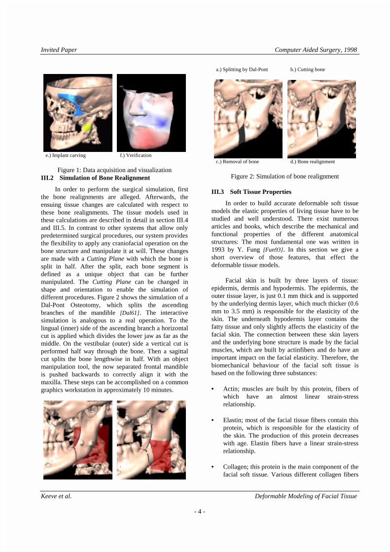
In order to perform the surgical simulation, firstthe bone realignments are alleged. Afterwards, theensuing tissue changes are calculated with respect tothese bone realignments. The tissue models used inthese calculations are described in detail in section III.4and III.5. In contrast to other systems that allow onlypredetermined surgical procedures, our system providesthe flexibility to apply any craniofacial operation on thebone structure and manipulate it at will. These changesare made with a Cutting Plane with which the bone issplit in half. After the split, each bone segment isdefined as a unique object that can be furthermanipulated. The Cutting Plane can be changed inshape and orientation to enable the simulation ofdifferent procedures. Figure 2 shows the simulation of aDal-Pont Osteotomy, which splits the ascendingbranches of the mandible [Dal61] . The interactivesimulation is analogous to a real operation. To thelingual (inner) side of the ascending branch a horizontalcut is applied which divides the lower jaw as far as themiddle. On the vestibular (outer) side a vertical cut isperformed half way through the bone. Then a sagittalcut splits the bone lengthwise in half. With an objectmanipulation tool, the now separated frontal mandibleis pushed backwards to correctly align it with themaxilla. These steps can be accomplished on a commongraphics workstation in approximately 10 minutes.
a.) Splitting by Dal-Pont b.) Cutting bone
c.) Removal of bone d.) Bone realignment
Figure 2: Simulation of bone realignment
III.3 Soft Tissue Properties
In order to build accurate deformable soft tissuemodels the elastic properties of living tissue have to bestudied and well understood. There exist numerousarticles and books, which describe the mechanical andfunctional properties of the different anatomicalstructures: The most fundamental one was written in1993 by Y. Fung [Fun93]. In this section we give ashort overview of those features, that effect thedeformable tissue models.
Facial skin is built by three layers of tissue:epidermis, dermis and hypodermis. The epidermis, theouter tissue layer, is just 0.1 mm thick and is supportedby the underlying dermis layer, which much thicker (0.6mm to 3.5 mm) is responsible for the elasticity of theskin. The underneath hypodermis layer contains thefatty tissue and only slightly affects the elasticity of thefacial skin. The connection between these skin layersand the underlying bone structure is made by the facialmuscles, which are built by actinfibers and do have animportant impact on the facial elasticity. Therefore, thebiomechanical behaviour of the facial soft tissue isbased on the following three substances:
• Actin; muscles are built by this protein, fibers ofwhich have an almost linear strain-stressrelationship.
• Elastin; most of the facial tissue fibers contain thisprotein, which is responsible for the elasticity ofthe skin. The production of this protein decreaseswith age. Elastin fibers have a linear strain-stressrelationship.
• Collagen; this protein is the main component of thefacial soft tissue. Various different collagen fibers
Invited Paper Computer Aided Surgery, 1998
Keeve et al. Deformable Modeling of Facial Tissue
- 5 -
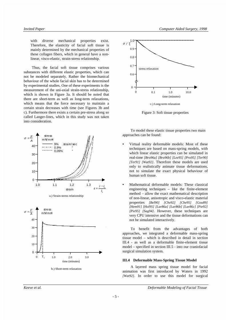
with diverse mechanical properties exist.Therefore, the elasticity of facial soft tissue ismainly determined by the mechanical properties ofthese collagen fibers, which in general have a non-linear, visco-elastic, strain-stress relationship.
Thus, the facial soft tissue comprises varioussubstances with different elastic properties, which cannot be modeled separately. Rather the biomechanicalbehaviour of the whole facial skin has to be determinedby experimental studies. One of these experiments is themeasurement of the uni-axial strain-stress relationship,which is shown in Figure 3a. It should be noted thatthere are short-term as well as long-term relaxations,which means that the force necessary to maintain acertain strain decreases with time (see Figures 3b andc). Furthermore there exists a certain pre-stress along socalled Langer-lines, which in this study was not takeninto consideration.
ε =
l − l0
l0
strain
1.1 1.2 1.31.0
10
20
30
40
σ = F
A
0
9% strain/sec0,9% “ 0,09% “
stressmN/mm2
a.) Strain-stress relationship
time (minutes)1.0 2.0 3.00
10
20
30
40
σ = F
A
0
stressmN/mm2
T 1
b.) Short-term relaxation
time (minutes)
stress relaxation
0.1 1.0 10.00
0
0.6
0.7
0.8
0.9
σ / T11.0
c.) Long-term relaxation
Figure 3: Soft tissue properties
To model these elastic tissue properties two mainapproaches can be found:
• Virtual reality deformable models: Most of thesetechniques are based on mass-spring models, withwhich linear elastic properties can be simulated inreal-time [Bro96a] [Bro96b] [Lee95] [Pro95] [Ter90][Ter91] [Wat92]. Therefore these models are usedonly to realistically animate tissue deformations,not to simulate the exact physical behaviour ofhuman soft tissue.
• Mathematical deformable models: These classicalengineering techniques – like the finite-elementmethod – allow the exact mathematical descriptionof non-linear, anisotropic and visco-elastic materialproperties [Bel90] [Che92] [Che95] [Gou89][Hem91] [Hol95] [Lar86a] [Lar86b] [Lar86c] [Pie92][Pie95] [Sag94]. However, these techniques arevery CPU intensive and the tissue deformations cannot be simulated interactively.
To benefit from the advantages of bothapproaches, we integrated a deformable mass-springtissue model – which is described in detail in sectionIII.4 - as well as a deformable finite-element tissuemodel – specified in section III.5 - into our craniofacialsurgical simulation system.
III.4 Deformable Mass-Spring Tissue Model
A layered mass spring tissue model for facialanimation was first introduced by Waters in 1992[Wat92]. In order to use this model for surgical
Invited Paper Computer Aided Surgery, 1998
Keeve et al. Deformable Modeling of Facial Tissue
- 6 -
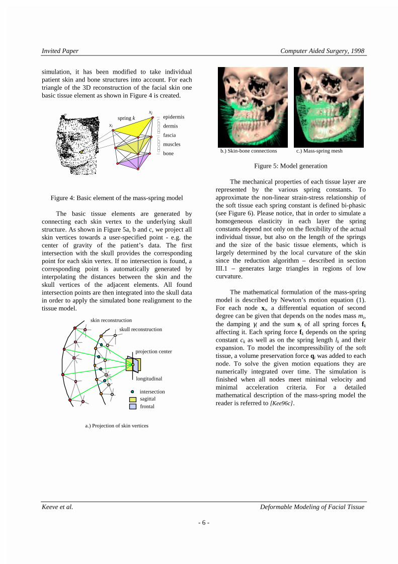
simulation, it has been modified to take individualpatient skin and bone structures into account. For eachtriangle of the 3D reconstruction of the facial skin onebasic tissue element as shown in Figure 4 is created.
Figure 4: Basic element of the mass-spring model
The basic tissue elements are generated byconnecting each skin vertex to the underlying skullstructure. As shown in Figure 5a, b and c, we project allskin vertices towards a user-specified point - e.g. thecenter of gravity of the patient’s data. The firstintersection with the skull provides the correspondingpoint for each skin vertex. If no intersection is found, acorresponding point is automatically generated byinterpolating the distances between the skin and theskull vertices of the adjacent elements. All foundintersection points are then integrated into the skull datain order to apply the simulated bone realignment to thetissue model.
a.) Projection of skin vertices
b.) Skin-bone connections c.) Mass-spring mesh
Figure 5: Model generation
The mechanical properties of each tissue layer arerepresented by the various spring constants. Toapproximate the non-linear strain-stress relationship ofthe soft tissue each spring constant is defined bi-phasic(see Figure 6). Please notice, that in order to simulate ahomogeneous elasticity in each layer the springconstants depend not only on the flexibility of the actualindividual tissue, but also on the length of the springsand the size of the basic tissue elements, which islargely determined by the local curvature of the skinsince the reduction algorithm – described in sectionIII.1 – generates large triangles in regions of lowcurvature.
The mathematical formulation of the mass-springmodel is described by Newton’s motion equation (1).For each node xi, a differential equation of seconddegree can be given that depends on the nodes mass mi,the damping γi and the sum si of all spring forces fk
affecting it. Each spring force fk depends on the springconstant ck as well as on the spring length lk and theirexpansion. To model the incompressibility of the softtissue, a volume preservation force qi was added to eachnode. To solve the given motion equations they arenumerically integrated over time. The simulation isfinished when all nodes meet minimal velocity andminimal acceleration criteria. For a detailedmathematical description of the mass-spring model thereader is referred to [Kee96c].
projection center
skin reconstruction
skull reconstruction
longitudinal
intersectionsagittal
frontal
epidermis
dermis
fascia
muscles
bone
spring kxi
xj
Invited Paper Computer Aided Surgery, 1998
Keeve et al. Deformable Modeling of Facial Tissue
- 7 -
ε =
l − l0
l0strain
1.1 1.2 1.31.0
10
20
30
40
σ =
FA
0
stressmN/mm2
c1
c2
Figure 6: Bi-phasic spring constants
mi
d2xi
dt2+γ i
dxi
dt+ si = 0 (1)
si = ck 1−lk
x j − x i
k∑ x j − xi( )+qi
(2)
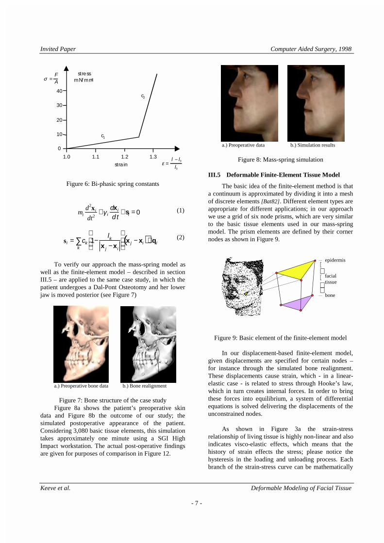
To verify our approach the mass-spring model aswell as the finite-element model – described in sectionIII.5 – are applied to the same case study, in which thepatient undergoes a Dal-Pont Osteotomy and her lowerjaw is moved posterior (see Figure 7)
a.) Preoperative bone data b.) Bone realignment
Figure 7: Bone structure of the case studyFigure 8a shows the patient’s preoperative skin
data and Figure 8b the outcome of our study; thesimulated postoperative appearance of the patient.Considering 3,080 basic tissue elements, this simulationtakes approximately one minute using a SGI HighImpact workstation. The actual post-operative findingsare given for purposes of comparison in Figure 12.
a.) Preoperative data b.) Simulation results
Figure 8: Mass-spring simulation
III.5 Deformable Finite-Element Tissue Model
The basic idea of the finite-element method is thata continuum is approximated by dividing it into a meshof discrete elements [Bat82]. Different element types areappropriate for different applications; in our approachwe use a grid of six node prisms, which are very similarto the basic tissue elements used in our mass-springmodel. The prism elements are defined by their cornernodes as shown in Figure 9.
Figure 9: Basic element of the finite-element model
In our displacement-based finite-element model,given displacements are specified for certain nodes –for instance through the simulated bone realignment.These displacements cause strain, which - in a linear-elastic case - is related to stress through Hooke’s law,which in turn creates internal forces. In order to bringthese forces into equilibrium, a system of differentialequations is solved delivering the displacements of theunconstrained nodes.
As shown in Figure 3a the strain-stressrelationship of living tissue is highly non-linear and alsoindicates visco-elastic effects, which means that thehistory of strain effects the stress; please notice thehysteresis in the loading and unloading process. Eachbranch of the strain-stress curve can be mathematically
epidermis
facialtissue
bone
Invited Paper Computer Aided Surgery, 1998
Keeve et al. Deformable Modeling of Facial Tissue
- 8 -
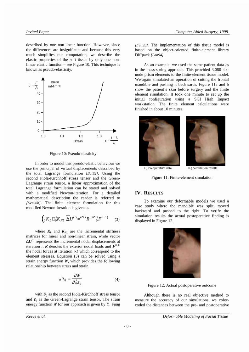
described by one non-linear function. However, sincethe differences are insignificant and because this verymuch simplifies our computation, we describe theelastic properties of the soft tissue by only one non-linear elastic function – see Figure 10. This technique isknown as pseudo-elasticity.
ε =
l − l0
l0strain
1.1 1.2 1.31.0
10
20
30
40
σ =
FA
0
stressmN/mm2
Figure 10: Pseudo-elasticity
In order to model this pseudo-elastic behaviour weuse the principal of virtual displacements described bythe total Lagrange formulation [Bat82]. Using thesecond Piola-Kirchhoff stress tensor and the Green-Lagrange strain tensor, a linear approximation of thetotal Lagrange formulation can be stated and solvedwith a modified Newton-iteration. For a detailedmathematical description the reader is referred to[Kee96b]. The finite element formulation for thismodified Newton-iteration is given as
0tKL+ 0
tKNL( )∆U ( i) =t +∆tR− 0t +∆tF (i −1)
(3)
where KL and KNL are the incremental stiffnessmatrices for linear and non-linear strain, while vector∆U(i) represents the incremental nodal displacements atiteration i. R denotes the exterior nodal loads and F(i-1)
the nodal forces at iteration i-1 which correspond to theelement stresses. Equation (3) can be solved using astrain energy function W, which provides the followingrelationship between stress and strain
0tSij =
∂W
∂ 0tε ij
(4)
with Sij as the second Piola-Kirchhoff stress tensorand εεij as the Green-Lagrange strain tensor. The strainenergy function W for our approach is given by Y. Fung
[Fun93]. The implementation of this tissue model isbased on the object-oriented finite-element libraryDiffpack [Lan94].
As an example, we used the same patient data asin the mass-spring approach. This provided 3,080 six-node prism elements to the finite-element tissue model.We again simulated an operation of cutting the frontalmandible and pushing it backwards. Figure 11a and bshow the patient’s skin before surgery and the finiteelement simulation. It took one minute to set up theinitial configuration using a SGI High Impactworkstation. The finite element calculations werefinished in about 10 minutes.
a.) Preoperative data b.) Simulation results
Figure 11: Finite-element simulation
IV. R ESULTS
To examine our deformable models we used acase study where the mandible was split, movedbackward and pushed to the right. To verify thesimulation results the actual postoperative finding isdisplayed in Figure 12.
Figure 12: Actual postoperative outcome
Although there is no real objective method tomeasure the accuracy of our simulations, we color-coded the distances between the pre- and postoperative
Invited Paper Computer Aided Surgery, 1998
Keeve et al. Deformable Modeling of Facial Tissue
- 9 -
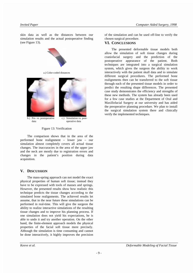
skin data as well as the distances between oursimulation results and the actual postoperative finding(see Figure 13).
a.) Color-coded distances
b.) Pre- vs. postoperative c.) Simulation vs. post-
data operative data
Figure 13: Verification
The comparison shows that in the area of theperformed bone realignment – lower jaw – oursimulation almost completely covers all actual tissuechanges. The inaccuracies in the area of the upper jawand the neck are mostly due to registration errors andchanges in the patient’s position during dataacquisition.
V. DISCUSSION
The mass-spring approach can not model the exactphysical properties of human soft tissue; instead theyhave to be expressed with tools of masses and springs.However, the presented results show how realistic thistechnique predicts the tissue changes according to thesimulated bone realignments. The achieved results letassume, that in the near future these simulations can beperformed in real-time. This will give the surgeon theability to realize interactive simulations of the resultingtissue changes and to improve his planning process. Ifone simulation does not yield his expectations, he isable to undo it and try another operation. On the otherhand, the finite-element approach models the physicalproperties of the facial soft tissue more precisely.Although the simulation is time consuming and cannotbe done interactively, it highly improves the precision
of the simulation and can be used off-line to verify thechosen surgical procedure.VI. C ONCLUSIONS
The presented deformable tissue models bothallow the simulation of soft tissue changes duringcraniofacial surgery and the prediction of thepostoperative appearance of the patient. Bothtechniques are integrated into a surgical simulationsystem, which gives the surgeon the ability to workinteractively with the patient skull data and to simulatedifferent surgical procedures. The performed bonerealignments then can be transferred to the soft tissuethrough each of the presented tissue models in order topredict the resulting shape differences. The presentedcase study demonstrates the efficiency and strengths ofthese new methods. The system has already been usedfor a few case studies at the Department of Oral andMaxillofacial Surgery at our university and has aidedthe preoperative planning procedure. We plan to installthe surgical simulation system there and clinicallyverify the implemented techniques.
Invited Paper Computer Aided Surgery, 1998
Keeve et al. Deformable Modeling of Facial Tissue
- 10 -
Acknowledgement: This work is supported by GermanScience Foundation research grant DFG Gi 198/2-1 to 5and German Academic Exchange Service research grantDAAD D/96/17570.
Invited Paper Computer Aided Surgery, 1998
Keeve et al. Deformable Modeling of Facial Tissue
- 11 -
References:
[Adv96] ADVANCED VISUAL SYSTEMS INC., http://www.avs.com1996.
[Ali96] A LIAS WAVEFRONT, SILICON GRAPHICS INC.http://www.alias.com, 1996.
[Alt93] A LTOBELLI D., KIKINIS R., MULLIKEN J., CLINE H.LORENSEN W., JOLESZ F., “Computer-Assisted Three-Dimensional Planning in Craniofacial Surgery,” Plasticand Reconstructive Surgery, Vol. 92, No. 2, pp. 576-5851993.
[Ath95] ATHANASIOU A. E. (ed.), “Orthodontic Cephalometry,”Mosby-Wolfe Verlag, London, 1995.
[Bat92] BATHE K.-J., “Finite Element Procedures in EngineeringAnalysis,” Prentice Hall, Englewood Cliffs, NJ, 1992.
[Bel90] BELEC L., “Computer Modeling of Total HipReplacement: Application to Join Geometry, StressAnalysis and Bone Remodeling,” Ph.D. Thesis, ThayerSchool of Engineering, Dartmouth College, Hanover, NH1990.
[Bil95] B ILL J. S., REUTHER J. F., DITTMANN W., KÜBLER N.MEIER J. L. PISTNER H., WITTENBERG G.“Stereolithography in Oral and Maxillofacial OperationPlanning,” Intern. J. of Oral Maxillofacial Surgery, Vol24, No. 1, Part II, pp. 98-101, 1995.
[Bla97] BLACK P., MORIARTY T., ALEXANDER E., STIEG P.WOODARD E., GLEASON P., MARTIN C., KIKINIS R.SCHWARTZ R., JOLESZ F., “The Development andImplementation of Intraoperative MRI and itsNeurosurgical Applications,“ Neurosurgery, Vol. 41, No4, pp. 831-842, October 1997.
[Bro96a] BRO-NIELSEN M., “Active Nets and Cubes,” InternalReport, Image Analysis Group, Institute of MathematicalModeling, Technical University of Denmark, LyngbyDenmark, 1996.
[Bro96b] BRO-NIELSON M., COTIN S., “Real-time VolumetricDeformable Models for Surgery Simulation using FiniteElements and Condensation,“ Proc. of Eurographics, Vol5, pp. 57-66, 1996.
[Che92] CHEN D. T., ZELTZER D., “Pump It Up: ComputerAnimation of a Biomechanically Based Model of Muscleusing the Finite Element Method,” Proc. ofSIGGRAPH’92, ACM Computer Graphics, Vol. 26, No2, pp. 89-98, July 1992.
[Che95] CHEN D. T., PIEPER S., ZELTZER D., “Computer Animationusing the Finite Element Method,” Internal Report, MITMedia Laboratory, Cambridge, MA, 1995.
[Cot96] COTIN S., DELINGETTE H., CLEMENT J., SOLER L., AYACHE
N., MARESCAUX J., “Geometrical and PhysicalRepresentations for a Simulator of Hepatic Surgery,“Proc. Medicine meets Virtual Reality IV, 1996.
[Cov93] COVER S. A., EZQUERRA N. F., O’BRIEN J. F., “InteractiveDeformable Models for Surgery Simulation,” IEEEComputer Graphics & Applications, Vol. 13, No. 6, pp68-75, Nov. 1993.
[Cyb98] CYBERWARE LABORATORY INCORPORATED (ed.)“Cyberware Model 3030RGB Digitizer Manual,”Monterey, CA, 1998.
[Dal61] DAL PONT G., “Retromolar Osteotomy for the Correctionof Prognathism,” J. of Oral Surgery, Vol. 19, No. 421961.
[Del94] DELINGETTE H., SUBSOL G., COTIN S., PIGNON J., “ ACraniofacial Surgery Testbed,” Rapport de recherchéNo. 2119, INRIA, Sophia-Antipolis, France, 1994.
[Den88] DENG X. Q., “A Finite Element Analysis of Surgery ofHuman Facial Tissue,” Ph.D. Thesis, ColumbiaUniversity, 1988.
[Euf94] EUFINGER H., KRUSE D., MACHTENS E., HEUSER L.“CAD/CAM-Rekonstruktion des zahnlosen Unterkieferszur Herstellung individueller Augmentate,” DtschZahnärztl. Z., Vol. 49, pp. 41-44, 1994.
[Fis96] FISHMAN E. K., KUSZYK B. S., GAO L., CABRAL B.“Surgical Planning for Liver Resection,” IEEEComputer, Vol. 29, No. 1, pp. 64-72, 1996.
[Fun93] FUNG Y. C., “Biomechanics: Mechanical Properties ofLiving Tissues,” Springer Verlag, New York, NY, 1993.
[Gib97] GIBSON S., et al., “Simulating Arthroscopic Knee Surgeryusing Volumetric Object Representations, Real-TimeVolume Rendering and Haptic Feedback,“ ProcCVRMed II and MRCAS II, March 1997.
[Gib98] GIBSON S., MIRTICH B., “A Survey of DeformableModeling in Computer Graphics,“ in preparation, 1998.
[Gou89] GOURRET J.-P., “Simulation of Object and Human SkinDeformations in a Grasping Task“, Proc. ofSIGGRAPH’89, ACM Computer Graphics, Vol. 23, No3, pp. 21-30, July 1989.
[Has95] HASSFELD S., MUEHLING J., ZOELLER J., “IntraoperativeNavigation in Oral and Maxillofacial Surgery,” Intern. Jof Oral Maxillofacial Surgery, Vol. 24, No. 1, Part II, pp111-116, 1995.
[Hem91] HEMMY D., HARRIS G. F., GANAPATHY V., “Finite ElementAnalysis of Craniofacial Skeleton Using Three-Dimensional Imaging as the Substrate,” in CARONNI E. P(ed.) Craniofacial Surgery, Proc. of the 2nd InternCongress of the Intern. Society of Cranio-Maxillo-FacialSurgery, Florence, Italy, 1991.
[Hol95] HOLLERBACH K., UNDERHILL K., RAINSBERGER R.“Automated Volumetric Grid Generation for FiniteElement Modeling of Human Joints,” BioengineeringConference ASME’95, Vol. 29, No. 29, pp. 49-50, 1995.
[Kee96a] KEEVE E., GIROD S., GIROD B., “Computer-AidedCraniofacial Surgery,” Proc. of Computer AssistedRadiology CAR’96, Paris, France, pp. 757-763, June 26-29, 1996.
[Kee96b] KEEVE E., GIROD S., PFEIFLE P., GIROD B., “Anatomy-Based Facial Tissue Modeling Using the Finite ElementMethod,” Proc. of IEEE Visualization’96, San FranciscoCA, Oct. 27 - Nov. 1, 1996.
[Kee96c] KEEVE E., “Visualisierungs- und Simulationsverfahrenzur interaktiven Planung kraniofazialer Korrektur-operationen,” Ph.D. thesis, University of Erlangen, Nov1996.
Invited Paper Computer Aided Surgery, 1998
Keeve et al. Deformable Modeling of Facial Tissue
- 12 -
[Kee97] KEEVE E., GIROD S., SCHALLER S., GIROD B., “AdaptiveSurface Data Compression,” Signal Processing, Vol. 59No. 2, pp. 211-220, June 1997.
[Koc96] KOCH R. M., GROSS M. H., BÜREN D. F., FRANKHAUSER
G., PARISH Y., CARLS F. R., “Simulating Facial SurgeryUsing Finite Element Models,” Proc. of SIGGRAPH’96New Orleans, LA; ACM Computer Graphics, Vol. 30Aug. 4-9. 1996.
[Krü85] KRÜGER E., “Fehlbildungen und Formveränderungen imKiefer-Gesichts-Bereich,” Lehrbuch der chirurgischenZahn-, Mund- und Kieferheilkunde, Quintessenz VerlagBerlin, 5. Auflage, 1985.
[Lan94] LANGTANGEN H. P., NIELSEN B. F., “Getting Started withDIFFPACK,” The DIFFPACK Report Series, SINTEFOslo, Norway, June 1994.
[Lar86a] LARRABEE W. F., “A Finite Element Model of SkinDeformation: I. Biomechanics of Skin and Soft Tissue: AReview,” Laryngoscope, Vol. 96, pp. 399-405, April1986.
[Lar86b] LARRABEE W. F., SUTTON D., “A Finite Element Model ofSkin Deformation: II. An Experimental Model of SkinDeformation,” Laryngoscope, Vol. 96, pp. 406-412, April1986.
[Lar86c] LARRABEE W. F., GALT J. A., “A Finite Element Model ofSkin Deformation: III. The Finite Element Model,”Laryngoscope, Vol. 96, pp. 413-419, April 1986.
[Lee95] LEE Y., TERZOPOULOS D., WATERS K., “RealisticModeling for Facial Animation,” Proc. ofSIGGRAPH’95, Los Angeles, CA, ACM ComputerGraphics, Vol. 29, pp. 55-62, Aug. 6-11, 1995.
[Lo94] LO L. J., MARSH J. L., VANNIER M. W., PATEL V. V.“Craniofacial Computer-Assisted Surgical Planning,”Clinics in Plastic Surgery, Vol. 21, No. 4, pp. 501-516Oct. 1994.
[Lor87] LORENSEN W. E., CLINE H. E., “Marching Cubes: A HighResolution 3D Surface Construction Algorithm,” Proc. ofSIGGRAPH’87, ACM Computer Graphics, Vol. 21, No4, pp. 163-169, July 1987.
[Nat98] NATIONAL LIBRARY OF MEDICINE, “The Visible HumanProject,” http://www.nlm.nih.gov/research, 1998.
[Par96] PARKE F., WATERS K., “Computer Facial Animation,”AK Peters, Wellesley, MA, 1996.
[Pat96] PATEL V. V., VANNIER M. W., MARSH J. L., LO L. J.“Assessing Craniofacial Surgical Simulation,” IEEEComputer Graphics & Applications, Vol. 16, No. 1, pp46-54, January 1996.
[Pie91] PIEPER S., “CAPS: Computer-Aided Plastic Surgery,”Ph.D. Thesis, MIT, Cambridge, MA, 1991.
[Pie92] PIEPER S., ROSEN J., ZELTZER D., “Interactive Graphicsfor Plastic Surgery: A Task-Level Analysis andImplementation,” Proc. of SIGGRAPH’92, ACMComputer Graphics, Vol. 25, No. 2, pp. 127-134, 1992.
[Pie95] PIEPER S., LAUB D., ROSEN J., “A Finite Element FacialModel for Simulating Plastic Surgery,” Plastic andReconstructive Surgery, Vol. 96, No. 5, pp. 1100-1105Oct. 1995.
[Pro95] PROVOT X., “Deformation Constraints in a Mass-SpringModel to Describe Rigid Cloth Behaviour,” Proc. ofGraphics Interface’95, Morgan Kaufman Publisher, PaloAlto, CA, pp. .147-154, May 1995.
[Sag94] SAGAR M. A., BULLIVANT D., MALLINSON G. D., HUNTER
P. J., HUNTER I., “A Virtual Environment and Model ofthe Eye for Surgical Simulation,” Proc. ofSIGGRAPH’94, ACM Computer Graphics, Vol. 28, No4, pp. 205-212, 1994.
[See92] SEEHOLZER H., WALKER R., “Kieferorthopädische undkieferchirurgische Behandlungsplanung mit demComputer am Beispiel des Dentofacial Planner,”Informationsschrift der Firma Gemetek, Erding, Germany1992.
[Ste94] STEINHÄUSER E. W., “Weichteilvorhersage bebimaxillären Operationen,” Fortschritte in der Kiefer-und Gesichts-Chirurgie, Sonderband: Die bimaxilläreChirurgie bei skelettalen Dysgnathien, Thieme-Verlag1994.
[Sze98] SZEKELY G., One-Day-Tutorial “Soft Tissue Deformationand Tissue Palpation” in conjunction with the FirstInternational Conference on Medical Image Computingand Computer-Assisted Intervention MICCAI’98,Boston, MA, October 12, 1998.
[Tan95] TANAKA I., KOBAYASHI M., FUJINO T., CHIYOKURA H.,“Simulation for Facial Lip Expression Using the FacialMuscle Model,” Proc. of Computer Assisted RadiologyCAR’95, Springer Verlag, Berlin, pp. 878-881, June 21-24, 1995.
[Tay96] TAYLOR R. H., LAVELLEE S., BURDEA G. C., MÖSGES R.“Computer Integrated Surgery: Technology and ClinicalApplications,” MIT Press, Cambridge, MA, 1996.
[Ter90] TERZOPOULOS D., WATERS K., “Physically-Based FacialModeling, Analysis and Animation,” J. of Visualizationand Computer Animation, Wiley & Sons, Vol. 1, pp. 73-80, 1990.
[Ter91] TERZOPOULOS D., WATERS K., “Techniques for RealisticFacial Modeling and Animating,” Proc. of ComputerAnimation’91, Springer Verlag, pp. 59-73, 1991.
[Van83] VANNIER M. W., MARSH J. L., WARREN J. O., “ThreeDimensional Computer Graphics for CraniofacialSurgical Planning and Evaluation,” Proc. ofSIGGRAPH’83, ACM Computer Graphics, Vol. 17, No3, pp. 263-273, July 1983.
[Wat92] WATERS K., “A Physical Model of Facial Tissue andMuscle Articulation Derived from Computer TomographyData,” SPIE Visualization in Biomedical ComputingVol. 1808, pp. 574-583. 1992.
[Wat95] WATERS K., FRISBIE J., “A Coordinated Muscle Model forSpeech Animation,” Proc. of Graphics Interface ‘95, pp163-170, Mai 1995.
[Wer94] WERNECKE J., “The Inventor Mentor: ProgrammingObject-Oriented 3D Graphics with Open Inventor,”Addison-Wesley, Reading, MA, 1994.
[Zei95] ZEILHOFER H. F., SADER R., HORCH H. H., DEPPE H.“Preoperative Visualization of Aesthetic Changes inOrthognatic Surgery,” Proc. of Computer AssistedRadiology CAR’95, Springer Verlag, Berlin, pp. 1369-1374, June 21-24, 1995.
Invited Paper Computer Aided Surgery, 1998
Keeve et al. Deformable Modeling of Facial Tissue
- 13 -
[Zon94] ZONNEVELD W., “Progress in Clinical Radiology: ADecade of Clinical Three-Dimensional Imaging - AReview, Part III: Image Analysis and Interaction, DisplayOptions, and Physical Models,” Investigative RadiologyVol. 29, No. 7, pp. 716-725, 1994.