Embed Size (px)
Citation preview
Craniosynostosis
Ron Grondin25 May 2006
Definition
• Heterogeneous group of disorders with premature fusion of cranial sutures– Non-syndromic most common– Also associated with >150 syndromes
Incidence
• 5-6 cases per 10,000 live births– Saggital synostosis most common– Followed by unilateral coronal, bilateral
coronal, metopic, then lambdoid

Etiology• Non-syndromic most common
– Syndromic in 10-20% of cases• Approx 14% of pedigrees with Craniosynostosis
demonstrated familial aggregation– Predominantly autosomal dominant with variable
penetrance– 60% penetrance and 61% sporadic incidence(Lajeunie et al, Am J Med Genet 55:500-504, 1995)
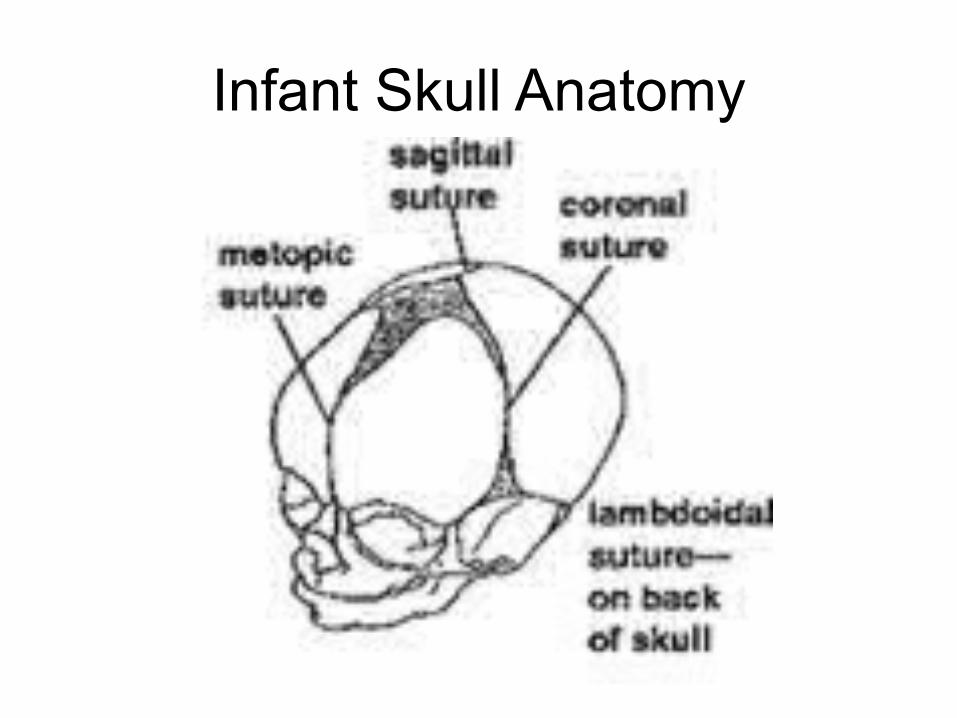
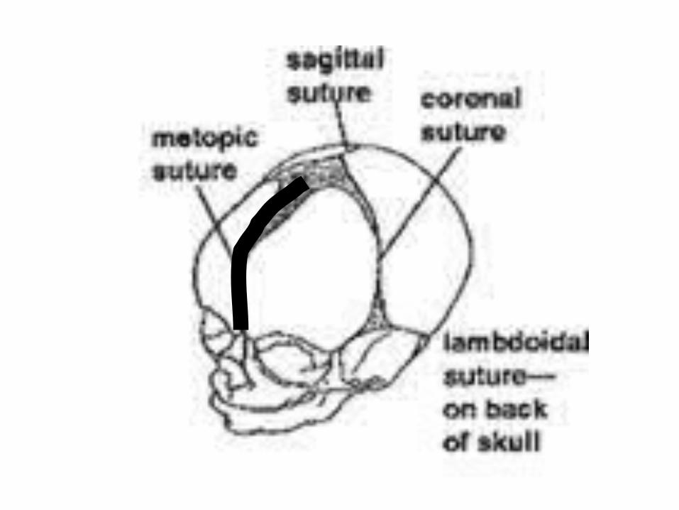
Infant Skull Anatomy
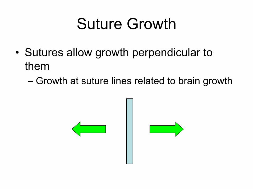
Suture Growth
• Sutures allow growth perpendicular to them– Growth at suture lines related to brain growth
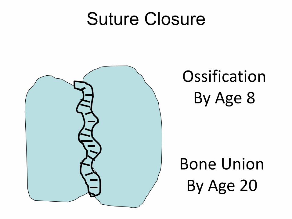
Suture Closure
Bone UnionBy Age 20
OssificationBy Age 8
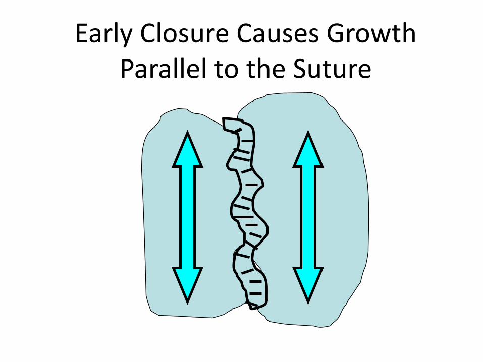
Early Closure Causes Growth Parallel to the Suture
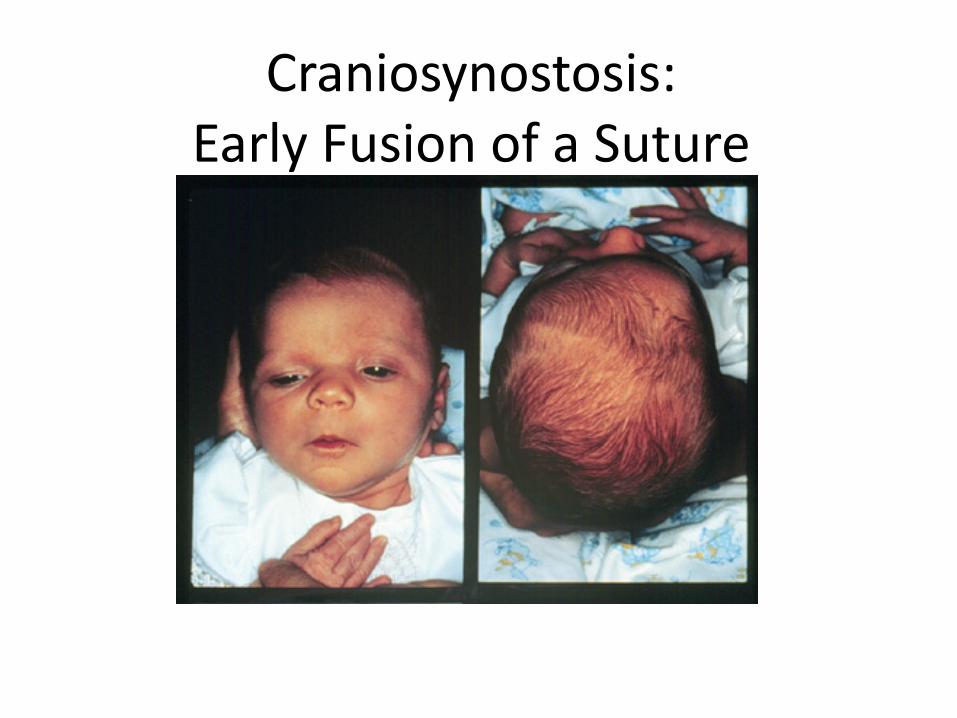
Craniosynostosis:Early Fusion of a Suture
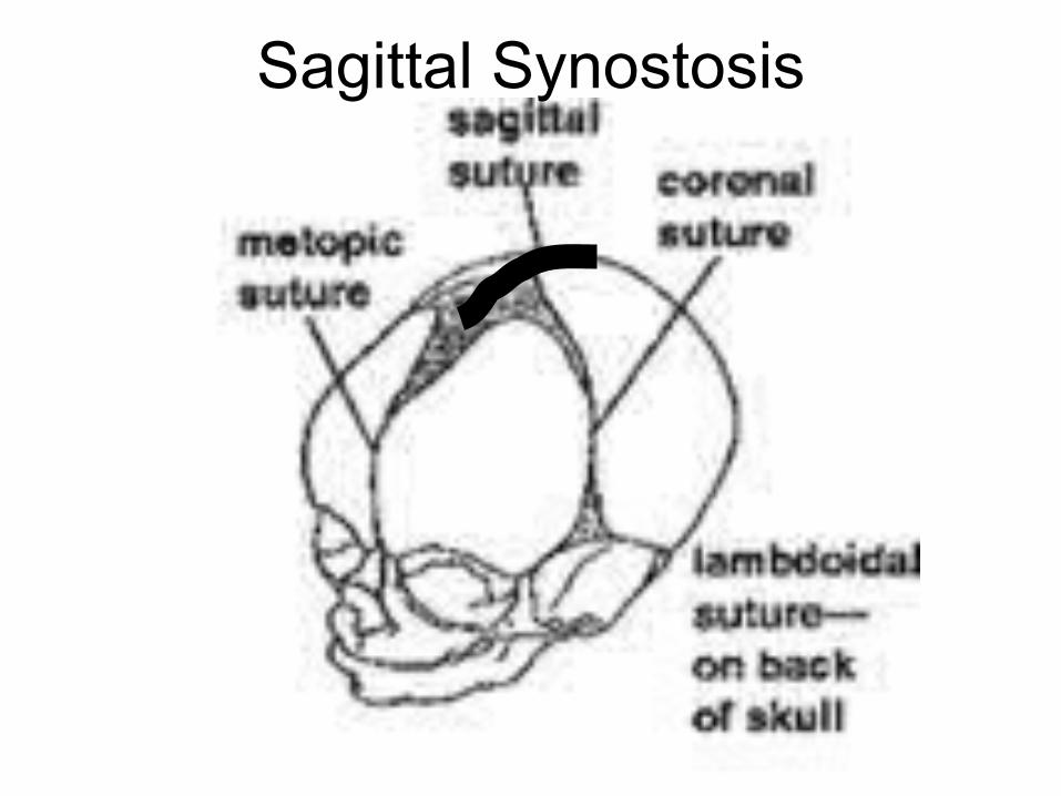
Sagittal Synostosis

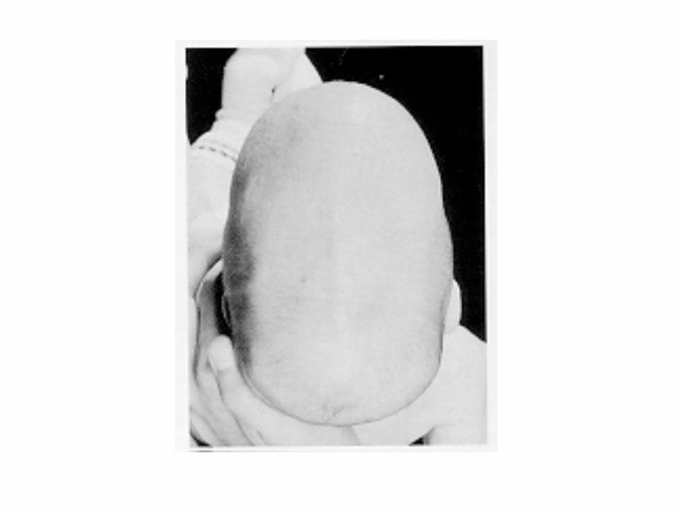
Sagittal Synostosis
“Boat-Head”(Scaphocephaly)
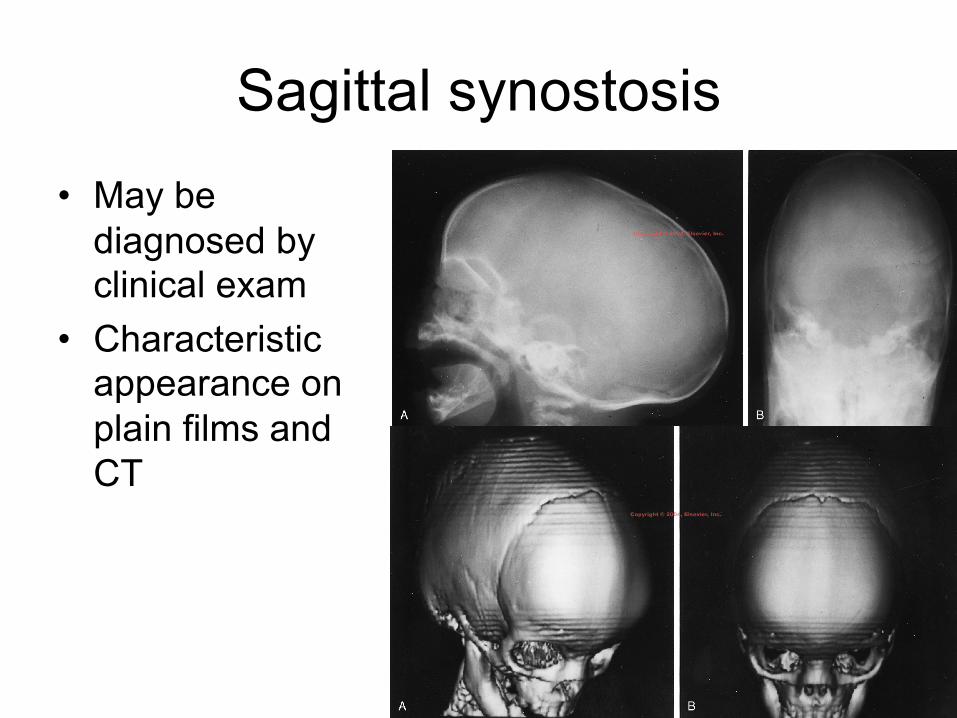
Sagittal synostosis• May be
diagnosed by clinical exam
• Characteristic appearance on plain films and CT
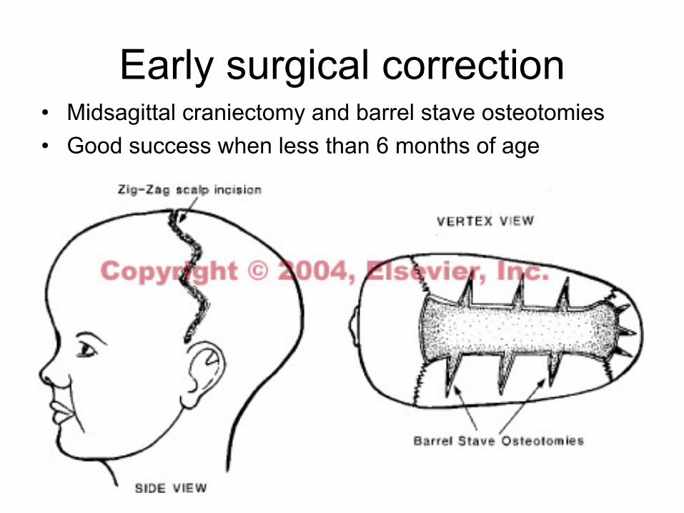
Early surgical correction• Midsagittal craniectomy and barrel stave osteotomies• Good success when less than 6 months of age

Late surgical correction• More extensive
cranial vault remodelling
• Usually in collaboration with plastic surgery
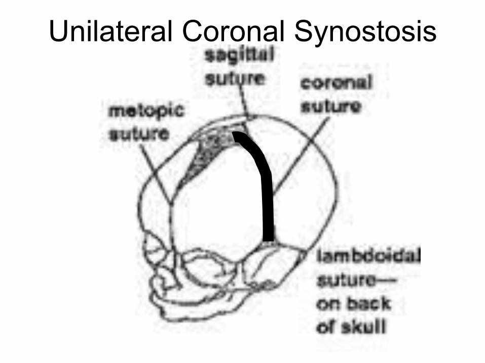
Unilateral Coronal Synostosis
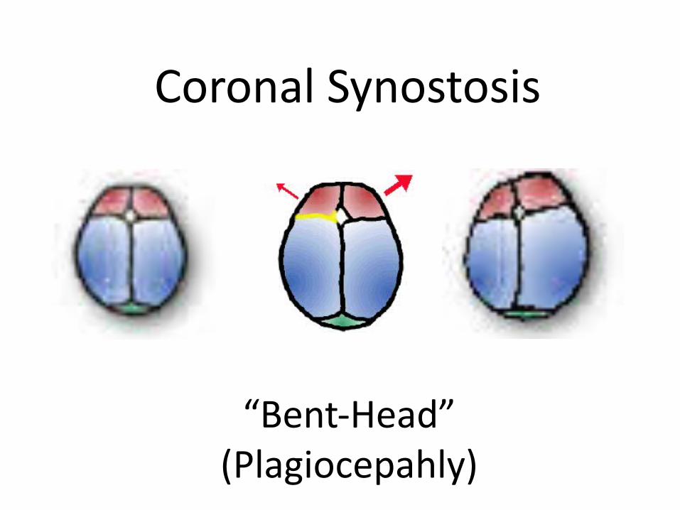
Coronal Synostosis
“Bent-Head”(Plagiocepahly)
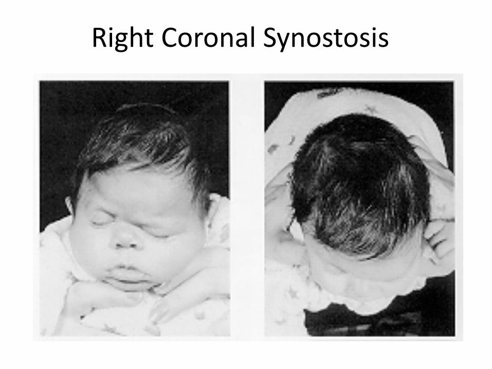
Right Coronal Synostosis
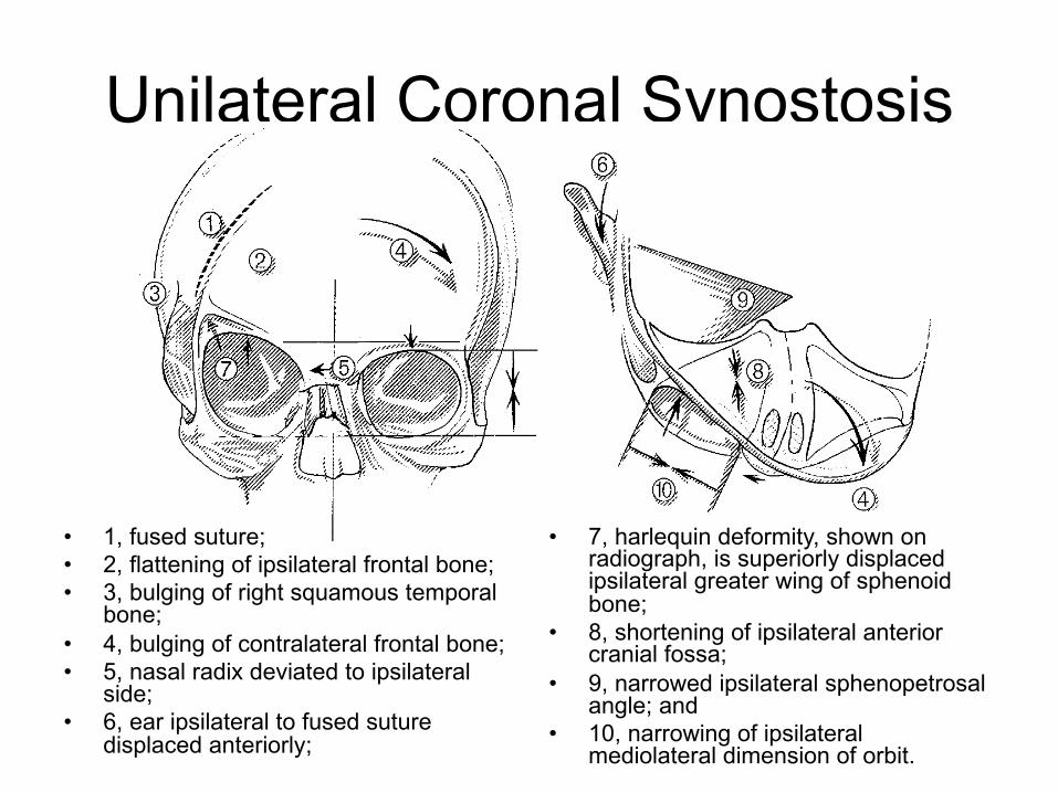
Unilateral Coronal Synostosis
• 7, harlequin deformity, shown on radiograph, is superiorly displaced ipsilateral greater wing of sphenoid bone;
• 8, shortening of ipsilateral anterior cranial fossa;
• 9, narrowed ipsilateral sphenopetrosal angle; and
• 10, narrowing of ipsilateral mediolateral dimension of orbit.
• 1, fused suture; • 2, flattening of ipsilateral frontal bone; • 3, bulging of right squamous temporal
bone; • 4, bulging of contralateral frontal bone; • 5, nasal radix deviated to ipsilateral
side;• 6, ear ipsilateral to fused suture
displaced anteriorly;
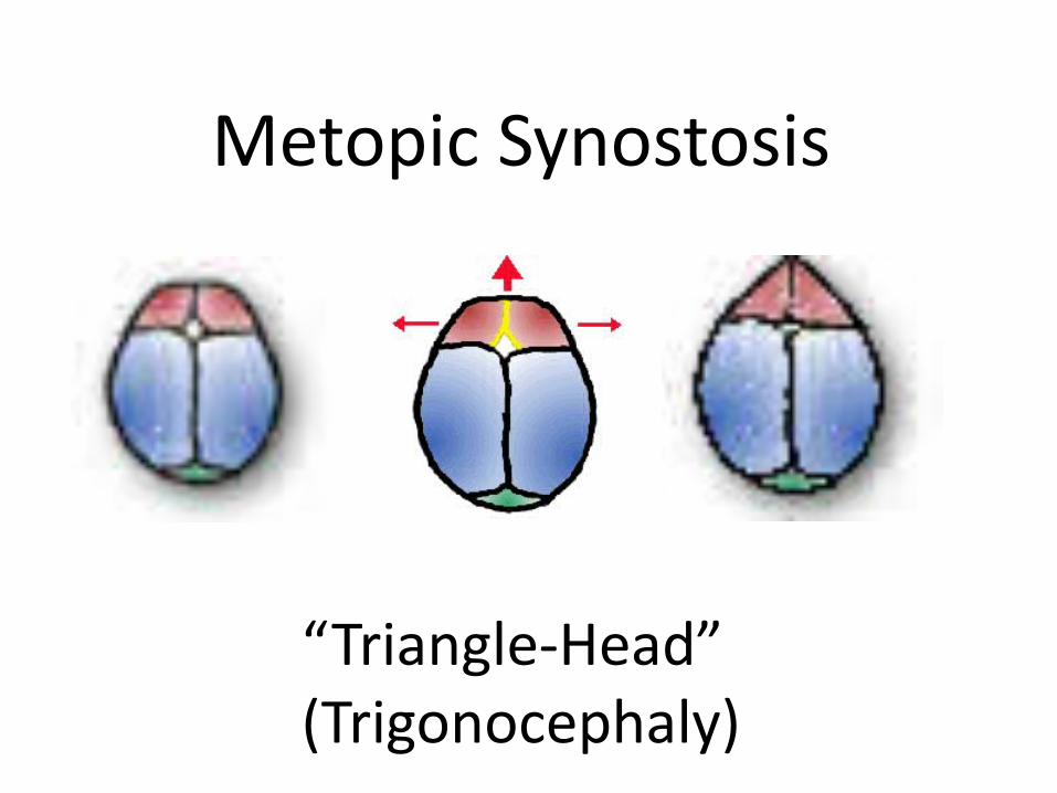
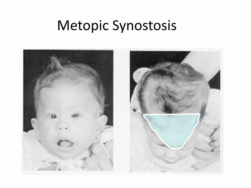
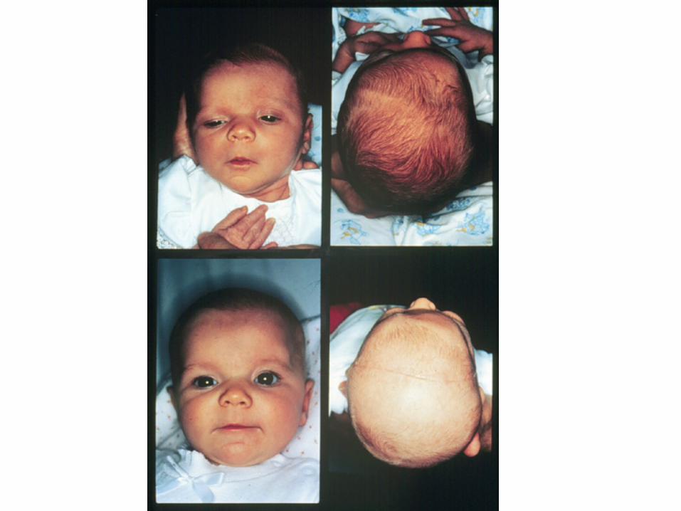
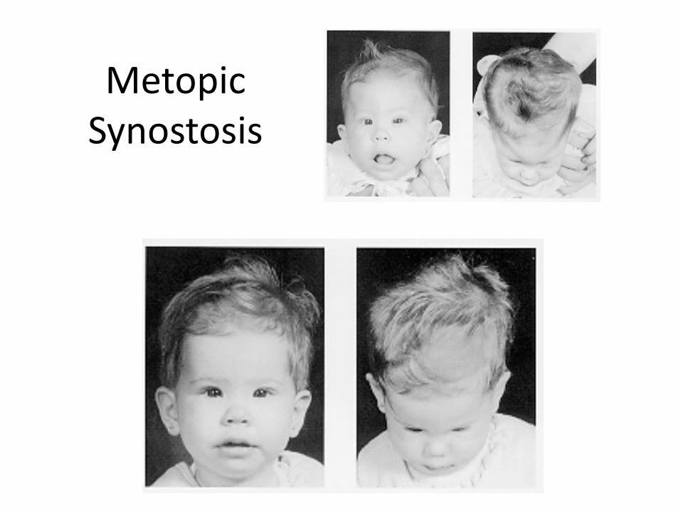
Metopic Synostosis
“Triangle-Head”(Trigonocephaly)
Metopic Synostosis

• 1, ridging of the fused suture; • 2, temporal narrowing; • 3, patent coronal suture
displaced anteriorly; • 4, compensatory bulging of the
parieto-occipital region, contributing to the pear-shaped appearance of skull;
• 5, narrowed bizygomatic dimension; and
• 6, posterior displacement of superolateral orbital rim.
Metopic Synostosis
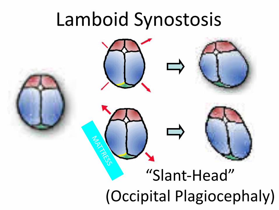
Lamboid Synostosis
“Slant-Head”(Occipital Plagiocephaly)
MATTRESS
Clinical Exam• Head circumference• Head shape (from above, side)• Ear and facial symmetry• Palpate suture lines & fontanelles
– Look for ridging• Look for associated anomalies• Skull X-ray or CT
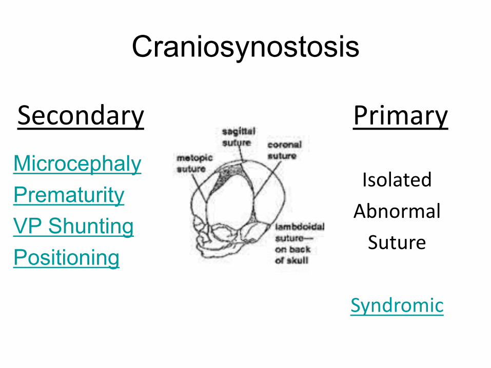
Craniosynostosis
MicrocephalyPrematurityVP ShuntingPositioning
Secondary Primary
IsolatedAbnormalSuture
Syndromic

• Deformational Scaphocephaly• Impaired mobility & prolonged
positioning• Persists until adulthood• Prevention:
– Donut-shaped head supports– waterbed mattresses
• Does not warrant intervention
Prematurity
VP Shunting
• Scaphocephaly• Chronic hydrocephalus thickens the skull• Once decompression with shunt, the
suture fuses• Surgery Indications:
– HC > 50 cm (4-5+ STDs)– When VPS performed during when VLBW
Microcephaly
• Surgical correction not indicated• Abnormal HC
– in primary craniosynostosis, HC remains normal yet oddly shaped
• Rare cases of multisutural craniosynostosis restricting head growth, but manifests with increased ICP
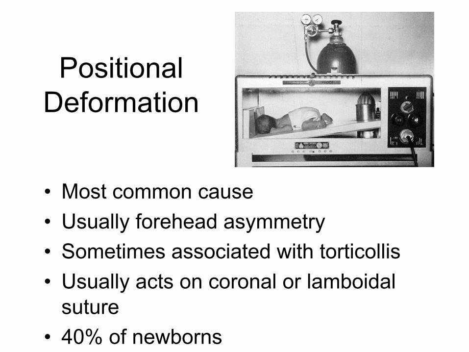
• Most common cause• Usually forehead asymmetry• Sometimes associated with torticollis• Usually acts on coronal or lamboidal
suture• 40% of newborns
PositionalDeformation
An Epidemic of Lamboidal Plagiocephaly
• 1992: Back to Sleep Campaign
• 1996: Tertiary Care Centers report rise in lamboidal plagiocephaly from 3% to 20%
Sorting out the “Epidemic”
• 102 Patients with occipital plagiocephaly over 4 year period
• Only 4 (3%) had true lamboidal synostosis• The rest were deformational
– Only 3 were progressive (required surgery)– Other responded to positioning or helmets
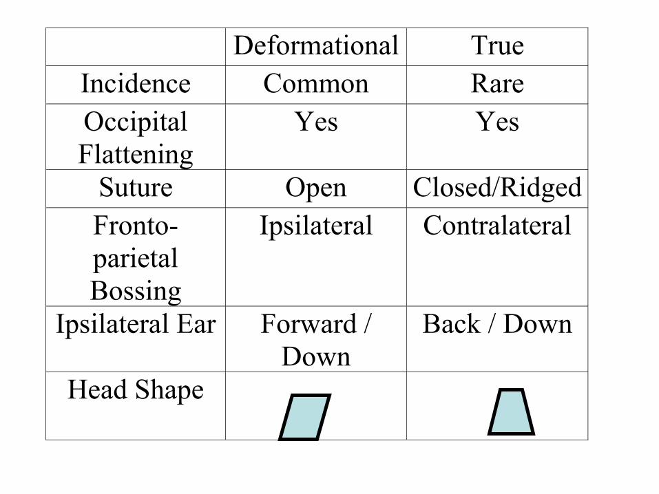
Deformational True Incidence Common Rare Occipital Flattening
Yes Yes
Suture Open Closed/Ridged Fronto-parietal Bossing
Ipsilateral Contralateral
Ipsilateral Ear Forward / Down
Back / Down
Head Shape
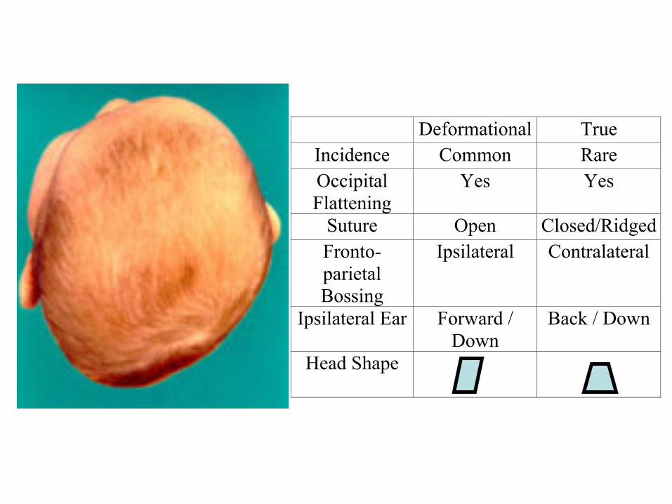
Deformational True Incidence Common Rare Occipital Flattening
Yes Yes
Suture Open Closed/Ridged Fronto-parietal Bossing
Ipsilateral Contralateral
Ipsilateral Ear Forward / Down
Back / Down
Head Shape

Syndromic Craniosynostosis
• 10-20 % of cases• Autosomal Dominant
– Linked to Chromosome 10q• Multi-sutural, complex cases
If a suture is fused,check hands, feet, big toe and thumb
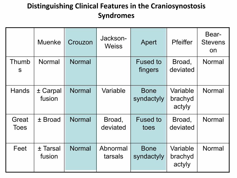
Distinguishing Clinical Features in the Craniosynostosis Syndromes
Muenke Crouzon Jackson-Weiss Apert Pfeiffer
Bear-Stevens
on
Thumbs
Normal Normal Fused to fingers
Broad, deviated
Normal
Hands ± Carpal fusion
Normal Variable Bone syndactyly
Variable brachydactyly
Normal
Great Toes
± Broad Normal Broad, deviated
Fused to toes
Broad, deviated
Normal
Feet ± Tarsal fusion
Normal Abnormal tarsals
Bone syndactyly
Variable brachydactyly
Normal
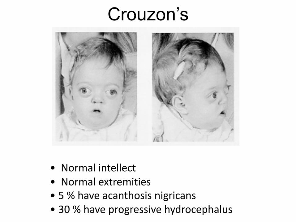
Crouzon’s
• Normal intellect• Normal extremities• 5 % have acanthosis nigricans• 30 % have progressive hydrocephalus
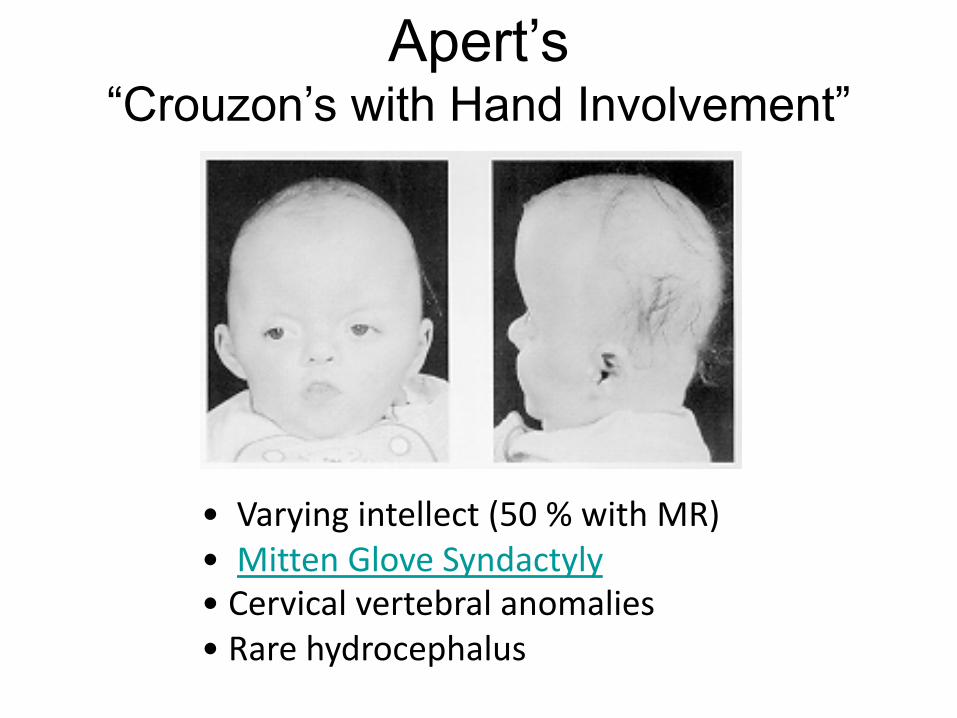
Apert’s“Crouzon’s with Hand Involvement”
• Varying intellect (50 % with MR)• Mitten Glove Syndactyly• Cervical vertebral anomalies• Rare hydrocephalus
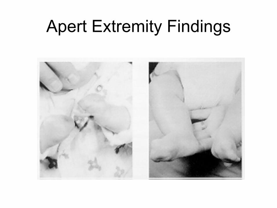
Apert Extremity Findings
True Craniosynostosis & Surgery
• Single Suture Synostosis: Confirm by exam and skull x-rays
• Complex cases: CT or 3D CT• X-Ray: Fused sutures have a broad ridge
of overgrowth of solid bone along a previous suture, or suture is completely obliterated
• Ridge is especially characteristic of fused sagittal suture
Management
• Surgery vs. Conservative Management

The Decision to Operate
• Raised ICP in 1/3 of cases, but no neuro impairment
• Cosmetic considerations usually most important– affects peer acceptance, parent-child bonding,
self-image and coping

Imaging• Skull X-ray• CT• 3-D CT
Surgery
• If not part of syndrome, the earlier the operation the better– At the latest 6-12 months (by 12 months, skull
is 85% of adult size)– For coronal suture, operate before 2 months
because of facial symmetry and visual system development
– Procedure depends on continuing skull growth
Surgery
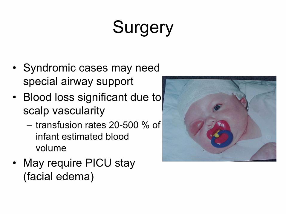
• Syndromic cases may need special airway support
• Blood loss significant due to scalp vascularity– transfusion rates 20-500 % of
infant estimated blood volume
• May require PICU stay (facial edema)
MetopicSynostosis
Surgery
• Unilateral coronal suture: difficult. Orbital relocation as well.
• Syndromic or multi-suture cases: staged repairs.

Apert: Post-Op
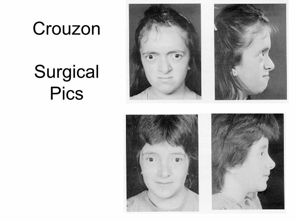
Crouzon
Surgical Pics
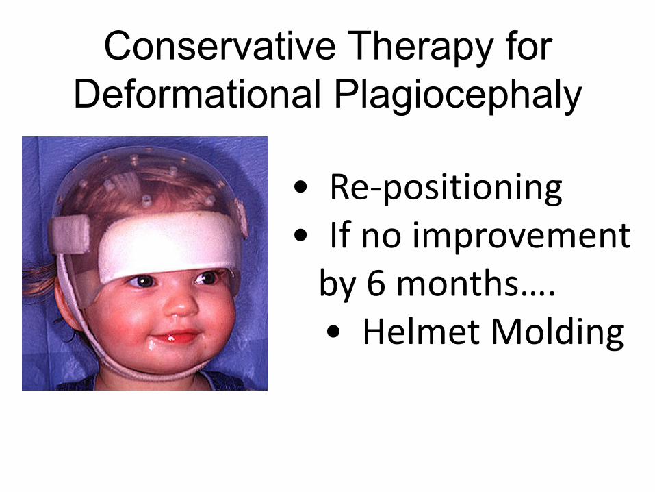
Conservative Therapy for Deformational Plagiocephaly
• Re-positioning• If no improvement
by 6 months….• Helmet Molding
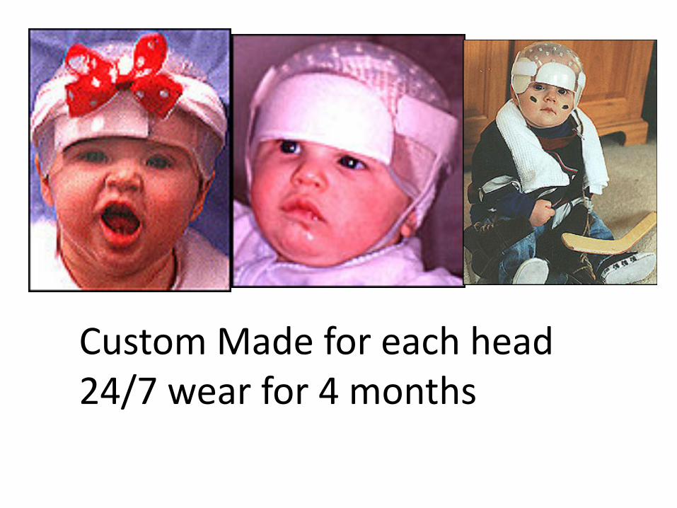
Custom Made for each head24/7 wear for 4 months