Embed Size (px)
Citation preview

Wayne State University
Wayne State University Dissertations
1-1-2015
Crebh, A Novel Liver Clock Keeper For EnergyMetabolismZe ZhengWayne State University,
Follow this and additional works at: http://digitalcommons.wayne.edu/oa_dissertations
Part of the Endocrinology Commons, Genetics Commons, and the Molecular BiologyCommons
This Open Access Dissertation is brought to you for free and open access by DigitalCommons@WayneState. It has been accepted for inclusion inWayne State University Dissertations by an authorized administrator of DigitalCommons@WayneState.
Recommended CitationZheng, Ze, "Crebh, A Novel Liver Clock Keeper For Energy Metabolism" (2015). Wayne State University Dissertations. Paper 1180.

CREBH, A NOVEL LIVER CLOCK KEEPER FOR
ENERGY METABOLISM
by
ZE ZHENG
DISSERTATION
Submitted to the Graduate School
of Wayne State University
Detroit, Michigan
in partial fulfillment of the requirements
for the degree of
DOCTOR OF PHILOSOPHY
2015
MAJOR: MOLECULAR BIOLOGY AND
GENETICS
Approved By:
Advisor Date

© COPYRIGHT BY
ZE ZHENG
2015
All Rights Reserved

ii
DEDICATION
This work is dedicated to my parents and my husband Daochun Sun
for their continuous support and understanding during the years of my
education, and to my lovely little daughter Amber for her joyful and peaceful
smiles to encourage me to complete this dissertation. I could not achieve
my goal without them.

iii
ACKNOWLEDGMENTS
I would like to express tremendous appreciation to my mentor, Dr.
Kezhong Zhang. His encouragement and guidance throughout these years
made this dissertation come true.
I would also like to thank to my committee members, Dr. Gregory
Kapatos for his sustained attention, inspiring discussions and generous
supports from my course studies, lab rotation, to my thesis project, Dr. Todd
Leff and Dr. Russel L. Finley for their informative class teaching,
understanding and suggestions during these years. I gratefully acknowledge
Dr. Maik Hüttemann and Dr. Li Li for their warm encouragements and
supports during the toughest time of my oversea study, Dr. Guohui Wang
and Dr. Chunbin Zhang for their patience and assistance with my
experiments at the very beginning, Dr. Xuebao Zhang and Dr. Hyunbae Kim
for teaching me the importance of precision in science. I sincerely recognize
the wonderful inspiring discussions with visiting professor Dr. Kenji
Fukudome, and the other two graduate students Mr. Roberto Mendez and
Mr. Aditya Dandekar in lab. I also want to thank Mrs. Suzanne Shaw from
CMMG administration, and Mrs. Becky Cai for their consideration and
assistance throughout my study, and making my overseas education feel like
home.

iv
TABLE OF CONTENTS
DEDICATION ...................................................................................................................ii
ACKNOWLEDGMENTS .................................................................................................. iii
LIST OF TABLES ............................................................................................................vi
LIST OF FIGURES ......................................................................................................... vii
LIST OF ABBREVIATIONS .............................................................................................ix
CHAPTER I: CREBH, a Liver Transcription Factor and More ......................................... 1
Introduction .................................................................................................................. 1
CREBH Gene Structure and Functions ........................................................................ 5
Transcriptional Regulation of CREBH ........................................................................ 10
CHAPTER II: Identification of CREBH as a Clock-regulated Diurnal Regulator in Liver 11
Summary.................................................................................................................... 11
Materials and Methods ............................................................................................... 13
Results ....................................................................................................................... 20
CHAPTER III: CREBH Functions under Circadian Rhythm ........................................... 28
Summary.................................................................................................................... 28
Materials and Methods ............................................................................................... 30
Results ....................................................................................................................... 33
v
CHAPTER IV: Crosstalk of CREBH and Other Circadian Regulators ........................... 52
Summary.................................................................................................................... 52
Materials and Methods ............................................................................................... 53
Results ....................................................................................................................... 55
REFERENCES .............................................................................................................. 72
ABSTRACT ................................................................................................................... 76
AUTOBIOGRAPHICAL STATEMENT ........................................................................... 78

vi
LIST OF TABLES
Table 1. Primers for gene expression qPCR. ................................................................ 14
Table 2. Primers for ChIP-qPCR. .................................................................................. 18
Table 3. CRE binding motifs in the promoter regions of mouse genes.......................... 41

vii
LIST OF FIGURES
Figure 1. Major conclusions of this dissertation. .............................................................. 4
Figure 2. Domain scheme of human and mouse CREBH. .............................................. 5
Figure 3. CREBH-mediated signaling pathways. ............................................................ 9
Figure 4. CREBH is a clock-controlled gene (CCG) in mouse liver ............................... 22
Figure 5. Illustration of E-boxes in mouse CrebH gene promoter region. ...................... 23
Figure 6. CrebH is directly regulated by BMAL1 in mouse liver .................................... 25
Figure 7. CREBH is not a typical CCG. ......................................................................... 27
Figure 8. CREBH regulates rhythmic levels of circulating lipids in mice. ....................... 34
Figure 9. Levels of hepatic TG in CREBH-null and WT control mice livers. .................. 34
Figure 10. CREBH regulates rhythmic expression of the genes involved in lipolysis, FA oxidation, and lipogenesis in mice under the circadian clock. ....................................... 36
Figure 11. Rhythmic protein levels of the key metabolic enzymes or regulators in the livers of CREBH-null and WT control mice. ................................................................... 37
Figure 12. Slight alterations of metabolic gene rhythmic expressions in the livers of CREBH-null and WT control mice. ................................................................................ 38
Figure 13. CREBH regulates rhythmic levels of lipids and expression of the genes involved in lipolysis, FA oxidation, and lipogenesis in mice under the circadian clock. . 40
Figure 14. CREBH regulates rhythmic levels of blood glucose and hepatic glycogen storage in mice under the circadian clock. .................................................................... 43
Figure 15. CREBH regulates rhythmic expression of the genes involved in gluconeogenesis and glycogenesis in mice under the circadian clock. ......................... 45
Figure 16. CREBH-null mice display hyper-locomotion and increased stereotypic activities during the night time. ...................................................................................... 48
Figure 17. CREBH deficiency leads to phase-shifted feeding behavior and increased metabolic rates. ............................................................................................................. 51
Figure 18. Rhythmic expression levels and amplitudes of core clock genes in the livers of CREBH-null and WT control mice. ............................................................................ 56

viii
Figure 19. Rhythmic regulation of core clock proteins by CREBH in the liver. .............. 57
Figure 20. Rhythmic regulation of circadian transcriptional activators and repressor by CREBH in the liver. ....................................................................................................... 60
Figure 21. Rhythmic interactions between CREBH, the circadian transcriptional activators PPARα and C/EBPβ, and the repressor E4BP4 in the liver. ......................... 63
Figure 22. Potential CREBH-, PPARα-, C/EBPβ-, and E4BP4- binding sequences in the promoter region of mouse Pck1 gene. .......................................................................... 65
Figure 23. Rhythmic interactions between CREBH, the circadian transcriptional activators PPARα and C/EBPβ, and the repressor E4BP4 in the liver. ......................... 65
Figure 24. Illustration of CREBH working model as a circadian metabolic oscillator. .... 67
Figure 25. Tissue-specific expression of the mouse CrebH mRNA. .............................. 71

ix
LIST OF ABBREVIATIONS
ACC1, Acetyl-CoA Carboxylase 1
Acot4, Acyl-CoA Thioesterase 4
Apoa4, apolipoprotein A-IV
ApoC2, Apolipoprotein C-II
APR, acute phase response
BDH1, 3-Hydroxybutyrate Dehydrogenase 1
BMAL1, Aryl hydrocarbon receptor nuclear translocator-like
bZIP, basic leucine zipper
CB1R, cannabinoid 1 receptor
CCG, clock-controlled genes
ChIP, chromatin immunoprecipitation
CLOCK, circadian locomotor output cycles kaput
CPT1α, carnitine palmitoyltransferase 1A
CREB3L3, cyclic-AMP-response-element-binding protein 3-like 3 (as known as CREBH)
CREBH, cyclic-AMP-response-element-binding protein, hepatic specific

x
CRP, C-reactive protein
CT, circadian time
DBP, D-element binding protein
DD, constant darkness
Dgat2, diacylglycerol O-acyltransferase 2
Dhcr24, 24-Dehydrocholesterol Reductase
E4BP4, nuclear factor interleukin-3-regulated protein
ECL, enhanced chemiluminescence
Elovl6, ELOVL fatty acid elongase 6
ER, endoplasmic reticulum
ERAD, ER-associated degradation
FA, fatty acids
FADS2, fatty acid desaturase 2
FGF21, fibroblast growth factor 21
G6pc, glucose-6-phosphatase
Gys2, glycogen synthase 2
Hmgcl, 3-hydroxymethyl-3-methylglutaryl-CoA lyase
xi
HPLC, high-performance liquid chromatography
IACUC, Institutional Animal Care and Use Committee
Lcat, Lecithin-Cholesterol Acyltransferase
LD, 12 hour light:12 hour dark
LXRα, liver X receptor α
NASH, non-alcoholic steatohepatitis
NFIL3, interleukin 3-regulated nuclear factor (also known as E4BP4)
NR1D1, nuclear receptor-subfamily 1, group D, member 1 (also known as REV-ERBα)
PAS, Periodic acid-Schiff
Pck1, phosphoenolpyruvate carboxykinase 1
PGC-1α, PPARγ coactivator-1α
PPARα, peroxisome proliferator-activated receptor α
qRT-PCR, Quantitative real-time PCR
RXR, retinoid X receptor
S1P, site-1 protease
S2P, site-2 protease
SAP, serum amyloid P-component

xii
SCN, suprachiasmatic nuclei
SNP, single nucleotide polymorphism
SREBPs, sterol regulatory element-binding proteins
TG, triglyceride
VCO2, carbon dioxide production
VO2, Oxygen consumption
WT, wild-type
KO, knockout
1
CHAPTER I: CREBH, a Liver Transcription Factor and More
Introduction
Circadian rhythms are biological processes that exhibit endogenous oscillations
over a 24-hour light-dark cycle and are entrainable by internal and external stimuli.
Circadian rhythms are generated at the level of gene transcription by a network of clock-
controlled genes (CCGs) that form an autoregulatory feedback loop. Genes that are
directly regulated by the CLOCK/BMAL1 core circadian transcription complex are referred
as first-order CCGs (Hughes, DiTacchio et al. 2009). The CLOCK/BMAL1 heterodimer
drives circadian expression of many other transcription factors, thereby extending and
enhancing other circadian regulatory functions. Local rhythms in peripheral organs, such
as the liver, are synchronized by master clock oscillators located in the suprachiasmatic
nuclei (SCN) of the anterior hypothalamus (Reppert and Weaver 2001). Circadian
oscillators in peripheral organs respond differently to entraining signals and control
different physiological outputs.
Dysregulation of circadian rhythm is closely associated with the development of
human metabolic disease, such as obesity and type-2 diabetes. The intimate and
reciprocal interaction between the circadian clock system and fundamental metabolic
pathways has demonstrated by many studies (Green, Takahashi et al. 2008; Bass and
Takahashi 2010; Feng and Lazar 2012; Hatori, Vollmers et al. 2012). Survey of nuclear
receptor mRNA profiles in metabolic tissues suggested that approximately half of the
known nuclear receptors or transcriptional regulators exhibit rhythmic expression (Yang,
Downes et al. 2006). BMAL1-binding sites are associated with carbohydrate and lipid

2
metabolism (Rey, Cesbron et al. 2011) has been revealed by genome-wide and phase-
specific DNA-binding rhythms for the core circadian transcriptional oscillators. In the liver,
nuclear receptors or transcription factors are inducible by metabolites or hormones, and
therefore, may serve as direct links between metabolic pathways and the circadian control
of gene expression. For example, the nuclear receptor PPARα, which binds fatty acid
ligands, and the core clock regulator BMAL1 reciprocally regulate each other to provide
a feedback loop that integrates lipid metabolic processes to circadian oscillations (Inoue,
Shinoda et al. 2005; Oishi, Shirai et al. 2005; Canaple, Rambaud et al. 2006). The clock-
controlled nuclear receptors REV-ERBs are key regulators of circadian lipid biosynthesis
in the liver, and ablation of REV-ERBs causes hepatic steatosis through de-repression of
lipogenesis (Feng, Liu et al. 2011; Bugge, Feng et al. 2012). PPARγ coactivator-1α (PGC-
1α) also serves a link between the clock and energy metabolism, as PGC-1α varies
rhythmically and has been shown to stimulate expression of Bmal1 and nuclear receptors
of the ROR family (Liu, Li et al. 2007). Furthermore, recent publications implicate that the
circadian clock synchronizes distinct signaling pathways, which play important roles in
circadian metabolism at the translational or post-transcriptional level (Cretenet, Le Clech
et al. 2010; Mauvoisin, Wang et al. 2014). For example, the circadian clock synchronizes
the rhythmic activation of the primary endoplasmic reticulum (ER) stress sensor IRE1α
(Cretenet, Le Clech et al. 2010). The disruption of rhythmic activation of the IRE1α
pathway which leads to impaired lipid metabolism through aberrant activation of the
sterol-regulated SREBP transcription factors is related to circadian clock malfunction.
We recently reported that an ER-tethered, liver-enriched transcription factor,
named CREBH, regulates energy homeostasis under metabolic stress. The expression

3
and activation of CREBH in the liver are influenced by a variety of inflammatory and
metabolic signals, such as the pro-inflammatory cytokine TNFα, saturated fatty acids,
insulin, fasting stress, and atherogenic high-fat diets (Zhang, Shen et al. 2006; Zhang,
Wang et al. 2012). Activated CREBH serves as a multifaceted activator of transcription
that induces expression of the genes involved in hepatic acute-phase response, fatty acid
(FA) oxidation, lipolysis, lipogenesis, and gluconeogenesis (Lee, Chanda et al. 2010; Lee,
Giannikopoulos et al. 2011; Zhang, Wang et al. 2012; Kim, Mendez et al. 2014). Non-
alcoholic steatohepatitis (NASH) and hypertriglyceridemia when fed an atherogenic high-
fat diet are profoundly developed in CREBH-null mice (Zhang, Wang et al. 2012). In
humans, patients with hypertriglyceridemia exhibit a high-rate of functional mutations of
the CREBH gene (Lee, Giannikopoulos et al. 2011). More recently, we demonstrated that
CREBH and PPARα function as binary transcriptional activators to regulate production of
fibroblast growth factor 21 (FGF21), a hepatic hormone that regulates whole body energy
homeostasis under metabolic stress (Kim, Mendez et al. 2014).
In this study, we demonstrate that CREBH is an organ-specific, diurnal regulator
of energy metabolism (Figure 1). CREBH plays an indispensable role in maintaining
glucose and lipid homeostasis under circadian control by regulating expression of the
genes involved in bi-directional metabolic pathways of both energy storage and utilization.
Loss-of-function of CREBH in mice leads to impaired rhythmic profiles of lipid and glucose,
hyper-locomotion, and time-shifted feeding behavior. Our finding that CREBH functions
as a liver metabolic circadian oscillator therefore has important implications in the
understanding of the molecular basis of circadian metabolism and the prevention and
treatment of metabolic disorders.

4
Figure 1. Major conclusions of this dissertation.

5
CREBH Gene Structure and Functions
The human CREBH gene is located at19p13.3 and spans 19,420 bp of genomic
DNA. It has 10 constitutive exons encoding a 2586 bp mRNA transcript, with a 1385 bp
open reading frame. The mouse CrebH gene is located at chromosome 10qC1 and spans
14540 bp of genomic DNA. It has 12 constitutive exons encoding a 2283 bp mRNA
transcript, with a 1439 bp open reading frame (Figure 2).
Figure 2. Domain scheme of human and mouse CREBH. TA, transcriptional activation domain; bZIP, basic leucine zipper domain; Tm, transmembrane domain; bp, nucleotide base pair number of the mRNA.

6
CREBH is a b-ZIP transcription factor conserved from Caenorhabditis elegans (C.
elegans) to human, and it is primarily expressed in hepatocytes, the parenchymal cell in
liver, as well as in small intestine and stomach tissues at lower expression levels (Omori,
Imai et al. 2001; Zhang, Shen et al. 2006; Fabbrini, Sullivan et al. 2010). In response to
regulated intramembrane proteolysis (RIP) induced by ER stress, acute inflammation,
and hepatic metabolic stress, CREBH can be translocated from ER membrane to Golgi,
cleaved by site-1 protease (S1P) and site-2 protease (S2P), and then trafficked into the
nucleus (Brown, Ye et al. 2000; Stirling and O'Hare 2006; Zhang, Shen et al. 2006; Zhang,
Wang et al. 2012). Under normal conditions, CREBH protein is regulated by the ER-
associated degradation (ERAD) pathway, and both full-length and cleaved CREBH have
half-lives of less than 1 or 2 hours (Bailey, Barreca et al. 2007). The full-length CREBH
protein molecular weight is ~75kDa. The cleaved 50 kDa CREBH fragment is gradually
increased in the nuclear fraction (Zhang, Shen et al. 2006) after ER stress challenge.
Several domains have been characterized, including transcriptional activation (TA)
domain, basic leucine zipper (bZIP) domain, transmembrane (Tm) domain, and ER
luminal domain (Chan, Kok et al. 2011). The interactions between CREBH and other bZIP
domain-containing transcription factors have been reported by previous studies. CREBH
forms a homodimer, or a heterodimer with activating transcription factor 6 (ATF6) through
the bZIP dimerization domain to activate the expression of acute phase response (APR)
genes, serum amyloid P-component (SAP), and C-reactive protein (CRP) upon ER stress
(Zhang, Shen et al. 2006). CREB-Zhangfei (CREBZF), as known as SMILE, repressively
heterodimerizes with CREBH through the bZIP domain and inhibits CREBH-mediated,
but not ATF6-mediated, transcriptional activity. This inhibitory effect is achieved by

7
competing with the CREBH binding ability to peroxisome proliferator activated receptor
gamma coactivator 1 alpha (PGC1α) (Misra, Chanda et al. 2011). Unlike ATF6, the ER
luminal tail of CREBH is not required for Golgi translocation, and it does not bind to BiP
(Llarena, Bailey et al. 2010). However, the N-linked glycosylation at the C-terminus of
CREBH ER luminal domain is required for the stress-induced cleavage and nuclear
transport (Chan, Mak et al. 2010).
In rodent models, the expression of CREBH can be greatly induced after 16 hours
fasting in the wild-type (WT) mouse liver (Lee, Chanda et al. 2010; Lee, Giannikopoulos
et al. 2011; Zhang, Wang et al. 2012), and in the liver tissue from db/db mice with insulin
resistance (Lee, Chanda et al. 2010). Interestingly, enhanced CREBH activation by
proteolysis without changing the CrebH mRNA level has been reported in the wild-type
mice fed an AHF diet for 6 months, which developed significant insulin resistance and
non-alcoholic steatohepatitis (NASH) (Zhang, Wang et al. 2012). This indicated a possible
adaption stage for CREBH expression after chronic high-fat dietary stress.
Many hepatocyte-enriched genes are transcriptionally regulated by CREBH.
Hepcidin, as well as the APR genes, SAP and CRP, are transcriptionally up-regulated by
CREBH upon ER stress (Zhang, Shen et al. 2006) (Figure 3). CREBH is also required to
activate the expression of the genes encoding functions involved in de novo lipogenesis,
fatty acid (FA) elongation and oxidation, triglyceride (TG) and cholesterol biosynthesis,
lipolysis, and lipid transport in liver. In hepatocytes, CREBH binds to the promoter of Fgf21,
apolipoprotein A-IV (Apoa4), apolipoprotein A-II (ApoC2), phosphoenolpyruvate
carboxykinase 1 (Pck1) and glucose-6-phosphatase (G6pc) and activates their
transcriptions in response to fasting-induced TG lipolysis and gluconeogenesis (Lee,

8
Chanda et al. 2010; Lee, Giannikopoulos et al. 2011; Zhang, Wang et al. 2012). CREBH
also activates transcriptions of other TG lipolysis regulators Apoa5, Apof, Lcat, Scp2,
Acot4, and G0s2, lipid droplet formation regulator Cidec, FA elongation enzymes Elovl2,
Elovl5, Elovl6, and Pecr, FA oxidation or cholesterol synthesis enzymes Cpt1a, Cyp4a10,
Cyp4a14, Cyp2b9, Cyp2b13, Fads1, Fads2, Acox1, Pparα, Dhcr24, and Acs1, and TG
synthesis enzymes Fasn, Acc1, Acc2, Scd1, and Dgat2 (Lee, Giannikopoulos et al. 2011;
Zhang, Wang et al. 2012).
Recent studies indicated association between CREBH and liver-related disorders
in human. The increased CrebH mRNA levels were found in liver biopsy samples of
chronic infection of hepatitis C virus (HCV) which can either directly induce hepatic
steatosis, or promote existing steatosis with fibrosis (Yoon and Hu 2006; Asselah, Bieche
et al. 2010). The association between multiple non-synonymous single nucleotide
polymorphism (SNP) mutations in CREBH N-terminal region with hypertriglyceridemia
was recently reported (Lee, Giannikopoulos et al. 2011; Johansen and Hegele 2012).
Taken together, CREBH is an important regulator of hepatic acute-phase
inflammation, fatty acid oxidation, lipogenesis, lipolysis, and gluconeogenesis under
metabolic stress conditions, and the dysfunction of CREBH is associated with the
pathogenesis of NASH, hypertriglyceridemia, and insulin resistance. Despite of this
progress, the general mechanism by which CREBH regulates the different metabolic
pathways remains unclear.

9
Figure 3. CREBH-mediated signaling pathways.

10
Transcriptional Regulation of CREBH
Transcription of CREBH is regulated by c-Jun in response to the activation of
cannabinoid 1 receptor (CB1R) (Chanda, Kim et al. 2011), as well as hepatocyte nuclear
factor 4α (HNF4α), which is a nuclear hormone receptor involved in the determination of
hepatocyte differentiation (Fabbrini, Sullivan et al. 2010) (Figure 3). The peroxisome
proliferator-activated receptor alpha (PPARα) agonist up-regulates CREBH expression in
both mouse and human hepatocytes (Rakhshandehroo, Hooiveld et al. 2009). PGC1α,
the transcriptional co-activator of PPARγ for the lipid metabolism, also enhances CREBH
expression in primary hepatocytes (Lee, Chanda et al. 2010). Increased FA uptake
activates CREBH expression in hepatocytes in vitro (Gentile, Wang et al. 2010; Zhang,
Wang et al. 2012), and this increased transcription can be blocked by insulin signals
(Gentile, Wang et al. 2010). Interestingly, the chronic AHF diet significantly promoted
CREBH activation without affecting the transcription levels (Zhang, Wang et al. 2012).
This suggests an adaptation of the transcription of CREBH, but an accumulation of
cleaved CREBH protein level after chronic metabolic stress. After chronic AHF diet,
CREBH-null mice display hypertriglyceridemia and massive accumulation of hepatic TG
compared to the WT mice (Lee, Giannikopoulos et al. 2011; Zhang, Wang et al. 2012).
On the other hand, fasting, which can lead to acute hepatic steatosis, FA oxidation,
lipolysis and gluconeogenesis, can significantly induce both mRNA transcription and
protein activation of CREBH (Lee, Chanda et al. 2010; Zhang, Wang et al. 2012). All
these observations suggest that CREBH is an important hepatic metabolic regulator.

11
CHAPTER II: Identification of CREBH as a clock-regulated regulator in liver
Summary
Circadian rhythm is the biological oscillation on the scale of 24 hours observed in
most living creatures on earth. The peripheral organs, such as liver and kidney, are known
to have local rhythms synchronized by the master clock oscillators located in the
suprachiasmatic nuclei (SCN) of the anterior hypothalamus to orchestrate rhythmic
physiology and behavior (Reppert and Weaver 2001). Approximately 2%-10% of all
genes exhibit circadian oscillations according to several microarray analyses for gene
expression in Drosophila, honey bees and mouse (Akhtar, Reddy et al. 2002; Lin, Han et
al. 2002; Duffield 2003; Keegan, Pradhan et al. 2007; Hughes, DiTacchio et al. 2009;
Doherty and Kay 2010; Rodriguez-Zas, Southey et al. 2012). Those genes are
categorized as clock-controlled genes (CCGs). The genes that are directly regulated by
the core circadian transcription factors CLOCK/BMAL1 heterodimer are referring to as
first-order CCGs. The second-order CCGs are defined as rhythmic genes regulated by
the first-order CCGs which are also transcription factors (Hughes, DiTacchio et al. 2009;
Lee and Sancar 2011). The hepatic metabolic homeostasis is regulated by rest-activity
cycle and feeding behavior, and it is coordinated by neural, hormonal and behavioral
signals (Stokkan, Yamazaki et al. 2001). Till now, several ER membrane-localized hepatic
metabolic regulators were found to be regulated by circadian clock, such as the activation
of IRE1α–XBP1 and SREBP-Insig2 pathways (Le Martelot, Claudel et al. 2009; Cretenet,
Le Clech et al. 2010). The role of circadian clock in liver metabolism is critical and
fundamental in controlling nutrient and energy homeostasis (Li and Lin 2009; Maury,

12
Ramsey et al. 2010). However, the communication between liver local clock and the
central clock for the synchronization of molecular oscillation still remain unclear.
In this dissertation, I first answered the questions of whether circadian clock
regulates the expression of CREBH in liver, and its proteolytic activation process. We
established animal models for circadian study by using CREBH-null and WT control mice,
and we found both CREBH expression and its proteolytic activation exhibit significant, but
distinct, circadian rhythmic patterns. Interestingly, although BMAL1 directly regulates
CREBH transcription in liver under normal physiological condition, other regulatory
mechanisms may be also involved in CREBH expression and activation under stresses,
such as prolonged fasting.
The transcriptional activation of CREBH mediated by circadian rhythm represents
an elegant signal transduction network. Delineation of this regulatory network increased
our understanding of the fundamental process and the synchronization of the liver local
clock and the hepatic metabolic pathways. CREBH protein requires activation,
inactivation, and degradation processes in response to the different circadian clock-
controlled physiological events. Identification of the regulatory mechanism of CREBH
activity and the sensing mechanism for the different circadian physiological events are
important addition to the knowledge of hepatic circadian metabolic regulation. Based on
these findings, the CREBH rhythmic gene regulation networks could be exploited as novel
therapeutic targets that modulate hepatic lipid and glucose homeostasis under circadian
rhythm disruption and metabolic syndromes.

13
Materials and Methods
Animal model
All animal experiments were performed with the approval of the Institutional Animal
Care and Use Committee (IACUC) of the Wayne State University. Male wild-type and
CREBH knockout C57BL6 mice of 4-month-old were housed in 12-hour light/12-hour dark
(LD) cycles with free access to food and water for at least 2 weeks before switching to
constant darkness (DD) for 24 hours to allow endogenous clocks to free run. Mice were
euthanized with isoflurane followed by rapid cervical dislocation. Liver samples from 3-5
mice per time point per genotype group were collected in constant darkness every 4 hours
for a 24-hour period. Circadian time 0 (CT0) is the onset of light phase. CT is shown in
hours.
Quantitative real-time reverse-transcription PCR (qRT-PCR) analysis
Total RNAs from mouse livers were isolated using TRIzol reagent (Invitrogen)
according to the manufacturer’s instruction. RNA was reverse-transcribed into cDNA
using a High-capacity cDNA Reverse Transcription Kit (Invitrogen). For quantitative real-
time PCR analysis, the reaction mixture containing cDNA template, primers, and SYBR
Green PCR Master Mix (Applied Biosystems) was run in a 7500 Fast Real-time PCR
System (Applied Biosystems, Carlsbad, CA). The sequences of real-time PCR primers
used in this study are described in Table 1. Fold changes of mRNA levels were
determined after normalization to internal control Arbp (acidic ribosomal phosphoprotein
P0) mRNA levels.

14
Table 1. Primers for gene expression qPCR.
Target region Organism 5'- Sequence -3'Product
length
ccgatctgcagacacacact
accctgaagtgctcgacatc
gctgctggaggacggttaca
cacaggtccccaggatgttg
TGTGCGATGATGATTCGTGA
GGTGAAGGTACGTTTGGTTTGC
ctactggctccctcacccagga
gacactcggctgctgtcttcca
ACTCCTGCAGGTTTAGCCGA
GGTCCCGCTCATTTTGGACA
AGATGCGGCTAGTGGCAAAG
CAGTTCCTTGACCCCAGCAT
CTGTCACCTGTGAGACCGGA
AGATGACGTTCAAACACCGGAA
CAGTAACCTGGTGAAGCTGGA
GCCAGACATGCTGGATCTCAT
GTGTCAGAGCCCGTGTCCG
AGGACTCTCTCATCCCCTCGT
GAGCAGAACCACGATAACCCA
AGGACTTCAGCCTCTCATCC
CAGGCACGTGAAAGAAAAGCA
GCCGTCTTCTGTGTGACTGA
GCAACTACAGTGGCCCTTTG
TCCACAGGATTTGACTGGGG
GGGAACTTAGAGGAGAGCCAAG
CCATGTTGGATGGATGTGGC
GCAATCCGGATCAAACGTGG
CCCGGCTGACAGTTACACG
ACTTTTCCTTAACGTGGGCCT
AGCATGTCTTCGATGTCGTTCA
ACGCGACAGTTTTGGTAGAGG
AACTCCGTTGCAGAATCAGGA
Comt Mus 187
E4bp4
Clock
Lxra Mus 117
118
Srebf1 Mus 147
Bmal1 Mus
Cebpb Mus 74
Arbp Mus
G6pc Mus 147
Bdh1 Mus 148
Hnf4a Mus 126
70MusPer2
Fgf21
RevERBa Mus 177
90
Mus 95
Acc1 Mus
Mus 118
Ppara Mus 145
142
Mus 108

15
Immunoblotting analyses
Total cell lysates were prepared from mouse livers or cultured cells using IPA cell
lysis buffer (1% NP-40; 50mM Tris-HCl, pH 8.0; 150mM NaCl; 5mM NaF; 1mM sodium
vanadate; 0.5% sodium deoxycholate; 0.1% SDS) supplemented with protease inhibitor
cocktail (Sigma, P2714). Protein concentration of the whole lysates was determined using
a Bradford assay (Bio-Rad). Denatured proteins were separated by SDS-PAGE on 8-15%
Tris-glycine polyacrylamide gels and transferred to a 0.45-mm PVDF membrane (GE
Healthcare). Membrane-bound antibodies were detected using an enhanced
chemiluminescence (ECL) detection reagent (GE Healthcare) and Bio-Rad imaging
system. Levels of β-actin, tubulin, or GAPDH were determined as loading controls. The
signal intensities were determined by Quantity One 4.6.7 (Bio-Rad). A rabbit polyclonal
CREBH antibody has been developed in our laboratory and was used to detect the
endogenous CREBH protein levels from mouse liver tissue (Kim, Mendez et al. 2014).
The commercially available antibodies were used to detect endogenous protein levels of
C/EBPβ, E4BP4, G6PC, FADS2, CPT1α, BDH1, ApoA4 (Santa Cruz Biotech), LXRα
(Invitrogen), HNF4α (Invitrogen), SREBP1c (Thermo Scientific), CLOCK (Cell Signaling),
BMAL1 (Novus Biologicals), PPARα (Millipore), PCK1 (Sigma), ACC1 (Epitomics),
FGF21 (R&D Systems), β-actin (Sigma), tubulin (Sigma), and GAPDH (Sigma),
respectively, in mouse liver lysates.
Preparation of membrane and nuclear protein fractions
Cellular membrane and nuclear protein fractions were prepared from mouse liver
tissues utilizing a Subcellular Protein Fractionation Kit (Thermo Scientific) according to
the manufacturer’s instruction. Male wild-type C57BL6 mice of 4-month-old were housed
in 12-hour light/12-hour dark (LD) cycles with free access to food and water for at least 2

16
weeks before switching to constant darkness (DD) for 24 hours to allow endogenous
clocks to free run. Liver tissues were collected from the mice every 4 hours over a 24-
hour circadian cycle. Equal amounts (weight) of liver tissues from the mice at each time
point (3 mice per time point) were pooled for the extraction of subcellular protein fractions.
The protein concentration of each fraction was determined by using the Bradford Protein
Assay (Bio-Rad).
Mouse liver nuclei preparation for ChIP assays
Mouse liver tissues were homogenized using a Teflon pestle in 1:10 (w:v) of ice-
cold NP-40 Lysis Buffer supplemented with protease inhibitor cocktail. The liberation of
nuclei was monitored by DAPI staining and fluorescence microscopy. To purify the intact
nuclei, lysates were then layered over 1M (bottom) and 0.68M (top) of sucrose, and spun
at 4620 g for 30 minutes at 4°C. Following a washing step, nuclear pellets were cross-
linked with 1% fresh formaldehyde in PBS for 10 minutes at room temperature. Cross-
linking was terminated by addition of 200mM Tris-HCl (pH 9.4) and 1mM DTT for 10
minutes and centrifuged at 1160 g for 15 minutes at 4°C. Nuclear pellets were suspended
in SDS lysis buffer containing protease inhibitors, incubated for 10 minutes on ice, and
sonicated in a cold-water bath using chiller circulator-equipped Bioruptor Sonication
Device (Diagenode) (Kapatos, Vunnava et al. 2007; Kfoury and Kapatos 2009).
ChIP assays with mouse liver chromatin
Mouse liver chromatin was fragmented to an average size of 500 bp by sonication
(see above), and then cleared of debris by centrifugation at 20000 g for 30 minutes at
8°C. The supernatant was harvested and diluted 10-fold with ChIP Dilution Buffer (0.01%
SDS; 1.1% Triton X-100; 1.2 mM EDTA; 167 mM NaCl; 16.7 mM Tris-HCl, pH8.0).
Approximate 10 μg of fragmented chromatin was pre-cleared by incubating with 2μg/mL

17
of rabbit IgG (Santa Cruz) for 1 hour at 4°C, followed by 1 hour of incubation with 50 μL
protein G agarose (Invitrogen) at 4°C with rotation. BMAL1- or CREBH-binding
complexes were pulled down by using 2 μg/mL of a rabbit anti-BMAL1 antibody (Novus
Biologicals, NB100-2288) or the rabbit anti-CREBH antibody developed in our laboratory
(Kim, Mendez et al. 2014). As controls, the pre-cleared chromatin samples were pulled
down using a rabbit anti-HA antibody (2μg/mL). Immunoprecipitated chromatin fragments
were reverse cross-linked, digested by proteinase K, and purified using QIAquick PCR
Purification Kit (Qiagen, Germantown, MD). Presence of BMAL1 or CREBH in gene
promoters under different circadian phases were quantified by qRT-PCR and expressed
relative to the input genomic DNA as previously described (Kapatos, Vunnava et al. 2007;
Kfoury and Kapatos 2009; Shimomura, Kumar et al. 2013). The sequences of the primers
used for the ChIP-PCR assay are described in Table 2.

18
Table 2. Primers for ChIP-qPCR.
Target region Organism 5'- Sequence -3'Product
length
CCTAATGCAGGTGCAAAGGC
TGTAGGAGCAAAGCAGGAGC
ATGAGGCCAAGGGTGAACTG
AGCAGCGATAAAGGCTCTGG
CTGGGTGTGGTGGTCAGC
CCCTGCTCCAGGTGTTACAG
CTTCTCCCTCCCTCACCCC
CTTCTTGGCCCTCAGCAGTG
CGC CCT GGC CAC GGT GGA AT
CTC CGG TGC CCA GCA GGG AT
CACCTAGTGAGGTAACACAC
TCATATGTTGCTGGCTGCAC
ATTCATCAGCCCAGGGACTG
CTTGTGAAGGCAGCAGCTGT
TACGTAAATCACCCTGAACATG
CAAGGCACAGACTGATAGCA
CTC ACC ACA ATC CAG CTT GTA C
CCT TTA GAC CAA ACT CCT ACA C
CACACTGTTTAGGAAAGGAGGCA
CTGCTGTACTCCACTCTTTCAC
CCAGCAGGGCTTAACTCCAT
AGGATCTTTCGAAGGCCAGC
TGCTTGCCAGAGGGTCAAAT
CGTGTTTGTCATCGAACGGG
GTTGGATGTGAAGGGAGCCA
GAGGTAGTGGGCAAGCTCAG
CAGAGAAGTTTACGGGCGGA
TAAGTCCCGAGCTTGCCAAC
CAAAGAGCCTCCAGGGTGAG
CCCTGTCCCCTACCCTCTAC
AATGGGCAAAAGGGTCCTGG
CAGTCCGCGTCCTTCTCTG
ATGTCTGGGCTGGGTCTAGT
GGGCCTTGGCTTCTTCTGTA
CAATGACGCGCACCGAC
AGCGGGAGGTTTATAAGGCG
GCAGTCCCTTCACCTAACCC
CTGGACGGCAGTGTCTGATT
Mus 151
Gys2 promoter Mus 207
158
117
Apoc2 promoter Mus 154
Mus 160E-boxes on Crebh promoter
Mus 137
Mus
Fads2 promoter Mus 89
E4BP4 promoter Mus 144
Pck1 promotor
LXRa promoter Mus
Rplp0 promoter Mus 179
E-box unrelated region on
Crebh promoter
E-box on Crebh promoter Mus
Bdh1 promoter Mus
G6P promotor
120
227
Mus
Fgf21 promotor Mus
SREBF1 promoter 75
143
88
Lipasin promoter Mus
172
Acc1 promotor Mus
Cpt1a promoter Mus
Cebpb promoter Mus 92
PPARa promoter Mus 140
96

19
Statistics
The results of experiments were analyzed by several statistical methods. Unpaired
Mann-Whitney U test was used for non-parametric comparisons. One-way ANOVA test
was used for parametric comparisons. Two-way ANOVA was used to distinguish the
effects of genotypes from the effects of circadian time on gene expression, levels of
mouse blood lipid and blood metabolites, and quantification of food intake. In all cases, p
value less than 0.05 was used to attribute statistical significance. When multiple testing
procedures were implemented (i.e. multiple t tests), the Bonferroni correction was used.

20
Results
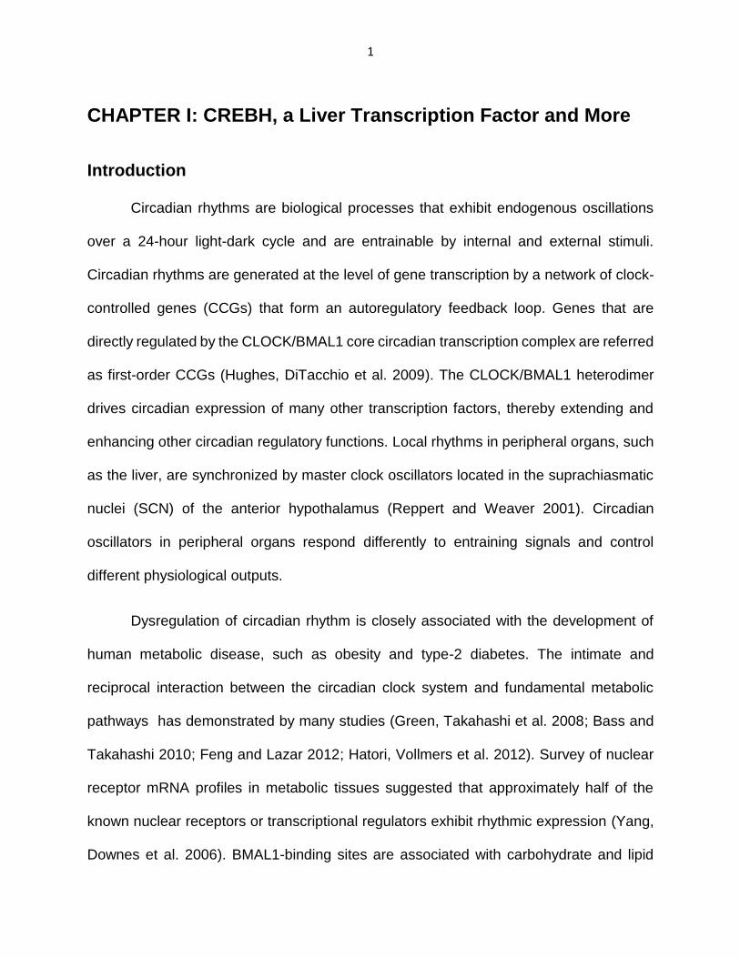
CREBH is a clock-controlled gene (CCG) in mouse liver
Previously, we demonstrated that inflammatory metabolic stimuli induce
expression and/or activation of CREBH in the liver (Zhang, Wang et al. 2012). To test
whether circadian rhythm for CREBH is present in mouse liver, we examined the 24-hour
expression profile of CREBH. Expression levels of the CrebH mRNA in the liver peaked
at circadian time (CT) 44 and reached a trough at CT56 (Figure 4A). Expression of the
CrebH mRNA in mouse liver exhibited a trend to increase during the late phase of daytime
and decrease during the late phase of nighttime. We next examined levels of precursor
and activated forms of CREBH proteins in mouse liver across the day-night cycle.
Production of the activated CREBH protein involves translocation of CREBH precursor
from the ER to the Golgi where it is cleaved by S1P and S2P protease (Zhang, Shen et
al. 2006), and therefore, levels of the activated form of CREBH can be evaluated by
examining cleaved CREBH proteins. Western blot analysis with total liver protein lysates
from animals under the circadian cycle demonstrated that levels of CREBH precursor
protein during the “daytime” phases were higher than those of the “nighttime” phases
(Figure 4B). In contrast, levels of cleaved CREBH protein during the “daytime” phases
were lower than those of the “nighttime” phases. To further delineate circadian rhythmic
levels of CREBH precursor and activated proteins, we performed Western blot analyses
with the membrane and nuclear protein fractions collected from pooled liver tissues of the
mice under the circadian circle. Levels of the membrane-bound CREBH precursor protein
peaked at CT44 and reached a trough at CT56 (Figure 4A, C-D), which is consistent with
the rhythmic mRNA expression profile. In an opposite manner, levels of the activated,

21
nuclear CREBH protein reached a bottom at CT44 and peaked at CT56, 12 hours after
the peak production of the CREBH precursor protein (Figure 4C-D). These data indicates
that the expression and proteolytic activation of CREBH in the liver are both rhythmically
regulated during the day-night cycle although they exhibit distinct circadian rhythmic
patterns.
22
Figure 4. CREBH is a clock-controlled gene (CCG) in mouse liver (A-B) Circadian oscillations of the CrebH mRNA expression (A) and protein levels (B) in male WT mouse liver tissues collected every 4 hours over a 24-hour period in constant darkness (DD), determined by qRT-PCR and Western blot analyses, respectively. Fold changes of mRNA levels are shown by comparing to the nadir mRNA levels at CT56. Each point denotes the mean ± SEM (n=3). Circadian time (CT) is shown in hours. (C) Western blot analysis of levels of membrane-bound CREBH precursor and nuclear proteins in mouse livers over the circadian circle. Cellular membrane and nuclear protein fractions were prepared from pooled liver tissues of WT mice collected every 4 hours over a 24-hour circadian cycle (n=3 mice per time point). (D) Quantification of the CREBH precursor and nuclear protein signals in the mouse livers under the circadian clock. CREBH protein signals in the pooled liver membrane and nuclear protein fractions were determined by Western blot densitometry. Fold changes of the protein levels are shown by comparing the protein signals to that at CT36 (defined as 1). Three mouse liver tissue samples per time point were pooled for the Western blot analysis, as described in panel C.
23
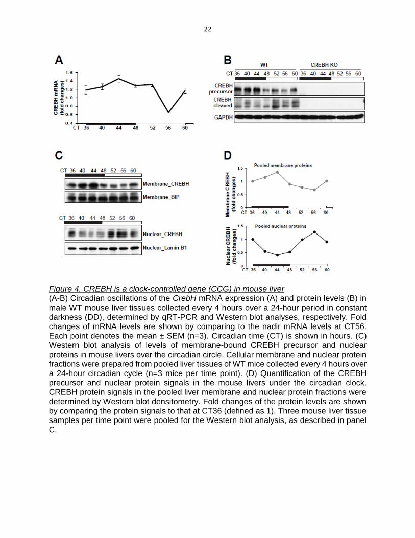
The expression of CREBH is rhythmically regulated by BMAL1 in liver
One of the key identifying features of clock-controlled genes (CCGs) is the
presence within the proximal promoter of one or more canonical E-box elements that bind
the core circadian CLOCK/BMAL1 heterodimer (Kumaki, Ukai-Tadenuma et al. 2008;
Ukai and Ueda 2010). There are several E-box binding elements found in the mouse
CrebH gene promoter regions (Figure 5).
Figure 5. Illustration of E-boxes in mouse CrebH gene promoter region. Non-canonical E-boxes E1 (CACATG) and E2 (CACTGC) locate in the promoter region of mouse CrebH gene.
24
To reveal whether BMAL1 regulates CREBH expression, we first examined
expression levels of the CrebH mRNA in liver-specific Bmal1 conditional knockout (Bmal1
LKO) and control mouse liver samples collected every 6 hours during a 24-hour circadian
period (Molusky, Ma et al. 2012). Expression levels of CrebH mRNA across the day-night
period were decreased in the livers of Bmal1 LKO mice, compared to those in the control
mouse liver (Figure 6A). To determine whether BMAL1 binds to the CrebH gene promoter,
chromatin immunoprecipitation (ChIP)-qPCR analysis was performed on mouse livers
from different circadian phases. ChIP-qPCR analysis demonstrated increased
enrichment of BMAL1 in the CrebH gene E-box-containing promoter region at CT8, a time
point when levels of the CrebH mRNA reached a nadir and began a sharp increase
(Figure 4A, 6B-C).

25
Figure 6. CrebH is directly regulated by BMAL1 in mouse liver (A) Expression of the CrebH mRNA in the livers of Bmal1 LKO and flox/flox control mice during different circadian phases. The liver samples from Bmal1 LKO and flox/flox control mice were collected every 6 hours over a 24-hour circadian period. The mean expression values were obtained with pooled liver cDNAs from 3-5 mice per time point per genotype as previously described (n= 3 experimental repeats) (Molusky, Ma et al. 2012). (B-C) ChIP analysis of the enrichment of BMAL1 in the CrebH gene promoter in mouse liver under different circadian phases. Presence of the endogenous BMAL1 to the E-boxes (E1-E2)-containing promoter region of the CrebH gene was quantified by ChIP-qPCR (F) in the WT mouse livers collected from CT8 and CT20 (n=3). A non-specific region of the CrebH gene distal from the promoter and a housekeeping gene Rplp0 promoter were amplified used as negative controls of ChIP assays. Quantification of BMAL1 enrichment in the CrebH gene promoter at different circadian phases was determined by comparing ChIP-qPCR signals from the samples pulled down by the anti-BMAL1 antibody to that pulled down by a rabbit anti-HA antibody. Each bar donates mean ± SEM (n=3 mice per time point). * p< 0.05 (CT8 vs CT20). The enrichment of BMAL1 in the CrebH gene promoter was confirmed by ChIP-PCR (C) utilizing the same templates and primers used for the ChIP-qPCR analysis.

26
Interestingly, this regulatory pattern is distinct from that of typical BMAL1-regulated
circadian genes whose expression levels are usually increased in few hours after BMAL1
enrichment in the promoter regions. One possible explanation is that other metabolic
trans-activators may also be involved in the transcriptional activation of the CrebH gene
upon energy fluctuations. This is supported by the observation that the diurnal expression
profile of the Bmal1 mRNA was distinguished from that of CrebH in the livers of wild-type
mice (Figure 7). Under metabolic stress, such as fasting, the diurnal expression profile of
the CrebH mRNA, but not the Bmal1 mRNA, was significantly altered (Figure 7A-B),
indicating that CREBH is not a typical CCG. Additionally, we examined expression and
activation of CREBH in livers of Bmal1 LKO and control mice under normal feeding
conditions or after a 24-hour period of fasting (Molusky, Ma et al. 2012). Levels of the
CrebH mRNA were significantly reduced in Bmal1 LKO livers under both fasting and
feeding conditions (Figure 7C). Immunoblotting analysis shown a slight decrease in the
precursor form of CREBH protein in the Bmal1 LKO liver, compared to the control liver,
under both feeding and fasting conditions (Figure 7D). The cleaved/activated CREBH
protein was however diminished in the livers of Bmal1 LKO mice under the feeding, but
not the fasting condition. These results confirm the regulation of CREBH by BMAL1 under
the normal physiological conditions and suggest that additional regulatory mechanisms
are also contributing to CREBH expression and activation.

27
Figure 7. CREBH is not a typical CCG. (A-B) Diurnal expression profiles of the Bmal1 and CrebH mRNAs in the wild-type mice under feeding or fasting conditions. 3-month old wild-type mice were subjected to fasting or feeding for 6, 12, or 24 hours. The experiment was started at 6 pm. The groups of animals were euthanized to collect liver samples at 12 am, 6 am, and 6 pm, respectively. Expression values of mRNAs were determined by qRT-PCR and normalized to the β-actin mRNA levels. Fold changes of mRNA levels are determined by comparing the expression values to that of one of the liver samples collected at 12 pm under the feeding condition (n=3 mice per time point under the fasting condition or 2 mice per time point under the feeding condition). (C-D) Levels of the CrebH mRNA (C) and protein (D) in the livers of Bmal1 LKO and flox/flox control mice under the feeding condition or after 16-hour overnight fasting. The levels of the CrebH mRNA were determined by qRT-PCR analysis, and the levels of the CREBH protein were determined by Western blot analysis. In A, expression values were normalized to the Arbp mRNA levels. Fold changes of mRNA levels are shown by comparing to that of one of the control mice under the feeding condition. Each bar donates mean ± SEM (n=4 mice per group).

28
CHAPTER III: CREBH Functions under Circadian Rhythm
Summary
The disruption of circadian rhythm is associated with the pathogenesis of hepatic
and gastrointestinal metabolic syndromes, such as non-alcoholic fatty liver diseases
(NAFLD) (Hoogerwerf 2009). CREBH transcriptionally regulated many hepatic metabolic
enzymes involved in lipogenesis, FA oxidation, lipolysis, and gluconeogenesis (Figure 3).
There are significant rhythmic patterns in the expression and activation of CREBH (Figure
4). These observations enable us to hypothesize that CREBH may function as an organ-
specific clock-controlled transcription factor that coordinates different pathways under
circadian rhythm to maintain hepatic metabolic homeostasis. By using circadian animal
models, we found that CREBH is required to keep circadian profiles of blood triglycerides,
fatty acids, and glucose as well as hepatic glycogen storage. Intriguingly, the expression
levels and amplitudes of the key genes regulated by CREBH are involved in bi-directional
metabolic pathways of both energy utilization and storage. CREBH deficiency leads to
increased metabolic rates, hyper-locomotion, and phase-shifted feeding behavior in mice.
These findings are significant and innovative because they indicate that the liver
local rhythm is critical for the maintenance of hepatic lipid and glucose homeostasis.
CREBH may sense the liver local clock and mediate the transcription of different
metabolic pathways, such as lipolysis, lipogenesis, FA oxidation, gluconeogenesis, and
glycogenesis in the different circadian phases. The new revealed functions of CREBH
result in a paradigm shift regarding our understanding of the molecular basis of liver

29
circadian rhythm, hepatic lipid/glucose homeostasis, and the pathological progression of
metabolic syndromes.

30
Materials and Methods
Rhythmic lipid and glucose profile analyses and food intake measurement
To profile circulating, approximate 20 μL blood serum per mouse per time point
was collected every 6 hours for 48 hours in constant darkness from the tail vein using
20μL K+ EDTA-containing microcapillary tubes. Blood serum TG and FFA were
measured by colorimetric assays (BioAssay Systems, Hayward, CA). To quantify hepatic
TG or glycogen, liver tissues from similar lobe regions of CREBH-null and WT control
mice under the circadian clock were collected and subjected to measurements of TG and
glycogen using commercial enzymatic kits following the manufacturer’s instructions
(BioAssay Systems, Hayward, CA). Levels of hepatic TG or glycogen were presented
after normalization to liver mass. The amount of animal chow left in the individual mouse
cages was carefully measured at each time point, and serial subtraction was calculated
for the measurement of food intake. Levels of blood glucose of the mice under constant
darkness were measured every 6 hours for 36 hours with an OneTouch Ultra Blood
Glucose Meter (LifeScan, Milpitas, CA).
Histological staining and quantitative analysis of hepatic glycogen
Periodic-acid staining of hepatic glycogen was performed according to the
standard protocol (Zhang, Wang et al. 2012; Zheng, Xu et al. 2013). Briefly, tissue
samples were collected from similar liver lobe regions of CREBH-null and WT control
mice under the circadian clock and then fixed in 10% formalin. Formalin-fixed, paraffin-
embedded liver tissue was sectioned on a cryostat, and sections were deparaffinized, re-
hydrated, and oxidized in 0.5% periodic acid solution for 5 minutes. The oxidized tissue
sections were incubated in Schiff’s reagent (Sigma) for 15 minutes. Biochemical

31
quantification of hepatic glycogen in the liver tissues of CREBH-null and WT control mice
under the circadian clock was performed using a commercial enzymatic kit (BioAssay
Systems, Hayward, CA) (Zhang, Wang et al. 2012; Zheng, Xu et al. 2013). Approximately
40 mg of liver tissue from similar lobe regions of CREBH-null and control mice were
homogenized in ice-cold citrate buffer (0.1M, pH 4.2). Homogenates were immediately
subjected to glycogen measurement using the glycogen assay kit following the
manufacturer’s instruction. Levels of hepatic glycogen were presented after normalization
to liver mass.
Locomotor activity
After 2 weeks on LD cycles, mice were released into DD for an additional 30 days
as previously described (Liu, Li et al. 2007; Siepka, Yoo et al. 2007). To collect locomotion
variable during 44 days in total, individual mice were housed in chambers surrounded by
an infrared photocell array interfaced with a computer running VersaMax/VersaDat
programs (AccuScan Instruments, Columbus, OH). Measures included distance travelled
(in centimeters) and stereotypy count (number of beam breaks at the same photocell
array) every 6 minutes over the 44 days (Bishop and Walker 2004). Activity level in certain
periods was calculated by averaging the total distance travelled per 6 minutes in
centimeters or the stereotypic movement counting of each mouse in the specific circadian
time period, as indicated in Figure 6, over 14 consecutive days during the LD cycles. The
period of rhythmic activities was calculated by the onset of major activities of each mouse
from two successive days over 30 days in DD.
Indirect calorimetry
Each mouse was monitored individually in the computer-controlled OxyScan open
circuit indirect calorimetry system (AccuScan Instruments, Columbus, OH) (Bishop and
32
Walker 2004) with free access to food and water. Oxygen consumption (VO2) and carbon
dioxide production (VCO2) were measured for 48 hours. Gas analyzers were calibrated
to room air drawn through each chamber at a rate of 0.5 L/minute.

33
Results
CREBH regulates circadian rhythmic levels of TG and FA by activating the genes
encoding functions in lipolysis, FA oxidation, and lipogenesis.
We recently demonstrated that CREBH is a key regulator of energy homeostasis
under metabolic stress (Zhang, Wang et al. 2012). To elucidate whether CREBH
regulates energy homeostasis under the day-night cycle, we characterized rhythmic
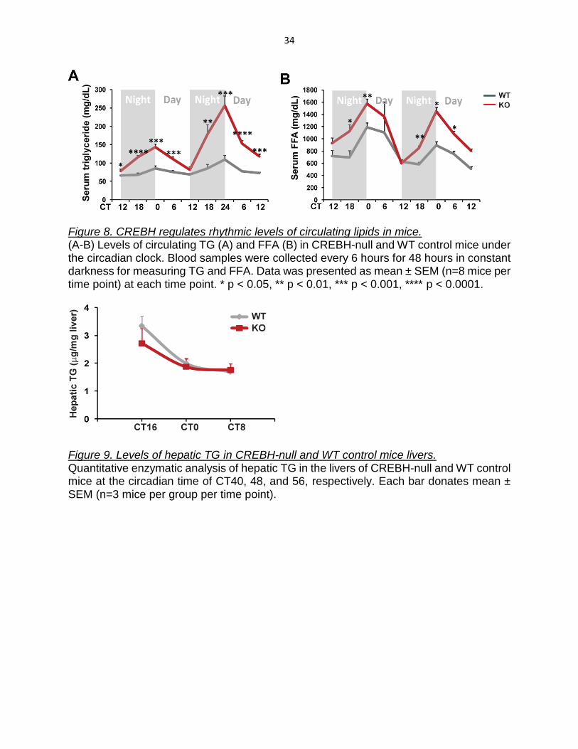
profiles of circulating lipids in CREBH-null and WT control mice. Compared to the WT
control mice, CREBH-null mice exhibited significantly higher levels of serum TG and FA
over a 48-hour period of constant darkness (Figure 8A-B). Hepatic TG levels in CREBH-
null mice were insignificantly lowered, compared to that in the control mice, at the night
time (CT40) when mice usually take most of their meals of the day (Figure 9). These
observations were consistent with the established roles of CREBH in TG lipolysis, FA
oxidation, and lipogenesis upon fasting or atherogenic high-fat feeding, as we previously
described (Zhang, Wang et al. 2012; Kim, Mendez et al. 2014).
34
Figure 8. CREBH regulates rhythmic levels of circulating lipids in mice. (A-B) Levels of circulating TG (A) and FFA (B) in CREBH-null and WT control mice under the circadian clock. Blood samples were collected every 6 hours for 48 hours in constant darkness for measuring TG and FFA. Data was presented as mean ± SEM (n=8 mice per time point) at each time point. * p < 0.05, ** p < 0.01, *** p < 0.001, **** p < 0.0001.
Figure 9. Levels of hepatic TG in CREBH-null and WT control mice livers. Quantitative enzymatic analysis of hepatic TG in the livers of CREBH-null and WT control mice at the circadian time of CT40, 48, and 56, respectively. Each bar donates mean ± SEM (n=3 mice per group per time point).

35
To check whether CREBH rhythmically regulates expression of the genes involved
in lipid metabolism in the liver, we determined rhythmic expression profiles of the genes
encoding key enzymes or regulators in lipolysis, FA oxidation, and lipogenesis in CREBH-
null and WT control mice under the endogenous circadian clock. Quantitative real-time
PCR (qRT-PCR) analysis indicated that rhythmic expression levels and amplitudes of the
following genes were altered in CREBH-null mice (Figure 10): 1) the gene encoding the
key enzyme in lipolysis, apolipoprotein C-II (ApoC2); 2) the genes encoding the key
enzymes or regulators in FA oxidation, including carnitine palmitoyltransferase 1A
(CPT1α), 3-hydroxybutyrate dehydrogenase 1 (BDH1), and FGF21; and 3) the genes
encoding the key enzymes in lipogenesis, including fatty acid desaturase 2 (FADS2) and
Acetyl-CoA Carboxylase 1 (ACC1). Consistent with the mRNA expression profiles,
protein levels of CPT1α, BDH1, FADS2, and ACC1 were decreased in the livers of
CREBH-null mice (Figure 11). Additionally, rhythmic expression levels of other key
metabolic genes involved in lipolysis, FA oxidation, and lipogenesis, including Dhcr24,
Lcat, Acot4, Hmgcl, Dgat2, and Elvol6, only insignificantly altered in the CREBH-null livers
(Figure 12).

36
Figure 10. CREBH regulates rhythmic expression of the genes involved in lipolysis, FA oxidation, and lipogenesis in mice under the circadian clock. Rhythmic expression levels of the CREBH-target genes involved in TG lipolysis, FA oxidation, and lipogenesis, including ApoC2, Bdh1, Cpt1a, Fgf21, Fads2, and Acc1 in CREBH-null and WT control mouse livers. Expression levels of mRNAs were determined by qRT-PCR. Fold changes of mRNA levels are shown by comparing to that of one of the wild-type control mice at the starting circadian time point. Each bar denotes mean ± SEM (n = 3-5 mice per time point).

37
Figure 11. Rhythmic protein levels of the key metabolic enzymes or regulators in the livers of CREBH-null and WT control mice. Rhythmic protein levels of the CREBH-target genes encoding PCK1, G6PC, FADS2, CPT1α, BDH1, and ACC1 in the livers of CREBH-null and WT control mice. The liver tissue samples from CREBH-null and WT control mice were collected every 4 hours in a 24-hour circadian period. Pooled liver protein lysates from 3-5 mice per genotype group per time point were used for the Western blot analyses. Levels of GAPDH were determined as loading controls.

38
Figure 12. Insignificant alterations of metabolic gene rhythmic expressions in the livers of CREBH-null and WT control mice. (B) Expression profiles of the genes encoding key enzymes involved in lipolysis, FA oxidation, and lipogenesis, including 24-Dehydrocholesterol Reductase (Dhcr24), Lecithin-Cholesterol Acyltransferase (Lcat), Acyl-CoA Thioesterase 4 (Acot4), 3-hydroxymethyl-3-methylglutaryl-CoA lyase (Hmgcl), diacylglycerol O-acyltransferase 2 (Dgat2), and ELOVL fatty acid elongase 6 (Elovl6), in the livers of CREBH-null and WT control mice under circadian clock. The liver samples from CREBH-null and WT control mice were collected every 4 hours over a 24-hour period. These RNAs were subjected to quantitative real-time RT-PCR analysis. Expression values were normalized to the Arbp mRNA levels. Fold changes of mRNA levels are shown by comparing to that of one of the WT control mice at the starting circadian time point. Asterisks indicate significant differences (* p < 0.05, ** p < 0.01) between WT and CREBH-null mice by post-hoc analyses followed by two-way ANOVA. Data represent mean ± SEM (n=3-5 mice per group per time point).

39
To determine whether CREBH directly regulates its target genes under the
circadian cycle, we performed ChIP-qPCR analysis to determine CREBH enrichment in
the promoter regions of metabolic genes whose rhythmic expression profiles were altered
in CREBH-null mouse livers. ChIP-qPCR analyses with WT mouse livers collected at
different circadian phases indicated that CREBH binds in a circadian phase-dependent
manner to the promoters of ApoC2, Bdh1, Cpt1a, Fgf21, Fads2, or Acc1 genes that
possess one or multiple CRE-binding elements (Figure 13, Table 3). Increased
enrichment of CREBH in the ApoC2 gene promoter was detectable at CT40 and peaked
at CT52, which is consistent with the rhythmic expression profile of the ApoC2 mRNA in
the liver. Similarly, consistent with the mRNA expression profiles, the enrichments of
CREBH in the Fads2 and Acc1 gene promoter reached peak levels at CT52, CT56, and
CT40, respectively (Figure 13). Taken together, these results indicate that CREBH
activates expression of genes involved in bi-directional metabolic pathways of both
energy utilization (lipolysis and FA oxidation) and storage (lipogenesis) depending upon
the circadian cycle.

40
Figure 13. CREBH regulates rhythmic levels of lipids and expression of the genes involved in lipolysis, FA oxidation, and lipogenesis in mice under the circadian clock. CREBH enrichment in the CREBH-target gene promoters in the WT mouse livers under different circadian phases determined by ChIP-qPCR. CREBH-null liver nuclei were used as negative control for the endogenous CREBH ChIP assays. Quantification of CREBH enrichment in the gene promoters at different circadian phases was determined by comparing ChIP-qPCR signals from the samples pulled down by the anti-CREBH antibody to that pulled down by a rabbit anti-IgG antibody. Each bar donates mean ± SEM (n=3 mice per time point).

41
Table 3. CRE binding motifs in the promoter regions of mouse genes.
Promoter sequences Nucleotide (nt) regions
Apoc2 promoter TGGCCTCTGACTGTCACTGT nt -117 to nt -113
Acc1 promoter
CTAACGCTGACCTTCTTTAC
CTTTCTCATGAACTTTATTT
nt -315 to nt -310
nt -271 to nt -265
Fgf21promoter
CCACTCCTGACGCGTGATAT nt -63 to nt -67
Bdh1 promoter
GTGAGGTGACCAATCCCCCT nt -452 to nt-434
Cpt1a promoter TCATTCTCTGATGTTAGACAAGC
TTCCTTACTGACCTCCTCCCCGCA
nt -568 to nt -562
nt -245 to nt -240
Fads2 promoter
AGGTCAGACACGTCGCCGACCG nt -599 to nt -594
Gys2 promoter
GTTGTACACTGACAAATACAGA
CATAATACTTGACATTTAAAAT
GATAGGGATTGACAATCAACCA
nt -591 to nt -587
nt -437 to nt -433
nt -375 to nt -371
Ppara promoter
ACAGGGGTGACGGGGGC nt -323 to nt -319
Cebpβ promoter
GGGCGGGCTGGCGTCACCCGC
ACCGCAGT
CGGGCAATGACGCGCACCGA
CCCAGCGTGACGCAGCCCGT
nt -344 to nt -337
nt -206 to nt -202
nt -160 to nt -156
Lxra promoter
GGAACGCTGACTCTGGAGGCT
GTGGGGGTGACTGAGAAGCAG
nt -184 to nt -180
nt -151 to nt -147
E4bp4 promoter CCGCCGCCCGTCACGGCGGGG
GCAGT
nt -160 to nt -156
G6pc promoter
CTGGATTGACCTACAGACTG nt -68 to nt -63
Pck1 promoter
CTTCTCATGACCTTTGGCCG
TGGGAGTGACACCTCACAGC
GGTGTTTTGACAACCAGCAG
nt -450 to nt -445
nt -431 to nt -427
nt -407 to nt -403
The binding motifs are highlighted (red underline). The complementary sequences (blue underline) are presented if the binding motifs locate in the negative strand.

42
CREBH regulates rhythmic hepatic glycogen storage and blood glucose levels by
activating the key genes involved in glycogenesis and gluconeogenesis.
We next examined whether CREBH regulates glucose homeostasis across the
day-night cycle. Periodic acid-Schiff (PAS) staining of hepatic glycogen of CREBH-null
and WT control mice under the endogenous circadian clock indicated that production of
hepatic glycogen in WT mice exhibited a circadian rhythmic pattern, which was increased
from CT40 to CT52 and depleted at CT56 (Figure 14A). In contrast, hepatic glycogen
storage in CREBH-null mice lost its rhythmic pattern, as the distribution and levels of
glycogen in the livers of CREBH-null mice exhibited marginal changes over the circadian
period. This observation was confirmed by quantitative enzymatic assay of hepatic
glycogen levels (Figure 14B). Further, we measured blood glucose levels of CREBH-null
and WT control mice across the day-night cycle. A phase-shifted rhythmic pattern of
serum glucose levels is revealed in CREBH-null mice (Figure 14C), and the blood glucose
levels in CREBH-null mice were lower than those in the control mice during the daytime
period. These phenotypes suggest that CREBH functions as a key regulator of glucose
homeostasis under the circadian control.

43
Figure 14. CREBH regulates rhythmic levels of blood glucose and hepatic glycogen storage in mice under the circadian clock. (A) Periodic-acid Schiff (PAS) staining of hepatic glycogen in the livers of CREBH-null and WT control mice at the circadian time of CT40, 44, 52, and 56, respectively (magnification: 200×). (B) Quantitative enzymatic analysis of hepatic glycogen in the livers of CREBH-null and WT control mice at the circadian time of CT40, 44, 52, and 56, respectively. Each bar donates mean ± SEM (n=3 mice per group per time point). (C) Levels of blood glucose in CREBH-null and WT control mice under the circadian clock. Blood glucose were measured every 6 hours for 36 hours in constant darkness. Data was presented as mean ± SEM (n=8 mice per time point) at each time point.

44
To understand the molecular basis for the altered rhythmic profile of hepatic
glycogen storage and blood glucose in CREBH-null mice, we examined expression of
phosphoenolpyruvate carboxykinase 1 (Pck1), glucose-6-phosphatase (G6pc), and
glycogen synthase 2 (Gys2), the rate limiting enzymes of hepatic gluconeogenesis and
glycogenesis, respectively (Roach, Depaoli-Roach et al. 2012), in CREBH-null and WT
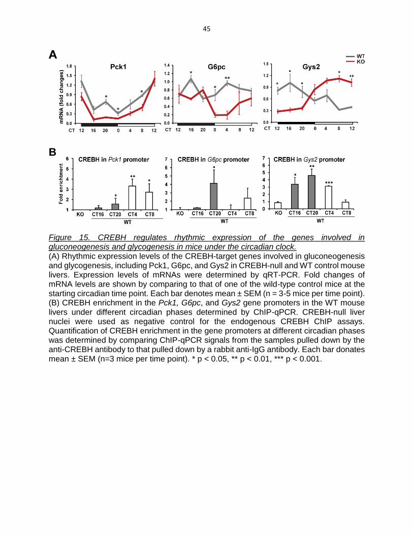
control mouse livers. Compared to WT mice, CREBH-null mice exhibited decreased
rhythmic expression of the Pck1 and G6pc genes in the livers (Figure 15A). Interestingly,
CREBH-null mice displayed an inverse rhythmic expression pattern of the Gys2 gene in
the liver (Figure 15A). Expression levels of the Gys2 mRNA in the WT mice peaked at
CT40 and reached a trough at CT56, whereas Gys2 mRNA expression in the CREBH-
null mice reached its nadir at CT40 and peaked at CT56. These results suggest that
CREBH is required to maintain the normal rhythmic expression of the Gys2 gene in the
liver. In the absence of CREBH, however, an alternative transcriptional mechanism likely
exists to enable expression of the Gys2 gene in a reverse rhythmic pattern. Our results
indicated that CREBH-null mice do not have sufficient Gys2 for hepatic glycogenesis
upon feeding in the night, and therefore, they display lower levels of glycogen during the
night time (Figure 14A-C). During the resting phases, however, CREBH-null mice produce
higher levels of Gys2 (due to inverse rhythmic expression) but lower levels of blood
glucose (due to the defect in gluconeogenesis). The combined effects of the altered Gys2
expression and the repressed gluconeogenesis may explain the loss of rhythm in hepatic
glycogen storage in CREBH-null mice (Figure 14A-C).
45
Figure 15. CREBH regulates rhythmic expression of the genes involved in gluconeogenesis and glycogenesis in mice under the circadian clock. (A) Rhythmic expression levels of the CREBH-target genes involved in gluconeogenesis and glycogenesis, including Pck1, G6pc, and Gys2 in CREBH-null and WT control mouse livers. Expression levels of mRNAs were determined by qRT-PCR. Fold changes of mRNA levels are shown by comparing to that of one of the wild-type control mice at the starting circadian time point. Each bar denotes mean ± SEM (n = 3-5 mice per time point). (B) CREBH enrichment in the Pck1, G6pc, and Gys2 gene promoters in the WT mouse livers under different circadian phases determined by ChIP-qPCR. CREBH-null liver nuclei were used as negative control for the endogenous CREBH ChIP assays. Quantification of CREBH enrichment in the gene promoters at different circadian phases was determined by comparing ChIP-qPCR signals from the samples pulled down by the anti-CREBH antibody to that pulled down by a rabbit anti-IgG antibody. Each bar donates mean ± SEM (n=3 mice per time point). * p < 0.05, ** p < 0.01, *** p < 0.001.

46
Many typical CRE-binding elements present in the promoter regions of the Pck1,
G6pc, and Gys2 genes (Table 3). To evaluate whether CREBH, as a transcriptional
activator, can directly target on the Pck1, G6pc, and Gys2 gene promoters under the
circadian cycles, ChIP-qPCR analysis were performed to quantify enrichment of CREBH
in the gene promoter regions in mouse liver tissues collected at different circadian phases.
ChIP-qPCR analyses indicated that enrichment of CREBH at the Pck1 and G6pc gene
promoters peaked at CT52 and CT44, respectively, consistent with the mRNA expression
profiles (Figure 15B). In the Gys2 gene promoter, enrichment of CREBH was increased
during the circadian night period from CT40 to CT44, a time of the day when mice usually
take their meals (Figure 15B). During the daytime period, enrichment of CREBH in the
Gys2 gene promoter was decreased at CT52 and not detectable at CT56. These results
suggest that CREBH maintains Gys2 rhythmic expression levels by directly regulating
transcription of the Gys2 gene. Additionally, in the absence of CREBH, an alternative
transcriptional mechanism likely exists to enable expression of the Gys2 gene in a reverse
rhythmic pattern. What the other transcription regulators of Gys2 expression and whether
CREBH interacts with these factors are interesting questions to be elucidated in the future.
Therefore, similar to the regulatory roles of CREBH in lipid metabolism, CREBH regulates
rhythmic expression of the key genes in bi-directional glucose metabolic pathways of both
energy utilization (gluconeogenesis) and storage (glycogenesis).

47
CREBH-null mice exhibit hyper-locomotion, increased metabolic rate, and phase-
shifted feeding behavior.
To further evaluate the physiological role of CREBH in the metabolic of whole
animal body, we examined locomotor activity, metabolic rate, and feeding behavior of
CREBH-null and WT control mice across the day-night cycle. We monitored locomotor
activity for 14 days during the normal light-dark cycle followed by 30 days in constant
darkness (Bishop and Walker 2004). Analysis of total distance travelled collected during
the 30 days in constant darkness showed that CREBH-null mice exhibited a 5.17-minute
shorting of the daily locomotor activity (23.86 hours/period), compared to WT control mice
(23.94 hours/period) (Figure 16A-B). Interestingly, there was no difference between
groups in respect to average distance travelled during the 12-hour light period (CT48-60)
(Figure 16A, C). However, CREBH-null mice exhibited significant hyper-locomotion
during the 12-hour dark period (CT36-48). We also monitored stereotypic movements,
which are characterized by small movements without travelling distance, such as
grooming, body shaking, and feeding. CREBH-null mice consistently exhibited increased
stereotypic movements during the second 6 hours of the dark period (CT42-48) (Figure
16A, D).

48
Figure 16. CREBH-null mice display hyper-locomotion and increased stereotypic activities during the night time. (A) Circadian rhythmic profiles of locomotor activities of CREBH-null and WT control mice under the circadian clock. Total distance travelled in centimeter (upper panel) and stereotypic movement (lower panel) of CREBH-null and WT control mice were monitored every 6 minutes under 12-hour/12-hour LD cycles for 14 days and under DD for 30 days (n=6 mice per group). (B) Lengths of circadian periods of CREBH-null and WT control mice calculated based on the circadian locomotor activities during 30 days in DD (n=6 mice per group). (C-D) Average activities of distance travelled (C) and stereotypic movement (D) of the CREBH-null and WT control mice over the circadian periods of CT36-48, CT48-56, or CT54-60 during the first 14 days in LD. Each bar donates mean ± SEM (n=6 mice per time point). ** p < 0.01, *** p < 0.001.
49
To further explore the pathophysiological effects related to the hyper-locomotion
of CREBH-null mice during the late dark phase, we characterized rhythmic feeding
behavior and metabolic rates in CREBH-null and WT control mice. A significant time-shift
in food intake was observed with CREBH-null mice across the 36-hour circadian period
(Figure 17A). Compared to the control mice, CREBH-null mice exhibited approximately a
6-hour delay in taking their biggest meal of the day (peak at CT42-48). The metabolic
rates, as reflected by the rates of oxygen consumption, of CREBH-null mice were
significantly higher than those of WT control mice across the 12-hour dark period, which
is consistent with the hyper-locomotion of CREBH-null mice during the 12-hour dark
period (CT36-48) (Figure 17B, 16C-D). Moreover, a dramatic increase in metabolic rates
was observed in CREBH-null mice during the second 6 hours of the dark period, concord
with the increased stereotypic movements and delayed feeding behavior of CREBH-null
mice during the same phase (Figure 17A-B, 16A, 16D). Additionally, we found that
compared to that of WT control mice, total food intake of CREBH-null mice over 48-hour
circadian period was modestly increased (Figure 17C). As CREBH is required for hepatic
glycogenesis and lipogenesis during the night (Figures 8-15), the impaired energy
catabolism may entrain CREBH-null mice to consume more dietary energy metabolites.
Additionally, high levels of blood TG and FFA may stimulate satiety-related signals that
lead to delayed food intake behavior and hyper-metabolic rates in the CREBH-null mice
during the late night time, an interesting question to be further elucidated in the future.
It is also interesting to discuss why Gys2 expression pattern is inversed, but
glycogen storage is not, in CREBH-null mice liver (Figure 14-15). It's explainable if
combining glucose and food intake data (Figure14-15, 17). In WT mice, upon feeding,

50
Gys2 increased after feeding to store free glucose from blood stream into glycogen in
liver. Meanwhile, CREBH null-mice don’t have enough Gys2 to process glycogenesis,
and therefore, they have lower glycogen during the nighttime. In WT mice, during resting
phase, glycogenesis slows down (low liver glycogen storage), but glycolysis and
gluconeogenesis increases to provide glucose as energy for body philological use.
Meanwhile CREBH null mice exhibited a delay in increasing blood glucose levels (due to
the defect in gluconeogenesis), and high levels of Gys2 which boost hepatic glycogen
levels when hepatic glycogen storage was depleted in the WT control mice.

51
Figure 17. CREBH deficiency leads to phase-shifted feeding behavior and increased metabolic rates. (A) Food intakes of CREBH-null and WT control mice under the circadian clock. Food intakes of individual animals were measured every 6 hours over 36-hour period in DD. Each point donates mean ± SEM (n=5 mice per group per time point). ** p < 0.01. (B) Metabolic rates, represented by oxygen consumption, of CREBH-null and WT control mice under the circadian clock. Oxygen consumption (VO2) normalized by body weight of individual mice was recorded every 10 minutes over a 36-hour period by a computer-controlled OxyScan open circuit indirect calorimetry systems. Average VO2 levels of each mouse over the phases of CT36-48, CT48-54, or CT54-60 were calculated for the statistical analysis (n=4 mice per group). * p < 0.05; ** p < 0.01; ns, non-significant. (C) Accumulative food intakes of CREBH-null and WT control mice over the 48-hour period. (D) Illustration of CREBH working model as a circadian metabolic oscillator.

52
CHAPTER IV: Crosstalk of CREBH and Other Circadian Regulators
Summary
The hypothesis in this dissertation is that CREBH may function as, or interact with,
the local first-order CCG to regulate the synchronization of rhythmic transcription of
hepatic metabolic enzymes. The transcriptional function of D-element binding protein
(DBP), a known local first-order CCG, can be repressed by the interleukin 3-regulated
nuclear factor (NFIL3, also known as E4BP4), a bZIP domain containing rhythmic
transcription factor regulated by the repressor REV-ERBa (Clayton, Kyriacou et al. 2001;
Reppert and Weaver 2002; Ueda, Chen et al. 2002). A recent study showed E4BP4
negatively regulates the rhythmic transcription of FGF21, a metabolic hormone that
regulates lipid and glucose metabolism, and this repressive regulation can be inhibited by
an unknown fasting-inducible factor (Tong, Muchnik et al. 2010). Since CREBH is a
fasting-inducible factor that promotes FGF21 transcription (Lee, Giannikopoulos et al.
2011; Zhang, Wang et al. 2012), we hypothesize that CREBH may bind to E4BP4 and
repress the negative regulation of E4BP4 to up-regulate the gluconeogenesis and TG
lipolysis pathways during the day time under circadian rhythm. To answer the questions,
we used both in vivo and in vitro methods to discover that CREBH regulates, and interacts
with, the circadian transcriptional activators PPARα and C/EBPβ or the repressor E4BP4
to modulate CREBH transcriptional activities.

53
Materials and Methods
Immunoprecipitation (IP)-Western blot analyses
Endogenous protein-protein interactions between CREBH and E4BP4, PPARα, or
CEBPβ in mouse livers across the day-night cycle were determined by IP-Western blot
analysis. Approximate 200 μg of liver protein lysates were incubated with 1μg of a rabbit
anti-mouse CREBH antibody overnight at 4°C, as indicated in Figure 5A. The rabbit
polyclonal CREBH antibody used for pulling down the endogenous CREBH protein was
developed in our laboratory (Kim et al. 2014). Protein complexes were
immunoprecipitated using Dynabeads Protein G (Novex), resolved by SDS-PAGE, and
then transferred to PVDF membrane. The assay was followed by Western blot analysis
with primary antibodies directed against E4BP4, PPARα, or CEBPβ. The protein
interaction signals were visualized by using HRP-conjugated Clean-Blot IP Detection
Reagents (Thermo Scientific), which can eliminate detection-interference from both
heavy-chain and light-chain IgG fragments of the antibodies used for the initial IP assay.
Conjugated HRP was then developed using an enhanced chemiluminescence (ECL)
detection reagent (GE Healthcare).
Luciferase gene expression reporter analysis
To construct the Pck1 gene promoter-driven expression reporter plasmid (pGL3-
Pck1), the 5′-flanking region from −385 nt to -36 nt of the mouse Pck1 gene was
amplified from mouse cDNA by PCR using the forward primer 5 ′ -
ATGGTACCGCAGCCAGCAACATATGAAG-3 ′ and the reverse primer 5 ′ -
ATGAGCTCATAGAAGGGAGGACAGCCCT-3′ . PCR products were digested using

54
KpnI and SacI restriction enzymes and cloned into the same sites of the pGL3-basic
vector (Promega, USA). The identity of the cloned plasmid was verified by sequence
analysis. For the luciferase assay, 1μg of pGL3-Pck1 plasmids and 0.1μg of pGL4.7
plasmids were transiently co-transfected into Hepa1-6 cells using the TransIT-2020
reagent (Mirus Bio, WI). After 24 hour, co-transfected cells were infected with adenovirus
expressing GFP (Ade-GFP), activated CREBH (Ade-CREBH), PPARα (Ade-PPARα),
E4BP4 (Ade-E4BP4), and/or C/EBPβ (Ade-C/EBPβ), as indicated in Figure 5B-C. Cells
were harvested and lysed at 24 hours after transfection. Luciferase activity was measured
using the Dual-Luciferase Assay System (Promega) according to the manufacturer's
instructions. The reporter expression readout was presented by normalizing Firefly
luciferase activities to Renilla luciferase activities (internal control). Each reporter assay
was performed in duplicate.

55
Results
Altered rhythmic expression of core clock genes in CREBH-null mice liver.
It is known that both the activation and function of circadian regulatory proteins are
regulated through intimate and reciprocal interactions with other circadian transcriptional
activators and repressors (Rutter, Reick et al. 2002; Bass and Takahashi 2010). To
understand whether CREBH, as a liver local circadian oscillator, regulates, and/or
interacts with, other circadian regulators, we examined expression of core clock genes in
the livers of CREBH-null and WT control mice. Deletion of CrebH resulted in insignificantly
altered rhythmic expression amplitudes of the genes encoding the core circadian
oscillators including Clock, Dbp, and Rev-erbα in mouse livers (Figure 18). Rhythmic
expression of Hnf4α, a previously identified regulator of CREBH (Luebke-Wheeler, Zhang
et al. 2008), was reduced in CREBH-null mice. Western blot analysis confirmed that
rhythmic expression of the circadian regulators BMAL1 and HNF4α were repressed in
CREBH-null livers (Figure 19).

56
Figure 18. Rhythmic expression levels and amplitudes of core clock genes in the livers of CREBH-null and WT control mice. Expression profiles of the clock genes, including Bmal1, Clock, Reverbα, Per2, Dbp, and Hnf4α, in the livers of CREBH-null and WT control mice under the circadian clock. The liver samples from the CREBH-null and WT control mice were collected every 4 hours over a 24-hour period. Expression values of mRNAs were determined by qRT-PCR and normalized to the Arbp mRNA levels. Fold changes of mRNA levels are shown by comparing to that of one of the wild-type control mice at the starting circadian time (CT) point. Asterisks indicate significant differences (* p < 0.05) between WT and CREBH-null mice by post-hoc analyses followed by two-way ANOVA. Data represent mean ± SEM (n=3 mice per group per time point).

57
Figure 19. Rhythmic regulation of core clock proteins by CREBH in the liver. Rhythmic levels of BMAL1, CLOCK, and HNFα proteins in CREBH-null and WT control mouse livers collected every 4 hours in a 24-hour period. Levels of proteins were determined by Western blot analysis. Pooled liver protein lysates from 3-5 mice per time point per genotype group were used. Levels of β-actin or GAPDH were included as loading controls.

58
CREBH rhythmically regulates circadian transcriptional activators C/EBPβ and
PPARα, and the repressor E4BP4.
To elucidate whether CREBH regulates, and/or interacts with, other circadian
regulators, we examined expression of core clock genes in the livers of CREBH-null and
WT control mice. PPARα is a liver-enriched, clock-regulated nuclear receptor that plays
key roles in regulating lipid metabolism during the starvation phase (Oishi, Shirai et al.
2005). C/EBPβ is a bZIP-containing transcriptional regulator known to rhythmically
regulate autophagy in the liver, facilitating degradation of glycogen and lipid droplets for
body energy (Ma, Panda et al. 2011). Liver X receptor α (LXRα) is a nuclear receptor that
regulates lipogenesis by forming heterodimers with members of the retinoid X receptor
(RXR) family (Willy, Umesono et al. 1995). Gene expression analyses showed that
rhythmic expression levels of the Pparα and Lxrα mRNAs were significantly repressed in
the CREBH-null livers, while rhythmic levels of the C/ebpβ mRNA in the CREBH-null mice
were only marginally changed, compared to those in the control mice (Figure 20A-C). The
mouse Pparα, C/ebpβ, and Lxrα gene promoters possess one or multiple CRE-binding
motifs (Table 3). ChIP-qPCR analysis indicated that in mouse livers CREBH binds in a
day-night dependent manner to the Pparα, C/ebpβ, or Lxrα gene promoters (Figure 20A-
C), suggesting that Pparα, C/ebpβ, and Lxrα are circadian-dependent targets of CREBH.
Moreover, Western blot analysis indicated that rhythmic levels of PPARα, C/EBPβ, and
LXRα proteins were decreased in the CREBH-null livers (Figure 20E). Note that the
discrepancy in C/EBPβ protein and mRNA levels in CREBH-null mice implies alternative
regulation of C/ebpβ gene expression independent of CREBH or potential involvement of
CREBH in C/EBPβ protein stability.

59
Further, we found that CREBH regulates rhythmic expression of E4BP4, a bZIP
domain-containing circadian transcriptional repressor (Clayton, Kyriacou et al. 2001;
Reppert and Weaver 2002; Ueda, Chen et al. 2002). Loss of CREBH resulted in a phase-
inversed expression pattern of hepatic E4bp4 mRNA (Figure 20D). Western blot analysis
showed that expression levels and the oscillation amplitude of E4BP4 protein were
repressed in CREBH-null livers (Figure 20E). ChIP-qPCR analysis of mouse liver
chromatins indicated that CREBH robustly binds to the E4bp4 gene promoter at CT4
(Figure 20D), suggesting that rhythmic expression of E4BP4 is regulated by CREBH in
mouse livers.

60
Figure 20. Rhythmic regulation of circadian transcriptional activators and repressor by CREBH in the liver. (A-C) Rhythmic expression levels and CREBH enrichment in the promoters of the Pparα, C/ebpβ, Lxrα and E4bp4 genes in the livers of CREBH-null and WT control mice under the circadian clock. Fold changes of the mRNA levels were determined by qRT-PCR (upper panel). Each bar denotes mean ± SEM (n = 3 mice per time point). Rhythmic enrichment of endogenous CREBH in the target gene promoters in the WT mouse livers under different circadian phases were determined by ChIP-qPCR (lower panel). Each bar donates mean ± SEM (n=4 mice per time point). * p < 0.05, ** p < 0.01, *** p < 0.001. (D) Rhythmic levels of PPARα, C/EBPβ, E4BP4, and LXRα proteins in CREBH-null and WT control mouse livers collected every 4 hours in a 24-hour period. Levels of proteins were

61
determined by Western blot analysis. Pooled liver protein lysates from 3-5 mice per time point per genotype group were used. Levels of β-actin or GAPDH were included as loading controls.

62
CREBH rhythmically interacts with circadian transcriptional activators and
repressor.
Next, we were interested about whether CREBH interacts with the circadian
transcriptional regulators to modulate its transcriptional activity. CREBH interaction with
PPARα and C/EBPβ were proved by IP-Western blot analyses using mouse livers
collected across the day-night cycle (Figure 21). The interaction between CREBH and
C/EBPβ peaked at CT56, a time period when lipolysis, FA oxidation, and
gluconeogenesis are highly activated upon energy demands. The CREBH-PPARα
interaction was detected from CT44 to CT52 (Figure 21). We were able to detect a robust
interaction between CREBH and the transcriptional repressor E4BP4, which began at
CT36, gradually increased over the night period, and peaked at CT48 (Figure 21).
Interestingly, the phase and intensity of the CREBH-C/EBPβ interaction roughly opposes
that of the CREBH-E4BP4 interaction, implying that C/EBPβ and E4BP4 may compete to
interact with CREBH in a circadian phase-dependent manner.

63
Figure 21. Rhythmic interactions between CREBH, the circadian transcriptional activators PPARα and C/EBPβ, and the repressor E4BP4 in the liver. Interactions between endogenous CREBH and PPARα, C/EBPβ, or E4BP4 in mouse livers under different circadian phases were determined by IP-Western blot analysis. Liver protein lysates pooled from 3 WT mice were pulled down by a rabbit anti-CREBH antibody and then probed with an antibody against PPARα, C/EBPβ, or E4BP4. As a loading control, the liver protein lysates pulled down by the anti-CREBH antibody was probed with the rabbit anti-IgG. Liver protein lysates pooled from WT or CREBH-null mice under the 24-hour circadian phases were included as positive and negative controls, respectively.

64
To explore the functional significance of the interactions between CREBH and
C/EBPβ, PPARα, or E4BP4, we performed reporter analysis with the Pck1 gene promoter,
a common target of CREBH, C/EBPβ, and PPARα (Nizielski, Arizmendi et al. 1996; Juge-
Aubry, Pernin et al. 1997; Lee, Chanda et al. 2010; Peeters and Baes 2010). Indeed, the
promoter region of the mouse Pck1 gene possesses multiple binding motifs for CREBH,
PPARα, C/EBPβ, and E4BP4, respectively (Figure 22). While over-expression of the
active form of CREBH, C/EBPβ, or PPARα alone can significantly increase Pck1 gene
promoter activity, co-expression of CREBH with C/EBPβ or PPARα further augmented
the reporter activity (Figure 23A). In contrast, co-expression of CREBH with E4BP4
significantly decreased Pck1 promoter activity either when compared to expression of
CREBH alone or co-expression of CREBH with GFP. These results suggest that C/EBPβ
and PPARα function as co-activators of CREBH in driving Pck1 gene transcription, while
E4BP4 acts as a repressor of CREBH-dependent Pck1 gene expression. Moreover, we
observed that co-expression of E4BP4 with the combination of CREBH and C/EBPβ or
PPARα repressed expression of the Pck1 gene reporter, compared to co-expression of
CREBH with C/EBPβ or PPARα (Figure 23B), thus supporting the suppressive effect of
E4BP4 on CREBH transcriptional activity through competition with the co-activator
C/EBPβ or PPARα. Given that the rhythmic expression of E4BP4 is decreased in CREBH-
null livers (Figure 21), the repressive effect of E4BP4 on CREBH activity may serve as a
negative feedback regulation of CREBH under the circadian constrain.
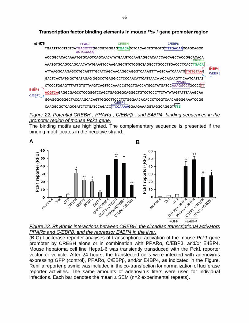
65
Figure 22. Potential CREBH-, PPARα-, C/EBPβ-, and E4BP4- binding sequences in the promoter region of mouse Pck1 gene. The binding motifs are highlighted. The complementary sequence is presented if the binding motif locates in the negative strand.
Figure 23. Rhythmic interactions between CREBH, the circadian transcriptional activators PPARα and C/EBPβ, and the repressor E4BP4 in the liver. (B-C) Luciferase reporter analyses of transcriptional activation of the mouse Pck1 gene promoter by CREBH alone or in combination with PPARα, C/EBPβ, and/or E4BP4. Mouse hepatoma cell line Hepa1-6 was transiently transduced with the Pck1 reporter vector or vehicle. After 24 hours, the transfected cells were infected with adenovirus expressing GFP (control), PPARα, C/EBPβ, and/or E4BP4, as indicated in the Figure. Renilla reporter plasmid was included in the co-transfection for normalization of luciferase reporter activities. The same amounts of adenovirus titers were used for individual infections. Each bar denotes the mean ± SEM (n=2 experimental repeats).

66
CHAPTER V: Conclusions and Significance
In this study, we demonstrated that CREBH functions as a circadian regulator of
lipid and glucose metabolism in the liver (Figure 24). Our major findings include: 1)
CREBH is a BMAL1-regulated diurnal regulator in the liver, and expression and
proteolytic activation of CREBH are both rhythmically regulated during the circadian cycle;
2) CREBH rhythmically regulates expression of the genes encoding key enzymes or
regulators of energy utilization processes, including lipolysis, FA oxidation, and
gluconeogenesis, and of energy storage processes, including lipogenesis and
glycogenesis; 3) CREBH controls rhythmic levels of circulating TG, FFA, and glucose as
well as hepatic glycogens; 4) CREBH rhythmically interacts with the circadian
transcriptional activators C/EBPβ and PPARα and the repressor E4BP4 to modulate
CREBH transcriptional activities; and 5) disruption of CREBH leads to phase-shifted
feeding behavior, increased metabolic rates, and hyper-locomotion during the dark period.
These findings demonstrate that CREBH is not only a key metabolic regulator but also a
liver circadian oscillator, and therefore, plays key roles in integrating energy metabolism
with circadian rhythm (Figure 24). The functional activity of CREBH as a peripheral
circadian metabolic regulator has profound impact on the measures of whole body
physiology, such as feeding, metabolic rate, and locomotor activity. Modulation of CREBH
activity may therefore have important implications in the prevention and treatment of
metabolic disorders.

67
Figure 24. Illustration of CREBH working model as a circadian metabolic oscillator.
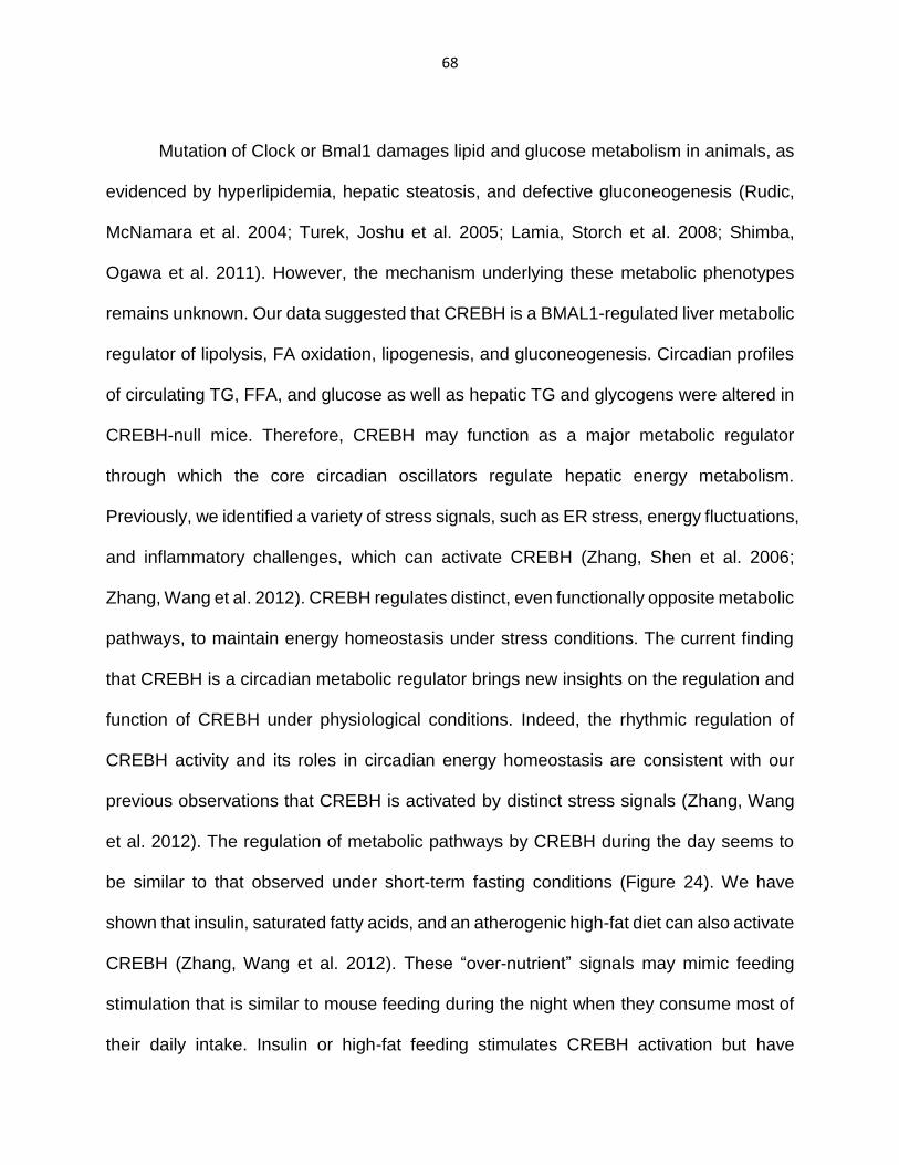
68
Mutation of Clock or Bmal1 damages lipid and glucose metabolism in animals, as
evidenced by hyperlipidemia, hepatic steatosis, and defective gluconeogenesis (Rudic,
McNamara et al. 2004; Turek, Joshu et al. 2005; Lamia, Storch et al. 2008; Shimba,
Ogawa et al. 2011). However, the mechanism underlying these metabolic phenotypes
remains unknown. Our data suggested that CREBH is a BMAL1-regulated liver metabolic
regulator of lipolysis, FA oxidation, lipogenesis, and gluconeogenesis. Circadian profiles
of circulating TG, FFA, and glucose as well as hepatic TG and glycogens were altered in
CREBH-null mice. Therefore, CREBH may function as a major metabolic regulator
through which the core circadian oscillators regulate hepatic energy metabolism.
Previously, we identified a variety of stress signals, such as ER stress, energy fluctuations,
and inflammatory challenges, which can activate CREBH (Zhang, Shen et al. 2006;
Zhang, Wang et al. 2012). CREBH regulates distinct, even functionally opposite metabolic
pathways, to maintain energy homeostasis under stress conditions. The current finding
that CREBH is a circadian metabolic regulator brings new insights on the regulation and
function of CREBH under physiological conditions. Indeed, the rhythmic regulation of
CREBH activity and its roles in circadian energy homeostasis are consistent with our
previous observations that CREBH is activated by distinct stress signals (Zhang, Wang
et al. 2012). The regulation of metabolic pathways by CREBH during the day seems to
be similar to that observed under short-term fasting conditions (Figure 24). We have
shown that insulin, saturated fatty acids, and an atherogenic high-fat diet can also activate
CREBH (Zhang, Wang et al. 2012). These “over-nutrient” signals may mimic feeding
stimulation that is similar to mouse feeding during the night when they consume most of
their daily intake. Insulin or high-fat feeding stimulates CREBH activation but have

69
marginal effects on CrebH gene expression (Zhang, Wang et al. 2012). This is consistent
with the CREBH expression and activation profiles and the regulatory roles of CREBH in
hepatic lipogenesis and glycogenesis during the night.
In this study, we provide expression levels and amplitudes for the CREBH-target
genes across the circadian cycle. Depending on the circadian phase and energy
demands, CREBH regulates expression of different target genes in lipid or glucose
metabolism. During the day time, CREBH regulates expression of the genes involved in
lipolysis, FA oxidation, and gluconeogenesis, including ApoC2, Badh1, Cpt1α, Fgf21,
Pck1, C/ebpβ, and Pparα (Figures 8-15). During the night, CREBH regulates expression
of the genes involved in lipogenesis and glycogenesis, including Acc1, Fads2, Lxrα, and
Gys2. Indeed, the functions of CREBH in mouse circadian metabolism are consistent with
the rhythmic activation of the CREBH protein, in which the levels of the activated, nuclear
CREBH protein peaked at the daytime (CT56) but reached a trough at the nighttime
period (CT44) (Figure 4C-D). Interestingly, the circadian expression pattern of the CrebH
mRNA was opposite to the rhythmic activation of the CREBH protein in the liver (Figure
4A-D). This observation was in line with recent rhythmic proteome study showing that
approximately one-half of rhythmic proteins are under significant translational or
posttranslational diurnal controls and have no corresponding rhythmic mRNAs
(Mauvoisin, Wang et al. 2014).
CREBH has reciprocal interactions with the circadian transcriptional regulators
PPARα and C/EBPβ as well as the circadian repressor E4BP4 (Figures 18-23). CREBH
regulates and interacts with PPARα or C/EBPβ to enhance CREBH transcriptional activity,
which oscillates in-phase with expression of the CREBH-target genes involved in lipolysis,

70
FA oxidation, and gluconeogenesis. On the other hand, CREBH interacts with E4BP4 to
repress CREBH transcriptional activity during the night-today transition period.
Interestingly, the phase of CREBH-C/EBPβ interaction is complimentary to that of
CREBH-E4BP4 interaction, suggesting that C/EBPβ and E4BP4 may compete to interact
with CREBH and thereby modulate CREBH activities during different circadian phases.
As a co-activator of CREBH, PPARα interacts with CREBH in the circadian phase that
partially overlaps with the C/EBPβ-CREBH interaction (Figure 21). It is possible that the
interaction between PPARα-CREBH may represent an enhancing mechanism that
facilitates CREBH peak activity during the phases of high-energy demands.
Peripheral clocks, such as in liver, are synchronized with the central clock located
in the SCN through a complicated regulatory network of neuronal, hormonal, behavioral,
and environmental signals (Ueda, Chen et al. 2002). The core circadian oscillators
regulate CREBH expression and activation through two layers: 1) BMAL1/CLOCK
regulates transcription of CrebH; and 2) E4BP4 or C/EBPβ interacts with activated form
of CREBH protein to exert suppressive or synergizing effect on CREBH activity. On the
other hand, CREBH regulates mRNA and/or protein levels of the core circadian regulators,
including E4BP4, C/EBPβ, PPARα, BMAL1, CLOCK, and HNF4α (Figure 18-20). The
reciprocal regulation between CREBH and the key circadian regulators may provide an
avenue by which local and central circadian regulators are integrated to influence whole
body physiology. Because expression of CrebH in mouse is highly restricted to liver and
small intestine (Luebke-Wheeler, Zhang et al. 2008) (Figure 25), the CREBH-null mouse
is an excellent model to study peripheral clock-originated feedback to the master clock.
Recent studies have found disturbance of circadian rhythm can lead to depression and

71
anxiety disorders (Salgado-Delgado, Tapia Osorio et al. 2011; Keers, Pedroso et al.
2012). CREBH-null mice exhibited increased stereotypic movements during the late night
period (Figure 16), which may partially reflect depressive activities associated with
metabolic and psychiatric disorders. Whether CREBH, as a liver circadian metabolic
oscillator, is crucial to maintaining metabolic and thereby psychiatric wellbeing is an
interesting question to be investigated in the future.
Figure 25. Tissue-specific expression of the mouse CrebH mRNA. CrebH mRNA expression profiles in mouse various organs were determined by qPCR. Each bar donate mean ± SEM (n=3 mice). Selection of brain regions was based on the brain regions described in Allen Brain Altas (http://www.brain-map.org/) (cortex, hippocampus, cerebellum, and olfactory bulb) and anterior hypothalamus where suprachiasmatic nuclei (SCN) locates. Female WT mice were deeply anesthetized with isoflurane followed by rapid decapitation. Brain was rapidly removed and placed on ice, and the specific brain regions were dissected under microscopic control. For dissecting anterior hypothalamus and motor cortex, section was made on ice-cold mouse brain matrix between posterior cut at Bregama-2.92mm and anterior cut at Bregama-0.22mm by an experienced neurologist.

72
REFERENCES
Akhtar, R. A., A. B. Reddy, et al. (2002). "Circadian cycling of the mouse liver transcriptome, as revealed by cDNA microarray, is driven by the suprachiasmatic nucleus." Curr Biol 12(7): 540-550.
Asselah, T., I. Bieche, et al. (2010). "In vivo hepatic endoplasmic reticulum stress in patients with chronic hepatitis C." J Pathol 221(3): 264-274.
Bailey, D., C. Barreca, et al. (2007). "Trafficking of the bZIP transmembrane transcription factor CREB-H into alternate pathways of ERAD and stress-regulated intramembrane proteolysis." Traffic 8(12): 1796-1814.
Bass, J. and J. S. Takahashi (2010). "Circadian integration of metabolism and energetics." Science 330(6009): 1349-1354.
Bishop, C. and P. D. Walker (2004). "Intranigral antagonism of neurokinin 1 and 3 receptors reduces intrastriatal dopamine D1 receptor-stimulated locomotion in the rat." Brain Res 1023(1): 126-133.
Brown, M. S., J. Ye, et al. (2000). "Regulated intramembrane proteolysis: a control mechanism conserved from bacteria to humans." Cell 100(4): 391-398.
Bugge, A., D. Feng, et al. (2012). "Rev-erbalpha and Rev-erbbeta coordinately protect the circadian clock and normal metabolic function." Genes Dev 26(7): 657-667.
Canaple, L., J. Rambaud, et al. (2006). "Reciprocal regulation of brain and muscle Arnt-like protein 1 and peroxisome proliferator-activated receptor alpha defines a novel positive feedback loop in the rodent liver circadian clock." Mol Endocrinol 20(8): 1715-1727.
Chan, C. P., K. H. Kok, et al. (2011). "CREB3 subfamily transcription factors are not created equal: Recent insights from global analyses and animal models." Cell Biosci 1(1): 6.
Chan, C. P., T. Y. Mak, et al. (2010). "N-linked glycosylation is required for optimal proteolytic activation of membrane-bound transcription factor CREB-H." J Cell Sci 123(Pt 9): 1438-1448.
Chanda, D., D. K. Kim, et al. (2011). "Cannabinoid receptor type 1 (CB1R) signaling regulates hepatic gluconeogenesis via induction of endoplasmic reticulum-bound transcription factor cAMP-responsive element-binding protein H (CREBH) in primary hepatocytes." J Biol Chem 286(32): 27971-27979.
Clayton, J. D., C. P. Kyriacou, et al. (2001). "Keeping time with the human genome." Nature 409(6822): 829-831.
Cretenet, G., M. Le Clech, et al. (2010). "Circadian clock-coordinated 12 Hr period rhythmic activation of the IRE1alpha pathway controls lipid metabolism in mouse liver." Cell Metab 11(1): 47-57.
Doherty, C. J. and S. A. Kay (2010). "Circadian control of global gene expression patterns." Annu Rev Genet 44: 419-444.
Duffield, G. E. (2003). "DNA microarray analyses of circadian timing: the genomic basis of biological time." J Neuroendocrinol 15(10): 991-1002.
Fabbrini, E., S. Sullivan, et al. (2010). "Obesity and nonalcoholic fatty liver disease: biochemical, metabolic, and clinical implications." Hepatology 51(2): 679-689.
Feng, D. and M. A. Lazar (2012). "Clocks, metabolism, and the epigenome." Mol Cell 47(2): 158-167. Feng, D., T. Liu, et al. (2011). "A circadian rhythm orchestrated by histone deacetylase 3 controls hepatic
lipid metabolism." Science 331(6022): 1315-1319. Gentile, C. L., D. Wang, et al. (2010). "Fatty acids regulate CREBh via transcriptional mechanisms that are
dependent on proteasome activity and insulin." Mol Cell Biochem 344(1-2): 99-107. Green, C. B., J. S. Takahashi, et al. (2008). "The meter of metabolism." Cell 134(5): 728-742.

73
Hatori, M., C. Vollmers, et al. (2012). "Time-restricted feeding without reducing caloric intake prevents metabolic diseases in mice fed a high-fat diet." Cell Metab 15(6): 848-860.
Hoogerwerf, W. A. (2009). "Role of biological rhythms in gastrointestinal health and disease." Rev Endocr Metab Disord 10(4): 293-300.
Hughes, M. E., L. DiTacchio, et al. (2009). "Harmonics of circadian gene transcription in mammals." PLoS Genet 5(4): e1000442.
Inoue, I., Y. Shinoda, et al. (2005). "CLOCK/BMAL1 is involved in lipid metabolism via transactivation of the peroxisome proliferator-activated receptor (PPAR) response element." J Atheroscler Thromb 12(3): 169-174.
Johansen, C. T. and R. A. Hegele (2012). "The complex genetic basis of plasma triglycerides." Curr Atheroscler Rep 14(3): 227-234.
Juge-Aubry, C., A. Pernin, et al. (1997). "DNA binding properties of peroxisome proliferator-activated receptor subtypes on various natural peroxisome proliferator response elements. Importance of the 5'-flanking region." J Biol Chem 272(40): 25252-25259.
Kapatos, G., P. Vunnava, et al. (2007). "Protein kinase A-dependent recruitment of RNA polymerase II, C/EBP beta and NF-Y to the rat GTP cyclohydrolase I proximal promoter occurs without alterations in histone acetylation." J Neurochem 101(4): 1119-1133.
Keegan, K. P., S. Pradhan, et al. (2007). "Meta-analysis of Drosophila circadian microarray studies identifies a novel set of rhythmically expressed genes." PLoS Comput Biol 3(11): e208.
Keers, R., I. Pedroso, et al. (2012). "Reduced anxiety and depression-like behaviours in the circadian period mutant mouse afterhours." PLoS One 7(6): e38263.
Kfoury, N. and G. Kapatos (2009). "Identification of neuronal target genes for CCAAT/enhancer binding proteins." Mol Cell Neurosci 40(3): 313-327.
Kim, H., R. Mendez, et al. (2014). "Liver-enriched transcription factor CREBH interacts with peroxisome proliferator-activated receptor alpha to regulate metabolic hormone FGF21." Endocrinology 155(3): 769-782.
Kumaki, Y., M. Ukai-Tadenuma, et al. (2008). "Analysis and synthesis of high-amplitude Cis-elements in the mammalian circadian clock." Proc Natl Acad Sci U S A 105(39): 14946-14951.
Lamia, K. A., K. F. Storch, et al. (2008). "Physiological significance of a peripheral tissue circadian clock." Proc Natl Acad Sci U S A 105(39): 15172-15177.
Le Martelot, G., T. Claudel, et al. (2009). "REV-ERBalpha participates in circadian SREBP signaling and bile acid homeostasis." PLoS Biol 7(9): e1000181.
Lee, J. H., P. Giannikopoulos, et al. (2011). "The transcription factor cyclic AMP-responsive element-binding protein H regulates triglyceride metabolism." Nat Med 17(7): 812-815.
Lee, J. H. and A. Sancar (2011). "Circadian clock disruption improves the efficacy of chemotherapy through p73-mediated apoptosis." Proc Natl Acad Sci U S A 108(26): 10668-10672.
Lee, M. W., D. Chanda, et al. (2010). "Regulation of hepatic gluconeogenesis by an ER-bound transcription factor, CREBH." Cell Metab 11(4): 331-339.
Li, S. and J. D. Lin (2009). "Molecular control of circadian metabolic rhythms." J Appl Physiol 107(6): 1959-1964.
Lin, Y., M. Han, et al. (2002). "Influence of the period-dependent circadian clock on diurnal, circadian, and aperiodic gene expression in Drosophila melanogaster." Proc Natl Acad Sci U S A 99(14): 9562-9567.
Liu, C., S. Li, et al. (2007). "Transcriptional coactivator PGC-1alpha integrates the mammalian clock and energy metabolism." Nature 447(7143): 477-481.
Llarena, M., D. Bailey, et al. (2010). "Different mechanisms of recognition and ER retention by transmembrane transcription factors CREB-H and ATF6." Traffic 11(1): 48-69.

74
Luebke-Wheeler, J., K. Zhang, et al. (2008). "Hepatocyte nuclear factor 4alpha is implicated in endoplasmic reticulum stress-induced acute phase response by regulating expression of cyclic adenosine monophosphate responsive element binding protein H." Hepatology 48(4): 1242-1250.
Ma, D., S. Panda, et al. (2011). "Temporal orchestration of circadian autophagy rhythm by C/EBPbeta." EMBO J 30(22): 4642-4651.
Maury, E., K. M. Ramsey, et al. (2010). "Circadian rhythms and metabolic syndrome: from experimental genetics to human disease." Circ Res 106(3): 447-462.
Mauvoisin, D., J. Wang, et al. (2014). "Circadian clock-dependent and -independent rhythmic proteomes implement distinct diurnal functions in mouse liver." Proc Natl Acad Sci U S A 111(1): 167-172.
Misra, J., D. Chanda, et al. (2011). "Curcumin differentially regulates endoplasmic reticulum stress through transcriptional corepressor SMILE (small heterodimer partner-interacting leucine zipper protein)-mediated inhibition of CREBH (cAMP responsive element-binding protein H)." J Biol Chem 286(49): 41972-41984.
Molusky, M. M., D. Ma, et al. (2012). "Peroxisomal localization and circadian regulation of ubiquitin-specific protease 2." PLoS One 7(11): e47970.
Nizielski, S. E., C. Arizmendi, et al. (1996). "Involvement of transcription factor C/EBP-beta in stimulation of PEPCK gene expression during exercise." Am J Physiol 270(5 Pt 2): R1005-1012.
Oishi, K., H. Shirai, et al. (2005). "CLOCK is involved in the circadian transactivation of peroxisome-proliferator-activated receptor alpha (PPARalpha) in mice." Biochem J 386(Pt 3): 575-581.
Omori, Y., J. Imai, et al. (2001). "CREB-H: a novel mammalian transcription factor belonging to the CREB/ATF family and functioning via the box-B element with a liver-specific expression." Nucleic Acids Res 29(10): 2154-2162.
Peeters, A. and M. Baes (2010). "Role of PPARalpha in Hepatic Carbohydrate Metabolism." PPAR Res 2010.
Rakhshandehroo, M., G. Hooiveld, et al. (2009). "Comparative analysis of gene regulation by the transcription factor PPARalpha between mouse and human." PLoS One 4(8): e6796.
Reppert, S. M. and D. R. Weaver (2001). "Molecular analysis of mammalian circadian rhythms." Annu Rev Physiol 63: 647-676.
Reppert, S. M. and D. R. Weaver (2002). "Coordination of circadian timing in mammals." Nature 418(6901): 935-941.
Rey, G., F. Cesbron, et al. (2011). "Genome-wide and phase-specific DNA-binding rhythms of BMAL1 control circadian output functions in mouse liver." PLoS Biol 9(2): e1000595.
Roach, P. J., A. A. Depaoli-Roach, et al. (2012). "Glycogen and its metabolism: some new developments and old themes." Biochem J 441(3): 763-787.
Rodriguez-Zas, S. L., B. R. Southey, et al. (2012). "Microarray analysis of natural socially regulated plasticity in circadian rhythms of honey bees." J Biol Rhythms 27(1): 12-24.
Rudic, R. D., P. McNamara, et al. (2004). "BMAL1 and CLOCK, two essential components of the circadian clock, are involved in glucose homeostasis." PLoS Biol 2(11): e377.
Rutter, J., M. Reick, et al. (2002). "Metabolism and the control of circadian rhythms." Annu Rev Biochem 71: 307-331.
Salgado-Delgado, R., A. Tapia Osorio, et al. (2011). "Disruption of circadian rhythms: a crucial factor in the etiology of depression." Depress Res Treat 2011: 839743.
Shimba, S., T. Ogawa, et al. (2011). "Deficient of a clock gene, brain and muscle Arnt-like protein-1 (BMAL1), induces dyslipidemia and ectopic fat formation." PLoS One 6(9): e25231.
Shimomura, K., V. Kumar, et al. (2013). "Usf1, a suppressor of the circadian Clock mutant, reveals the nature of the DNA-binding of the CLOCK:BMAL1 complex in mice." Elife 2: e00426.

75
Siepka, S. M., S. H. Yoo, et al. (2007). "Circadian mutant Overtime reveals F-box protein FBXL3 regulation of cryptochrome and period gene expression." Cell 129(5): 1011-1023.
Stirling, J. and P. O'Hare (2006). "CREB4, a transmembrane bZip transcription factor and potential new substrate for regulation and cleavage by S1P." Mol Biol Cell 17(1): 413-426.
Stokkan, K. A., S. Yamazaki, et al. (2001). "Entrainment of the circadian clock in the liver by feeding." Science 291(5503): 490-493.
Tong, X., M. Muchnik, et al. (2010). "Transcriptional repressor E4-binding protein 4 (E4BP4) regulates metabolic hormone fibroblast growth factor 21 (FGF21) during circadian cycles and feeding." J Biol Chem 285(47): 36401-36409.
Turek, F. W., C. Joshu, et al. (2005). "Obesity and metabolic syndrome in circadian Clock mutant mice." Science 308(5724): 1043-1045.
Ueda, H. R., W. Chen, et al. (2002). "A transcription factor response element for gene expression during circadian night." Nature 418(6897): 534-539.
Ukai, H. and H. R. Ueda (2010). "Systems biology of mammalian circadian clocks." Annu Rev Physiol 72: 579-603.
Willy, P. J., K. Umesono, et al. (1995). "LXR, a nuclear receptor that defines a distinct retinoid response pathway." Genes Dev 9(9): 1033-1045.
Yang, X., M. Downes, et al. (2006). "Nuclear receptor expression links the circadian clock to metabolism." Cell 126(4): 801-810.
Yoon, E. J. and K. Q. Hu (2006). "Hepatitis C virus (HCV) infection and hepatic steatosis." Int J Med Sci 3(2): 53-56.
Zhang, C., G. Wang, et al. (2012). "Endoplasmic reticulum-tethered transcription factor cAMP responsive element-binding protein, hepatocyte specific, regulates hepatic lipogenesis, fatty acid oxidation, and lipolysis upon metabolic stress in mice." Hepatology 55(4): 1070-1082.
Zhang, K., X. Shen, et al. (2006). "Endoplasmic reticulum stress activates cleavage of CREBH to induce a systemic inflammatory response." Cell 124(3): 587-599.
Zheng, Z., X. Xu, et al. (2013). "Exposure to ambient particulate matter induces a NASH-like phenotype and impairs hepatic glucose metabolism in an animal model." J Hepatol 58(1): 148-154.

76
ABSTRACT
CREBH, A NOVEL LIVER CLOCK KEEPER FOR ENERGY METABOLISM
by
ZE ZHENG
May 2015
Advisor: Dr. Kezhong Zhang
Major: Molecular Biology and Genetics
Degree: Doctor of Philosophy
Circadian rhythms play crucial roles in orchestrating diverse physiological
processes that are critical for health and disease. Cyclic AMP responsive element
binding protein 3-like 3 (CREB3L3, also known as CREBH) is a liver-enriched,
endoplasmic reticulum (ER)-tethered transcription factor known to regulate hepatic
acute-phase response and energy homeostasis under stress conditions. Here, we
demonstrate that CREBH is regulated by the circadian clock and functions as a diurnal
regulator of hepatic lipid and glucose metabolism. CREBH is required to maintain
circadian profiles of blood triglycerides, fatty acids, and glucose as well as hepatic
glycogen storage. CREBH rhythmically regulates expression levels and amplitudes of
the key genes involved in bi-directional metabolic pathways of both energy utilization
and storage. CREBH regulates, and interacts with, the circadian transcriptional
activators PPARα and C/EBPβ or the repressor E4BP4 to modulate CREBH
transcriptional activities. CREBH deficiency leads to hyper-locomotion, increased

77
metabolic rates, and phase-shifted feeding behavior in mice. In summary, our studies
reveal that CREBH functions as a liver metabolic regulator that integrates energy
metabolism with circadian rhythm.
78
AUTOBIOGRAPHICAL STATEMENT
ZE ZHENG
EDUCATION 2011 – 2015 Ph.D. program at the Center for Molecular Medicine and Genetics Wayne State University School of Medicine, Detroit, MI, USA 2009 – 2011 M.A. program at Department of Biological Sciences, Wayne State University 2008 – 2009 English Language Institute, Wayne State University 2003 – 2008 Medical Degree (B.S. of Clinical Medicine) Jiamusi University School of Medicine, P.R.China
PROFESSIONAL TRAININGS 2009 – 2015 Graduate Research Assistant at the Center for Molecular Medicine and Genetics Advisor: Kezhong Zhang, Ph.D. 2007 – 2008 Medical Intern, the Central hospital in the City of Jiamusi, P.R.China
AWARDS 2012 Travel award for Keystone Symposia – Fibrosis 2011 1st place of Oral Presentation Competition in Wayne State University Graduate
Student research Day XV 2010 2nd place & travel award of Poster Competition in the Center for Molecular Medicine
and Genetics
SELECTED PUBLICATIONS 1. Kim H*, Mendez R*, Zheng Z, et al. Liver-enriched transcription factor CREBH interacts
with PPARα to regulate metabolic hormone FGF21, Endocrinology 2014.
2. Zheng Z*, Zhang C*, Yan J*, Ruan Y, et al. Diabetes Mellitus is associated with hepatocellular carcinoma: a retrospective case-control study in hepatitis endemic area, PLoS One 2013. *Contribute equally.
3. Zheng Z, Xu X, et al. Exposure to ambient particulate matter induces a NASH-like phenotype and impairs hepatic glucose metabolism in an animal model. J Hepatol 2013.
4. Qiu Q, Zheng Z, Chang L, et al. Toll-like receptor-mediated IRE1alpha activation as a
therapeutic target for inflammatory arthritis. EMBO J 2013.
5. Mendez R, Zheng Z, et al. Exposure to fine airborne particulate matter induces macrophage infiltration, unfolded protein response, and lipid deposition in white adipose tissue. Am J Transl Res 2013.
6. Zhang C, Wang G, Zheng Z, et al. Endoplasmic reticulum-tethered transcription factor
CREBH regulates hepatic lipogenesis, fatty acid oxidation, and lipolysis upon metabolic
stress in mice. Hepatology 2012.
7. Lee JS*, Zheng Z*, Mendez R, et al. Pharmacologic ER stress induces non-alcoholic
steatohepatitis in an animal model. Toxicol Lett 2012. *Contribute equally.
8. Zheng Z, Zhang C, Zhang K. Measurement of ER stress response and inflammation in
the mouse model of nonalcoholic fatty liver disease. Methods Enzymol 2011.
9. Zheng Z, Zhang C, Zhang K. Role of unfolded protein response in lipogenesis. World J
Hepatol 2010.
10. Laing S, Wang G, Briazova T, Zhang C, Wang A, Zheng Z, et al. Airborne particulate
matter selectively activates endoplasmic reticulum stress response in the lung and liver
tissues. Am J Physiol Cell Physiol. 2010.